

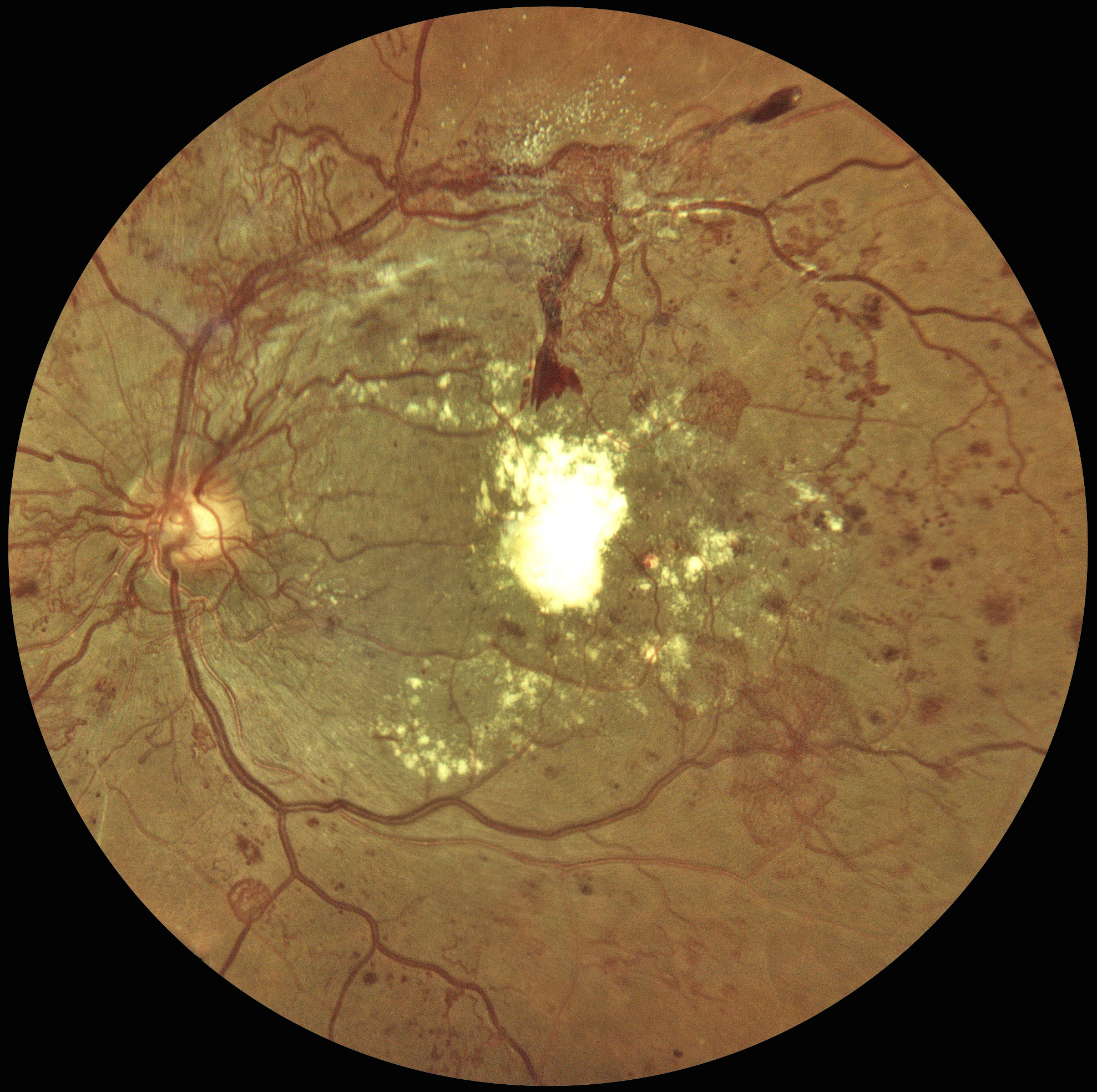
This 48YO male with a history of poorly controlled type 2 DM was seen 4 years earlier for an eye examination elsewhere but was immediately lost to follow-up. He presented to us with hand-motion vision in his left eye.
Color fundus photography shows florid disc and peripheral neovascularization variably coating the posterior pole. A large plaque of subfoveal lipid is also noted.
Learning Points:
These findings would have been caught earlier if he had not been lost to follow-up, which, unfortunately, is quite common among diabetic patients (Obeid et al, Ophthalmology 2018;125:1386-1392; Gao et al, Ophthalmology Retina 2019;3:230-236; Green et al, AJO 2020;216:18-27; Ophthalmology Retina 2025;9:1167-1174, etc, etc).
At this stage, our patient will not recover central vision due to the subfoveal lesion, and our hope is to prevent further vision loss. Anti-VEGF therapy, panretinal photocoagulation, and macular photocoagulation were suggested. He is at significant risk for traction retinal detachment as the neovascularization involutes (crunch), and we will therefore follow him very closely.



