

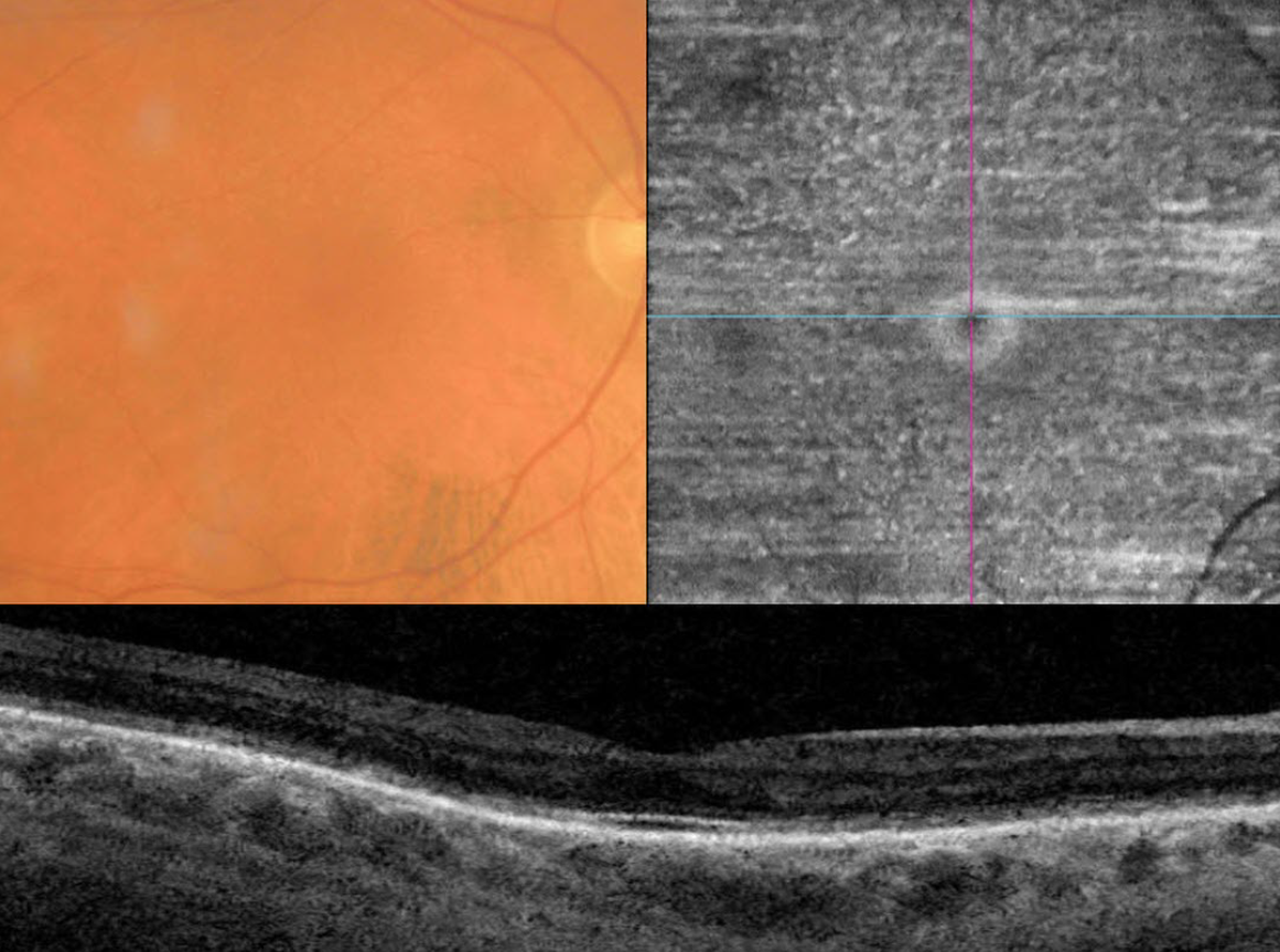
This 70YO male presented with six months of vision loss. He has a history of severe Crohn’s disease. He has undergone 9 bowel surgeries resulting in short bowel syndrome being treated with total parenteral nutrition. Vision was 20/50 OD and 20/40 OS.
Triton color imaging shows numerous smaller subretinal yellow-white dots. These dots are well-visualized on en face swept-source OCT through the outer retina. OCT B scan shows outer retinal thinning and loss of the hyperreflective bands detail.
Fasting serum vitamin A was severely low at 2.5 (normal 20-60), and he was started on parenteral vitamin A therapy.
Learning Points:
Vitamin A deficiency is rare in the United States and is sometimes related to malabsorption in chronic pancreatitis or inflammatory bowel disease. It leads to several well-documented ocular complications including nyctalopia, Bitot spots and xanthopsia. Outer nuclear layer thinning with disruption of the outer retinal bands can be visualized on OCT (Berkenstock et al, Int J Retin Vitr 2020;6:23). The outer retinal white dots colocalize to OCT outer retinal hyperreflective lesions (Aleman et al, Doc Ophthalmol 2013;127:239-243). Oral or intramuscular vitamin A replacement has been shown to reverse the retinal changes if initiated early enough. We are hoping that our patient’s vision and retinal findings will improve with continued treatment.



