

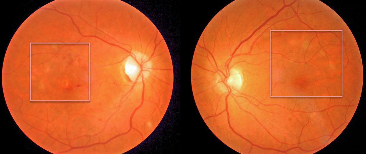
Our patient presented with macular pigmentary changes, which were most pronounced in the right eye. Vision was counting fingers OD and normal OS.
On further questioning, they gave a history of vision loss following West Nile encephalitis 6 months earlier. Although we can’t say for sure, these findings are most likely the sequelae of resolved West Nile chorioretinitis.
Learning Points:
West Nile virus is an RNA virus transmitted by the bite of a mosquito that has fed on an infected bird. Only 1 in 150 of infected persons experience encephalitis, and far fewer will have ocular effects.
Chorioretinitis in a linear pattern is the most common ocular finding, although retinal vasculitis and optic nerve swelling can also occur. There is no proven treatment for West Nile virus infection.
See a great review on this topic by Garg and Jampol (Surv Ophthalmology 2005;50:3-13).



