

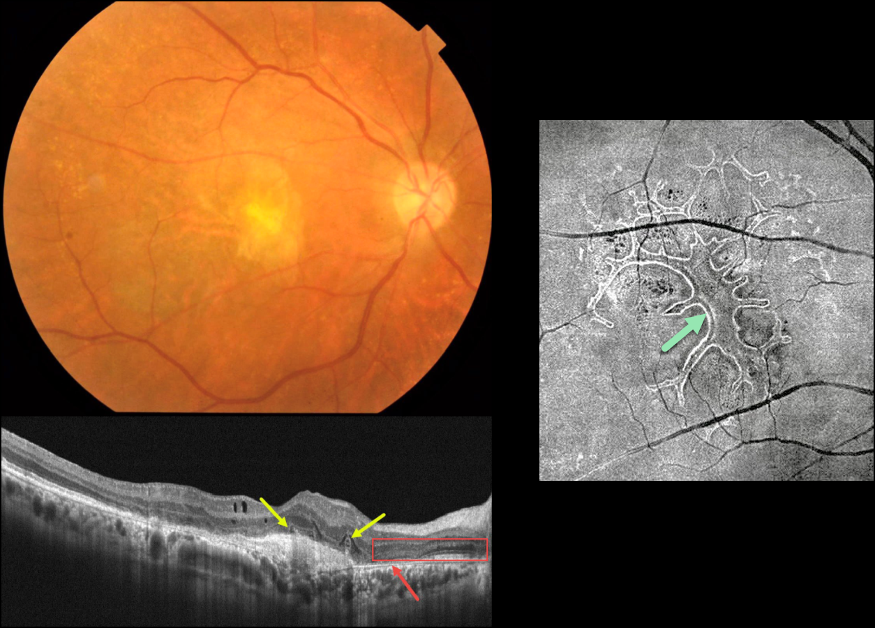
This 63YO male received several intravitreal anti-VEGF injections for neovascular AMD in his right eye, the most recent being 6 months earlier. Vision is counting fingers.
Color photography shows an inactive atrophic foveal scar. OCT scanning shows a hyperreflective macular neovascularization (MNV). The outer retina overlying the MNV is disorganized with several overlying hyporeflective lesions with surrounding hyperreflective rims. More nasally, there is complete RPE loss with bare Bruch’s membrane. The overlying retina is anomalous with additional hypo- and hyperreflective bands.
En face OCT scanning through the outer retina provides a more dramatic view of the extensive tubulation-branching network.
Learning Points:
Outer retinal tubulation (ORT) is often noted overlying inactive MNV with ongoing anti-VEGF therapy and should not be confused with exudative fluid or cysts, which lack a hyperreflective border. The outer hyperreflective band likely represents inner segment mitochondria undergoing fission and translocation toward the nucleus (Litts et al, Retina 2018;38:445-461).
ORT, initially described by Zweifel et al (Arch Ophthalmol 2009;127:1596-1602), is a neurodegenerative condition of the photoreceptors and Muller cells associated with atrophy affecting the outer retina and retinal pigment epithelium, including advanced AMD and inherited retinal diseases.



