

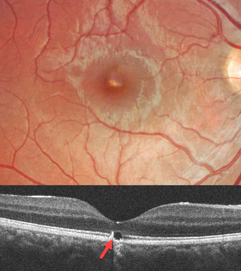
This 17YO male was in a car accident several weeks earlier with a whiplash injury. He noticed mild vision loss in his right eye since. Vision was 20/30.
Color imaging shows a somewhat linear area of foveal pigment loss. OCT scanning of this region shows an outer foveal microdefect with adjacent hyperreflectivity along its temporal edge.
Learning Points:
Initially described by Kelley et al (Arch Ophthalmol 1978;96:834-835), whiplash maculopathy is thought to occur from a vitreous coup-contrecoup injury following a motor vehicle accident. This can cause contusive RPE injury with transient macular edema (AJO 2007;143:348-350) or, more typically, a solar retinopathy-like defect in the outer retina.
Outer foveal microdefects (Cohen et al, Ophthalmology Retina 2021;5:553-561) have been described in numerous conditions, including macular telangiectasia, tamoxifen use, ABCA4 disorders (cone-rod dystrophies, Stargardt disease, and fundus flavimaculatus), phototoxicity, trauma, and vitreomacular traction disorders.



