

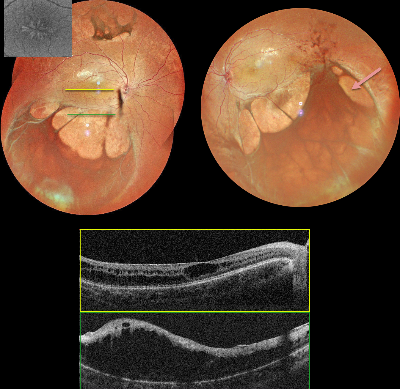
This 17YO boy presented with a few days of mild bilateral blurred vision. Vision was 20/40 OU.
MultiColor imaging shows central radiating foveal schisis, and larger areas of schisis extend beyond the arcades. OCT scanning confirms foveal schisis and more peripheral macular schisis. Identical findings were noted in the left macula (not shown).
Learning Points:
Sex-linked retinoschisis is caused by a mutation of the RS1 gene which is located on the X chromosome and encodes for retinoschisin, a protein secreted by photoreceptors that is involved in intercellular adhesion and likely retinal cellular organization (Heymann et al, Progress Retinal Eye Research 2023;95:101147). Although usually transmitted as an X-linked recessive disorder in males with an incidence of 1 in 15,000 to 30,000, it can also sometimes affect females.
The characteristic features include foveoschisis and peripheral schisis with vitreous veils. Over time, the macular schisis flattens with secondary atrophy, and vision usually levels off at about 20/100. Patients can develop spontaneous vitreous hemorrhage and rhegmatogenous retinal detachment from peripheral schisis. Although there is no specific treatment, topical carbonic anhydrase inhibitors may decrease the foveal thickness and possibly help minimize foveal atrophy (see Andreuzzi et al Retina 2017;37:1555-1561).



