

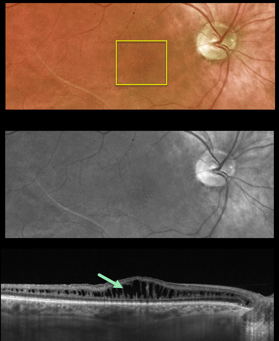
This 39YO male presented to our clinic with a known history of sex-linked retinoschisis. Vision was 20/100 OU.
Optos color and green channel images show faint radiating foveal cystic changes bilaterally (only right eye shown).
Swept-source OCT scanning shows prominent splitting of the inner nuclear layer.
Learning Points:
Although historically taught as involving the nerve fiber layer, sex-linked retinoschisis in the posterior pole causes schisis throughout the macula including the inner nuclear, outer nuclear and outer plexiform layers (see Yu et al, AJO 2010;149:973-978).
With time, the schisis often collapses with secondary atrophy (see Menke et al, Retina 2011;31:1186-1192).



