

05 Retinal Vascular Disorders: Misc
<< Back to Cases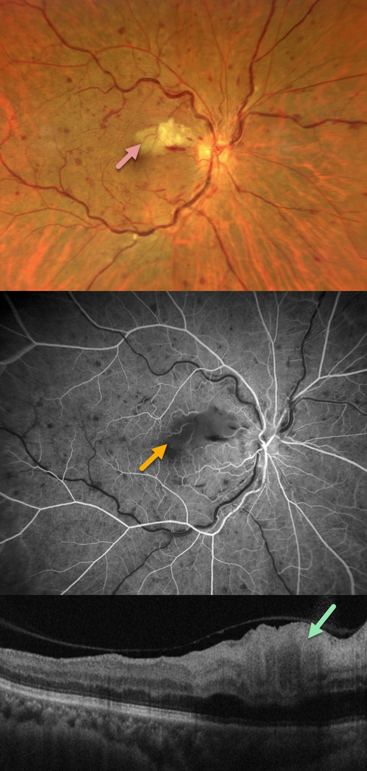
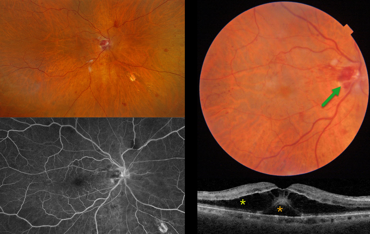
CRVO WITH SECONDARY CILIORETINAL ARTERY OCCLUSION
Francis Ezra Laxamana, Bianca Teresa Suarez, Jesus Jacinto Bautista and Carlo Ladores.
Originally posted on @retina.rocks September 7, 2026
This 58YO female with a history of hypertension and diabetes presented with sudden painless vision loss in her right eye. Vision was 20/25 OD and 20/20 in her normal OS.
Fundus photography shows a non-ischemic central retinal vein occlusion (CRVO) with dilated tortuous veins, mild scattered retinal hemorrhages, and a superonasal macular branch retinal artery occlusion (BRAO). The BRAO shows inner retinal hyperreflectivity on OCT. Fluorescein angiography shows good perfusion throughout the distribution of the CRVO, with a filling defect from the BRAO.
Learning Points:
Some patients with CRVO will develop a secondary non-embolic cilioretinal artery occlusion caused by either compression from optic nerve edema or from a sudden rise in intraluminal retinal capillary bed pressure (Hayreh et al, Retina 2008;28;581-594).
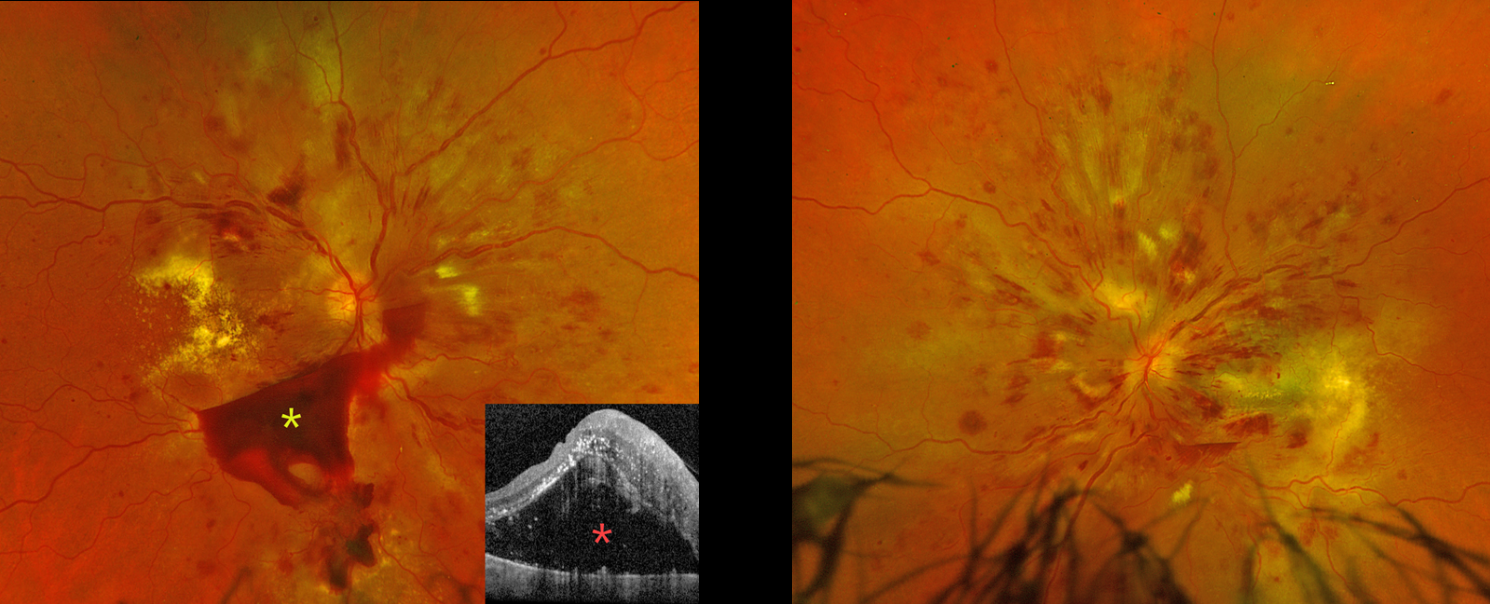
HYPERTENSIVE RETINOPATHY
Originally posted on @retina.rocks September 2, 2026
This 42YO undomiciled female presented with several months of bilateral vision loss. She has a complex medical history of hypertension, type 2 diabetes (last A1c 9.9), end-stage renal disease on dialysis, chronic obstructive pulmonary disorder, and bipolar disorder. The day prior to presenting to our clinic, she was seen at an outside emergency department for intractable vomiting in the setting of 3 consecutively missed dialysis sessions. Her blood pressure in the ED was 199/105 after self-stopping blood pressure medications because of vomiting. She had left the ED against medical advice. Vision was counting fingers OD and hand motion OS.
Optos color RG imaging OD shows diffuse intraretinal hemorrhages, multiple subhyaloid hemorrhages, cotton-wool spots (CWS), and extensive intraretinal and subretinal exudation. OCT shows marked exudative subretinal fluid with intraretinal hyperreflectivity and thickened inner retinal layers. Similar findings are noted OS.
Learning Points:
Malignant hypertension, defined as blood pressure above 180 systolic and/or 120 diastolic, is a life-threatening medical emergency. Eye doctors are often uniquely positioned to diagnose this condition (Shukla et al, Indian J Ophthalmol 2025;73:1552-1555). Patients will often present with bilateral optic nerve swelling, flame-shaped retinal hemorrhages, CWS, and, with more chronic disease, lipid precipitates in the nasal macular outer plexiform layer.
Our patient’s funduscopic findings were more likely hypertensive than from proliferative diabetic retinopathy. She was instructed to return to the emergency room for blood pressure control, and a poor visual prognosis was discussed.
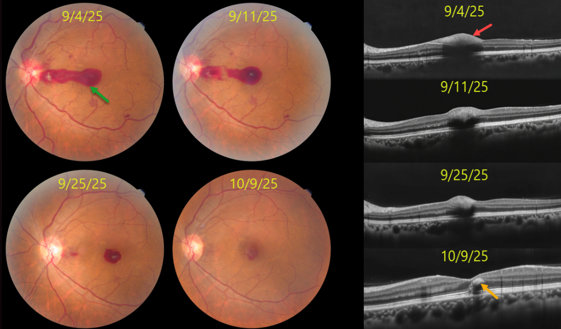
HYPERTENSIVE RETINOPATHY WITH SUB-ILM HEMORRHAGE
Abhishek Karra and Ravindra Karra
Originally posted on @retina.rocks August 31, 2026
This 44YO female presented with sudden vision loss OS with uncontrolled hypertension (180/110 mmHg). Vision was 20/20 OD and 20/200 OS.
Color photography shows a pre-retinal hemorrhage extending from the temporal edge of the disc through the central macula, disc and peripapillary inner retinal hemorrhages, and blot and nerve fiber layer hemorrhages. The pre-retinal blood localizes to the sub-internal limiting membrane (ILM) space on OCT. The retinal findings normalized over the next 4-5 weeks with visual improvement to 20/20. OCT at the final visit shows a small amount of mid-retinal foveal blood.
Learning Points:
Malignant hypertension, defined as blood pressure above 180 systolic and/or 120 diastolic, is a life-threatening medical emergency. Eye doctors are often uniquely positioned to diagnose this condition (Shukla et al, Indian J Ophthalmol 2025;73:1552-1555). Patients will often present with bilateral optic nerve swelling, flame-shaped retinal hemorrhages, CWS, and, with more chronic disease, lipid precipitates in the nasal macular outer plexiform layer.
Sub-ILM hemorrhage, to the best of our knowledge, has not been previously described in hypertensive retinopathy. It is usually associated with retinal arterial macroaneurysm, Valsalva retinopathy, trauma, and blood dyscrasias. In our patient’s case, we hypothesize that the sub-ILM likely originated from the disc and dissected temporally across the macula.
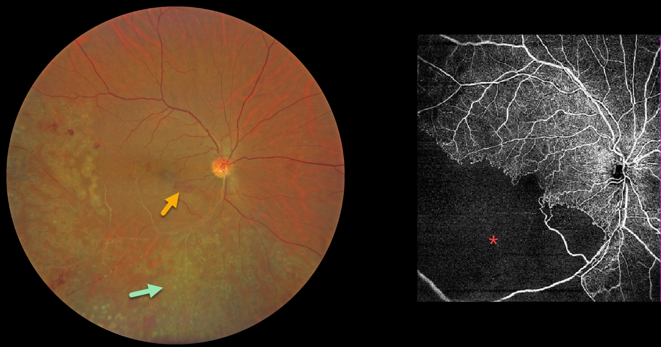
BRVO WITH RETINAL NEOVASCULARIZATION
Abhishek Karra and Ravindra Karra
Originally posted on @retina.rocks August 10, 2026
This 42YO male presented with a 3-month history of decreased vision in his right eye. He had received laser treatment in this eye elsewhere. Vision was 20/60 OD.
Color photography shows an old ischemic inferotemporal branch retinal vein occlusion (BRVO) with heavy scatter laser throughout its distribution. Collateral vessels are noted in the inferonasal macula. OCT angiography shows profound capillary loss. There was no macular edema, and observation was recommended.
Learning Points:
In 1986, the Branch Vein Occlusion Study Group reported that scatter laser decreased the risk of vitreous hemorrhage from retinal neovascularization in BRVO and suggested that laser should be applied once neovascularization develops (Arch Ophthalmol 1986;104:34-41). Thirty-five years later, scatter laser remains the gold standard, although some may prefer to stabilize the neovascularization with anti-VEGF therapy before laser.
RETINAL VENOUS MALFORMATION
Ananya Kaginalkar, Vaishali Vasavada and Samaresh Srivastava
Originally posted on @retina.rocks July 10, 2026
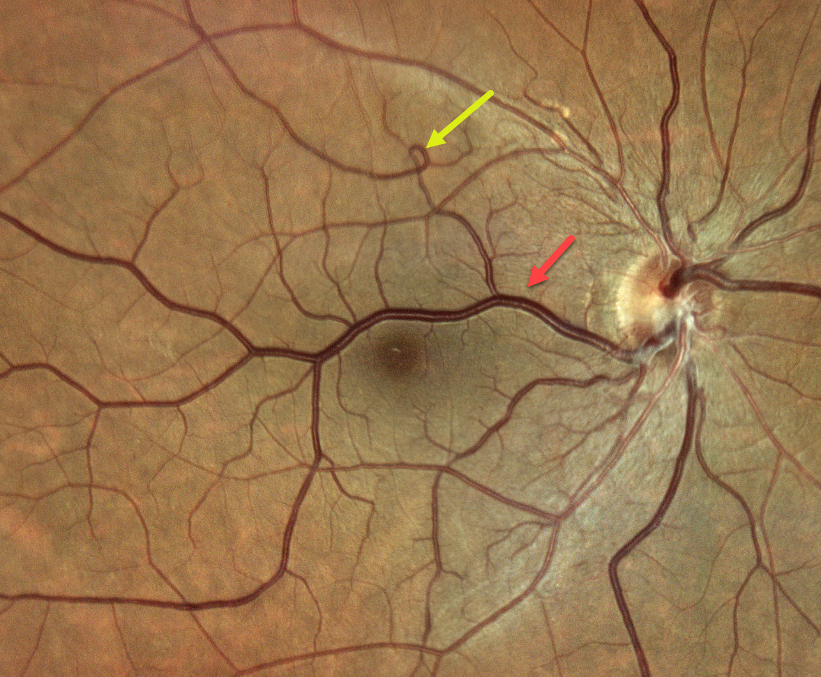
This 57YO male presented with 3 weeks of blurred vision in his left eye. Vision was 20/20 OD and 20/40 OS. Anterior segments showed a moderate nuclear sclerotic cataract OS, accounting for his acuity. The left fundus was normal.
Color photography shows a large aberrant retinal vein, which crosses the horizontal meridian and drains nearly the entire right macula. There is a small vascular loop superiorly.
Learning Points:
Retinal vessels virtually always respect the horizontal meridian. Retinal venous malformations are congenital anomalous vessels that, by definition, cross the horizontal. Originally described by Gary Brown et al as congenital retinal macrovessels (Arch Ophthalmology 1982;100:1430-1436), these vessels are usually located in the macular region and are virtually always venous. These lesions are now called retinal venous malformations, since about one-quarter of patients have associated venous anomalies in the brain (Pichi et al, JAMA Ophthalmol 2018;136:372-379).
CRVO WITH CENTRAL BOUQUET HEMORRHAGE
Originally posted on @retina.rocks June 26, 2026
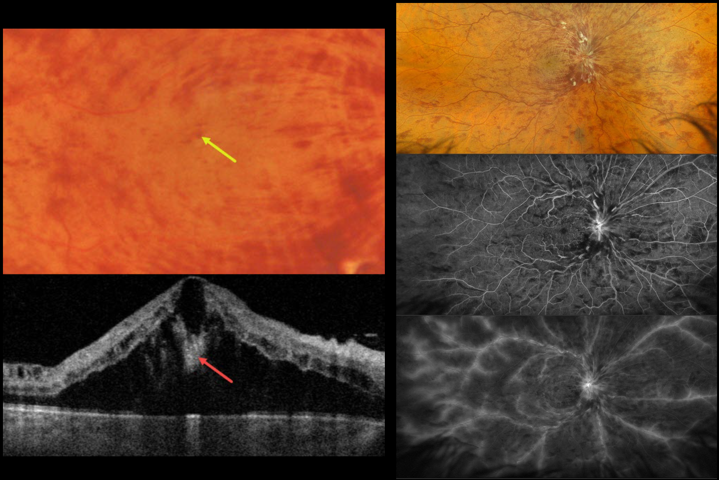
This healthy 52YO male presented with recent severe vision loss in his right eye. Vision was 20/400 OD and 20/20 in his normal OS.
Color photography and Optos RGB imaging show a central retinal vein occlusion (CRVO) with marked retinal hemorrhages and macular edema. Inner blood-filled retinal cysts are noted on funduscopy and swept-source OCT. On fluorescein angiography, the occlusion is fairly well perfused with late leakage from the nerve, macula, and retinal veins. Monthly anti-VEGF therapy was started.
Learning Points:
Cicinelli et al recently reported that central bouquet hemorrhages in retinal vein occlusion are associated with poor visual outcomes due to localized effects of elevated venous pressure and macular ischemia (IOVS 2025;66(12):62).
Prior to the introduction of intravitreal injections in the early 2000s, there were essentially no treatments for CRVO-related macular edema. Anti-VEGF injections are now generally the initial treatment of choice, with about 50-60% of eyes gaining at least 3 lines of vision. Intravitreal steroids, usually Ozurdex, are more often used as secondary treatment. Macular grid laser photocoagulation is not performed because it has been shown to confer no visual benefit in the Central Vein Occlusion Study Group (Ophthalmology 1995;102:1425-1433).
RETINAL ARTERIAL MACROANEURYSM (RAM)
Rohit Agrawal
Originally posted on @retina.rocks May 22, 2026
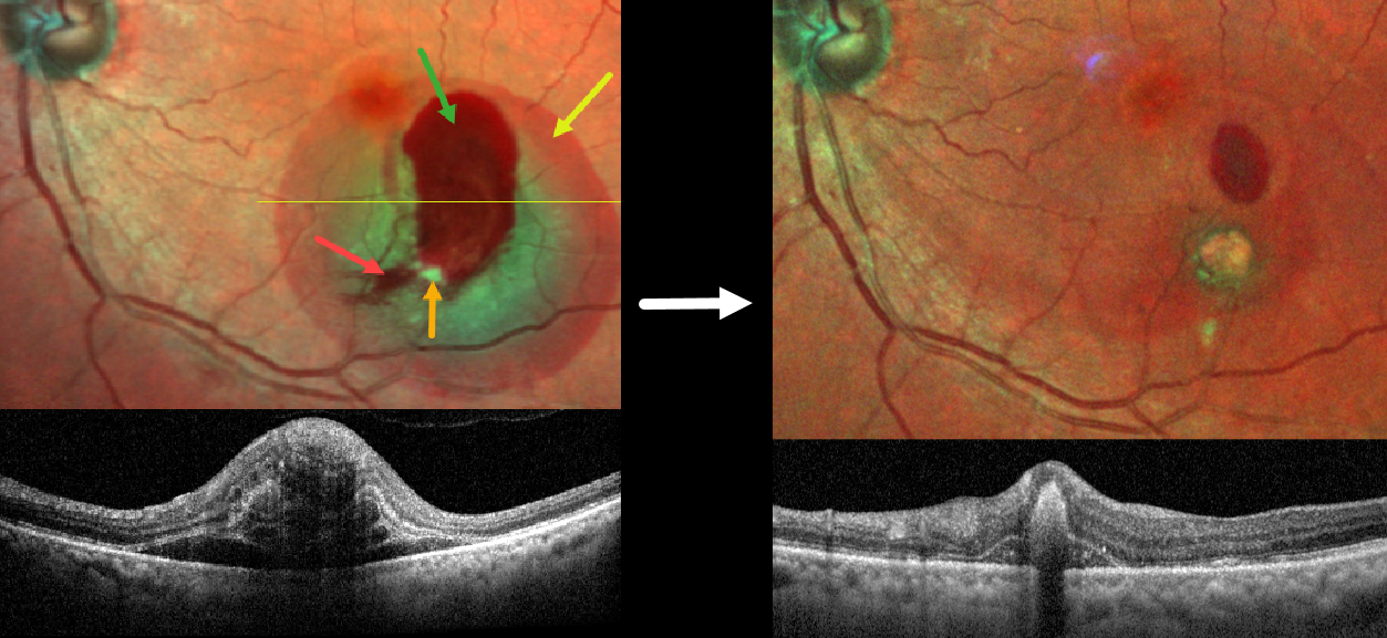
This 78YO female presented with sudden blurred vision in her left eye. Vision was 20/40.
MultiColor SLO imaging shows subretinal, intraretinal and preretinal blood centered in the inferotemporal macula. A fibrosed retinal arterial macroaneurysm is seen along the inferior edge of the premacular blood. OCT shows a complex laminated pattern of subretinal and intraretinal blood.
A RAM is thought to occur in a weakened arterial wall from arteriosclerosis and appears as a yellow-white dilated lesion along a retinal artery within the first 3 bifurcations from the optic nerve. There are 2 distinct presentations, hemorrhagic or exudative. The hemorrhagic type often presents, as in this case, with pathognomonic multiple layers of blood. Acute macular exudation can be treated with observation, anti-VEGF injections, or thermal laser.
Our patient was treated with a combination of an anti-VEGF injection followed by thermal laser. Five weeks later, vision improved to 20/30 with marked improvement in the blood and macular thickening.
COATS DISEASE
Rajashree Salvi, Apoorva Ayachit, Guruprasad Ayachit, and Shrinivas Joshi
Originally posted on @retina.rocks May 15, 2026
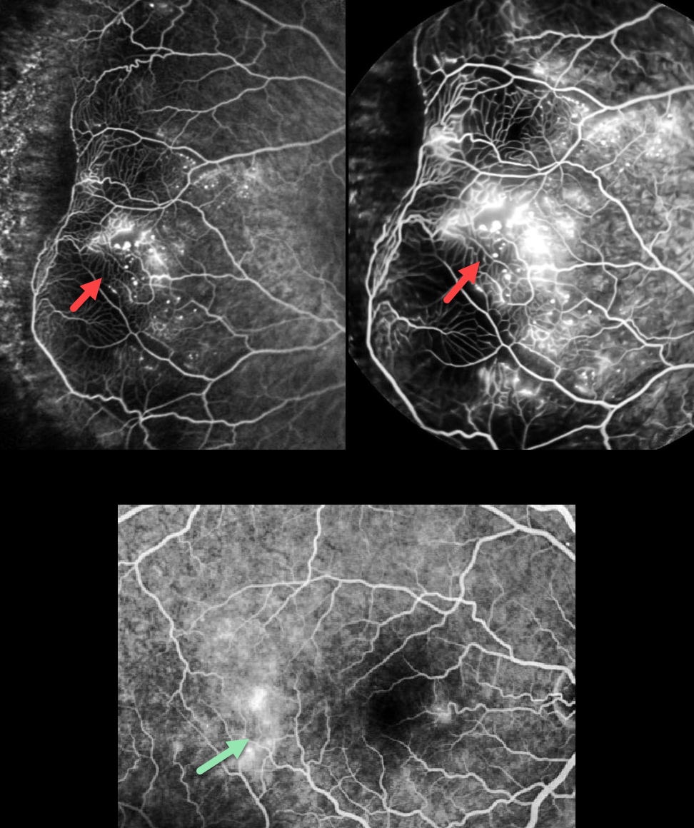
This 32YO male complained of decreased vision in his right eye for 2 weeks. Vision was 20/20 OD and 20/20 in his normal OS.
Fundus fluorescein angiography shows a broad zone of temporal midperipheral leaking retinal telangiectasia with capillary dropout and distal temporal ischemia. Larger retinal vessel anastomoses are present at the border of the perfused and nonperfused retina. There is also late temporal macular leakage. Color photography is unfortunately not available, and our patient was immediately lost to follow-up.
Learning Points:
Described by George Coats in 1908, Coats disease is an idiopathic disorder, usually unilateral, in males. It is characterized by retinal vascular telangiectasia involving capillaries, arterioles, and venules. Patients can present with a wide range of findings, from minimal exudation to total exudative retinal detachment. The leaking vessels are usually treated to prevent exudative complications.
Thermal laser photocoagulation is usually the initial treatment modality, but anti-VEGF injections can also be used to stabilize the vascular leakage. Treatment involves a combination of focal laser to the leaking vascular abnormalities along with scatter laser into areas of ischemia. Patients will often require multiple rounds of treatment. See Sigler et al for a review of the current management (Survey Ophthalmology 2014;59:30-46).
RETINAL ARTERIAL MACROANEURYSM (RAM)
Shilpi Kapoor, Abhishek Sachdeva and Sanjay Sachdeva
Originally posted on @retina.rocks May 11, 2026
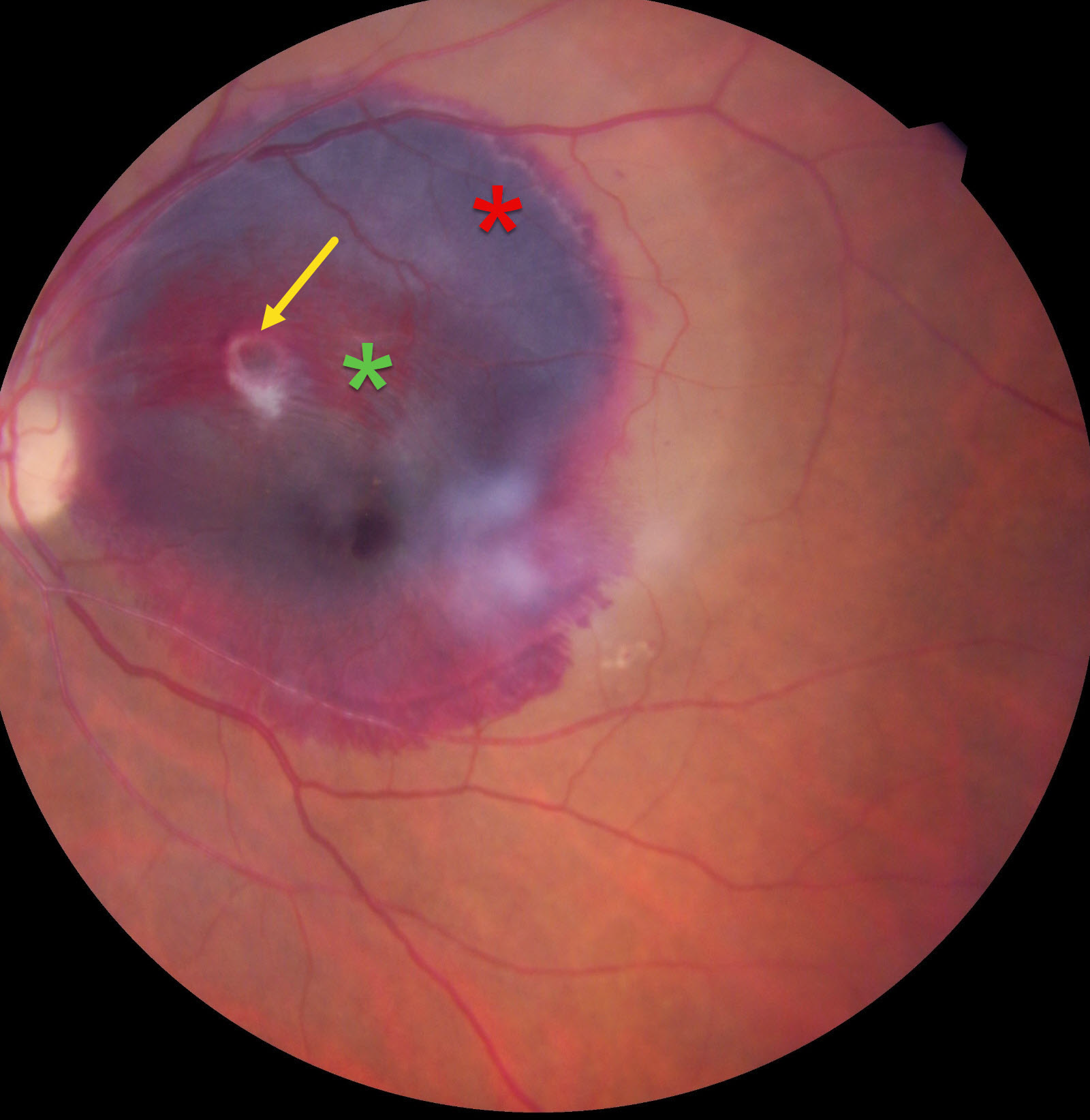
This 65YO female presented with 5 days of acute vision loss in her left eye. Vision was 20/20 in her normal OD and 20/400 OS.
Color photography shows a large subretinal hemorrhage occupying the entire macula, along with a smaller preretinal hemorrhage. A somewhat fibrosed retinal arterial macroaneurysm (RAM) is noted within the preretinal blood. An intravitreal ranibizumab injection was given, but the patient was unfortunately immediately lost to follow-up.
Learning Points:
A RAM is thought to occur in a weakened arterial wall from arteriosclerosis and appears as a yellow-white dilated lesion along a retinal artery within the first 3 bifurcations from the optic nerve. There are 2 distinct presentations, hemorrhagic or exudative. The hemorrhagic type often presents, as in this case, with pathognomonic multiple layers of blood. Acute macular exudation can be treated with observation, anti-VEGF injections, or thermal laser.
SICKLE CELL RETINOPATHY
Originally posted on @retina.rocks May 5, 2026
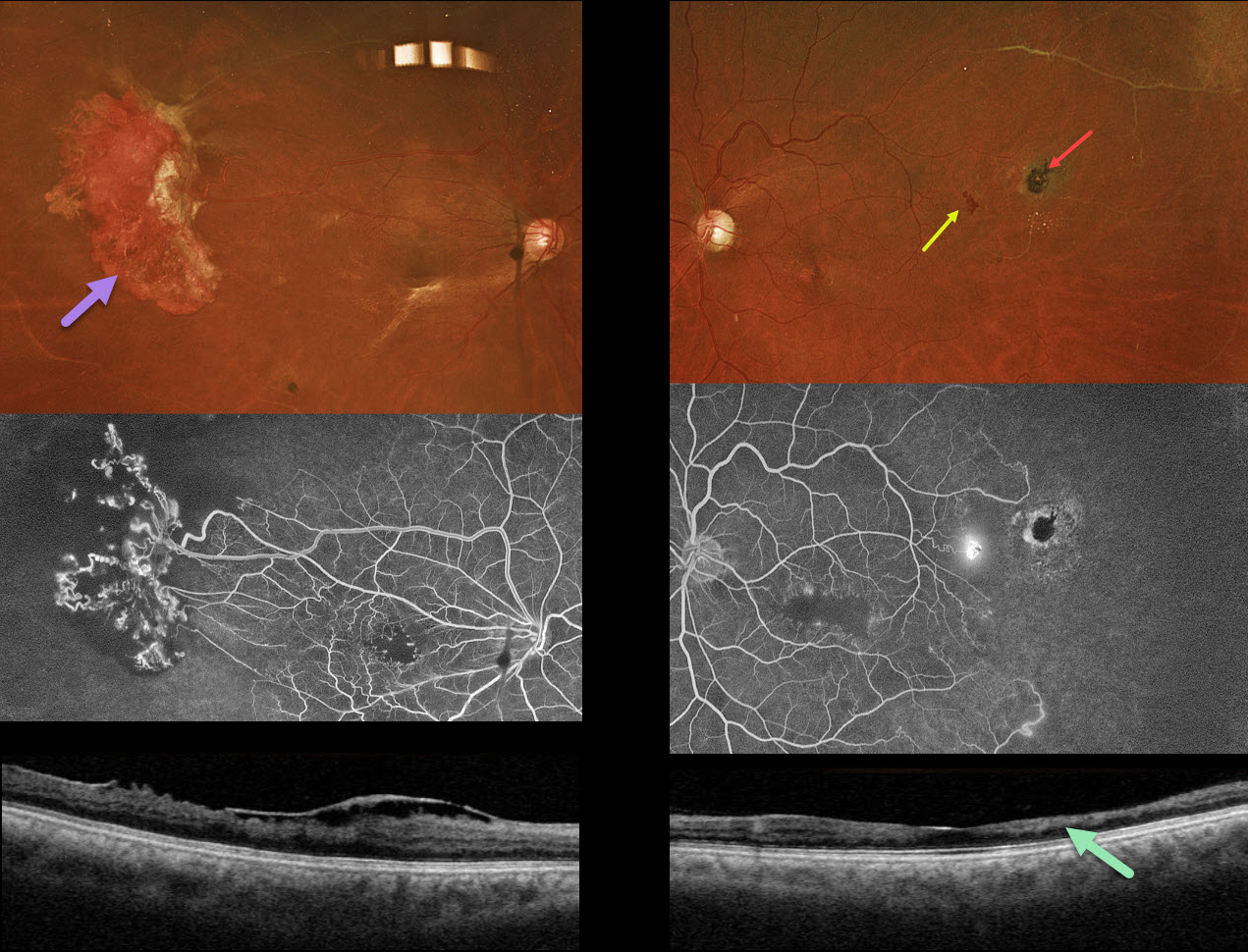
This 44YO male with a known history of sickle cell disease was referred for a possible detached retina. Vision has decreased over the past few years. Vision was 20/200 OU.
Optos color RGB imaging shows a large area of active neovascularization in the right temporal midperiphery associated with extensive angiographic retinal ischemia. Similar ischemia is noted OS with a small patch of leaking neovascularization. There is a small, more distal black sunburst lesion. The foveal avascular zones are enlarged with surrounding telangiectasia. This is reflected in bilateral inner retinal thinning with disorganization of inner retinal layers on OCT scanning (DRIL). An epimacular membrane is also noted OD.
Learning Points:
Patients with sickle cell disease develop symptoms throughout their bodies as stiffened, sickled red blood cells block capillary beds. In the eye, this can cause proliferative sickle cell retinopathy (PSR) with sea-fan-shaped areas of peripheral retinal neovascularization developing at the junction of perfused and ischemic retina. Increasing macular ischemia correlates with peripheral angiographic nonperfusion, as seen in our patient (Han et al., Ophthalmology Retina 2019;2:599-605). Complications include vitreous hemorrhage and tractional retinal detachment. Although patients with SS disease tend to have more systemic complications, SC disease is usually associated with more severe ocular findings.
Treatment with sectoral (Farber et al Arch Ophthalmology 1991;109:363-367) or circumferential (Kimmel et al Ophthalmology 1986;93:1429-1434) scatter laser photocoagulation is somewhat controversial since, unlike more typical proliferative retinopathies (ie from diabetes and branch retinal vein occlusions), these new vessels tend to auto-infarct with a good natural history for untreated disease (Downes et al Ophthalmology 2005;112:1869-1875). Based on the active appearance of the large temporal neovascularization OD, we elected for prophylactic scatter laser throughout the ischemic retina.
RADIATION RETINOPATHY
Ayushi Gupta and Vishal Agrawal
Originally posted on @retina.rocks April 21, 2026
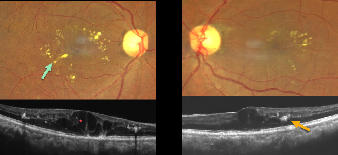
This 53YO female presented with gradual bilateral vision loss. Three years earlier, she was treated elsewhere for metastatic breast carcinoma, which included a mastectomy followed by radiotherapy to the supraclavicular region and brain. She denied having diabetes. Vision was 20/60 OD and 20/30 OS.
Fundus photography shows bilateral macular telangiectasia with radiating macular lipid flecks. OCT scanning shows diffuse cystic retinal thickening with hyperreflective lipid centered around the outer plexiform layer. Anti-VEGF therapy was recommended.
Learning Points:
Radiation retinopathy usually occurs 3 months to 3 years after external-beam radiation or plaque radiotherapy. The ocular findings and pathogenesis closely resemble those seen with diabetic retinopathy. Radiation optic neuropathy can also develop (Carey et al, Br J Ophthalmol 2023;107:743-749).
RETINAL ARTERIOVENOUS MALFORMATION
The European VitreoRetinal Society (EVRS) and Rajiv Gandhi
Originally posted on @retina.rocks April 17, 2026
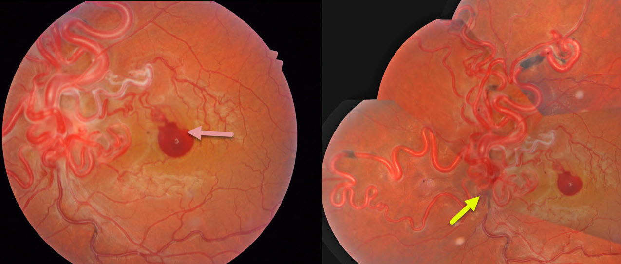
This healthy 34YO man noticed sudden vision loss in his left eye following heavy lifting 2 days earlier. Vision was 20/20 in his normal OD and 20/200 OS.
Color photography shows a diffuse network of markedly dilated, tortuous vessels involving the optic disc and posterior pole, extending into the peripheral retina, with an associated small premacular hemorrhage. The vessels are sheathed superonasal to this blood. Unfortunately, our patient was lost to follow-up immediately.
Learning Points:
A retinal arteriovenous malformation (AVM) is a congenital connection between an artery and a vein without an intervening capillary network. These lesions appear along a spectrum ranging from small, isolated lesions to the large ‘bag of worms’ appearance. These more extensive AVMs are usually part of the Wyburn-Mason Syndrome, which includes ocular and midbrain vascular lesions. Although retinal AVMs are usually stable lesions, bleeding or secondary venous occlusions may occur. Our patient’s vascular sheathing may indicate prior occlusion.
RADIATION RETINOPATHY
The European VitreoRetinal Society (EVRS) and Abhishek Upadhyaya
Originally posted on @retina.rocks April 3, 2026
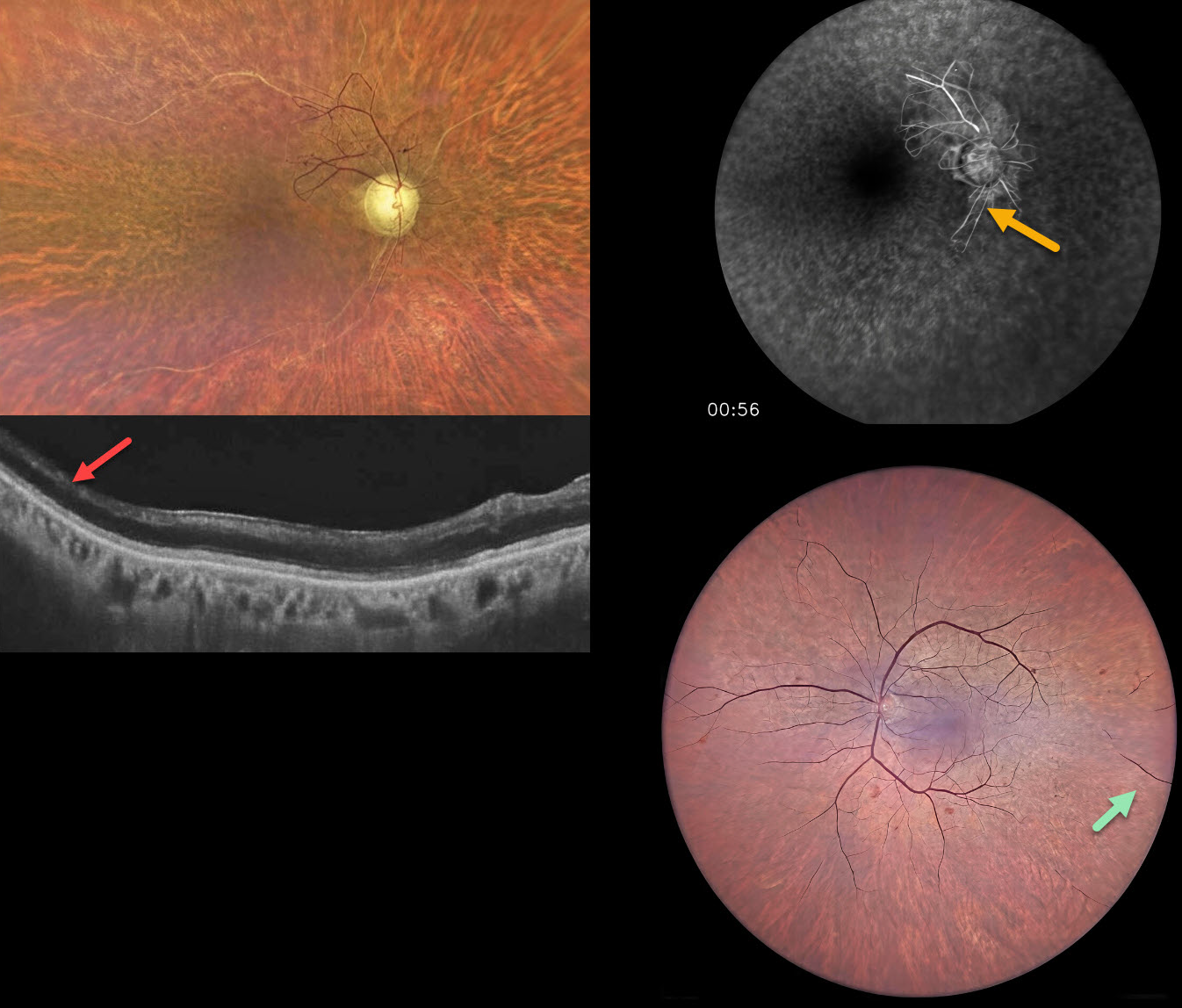
This 65YO male presented for evaluation for cataract surgery. He underwent external beam radiation for throat cancer, predominantly on the right side, 20 years earlier. Vision was hand motion OD and 20/200 OS.
Color photography OD shows severe panretinal ischemia with the major vessels showing absent blood columns except for some abnormal preserved vessels with anastomoses just superotemporal to the nerve. OCT shows temporal inner retinal atrophy. The left posterior pole appears more normal with severe more peripheral ischemia. Fluorescein angiography confirms the profound ischemia noted funduscopically.
Learning Points:
Radiation retinopathy usually occurs 3 months to 3 years following external beam radiation or plaque radiotherapy. The ocular findings closely resemble those seen with diabetic retinopathy. Radiation optic neuropathy can also develop (Carey et al, Br J Ophthalmol 2023;107:743-749) .
Panretinal photocoagulation was recommended for the right eye.
COATS DISEASE
Originally posted on @retina.rocks March 12, 2026
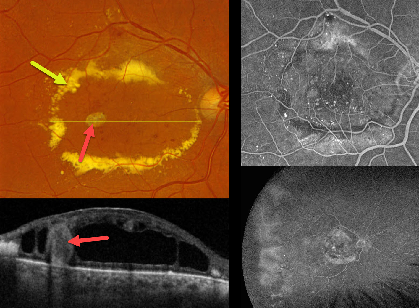
This 71YO male presented with a vague history of decreased vision in his right eye. He gave a history of diet-controlled type 2 diabetes. Vision was 20/200 OD and 20/20 in his normal OS.
Optos color RGB imaging shows an oval ring of subretinal lipid surrounding the central macula. Scattered telangiectatic vessels are noted. A large fibrosed macroaneurysm is seen temporally. On Triton swept-source OCT, this macroaneurysm spans nearly the entire retinal thickness and is surrounded by a large mid-retinal cystic cavity. Fundus fluorescein angiography (FFA) shows numerous macular microaneurysms with relative blockage from the subretinal lipid. There is diffuse leakage from the temporal vessels with ischemic retina more peripherally associated with some telangiectatic vessels. FFA was normal in his left eye (not shown). Intravitreal anti-VEGF therapy was started.
Learning Points:
Described by George Coats in 1908, Coats disease is an idiopathic disorder, usually unilateral, in males. It is characterized by retinal vascular telangiectasia involving capillaries, arterioles, and venules. Patients can present with a wide range of findings, from minimal exudation to total exudative retinal detachment. The leaking vessels are usually treated to prevent exudative complications.
Thermal laser photocoagulation is usually the initial treatment modality, but anti-VEGF injections can also be used to stabilize the vascular leakage. Treatment involves a combination of focal laser to the leaking vascular abnormalities along with scatter laser into areas of ischemia. Patients will often require multiple rounds of treatment. See Sigler et al for a review of the current management (Survey Ophthalmology 2014;59:30-46).
HYPERTENSIVE RETINOPATHY AND CHOROIDOPATHY
Originally posted on @retina.rocks February 18, 2026
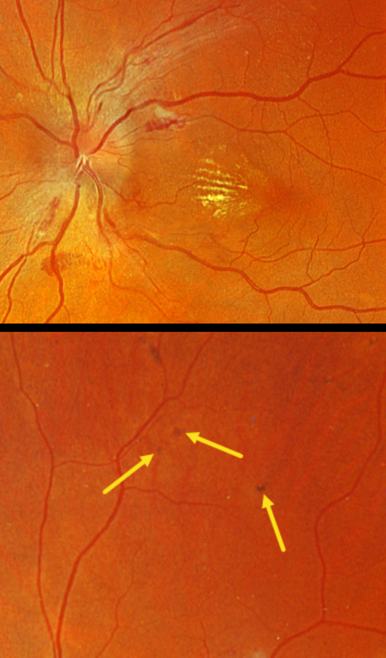
This 46YO male presented to his local optometrist 1 week earlier with a few days of bilateral vision loss. His doctor diagnosed malignant hypertension due to retinal findings and a blood pressure of about 280/160. He was hospitalized for a few days and presented to us for further evaluation. Vision was 20/30 OD and 20/50 OS.
Optos color RGB imaging shows optic nerve edema, peripapillary hemorrhages, and a nasal macular lipid star. Several small pigmented Elschnig spots are noted superiorly OS. He was immediately lost to follow-up.
Learning Points:
Malignant hypertension, defined as blood pressure above 180 systolic and/or 120 diastolic, is a life-threatening medical emergency. Eye doctors are in a unique position to often diagnose this condition. Patients will often present with bilateral optic nerve swelling, flame-shaped retinal hemorrhages, and, with more chronic disease, lipid precipitates in the nasal macular outer plexiform layer. Focal (Elschnig spots) or linear (Siegrist streaks) choroidal pigmentary changes develop from hypertensive choroidopathy.
BRVO WITH RETINAL NEOVASCULARIZATION AND COLLATERALS
Originally posted on @retina.rocks February 11, 2026
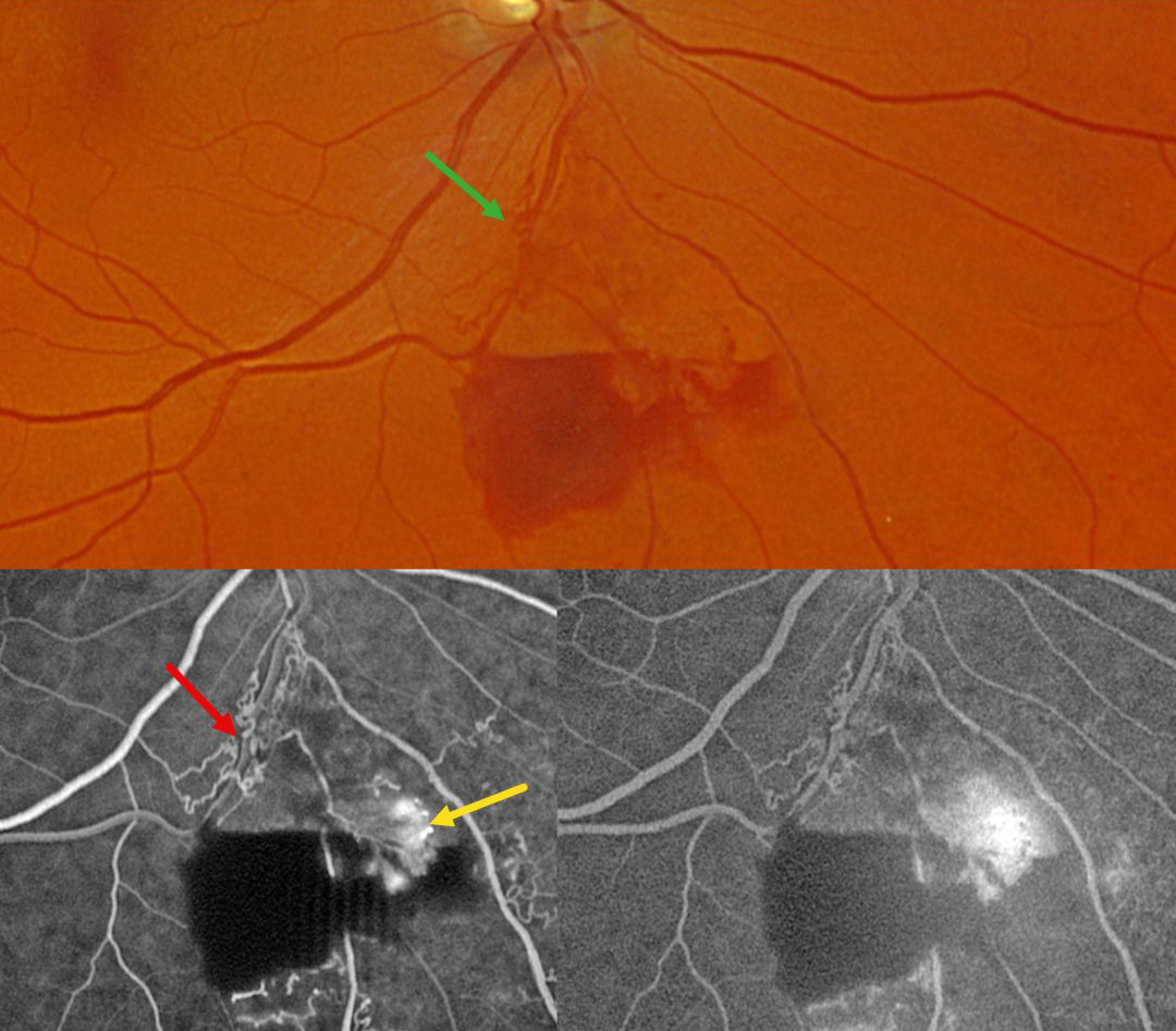
This 59YO male was referred for asymptomatic retinal findings in his right eye. Vision was 20/30.
Optos color RG imaging shows an inferior branch retinal vein occlusion with midperipheral preretinal blood. Collateral vessels are noted at the site of the occlusion. These collaterals show no leakage angiographically, although the adjacent retinal neovascularization does leak.
Learning Points:
Collateral vessels are dilated, pre-existing vessels that bypass an occlusion. These are most commonly seen in the posterior pole following retinal venous occlusions and are exceedingly rare with arterial occlusions. The endothelial tight junctions are intact, so they don’t leak angiographically. This helps differentiate them from neovascularization, which does leak. Our patient’s eye shows us a wonderful example of both.
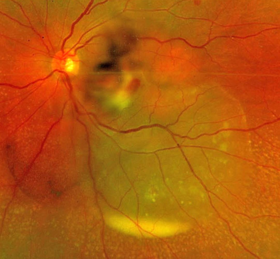
SEPTIC RETINAL EMBOLUS WITH SUBRETINAL HYPOPYON
The European VitreoRetinal Society (EVRS) and Mohit Dogra
Originally posted on @retina.rocks February 6, 2026
This previously healthy 37YO female presented to us for a second opinion regarding 4 days of decreased vision in her left eye. There was a recent history of a urinary tract infection secondary to E. coli. Vision was 20/20 in her normal OD and 20/400 OS. There was a moderate non-granulomatous uveitis with vitreous cells.
Optos color RG imaging shows opaque white retina in the inferonasal macula with overlying retinal and vitreous blood. An exudative retinal detachment extends inferotemporally with an inferior subretinal hypopyon.
She was referred back to the initially treating ophthalmologist and was subsequently lost to follow-up.
Learning Points:
Posterior segment chorioretinitis from septic embolization is a rare event, most commonly found in patients with bacterial endocarditis. Our patient presented with a constellation of findings, including retinitis, retinal and vitreous hemorrhage, and an exudative retinal detachment with an inferior subretinal hypopyon.
Subretinal hypopyon is characterized by yellowish inflammatory material settling inferiorly in the subretinal space. It has been documented in infectious (bacterial and fungal endophthalmitis, acute retinal necrosis, tuberculosis, syphilis), inflammatory (sympathetic ophthalmia), and neoplastic (leukemia, lymphoma) disorders.
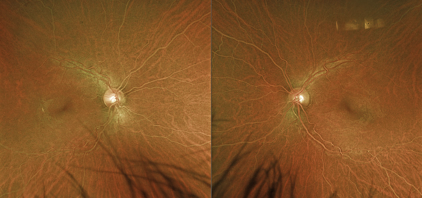
LIPEMIA RETINALIS
Hailey Dunlow
Originally posted on @retina.rocks February 4, 2026
This previously healthy 38YO male was referred for asymptomatic retinal findings. Vision was 20/25 OD and 20/20 OS. Optos color RG imaging shows symmetric, creamy-colored blood vessels bilaterally. Emergent bloodwork showed total cholesterol 1093 mg/dL, triglycerides 9644 mg/dL, and HDL 4 mg/dL. He was immediately referred to his PCP for emergent medical management. He was immediately lost to follow up.
Learning Points:
Lipemia retinalis is a rare retinal manifestation of severe hypertriglyceridemia, characterized by creamy white discoloration of the retinal vasculature. In severe cases, the fundus can turn salmon-colored. Patients are asymptomatic, and vision is normal. However, they are at obvious cardiovascular risk without systemic treatment.
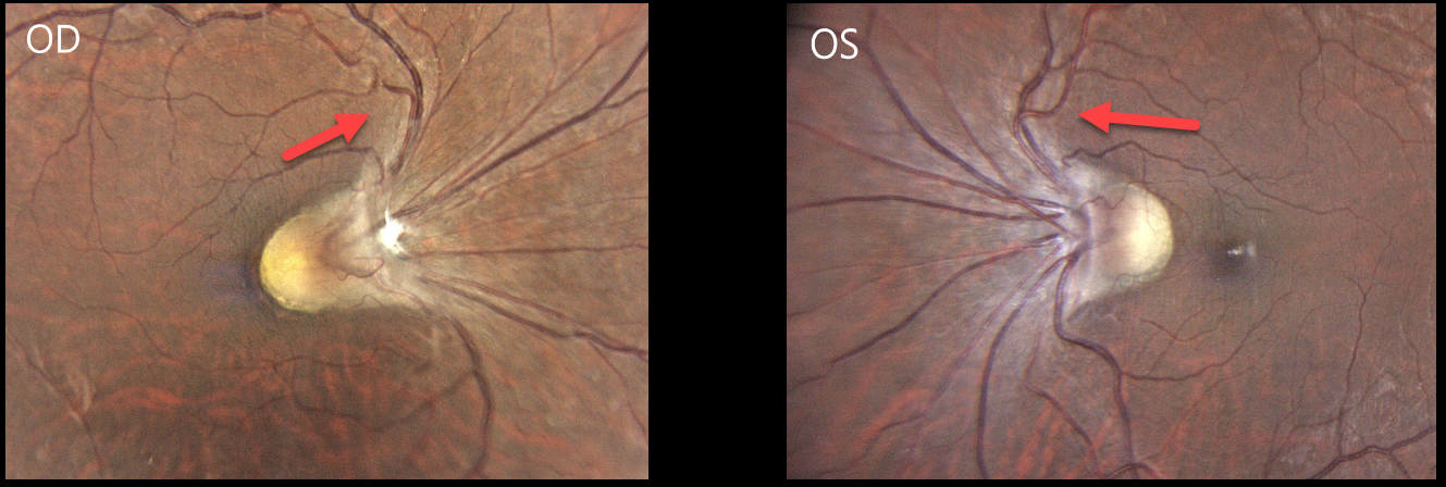
SITUS INVERSUS
Gil Calvão-Santos
Originally posted on @retina.rocks January 15, 2026
This healthy 38YO highly myopic female presented with 6 months of decreased vision in her right eye. Her current refraction was -15.00 D OD and -11.00 -2.25 × 10° OS. Vision was 20/200 OD and 20/25 OS.
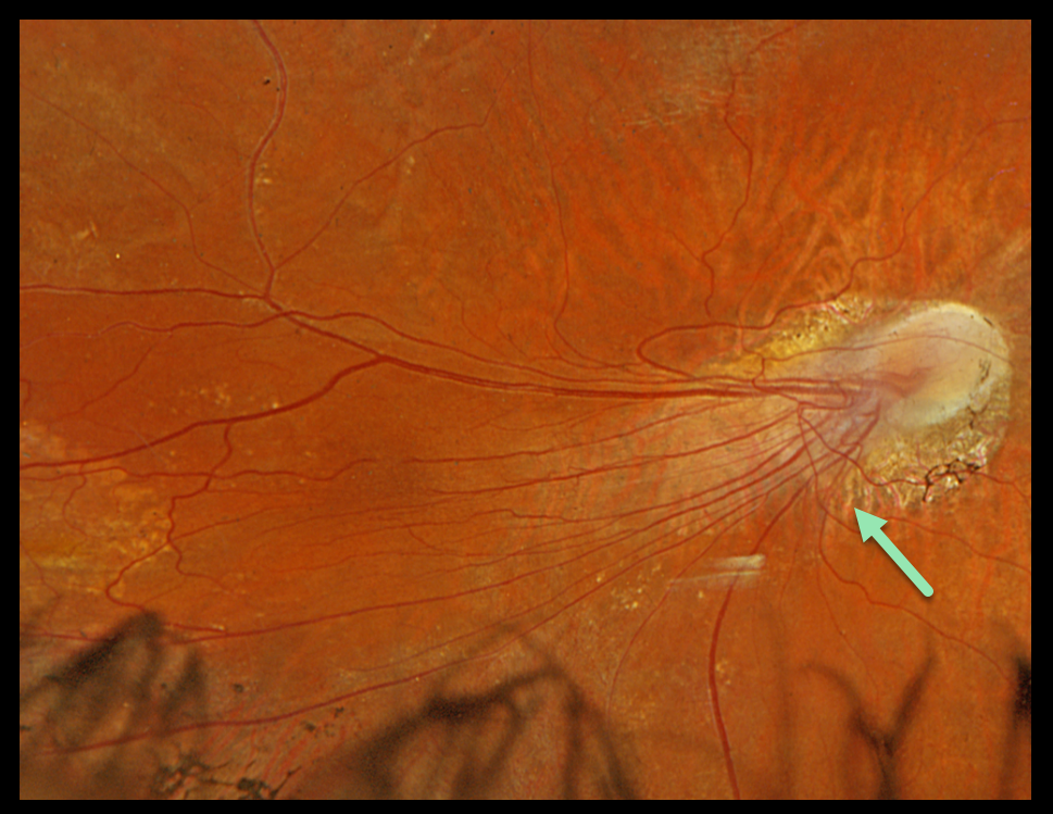
Color photography shows the retinal vessels emerge from slightly tilted discs to initially course nasally and superiorly before turning temporally toward the macula. The macular center is normally located about 2-disc diameters temporal to the nerve. The right fovea cannot be visualized and likely lies just temporal to the edge of the nerve, and about one-half disc diameter temporal to the left nerve.
Learning Points:
Situs inversus occurs in 5% of normal eyes and 70% of eyes with tilted discs (Witmer et al, Surv Ophthalmology 2010;55:403-428) and presents with the retinal vessels exiting the optic disc nasally. Vision is usually normal unless other pathology is present.
Despite the abnormal macular architecture, our patient reportedly had normal vision until recently, and was found to have a cataract, which was consistent with her acuity. Cataract surgery was recommended, and vision improved to 20/25 postoperatively.
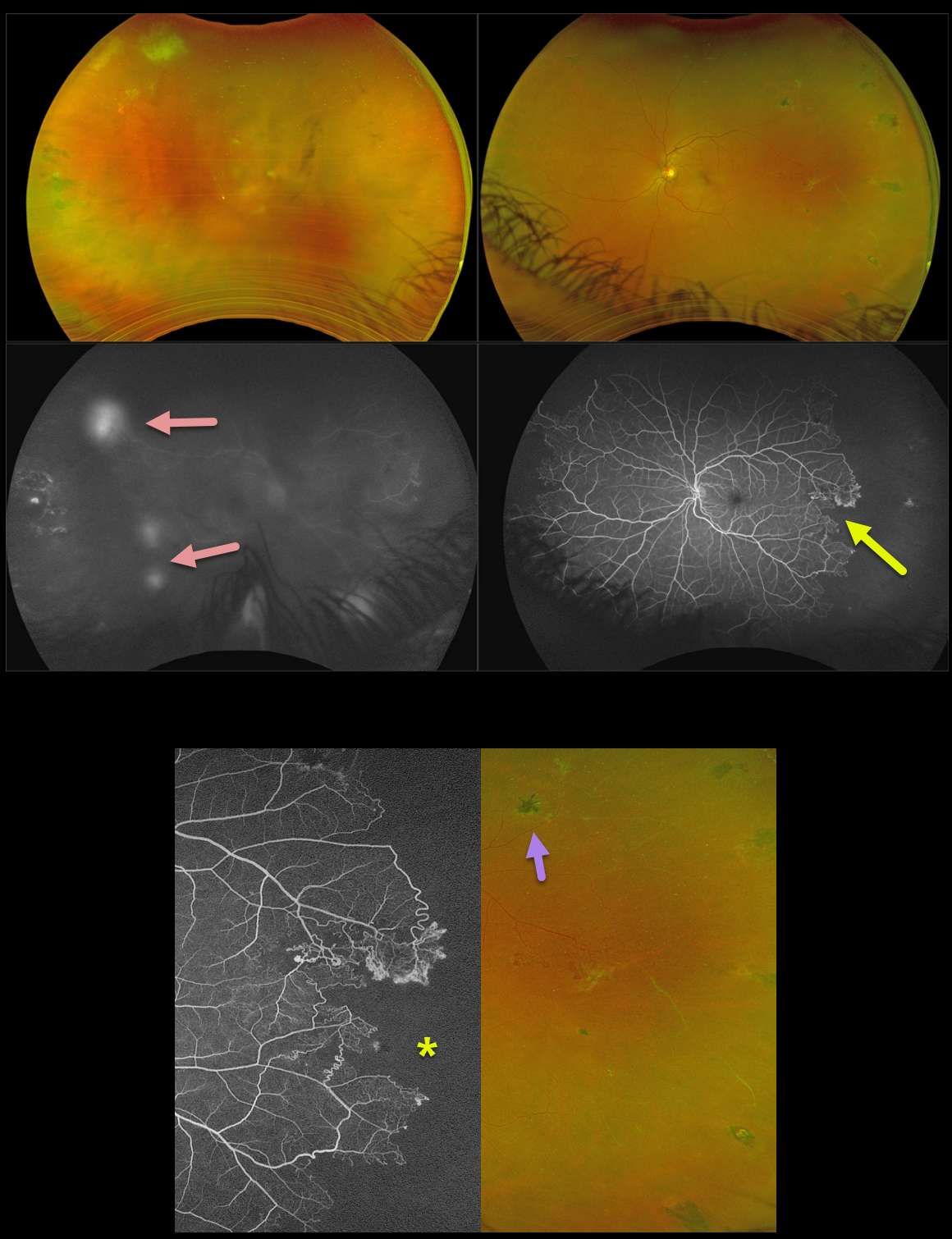
SICKLE CELL RETINOPATHY
Originally posted on @retina.rocks January 1, 2026
This 45YO male with a known history of sickle cell SC disease presented with new floaters and blurred vision in his right eye. Vision was 20/70 OD and 20/30 OS.
Optos color RG imaging shows a fairly dense vitreous hemorrhage OD, with variably fibrosed peripheral retinal neovascularization superotemporally. The left fundus shows temporal retinal ischemia with white vessels and mostly active retinal neovascularization along its posterior extent. Scattered black sunburst lesions are noted temporally as well. Fluorescein angiography OS shows an irregular, abrupt margin of peripheral complete capillary nonperfusion, especially temporally. The neovascularization in his OD shows late leakage.
Avastin was injected OD. Given the active vessels in his OS, along with the recent vitreous hemorrhage OD, we decided to prophylactically treat the left ischemic retina with scatter laser.
Learning Points:
Patients with sickle cell disease develop symptoms throughout their bodies as stiffened, sickled red blood cells block capillary beds. In the eye, this can cause proliferative sickle cell retinopathy (PSR) with sea-fan-shaped areas of peripheral retinal neovascularization developing at the junction of perfused and ischemic retina. The black sunburst lesions are thought to occur from intraretinal RPE migration in response to photoreceptor damage from intraretinal and subretinal hemorrhage (Asdourian et al, BJO 1975;59:710-716).
Complications include vitreous hemorrhage and traction retinal detachment. Although patients with SS disease tend to have more systemic complications, SC disease is usually associated with more severe ocular findings. Treatment with peripheral scatter laser photocoagulation is somewhat controversial since, unlike more typical proliferative retinopathies (ie from diabetes and branch retinal vein occlusions), these new vessels tend to auto-infarct.
For a great review article on sickle cell disease and the eye, see Elagouz et al, Surv Ophthalmology 2010;55;359-377.
CILIORETINAL ARTERY OCCLUSION
Malvika Singh and Manish Nagpal
Originally posted on @retina.rocks December 30, 2025
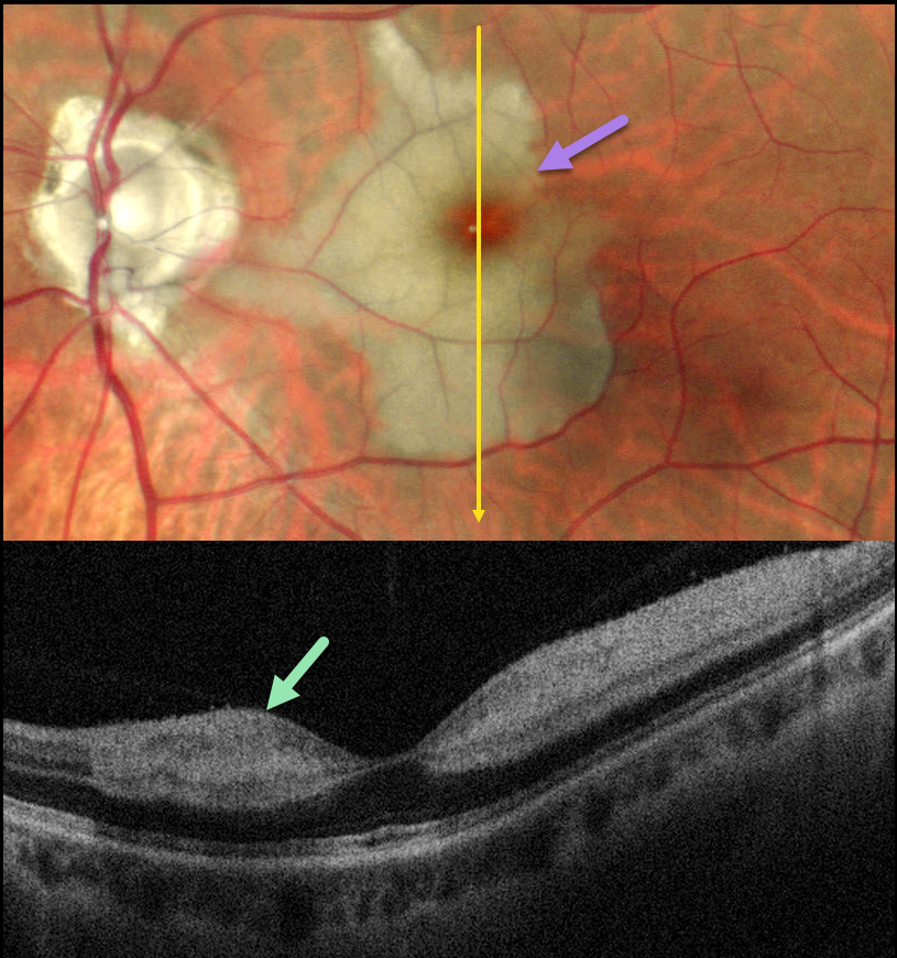
This healthy 46YO male presented with an 8-day history of a central scotoma in his left eye. Vision was 20/20 in his normal OD and 20/400 OS.
Pseudocolor SLO imaging shows an acute cilioretinal artery occlusion with foveal sparing. OCT scanning shows marked inner retinal hyperreflectivity with posterior shadowing.
Learning Points:
The inner two-thirds of the neurosensory retina is supplied by the central retinal artery, with the choroid supplying the RPE and photoreceptor layer. Cilioretinal arteries, which arise from the posterior choroidal circulation, are present in up to 50% of individuals, and bilateral in about 25% (Schneider et al, Acta Ophthalmologica 2021;99:e310-e318).
Their clinical significance usually arises from foveal preservation in eyes with concomitant retinal artery occlusions (Brown and Shields, Arch Ophthalmology 1979;97:84-92). However, they can also be a rare cause of primary branch retinal artery occlusion, either in isolation (our patient) or in association with other entities, including central retinal vein occlusions (Hayreh et al, Retina 2008;28:581-594).
COATS DISEASE
Shraddha Raj Shrivastava and Manish Nagpal
Originally posted on @retina.rocks December 15, 2025
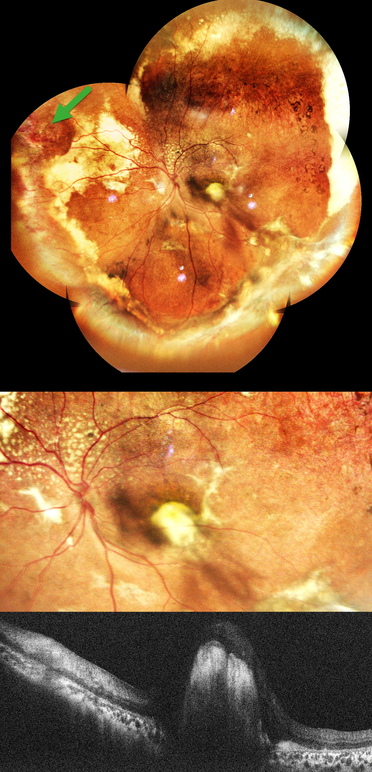
This healthy 11YO boy was brought in by his parents with a 1.5-year history of vision loss and outward deviation. Vision was 20/15 in his normal OD and light perception OS. Xanthocoria was noted on the pupillary light reflex.
Pseudocolor SLO imaging shows extensive peripheral subretinal lipid in all quadrants with telangiectatic and aneurysmal capillary dilations, most prominent in the nasal periphery. A superonasal orange-red elevated vasoproliferative mass is noted in the superonasal midperiphery. Pigmentary changes and RPE atrophy are observed in the superotemporal mid-peripheral retina. There is a large subfoveal lipid plaque that appears as a hyperreflective nodule on OCT. An exudative subtotal retinal detachment involving the inferior and nasal quadrants was noted clinically.
Learning Points:
Coats disease is characterized by vascular changes throughout the retinal vasculature, including the arteries, the capillary bed, and the veins. Larger dilated vessels in Coats can sometimes mimic angiomas, vascular tumors, or exudation from retinoblastoma. The presence of retinal telangiectasia can help establish the diagnosis of Coats in these cases.
Coats is usually unilateral in males. However, recent ultra-widefield angiographic studies indicate subtle vascular changes mostly in the temporal periphery of the fellow eye (Brockmann et al, BJO 2021;105:1444-1453).
Laser photocoagulation can be used to directly treat leaking telangiectatic vessels and ischemic retina. Intravitreal anti-VEGF injections can also be used as an adjunctive treatment. Pars plana vitrectomy (PPV) with external drainage of subretinal fluid plays an important role in the management of advanced Coats disease, particularly in cases with extensive exudative retinal detachment.
In our patient, pars plana vitrectomy with external drainage of subretinal fluid, along with endolaser/cryotherapy to leaking aneurysms, and an intravitreal anti-VEGF injection was advised.
HYPERTENSIVE RETINOPATHY
Shraddha Raj Shrivastava and Manish Nagpal
Originally posted on @retina.rocks December 11, 2025
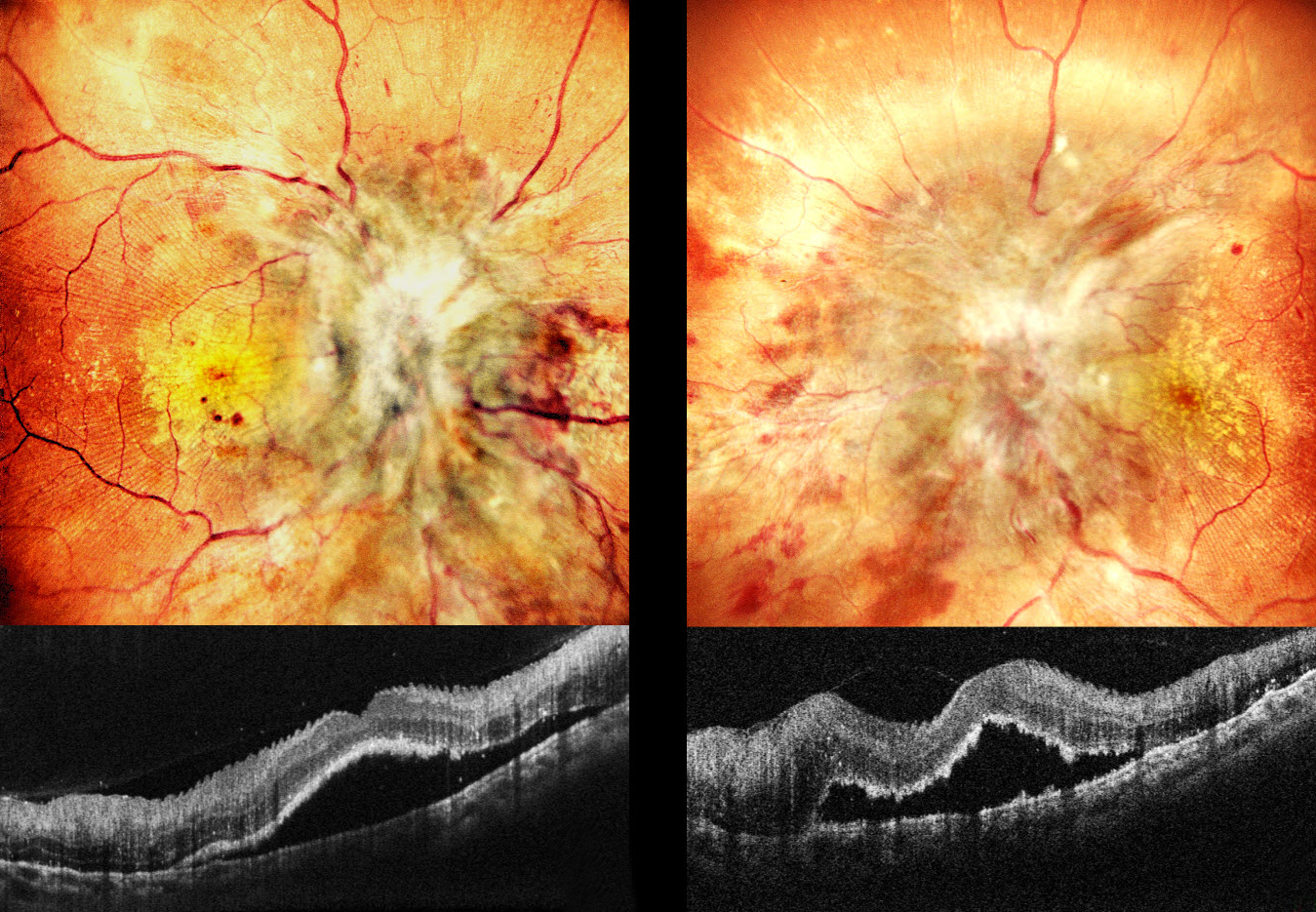
This 20YO male has a known history of chronic kidney disease. Despite hemodialysis, he’s had multiple episodes of malignant hypertension over the prior 2 months. He presented with 1 month of severe vision loss of counting fingers at 2 meters OU.
Pseudocolor SLO imaging shows profound bilateral disc edema with more peripheral nerve fiber layer infarcts (cotton-wool spots, CWS), retinal hemorrhages, and macular lipid. Fine inner retinal radiating striae are noted temporally. OCT scanning shows subfoveal fluid with hyperreflective ragged outer segments. Some small dots of outer retinal hyperreflectivity likely represent lipid.
Learning Points:
Malignant hypertension, defined as blood pressure above 180 systolic and/or 120 diastolic, is a life-threatening medical emergency. Eye doctors are often uniquely positioned to diagnose this condition (Shukla et al., Indian J Ophthalmol 2025;73:1552-1555). Patients will often present with bilateral optic nerve swelling, flame-shaped retinal hemorrhages, CWS, and, with more chronic disease, lipid precipitates in the nasal macular outer plexiform layer.
Posterior segment findings include exudative retinopathy, choroidopathy, and neuropathy. Retinopathy, due to breakdown of the inner retinal blood-retinal barrier, results in retinal hemorrhages and CWS. Choroidopathy, caused by choroidal fibrinoid necrosis, acutely creates exudative retinal detachment and, upon resolution, can leave behind generalized coarse, focal (Elschnig spots) or linear (Siegrist streaks) pigmentary changes. Papilledema is defined as bilateral disc swelling due to raised intracranial pressure. The nerve findings in malignant hypertension are likely due more to localized ischemic changes than to elevated intracranial pressure (Kishi et al, Arch Ophthalmology 1985:1198-1206).
RETINAL ARTERIOVENOUS MALFORMATION
Seif Anwar
Originally posted on @retina.rocks December 1, 2025
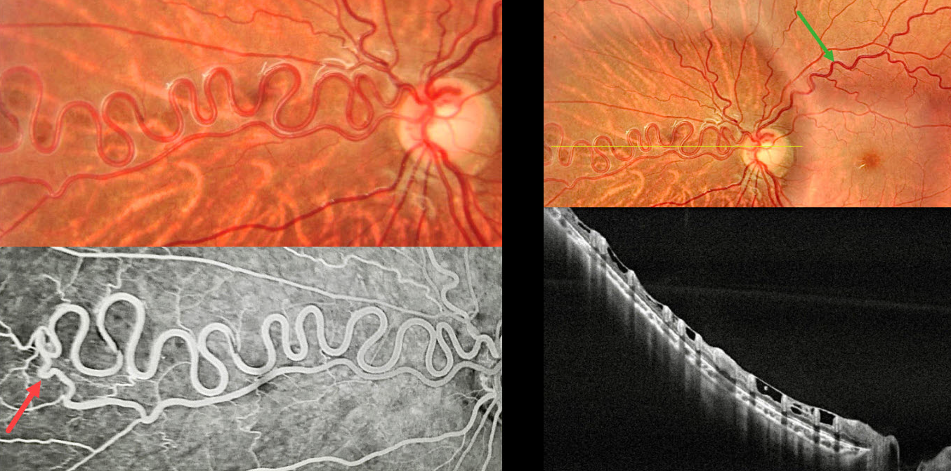
This healthy 12YO boy was referred for asymptomatic retinal findings. Vision was 20/20 in his normal OD and 20/20 OS.
Color photography shows a dilated multi-corkscrewed arteriole extending into the nasal midperiphery. The major superotemporal vein is also somewhat dilated and tortuous. On OCT, these anomalous vessels are hyperreflective and extend through the full thickness of the retina. ILM draping spans between each vessel. Angiographically, the artery directly connects to a paired vein. There is no angiographic leakage.
Learning Points:
A retinal arteriovenous malformation (AVM) is a congenital connection between an artery and a vein without an intervening capillary network. These lesions appear along a spectrum ranging from small, isolated lesions to the large ‘bag of worms’ appearance. These more extensive lesions are usually part of the Wyburn-Mason Syndrome, which includes ocular and midbrain vascular lesions. Although retinal AVMs are usually stable lesions, secondary venous occlusions may occur. Our patient has an isolated retinal AVM due to the lack of intracranial lesions on neurologic imaging.
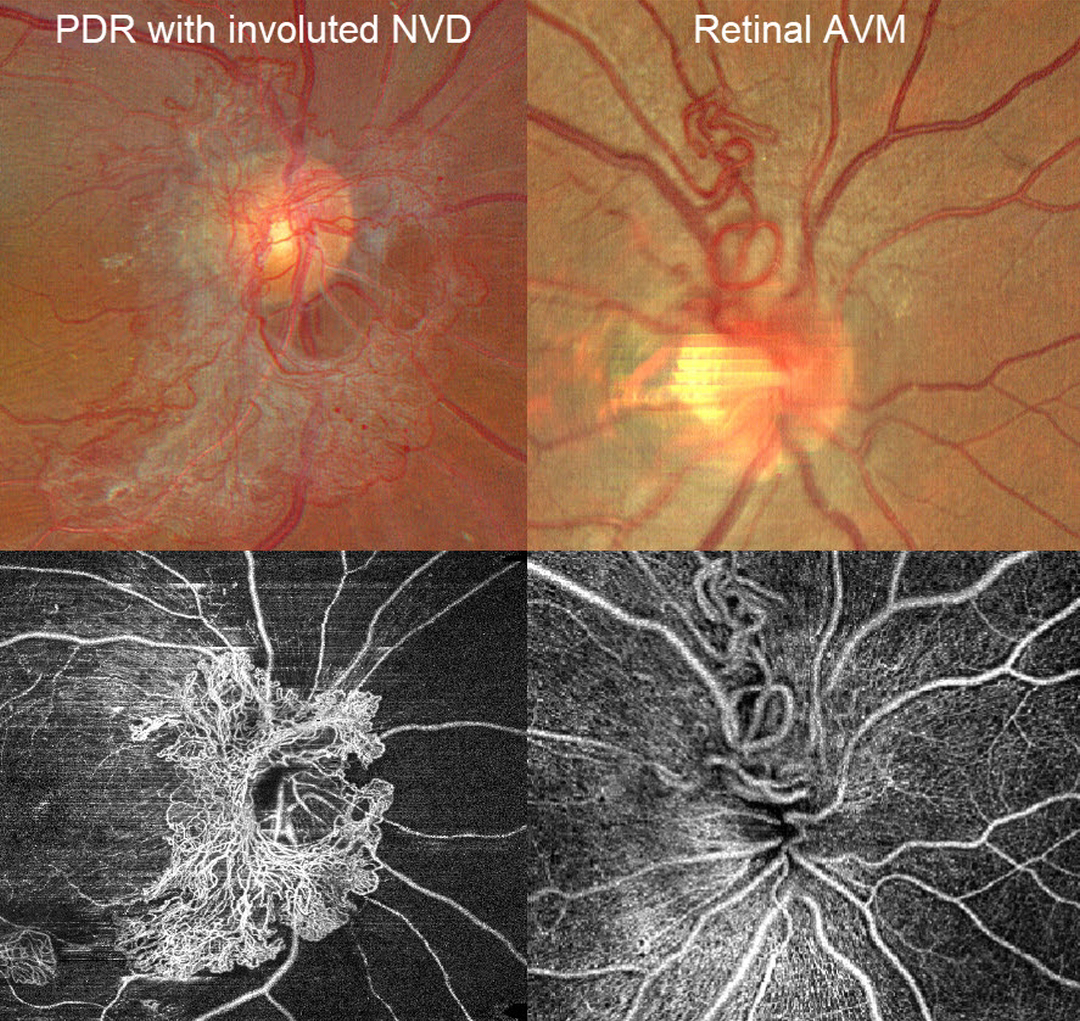
PROLIFERATIVE DIABETIC RETINOPATHY NVD VS RETINAL ARTERIOVENOUS MALFORMATION
Ayushi Gupta and Vishal Agrawal.
Originally posted on @retina.rocks November 12, 2025
A patient with disc neovascularization (NVD) due to proliferative diabetic retinopathy (PDR) following full panretinal photocoagulation is compared with a patient with an asymptomatic retinal arteriovenous malformation (AVM).
Color photography of the PDR patient shows diffuse flat NVD with variable tight capillary networks of new vessels, admixed with larger vessels and fibrosis. OCT angiography (OCTA) confirms flow signals within these vessels, along with diffuse capillary loss superior, nasal, and inferior to the nerve. Color photography and OCTA of the retinal AVM show a tangle of arterioles and veins extending superiorly from the nerve. Fluorescein angiography, if performed, would show leakage from the NVD but not from the AVM.
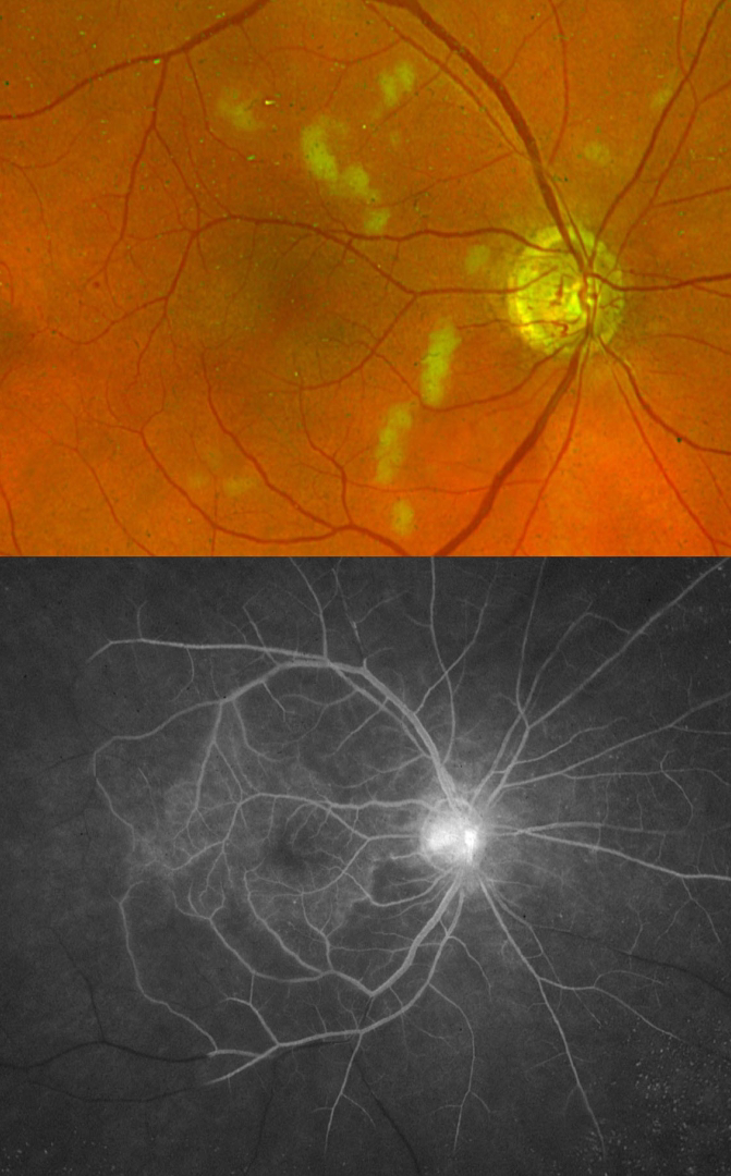
IDIOPATHIC RETINAL VASCULITIS ANEURYSMS AND NEURORETINITIS (IRVAN)
Mattie Adams
Originally posted on @retina.rocks November 5, 2025
This healthy 63YO male presented with an ill-defined history of blurriness in his right eye upon first waking in the morning. Vision was 20/100 OD and 20/20 in his normal OS. There was diffuse rubeosis OD on slit lamp examination, and intraocular pressure was 28 mmHg.
Optos color RG imaging shows scattered nerve fiber layer infarcts with a rare retinal hemorrhage. Fluorescein angiography shows complete capillary nonperfusion distal to the peripheral macula with late optic nerve leakage. Carotid Doppler and CT angiography were negative.
Intravitreal Avastin was given, followed by full panretinal photocoagulation. He subsequently developed florid rubeosis with neovascular glaucoma requiring an Ahmed tube.
Learning Points:
Idiopathic Retinitis, Vasculitis, Aneurysms, and Neuroretinitis (IRVAN) is a rare disease of unknown etiology characterized by retinal vasculitis, aneurysmal arterial dilations, and neuroretinitis. The diagnosis is clinical, as there are no systemic associations or diagnostic laboratory tests. Early panretinal photocoagulation (PRP) to areas of widespread retinal nonperfusion is recommended to prevent neovascular complications and vitreous hemorrhage (Samuel et al Ophthalmology 2007;114:1526-1529).
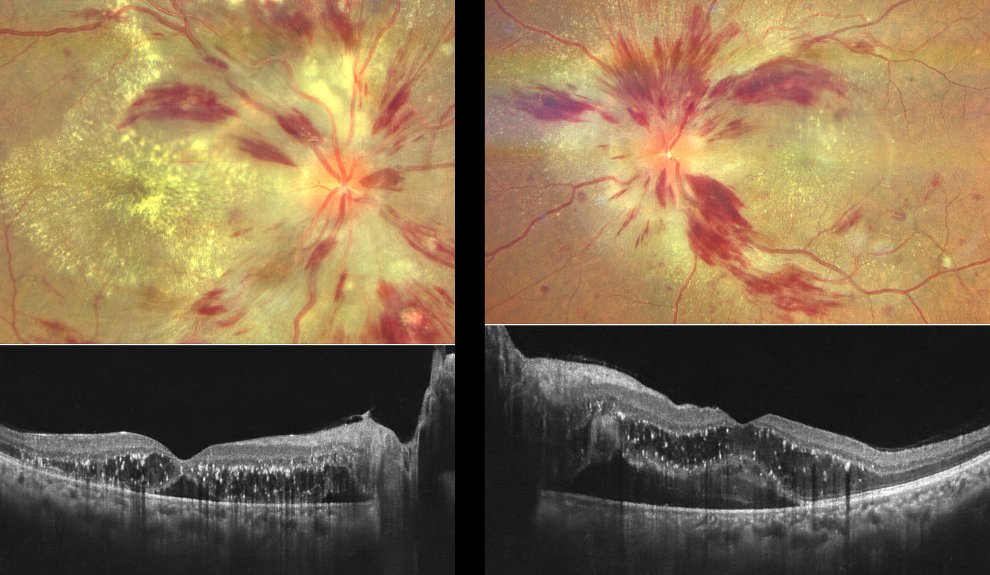
HYPERTENSIVE RETINOPATHY
Ayushi Gupta and Vishal Agrawal
Originally posted on @retina.rocks October 27, 2025
This previously healthy 40YO female presented with 2 months of painless bilateral vision loss. She had occasional headaches, which she self-medicated with painkillers. Vision was counting fingers OD and 20/120 OS.
Color photography in her right eye shows a mildly swollen nerve, extensive hemorrhages in the nerve fiber layer, nerve fiber layer infarcts (cotton-wool spots), and a macular lipid star. OCT scanning shows outer retinal fluid with hyperreflective lipid flecks and foveal subretinal fluid. Similar findings are present in her left eye.
Her blood pressure in our office was 166/122 mmHg. She was immediately referred for emergent medical care.
Learning Points:
Malignant hypertension, defined as blood pressure above 180 systolic and/or 120 diastolic, is a life-threatening medical emergency. Eye doctors are in a unique position to often diagnose this condition. Patients will often present with bilateral optic nerve swelling, flame-shaped retinal hemorrhages, and, with more chronic disease, lipid precipitates in the nasal macular outer plexiform layer. Since the macular fluid emanates from the optic nerve, the macular lipid and thickening are virtually always worse nasally and are an important clinical clue for this diagnosis.
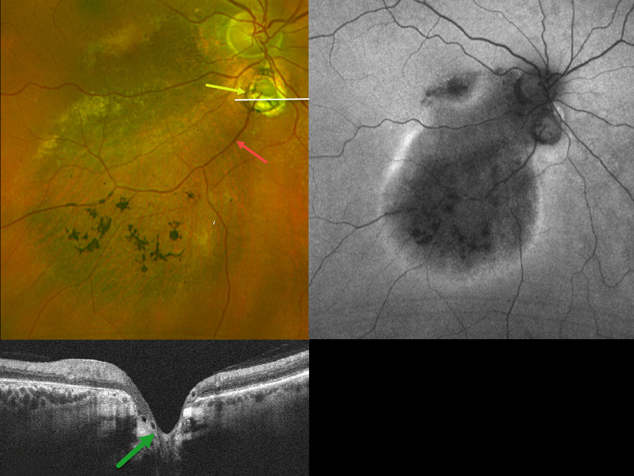
CHOROIDAL COLOBOMA WITH OPTIC NERVE PSEUDODUPLICATION
Originally posted on @retina.rocks October 8, 2025
This 60YO female was referred for asymptomatic unilateral fundus findings in her right eye. Vision was 20/30 OD.
Optos color RG imaging shows a choroidal coloboma just inferior to the disc, giving the appearance of a pseudoduplicated optic nerve. Coarse pigmentary changes with some pigment migration extend from the macula into the inferior midperiphery. An inferotemporal retinal vein drains directly into the choroid through the coloboma. Triton swept-source OCT shows that the coloboma overlies an area of ectatic sclera. Fundus autofluorescence (FAF) shows variable hypo-FAF within the area of clinical pigmentary changes, with a surrounding rim of hyper-FAF.
Learning Points:
Choroidal colobomas are most often due to failure of the optic vesicle and choroidal fissure to close during fetal development. Lesions like this may also be from other causes, including toxoplasmosis. Choroidal coloboma can present as solitary or multiple lesions. Visual prognosis depends on the location and involvement of the optic nerve and macula.
Pseudoduplication of the optic nerve occurs when a round choroidal coloboma or chorioretinal scar about the size of the optic nerve is associated with overlying radiating blood vessels (Bloom et al, Retinal Cases 2022;16:174-176). In our experience, it is most common for toxoplasmosis scars to be located near the optic nerve.
Under normal conditions, the retinal and choroidal circulations remain separate and distinct. Rarely, the two circulations connect directly through a chorioretinal anastomosis. These can occur in chorioretinal scars (typically from inactive toxoplasmosis lesions), in macular telangiectasia, and in disciform scars.
The prominent gutter of inferior pigmentary changes indicates a prior retinal detachment, most likely related to spontaneously resolved fluid associated with the coloboma (Tanaka et al, Ophthalmology Retina 2021;5:702-710). Similar pigmentary changes are seen in central serous retinopathy, although our patient’s macular OCTs showed no pachychoroid features (not shown). Finally, this may represent previously resolved pit-like macular fluid, which is rarely found in the absence of a pit or severe glaucomatous cupping (Fujimoto et al, Ophthalmology Retina 2023;7:811-818).
RETINAL VENOUS MALFORMATION
Ayushi Gupta and Vishal Agrawal
Originally posted on @retina.rocks October 7, 2025
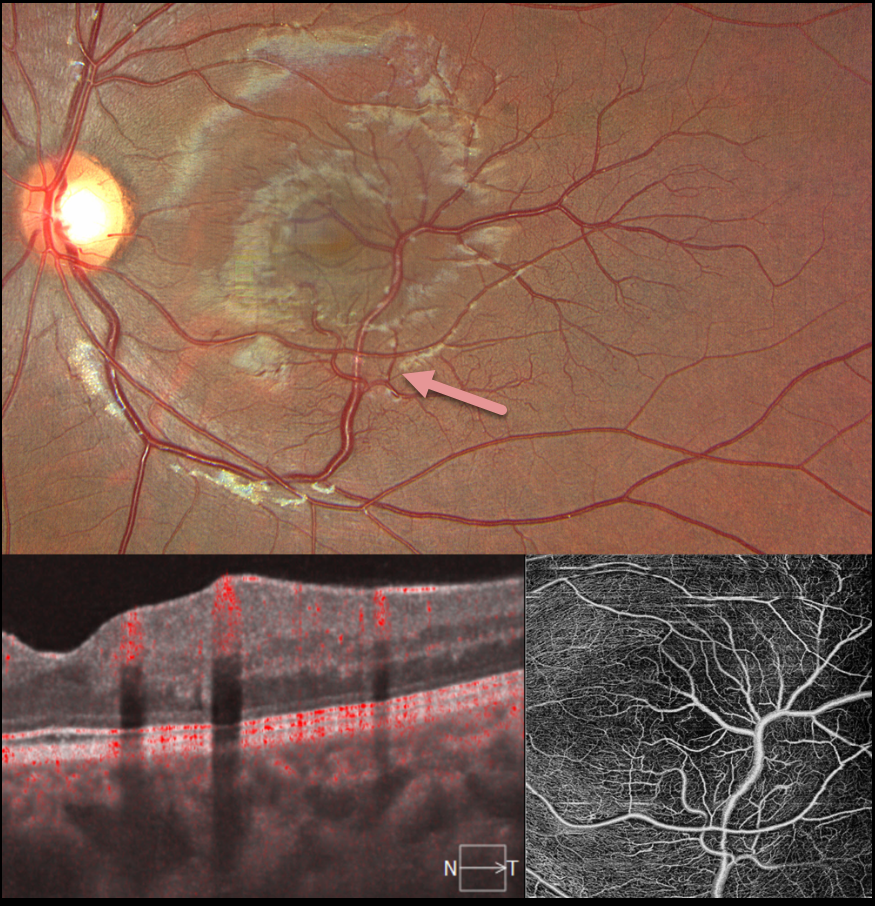
This 17YO female presented for a routine eye examination. Vision was 20/20 in her normal OD and 20/20 OS. Color imaging shows an inferior retinal venous malformation that drains the inferior and superotemporal macula. En face and B-scan OCT angiography show these vessels to lie in the inner retina.
Learning Points:
Retinal vessels virtually always respect the horizontal meridian. Retinal venous malformations are congenital anomalous vessels that, by definition, cross the horizontal. Originally described by Gary Brown et al as congenital retinal macrovessels (Arch Ophthalmology 1982;100:1430-1436), these vessels are usually located in the macular region and are virtually always venous. These lesions are now called retinal venous malformations, since about one-quarter of patients have associated venous anomalies in the brain (Pichi et al, JAMA Ophthalmol 2018;136:372-379).
RETINAL ARTERIAL MACROANEURYSM
Originally posted on @retina.rocks September 22, 2025
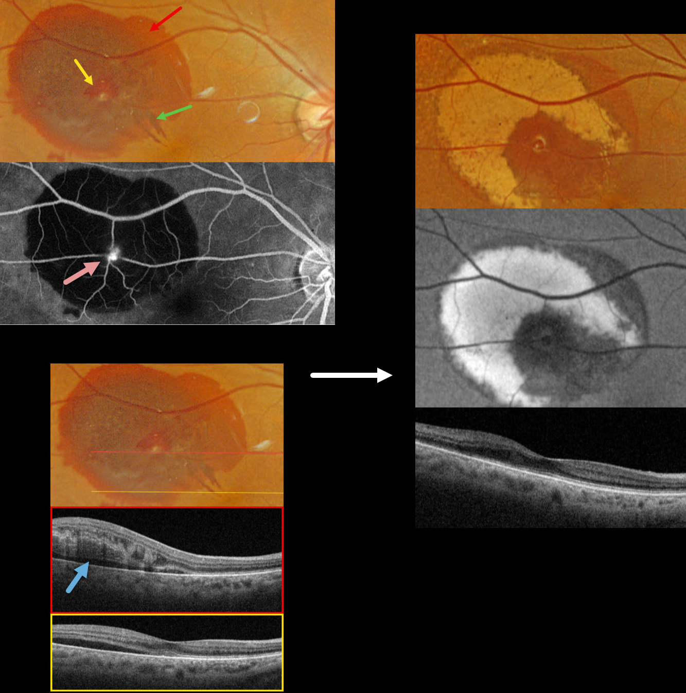
This 83YO female presented with recent blurred vision in her right eye. Vision was 20/40.
Optos color RGB imaging shows subretinal blood in the superotemporal macula. A small amount of inner vs preretinal blood surrounds a small yellow-white retinal arterial macroaneurysm (RAM). In addition, some radiating outer plexiform blood radiates towards the macular center. Fluorescein angiography shows a hyperreflective, leaking RAM with blockage by surrounding subretinal blood. Swept source OCT shows variable outer retinal edema, hyperreflective blood, and subretinal fluid.
Observation was recommended. Six weeks later, the blood is absorbing and turning yellow superiorly and temporally. This devitalized blood is markedly hyper-autofluorescent. The foveal fluid resolved, and vision improved to 20/30.
Learning Points:
A RAM is thought to occur in a weakened arterial wall from arteriosclerosis and appears as a yellow-white dilated lesion along a retinal arterial within the first 3 bifurcations from the optic nerve. There are 2 distinct presentations, hemorrhagic or exudative. The hemorrhagic type often presents, as in this case, with pathognomonic multiple layers of blood. Acute macular exudation can be treated with observation, anti-VEGF injections, or thermal laser
RETINAL VASCULAR LOOP
Kanwaljeet Harjot Madan
Originally posted on @retina.rocks September 16, 2025
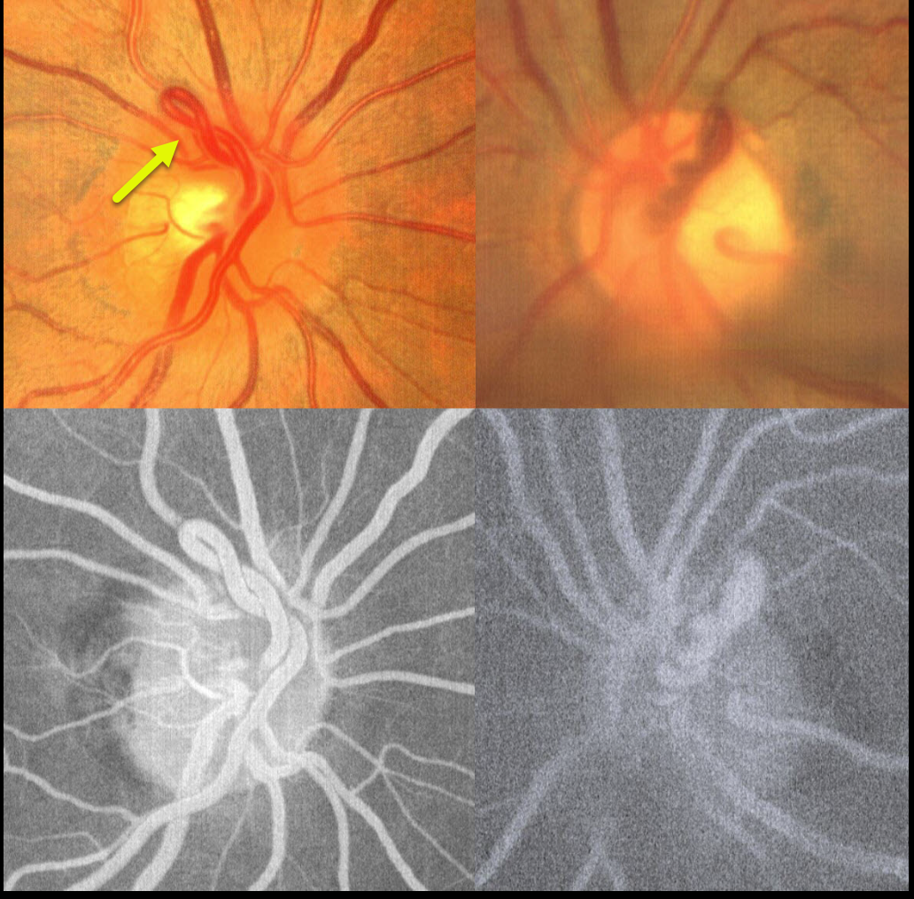
This healthy 32YO female was referred for possible asymptomatic disc neovascularization. Vision was 20/20 OU. Fundus photography shows bilateral vascular loops that do not leak angiographically.
Learning Points:
A retinal vascular loop is a benign congenital anomaly that presents as an elevated and twisted bundle of vessels projecting from the disc into the vitreous cavity. Is it usually unilateral and arterial. Although usually just an incidental finding, these can rarely cause a branch arterial or venous occlusion (see Retinal Cases Brief Rep 2014;8;124-126 and Ophthalmology 2020;127;124-126). They should also not be confused with disc neovascularization, which consists of much smaller caliber vessels that leak on fluorescein angiography.
RETINITIS PIGMENTOSA WITH CME
Rohan Jain and Manish Nagpal
Originally posted on @retina.rocks September 15, 2025
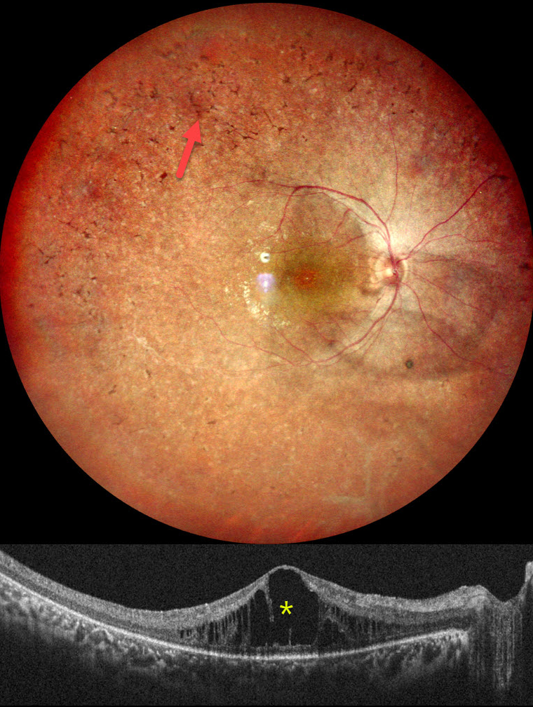
This healthy 18YO female complained of night blindness for a few years. Her mother and sister have a history of retinitis pigmentosa (RP). Vision was 20/90 OU.
Pseudocolor SLO imaging of her right eye shows typical RP scarring with intraretinal pigment migration extending from the macula into the retinal periphery. There is loss of the foveal reflex and cystoid macular edema (CME). Identical findings were noted in the OS (not shown).
Learning Points:
RP is a clinically and genetically heterogeneous group of inherited retinal disorders, which can present sporadically or with any inheritance pattern (autosomal dominant, autosomal recessive, sex-linked, or mitochondrial). It is characterized by diffuse, progressive dysfunction of predominantly rod photoreceptors, followed by RPE degeneration with intraretinal pigment migration. Visual impairment usually presents as night blindness and progressive visual field loss.
CME is observed in up to 20% of RP patients, often without angiographic leakage. Pathological mechanisms include vitreous traction/epiretinal membranes or a breakdown of the blood-retinal barrier due to inflammation or RPE/Muller cell dysfunction (Gaudric et al, Progress in Retinal and Eye Research 2022;91:101092). First-line treatment is topical or oral carbonic anhydrase inhibitors (CAIs). Other options for refractory CME include steroids (topical, periocular, or intravitreal), anti-VEGF injections, or vitrectomy for a tractional component. Our patient was given topical CAIs but was immediately lost to follow-up.
CRAO
Tejaswita Verma and Manish Nagpal
Originally posted on @retina.rocks September 1, 2025
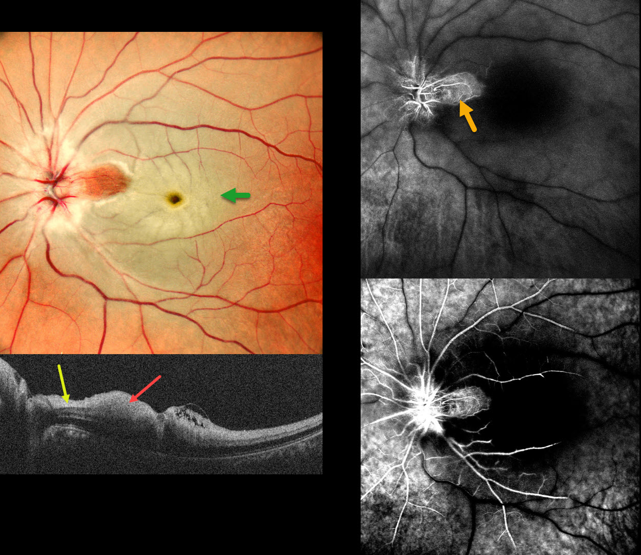
This 37 YO male presented with acute vision loss in his left eye 3 days earlier. Vision was 20/20 in his normal OD and 20/200 in his OS.
Pseudocolor SLO imaging shows a fresh central retinal artery occlusion (CRAO) with sparing of a small area of temporal peripapillary retina supplied from a patent cilioretinal artery. OCT scanning shows opaque, hyperreflective, thickened inner retinal layers, except for a small area of more normal retina within the cilioretinal sparing.
Fluorescein angiography shows profound diffuse retinal nonperfusion and delayed filling except for the nasal macula. Workup elsewhere included an MRI showing left ICA and left MCA chronic infarcts.
Learning Points:
The inner two-thirds of the neurosensory retina is supplied by the central retinal artery, with the choroid supplying the RPE and photoreceptor layer. Acute CRAO, therefore, presents with a white edematous inner retina that spares the fovea. Cilioretinal arteries, which arise from the posterior choroidal circulation, are present in up to 50% of individuals, and bilateral in about 25% (Schneider et al, Acta Ophthalmologica 2021; 99:e310-e318).
RETINAL VENOUS MALFORMATION
The European VitreoRetina Society (EVRS) and P.C. Ranjan
Originally posted on @retina.rocks August 29, 2025
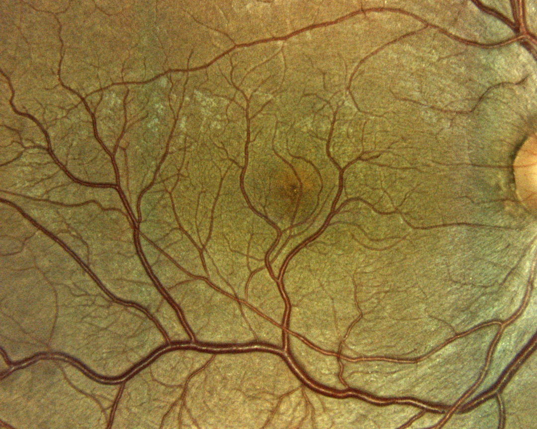
This 50YO male presented for a routine eye examination. Vision was 20/20 OD and 20/20 in his normal OS. Color imaging shows an inferior retinal venous malformation that drains nearly the entire macula.
Learning Points:
Retinal vessels virtually always respect the horizontal meridian. Retinal venous malformations are congenital anomalous vessels that, by definition, cross the horizontal. Originally described by Gary Brown et al as congenital retinal macrovessels (Arch Ophthalmology 1982;100:1430-1436), these vessels are usually located in the macular region and are virtually always venous. These lesions are now called retinal venous malformations, since about one-quarter of patients have associated venous anomalies in the brain (Pichi et al, JAMA Ophthalmol 2018;136:372-379).
COATS DISEASE
Ayushi Gupta and Vishal Agrawal
Originally posted on @retina.rocks August 26, 2025
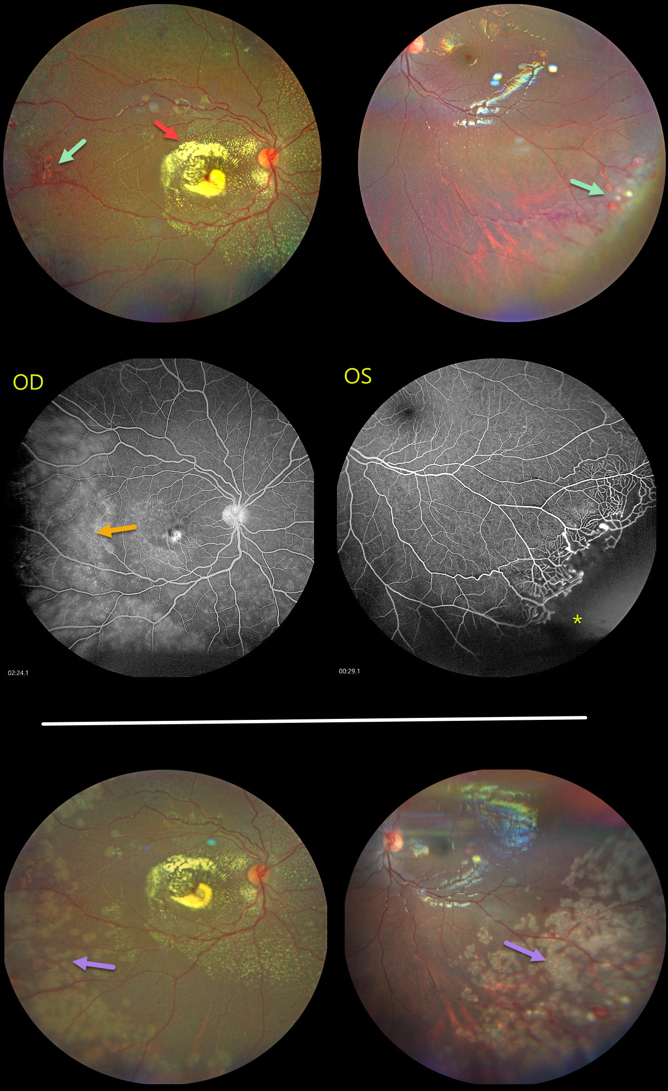
This healthy 6YO girl’s parents noted that her right eye had been deviating outward for the past several weeks. Vision was light perception OD and 20/20 OS.
Color photography shows fairly dense subfoveal lipid with more diffuse specks of subretinal lipid radiating outwards from the disc. Telangiectatic vessels are noted temporally OD and also in the left inferotemporal periphery. Fluorescein angiography shows diffuse leakage from these vessels OD, with distal retinal ischemia OS.
Scatter laser to the ischemic retina, along with focal treatment of the larger telangiectatic vessels, was applied. Nine months later, vision was unchanged, with consolidation of the subfoveal lipid scar OD along with persistent telangiectasia OS.
Learning Points:
Described by George Coats in 1908, Coats disease is an idiopathic disorder typically unilateral in males. It is characterized by retinal vascular telangiectasia involving capillaries, arterioles, and venules. Patients can present with a wide range of findings, from minimal exudation to total exudative retinal detachment. The leaking vessels are usually treated to prevent exudative complications. Thermal laser photocoagulation is usually the initial modality, but anti-VEGF injections can also be used to stabilize the vascular leakage. Patients will often require multiple rounds of treatment. See Sigler et al for a review of the current management (Survey Ophthalmology 2014;59:30-46).
Dyskeratosis congenita or familial exudative vitreoretinopathy should be considered for patients with frank bilateral disease (Teixeira et al, Arch Ophthalmol 2008;126:134-135), in women with Coats-like findings (Johnson et al, Genetics 2009;30:181-184), or in patients with a family history of similar findings. Our patient’s family history was negative, and her nail beds were normal. Genetic testing was not possible due to cost concerns.
BRVO WITH COLLATERALS AND DRIL
Malvika Singh, Akansha Sharma and Manish Nagpal
Originally posted on @retina.rocks August 19, 2025
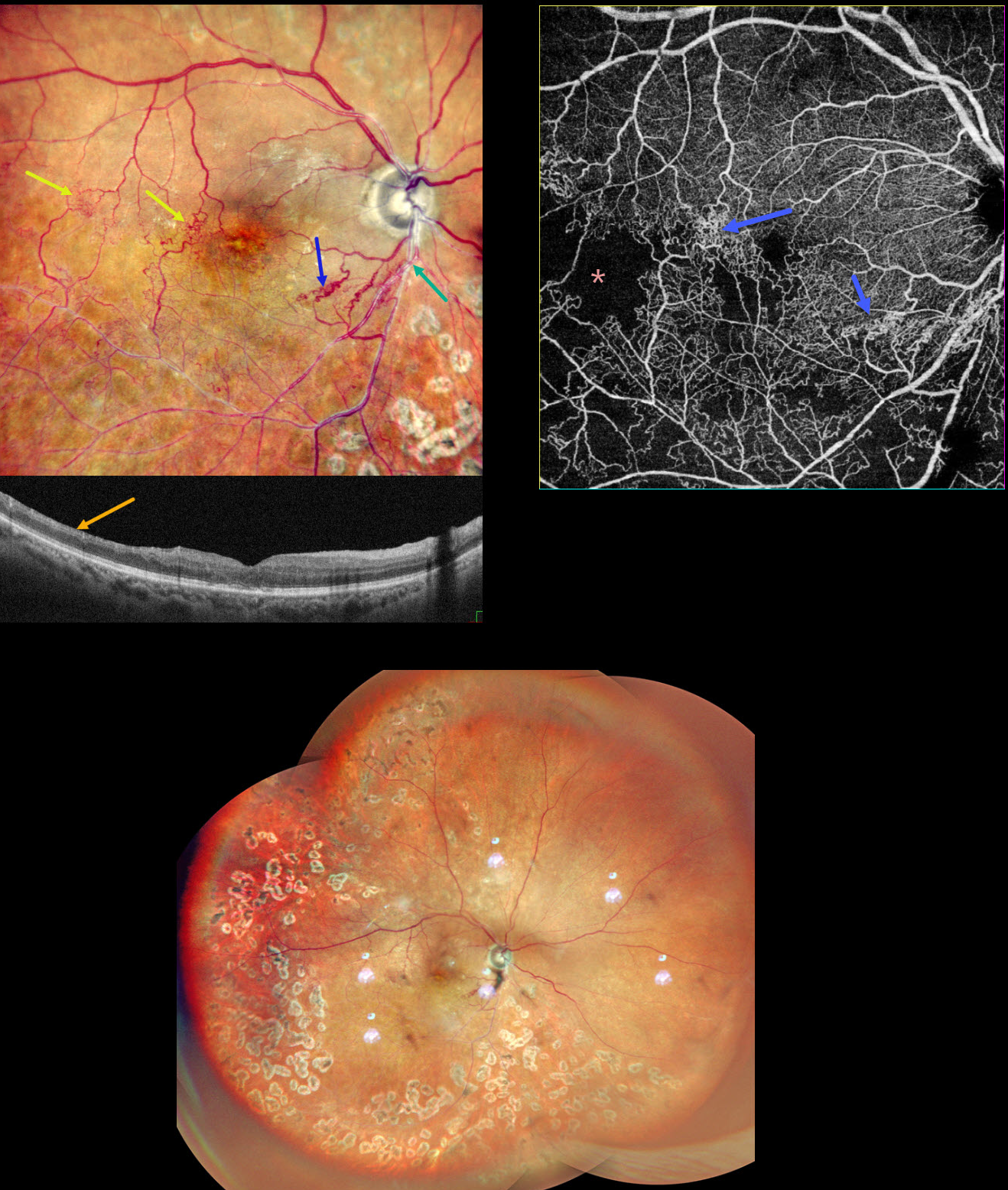
This 65YO male presented with a branch retinal vein occlusion (BRVO) in his right eye and 20/40 vision. He was treated elsewhere about a year ago with 3 intravitreal injections and scatter laser 7 years earlier.
Pseudocolor SLO shows an old major inferotemporal ischemic BRVO, which arises from an arteriovenous crossing just inferior to the disc. Extensive collateral vessels bypass the occlusion and cross the temporal horizontal raphe. OCT scanning shows thinned, featureless inner retina (disorganization of retinal inner layers, DRIL). Scatter laser is noted temporally and inferiorly. OCT angiography shows variable capillary loss throughout the BRVO distribution and highlights the collateral vessels.
Learning Points:
Collateral vessels are dilated, pre-existing vessels that bypass an occlusion. These are most commonly seen in the posterior pole following retinal venous occlusions and are exceedingly rare with arterial occlusions. The endothelial tight junctions are intact, so they don’t leak angiographically. This helps differentiate them from neovascularization, which does leak.
DRIL is seen in various retinal vascular disorders, including diabetic macular edema and retinal vein occlusions. It is generally associated with worse vision and increased anti-VEGF treatment burden (see Babiuch et al, JAMA Ophthalmol 2019;137:38-46).
ANEMIC PRERETINAL HEMORRHAGES
The European VitreoRetina Society (EVRS) and Rajiv Gandhi
Originally posted on @retina.rocks August 15, 2025
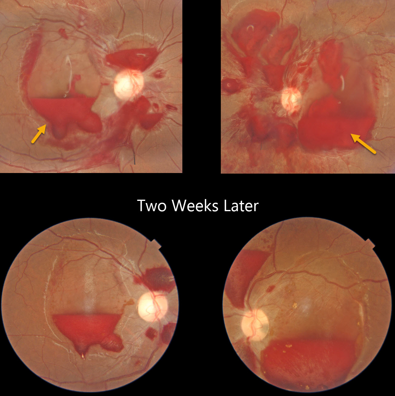
This healthy 16YO female presented with 2 days of acute bilateral vision loss. She denied any trauma or Valsalva. Vision was 20/400 OU.
Color photography shows extensive bilateral preretinal and sub-internal limiting membrane hemorrhages. Some intraretinal white-centered hemorrhages are also noted. She was found to have profound iron-deficiency anemia due to very heavy menstruation with a hemoglobin of 5.2 gm/dl.
The blood was improving bilaterally two weeks later. Following multiple blood transfusions, the posterior segment hemorrhages spontaneously resolved (not shown).
RETINAL ARTERIOVENOUS MALFORMATION
Rohan Jain and Manish Nagpal
Originally posted on @retina.rocks August 12, 2025
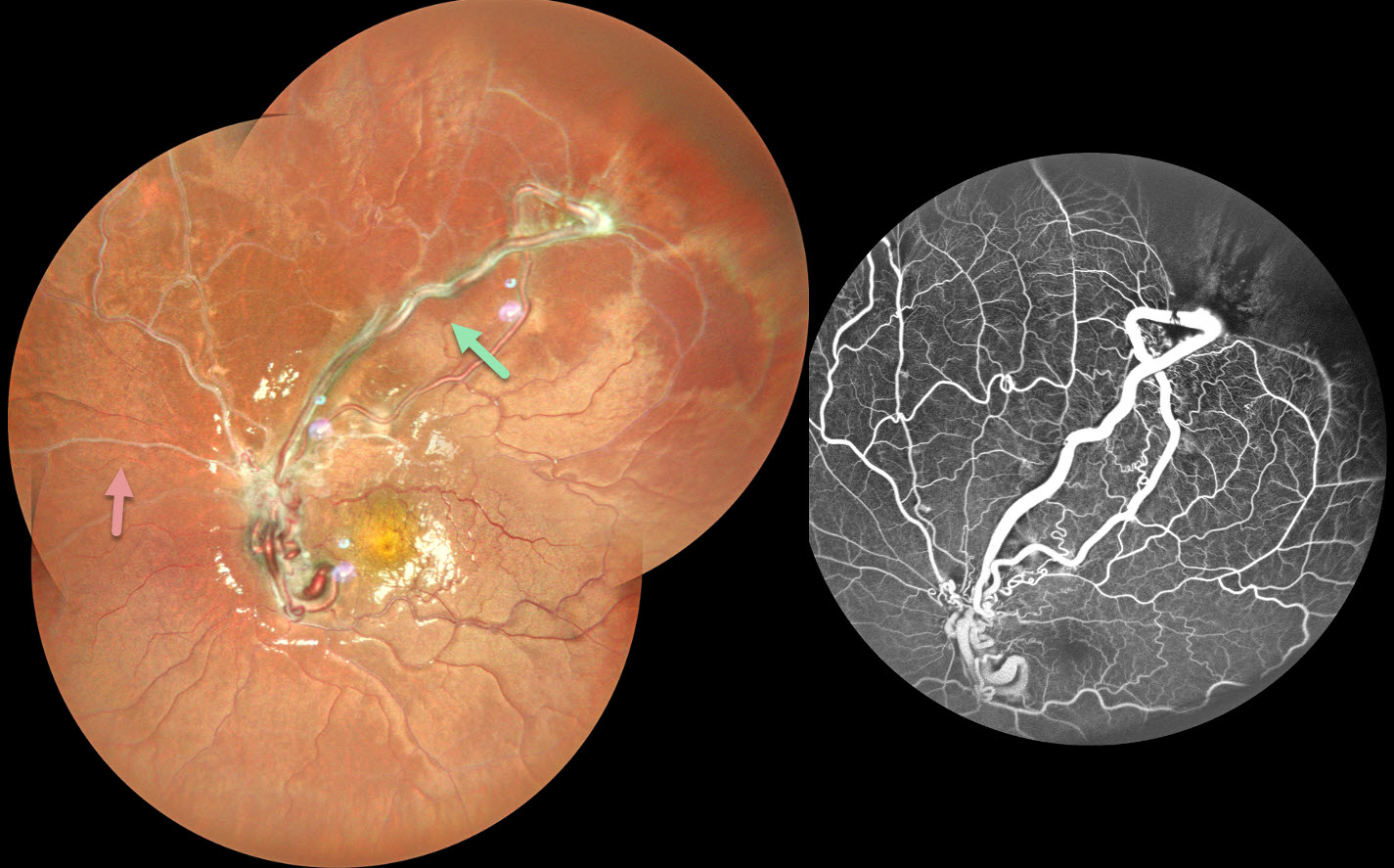
This healthy 10YO female presented with a 3-month history of decreased vision in her left eye. Vision was 20/15 in her normal OD and 20/200 OS.
Pseudocolor SLO imaging shows tangles of markedly dilated and tortuous vessels, which extend into the superotemporal midperiphery and obscure the optic disc. The retinal vessels are sheathed nasally and superiorly. Fluorescein angiography shows no leakage with ischemic retina distal to the anomalous vessels. Neurologic imaging was negative.
Learning Points:
A retinal arteriovenous malformation (AVM) is a congenital connection between an artery and a vein without an intervening capillary network. These lesions appear along a spectrum ranging from small, isolated lesions to the large ‘bag of worms’ appearance as seen in our patient. These more extensive lesions are usually part of the Wyburn-Mason Syndrome, which includes vascular lesions in the eyes and midbrain.
Retinal venous malformations, also known as retinal macrovessels, are now thought to represent an AVM (Pichi et al., JAMA Ophthalmol 2018;136:372-379). These lesions can rarely bleed or cause retinal venous occlusions. The diffuse vascular sheathing may represent such a prior occlusion.
LEUKEMIA WITH PSEUDO-BILATERAL CRVO
Mattie Adams
Originally posted on @retina.rocks July 28, 2025
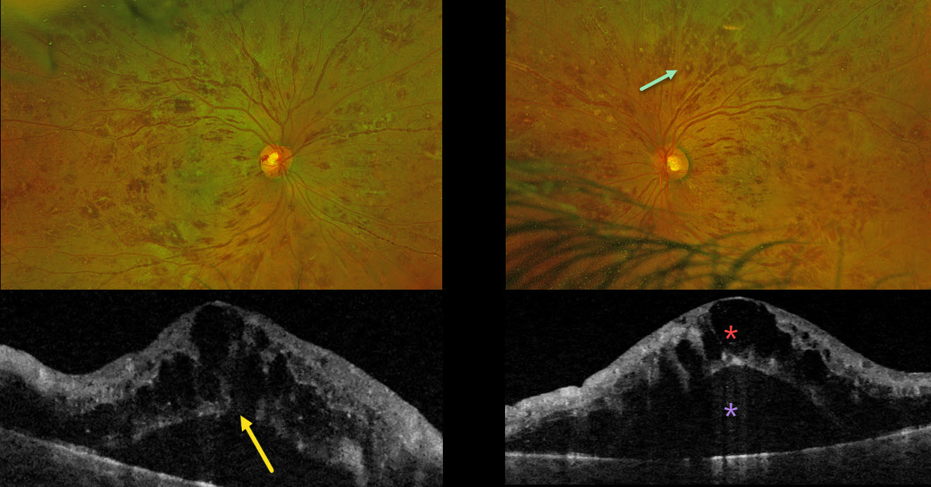
This previously healthy 38YO male presented with 1 month of bilateral blurriness. Vision was 20/60 OD and 20/80 OS.
Optos color RG imaging shows severe, bilateral, mostly white-centered retinal hemorrhages extending from the maculae into the retinal peripheries. Triton swept-source OCT shows marked bilateral cystic foveal swelling with subretinal fluid. An outer macular hole is probably present OD. His white blood cell count was markedly elevated at 206,400. Subsequent emergent oncologic workup led to the diagnosis of chronic myelogenous leukemia.
Learning Points:
The term “Roth spot” was named after Dr. Moritz Roth, who, in 1872, noted white-centered hemorrhages in patients with bacterial endocarditis. He felt that these white spots were septic emboli that originated from an infected cardiac valve. However, similar lesions can be seen in many other disorders, including leukemia, hyperviscosity, anemia, diabetes, and hypertension (Duane et al Ophthalmology 1980;87:66-69). The white material may represent a variety of causes, including fibrin. So, these lesions are best called white-centered hemorrhages, unless one is specifically referring to those found associated with bacterial endocarditis.
In our patient’s case, the severity of the retinal hemorrhages gave an appearance resembling bilateral central retinal vein occlusions (CRVO). However, CRVO hemorrhages are located predominantly in the nerve fiber layer (our patients’ hemorrhages were deep and white-centered), and the optic nerve is usually swollen with this degree of hemorrhage (our patients’ nerves were flat).
SICKLE CELL RETINOPATHY
Originally posted on @retina.rocks July 10, 2025
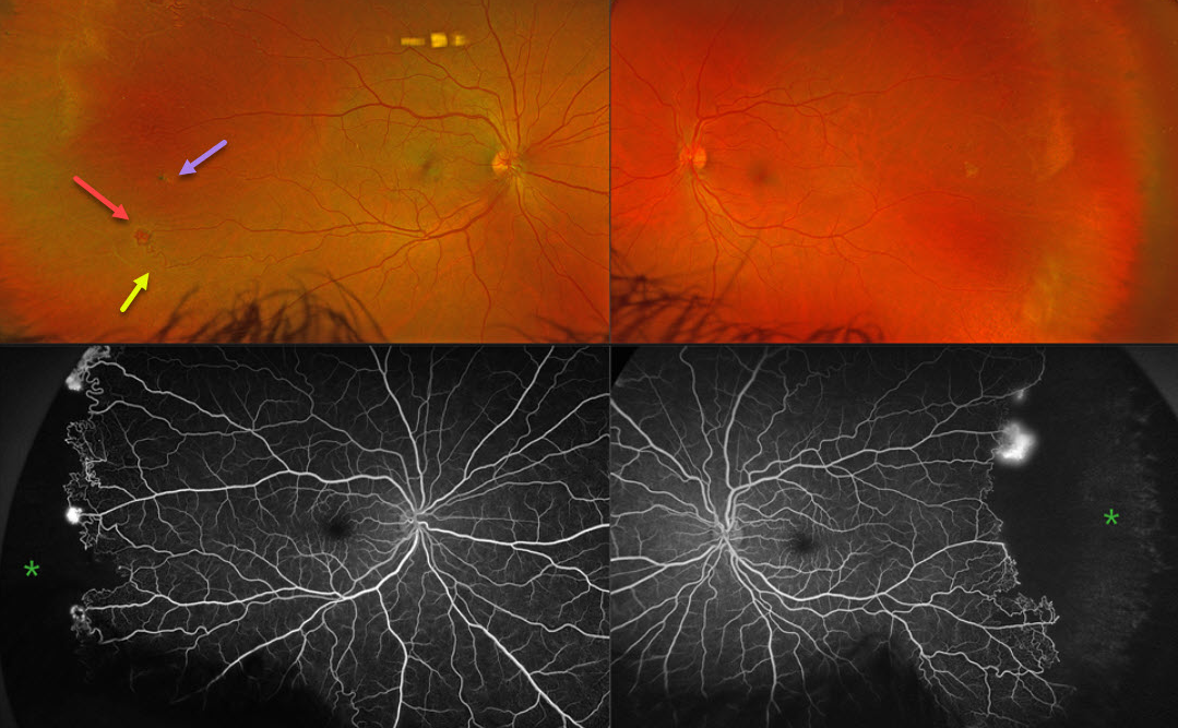
This 43YO male was referred for asymptomatic retinal findings. He has a known history of sickle cell disease. Vision was 20/20 OU.
Optos color RG imaging shows bilateral peripheral temporal and featureless retinas with white vessels. There is a small temporal pigmented chorioretinal scar OD, indicating a possible black sunburst lesion. The retinal vascular bed is remodeled along the border of perfused and ischemic retina with retinal arteriovenous shunting and neovascularization. The complete temporal nonperfusion with bilateral neovascularization is confirmed on fluorescein angiography.
Learning Points:
Patients with sickle cell disease develop symptoms throughout their bodies as stiffened, sickled red blood cells block capillary beds. In the eye, this can cause proliferative sickle cell retinopathy (PSR) with sea-fan-shaped areas of peripheral retinal neovascularization developing at the junction of perfused and ischemic retina. Complications include vitreous hemorrhage and tractional retinal detachment. Although patients with SS disease tend to have more systemic complications, SC disease is usually associated with more severe ocular findings.
Treatment with sectoral (Farber et al Arch Ophthalmology 1991;109:363-367) or circumferential (Kimmel et al Ophthalmology 1986;93:1429-1434) scatter laser photocoagulation is somewhat controversial since, unlike more typical proliferative retinopathies (ie from diabetes and branch retinal vein occlusions), these new vessels tend to auto-infarct with a good natural history for untreated disease (Downes et al Ophthalmology 2005;112:1869-1875). We elected to closely follow him for now with observation.
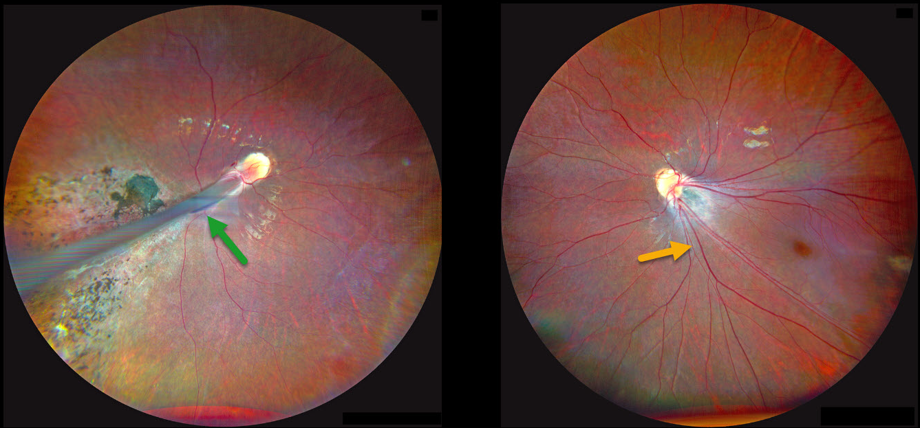
RETINOPATHY OF PREMATURITY
Kanwaljeet Harjot Madan
Originally posted on @retina.rocks June 23, 2025
This 4YO girl’s parents noticed that her right eye was deviating outwards for several months, along with decreased vision bilaterally. Her mother gave a history of prematurity at 28 weeks of gestational age with a low birth weight of 1175 g and oxygen exposure. Vision was counting fingers OD and 20/80 OS. There was a large right exotropia.
Fundus photography OD shows a prominent retinal fold extending from the optic disc into the inferotemporal periphery. Diffuse chorioretinal scarring is noted surrounding the retinal fold. The retinal vessels OS are dragged inferotemporally with marked foveal ectopia.
Learning Points:
Retinopathy of prematurity (ROP) is a potentially blinding vasoproliferative disease occurring in premature infants. Although multifactorial, current screening guidelines are based primarily on low birth weight and gestational age (Kim et al, Surv Ophthalmology 2018;63:618-637). Screening and treatment for these eyes is extremely subspecialized, with treatment options primarily including timely scatter laser photocoagulation and intravitreal anti-VEGF injections (Yonekawa et al, Retina 2017:37:2208-2225).
Patients with ROP have an increased risk for vision-threatening complications throughout their life, including vitreous hemorrhage and retinal detachment (Hsu et al, Ophthalmology Retina 2023;7:72-80). We will therefore monitor our patient yearly.
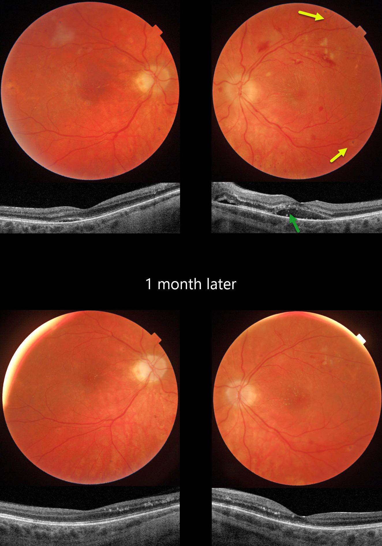
HYPERTENSIVE RETINOPATHY FROM METHAMPHETAMINE
Originally posted on @retina.rocks May 22, 2025
This 21YO female presented with bilateral vision loss. She was admitted to the hospital 2 weeks earlier for malignant hypertension (HTN), likely from methamphetamine abuse. Blood pressure at that time was 292/208. Vision was hand motion OD and 20/80 OS.
Triton color photography of her left eye shows a somewhat pale and swollen nerve, mostly inner retinal hemorrhages, and resolving nerve fiber layer infarcts (cotton-wool spots, CWS). Faint lipid exudates radiate nasally and superiorly from the macular center. Multifocal small hyperpigmented choroidal lesions are noted outside the arcades. Swept-source OCT shows subretinal fluid with hyperreflective lipid in the subretinal space and outer nuclear layer. Similar but much milder findings are noted in her right eye.
When examined 1 month later, vision remained at hand motion OD and improved to 20/40 OS with improved funduscopic and OCT findings.
Learning Points:
Malignant hypertension, defined as blood pressure above 180 systolic and/or 120 diastolic, is a life-threatening medical emergency. Eye doctors are in a unique position to often diagnose this condition. Patients will often present with bilateral optic nerve swelling, flame-shaped retinal hemorrhages, CWS, and, with more chronic disease, lipid precipitates in the nasal macular outer plexiform layer.
Acute hypertensive choroidopathy outside the setting of pre-eclampsia, in our experience, is quite rare. Independent of the disc and neurosensory retinal findings, patients present with localized multifocal serous retinal detachments (de Venecia and Jampol, Arch Ophthalmol 1984;102:68-73). With blood pressure control, these resolve often with minimal funduscopic changes. Focal (Elschnig spots) or linear (Siegrist streaks) choroidal pigmentary changes may result from more severe disease.
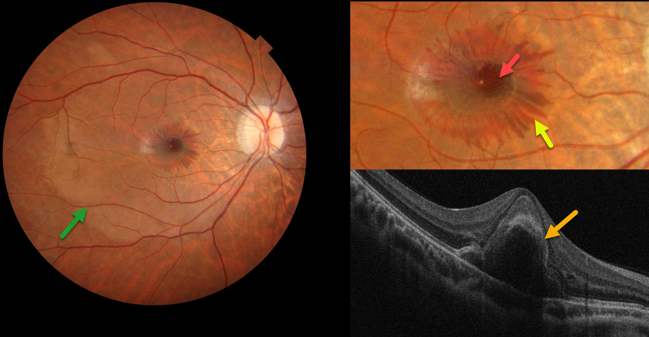
CENTRAL BOUQUET HEMORRHAGE
Evgenii Chernov
Originally posted on @retina.rocks May 13, 2025
This 24YO male woke with distorted vision in his right eye the day he presented to our clinic. Over the next 2 hours, a central spot appeared in his vision. He recalled an active tennis game the previous day but denied any direct ocular trauma. Vision was 20/200 OD and 20/25 in his normal OS.
Color photography shows central foveal blood, surrounding radiating outer plexiform (OPL) blood, and a deep vs subretinal whitish discoloration of the macula. On OCT, the central blood is located in the outer retina.
Learning Points:
Radiating deep macular hemorrhages are seen in multiple conditions, including Valsalva and trauma. Their unique radiating appearance is caused by blood accumulating in Henle’s layer. Kon Graversen et al described hemorrhagic unilateral retinopathy, an idiopathic disorder with a similar appearance (Retina 2014;34:483-489).
Ramthohul et al recently described a central bouquet hemorrhage with a round intraretinal blood spot surrounded by these OPL hemorrhages (Retina 2024;44:551-557). Various etiologies included neovascular AMD, myopic lacquer cracks, macular telangiectasia, and trauma associated with angioid streaks. Despite the macular changes consistent with commotio retinae, our patient adamantly denied ocular trauma.
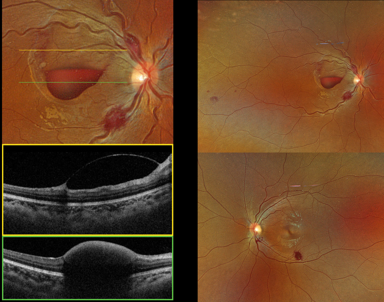
LEUKEMIA
Hailey Dunlow
Originally posted on @retina.rocks May 12, 2025
This previously healthy 14YO female presented with two weeks of vision loss in her right eye. Vision was hand motion OD and 20/25 OS.
Optos color RGB imaging shows bilateral inner and deep retinal hemorrhages, along with an inferior macular sub-internal limiting membrane (ILM) hemorrhage OD. Triton swept-source OCT confirms the location of the macular blood OD.
She was immediately sent to the emergency room. Complete blood count revealed pancytopenia with HgB 3.8, WBC 1.4, and platelets 54, and the patient was subsequently diagnosed with acute myelogenous leukemia. She was immediately lost to follow-up.
Learning Points:
Ocular leukemic manifestations are primarily caused by accompanying hematologic abnormalities, including hyperviscosity, anemia, and thrombocytopenia (Soman et al, Ophthalmology Retina 2018;2:17-23). Posterior segment findings include retinal hemorrhages (sub-ILM, nerve fiber layer, or deeper, white-centered), cotton-wool spots, dilated retinal veins, and serous retinal detachment. Leukemic infiltration of the optic nerve, retina, and choroid can also occur. Sub-ILM hemorrhages are usually self-limiting and resolve within several months of treatment of the underlying disease (Ophthalmology Retina 2018;2:494-501).
MULTIPLE MYELOMA
Originally posted on @retina.rocks May 5, 2025
This 81YO male presented with two days of vision loss OD. He has a history of diet-controlled type 2 diabetes and a 5-year history of multiple myeloma. He was examined by us 2 weeks earlier for a routine visit for inactive herpes simplex iridocyclitis. Vision was 20/40 on that visit with a normal OCT (not shown), but had decreased to counting fingers at the current visit.
Triton color imaging shows a faint, round serous macular detachment with scattered small inner retinal and nerve hemorrhages. Swept-source OCT shows subfoveal fluid with overlying cystic edema in the outer nuclear layer. Optos color RGB imaging shows scattered retinal hemorrhages throughout the right posterior pole, with a normal left fundus. Fluorescein angiography shows no leakage.
Learning Points:
Monoclonal gammopathies (paraproteinemia) result from the production of monoclonal gamma-globulins in the blood, including multiple myeloma (IgG or IgA) and Waldenstrom macroglobulinemia (IgM). Retinal findings are often caused by hyperviscosity (Rajagopal et al, Surv Ophthalmology 2016;61:236-247) and include intraretinal or deep retinal hemorrhages, dilated vessels, or vascular occlusions.
A unique retinopathy found in these disorders is termed paraproteinemic maculopathy (Mansour et al, Ophthalmology 2014;121:1925-1932). Somewhat simulating optic pit maculopathy, patients develop serous macular detachments with overlying cystic outer retinal edema and occasionally outer macular holes. There is no angiographic leakage. The macular fluid, which is usually refractory to laser photocoagulation, intravitreal injections, and photodynamic therapy, is treated primarily by decreasing blood immunoglobulin levels.
Unfortunately, our patient died 2 months after our examination.
RETINOPATHY OF PREMATURITY
Originally posted on @retina.rocks April 23, 2025
This 70YO female presented with stable 20/100 vision in her right eye. Her left eye was enucleated almost 60 years previously due to complications of retinopathy of prematurity (ROP).
Optos color RGB imaging shows marked temporal macular dragging with peripapillary and distal temporal chorioretinal scarring.
Learning Points:
Patients with ROP have an increased risk for vision-threatening complications throughout their life, including vitreous hemorrhage and retinal detachment (Hsu et al, Ophthalmology Retina 2023;7:72-80). We will therefore continue to monitor our patient yearly.
RETINAL ARTERIOVENOUS MALFORMATION
Mattie Adams
Originally posted on @retina.rocks April 15, 2025
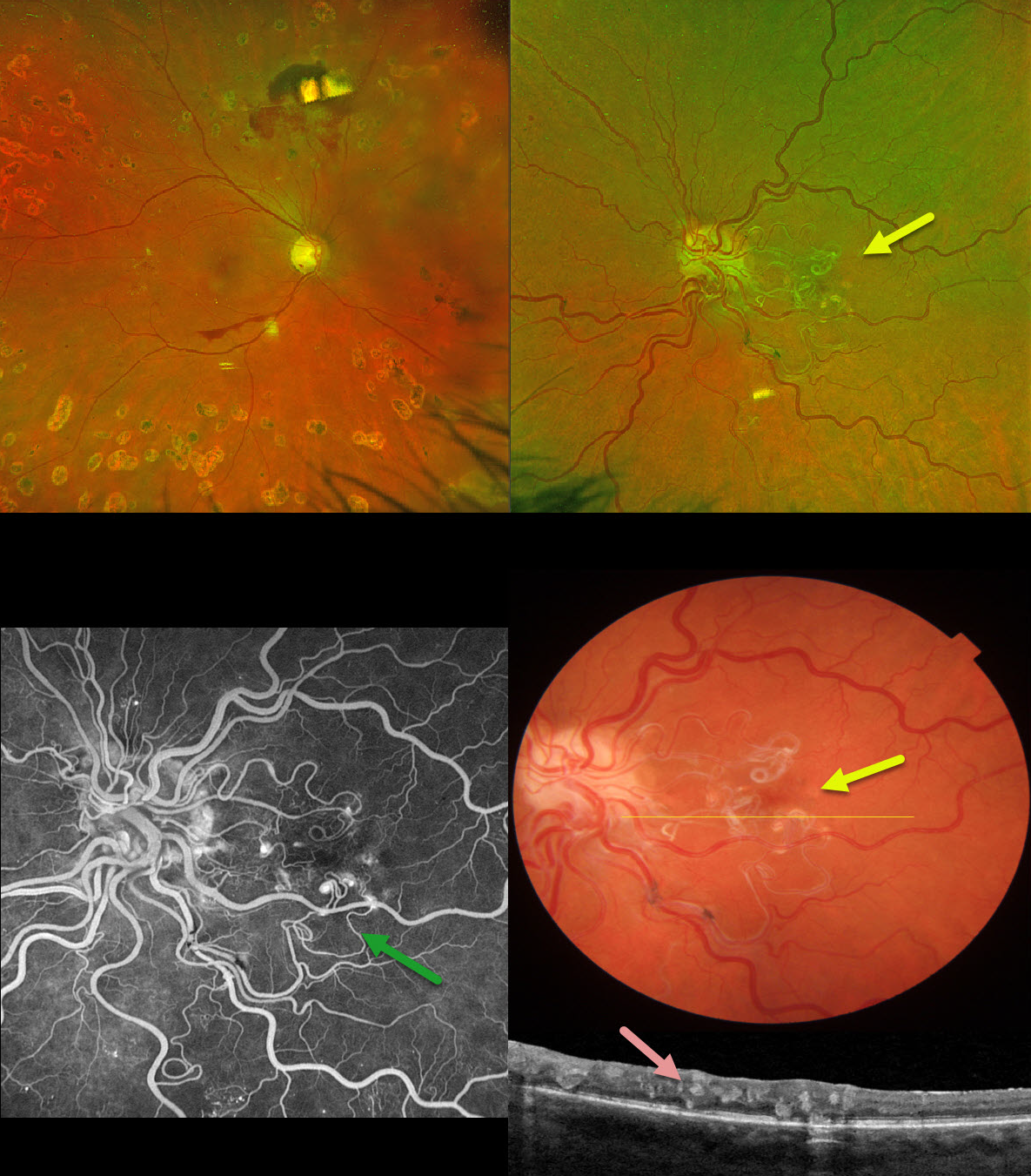
This 53YO female with a history of type 2 diabetes was referred for a symptomatic vitreous hemorrhage in her right eye from proliferative diabetic retinopathy. She received prior panretinal photocoagulation (PRP) in this eye elsewhere. Vision was 20/60 OD and 20/70 OS.
Optos color RG imaging OD shows preretinal blood in the inferior macula, and PRP scarring is noted superior to the nerve. The left eye shows a tangle of dilated and partially sheathed vessels extending from the nerve into the central macula. The more peripherally located vessels are dilated and tortuous.
On B-scan OCT, these vessels are noted in all retinal layers. These vessels did not leak on fluorescein angiography. Neurologic imaging was negative.
Learning Points:
A retinal arteriovenous malformation (AVM) is a congenital connection between an artery and a vein without an intervening capillary network. These lesions appear along a spectrum ranging from small, isolated lesions to the large ‘bag of worms’ appearance as seen in our patient. These more extensive lesions are usually part of the Wyburn-Mason Syndrome, which includes ocular and midbrain vascular lesions. Our patient has an isolated retinal AVM, as there are no intracranial lesions.
Unlike diabetic neovascularization that proliferates from the retinal surface into the cortical vitreous, the vessels in an AVM are intraretinal. Even though the AVM somewhat mimics diabetic disc neovascularization, its presence in our diabetic patient is purely coincidental. Although retinal AVMs are usually stable lesions, retinal hemorrhages, vitreous hemorrhage, and venous occlusions may occur.
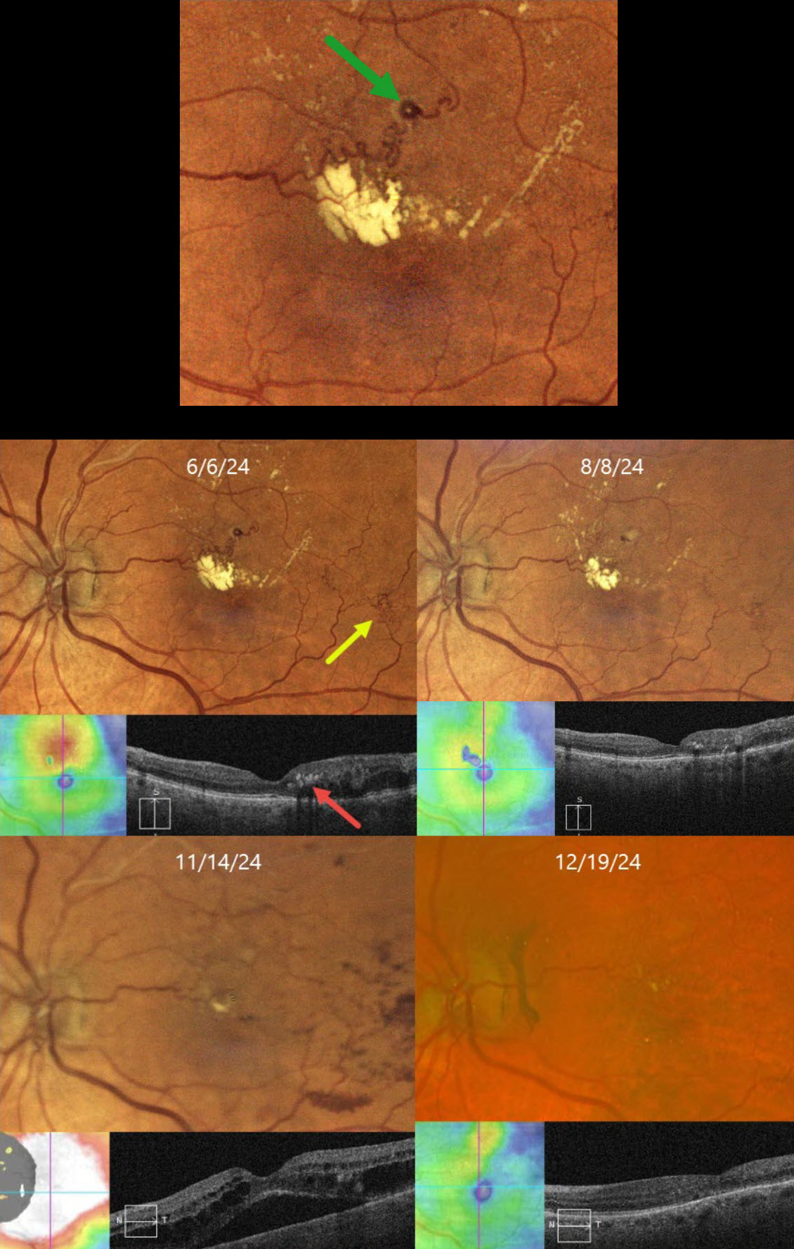
BRVO WITH RETINAL VENOUS MACROANEURYSM
Originally posted on @retina.rocks April 14, 2025
This 72YO female has been followed since 2017 for a branch retinal vein occlusion in her left eye. She has required ongoing anti-VEGF therapy for macular edema and failed macular laser for treatment burden several times. Although her eye was stable for years, on 6/6/24, there was new macular lipid with increased superior macular edema. Vision was stable at 20/25.
Color imaging shows a major superotemporal branch retinal vein occlusion with extensive collateral vessels and superior macular edema. Confluent lipid exudates threaten the macular center and appear as hyperreflective deposits in the mid-retina on OCT. A retinal venous macroaneurysm (RVM) is noted in the superior macula.
An intravitreal Eylea injection was administered, followed by focal thermal laser treatment of the RVM. Two months later, on 8/8/24, vision was 20/30. The lipid was decreasing, and the edema resolved. On 11/14/24, vision decreased to 20/60. Although the lipid was almost resolved, recurrent macular fluid persisted. Following a single Avastin injection, vision improved to 20/30, and the edema resolved. Macular grid laser was applied in hopes of reducing the treatment burden. We will continue to follow her closely and treat her as needed.
Learning Points:
In 1990, Cousins et al reported that RVMs can be an additional finding in occlusive venous disease, always in association with collateral vessels or capillary or arterial macroaneurysms (AJO 1990;109:567-570). These vascular abnormalities, also known as retinal capillary macroaneurysms or telangiectatic capillaries (TelCaps), tend to have a higher rate of intraretinal lipid (40%) but are less likely to affect central vision since they are usually outside the macula. They tend to have a higher association with retinal ischemia, thus being at a higher rate for developing retinal neovascularization (32%). Although these lesions will usually involute spontaneously, we treated our patient out of concern that the lipid might extend into the foveal center.
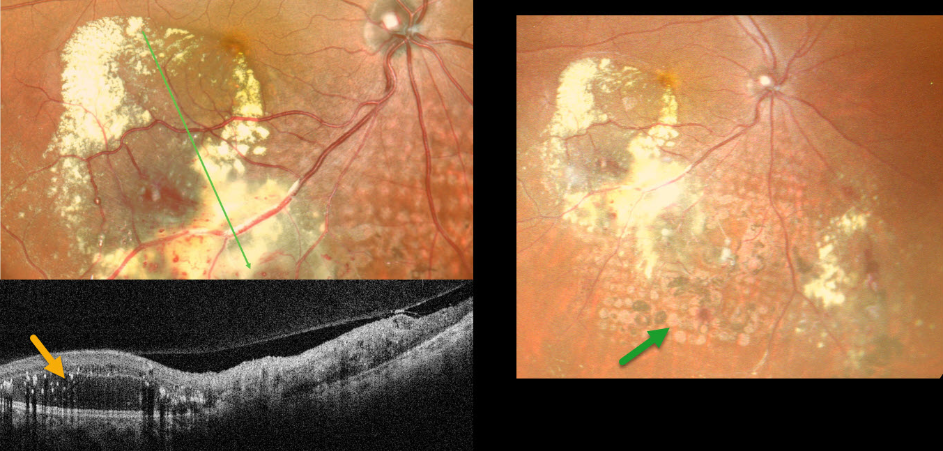
COATS DISEASE
Tejaswita Verma and Manish Nagpal
Originally posted on @retina.rocks April 2, 2025
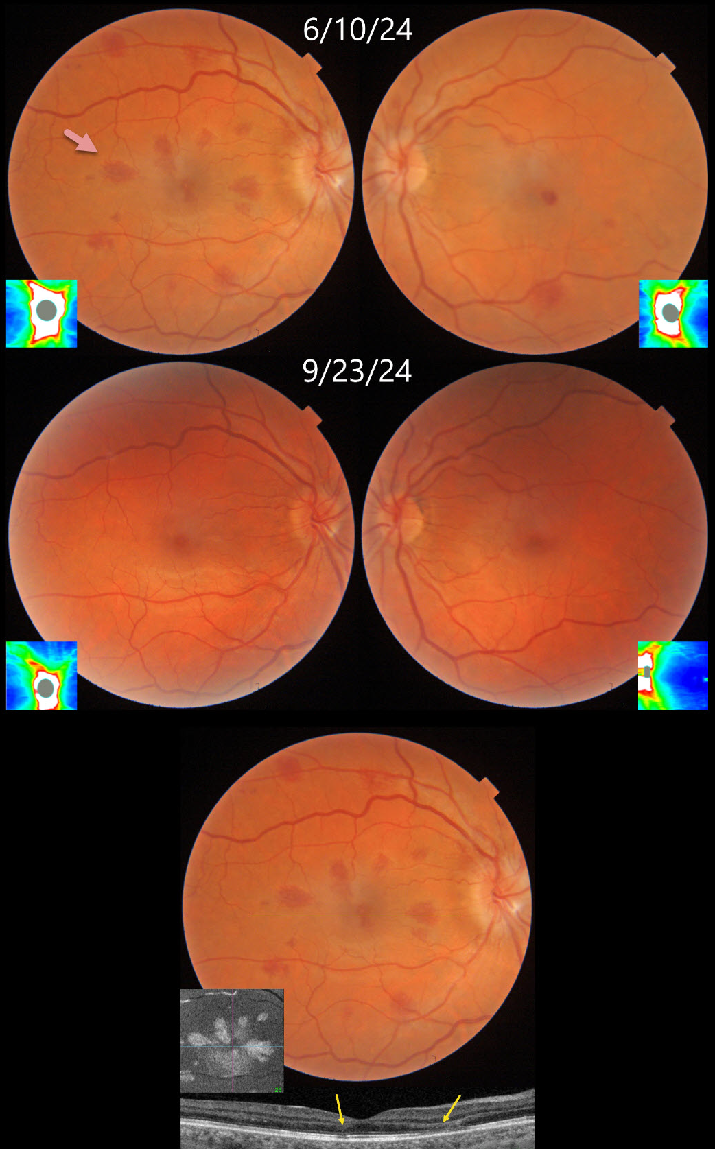
ANEMIA WITH BILATERAL OPTIC NERVE EDEMA
Fraser McKay
Originally posted on @retina.rocks March 27, 2025
This 34YO female underwent uterine ablation 2 weeks earlier for heavy menstruation and severe secondary iron deficiency anemia. Although we don’t know what her hemoglobin level was prior to the procedure, it had risen to 10.7 postoperatively. Vision was 20/30 OD and 20/50 OS.
Triton color photography shows bilateral teardrop-shaped macular outer plexiform layer (OPL) hemorrhages, with greater involvement in her right eye. Numerous more peripheral deep and white-centered hemorrhages are also noted. Swept-source OCT confirms the location of the macular blood. En face OCT shows a more dramatic view of their petaloid shape. There is also mild optic nerve edema, especially in the right eye. The hemorrhages and nerve swelling mostly resolved with iron supplementation several months later.
Learning Points:
Anemia-related retinal findings most commonly include hemorrhages in the retina and sub-internal limiting membrane. Severe anemia is a rare cause for optic nerve swelling and should always be considered in patients with atypical idiopathic intracranial hypertension (Biousse et al, AJO 2003;135:437-446). The underlying pathogenesis of anemia-induced optic nerve swelling remains unknown.
Radiating deep macular hemorrhages are seen in multiple conditions, including Valsalva and trauma (Baumal et al, Br J Ophthalmol 2021;105:374-380). We have not previously seen these hemorrhages in isolated anemia. Their unique radiating appearance is caused by blood accumulating in Henle’s layer. Kon Graversen et al described hemorrhagic unilateral retinopathy, an idiopathic disorder with a similar appearance (Retina 2014;34:483-489).
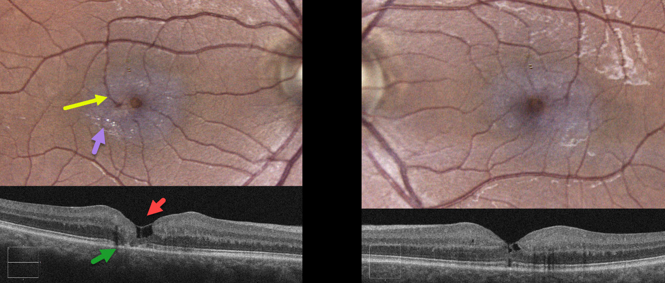
MACULAR TELANGIECTASIA
Originally posted on @retina.rocks March 26, 2025
This healthy 43YO male was referred for asymptomatic retinal findings. Vision was 20/40 OD and 20/30 OS.
Color imaging of the right eye shows a foveal cyst with pericentral opaque whitened retina and numerous inner retinal crystals. A right-angle venule is noted superotemporally.
OCT scanning shows inner foveal cavitations with variable loss of the outer retinal bands. Temporal foveal outer retinal atrophy is also noted. The left macula shows an encircling, pericentral, opaque retina with mild crystals. OCT reveals milder inner cavitations with an outer foveal microdefect.
Learning Points:
Originally described by Gass (Arch Ophthalmology 1982;100:769-780), macular telangiectasia type 2 (MacTel2) is a neurodegenerative disorder, most likely originating from Muller cell dysfunction. The term telangiectasia is misleading, as the funduscopic findings are mostly non-vascular. The refractile inner retinal ‘crystals’ are thought to be Muller cell footplates. The foveal retina has a whitish discoloration, most likely due to retinal opacification from dysfunctional Muller cells. Photoreceptor loss allows RPE cells to migrate along intraretinal capillaries, causing the coarse clinical pigment clumping (not seen in our patient).
Most patients will also have subclinical chorioretinal anastomosis in association with right-angle venules (Spaide et al, Retina 2018;38:1920-1929). Secondary macular neovascularization can be successfully treated per age-related macular degeneration protocols.
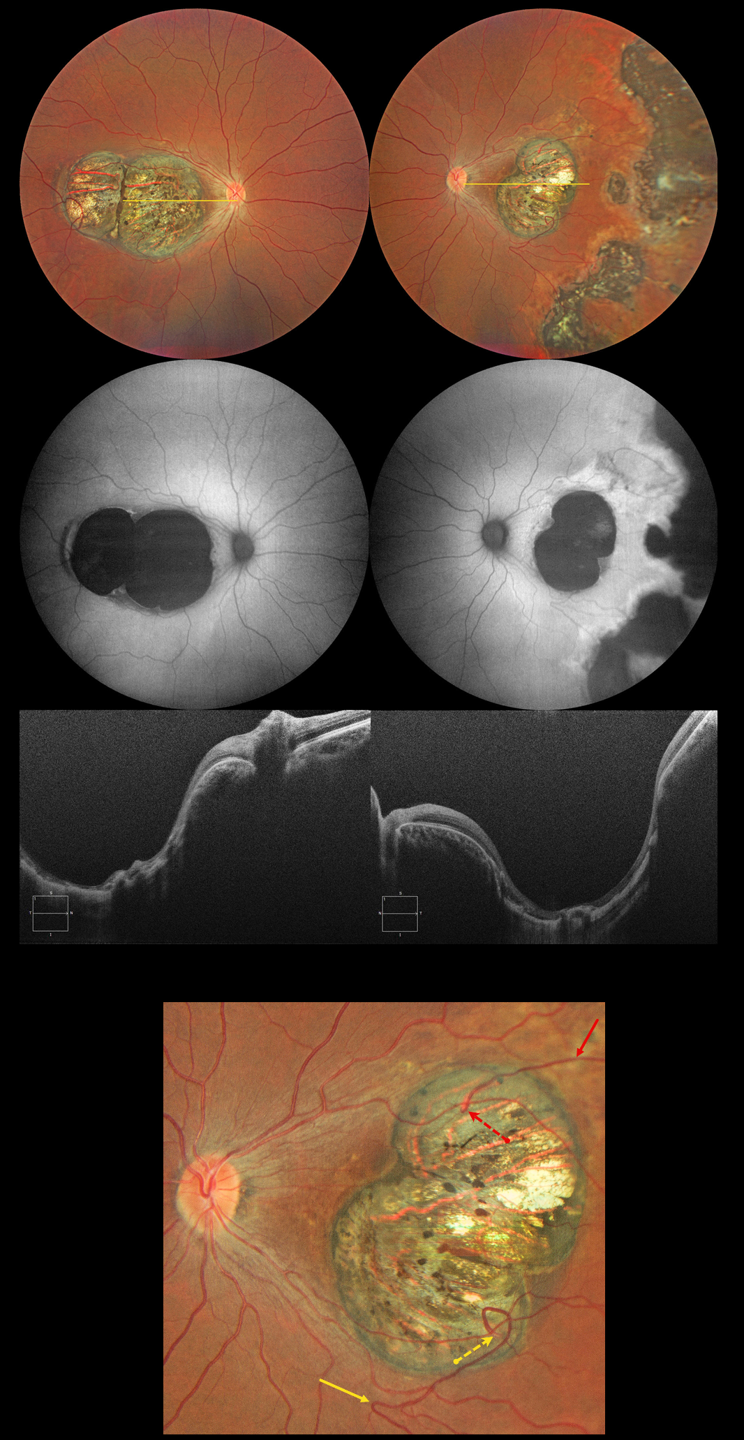
CHOROIDAL COLOBOMA
Evgenii Chernov
Originally posted on @retina.rocks March 18, 2025
This 32YO female has a history of congenital nystagmus and poor vision since birth. Vision was 20/130 OU.
Color photography shows bilateral macular colobomas with more peripheral multifocal variably pigmented colobomatous scarring in her left eye. These lesions are hypoautofluorescent. OCT scanning through the macular lesions shows thinned hyperreflective retina draped across the posteriorly bowed sclera.
The right macular coloboma also contains several retinal choroidal anastomoses. The major inferotemporal vein acutely bends superotemporally to enter the coloboma and drain into the choroidal circulation. A superotemporal vein also enters the coloboma to drain into a visible choroidal vortex.
Learning Points:
Choroidal colobomas are most commonly caused by failure of the optic vesicle and choroidal fissure to close during embryonic development and are usually found inferonasally. They can present as solitary or multiple lesions. The differential diagnosis for congenital macular coloboma also includes post inflammatory lesions (toxoplasmosis, cytomegalovirus, and zika) and retinal dystrophies including North Carolina macular dystrophy. The retina within the coloboma is variably thinned, which increases the risk for rhegmatogenous retinal detachment.
Under normal conditions the retinal and choroidal circulations remain separate and distinct. Rarely, the two circulations directly connect with each other through a chorioretinal anastomosis. These can occur in chorioretinal scars (typically from inactive toxoplasmosis lesions), in macular telangiectasia, and in disciform scars.
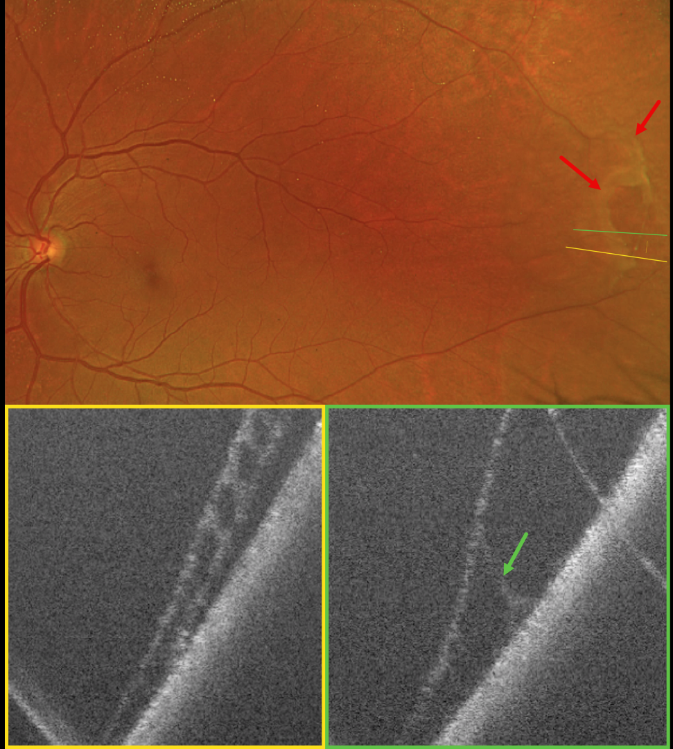
DEGENERATIVE RETINOSCHISIS + SITUS INVERSUS
Sayena Jabbehdari and Ahmed Sallam
Originally posted on @retina.rocks March 5, 2025
This 64YO female was referred for a possible asymptomatic rhegmatogenous retinal detachment (RRD) in her left eye. Vision was 20/20 in her normal OD and 20/20 OS.
Optos color RG imaging shows superotemporal degenerative retinoschisis with a few outer-layer holes (OLH). OCT confirms the schisis as well as the outer layer hole.
Learning Points:
Clinically, degenerative retinoschisis usually appears as a smooth, dome-shaped peripheral retinal elevation without the outer retinal corrugations classically seen in RRD (Oquendo et al, AJO 2024;268:212-221). Degenerative retinoschisis is virtually never prophylactically treated (Byer 1986;93:1127-1137). Although the schisis cavities can enlarge, prophylactic treatment, including thermal laser, does not prevent further spread and should not be performed (Ness et al, Surv Ophthalmology 2022;67:892-907).
Situs inversus of the optic disc was identified as a benign incidental finding in our patient. This occurs in 5% of normal eyes and 70% of eyes with tilted discs (Witmer et al, Surv Ophthalmology 2010;55:403-428). It can be mistaken for other congenital disc anomalies.
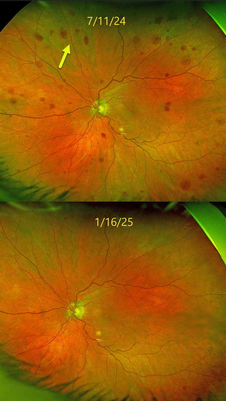
OCULAR ISCHEMIA
Originally posted on @retina.rocks March 3, 2025
This 83YO male was referred for asymptomatic changes in his left fundus. Vision was 20/30 in his healthy OD and 20/100 OS.
Optos color RG imaging shows scattered deep, variably round retinal hemorrhages extending from the peripheral macula into the midperiphery. Carotid Doppler testing revealed a critical stenosis of the left internal carotid artery, and he was emergently referred to his cardiologist. The findings dramatically improved within months of carotid surgery.
Learning Points:
The retinal hemorrhages in the ocular ischemic syndrome (OIS) are unique. Retinal vascular disorders like diabetic retinopathy, retinal vein occlusions, and hypertensive retinopathy usually have flame- and dot-shaped inner retinal hemorrhages concentrated around the posterior pole.
Our patient’s findings are classic for OIS, with hemorrhages in the deeper peripheral retina. The retinal periphery is literally the end of the line for the retinal arterial circulation. In an eye receiving less blood supply due to carotid occlusive disease, the more proximal retina has first dibs on the available oxygen. There are penetrating capillaries that dive radially from the nerve fiber and ganglion cell layers into the deeper retina. The deep vascular complex that supplies the inner and outer plexiform layers is thus furthest downstream, and these endothelial cells are likely damaged by OIS. Blood cells leaking into this space accumulate, giving the large outer blot hemorrhages.
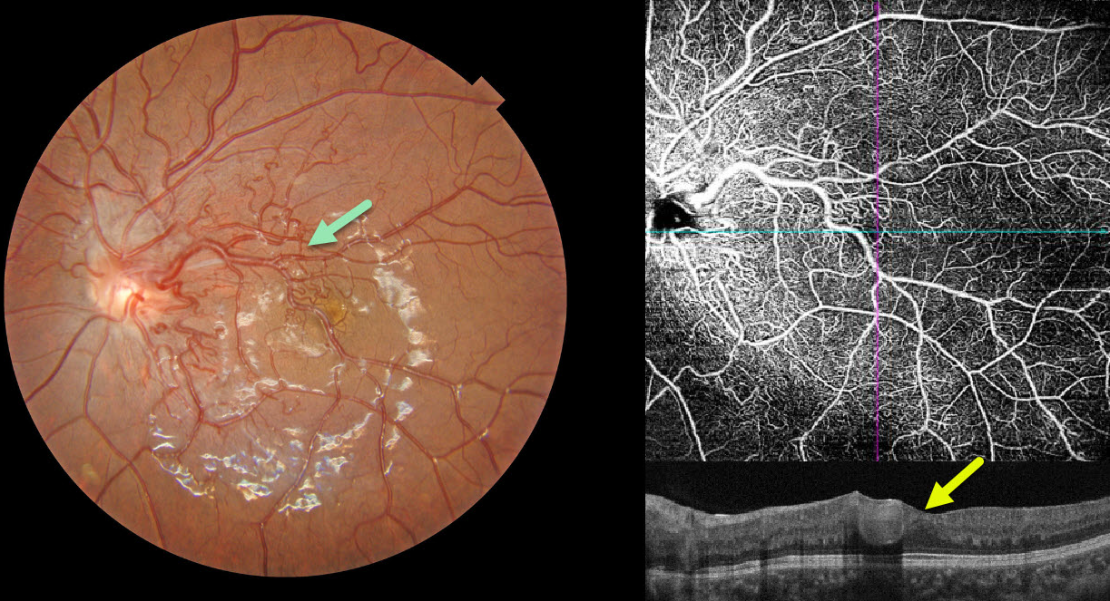
RETINAL ARTERIOVENOUS MALFORMATION
Evgenii Chernov
Originally posted on @retina.rocks February 17, 2025
This 10YO girl presented with asymptomatic retinal findings in her left eye. Vision was 20/25 in her normal OD and 20/30 OS.
Color photography shows a retinal arteriovenous malformation (AVM) composed of dilated, tortuous vessels, mostly in the macular area. The foveal pit is crossed by a large tortuous vessel. OCT angiography highlights the profoundly abnormal macular vasculature, and an OCT B-scan shows the markedly dilated inner foveal vessel. MRI scanning of the brain and orbits was normal.
Learning Points:
A retinal AVM is a congenital connection between an artery and a vein without an intervening capillary network. An AVM appears along a spectrum ranging from small, isolated lesions to the large ‘bag of worms’ appearance. These more extensive lesions are usually part of the Wyburn-Mason Syndrome, which includes ocular and midbrain vascular lesions. Our patient has an isolated retinal AVM, as there are no intracranial lesions.
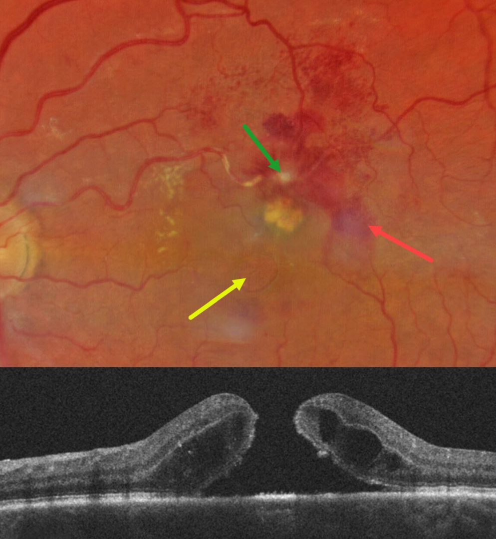
RETINAL ARTERIAL MACROANEURYSM (RAM) WITH MACULAR HOLE
Will Gibson
Originally posted on @retina.rocks January 13, 2025
This 67YO white female presented for her six-month diabetic follow-up with a few weeks of vision loss in her left eye. Vision was 20/80 OS and 20/20 in her normal OD.
Color photography shows a fibrosed retinal arterial macroaneurysm (RAM), in the superior macula. Faint subretinal blood is noted temporally, with mostly inner retinal blood superotemporally. The artery is sheathed just proximal to the RAM. A large macular hole with surrounding subretinal fluid is present, accompanied by a lipid clump between the hole and the RAM. Faint lipid is also noted in the nasal macula. She was referred to a retinal specialist for further management.
Learning Points:
Macular hole is a rare finding with retinal arterial macroaneurysms (Sagara et al, Br J Ophthalmol 2009;93:1337-1340). Subretinal and sub-internal limiting membrane blood appears to increase the risk of macular hole.
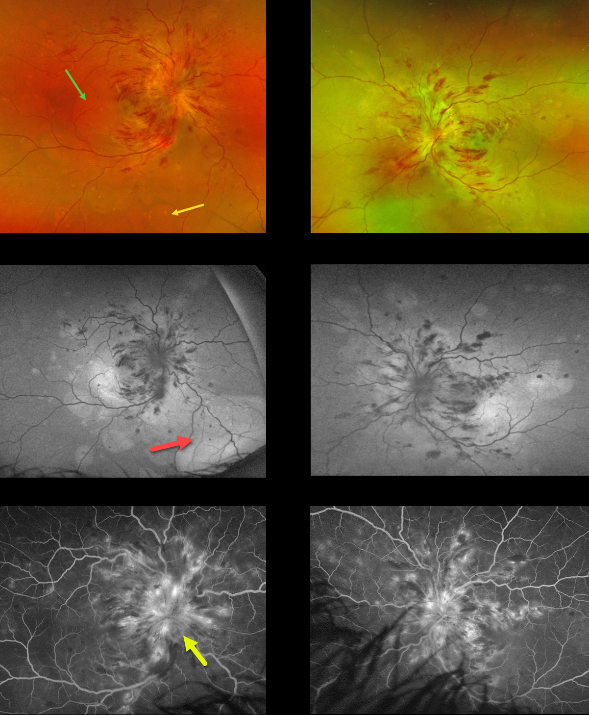
HYPERTENSIVE RETINOPATHY AND CHOROIDOPATHY
Yuenpang Cheung
Originally posted on @retina.rocks December 25, 2024
This 21YO female was admitted to the hospital 2 weeks earlier for malignant hypertension. She underwent prior failed renal transplantation due to tubulocystic renal disease and was currently on dialysis. Vision was counting fingers bilaterally.
Optos color RG imaging shows marked bilateral papilledema with mostly inner retinal hemorrhages radiating into the midperipheries. Multifocal blisters of exudative serous retinal detachments are scattered throughout each posterior pole, including the macula and midperiphery. These exudative detachments are best imaged on fundus autofluorescence (FAF), where they are hyper-FAF. Fluorescein angiography shows marked optic nerve leakage and scattered more peripheral subretinal leakage.
Learning Points:
Malignant hypertension, defined as blood pressure above 180 systolic and/or 120 diastolic, is a life-threatening medical emergency. Eye doctors are in a unique position to often diagnose this condition. Patients will often present with bilateral optic nerve swelling, flame-shaped retinal hemorrhages, and, with more chronic disease, lipid precipitates in the nasal macular outer plexiform layer.
Acute hypertensive choroidopathy outside the setting of pre-eclampsia, in our experience, is quite rare. Independent of the disc and neurosensory retinal findings, patients present with localized multifocal serous retinal detachments (de Venecia and Jampol, Arch Ophthalmol 1984;102:68-73). With blood pressure control, these resolve often with minimal funduscopic changes. Focal (Elschnig spots) or linear (Siegrist streaks) choroidal pigmentary changes may result from more severe disease. The acute findings are thought to result from choroidal arterial constriction with secondary choriocapillaris necrosis (Hayreh et al, Ophthalmology 1986;93:1383-1400). This causes ischemic injury to the overlying RPE with a breakdown of the outer blood-retinal barrier and exudative subretinal fluid.
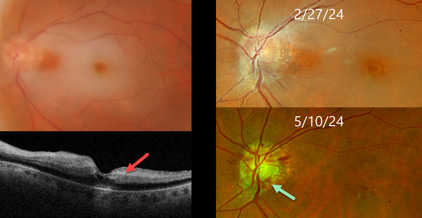
CRAO
Originally posted on @retina.rocks December 23, 2024
This 75YO female presented with an acute counting fingers central retinal artery occlusion. Triton color imaging shows a small area of papillomacular sparing, and swept-source OCT shows inner retinal hyperreflectivity. The retinal opacification completely resolved 10 weeks later with the development of extensive disc collaterals. Vision at counting fingers.
Learning Points:
Collateral vessels are dilated, pre-existing vessels that bypass an occlusion. These are seen in the posterior pole most commonly following retinal venous occlusions. Disc collaterals are exceedingly rare with arterial occlusions and were first described by Nettleship in 1891 (Ragge and Hoyt, Br J Ophthalmology 1992;76:186-188). The endothelial tight junctions are intact so they don’t leak angiographically. This helps to differentiate them from neovascularization which does leak.
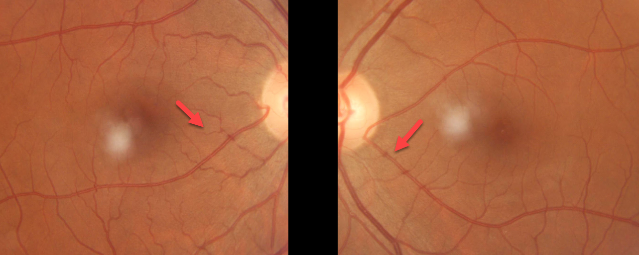
CILIORETINAL ARTERIES
Sajal Golchha and Sarthak Bafna
Originally posted on @retina.rocks December 3, 2024
This 46YO visually asymptomatic male presented with bilateral cilioretinal arteries.
The inner two-thirds of the neurosensory retina is supplied by the central retinal artery, with the choroid supplying the RPE and photoreceptor layer. Cilioretinal arteries, which arise from the posterior choroidal circulation, are present in up to 50% of individuals, and bilateral in about 25% (Schneider et al, Acta Ophthalmologica 2021;99:e310-e318).
Our patient is unique in that their left cilioretinal artery supplies virtually the entire macula. If he were unlucky enough to be the one in 50,000 people per year who develops a central retinal artery occlusion (Leavitt et al, AJO 2011;152:820-823), these vessels would most likely preserve macular function.
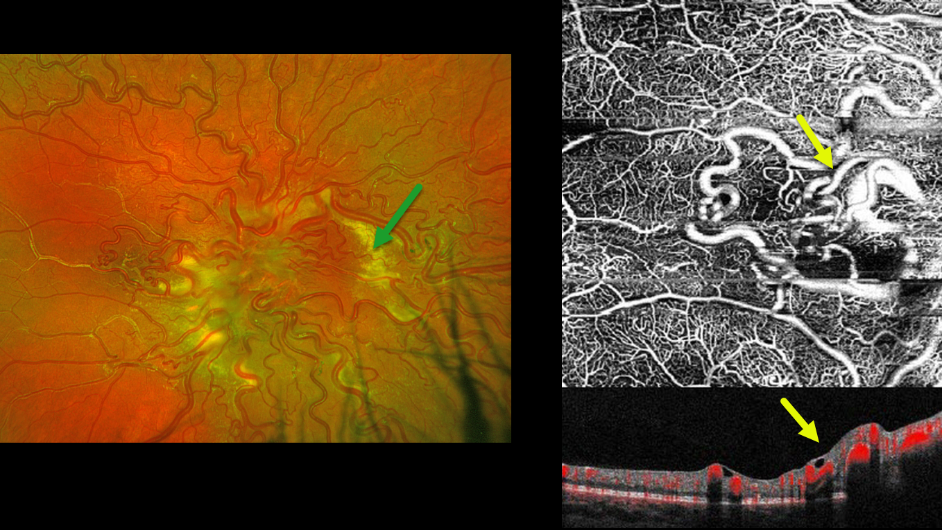
RETINAL ARTERIOVENOUS MALFORMATION
Mattie Adams and Jonathan Bunnell
Originally posted on @retina.rocks November 28, 2024
This 10YO boy recently moved to the area with a prior diagnosis of Wyburn-Mason syndrome. Prior neurologic imaging was negative, and he was otherwise healthy. Vision was 20/400 OD and 20/20 in his normal left eye.
Optos color RG imaging of his right eye shows tangles of markedly dilated and tortuous vessels covering the optic nerve and posterior pole. The vessels are sheathed superiorly and temporally. Subretinal lipid surrounds the posterior pole. En face OCT angiography highlights these dilated inner retinal vessels. Observation was recommended.
Learning Points:
A retinal arteriovenous malformation (AVM) is a congenital connection between an artery and a vein without an intervening capillary network. These lesions appear along a spectrum ranging from small, isolated lesions to the large ‘bag of worms’ appearance as seen in our patient. These more extensive lesions are usually part of the Wyburn-Mason Syndrome, which includes ocular and midbrain vascular lesions. Our patient has an isolated retinal AVM, with no intracranial lesions.
Although retinal AVMs are usually stable lesions, secondary venous occlusions may occur, and the peripheral vascular sheathing in our patient likely indicates a prior occlusion. The subretinal lipid in our patient is a very atypical finding in our experience.
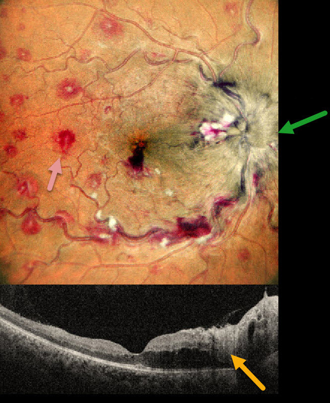
ANEMIA
Anand Temkar and Manish Nagpal
Originally posted on @retina.rocks October 24, 2024
This 35YO male presented with 1 month of vision loss in his right eye. There was no past medical history. Vision was 20/40 OD and 20/20 in his normal OS.
MultiColor imaging shows a markedly swollen optic nerve with extensive multifocal deep, white-centered retinal hemorrhages. OCT shows marked mostly outer retinal edema extending from the nerve towards the macular center.
Extensive laboratory evaluation was negative except for a microcytic anemia with a hemoglobin of 2.9 g/dl. This was felt to be caused by an iron deficiency due to poor diet. Intravitreal anti-VEGF injections were recommended, along with oral prednisone for the optic nerve swelling. Unfortunately, he was immediately lost to follow-up.
Learning Points:
Anemia-related retinal findings most commonly include hemorrhages in the retina and sub-internal limiting membrane. Severe anemia is a rare cause for papilledema and should always be considered in patients with atypical idiopathic intracranial hypertension (Biousse et al, AJO 2003;135:437-446). The underlying pathogenesis of anemia-induced optic nerve swelling remains unknown. It is unclear why our patient had unilateral findings from a systemic disorder.
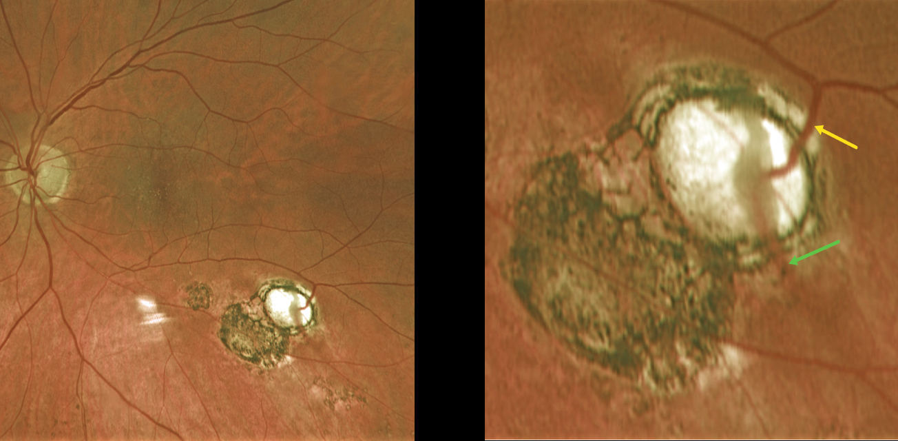
TOXOPLASMOSIS WITH RETINAL CHOROIDAL ANASTOMOSIS
Originally posted on @retina.rocks October 21, 2024
This 60YO female was referred for asymptomatic scarring in her left eye. Vision was 20/50 OD and 20/40 OS due to early bilateral cataracts. Small macular drusen were noted bilaterally.
Optos color RG imaging of her left shows multifocal variably pigmented scars below the inferotemporal arcade. A magnified inferotemporal view shows a retinal vein diving into an atrophic scar and directly connecting to a choroidal vein.
Learning Points:
Under normal conditions the retinal and choroidal circulations remain separate and distinct. Rarely, the two circulations directly connect with each other through a chorioretinal anastomosis. These can occur in chorioretinal scars (typically from inactive toxoplasmosis lesions as in this patient), in macular telangiectasia, and in disciform scars. We felt our patient’s findings most consistent with inactive toxoplasmosis scarring.
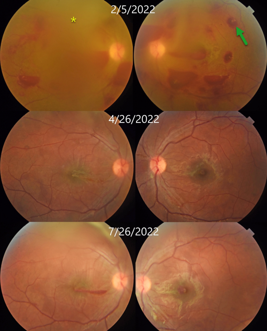
MEGALOBLASTIC ANEMIA
Alay Banker and Mauli Shah
Originally posted on @retina.rocks October 17, 2024
This 17YO male presented with 1 week of bilateral vision loss. Vision was counting fingers OU.
Bilateral white-centered and preretinal hemorrhages are noted. The fundus detail is decreased bilaterally due to breakthrough vitreous blood. Bloodwork revealed severe anemia with a hemoglobin of 5.3 g/dl. Vitamin B12 level was decreased at 160 pg/ml (normal 187-833). Based on the peripheral blood smear, which showed macrocytes, anisocytosis, poikilocytosis, target cells, and nucleated red blood cells, he was diagnosed with megaloblastic anemia.
His B12 deficiency resulted from a poorly balanced vegetarian diet. Following several blood transfusions and injectable vitamin B12 supplementation, as well as oral sublingual B12 and dietary modification, his anemia improved.
Two months later, vision improved to 20/200 OD and 20/80 OS; the hemoglobin rose to 12 g/dL; and funduscopic findings were improving. Five months following presentation, vision improved to 20/40 OU, and the retinal findings continued to normalize.
Learning Points:
Megaloblastic anemia is characterized by low hemoglobin in the presence of megaloblasts (large and immature nucleated red blood cells). It is caused by B12 or B9 (folate) deficiency, either from poor dietary intake (as in our patient) or from structural/functional gastrointestinal disorders that impair vitamin B malabsorption.
White-centered retinal hemorrhages and sub-internal limiting membrane/preretinal blood are commonly associated with anemia and blood dyscrasias. Other causes include bacterial endocarditis, hypertension, diabetic retinopathy, HIV retinopathy, and shaken baby syndrome. Hence, comorbidity must be questioned in such cases, and the patient should be screened for systemic diseases.
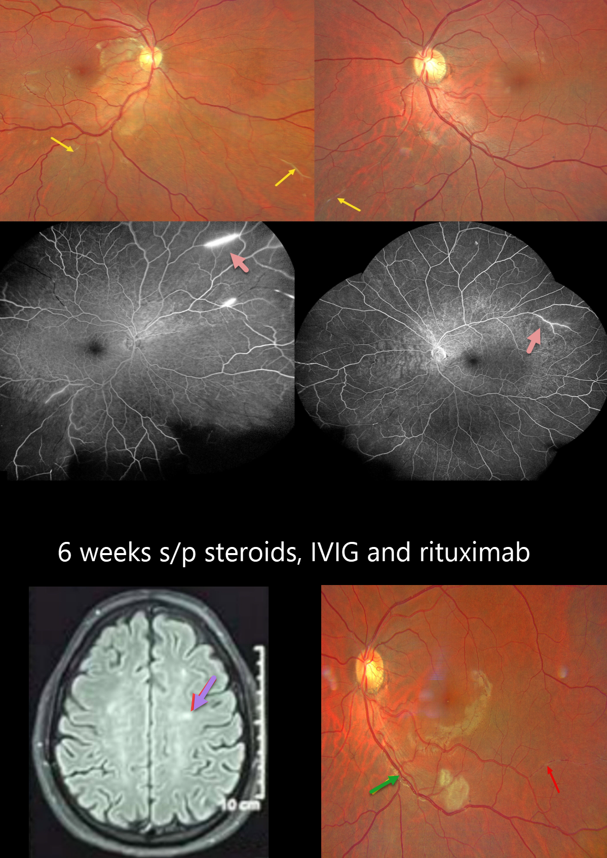
SUSAC SYNDROME
Ayushi Gupta and Vishal Agrawal
Originally posted on @retina.rocks October 15, 2024
This healthy 31YO male presented with about 2 weeks of vision loss in his right eye. He also complained of mild headaches over the prior month. Vision was counting fingers OD and 20/20 OS.
Fundus photography OD shows an opaque ischemic retina temporal to the disc, just sparing the fovea. Evidence of prior arterial occlusions is seen as bilaterally sclerosed vessels. Fluorescein angiography shows focal areas of scattered retinal ischemia with segmental areas of vascular staining.
MRI scanning shows multiple small, discrete, snowball-like lesions, and audiologic evaluation revealed bilateral mild sensorineural hearing loss, establishing the diagnosis of Susac syndrome. He was started on a 5-day course of intravenous immunoglobulins and steroids followed by maintenance rituximab therapy.
When he returned 6 weeks later, his vision was 20/80 OD and 20/20 OS. The nasal macular opacification is improving, but new areas of acute retinal ischemia are present in the inferior right and left maculae. Refractile deposits line an occluded arteriole along the left proximal inferotemporal arcade, and a new sclerosed arteriole is noted inferotemporally.
Learning Points:
Retinocochleocerebral vasculopathy, more commonly known as Susac syndrome, is a rare, microangiopathic disease of debated pathophysiology characterized by the classic triad of encephalopathy, sensorineural hearing loss, and branch retinal artery occlusions (Heng et al, Survey Ophthalmology 2019;64:477-485). The full triad may take months to evolve, frustrating the diagnosis. Retinal arterial wall atheromatous plaques, also known as Gass plaques, are often seen in later stages. They are a result of the occlusion, not the cause. The development of wide-field angiography has allowed for earlier detection and treatment. Although the findings are usually self-limiting while they wax and wane, systemic treatment with steroids and immunosuppression can be considered for more severe cases.
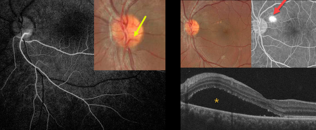
CILIORETINAL ARTERY + CENTRAL SEROUS CHORIORETINOPATHY
Mayelinne García, Luis Montero and Alberto Ruiz
Originally posted on @retina.rocks September 24, 2024
This healthy 25YO male presented with 3 weeks of vision loss in his left eye from active idiopathic central serous chorioretinopathy (ICSC). Vision was 20/20 in his normal OD and 20/110 OS.
A cilioretinal vessel originates within the inferotemporal aspect of the nerve and arches inferiorly. Fluorescein angiography reveals that this cilioretinal artery supplies the entire inferotemporal retina. Color imaging of the macula shows a large serous detachment with some drusen-like changes. This detachment is confirmed on OCT scanning. A blot leak is seen angiographically. Due to the patient’s symptoms, thermal laser was applied to the angiographic leak.
Learning Points:
The inner two-thirds of the neurosensory retina are supplied by the central retinal artery, with the choroid supplying the RPE and photoreceptor layer. Cilioretinal arteries, which arise from the posterior choroidal circulation, are present in up to 50% of individuals and are bilateral in about 25% (Schneider et al, Acta Ophthalmologica 2021;99:e310-e318).
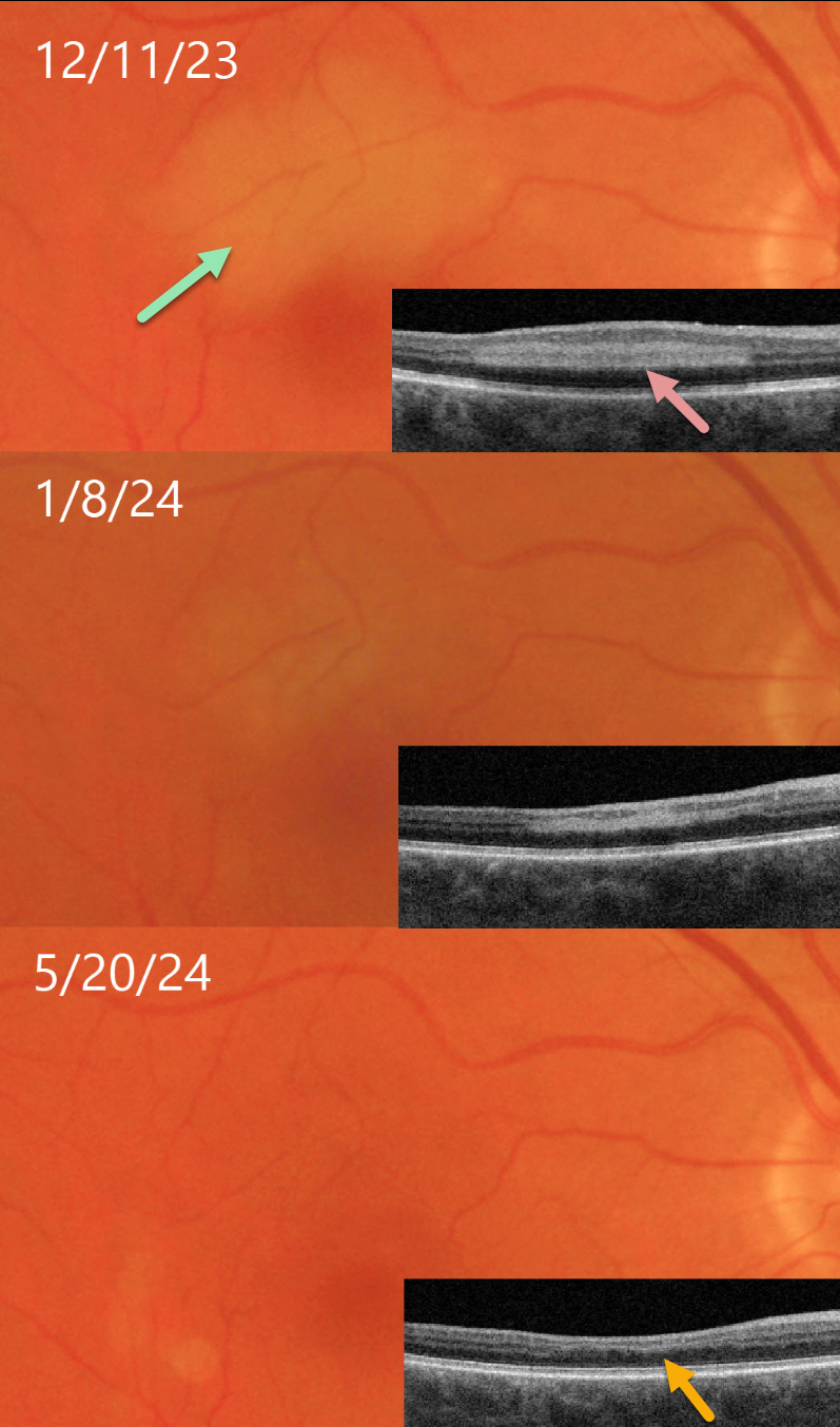
PARACENTRAL ACUTE MIDDLE MACULOPATHY (PAMM)
Originally posted on @retina.rocks September 11, 2024
This 68YO male presented on 12/11/23 with a several-day history of a paracentral scotoma in his right eye. Vision was 20/30 OD and 20/25 in his normal OS.
Triton color imaging shows an area of retinal opacification in the superior macula. This area is hyperreflective on swept-source OCT. The lesion gradually resolved over several months. When last examined on 5/20/24, the funduscopic appearance normalized. The area of prior mid-retinal hyperreflectivity was replaced by retinal thinning, with the inner plexiform/inner nuclear/outer plexiform layers compressed into a single hyperreflective line.
Learning Points:
Paracentral acute middle maculopathy (PAMM) is a phenotype caused by acute mid-retinal ischemia due to obstruction of the deep and intermediate capillary plexi. PAMM is associated with an increasing constellation of conditions, including retinal vein and artery occlusions. It is unclear if patients with a pure PAMM phenotype require an immediate stroke protocol referral (Bousquet et al, Retina 2023;43:1827-1832).
In our practice, we have seen several elderly patients who developed a complete CRAO within days of presenting with PAMM, so we do send these patients for emergent evaluation. We do not believe that typical PAMM in younger patients or found as a secondary finding in other ocular conditions requires a stroke workup. Our patient’s medical evaluation was fortunately negative.
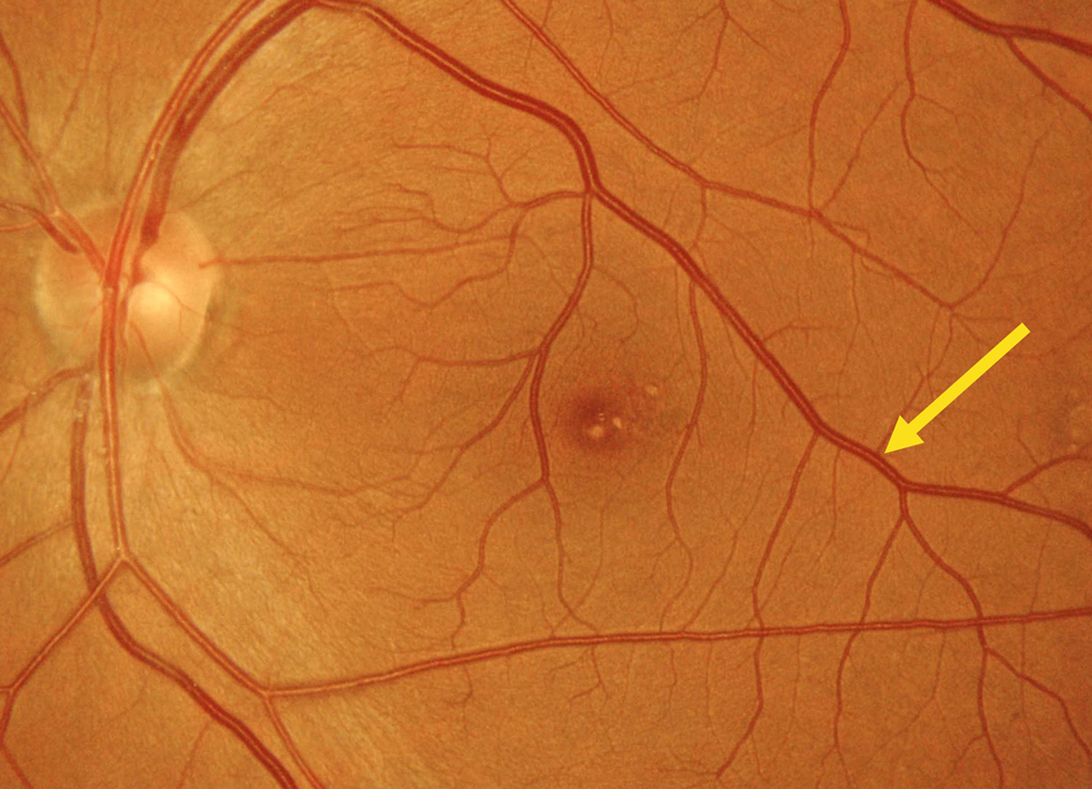
RETINAL VENOUS MALFORMATION
Evgenii Chernov
Originally posted on @retina.rocks September 10, 2024
This 28YO female was referred for an asymptomatic funduscopic finding. Vision was 20/20 OU.
Color imaging shows a superior retinal venous malformation. This vessel drains nearly the entire macula as it extends into the inferotemporal midperiphery. Some mixed foveal drusen are also noted.
Learning Points:
Retinal vessels virtually always respect the horizontal meridian. Retinal venous malformations are congenital anomalous vessels that, by definition, cross the horizontal. Originally described by Gary Brown et al as congenital retinal macrovessels (Arch Ophthalmology 1982;100:1430-1436), these vessels are usually located in the macular region and are virtually always venous.
These lesions are now called retinal venous malformations since about one-quarter of patients will have associated venous anomalies in the brain (Pichi et al JAMA Ophthalmol 2018;136:372-379).
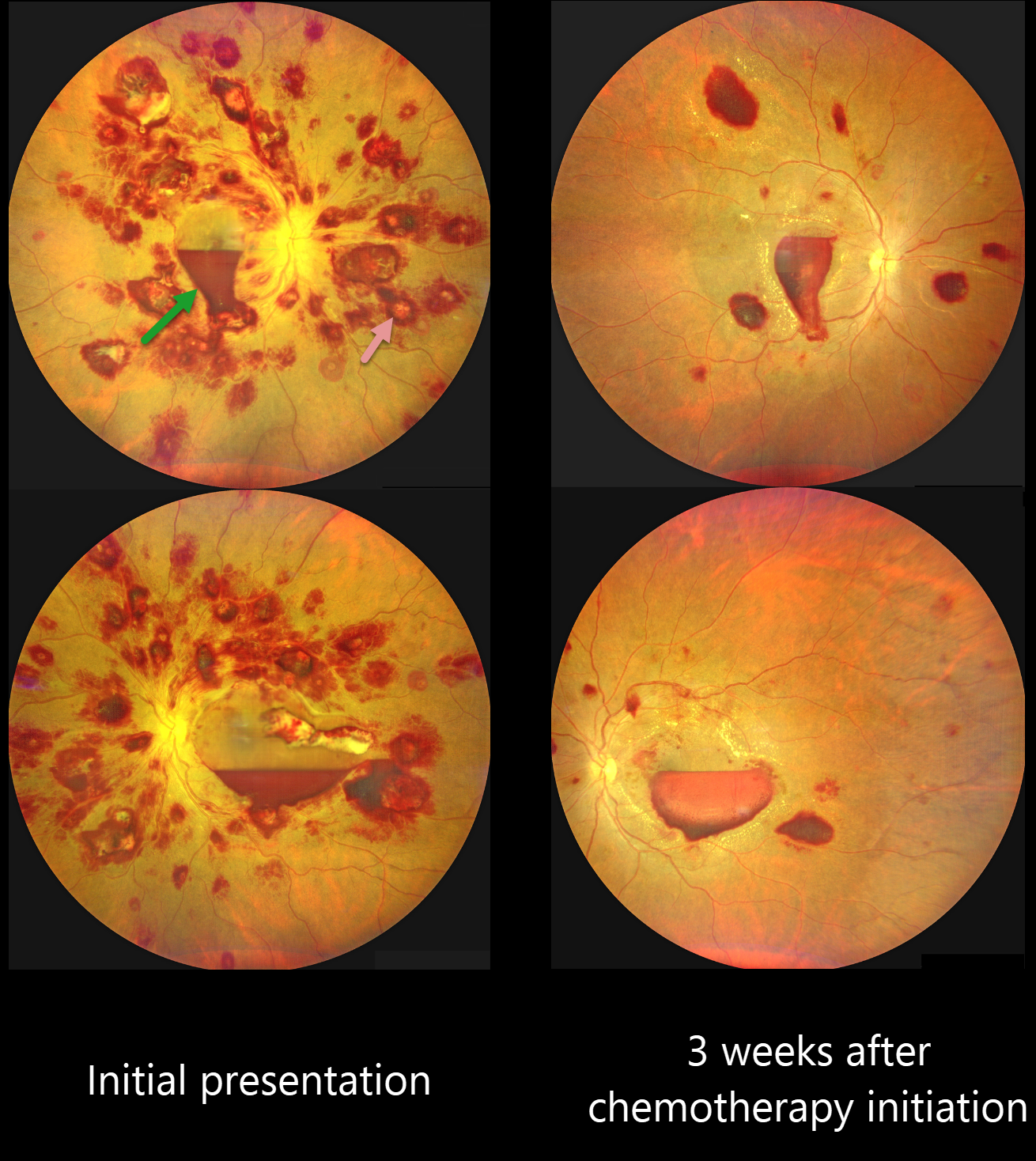
LEUKEMIA
Kanwaljeet Harjot Madan
Originally posted on @retina.rocks August 28, 2024
This 42YO previously healthy male presented with one week of bilateral decreased vision. Vision was 20/100 OD and 20/200 OS.
Color photography shows an extensive bilateral nerve fiber layer and deep white-centered retinal hemorrhages. Layered macular sub-internal limiting membrane (ILM) hemorrhages are noted as well, with some yellow devitalized blood in the temporal left macula. Hematologic workup revealed elevated blast cells, severe anemia (HgB 5.4) and thrombocytopenia.
He was diagnosed with acute myeloid leukemia and referred to oncology. He started chemotherapy, and 3 weeks later, his vision improved to 20/70 OU. The HgB was 8.2 with decreased blast cells. The bilateral retinal hemorrhages were markedly decreased.
This case was submitted by Kanwaljeet Harjot Madan.
Learning Points:
The term “Roth spot” was named after Dr. Moritz Roth, who, in 1872, noted white-centered hemorrhages in patients with bacterial endocarditis. He felt that these white spots were septic emboli that originated from an infected cardiac valve. However, similar lesions can be seen in many other disorders, including leukemia, hyperviscosity, anemia, diabetes, and hypertension (Duane et al Ophthalmology 1980;87:66-69). The white material may represent a variety of causes, including fibrin. So, these lesions are best called white-centered hemorrhages, unless one is specifically referring to those found associated with bacterial endocarditis.
Ocular leukemic manifestations are primarily caused by accompanying hematologic abnormalities, including hyperviscosity, anemia, and thrombocytopenia (Soman et al, Ophthalmology Retina 2018;2:17-23). Posterior segment findings include retinal hemorrhages (sub-ILM, nerve fiber layer or deeper, white-centered), cotton-wool spots, dilated retinal veins, and serous retinal detachment. Leukemic infiltration of the optic nerve, retina, and choroid can also occur. The sub-ILM hemorrhages are usually self-limiting and resolve within several months of treating the underlying disease (Ophthalmology Retina 2018;2:494-501).
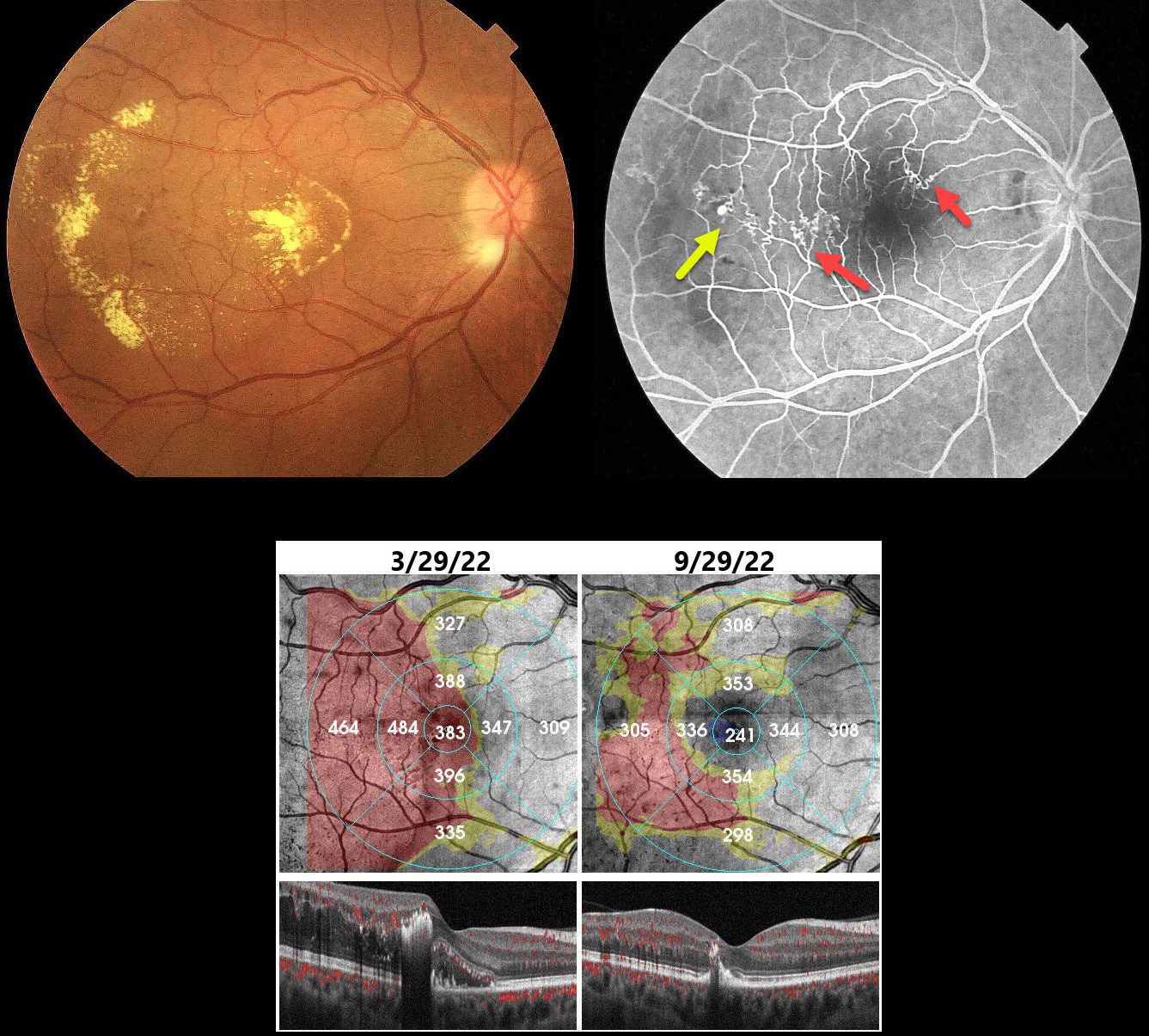
BRVO WITH RETINAL VENOUS MACROANEURYSM
Seif Anwar
Originally posted on @retina.rocks August 22, 2024
This 40YO hypertensive male presented with gradual vision loss in his right eye. Vision was 20/200 OD and 20/20 in his normal OS.
Color imaging shows central and temporal macular lipid and telangiectasia. Fundus fluorescein angiography shows a superotemporal branch retinal vein occlusion (BRVO) with superonasal and temporal collateral vessels. A retinal venous macroaneurysm (RVM) is noted distally.
Our patient was initially treated unsuccessfully with several monthly Lucentis injections. The macular edema and lipid improved following macular grid laser with focal treatment of the RVM.
Learning Points:
In 1990 Cousins et al reported that RVMs can be an additional finding in occlusive venous disease, always in association with collateral vessels or capillary or arterial macroaneurysms (AJO 1990;109:567-570). These vascular abnormalities tend to have a higher rate of intraretinal lipid (40%) but are less likely to affect central vision since they are usually outside the macula. They tend to be associated with retinal ischemia, thus increasing the risk of developing retinal neovascularization (32%).
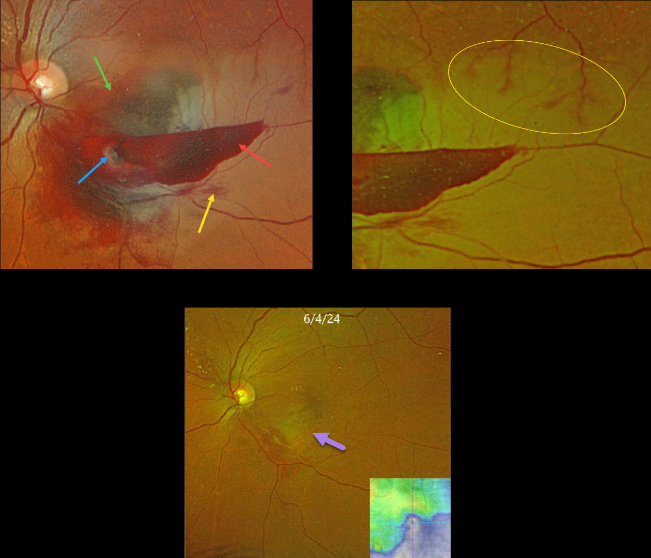
RETINAL ARTERIAL MACROANEURYSM (RAM) WITH BRAO
Originally posted on @retina.rocks August 13, 2024
This 59YO female presented with acute vision loss in her left eye. Vision was counting fingers.
Optos color RGB shows a multi-layered macular hemorrhage, including preretinal, intraretinal, and subretinal blood. The causative ruptured retinal arterial macroaneurysm (RAM) is noted within the preretinal blood inferonasally. The surrounding neurosensory retina is somewhat opaque from a secondary inferior branch retinal artery occlusion (BRAO).
Optos RG color imaging shows some periarterial sparing along the superotemporal edge of the BRAO. The perfused superior retinal arteriole, as it enters the region of the occlusion, still has some oxygen that is diffusing into the perivascular tissue.
The patient was treated with several monthly Avastin injections in hopes of more rapidly resolving the surrounding fluid to help relieve arterial compression. Although the blood and fluid resolved, secondary central and inferotemporal macular atrophy resulted from the prior BRAO. Vision was 20/400.
Learning Points:
A BRAO can be a rare complication of direct thermal laser of a RAM (Russell and Folk, AJO 1987;104:186-187), which is one reason it is almost never performed anymore. In this case, we believe the BRAO was secondary to mechanical arterial compression from the surrounding blood.
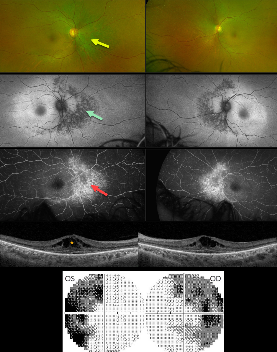
BITEMPORAL HEMIANOPIA WITH RETINITIS PIGMENTOSA
Mattie Adams
Originally posted on @retina.rocks August 12, 2024
This 54YO female was referred for asymptomatic retinal changes. Vision was 20/30 OD and 20/20 OS.
Visual fields showed bitemporal hemianopia. What is your diagnosis?
Optos color RG imaging shows pigmentary changes with intraretinal pigment migration extending superiorly, nasally, and inferiorly from the optic nerves. These changes are hypoautofluorescent and appear as angiographic window defects. Bilateral cystoid edema is seen on Triton swept-source OCT.
Learning Points:
Although bitemporal hemianopia usually signifies retrobulbar pathology at the optic chiasm, retinal disease can sometimes also cause similar field loss. Our patient’s retinitis pigmentosa (RP) changes were centered around each optic nerve, causing the hemianopia. Genetic testing revealed double heterozygous pathogenic ARSG mutations. This genotype has been associated with atypical Usher syndrome (Fowler et al, Ophthalmic Genetics 2021;42:338-343).
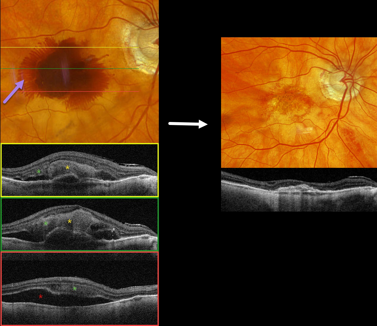
NEOVASCULAR AMD WITH HENLE LAYER HEMORRHAGE
Originally posted on @retina.rocks July 29, 2024
This 97YO male has a history of counting-finger vision in his left eye due to foveal macular atrophy. On 2/29/24, vision was 20/40 in his non-exudative right eye. He presented on 3/12/24 with sudden vision loss OD and new hand motions vision.
Optos color RGB imaging shows fairly dense central subretinal blood, with surrounding radiating blood in the deeper Henle’s layer (outer plexiform layer). Triton swept-source OCT shows variable thick subretinal blood and more inferior subretinal fluid. Henle’s blood is seen as a diffuse hyperreflective obscuration of the outer retinal layers. Vitrectomy with subretinal TPA was performed two days later. Two months later, vision was counting fingers, and the macula was completely free of fluid and blood.
Learning Points:
Radiating deep macular hemorrhages are seen in multiple conditions, including Valsalva and trauma. We have not previously seen these hemorrhages in neovascular AMD. Their unique radiating appearance results from blood accumulating in Henle’s layer. Kon Graversen et al described hemorrhagic unilateral retinopathy, an idiopathic disorder with a similar appearance (Retina 2014;34:483-489).
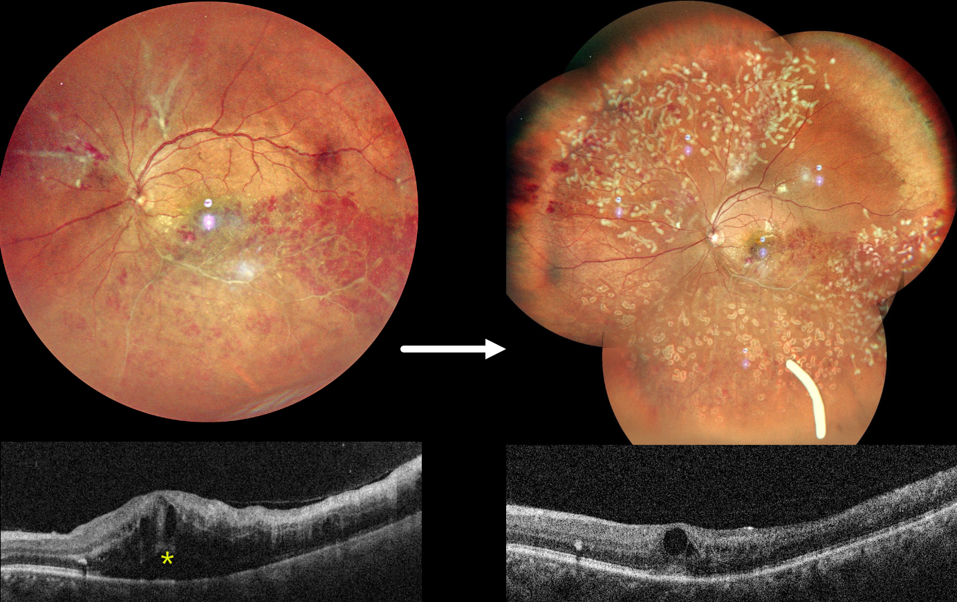
OCCLUSIVE RETINAL VASCULITIS
Anjana Mirajkar and Manish Nagpal
Originally posted on @retina.rocks July 23, 2024
This healthy 32YO male presented on 4/23/24 with 2 months of decreased vision in his left eye. Family history was negative. A prior workup by a prior doctor was negative, including protein electrophoresis and homocysteine levels. He failed a short trial of oral steroids. Vision was 20/20 in his healthy right eye and counting fingers in his left eye.
Fundus photography shows an ischemic inferotemporal branch retinal vein occlusion with moderate, mostly peripheral retinal hemorrhages. The superotemporal retinal vein is diffusely beaded, and an apparent independent ischemic BRVO is seen superonasally, with probable preretinal blood. Marked macular edema is seen on OCT. Unfortunately, fluorescein angiography is not available.
An Ozurdex injection was given for macular edema, and two sessions of panretinal photocoagulation were performed for proliferative disease. On 6/3/24, vision improved to 20/120, and the macular edema was improving.
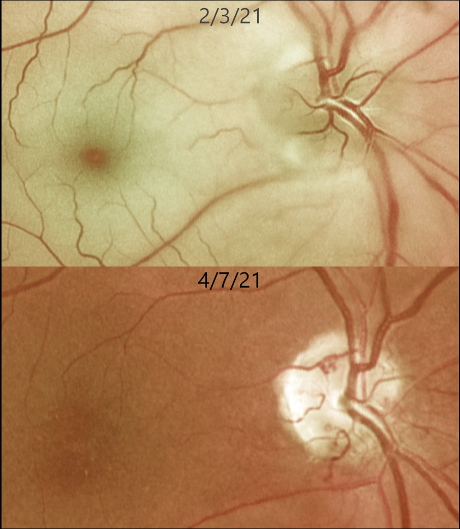
CRAO
Originally posted on @retina.rocks July 16, 2024
This 62YO female presented with an acute counting-fingers central retinal artery occlusion. Emergent stroke workup revealed a silent acute lacunar infarct. The retinal opacification completely resolved 2 months later. New fine collateral vessels were noted on the nerve. Vision remained at counting fingers.
Learning Points:
Collateral vessels are dilated, pre-existing vessels that bypass an occlusion. These are most commonly seen at the posterior pole following retinal venous occlusions and are exceedingly rare with arterial occlusions. The endothelial tight junctions are intact, so they don’t leak angiographically. This helps differentiate them from neovascularization, which does leak.
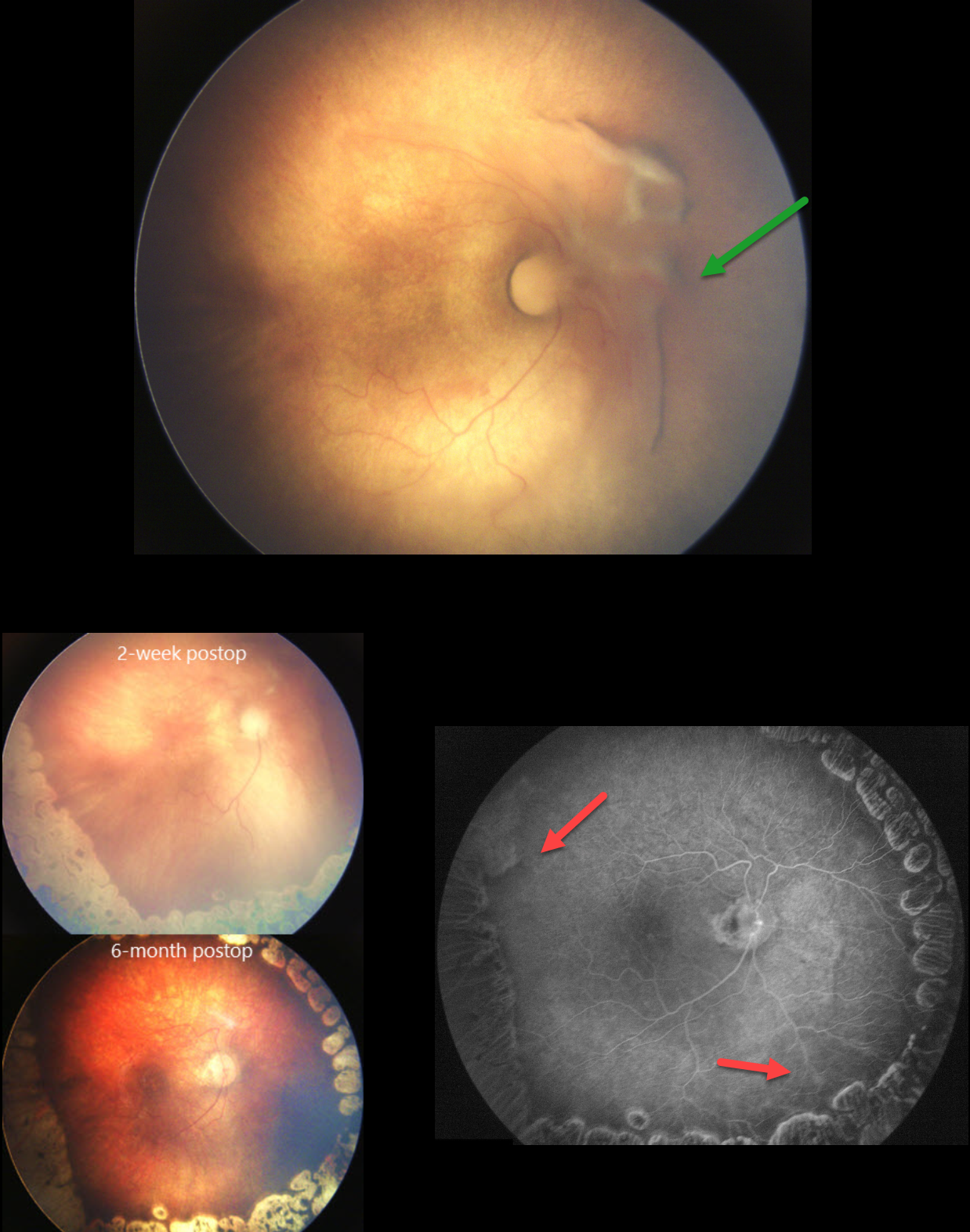
RETINOPATHY OF PREMATURITY
Şengül Özdek
Originally posted on @retina.rocks July 1, 2024
A prematurely born baby at a gestational age of 30 weeks and birth weight of 1120 grams was referred to our clinic at postmenstrual age of 37 weeks for the treatment of stage 4A ROP in her right eye. It was a zone 1 disease, and even the macula was not yet vascularized in either eye. There was a tractional retinal detachment at the nasal side of the right optic disc.
A lens-sparing vitrectomy was done with laser ablation of zone 2. Zone 1 (macula) was not lasered, and an anti-VEGF injection was given at the end of the surgery. The retina was completely attached at both 2-weeks and 6-months postoperatively. The retina was finally completely vascularized in zone 1 six months postoperatively.
Learning Points:
The current ROP classification system is based on the International Classification of Retinopathy of Prematurity, Third Edition (ICROP 3; Ophthalmology 2021;128:e51-e68), and there is no zone half in the zone descriptions. However, especially in developing countries with limited resources in neonatal intensive care units, ROP with avascular macula (even less than zone I, which could be called zone half!), as in the present case, can be seen. Such cases are usually called Aggressive ROP, which can progress to stage 4-5 very quickly.
It is generally recommended to laser the avascular retina during vitrectomy to control active disease. However, in zone 1 cases where the macular vascularization is not yet completed, as in our patient, we leave the zone 1 non-lasered to give a chance for macular vascularization after surgery. An anti-VEGF injection at the end of the surgery was therefore given to control possible disease activation due to the lack of laser treatment to the posterior nonperfused retina. To the best of our knowledge, this is the first presentation of such a treatment approach in the literature.
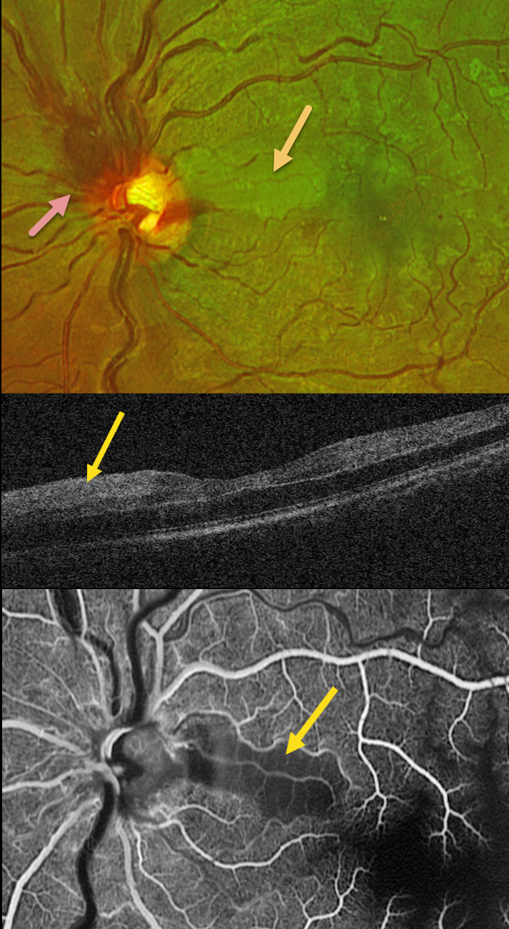
CRVO + CILIORETINAL ARTERY OCCLUSION
Amit Nandan Tripathi and Vipin Rana
Originally posted on @retina.rocks June 18, 2024
This healthy 30YO male presented with sudden vision loss in his left eye. Vision was 20/20 in his healthy right eye and counting fingers in his left eye.
Optos color RG imaging shows an acute non-ischemic central retinal vein occlusion (CRVO) with mild inner retinal hemorrhages scattered in all quadrants. The optic nerve is somewhat swollen superiorly, and there is an acute cilioretinal artery occlusion in the superior papillomacular bundle.
This area is hyperreflective on OCT. Fluorescein angiography shows a well-perfused CRVO except for a localized area of ischemia within the nasal occlusion.
Learning Points:
Some patients with CRVO will develop a secondary non-embolic cilioretinal artery occlusion caused by either compression from optic nerve edema or from a sudden rise in intraluminal retinal capillary bed pressure (Hayreh et al, Retina 2008;28:581-594). Observation was recommended, and vision improved to 20/60 five days after symptom onset.
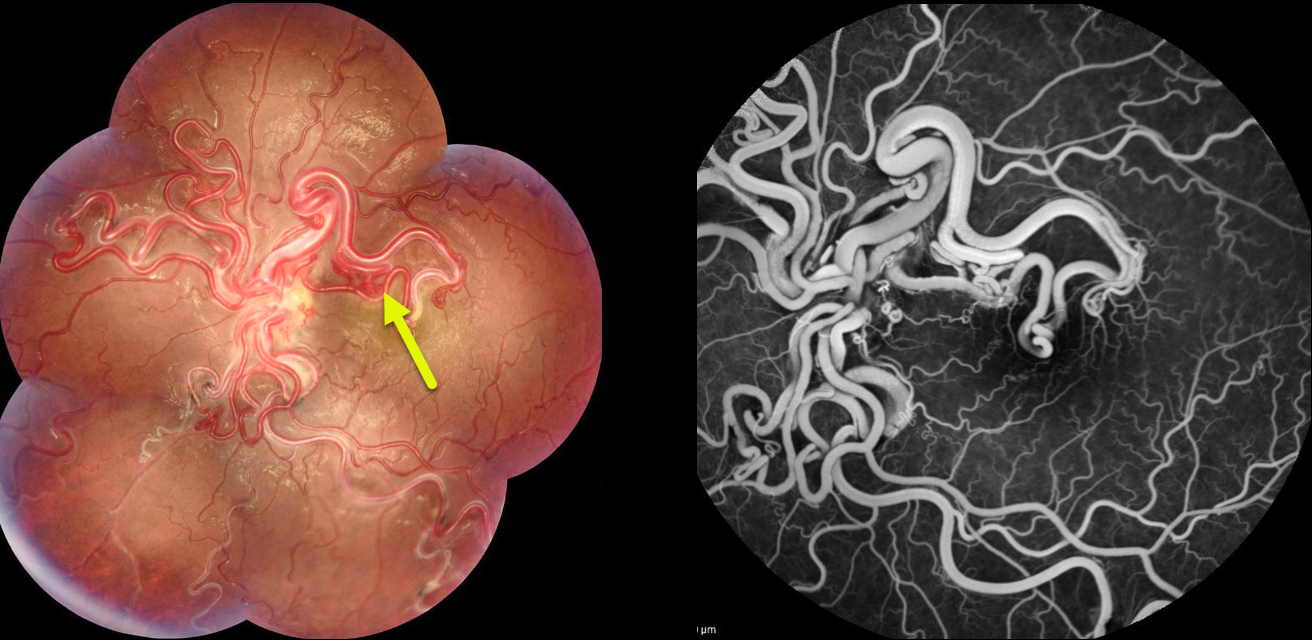
RETINAL ARTERIOVENOUS MALFORMATION
Şengül Özdek and Ece Özdemir Zeydanlı
Originally posted on @retina.rocks May 8, 2024
This healthy 16YO girl presented with a long history of poor vision in her left eye. Vision was 20/100 in her left eye.
Fundus photography of her left eye shows tangles of markedly dilated and tortuous vessels covering the posterior pole. Fluorescein angiography shows no leakage. Brain MRI revealed a cerebral hemangioma next to the optic chiasm.
Learning Points:
A retinal arteriovenous malformation (AVM) is a congenital connection between an artery and a vein without an intervening capillary network. These lesions appear along a spectrum ranging from small, isolated lesions to the large ‘bag of worms’ appearance as seen in our patient. These more extensive lesions are usually part of the Wyburn-Mason Syndrome, which includes vascular lesions in the eyes and midbrain. Retinal venous malformations, aka retinal macrovessels, are now thought to represent an AVM (Pichi et al JAMA Ophthalmol 2018;136:372-379).
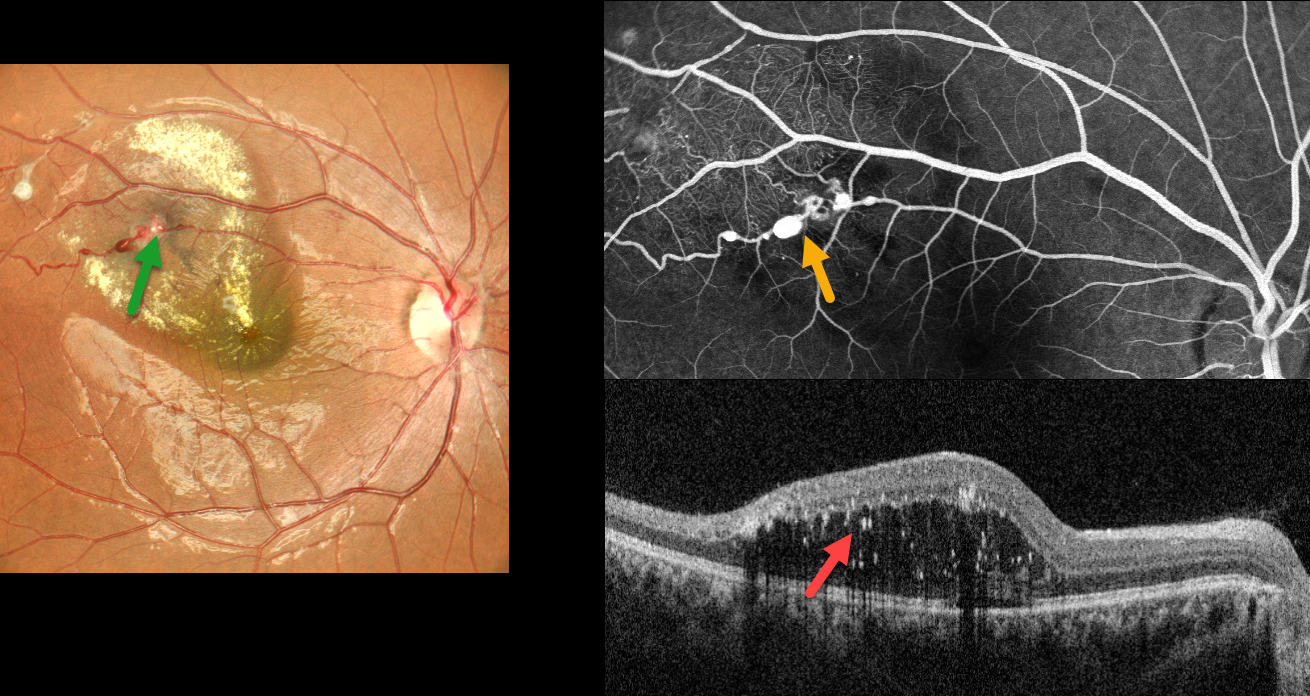
COATS DISEASE
Anjana Mirajkar and Manish Nagpal
Originally posted on @retina.rocks May 2, 2024
This healthy 19YO female presented with 1 week of blurred vision in her right eye. Vision was 20/80 OD and 20/20 in her normal OS.
Color imaging shows multiple superotemporal variably fibrosed macroaneurysms with surrounding lipid that extends towards the central macula. More subtle telangiectasia and lipid were seen in the superior periphery (not shown).
Fluorescein angiography shows multiple patent macroaneurysms along the superotemporal arteriole with more distal telangiectasia. OCT shows outer retinal fluid with numerous suspended hyperreflective lipid particles. Thermal laser followed by 3 monthly anti-VEGF injections was given.
Learning Points:
Described by George Coats in 1908, Coats disease is an idiopathic disorder that is usually unilateral in males. It is characterized by retinal vascular telangiectasia involving capillaries, arterioles, and venules. Patients can present with a wide variety of findings ranging from minimal exudation to total exudative retinal detachment. The leaking vessels are usually treated to prevent exudative complications.
Thermal laser photocoagulation is usually the initial modality, but anti-VEGF injections can also be used to stabilize the vascular leakage. Patients will often require multiple rounds of treatment. See Sigler et al for a review of the current management (Survey Ophthalmology 2014;59:30-46).
Dyskeratosis congenita or familial exudative vitreoretinopathy should be considered for patients with frank bilateral disease (Teixeira et al, Arch Ophthalmol 2008;126:134-135), in women with Coats-like findings (Johnson et al, Genetics 2009;30:181-184), or in patients with a family history of similar findings. Since our patient’s findings were unilateral and her nail beds were normal, we deferred genetic testing.
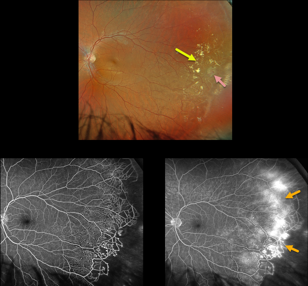
COATS DISEASE
Originally posted on @retina.rocks April 3, 2024
This 28YO healthy male was referred for asymptomatic findings on retinal examination in his left eye. Vision was 20/20 in his normal OD and 20/20 OS.
Optos color RGB imaging shows scattered temporal lipid and telangiectatic vessels. A large macroaneurysm is noted. The retina distal to the lipid is featureless and clinically ischemic.
Fluorescein angiography shows the telangiectasia becoming more prominent and ischemic as it extends from the distal temporal macula into the periphery. There is marked leakage at the junction of the perfused and ischemic retina. Scatter laser to the ischemic retina, along with a focal laser to the macroaneurysm, was scheduled.
Learning Points:
Described by George Coats in 1908, Coats disease is an idiopathic disorder that is usually unilateral in males. It is characterized by retinal vascular telangiectasia involving capillaries, arterioles, and venules. Patients can present with a wide variety of findings ranging from minimal exudation to total exudative retinal detachment.
The leaking vessels are usually treated to prevent exudative complications. Thermal laser photocoagulation is usually the initial modality, but anti-VEGF injections can also be used to stabilize the vascular leakage. Patients will often require multiple rounds of treatment.
See Sigler et al for a review of the current management (Survey Ophthalmology 2014;59:30-46).
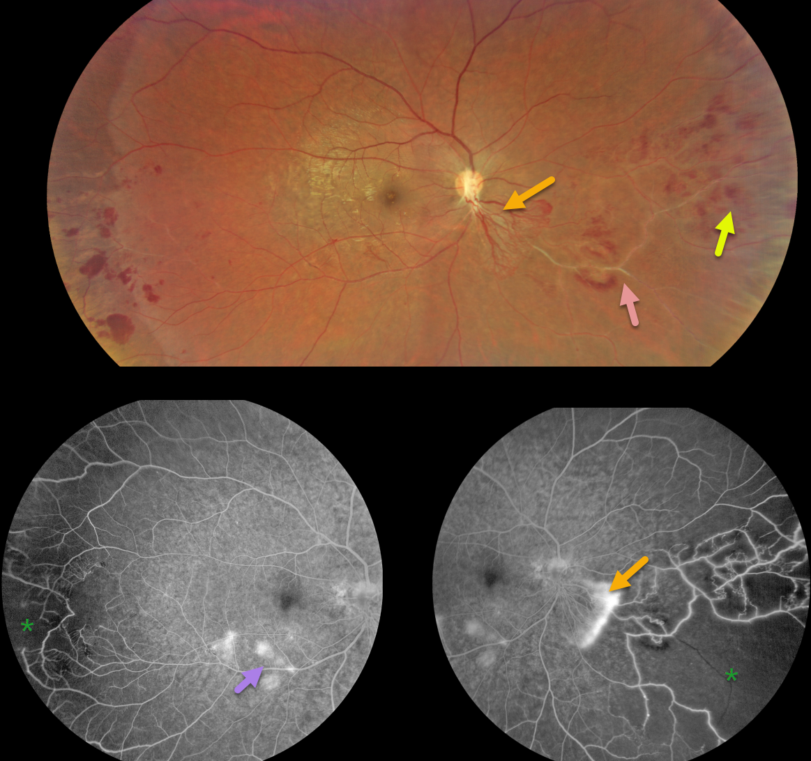
EALES DISEASE
Narciso Atienza Jr.
Originally posted on @retina.rocks March 28, 2024
This healthy 31YO male presented with 6 months of blurred vision. He was previously diagnosed by a general ophthalmologist with a branch vein occlusion in the OD. Vision was 20/30 in his right eye and 20/20 in his left eye.
Color imaging shows active neovascularization extending inferonasally from the nerve, with more distal retinal hemorrhages and vascular sheathing.
Fluorescein angiography shows leaking neovascularization with retinal ischemia nasally and temporally. Some vascular leakage is noted in the inferotemporal macula.
Learning Points:
First described by British ophthalmologist Henry Eales in 1880, Eales disease is characterized by sharply circumscribed peripheral regions of distal ischemia, often with retinal neovascularization at the junction of the perfused and ischemic retina. It is more common in males in their 20’s to 30’s and may be associated with a prior exposure to tuberculosis. Most patients are asymptomatic unless they develop a vitreous hemorrhage. Scatter laser to ischemic retina and intravitreal anti-VEGF injections are effective for retinal neovascularization.
It is likely a heterogeneous disorder and is a diagnosis of exclusion, needing first to rule out the multitude of other disorders that can cause peripheral retinal ischemia and neovascularization. The location of our patient’s neovascularization is a bit unusual for Eales, which is usually more peripheral. However, since his systemic work-up, including chest X-ray, was normal except for a positive PPD, we feel Eales is a reasonable diagnosis. A scatter laser to the ischemic retina has been planned and scheduled.
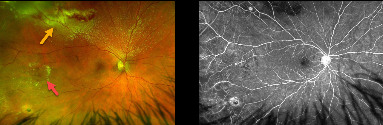
COATS DISEASE
Will Gibson
Originally posted on @retina.rocks March 19, 2024
This 36YO male presented with asymptomatic findings on fundus examination in his right eye. Vision was 20/25 in his right eye and 20/20 in his normal left eye.
Optos color RG imaging shows telangiectatic vessels in the temporal and superotemporal midperipheries. Subretinal lipid is most dense superotemporally.
Fluorescein angiography beautifully shows the telangiectasia, including capillary loss, microaneurysms, and macroaneurysms. He was referred to a retinal specialist for further management.
Learning Points:
Described by George Coats in 1908, Coats disease is an idiopathic disorder that is usually unilateral in males. It is characterized by retinal vascular telangiectasia involving capillaries, arterioles, and venules. Patients can present with a wide variety of findings ranging from minimal exudation to total exudative retinal detachment. The leaking vessels are usually treated to prevent exudative complications.
Thermal laser photocoagulation is usually the initial modality, but anti-VEGF injections can also be used to stabilize the vascular leakage. Patients will often require multiple rounds of treatment.
See Sigler et al for a review of the current management (Survey Ophthalmology 2014;59:30-46).
Atypical cases, including positive family history, bilateral findings, or in females, should be considered for genetic testing to rule out inherited telomere disorders, including Coats plus syndrome (Teixeira et al, Arch Ophthalmology 2008;126:134-135) and dyskeratosis congenita (Johnson et al, Ophthalmic Genetics 2009;30:181-184).
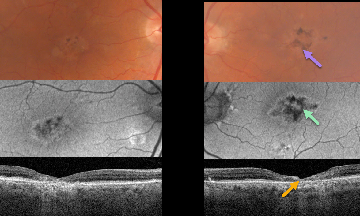
MACULAR TELANGIECTASIA
Originally posted on @retina.rocks March 4, 2024
This 80YO male was referred for bilateral macular findings. He was without new visual complaints. Vision was 20/70 OD and 20/200 OS.
Optos color RG imaging shows bilateral foveal pigment loss with coarse intraretinal pigment migration. The changes are mostly hypo-autofluorescent.
Triton swept-source OCT shows atrophy and disorganization of all retinal layers in the central and temporal foveas.
Learning Points:
Originally described by Gass (Arch Ophthalmology 1982;100:769-780), macular telangiectasia type 2 (MacTel2) is a neurodegenerative disorder, most likely originating from Muller cell dysfunction.
The term telangiectasia is misleading, as the funduscopic findings are mostly non-vascular. The refractile inner retinal ‘crystals,’ not seen in our patient, are thought to be Muller cell footplates. The foveal retina shows a whitish discoloration, most likely due to retinal opacification caused by dysfunctional Muller cells. Photoreceptor loss allows RPE cells to migrate along intraretinal capillaries, causing the coarse clinical pigment clumping.
The majority of patients will also have subclinical chorioretinal anastomosis associated with right-angle venules (Spaide et al, Retina 2018;38:1920-1929). Secondary macular neovascularization can be successfully treated per age-related macular degeneration protocols.
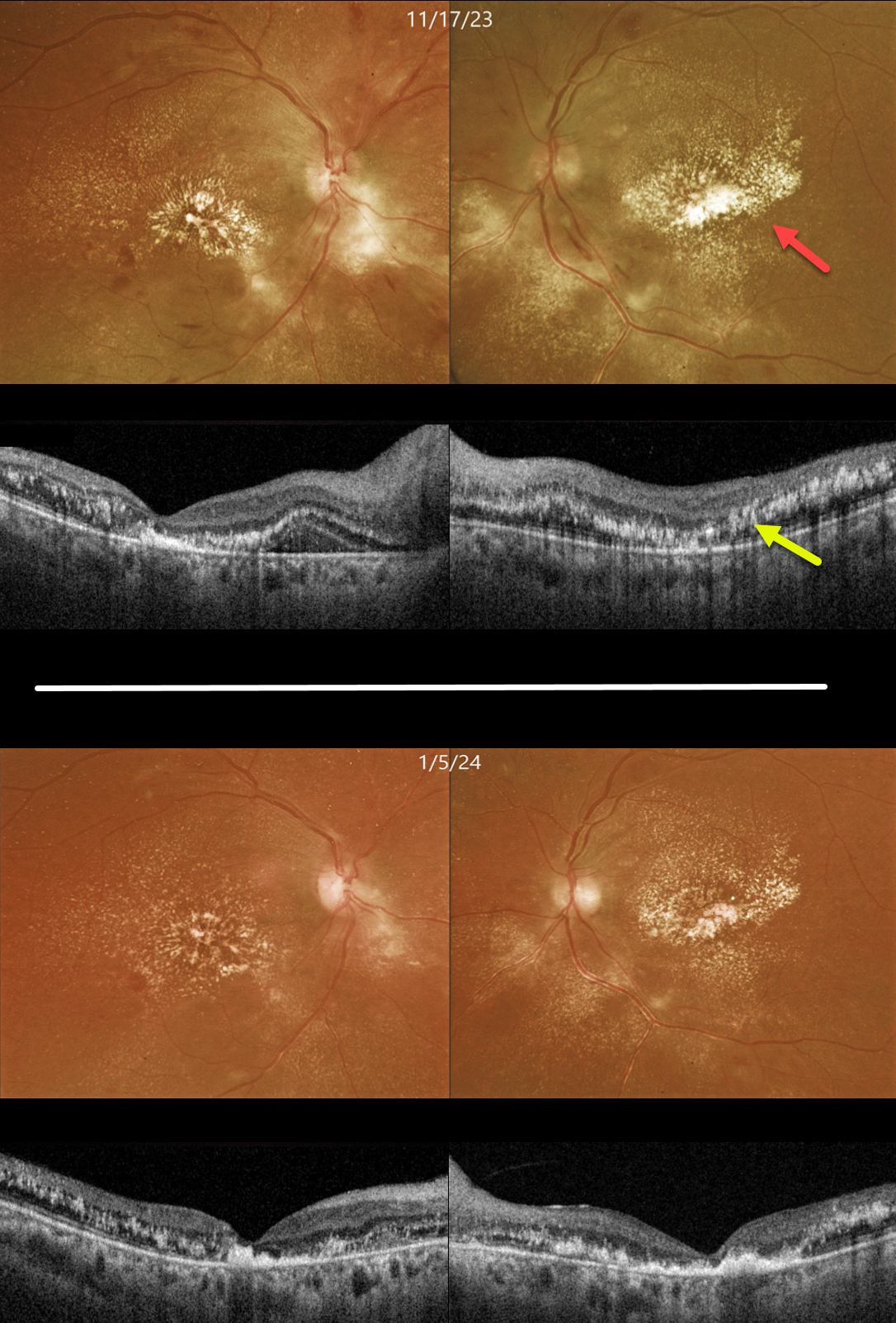
HYPERTENSIVE RETINOPATHY
Originally posted on @retina.rocks February 20, 2024
This 43YO female was recently discharged from the hospital for severe hypertension. She told us she was hospitalized for about a month and a half, with treatment including a medically induced coma. Vision was 20/200 OU.
Color imaging shows bilateral retinal lipid extending from the maculas into the midperipheries. Swept source OCT shows bilateral central outer retinal thinning and scattered hyperreflective lipid extending from the outer plexiform layer to above the RPE.
When she returned 6 weeks later, vision remained at 20/200 OU, and the macular lipid was slowly regressing.
Learning Points:
Malignant hypertension, defined as blood pressure above 180 systolic and/or 120 diastolic, is a life-threatening medical emergency. Eye doctors are in a unique position to often diagnose this condition.
Patients will often present with bilateral optic nerve swelling, flame-shaped retinal hemorrhages, and, with more chronic disease, lipid precipitates in the nasal macular outer plexiform layer.
Since the macular fluid emanates from the optic nerve, macular lipid and thickening are virtually always worse nasally, which is an important clinical clue for this diagnosis.
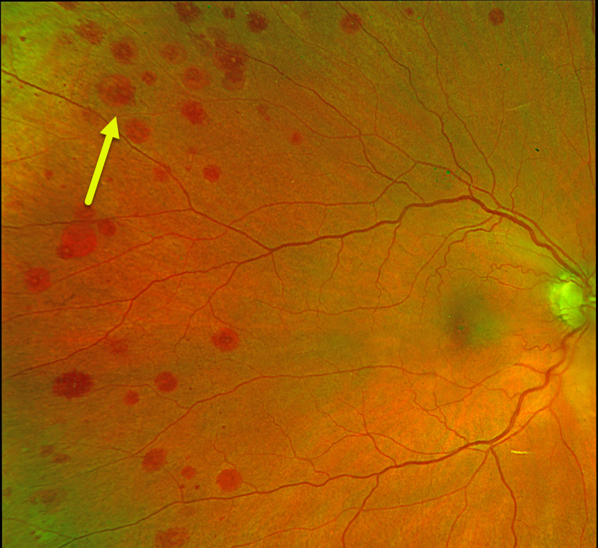
RETINAL HEMORRHAGES
Originally posted on @retina.rocks February 12, 2024
This 79YO female was referred for asymptomatic retinal findings. She has a history of diet-controlled type 2 diabetes.
Optos color RG imaging shows scattered, round, deep retinal hemorrhages, some with white centers. Most of the midperipheral hemorrhages are much more extensive in her right eye.
Bloodwork, including CBC, serum viscosity, and serum protein electrophoresis, was normal. We suspect ocular ischemia as the cause of her findings, but despite numerous attempts, the patient continues to refuse carotid testing.
Learning Points:
The term “Roth spot” was named after Dr. Moritz Roth, who, in 1872, noted white-centered hemorrhages in patients with bacterial endocarditis. He felt that these white spots were septic emboli that originated from an infected cardiac valve. However, similar lesions can be seen in many other disorders, including leukemia, hyperviscosity, anemia, diabetes, and hypertension (Duane et al Ophthalmology 1980;87:66-69).
The white material may represent a variety of causes, including fibrin. So these lesions are best called white-centered hemorrhages, unless one is specifically referring to one of these lesions in a patient with bacterial endocarditis.
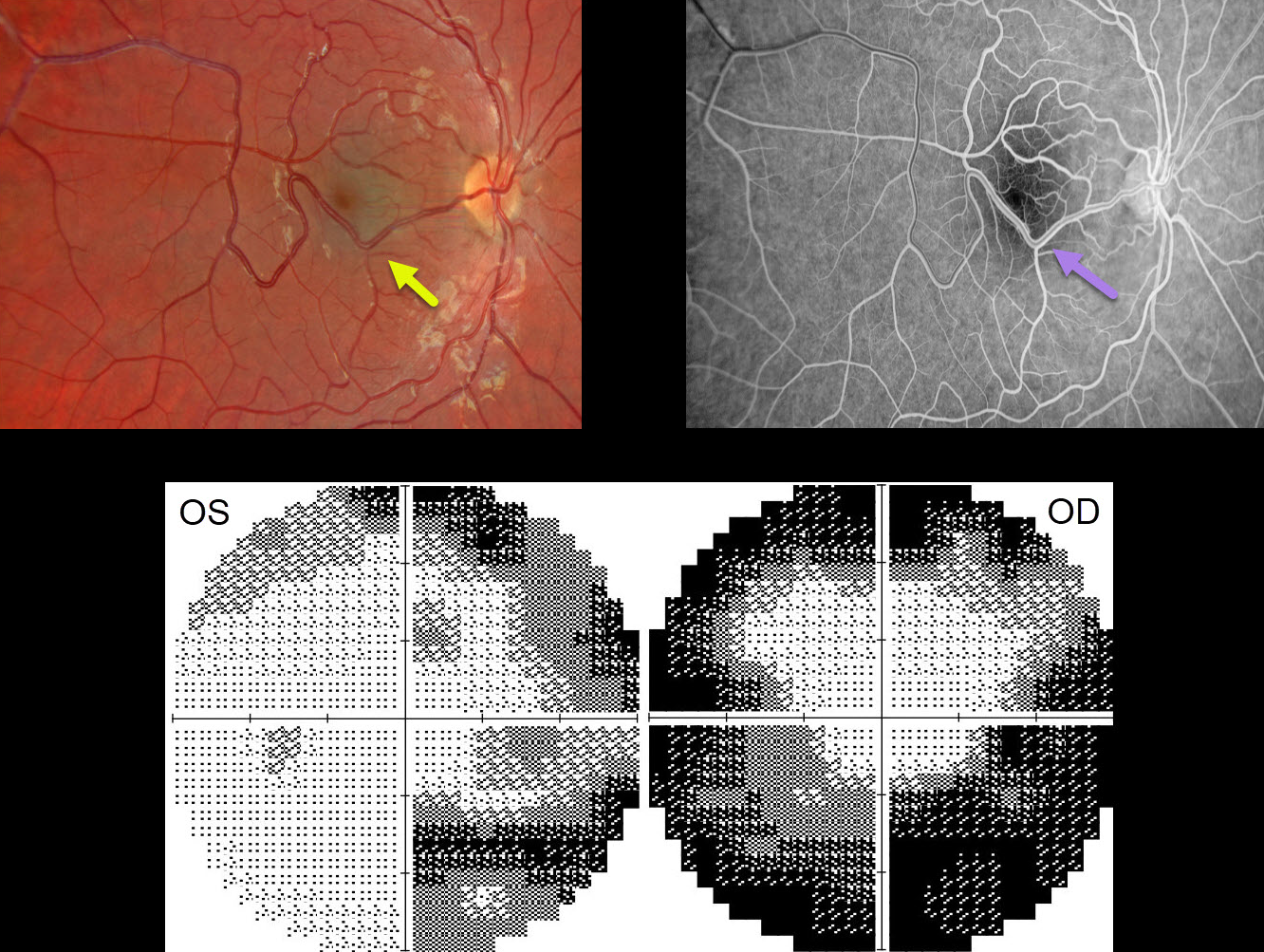
RETINAL VENOUS MALFORMATION
Will Gibson
Originally posted on @retina.rocks January 22, 2024
This 17YO male presented with bilateral vision loss and pain on eye movements 1 week following a COVID-19 infection. There was no prior ocular or medical history. Vision was 20/70 OD and 20/50 OS.
Color imaging of the right eye shows a large temporal retinal venous malformation. This vessel shows angiographic laminar filling without leakage. The right and left nerves were clinically normal, and the left fundus was normal (not shown).
OCT retinal nerve fiber layer analysis was normal bilaterally. Visual fields show moderate generalized constriction OD and nasal loss OS with a possible vertical cut inferiorly. Subsequent neurologic imaging was consistent with optic neuritis. Additional workup was unremarkable, and neuro-ophthalmic consultation led to the diagnosis of bilateral retrobulbar optic neuritis secondary to COVID-19.
Observation was recommended, and symptoms began to improve within a week. Two months later, all visual symptoms resolved, vision returned to 20/25 OU, and the visual fields normalized.
Learning Points:
Retinal vessels virtually always respect the horizontal meridian. Retinal venous malformations are congenital anomalous vessels that, by definition, cross the horizontal.
Originally described by Gary Brown et al as congenital retinal macrovessels (Arch Ophthalmology 1982;100:1430-1436), these vessels are usually located in the macular region and are virtually always venous.
These lesions are now called retinal venous malformations, since about one-quarter of patients have associated venous anomalies in the brain (Pichi et al, JAMA Ophthalmol 2018;136:372-379).
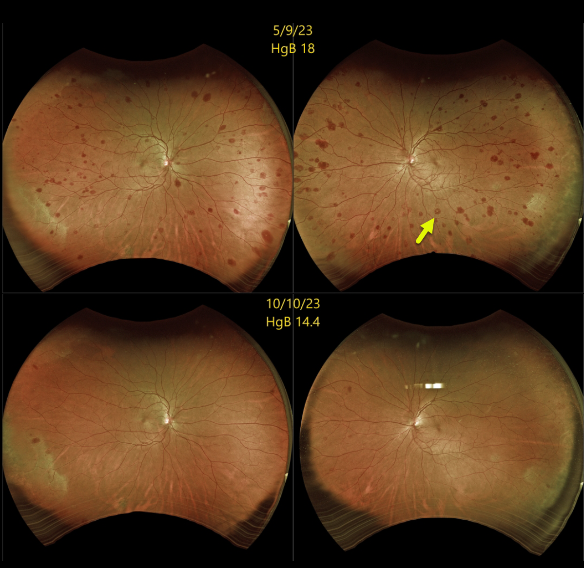
SECONDARY POLYCYTHEMIA
Originally posted on @retina.rocks December 18, 2023
This 51YO female was referred for asymptomatic retinal hemorrhages in each eye. She had a history of pulmonary hypertension. Vision was 20/20 bilaterally.
Optos color RG imaging shows scattered retinal hemorrhages in all quadrants, most of which are deep with white centers. A complete blood count revealed a hemoglobin of 18.0 g/dl, and she was immediately referred for a hematology consult.
Subsequent hematologic workup was negative, and she was felt to have secondary polycythemia from her pulmonary hypertension. She has since been treated with serial phlebotomies.
When last examined 5 months later, the retinal hemorrhages were markedly decreased, and her hemoglobin had decreased to 14.4 g/dl.
Learning Points:
The term “Roth spot” was named after Dr. Moritz Roth, who, in 1872, noted white-centered hemorrhages in patients with bacterial endocarditis. He felt that these white spots were septic emboli that originated from an infected cardiac valve.
However, similar lesions can be seen in many other disorders, including leukemia, hyperviscosity, anemia, diabetes, and hypertension (Duane et al Ophthalmology 1980;87:66-69). The white material may represent a variety of causes, including fibrin. So these lesions are best called white-centered hemorrhages, unless one is specifically referring to those found associated with bacterial endocarditis.
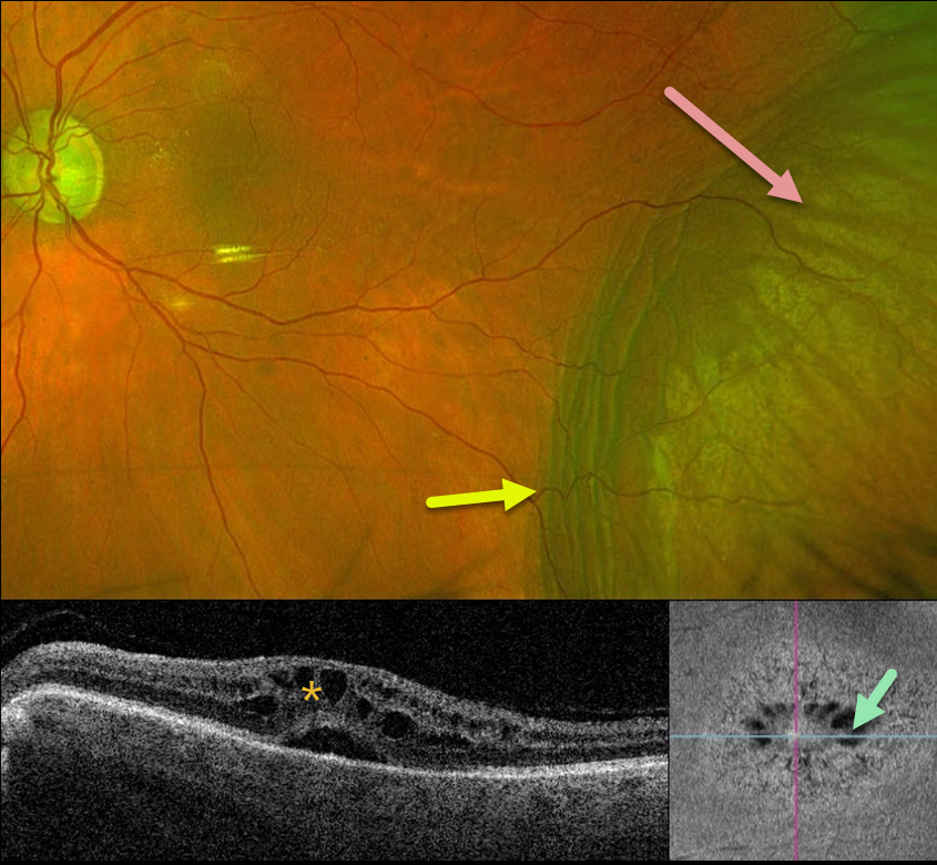
CHOROIDAL DETACHMENT
Originally posted on @retina.rocks November 30, 2023
This 84YO male has a long history of advanced chronic open-angle glaucoma with numerous surgical procedures. For several years, his left eye has had stable hypotony, with an intraocular pressure (IOP) of about 3-6 mmHg. Vision is 20/60.
Optos color RG imaging shows an inferotemporal choroidal detachment with chorioretinal folds along its posterior margin. Triton swept-source OCT shows cystoid edema with subretinal fluid, and en face imaging shows radiating mid-retina foveal cysts.
Since his symptoms, vision, IOP, and retinal findings remain stable, we are continuing to follow him without further intervention.
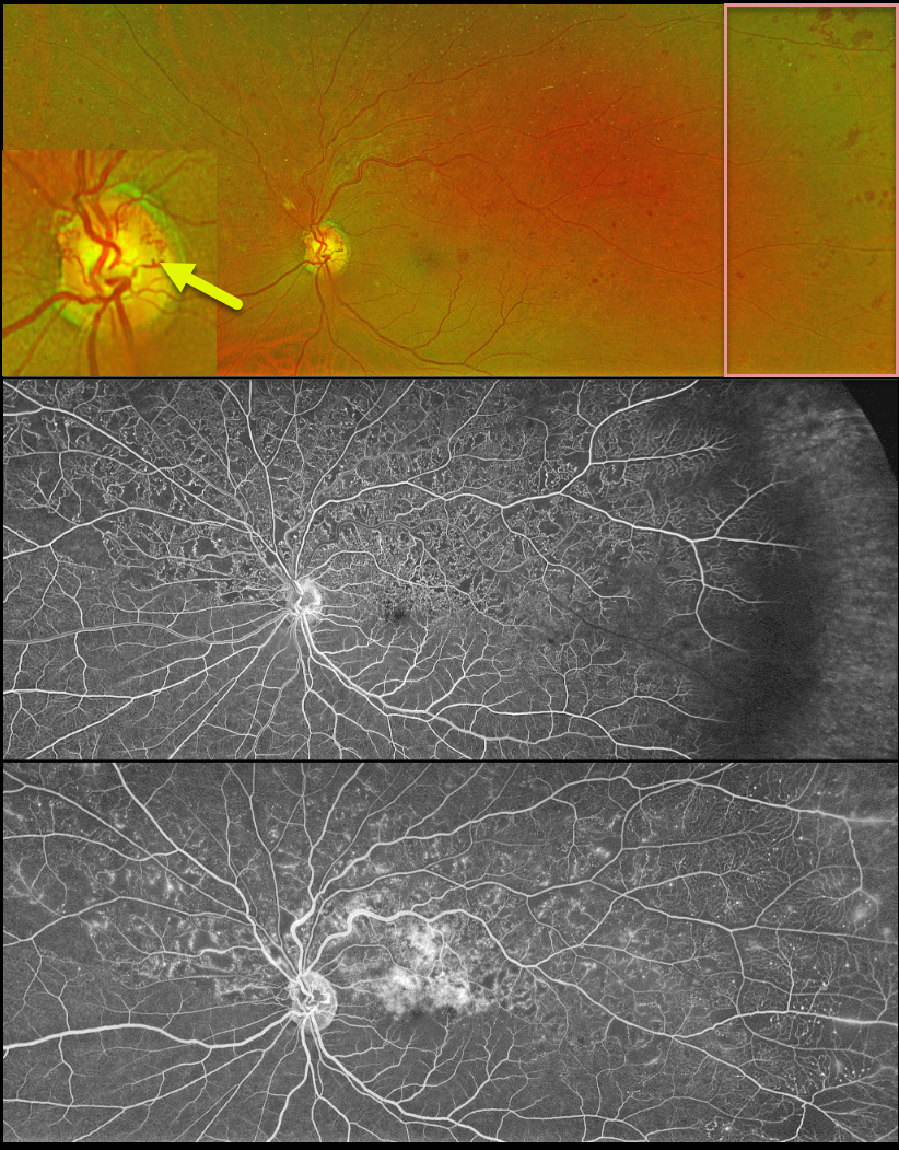
HRVO
Originally posted on @retina.rocks November 8, 2023
This 63YO male was referred for relatively asymptomatic retinal findings in his left eye. Vision was 20/60.
Optos color RG imaging shows a superior hemiretinal vein occlusion (HRVO) with mild hemorrhages, mostly in the temporal periphery. The occlusion is well-perfused without retinal neovascularization on fluorescein angiography. Prominent collateral vessels are noted on the optic disc superiorly. Observation was recommended.
Learning Points:
Branch retinal vein occlusions are virtually always caused by a retinal artery compressing a retinal vein within its common adventitial sheath. Some people are born with an anomalous two-trunked central retinal vein that independently drains the superior and inferior retinal hemispheres.
HRVO, similar to central retinal vein occlusion, is caused by compression of the retinal vein posterior to the lamina cribrosa (see Hayreh, Arch Ophthalmol 1980;98:1600-1609).
Collateral vessels are dilated, pre-existing vessels that bypass an occlusion. These are seen in the posterior pole most commonly following retinal venous occlusions, but can also occur on the optic disc with optic nerve tumors. The endothelial tight junctions are intact, so they don’t leak on angiography. This helps differentiate them from neovascularization, which does leak.
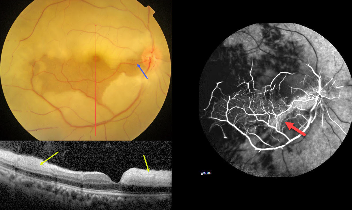
CRAO WITH CILIORETINAL ARTERY SPARING
Anjana Mirajkar and Manish Nagpal
Originally posted on @retina.rocks November 2, 2023
This 45YO male presented with acute vision loss in his right eye 4-5 days earlier. Vision was 20/80 in his right eye and 20/20 in his normal left eye.
Color photography shows an acute central retinal artery occlusion (CRAO) with sparing of the central and inferior macula due to a large cilioretinal artery.
The vertical OCT B-scan shows opaque hyperreflective paracentral inner retina with normal central and inferior foveal layers. Fluorescein angiography confirms preserved central and inferior macular arterial flow.
Workup elsewhere included a normal brain MRI and cardiac 2D Echo. Carotid Doppler showed a plaque in the right internal carotid artery.
Learning Points:
The inner two-thirds of the neurosensory retina is supplied by the central retinal artery, with the choroid supplying the RPE and photoreceptor layer. Acute CRAO, therefore, presents with a white edematous inner retina that spares the fovea.
Cilioretinal arteries, which arise from the posterior choroidal circulation, are present in up to 50% of individuals, and bilateral in about 25% (Schneider et al, Acta Ophthalmologica 2021;99:e310-e318). Our patient was quite fortunate to have a large cilioretinal vessel that relatively preserved central vision.
Patients with acute retinal ischemia (defined as transient monocular vision loss, acute BRAO, or acute CRAO) need to be emergently referred to a stroke center. This is especially urgent with an acute CRAO since about 75% of patients have already developed a recent stroke.
If the patient presents immediately after occlusion, measures to acutely lower intraocular pressure to move the causative embolus downstream can be performed, including ocular massage, paracentesis, and breathing into a bag. Unfortunately, most patients present outside this window (Shah et al, Ophthalmology Retina 2023;7:527-531), and even then, these measures are usually ineffective.
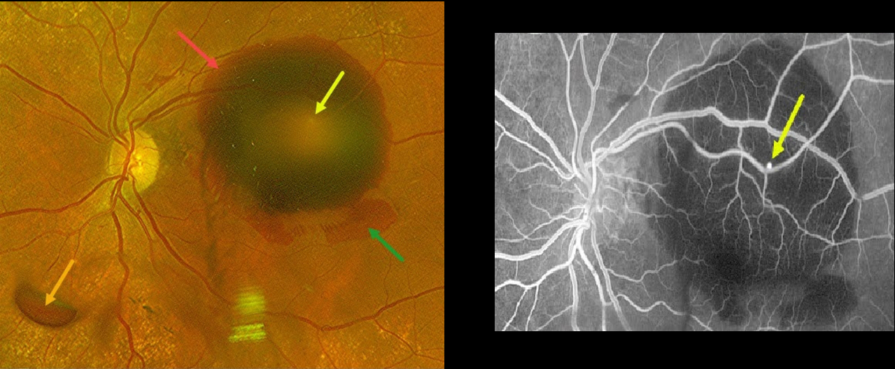
RUPTURED RETINAL ARTERY MACROANEURYSM (RAM)
Mohammad Abbas
Originally posted on @retina.rocks November 1, 2023
This 78YO hypertensive female presented with sudden vision loss in her left eye. Vision was 20/40 in her normal right eye and counting fingers in her left eye.
Optos color RG imaging shows multiple layers of blood, including a large submacular hemorrhage, intraretinal blood, and preretinal blood.
A tiny retinal arterial macroaneurysm is noted in the superotemporal macula clinically as well as on fluorescein angiography.
This case was submitted by Mohammad Abbas.
Learning Points:
A RAM is thought to occur in a weakened arterial wall from arteriosclerosis, and appears as a yellow-white dilated lesion along a retinal artery within the first 3 bifurcations from the optic nerve.
There are 2 distinct presentations, hemorrhagic and exudative. The hemorrhagic type often presents, as in this case, with pathognomonic multiple layers of blood. Acute macular hemorrhage can be treated with observation, anti-VEGF injections, or thermal laser.
Due to the thick submacular blood, vitrectomy with subretinal TPA was recommended. However, she declined surgery and is being followed for now.
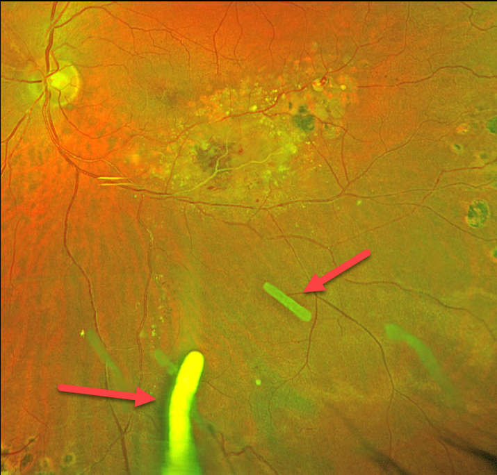
TRIAMCINOLONE-MOXIFLOXACIN (TriMoxi)
Originally posted on @retina.rocks October 30, 2023
This 41YO male has been followed for years with Coats disease in his left eye. Numerous treatment modalities have been used, including thermal laser, intravitreal anti-VEGF, and Ozurdex (dexamethasone 0.7mg) injections. Most recently, he has received Ozurdex every 3-4 months for the past 2 years. Vision is stable at 20/30.
Optos color RG imaging shows multiple Ozurdex ghosts in variable stages of absorption.
Learning Points:
The Ozurdex implant slowly dissolves while releasing its dexamethasone. Although the clinical effect usually lasts about 2 to 3 months, remnants can remain in the eye for over a year (Kim et al, Retina 2020;40:2226-2231).
Approved for diabetic and retinal vein occlusion macular edema and posterior uveitis, it is also often used off-label for other causes of macular edema, including uveitic and pseudophakic CME. For our patient, Ozurdex has been the most successful modality at keeping his central macula dry.
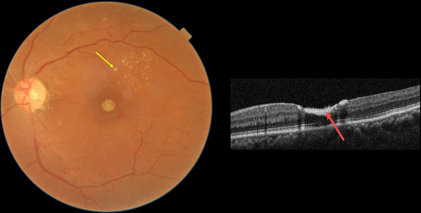
RETINAL ARTERIAL MACROANEURYSM (RAM)
Asma Samsudeen and Ashish Sharma
Originally posted on @retina.rocks October 26, 2023
This 54YO female presented with 3 weeks of vision loss in her left eye. Vision was 20/20 in her normal OD and 20/200 OS.
Fundus photography shows devitalized yellow prefoveal blood, some lipid flecks centered in the superotemporal macula, and a tiny fibrosed superior retinal arterial macroaneurysm (RAM).
OCT scanning through the fovea confirms the sub-internal limiting membrane (ILM) location of the hyperreflective blood.
This case was submitted by Asma Samsudeen and Ashish Sharma.
Learning Points:
A RAM is thought to occur in a weakened arterial wall from arteriosclerosis, and appears as a yellow-white dilated lesion along a retinal artery within the first 3 bifurcations from the optic nerve.
There are 2 distinct presentations, hemorrhagic or exudative, and our patient has features of both. Although she reported only a several-week history of vision loss, the retinal findings suggest a much longer course.
The devitalized blood usually takes months to turn yellow, and was likely much larger in extent originally. The blood elsewhere has largely resolved. There was also likely macular thickening and possibly subretinal fluid, which spontaneously resolved, leaving behind residual peripheral macular lipid.
Observation was recommended, and we expect her vision to significantly improve, as the retina appears structurally fairly normal on OCT deep to the blood. Subretinal blood, especially devitalized, is toxic to the outer retina, which is why removal and/or displacement of thick subretinal blood is often recommended. Blood in the sub-ILM or preretinal space does not seem to damage the retina, so it can more safely be observed (see Bloom and Spaide, Retinal Cases 2022;16;401-402).
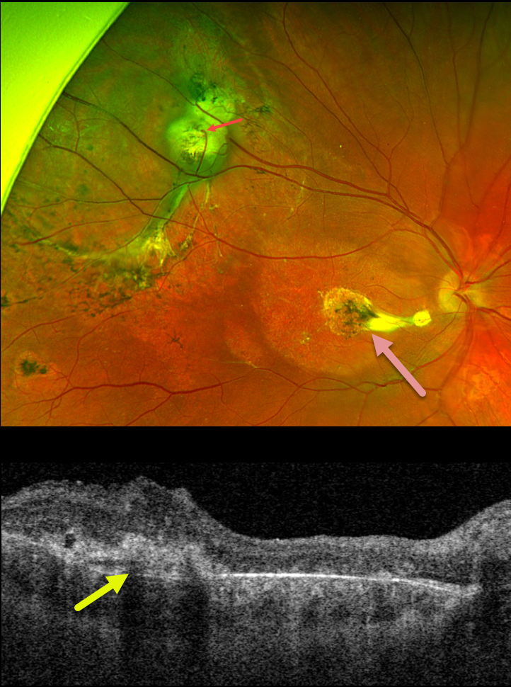
COATS DISEASE
Originally posted on @retina.rocks October 19, 2023
This 14YO boy received thermal laser photocoagulation for an exudative retinal detachment in his right eye from Coats disease about 10 years earlier. There were no new complaints, and vision was stable at 20/400. His normal left eye was 20/20.
Optos color RG imaging shows peripheral superotemporal scarring and subretinal fibrosis. A venous retinal-choroidal anastomosis is noted. Some variably pigmented laser scars are seen, and there are no active Coats lesions. A nematode-like area of subfoveal scarring is seen more centrally with variable outer retinal thinning and edema.
Learning Points:
Described by George Coats in 1908, Coats disease is an idiopathic disorder that is usually unilateral in males. It is characterized by retinal vascular telangiectasia involving capillaries, arterioles, and venules.
Patients can present with a wide variety of findings ranging from minimal exudation to total exudative retinal detachment. The leaking vessels are usually treated to prevent exudative complications.
Thermal laser photocoagulation is usually the initial modality, but anti-VEGF injections can also be used to stabilize the vascular leakage. Patients will often require multiple rounds of treatment.
See Sigler et al for a review of the current management (Survey Ophthalmology 2014;59:30-46).
Under normal conditions, the retinal and choroidal circulations remain separate and distinct. Rarely, the two circulations connect directly through a chorioretinal anastomosis. These can occur in chorioretinal scars, typically from inactive toxoplasmosis lesions, macular telangiectasia, and disciform scars.
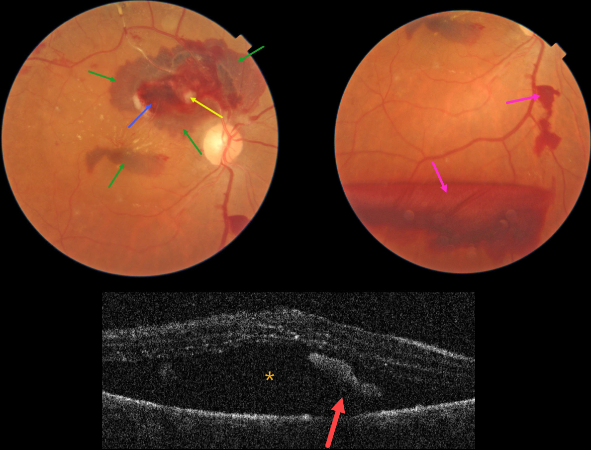
RETINAL ARTERIAL MACROANEURYSM (RAM)
Asma Samsudeen and Ashish Sharma
Originally posted on @retina.rocks September 28, 2023
This 76YO female presented with blurred vision in her right eye for one month. Vision was 20/60 in her right eye and 20/30 in her left eye.
Color photography shows a ruptured retinal arterial macroaneurysm (RAM) causing multiple layers of blood. The blood is subretinal, intraretinal, and preretinal. Some scattered lipid flecks are noted centrally and temporally, and the arterial distal to the RAM is sclerosed.
OCT scanning shows marked subretinal fluid and some hyperreflective subretinal blood.
Learning Points:
A RAM is thought to occur in a weakened arterial wall from arteriosclerosis, and appears as a yellow-white dilated lesion along a retinal artery within the first 3 bifurcations from the optic nerve.
There are 2 distinct presentations, hemorrhagic or exudative. The hemorrhagic type often presents, as in this case, with pathognomonic multiple layers of blood. Acute macular exudation can be treated with observation, anti-VEGF injections, or thermal laser.
Due to her symptomatic subretinal fluid, a single intravitreal Avastin injection was given. Four months later, vision improved to 20/30, the macular fluid resolved, and most of the blood layers were gone.
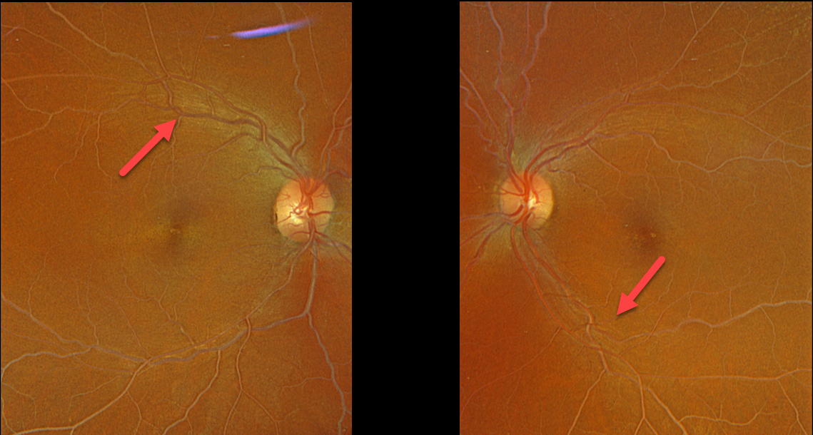
LIPEMIA RETINALIS
Originally posted on @retina.rocks August 21, 2023
This 47YO female presented with bilateral symmetric creamy colored blood vessels.
Her lipid panel results were: Cholesterol 1039, Triglyceride 821, HDL 7, and LDL too high to measure.
She was immediately referred for medical management of her previously undiagnosed hyperlipidemia.
Learning Points:
Lipemia retinalis is a rare retinal manifestation of severe hypertriglyceridemia, characterized by creamy white discoloration of the retinal vasculature. In severe cases, the fundus can turn salmon-colored.
Patients are asymptomatic and have normal vision. However, they are at obvious cardiovascular risk without systemic treatment.
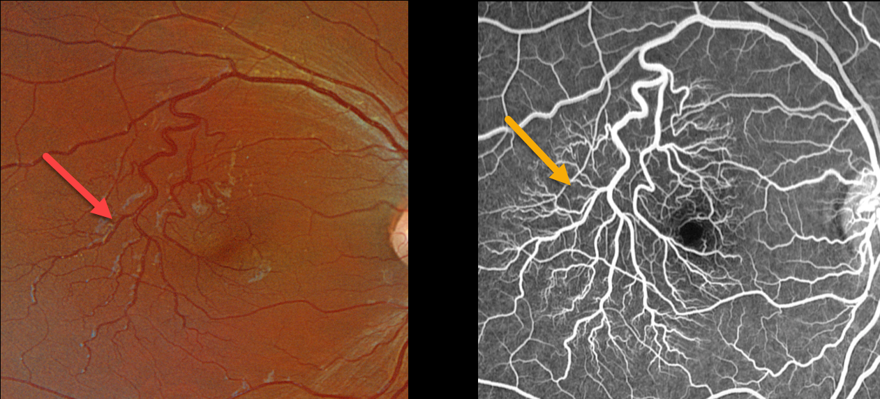
RETINAL VENOUS MALFORMATION
Originally posted on @retina.rocks August 14, 2023
This healthy 13YO female was referred for asymptomatic vascular changes in her right eye. Vision was 20/20 OU.
Optos color RGB imaging shows a prominent superotemporal retinal vein that crosses into the inferotemporal retina. As it descends temporally, it arborizes into numerous smaller branches. There was no leakage angiographically.
Neurologic imaging of our patient was fortunately normal, and observation was recommended.
Learning Points:
Retinal vessels virtually always respect the horizontal meridian. Retinal venous malformations are congenital anomalous vessels that, by definition, cross the horizontal.
Originally described by Gary Brown et al as congenital retinal macrovessels (Arch Ophthalmology 1982;100:1430-1436), these vessels are usually located in the macular region and are virtually always venous.
These lesions are now called retinal venous malformations, since about one-quarter of patients have associated venous anomalies in the brain (Pichi et al, JAMA Ophthalmol, 2018;136:372-379).
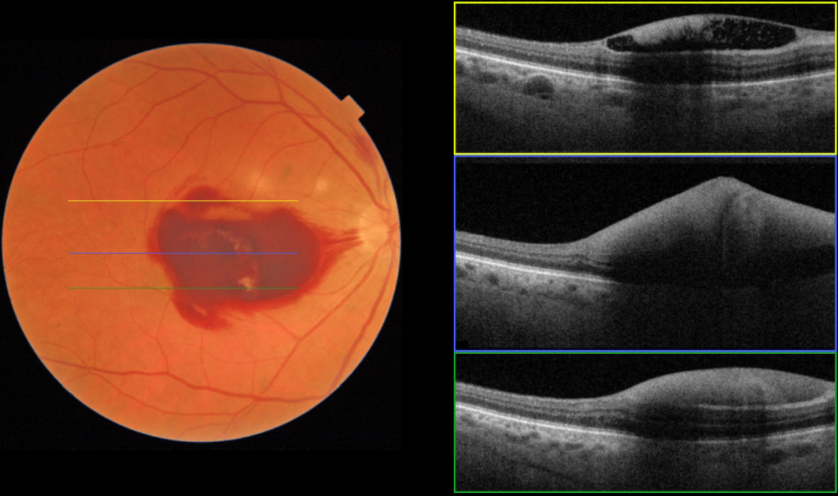
LEUKEMIA
Originally posted on @retina.rocks August 7, 2023
This 63YO male, who is undergoing treatment for leukemia, presented with one day of acute vision loss in his right eye. He was recently discharged from the hospital for chemotherapy-induced pancytopenia. His CBC showed: WBC 0.5, HgB 5.6, and Platelets 2. Vision was counting fingers OD.
Fundus imaging shows sub-internal membrane (ILM) hemorrhage. The OCT scans confirm hemorrhage in the sub-ILM space. We expect our patient’s macular hemorrhage to resolve as his blood counts normalize
Learning Points:
Ocular leukemic manifestations are primarily caused by accompanying hematologic abnormalities, including hyperviscosity, anemia, and thrombocytopenia (Soman et al, Ophthalmology Retina 2018;2:17-23).
Posterior segment findings include retinal hemorrhages (sub-ILM, nerve fiber layer, or deeper, white-centered), cotton-wool spots, dilated retinal veins, and serous retinal detachment. Leukemic infiltration of the optic nerve, retina, and choroid can also occur.
The sub-ILM hemorrhages are usually self-limiting and resolve within several months of treating the underlying disease (Ophthalmology Retina 2018;2:494-501).
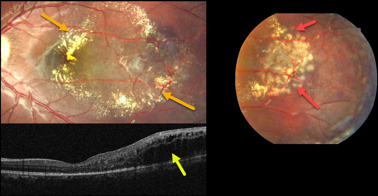
COATS DISEASE
Asma Samsudeen and Ashish Sharma
Originally posted on @retina.rocks July 27, 2023
This healthy 8YO boy presented for a routine eye examination. Vision was 20/20 in his normal right eye and 20/30 in his left eye.
Fundus photography shows telangiectatic vessels in the temporal macula associated with surrounding lipid. The lipid is particularly dense centrally.
OCT scanning shows temporal, mostly outer retinal edema. The macular center looks remarkably normal given the fundoscopic appearance. Focal macular laser to the telangiectatic lesions was applied
Learning Points:
Described by George Coats in 1908, Coats disease is an idiopathic disorder typically unilateral in males. It is characterized by retinal vascular telangiectasia involving capillaries, arterioles, and venules.
Patients can present with a wide range of findings, from minimal exudation to total exudative retinal detachment. The leaking vessels are usually treated to prevent exudative complications.
Thermal laser photocoagulation is usually the initial modality, but anti-VEGF injections can also be used to stabilize the vascular leakage. Patients will often require multiple rounds of treatment.
See Sigler et al for a review of the current management (Survey Ophthalmology 2014;59:30-46).
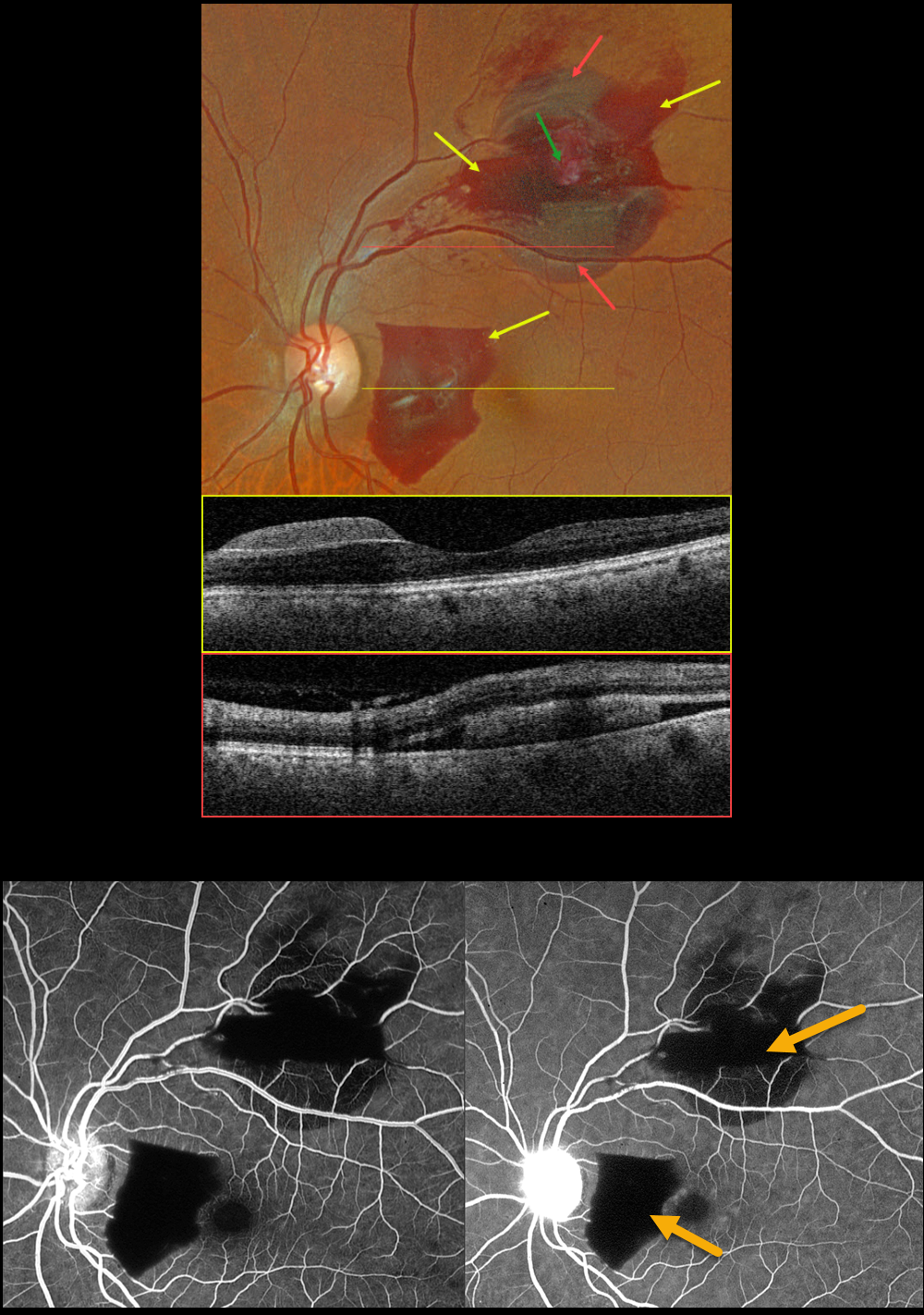
RETINAL ARTERIAL MACROANEURYSM (RAM)
Originally posted on @retina.rocks July 24, 2023
This 65YO female presented with 2 weeks of paracentral vision loss in her left eye. Vision was 20/25.
Optos color RGB imaging shows multilayered hemorrhages in the nasal and superior macula, including preretinal and subretinal blood.
Swept-source OCT through the preretinal and subretinal areas confirms the locations of the blood. Fluorescein angiography shows a blockage in the blood. The retinal arterial macroaneurysm, which is noted as a yellowish mass within the superior blood clinically, is obscured by the blood angiographically.
Learning Points:
A RAM is thought to occur in a weakened arterial wall from arteriosclerosis, and appears as a yellow-white dilated lesion along a retinal artery within the first 3 bifurcations from the optic nerve.
There are 2 distinct presentations, hemorrhagic and exudative. The hemorrhagic type often presents, as in this case, with pathognomonic multiple layers of blood. Acute macular hemorrhage can be treated with observation, anti-VEGF injections, or thermal laser.
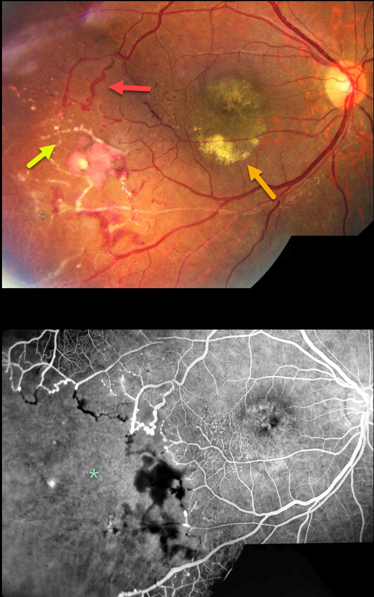
COATS DISEASE
Marianna Kavalarak
Originally posted on @retina.rocks July 5, 2023
This 19YO female presented with a chronic history of unilateral vision loss in her right eye. Vision was 20/100 OD and 20/20 in her healthy left eye.
Color imaging shows extensive vascular changes in the inferotemporal retina, including retinal telangiectasia, macroaneurysms, vascular beading, sheathed vessels, and featureless ischemic retina. The central macula is hyperpigmented with variable subretinal lipid.
Fluorescein angiography shows marked distal capillary loss. The vessels bordering the perfused and ischemic retina are irregularly dilated with anastomoses. Fine telangiectatic vessels are noted in the inferotemporal macula.
Learning Points:
Coats disease is characterized by vascular changes throughout the retinal vasculature, including the arteries, the capillary bed, and the veins. Larger dilated vessels in Coats can sometimes mimic angiomas, vascular tumors, or exudation from retinoblastoma. The presence of retinal telangiectasia can help establish the diagnosis of Coats in these cases.
Coats are usually unilateral in males. However, recent ultra-widefield angiographic studies indicate subtle vascular changes mostly in the temporal periphery of the fellow eye (Brockmann et al, BJO 2021;105:1444-1453).
Dyskeratosis congenita or familial exudative vitreoretinopathy (FEVR) should be considered for patients with frank bilateral disease (Teixeira et al, Arch Ophthalmol 2008;126:134-135), in women with Coats-like findings (Johnson et al, Genetics 2009;30:181-184), or in patients with a family history of similar findings.
Laser photocoagulation can be used to directly treat leaking telangiectatic vessels, and a scatter laser can be placed into the ischemic retina. Anti-VEGF injections can also be used as primary or secondary treatment.
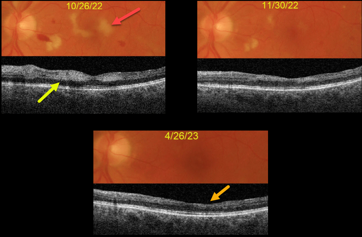
HYPERTENSIVE RETINOPATHY
Originally posted on @retina.rocks July 3, 2023
This 63YO male complained of vision loss in his left eye following left carotid endarterectomy. His blood pressure was markedly elevated perioperatively, but he reported feeling better. Blood pressure was 175/101 when seen by his doctor one week earlier. Vision was 20/25 OD and 20/200 OS.
Hypertensive retinopathy was noted bilaterally, with scattered retinal hemorrhages and nerve fiber layer infarcts (cotton-wool spots, CWS).
Color imaging of the left eye shows CWS virtually involving the macular center. Swept source OCT shows inner retinal hyperreflectivity from these lesions.
Over the following months, both his blood pressure and macular appearance normalized. When last seen on 4/26/23, vision was 20/40, although he was still quite symptomatic from paracentral scotomas due to inner retinal atrophy from the resolved CWS.
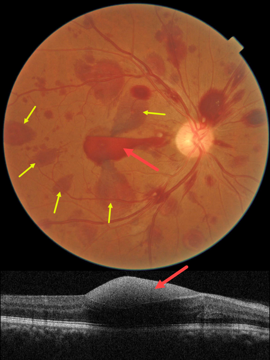
HEMORRHAGIC UNILATERAL RETINOPATHY
Asma Samsudeen and Ashish Sharma
Originally posted on @retina.rocks May 25, 2023
This healthy 52YO male noticed acute vision loss in his right eye two days earlier. He denied Valsalva or trauma. Vision was counting fingers in his right eye and 20/20 in his normal left eye. He has a history of untreated hypertension, and his BP in the office was 180/110.
Color imaging shows multifocal, mostly deep, retinal hemorrhages scattered throughout the right posterior pole. Some of these hemorrhages are radiating outwards from the macular center in Henle’s layer. A subhyaloid vs sub-ILM hemorrhage is also noted.
Learning Points:
Named by Kon Graversen et al (Retina 2014;34:483-489), hemorrhagic unilateral retinopathy presents with unilateral, deep intraretinal hemorrhages and sudden vision loss ranging from 20/20 to hand motion.
There are no associated systemic conditions, nor a history of trauma or Valsalva maneuver. It is most common in females. Vision usually recovers completely within 6 months.
We do not believe our patient’s findings were caused by his hypertension since they were unilateral and predominated by deeper retinal findings. Observation was recommended.
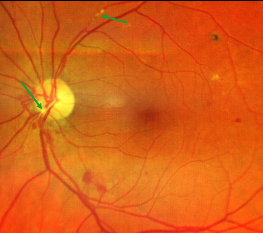
RETINAL EMBOLI
Natasa Draca and Emma Oreskovic
Originally posted on @retina.rocks May 17, 2023
This 77YO male presented with unilateral left superior field loss in his left eye one day earlier. Vision was 18/20 bilaterally.
Color imaging shows retinal emboli along the proximal superotemporal arcade and overlying the inferonasal nerve heads. Some scattered retinal hemorrhages are also seen. The right fundus was normal. Visual field testing was normal.
He was immediately referred for a stroke evaluation, which was fortunately negative. Carotid Doppler showed bilateral 30% internal carotid artery stenosis. He was found to be in new-onset atrial fibrillation, which was the presumed source for the emboli, and was placed on oral anticoagulation therapy.
Learning Points:
Retinal emboli, which generally arise from either the heart or carotid arteries, can be calcific, platelet-fibrin, or cholesterol (Hollenhorst plaques). If large enough, they can obstruct a retinal arteriole and cause acute retinal ischemia, which is defined as transient monocular vision loss (TMVL, formerly known as amaurosis fugax), acute branch retinal artery occlusion, or acute central retinal artery occlusion.
TMVL requires an immediate referral to the nearest stroke center. Asymptomatic emboli can be more effectively evaluated for an embolic source with carotid Doppler and cardiac B-scan ultrasonography.
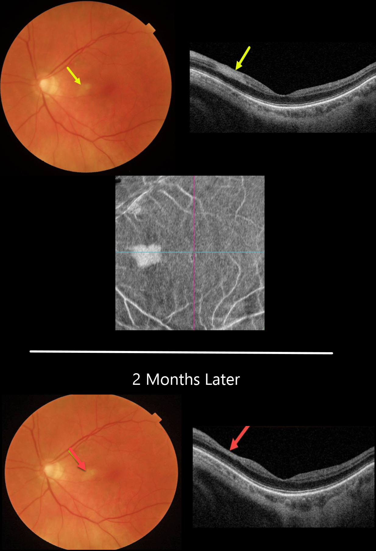
PARACENTRAL ACUTE MIDDLE MACULOPATHY (PAMM)
Originally posted on @retina.rocks April 25, 2023
This 81YO female presented with 1 week of a temporal paracentral scotoma in her left eye. Vision was 20/50 OS.
Color imaging shows an area of inner retinal opacification in the nasal macula. This area is hyperreflective on swept-source OCT. En face imaging shows another perspective for this lesion.
Two months later, the retinal opacification was smaller with a small area of secondary inner retinal thinning.
Due to our patient’s age and somewhat atypical appearance of the PAMM lesion, which involved both the inner and middle retinal layers, she was immediately referred to the nearest stroke center.
This revealed a critical ipsilateral internal carotid artery stenosis, which was successfully repaired with emergent surgery.
Learning Points:
Paracentral acute macular maculopathy (PAMM) is a phenotype caused by acute mid-retinal ischemia due to obstruction of the deep and intermediate capillary plexi. PAMM is associated with an increasing constellation of conditions, including retinal vein and artery occlusions. It is unclear if patients with a pure PAMM phenotype require an immediate stroke protocol referral.
In our practice, we have seen several elderly patients who developed a complete CRAO within days of presenting with PAMM, so we do send these patients for emergent evaluation. We do not believe that typical PAMM in younger patients or found as a secondary finding in other ocular conditions requires a stroke workup.
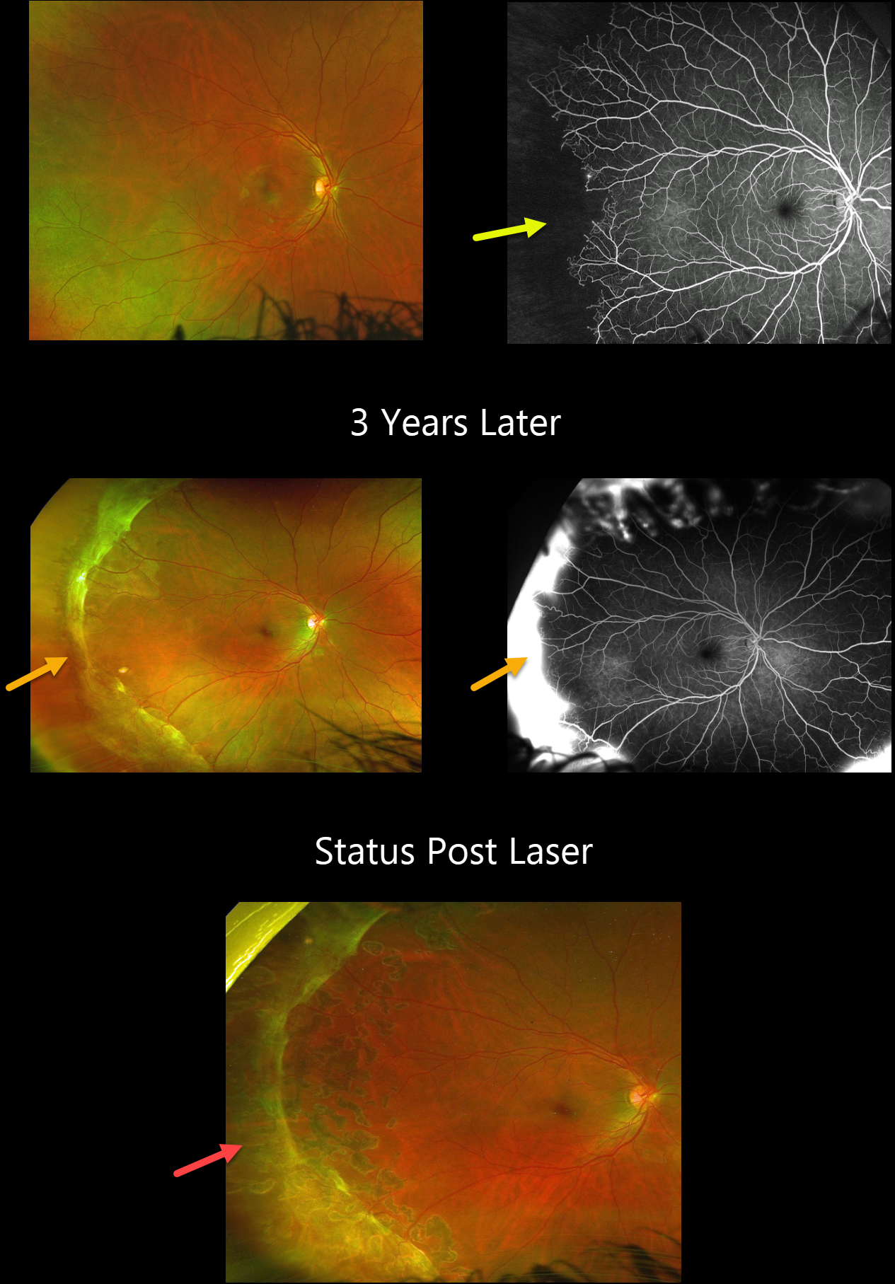
SICKLE CELL RETINOPATHY
Will Gibson
Originally posted on @retina.rocks April 24, 2023
This 19YO male has a known history of sickle disease (Hb SC). On 7/30/20, vision was 20/25 OD.
Optos color imaging shows temporal vascular whitening, and angiography shows marked temporal ischemia.
He was lost to follow-up until 1/10/23 when he presented with hand motions vision from a vitreous hemorrhage OS (not shown). Although vision was still 20/20 OD, a broad band of fibrosed neovascularization was noted in the periphery. A peripheral scatter laser was applied to the right eye at the time of OS vitrectomy, and 6 weeks later, all neovascularization had regressed.
Learning Points:
Patients with sickle cell disease develop symptoms throughout their bodies as stiff, sickled red blood cells block capillary beds. In the eye, this can cause proliferative sickle cell retinopathy with sea-fan shaped areas of peripheral retinal neovascularization developing at the junction of perfused and ischemic retina. Complications include vitreous hemorrhage and traction retinal detachment. Although patients with SS disease tend to have more systemic complications, SC disease is usually associated with more severe ocular findings.
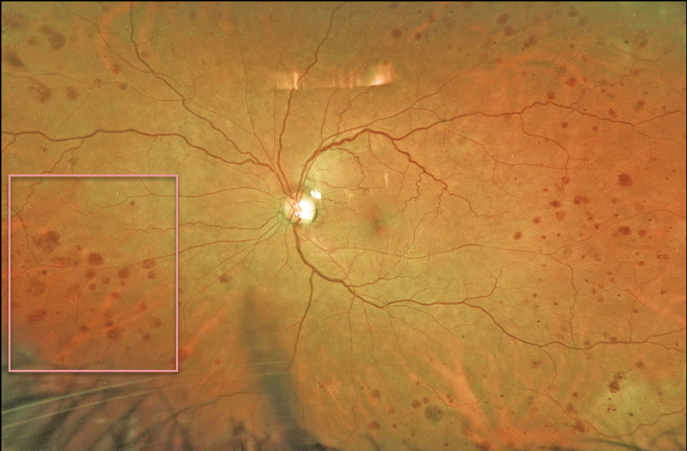
OCULAR ISCHEMIC SYNDROME
Originally posted on @retina.rocks April 10, 2023
This 70YO female was referred for asymptomatic unilateral retinal hemorrhages in her left eye. Vision was 20/30. Optos imaging shows encircling midperipheral deep outer retinal hemorrhages.
Since our patient had a known history of complete left internal carotid artery blockage, no further testing was ordered. We will follow her several times yearly due to the risk of ocular neovascularization.
Learning Points:
The retinal hemorrhages in the ocular ischemic syndrome (OIS) are unique. Retinal vascular disorders like diabetic retinopathy, retinal vein occlusions, and hypertensive retinopathy usually have flame- and dot-shaped inner retinal hemorrhages concentrated around the posterior pole.
Our patient’s findings are classic for OIS, with hemorrhages in the deeper peripheral retina. The retinal periphery is literally the end of the line for the retinal arterial circulation.
In an eye receiving less blood supply due to carotid occlusive disease, the more proximal retina has first dibs on the available oxygen. There are penetrating capillaries that dive radially from the nerve fiber and ganglion cell layers into the deeper retina. The deep vascular complex that supplies the inner and outer plexiform layers is thus furthest downstream, and these endothelial cells are likely damaged in OIS. Blood cells leaking into this space accumulate, causing the large outer blot to hemorrhage.
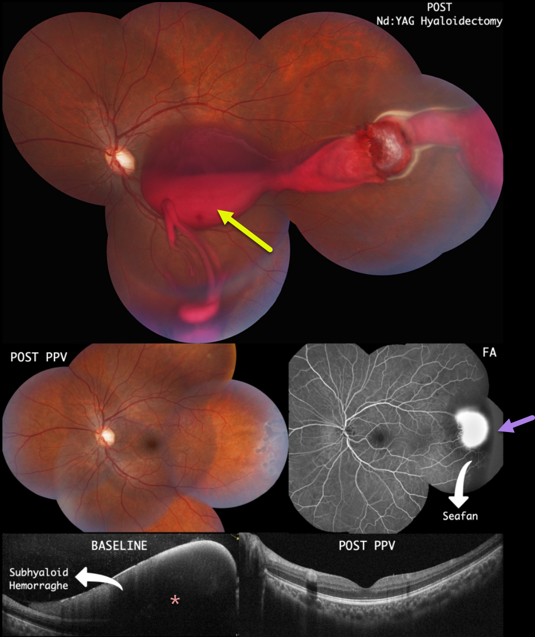
SICKLE CELL RETINOPATHY
Lucas Zago, Julia Jiquilin Carvalho and Mauricio Maia
Originally posted on @retina.rocks April 3, 2023
This 22YO male presented with 3 days of vision loss in his left eye. There was a past medical history of Hemoglobin SC Disease. He denied any Valsalva maneuvers. He remembered seeing a retinal doctor several months earlier with the diagnosis of proliferative sickle retinopathy. Vision was counting fingers OS.
Color imaging shows a large subhyaloid/vitreous hemorrhage extending from the temporal midperiphery into the macula. The blood seemed to originate from a probable area of neovascularization (sea fan). OCT scanning confirmed the subhyaloid location of the blood.
Immediately following Nd:YAG laser hyaloidotomy, the blood began to drain into the inferior vitreous. Peripheral scatter laser into the temporal ischemic retina was also performed.
However, he subsequently developed a dense vitreous hemorrhage requiring vitrectomy with endolaser. One month after surgery, the vitreous was clear, the neovascularization had regressed, and the macular appearance had normalized. Vision was 20/20.
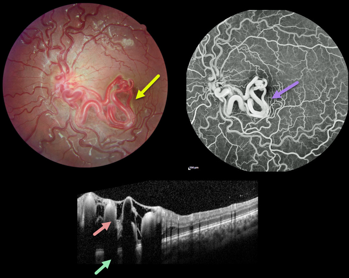
RETINAL ARTERIOVENOUS MALFORMATION
Şengül Özdek and Ece Özdemir Zeydanl
Originally posted on @retina.rocks March 24, 2023
This healthy 11YO girl was seen for a routine ophthalmological examination. Vision was 20/20 OU.
Fundus photography of her left eye shows unilateral tangles of markedly dilated and tortuous vessels extending from the disc into the inferior macula and fovea. The retinal vessels elsewhere are also somewhat dilated and tortuous. OCT scanning through the inferior macula shows these hyperreflective vessels to variably extend through the full-thickness retina with posterior shadowing.
The early-phase fluorescein angiogram shows rapid filling of the dilated artery and vein, with no intervening capillaries (not shown). The late phase shows no angiographic leakage.
Learning Points:
A retinal arteriovenous malformation (AVM) is a congenital connection between an artery and vein without an intervening capillary network. These lesions appear along a spectrum ranging from small isolated lesions to the large ‘bag of worms’ appearance as seen in our patient.
These more extensive lesions are usually part of the Wyburn-Mason Syndrome, which includes vascular lesions in the eyes and midbrain. Retinal venous malformations, also known as retinal macrovessels, are now thought to represent an AVM (Pichi et al., JAMA Ophthalmol 2018;136:372-379).
Fortunately, our patient’s MRI was normal. The AVM should likely remain stable throughout life and should not require any treatment.
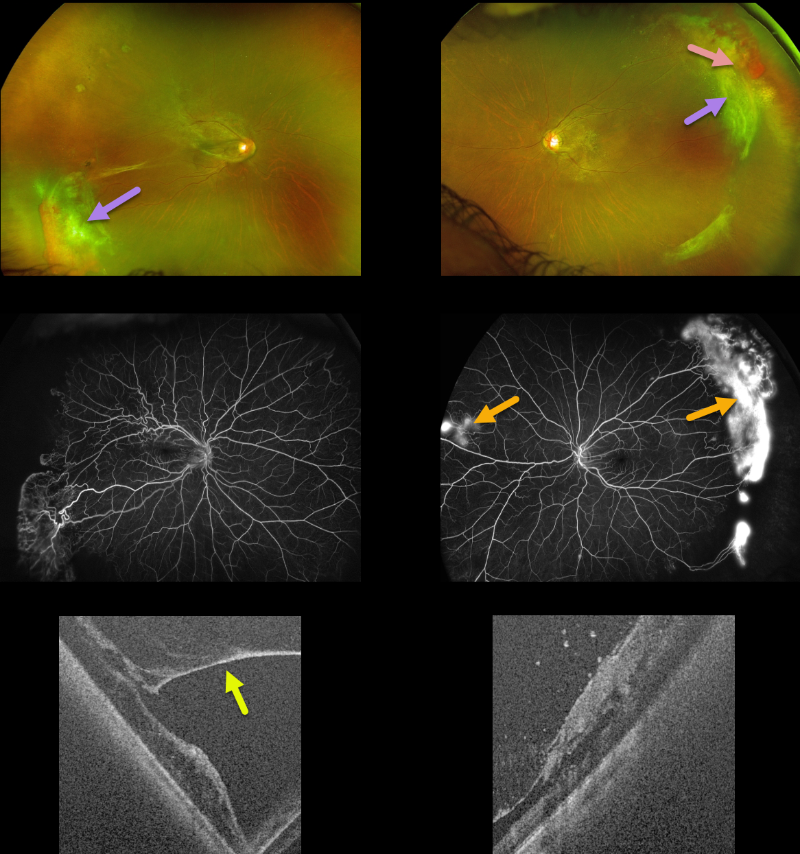
SICKLE CELL RETINOPATHY
Paulo Eduardo Stanga, Javier Valentin Bravo, Sebastian Stanga, and Ursula Reinstein
Originally posted on @retina.rocks March 13, 2023
This 45YO female with a known history of sickle SC disease presented with intermittent bilateral floaters following a COVID infection four months earlier. Vision was 20/40 OD and 20/16 OS.
Optos color imaging shows bilateral temporal fibrosed retinal neovascularization (sea fans) associated with some localized vitreous blood. The right eye had a significant epimacular membrane with traction, with a milder adherent membrane in her left eye.
Fluorescein angiography shows bilateral far temporal ischemia with leaking neovascularization. Some nasal neovascularization is also noted in her left eye. Ultrawidefield OCT shows vitreous traction adherent to the neovascularization.
We elected to closely follow her for now with observation.
Learning Points:
Patients with sickle cell disease develop symptoms throughout their bodies as stiff, sickled red blood cells block capillary beds. In the eye, this can cause proliferative sickle cell retinopathy (PSR) with sea-fan-shaped areas of peripheral retinal neovascularization developing at the junction of perfused and ischemic retina.
Complications include vitreous hemorrhage and traction retinal detachment. Although patients with SS disease tend to have more systemic complications, SC disease is usually associated with more severe ocular findings.
Treatment with sectoral (Farber et al Arch Ophthalmology 1991;109:363-367) or circumferential (Kimmel et al Ophthalmology 1986;93:1429-1434) scatter laser photocoagulation is somewhat controversial since, unlike more typical proliferative retinopathies (i.e., from diabetes and branch retinal vein occlusions), these new vessels tend to auto-infarct with a good natural history for untreated disease (Downes et al Ophthalmology 2005;112:1869-1875).
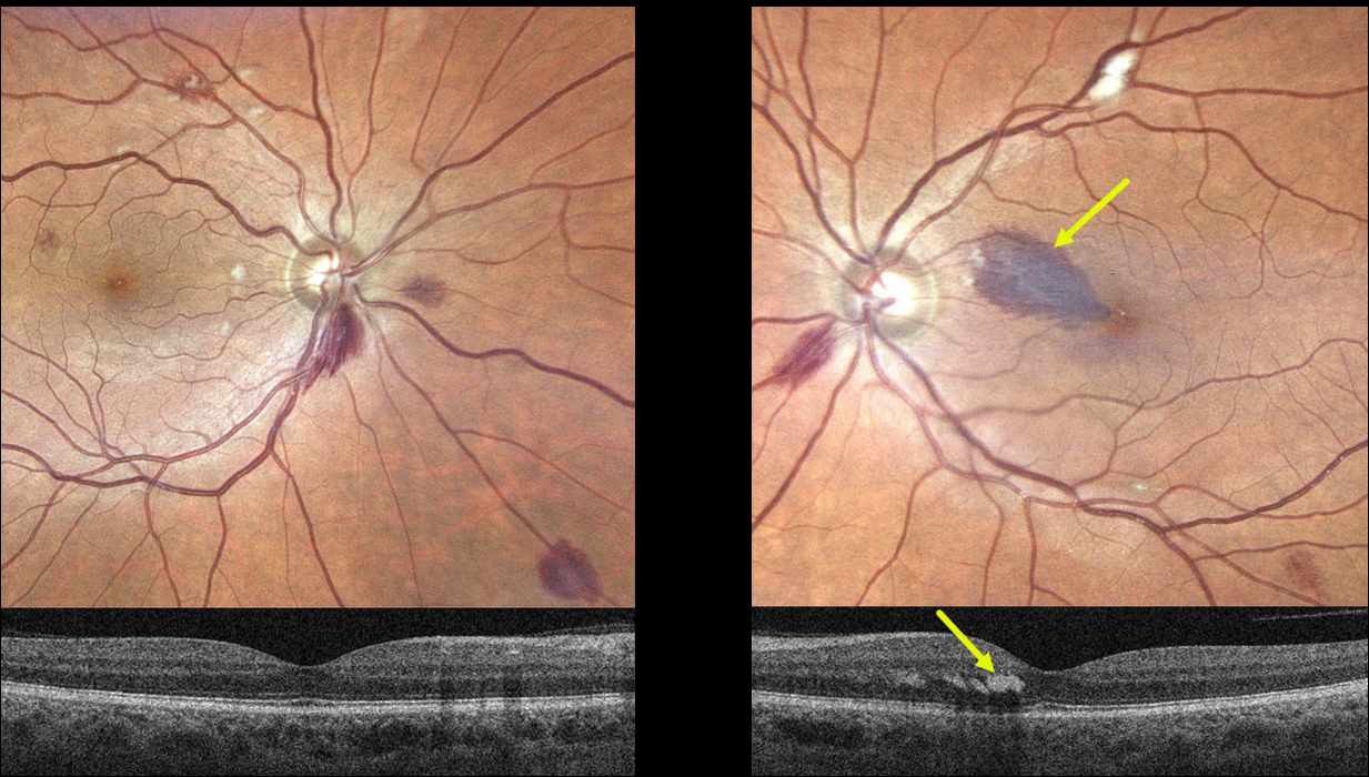
ANEMIA
Originally posted on @retina.rocks February 28, 2023
This 55YO male presented with a one-week history of a paracentral scotoma in his left eye. Vision was 20/40 OD and 20/60 OS. He gave a history of aplastic anemia, and his hemoglobin was 6.2 when he developed these symptoms. Blood transfusion brought his hemoglobin to over 8. He told us that his blood pressure runs low to normal.
Optos imaging shows scattered bilateral deep and superficial retinal hemorrhages with a few cotton wool spots. A tear-drop-shaped, larger hemorrhage is noted in the superonasal left macula. This hemorrhage localizes to the outer plexiform layer on swept-source OCT.
Learning Points:
The central retinal artery supplies the inner two-thirds of the retina, while the choroid supplies the outer third (the photoreceptor layers), the RPE, and the choroid. The outer plexiform layer (OPL) is the ‘end of the line’ for the inner retinal circulation.
In severe anemia, the more proximal inner retina removes whatever oxygen is available, leaving the deeper layers relatively ischemic. This can cause dysfunction of the retinal vascular endothelial tight junctions, which allows red blood cells to enter and stain the OPL, appearing clinically as deep retinal blot hemorrhages.
Deep blot OPL hemorrhages also appear for similar reasons in the temporal retinal periphery in ocular ischemia.
We advised observation, and expect his findings and symptoms to improve as long as his anemia is controlled.
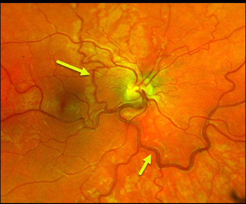
RETINAL VENOUS MALFORMATION
Ryan Kern
Originally posted on @retina.rocks February 7, 2023
This healthy 24YO female presented for a general eye examination. There was no past ocular or medical history.
Color imaging of her right eye shows a large dilated retinal vein that drains the entire fundus except superonasally. It also crosses the temporal horizontal raphe into the superotemporal quadrant. Her left eye was normal.
Learning Points:
Retinal vessels virtually always respect the horizontal meridian. Retinal venous malformations (Pichi et al JAMA Ophthalmol 2018;136:372-379) are congenital anomalous vessels that by definition cross the horizontal.
Originally described by Gary Brown et al as congenital retinal macrovessels (Arch Ophthalmology 1982;100:1430-1436), these vessels are usually located in the macular region and are virtually always venous.
The findings in our patient are unusual, as the anomalous vessel supplies the majority of the retinal venous drainage.
These vessels are technically arterio-venous malformations (AVM) and are associated with venous anomalies in the brain (24% compared to up to 6% of the population). Brain MRI is therefore recommended to rule out any intracranial abnormalities. Neurologic imaging in our patient was fortunately normal.
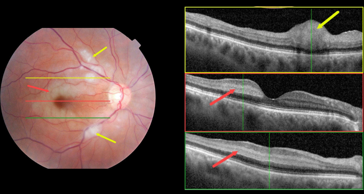
CRAO
Ryan Kern
Originally posted on @retina.rocks January 10, 2023
This 70 YO female presented with acute vision loss in her right eye starting 1 day earlier. Vision was 20/70 OD and 20/25 in her normal left eye.
Color fundus photography shows an incomplete central retinal artery occlusion (CRAO) with focal areas of acute inner retinal infarcts along with paracentral areas of deeper retinal opacification.
OCT scanning through the superior macula shows a focal area of retinal opacification and thickening that extends from the outer plexiform layer to the internal limiting membrane. Scans of the central and inferior macula show hyperreflectivity involving the middle retinal layers (outer plexiform, inner nuclear, and inner plexiform).
Blood pressure was 183/110. Emergent stroke evaluation was fortunately negative. Her blood pressure was stabilized, and baby aspirin was started. Visual acuity improved to 20/30 one month later.
This case was submitted by Ryan Kern.
Learning Points:
Paracentral acute macular maculopathy (PAMM) is a phenotype caused by acute mid-retinal ischemia due to obstruction of the deep (DCP) and intermediate (ICP) capillary plexi. PAMM is associated with an increasing constellation of conditions, including retinal vein and artery occlusions.
It is unclear if patients with a pure PAMM phenotype require an immediate stroke protocol referral. In our practice, we have seen several elderly patients who developed a complete CRAO within days of presenting with PAMM, so we do send these patients for emergent evaluation. We do not believe that typical PAMM in younger patients or found as a secondary finding in other ocular conditions requires a stroke workup.
Our patient’s fundus appearance has more of a Purtscher ’s-like appearance with focal areas of cotton wool spot-like inner retinal ischemia (although on OCT these involved the middle retinal layers as well) and mid-retinal ischemia.
However, given her age and history, acute onset, and unilateral presentation, her diagnosis is most consistent with an incomplete CRAO.
For a great recent PAMM review, see Scharf et al, Progress in Retinal and Eye Research 2021;81;100884.
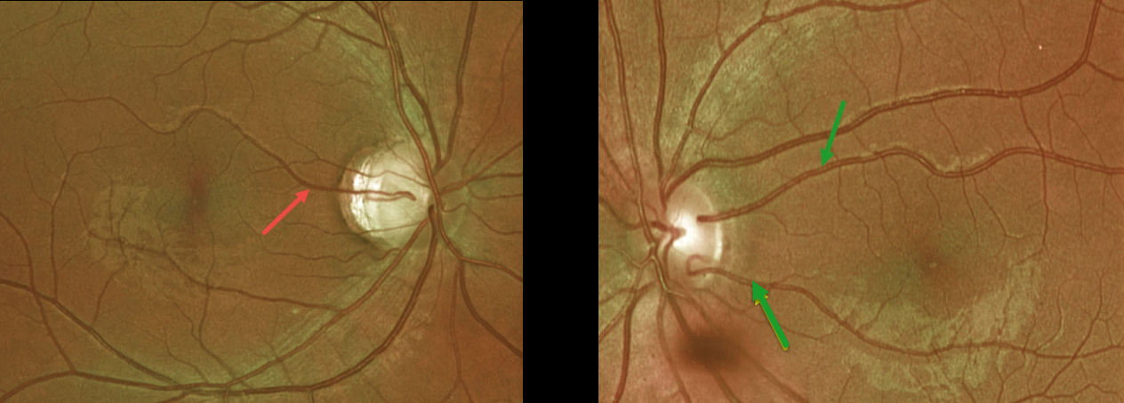
CILIORETINAL ARTERY
Originally posted on @retina.rocks December 27, 2022
This healthy 23YO female presented with incidental findings of bilateral prominent cilioretinal arteries.
Optos imaging shows a superior cilioretinal vessel in her right eye. There are two cilioretinal vessels in her left eye, which collectively seem to supply the entire central macular region.
Learning Points:
The inner two-thirds of the neurosensory retina is supplied by the central retinal artery, with the choroid supplying the RPE and photoreceptor layer.
Cilioretinal arteries, which arise from the posterior choroidal circulation, are present in up to 50% of individuals, and bilateral in about 25% (Schneider et al, Acta Ophthalmologica 2021;99:e310-e318).
Our patient is unique in that she has multiple vessels in her left eye (Leavitt et al, AJO 2011;152:820-823), which seem to supply the arterial needs for her entire macula. If she were unlucky enough to be the one in 50,000 people per year to develop a central retinal artery occlusion (Leavitt et al, AJO 2011;152:820-823), these vessels would most likely preserve macular function.
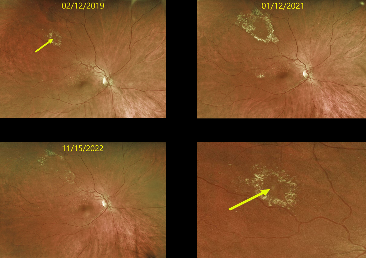
RETINAL VENOUS MACROANEURYSM
Originally posted on @retina.rocks December 15, 2022
This 63 YO female originally presented in 2019 with a history of a major superotemporal branch retinal vein occlusion (BRVO). Successful macular laser was performed for macular edema.
When initially examined, a midperipheral retinal venous macroaneurysm (RVM) with some surrounding lipid was noted. This RVM has been observed due to its distant location from the fovea. The series of photos over several years shows the exudation to wax and wane. At the last examination on 11/15/22, there was almost complete lipid resolution. Vision remains at 20/25.
Learning Points:
In 1990 Cousins et al reported that RVMs can be an additional finding in occlusive venous disease, always in association with collateral vessels or capillary or arterial macroaneurysms (AJO 1990;109;567-1700).
These vascular abnormalities tend to have a higher rate of intraretinal lipid (40%) but are less likely to affect central vision since they are usually outside the macula.
They tend to have a higher association with retinal ischemia, thus being at a higher rate of developing retinal neovascularization (32%). The lesions will usually involute, as was the case in our patient.
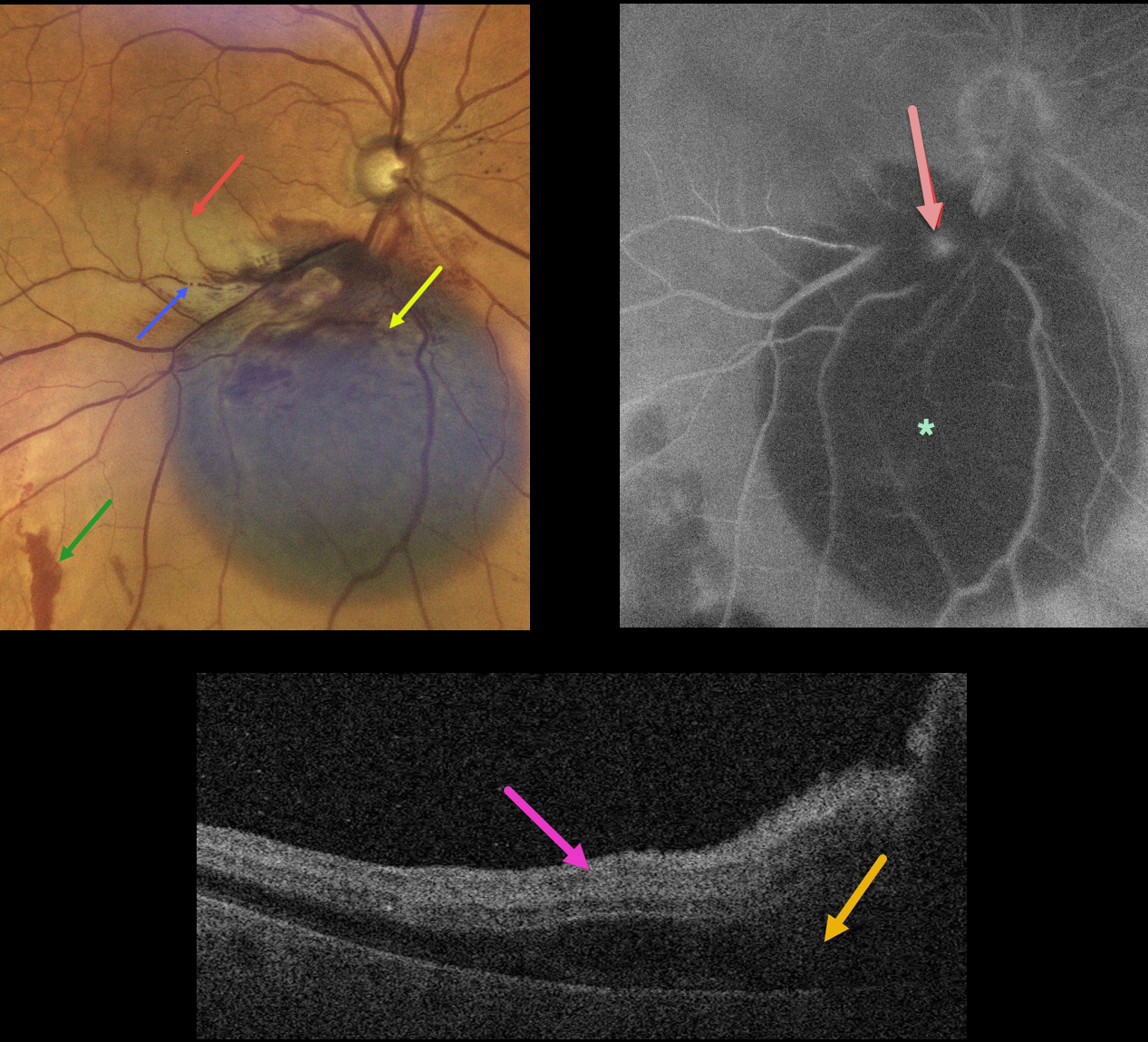
RETINAL ARTERIAL MACROANEURYSM WITH SECONDARY BRAO
Originally posted on @retina.rocks December 8, 2022
This 79YO female presented with acute inferior paracentral vision loss in her right eye. Vision was 20/50.
A large blister of dark subretinal blood was noted just inferior to the optic nerve. A few small areas of more distal red subretinal blood were seen. An inferior macular branch retinal artery occlusion likely accounted for her symptoms. The blood column is stagnant within the inferior portion of the acute occlusion.
OCT scanning through the BRAO shows hyperreflective inner retina with subretinal fluid.
Fluorescein angiography shows blockage from the subretinal blood, along with a leaking retinal arterial macroaneurysm (RAM).
Learning Points:
A BRAO can be a rare complication from direct thermal laser of a RAM (Russell and Folk, AJO 1987;104:186-187), which is one reason why this is almost never performed anymore. In this case, we believe the BRAO was secondary to mechanical arterial compression from the surrounding blood.
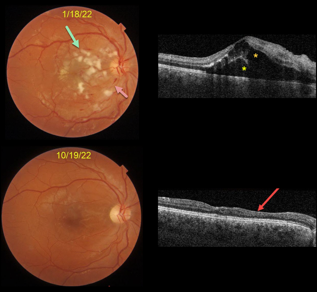
HYPERTENSIVE RETINOPATHY
Originally posted on @retina.rocks November 24, 2022
This is a follow-up from our 2/4/22 post of a patient with malignant hypertension.
This 22YO male presented on 1/18/22 with subacute vision loss of 20/200 OD and 20/50 OS. Blood pressure was 169/135. Color imaging shows nerve fiber layer infarcts (cotton-wool spots) and mild inner retinal hemorrhages.
OCT scanning shows mostly outer macular edema emanating from the nerve, with foveal subretinal fluid.
Nine months later, the macular and OCT findings have mostly normalized. OCT scanning shows thinning with disorganization of retinal inner layers (DRIL) nasally OD.
Learning Points:
Malignant hypertension, defined as blood pressure above 180 systolic and/or 120 diastolic, is a life-threatening medical emergency. Eye doctors are in a unique position to often diagnose this condition.
Patients will often present with bilateral optic nerve swelling, flame-shaped retinal hemorrhages, and, with more chronic disease, lipid precipitates in the nasal macular outer plexiform layer.
Since the macular fluid emanates from the optic nerve, the macular thickening is always worse nasally (as in our case), and is an important clinical clue for this diagnosis.
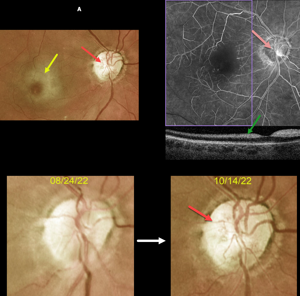
CRAO
Originally posted on @retina.rocks November 21, 2022
This 63YOF initially presented with an acute central retinal artery occlusion in her right eye. Emergency stroke referral revealed two prior strokes with a new asymptomatic stroke.
She returned two months later. There was subjective visual improvement, although vision remained at hand motion. Although the retinal opacification in general was improving, the perifoveal retina was more opaque with a pronounced cherry red spot.
New fine vessels were noted on the nerve. These vessels did not leak on fundus fluorescein angiography. Marked angiographic ischemia is also noted. OCT scanning shows increased reflectivity from the ischemic inner retina.
Learning Points:
Collateral vessels are dilated, pre-existing vessels that bypass an occlusion. These are seen in the posterior pole most commonly following retinal venous occlusions and are exceedingly rare with arterial occlusions.
The endothelial tight junctions are intact, so they don’t leak angiographically. This helps differentiate them from neovascularization, which does leak.
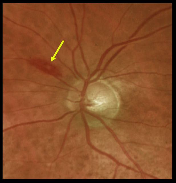
LYMPHOMA WITH WHITE-CENTERED RETINAL HEMORRHAGE
Originally posted on @retina.rocks November 10, 2022
This 84YO female was found to have a white-centered retinal hemorrhage superonasal to the optic nerve disc.
Complete blood count revealed a white blood count of 19,000 and a hemoglobin of 7.9. She was subsequently diagnosed with atypical diffuse large B-cell lymphoma.
Learning Points:
The term “Roth spot” was named after Dr. Moritz Roth, who, in 1872, noted white-centered hemorrhages in patients with bacterial endocarditis.
He felt that these white spots were septic emboli that originated from an infected cardiac valve. However, similar lesions can be seen in many other disorders, including leukemia, hyperviscosity, anemia, diabetes, and hypertension.
The white material may represent a variety of causes, including fibrin. These lesions are best called white-centered hemorrhages, unless one is specifically referring to a hemorrhage in a patient with bacterial endocarditis.
For a review of white-centered retinal hemorrhages, see Duane et al Ophthalmology 1980;87:66-69.
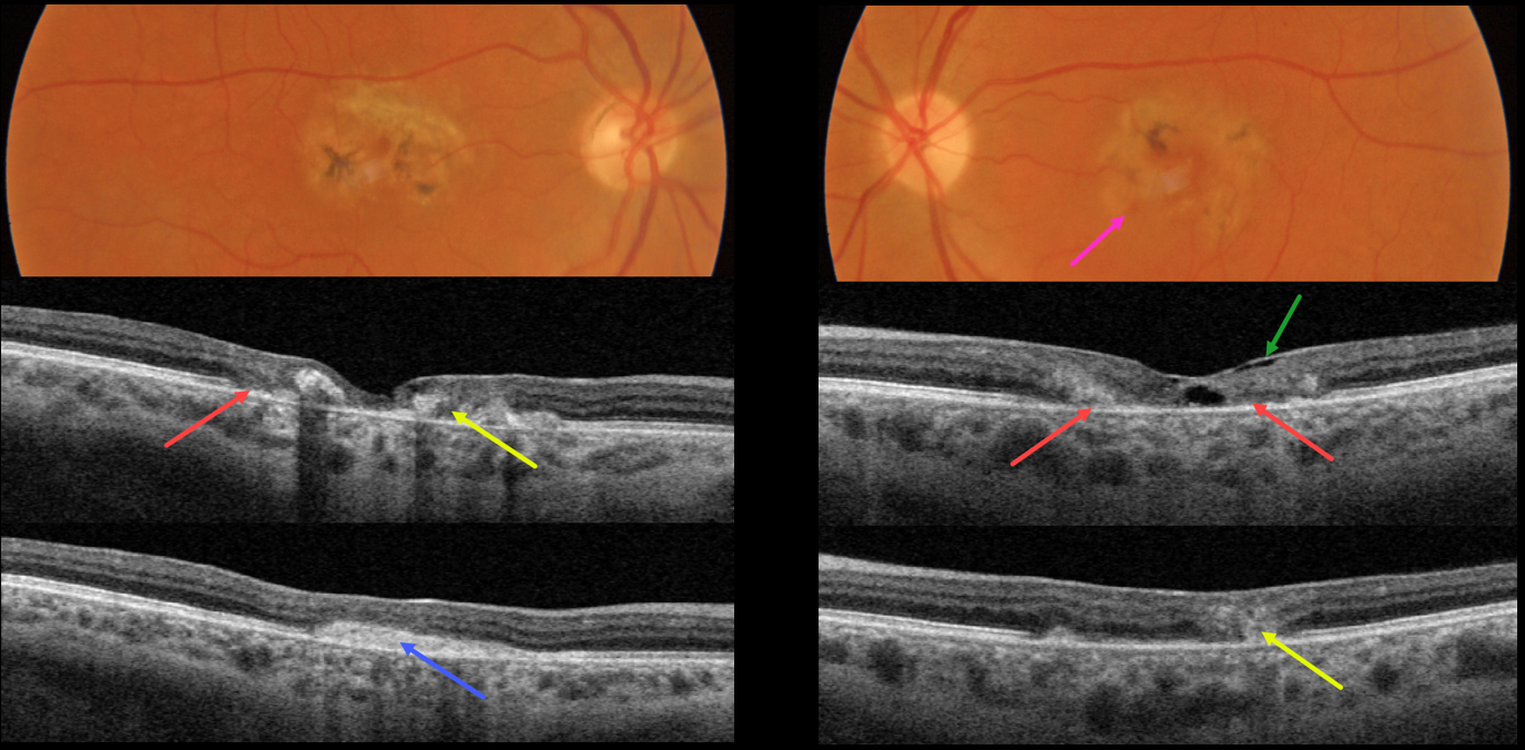
MACULAR TELANGIECTASIA
Originally posted on @retina.rocks November 8, 2022
This 49YO female presented with several weeks of vision loss in her left eye. A macular neovascularization (MNV) in her right eye was treated 4 years earlier with intravitreal Avastin. Vision was 20/200 OD and 20/70 OS.
Both maculas had central pigment loss with intraretinal pigment migration. A small dot of blood was noted inferonasally in the left macula.
OCT scanning shows outer retinal atrophy, pericentral retinal disorganization, a fibrosed, inactive MNV in the right macula, and hyporeflective loss of retinal tissue (cavitation) in the left outer macula. Faint loss of the temporal inner retinal tissue is highlighted by intact internal limiting membrane (ILM drape sign).
Anti-VEGF therapy was started for a presumed MNV in her left eye.
Learning Points:
Originally described by Gass (Arch Ophthalmology 1982;100:769-780), MacTel2 is a neurodegenerative disorder, most likely originating from Muller cell dysfunction.
The term, telangiectasia, is misleading in that the funduscopic findings are mostly non-vascular. The refractile inner retinal ‘crystals’ are thought to be Muller cell footplates. The foveal retina shows a whitish discoloration, most likely due to retinal opacification caused by dysfunctional Muller cells.
Photoreceptor loss allows RPE cells to migrate along intraretinal capillaries, causing the coarse clinical pigment clumping.
All of these changes are usually in the temporal fovea, so our patient is atypical in that they involve the entire central macular regions.
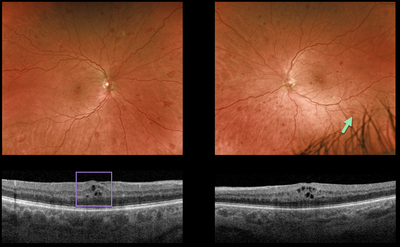
LEUKEMIA
Originally posted on @retina.rocks October 28, 2022
This 50YO male presented with bilateral asymptomatic retinal hemorrhages. He gave a 5-year history of well-controlled type 2 diabetes with a recent A1C of 6.3%, and was previously diagnosed with only mild nonproliferative diabetic retinopathy.
Optos imaging shows scattered retinal hemorrhages in all quadrants, most of which are deep with white centers. Mild bilateral macular edema is seen on OCT scanning.
The deeper, round retinal hemorrhages were felt to be atypical for diabetes. We therefore ordered a complete blood count (CBC), which revealed a white blood cell count over 80,000 u/L and polychromatic red blood cells.
Immediate referral to oncology revealed undiagnosed chronic myelogenous leukemia, and treatment was initiated.
Learning Points:
The term “Roth spot” was named after Dr. Moritz Roth, who, in 1872, noted white-centered hemorrhages in patients with bacterial endocarditis. He felt that these white spots were septic emboli that originated from an infected cardiac valve.
However, similar lesions can be seen in many other disorders, including leukemia, hyperviscosity, anemia, diabetes, and hypertension.
The white material may represent a variety of causes, including fibrin. So these lesions are best-called white-centered hemorrhages, unless one is specifically referring to a patient with bacterial endocarditis. See Duane et al Ophthalmology 1980;;87:66-69.
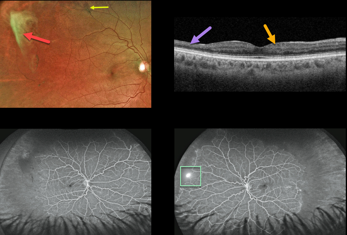
SICKLE CELL RETINOPATHY
Originally posted on @retina.rocks October 21, 2022
This 39YO African American male presented with asymptomatic proliferative sickle cell retinopathy with spontaneously avulsed bilateral superotemporal peripheral fibrosed retinal neovascularization (sea fans). A small midperipheral black sunburst lesion is seen superotemporally in his right eye.
Swept-source OCT shows variable inner retinal thinning and disorganization of the retinal inner layers (DRIL).
Fluorescein angiography shows a patch of retinal neovascularization OS at the junction of perfused and non-perfused retina.
Since these lesions often auto-infarct, as they did in our patient, we elected to observe the small area of neovascularization in his right eye. He is also at low risk for a vitreous hemorrhage or traction detachment since the vitreous is already detached in this area.
Learning Points:
Patients with sickle cell disease develop symptoms throughout their bodies as stiff, sickled red blood cells block capillary beds. In the eye, this can cause proliferative sickle cell retinopathy (PSR) with sea-fan-shaped areas of peripheral retinal neovascularization developing at the junction of perfused and ischemic retina.
The variable macular thinning noted on OCT is caused by microinfarcts to the retinal microcirculation. Enlargement of the foveal avascular zone is a common OCT angiographic finding (Fares, AJO 2021;224:7-17).
The black sunburst lesions are thought to occur from intraretinal RPE migration in response to photoreceptor damage from intraretinal and subretinal hemorrhage (see Ausdourian et al, BJO 1975;59:710-716).
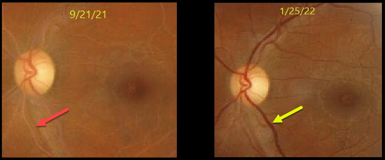
LIPEMIA RETINALIS
Nayara Pinto and Romano Vasconcelos
Originally posted on @retina.rocks October 11, 2022
This 28YO female presented with bilateral symmetric creamy colored blood vessels (only left eye shown). Triglycerides were markedly elevated above 1000.
The vascular appearance normalized 4 months following systemic treatment with a relatively normal triglyceride level of 189.
Learning Points:
Lipemia retinalis is a rare retinal manifestation of severe hypertriglyceridemia, characterized by creamy white discoloration of the retinal vasculature. In severe cases, the fundus can turn salmon-colored.
Patients are asymptomatic, and vision is normal. However, they are at obvious cardiovascular risk without systemic treatment.
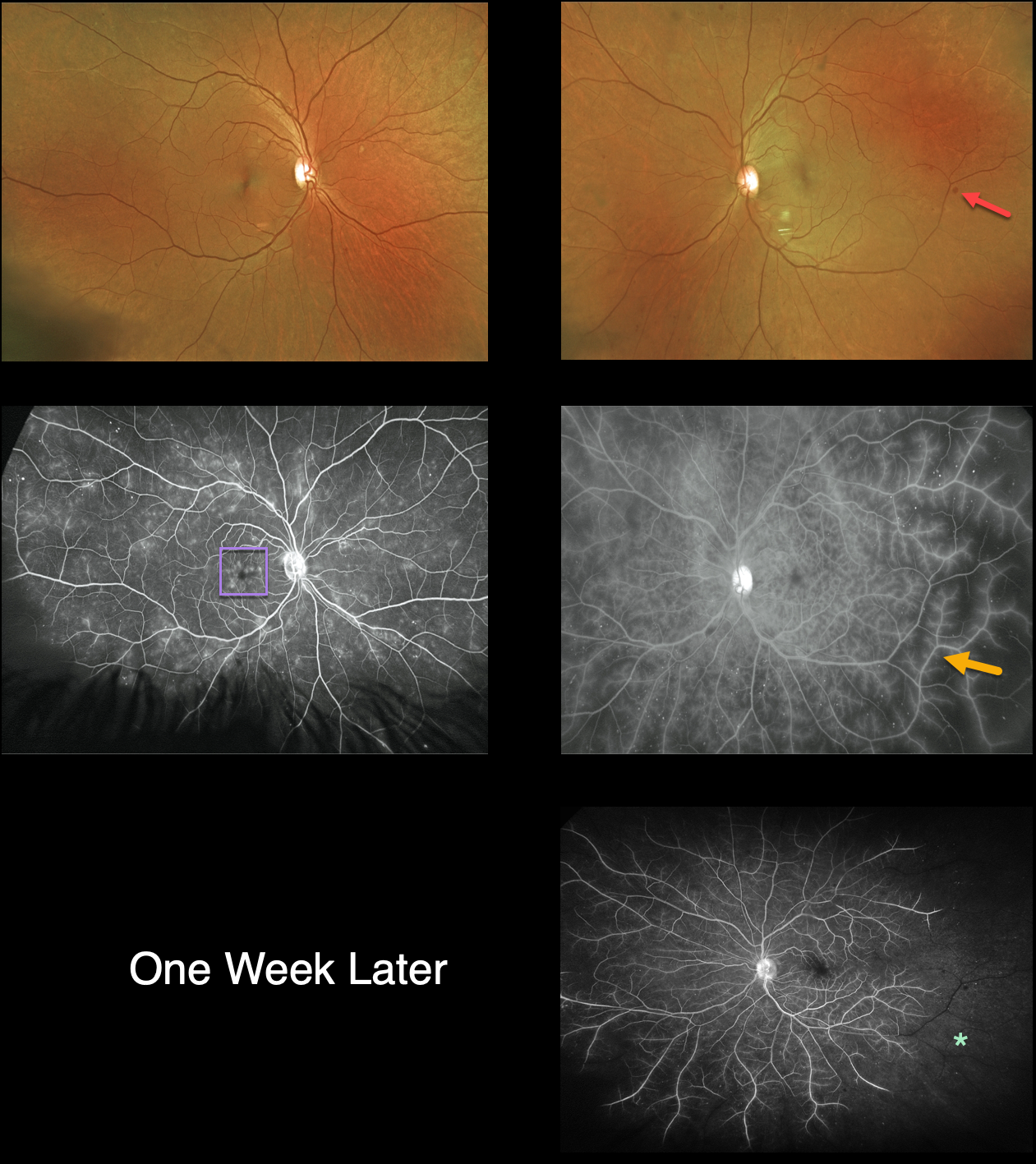
UNKNOWN RETINAL ISCHEMIA
Originally posted on @retina.rocks October 6, 2022
This 57YO female presented with 2 weeks of blurred vision. There was a 10-year history of type 2 diabetes and prior use of intravenous crack cocaine, though she claimed to have been clean for 18 years. Vision was 20/30 OD and 20/60 OS. The intraocular pressures were normal, but diffuse rubeosis was seen in her left eye.
Optos color imaging is fairly unremarkable, except for a few scattered bilateral dot-blot retinal hemorrhages.
Fluorescein angiography, however, shows diffuse vascular staining and ischemia, especially temporarily in her left eye. Cystoid leakage is also seen in her right macula.
Intravitreal Avastin was injected into her left eye. One week later, vision decreased to counting fingers OS, and the rubeosis had completely resolved. However, there was a marked increase in retinal ischemia. Unfortunately, she was then lost to follow-up.
Learning Points:
The rapid progression of the retinal ischemia in her left eye would be quite unusual for diabetes, especially immediately following intravitreal Avastin.
Although we are not certain of the underlying etiology, possible concurrent cocaine use in combination with diabetic endothelial damage may have been a contributing factor.
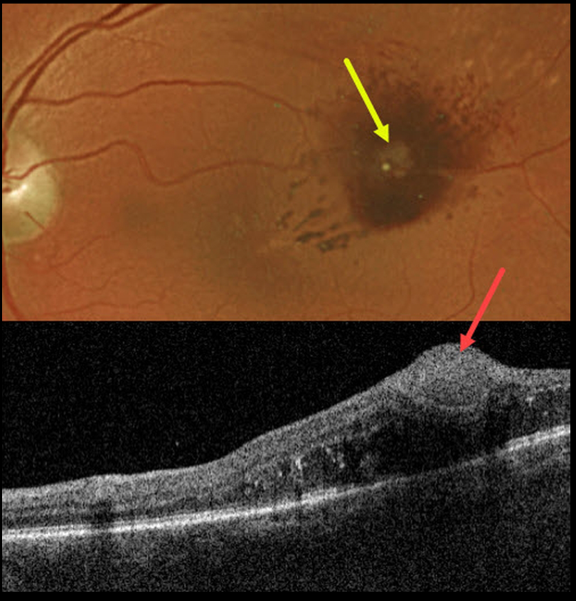
RETINAL ARTERIAL MACROANEURYSM (RAM)
Originally posted on @retina.rocks September 21, 2022
This 70YO female presented with mild vision loss in her left eye. Vision was 20/50.
Optos imaging shows a retinal arterial macroaneurysm (RAM) with surrounding blood. Swept-source OCT shows the hyperreflective RAM in the inner retina. The underlying retina is somewhat thickened.
Learning Points:
A RAM is thought to occur in a weakened arterial wall from arteriosclerosis, and usually appears as a yellow-white dilated lesion along a retinal arteriole within the first 3 bifurcations from the optic nerve.
Although most RAMs spontaneously resolve, chronic central edema or lipid can cause long term vision loss.
Mansour et al, Retina 2019;39;1133-1141, showed that anti-VEGF treatment is effective in reducing macular exudation and improving vision.
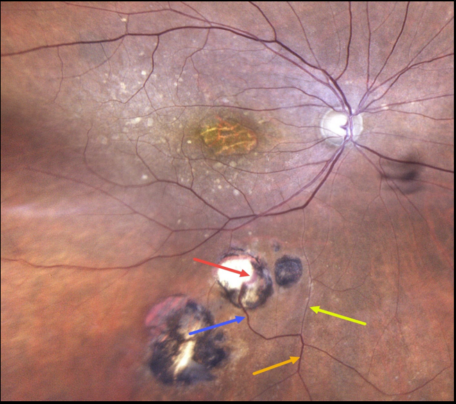
TOXOPLASMOSIS WITH RETINAL CHOROIDAL ANASTOMOSIS
Originally posted on @retina.rocks September 20, 2022
This 86YO female has a history of atrophic age-related macular degeneration with vision of 20/400 OD and 20/60 OS. Optos imaging of her right eye shows foveal atrophy with surrounding mixed drusen.
Multifocal inactive toxoplasmosis scars are noted inferiorly. An inferior retinal vein bifurcates, with a markedly attenuated branch continuing on its path towards the optic nerve. The other branch has a more normal caliber, dives into one of the toxoplasmosis scars, and drains into the choroid.
Learning Points:
Under normal conditions, the retinal and choroidal circulations remain separate and distinct. Rarely, the two circulations connect directly through a chorioretinal anastomosis.
These can occur in chorioretinal scars (typically from inactive toxoplasmosis lesions, as in this patient), in macular telangiectasia, and in disciform scars.
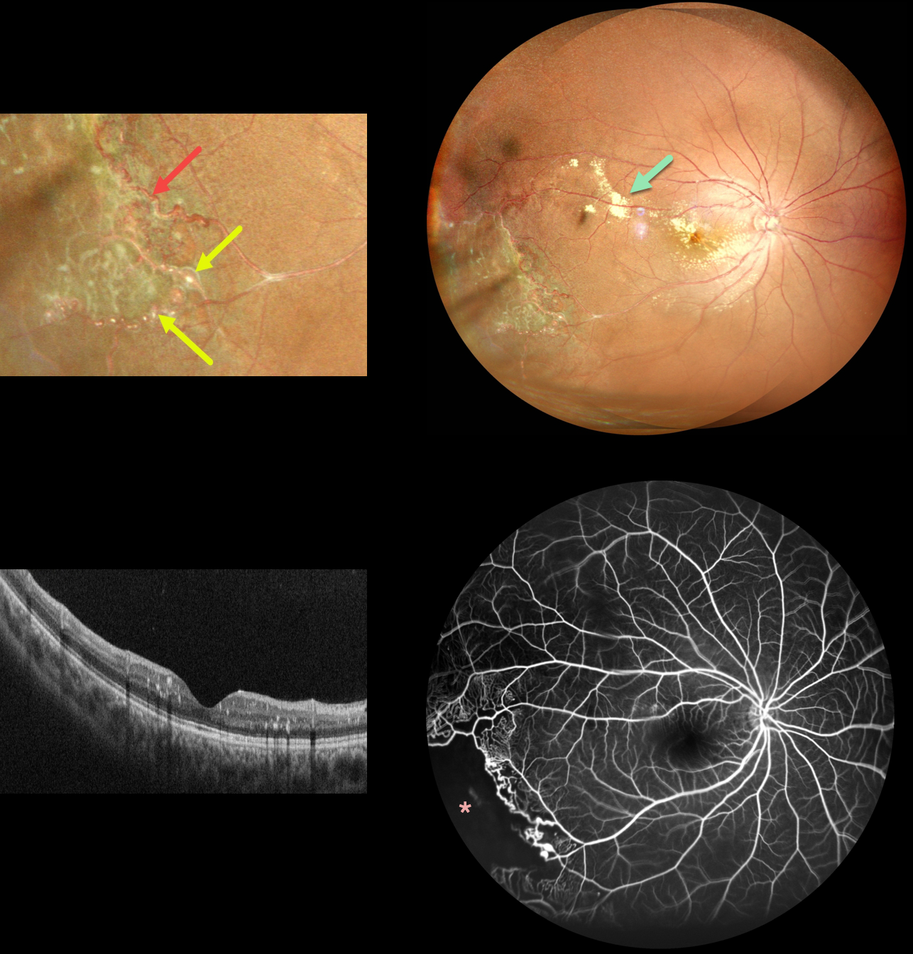
COATS DISEASE
Pramod Suman and Manish Nagpal
Originally posted on @retina.rocks September 5, 2022
This 11YO boy presented with these classic unilateral findings for Coats disease. An ultrawidefield image of the right eye shows scattered submacular and subretinal lipid posteriorly with distal featureless ischemic retina inferotemporally.
More peripherally, there are both large vessel changes, including beading and multiple macroaneurysms, with small vessel telangiectatic changes.
OCT scanning is remarkably normal except for some scattered specks of hyperreflective intraretinal lipid. Fluorescein angiography shows peripheral temporal telangiectasia with more distal ischemia, along with some subtle superotemporal macular telangiectatic changes.
Sector scatter laser was recommended.
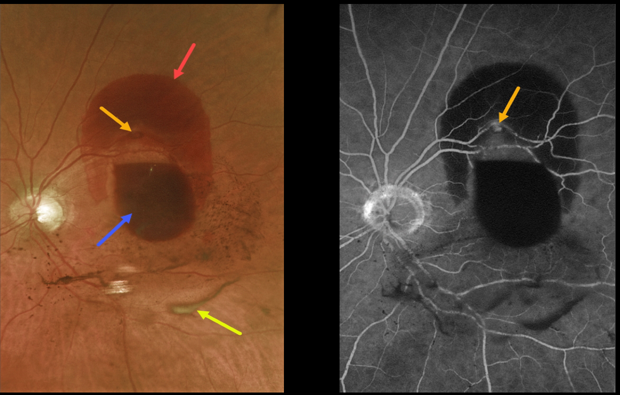
RETINAL ARTERIAL MACROANEURYSM (RAM)
Originally posted on @retina.rocks September 1, 2022
This 85 YO female presented with a retinal arterial macroaneurysm (RAM) with counting fingers vision.
The macroaneurysm has ruptured, causing multiple levels of blood shown on fundus photography and fluorescein angiography. The blood is subretinal superiorly, subhyaloid more centrally, and in the vitreous (both red and devitalized). We are observing this patient.
Learning Points:
A RAM is thought to occur in a weakened arterial wall from arteriosclerosis, and appears as a yellow-white dilated lesion along a retinal arterial within the first 3 bifurcations from the optic nerve.
There are 2 distinct presentations, hemorrhagic or exudative. The hemorrhagic type often presents, as in this case, with pathognomonic multiple layers of blood.
Acute macular hemorrhage can be treated with observation, anti-VEGF injections, or thermal laser.
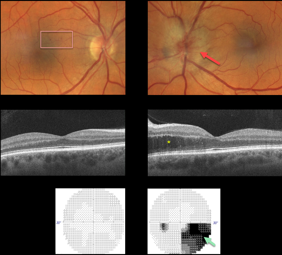
HYPERTENSIVE RETINOPATHY vs AION
Originally posted on @retina.rocks August 16, 2022
This 57YO male presented with unilateral optic nerve swelling in his left eye and a few faint nasal macular retinal hemorrhages in his right eye. Vision was 20/40 OD and 20/25 OS.
OCT was normal OD and thickened in the nasal outer nuclear layer OS. Visual fields were normal OD and inferiorly depressed, especially inferonasally, OS.
Blood pressure was 190/110, and he was immediately referred to the emergency room.
Learning Points:
Malignant hypertension, defined as blood pressure above 180 systolic and/or 120 diastolic, is a life-threatening medical emergency. Eye doctors are in a unique position to often diagnose this condition.
Patients will often present with bilateral optic nerve swelling, flame-shaped retinal hemorrhages, and, with more chronic disease, lipid precipitates in the nasal macular outer plexiform layer. However, rarely, the ocular findings may be asymmetric or even unilateral.
Since the macular fluid emanates from the optic nerve, the macular thickening is always worse nasally (as in our case), and is an important clinical clue for this diagnosis.
Our patient may also have had an isolated anterior ischemic optic neuropathy in his left eye and mild hypertensive retinopathy in his right eye. The partial altitudinal deffect is more common with this diagnosis than hypertension.
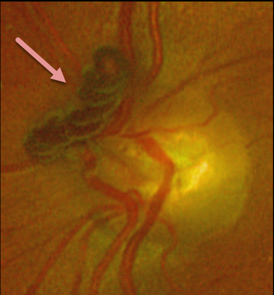
RETINAL VASCULAR LOOP
Originally posted on @retina.rocks August 2, 2022
This 77YO female presented with this completely asymptomatic yet prominent retinal vascular loop. Observation was recommended.
Learning Points:
These benign congenital prepapillary vascular loops can be unilateral or bilateral. Although usually just an incidental finding, they can rarely cause a branch arterial or venous occlusion (see Retinal Cases Brief Rep 2014:8;124-126 and Ophthalmology 2020;127:124-126).
They should also not be confused with disc neovascularization, which consists of much smaller caliber vessels that leak on fluorescein angiography.
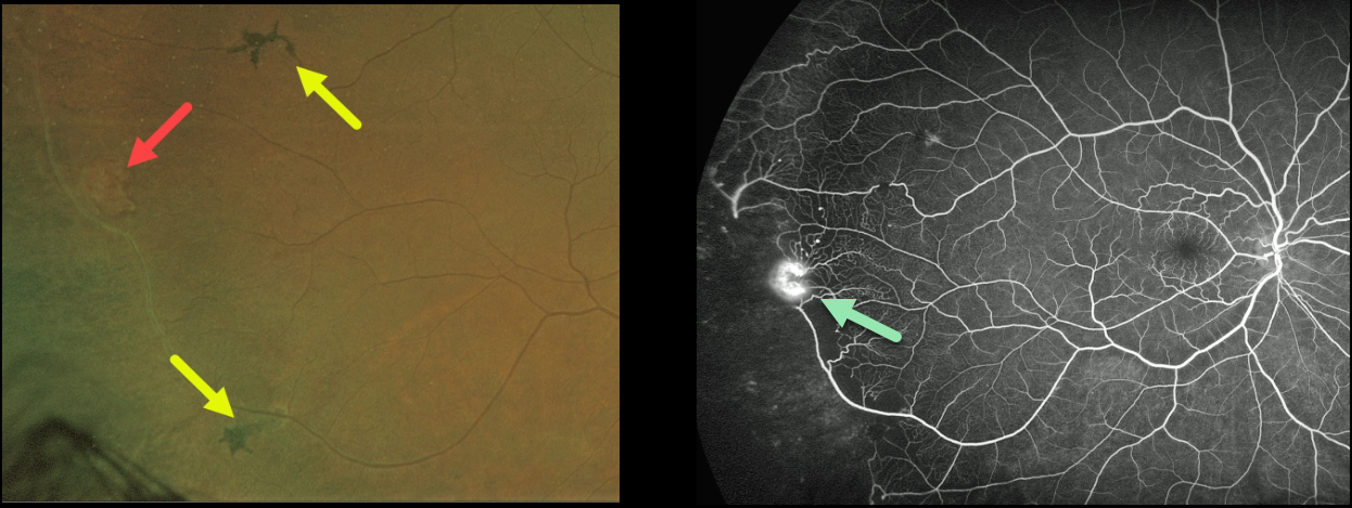
SICKLE CELL RETINOPATHY
Originally posted on @retina.rocks August 1, 2022
This 56YO African American male presented with floaters in his right eye and a history of sickle cell anemia. Vision was 20/30 OU.
A mild vitreous hemorrhage was noted inferiorly, along with an area of suspected peripheral retinal neovascularization. There were several areas of midperipheral black sunburst lesions. Fluorescein angiography confirmed a patch of retinal neovascularization at the junction of perfused and non-perfused retina. Due to the symptomatic vitreous hemorrhage, scatter laser to the peripheral ischemic retina was recommended.
Learning Points:
Patients with sickle cell disease develop symptoms throughout their bodies as stiff, sickled red blood cells block capillary beds. In the eye, this can cause proliferative sickle cell retinopathy (PSR) with sea-fan-shaped areas of peripheral retinal neovascularization developing at the junction of perfused and ischemic retina.
The black sunburst lesions are thought to occur from intraretinal RPE migration in response to photoreceptor damage from intraretinal and subretinal hemorrhage (see Ausdourian et al, BJO 1975;59:710-716). Complications include vitreous hemorrhage and traction retinal detachment. Although patients with SS disease tend to have more systemic complications, SC disease is usually associated with more severe ocular findings.
Treatment with peripheral scatter laser photocoagulation is somewhat controversial because, unlike in more typical proliferative retinopathies (e.g., those from diabetes and branch retinal vein occlusions), these new vessels tend to auto-infarct.
For a great review article on sickle cell disease and the eye, see Elagouz et al, Surv Ophthalmology 2010;55:359-377.
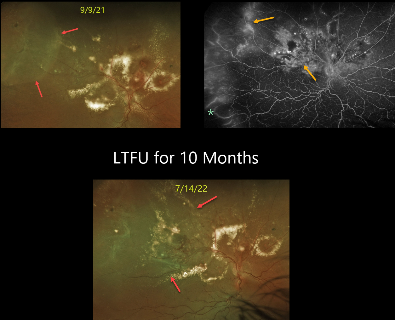
COATS DISEASE
Originally posted on @retina.rocks July 25, 2022
This 54YO male patient has a long history of Coats disease. Vision was 20/400 when he was referred to our practice on 9/9/21. Unfortunately, he was lost to follow-up for almost a year because of his fear of doctors. Vision decreased to hand motion.
Optos color imaging shows his funduscopic appearance at the initial and subsequent visits. Fluorescein angiography shows extensive leakage from the various vascular abnormalities, as well as peripheral temporal ischemia. Although the posterior pole lipid mostly decreased during the prior year, the exudative detachment enlarged, now spreading through the macula.
Vitrectomy is scheduled in hopes of preventing further vision loss.
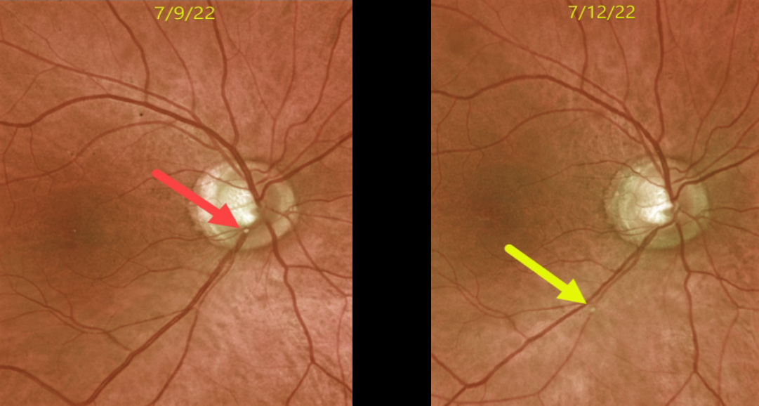
RETINAL EMBOLUS
Originally posted on @retina.rocks July 22, 2022
This 72YO male presented with acute superior field loss from an acute branch retinal artery occlusion (BRAO) in his right eye.
The causative calcific embolus is seen originally along the inferior optic nerve margin. Three days later, the plaque moved further downstream.
This patient had acute retinal ischemia (defined as acute BRAO, central retinal artery occlusion, or transient monocular vision loss), and was immediately referred to the nearest stroke center, which is the standard of care.
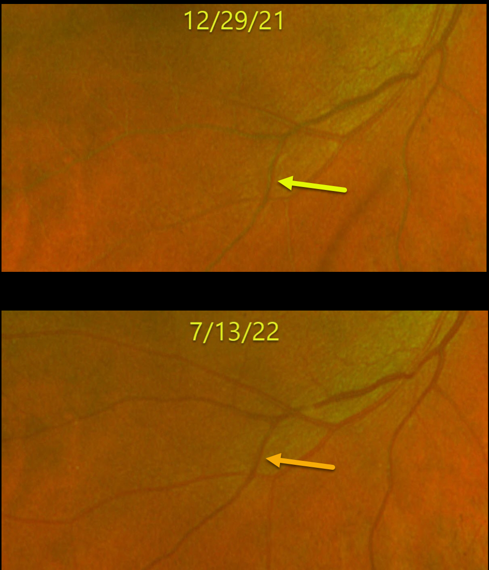
LIPEMIA RETINALIS
Originally posted on @retina.rocks July 21, 2022
This 51YO male’s retinal vasculature turned creamy white from very high triglycerides. His original triglyceride levels were around 4,200 mg/dL.
After 6 months of systemic treatment, his triglyceride levels decreased to 3,100 mg/dL. His blood returned to a normal red color, and his retinal vessels normalized in appearance.
Learning Points:
Lipemia retinalis is a rare retinal manifestation of severe hypertriglyceridemia, characterized by creamy white discoloration of the retinal vasculature. In severe cases, the fundus can turn salmon-colored.
Patients are asymptomatic, and vision is normal. However, they are at obvious cardiovascular risk without systemic treatment.
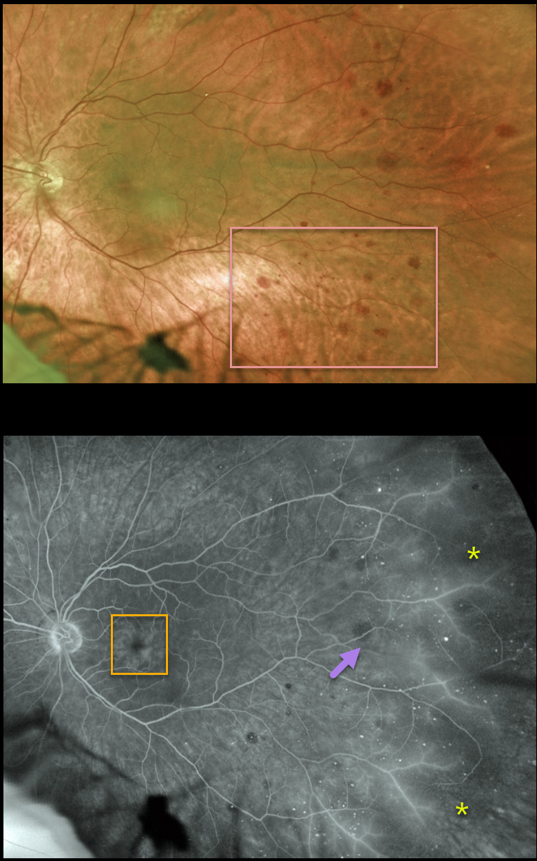
OCULAR ISCHEMIA
Originally posted on @retina.rocks July 18, 2022
This 69YO male presented with a few months of blurred vision in his left eye. Vision was 20/40 in his normal right eye and 20/100 in his left eye.
Scattered deep retinal hemorrhages are noted temporally. Fluorescein angiography shows temporal midperipheral microaneurysms, more distal ischemia with vascular leakage, and moderate cystoid macular leakage.
Since our patient had a history of a complete left internal carotid artery blockage, no further testing was ordered. He is currently monitored regularly by a vascular surgeon.
Learning Points:
The retinal hemorrhages in the ocular ischemic syndrome (OIS) are unique. Retinal vascular disorders like diabetic retinopathy, retinal vein occlusions, and hypertensive retinopathy usually have flame- and dot-shaped inner retinal hemorrhages concentrated around the posterior pole.
Our patient’s findings are classic for OIS, with hemorrhages in the deeper peripheral retina. The retinal periphery is literally the end of the line for the retinal arterial circulation. In an eye receiving less blood supply due to carotid occlusive disease, the more proximal retina has first dibs on the available oxygen.
There are penetrating capillaries that dive radially from the nerve fiber and ganglion cell layers into the deeper retina. The deep vascular complex that supplies the inner and outer plexiform layers is thus furthest downstream, and these endothelial cells are likely damaged in OIS. Blood cells leaking into this space accumulate, causing the large outer blot to hemorrhage.
These patients need to have carotid Doppler and ultrasound and referred for medical or surgical intervention if they have a critical stenosis. They may also develop anterior or posterior segment neovascularization requiring photocoagulation and/or anti-VEGF injections.
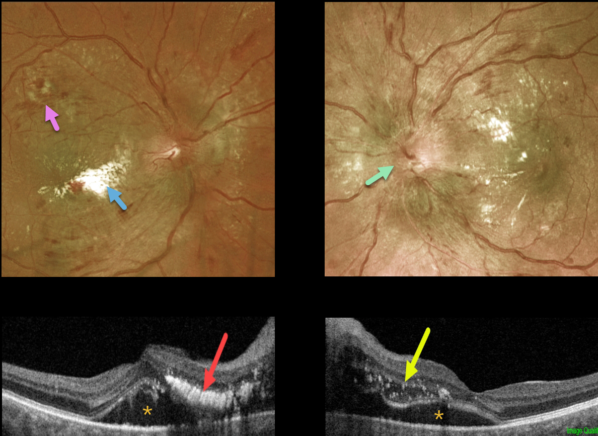
HYPERTENSIVE RETINOPATHY
Originally posted on @retina.rocks July 11, 2022
This 44YO male patient presented with a 2-week history of bilateral vision loss. He denied any past medical history.
Vision was 20/150 OD and 20/50 OS. Optos imaging shows bilateral optic nerve swelling, flame-shaped retinal hemorrhages, and macular lipid.
Swept-source OCT shows bilateral subretinal fluid extending from the nerve into the macular centers. A plaque of outer retinal hyperreflective lipid is noted in the nasal right macula, with hyperreflective dots of suspended lipid within the outer retinal edema in his left eye.
Blood pressure was 242/136. He was immediately sent to the emergency room for treatment of his malignant hypertension.
Learning Points:
Malignant hypertension, defined as blood pressure above 180 systolic and/or 120 diastolic, is a life-threatening medical emergency. Eye doctors are in a unique position to often diagnose this condition.
Patients will often present with bilateral optic nerve swelling, flame-shaped retinal hemorrhages, and, with more chronic disease, lipid precipitates in the nasal macular outer plexiform layer. Since macular fluid originates from the optic nerve, macular thickening is always worse nasally (as in our case) and is an important clinical clue for this diagnosis.
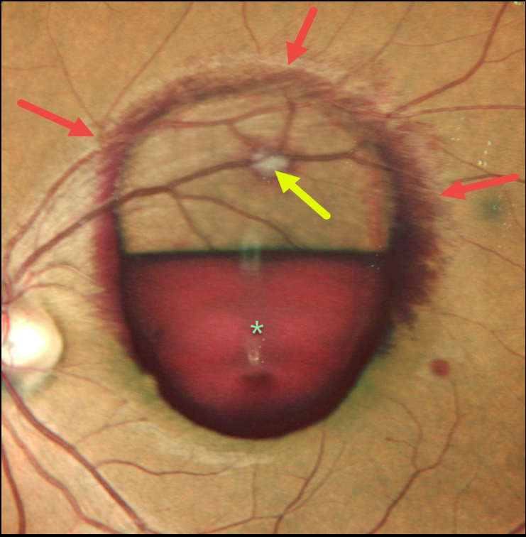
RETINAL ARTERIAL MACROANEURYSM (RAM)
Nivesh Gupta
Originally posted on @retina.rocks June 23, 2022
This 34YO female presented with a symptomatic ruptured retinal arterial macroaneurysm (RAM).
Vision was counting fingers due to the inferior sub-internal limiting membrane blood obscuring the underlying fovea. More peripherally, blood is staining the surrounding nerve fiber layer. The fibrosed RAM is visible superiorly.
Learning Points:
A RAM is thought to occur in a weakened arterial wall from arteriosclerosis, and usually appears as a yellow-white dilated lesion along a retinal arteriole within the first 3 bifurcations from the optic nerve.
There are 2 distinct presentations, hemorrhagic or exudative. The hemorrhagic type often presents with pathognomonic preretinal, intraretinal, and subretinal blood. Acute macular hemorrhage can be treated with observation, anti-VEGF injections, or thermal laser to the RAM.
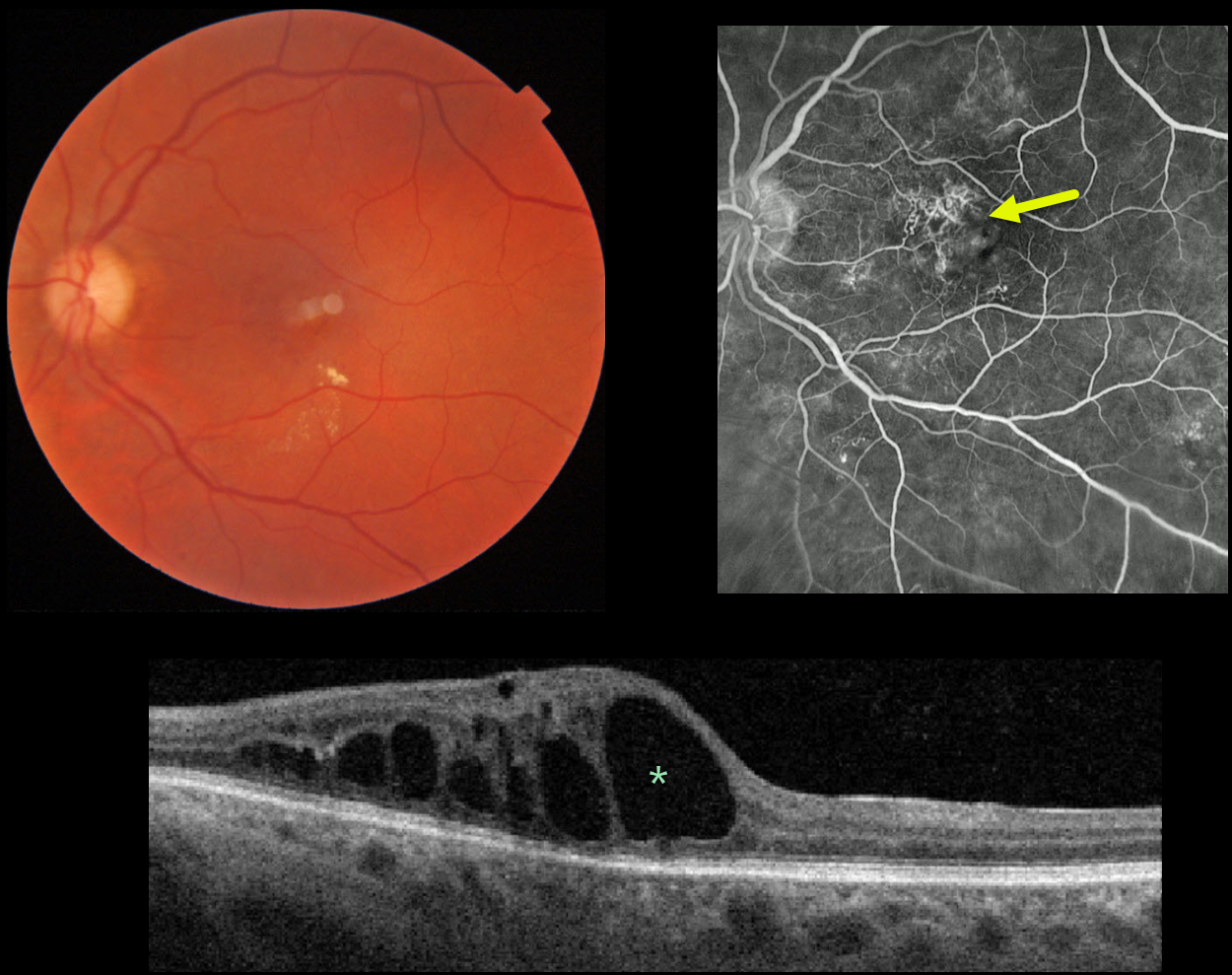
RADIATION RETINOPATHY
Originally posted on @retina.rocks June 15, 2022
This 60YO male presented with 20/400 vision in his left eye due to radiation retinopathy caused by prior photon radiation treatment for brain cancer 10 years earlier.
Fundus photography shows some small scattered inner retinal hemorrhages and exudates. Fluoroscein angiography shows superonasal foveal retinal telangiectasia, with mild scattered inferior microvascular changes. OCT shows central and nasal thickening from large outer retinal cysts. Anti-VEGF therapy was started.
Learning Points:
Radiation retinopathy usually occurs 3 months to 3 years after external beam radiation (e.g., for facial or orbital cancers) or plaque radiotherapy. The ocular findings closely resemble those seen with diabetic retinopathy.
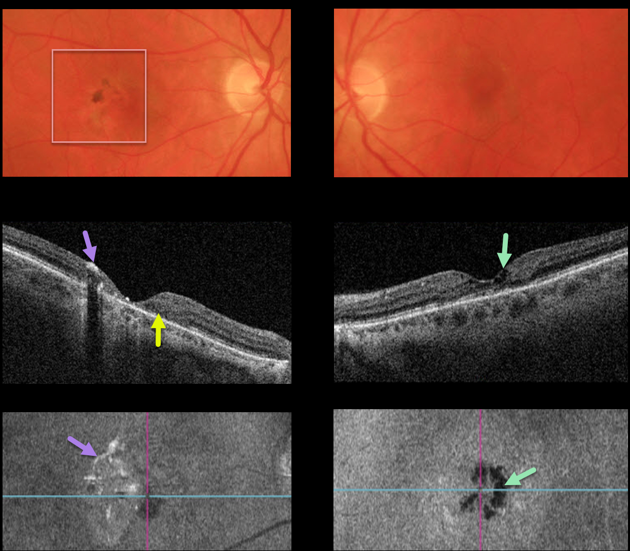
MACULAR TELANGIECTASIA
Originally posted on @retina.rocks June 7, 2022
This 60YO female presented with 20/70 vision OD and 20/50 vision OS, along with classic findings of macular telangiectasia type 2 (MacTel2).
The perifoveal retina is somewhat opaque with significant pigmentary changes, particularly in the right eye.
OCT scanning of the right eye shows significant central outer retinal atrophy with hyper-reflective pigment migration. The left OCT shows milder atrophy, central inner cavitations, but no pigment migration.
En face imaging helps highlight some of the findings described above. The right en face image shows scattered patchy hyperreflective areas corresponding to the pigment migration, while the left en face image highlights the hyporeflective cavitations.
Learning Points:
Originally described by Gass (Arch Ophthalmology 1982;100:769-780), MacTel2 is a neurodegenerative disorder, most likely originating from Müller cell dysfunction. The term telangiectasia is misleading because the funduscopic findings are mostly nonvascular.
The refractile inner retinal ‘crystals’ are thought to be Müller cell footplates. The foveal retina shows a whitish discoloration, most likely due to retinal opacification resulting from Müller cell dysfunction.
Photoreceptor loss allows RPE cells to migrate along intraretinal capillaries, causing the coarse clinical pigment clumping (not seen in our patient).
The majority of patients will also have subclinical chorioretinal anastomosis associated with right-angle venules (Spaide et al, Retina 2018;38:1920-1929).
Secondary macular neovascularization can be successfully treated according to age-related macular degeneration protocols.
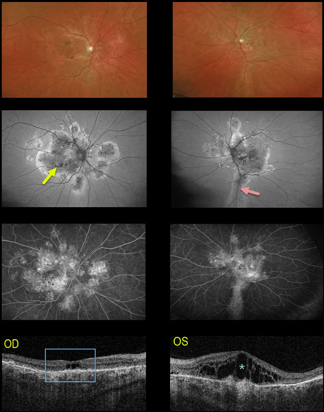
IDIOPATHIC CENTRAL SEROUS CHORIORETINOPATHY
Originally posted on @retina.rocks May 25, 2022
This 64YO male received prior photodynamic therapy in one of his eyes for idiopathic central serous chorioretinopathy (ICCS) and was subsequently lost to follow-up for 10-15 years. He presented with 20/400 vision bilaterally.
Multimodal imaging shows classic findings for chronic ICSC. Optos color imaging shows macular and peripapillary pigmentary changes with a suggestion of an inferior gutter OS.
Fundus autofluorescence better shows the bilateral pigmentary changes and inferior gutter OS, and fluorescein angiography shows variable chorioretinal staining.
Triton swept-source OCT shows bilateral thickened choroid, dry central cavitation-like changes OD, and marked cystoid macular edema OS.
Photodynamic therapy was performed for the left eye, although we are not certain this will improve his vision, given the chronicity of these findings.
Learning Points:
Macular edema is an uncommon finding with typical ICSC, likely due at least in part to the external limiting membrane acting as a barrier to subretinal fluid migrating into the retina. Eyes with chronic ICSC, however, can develop retinal edema as in this case.
Gutters are commonly seen in pathology with chronic subretinal leakage, including central serous retinopathy, choroidal hemangioma, choroidal nevus, and uveal melanoma. These pigmentary changes are usually best imaged with FAF.
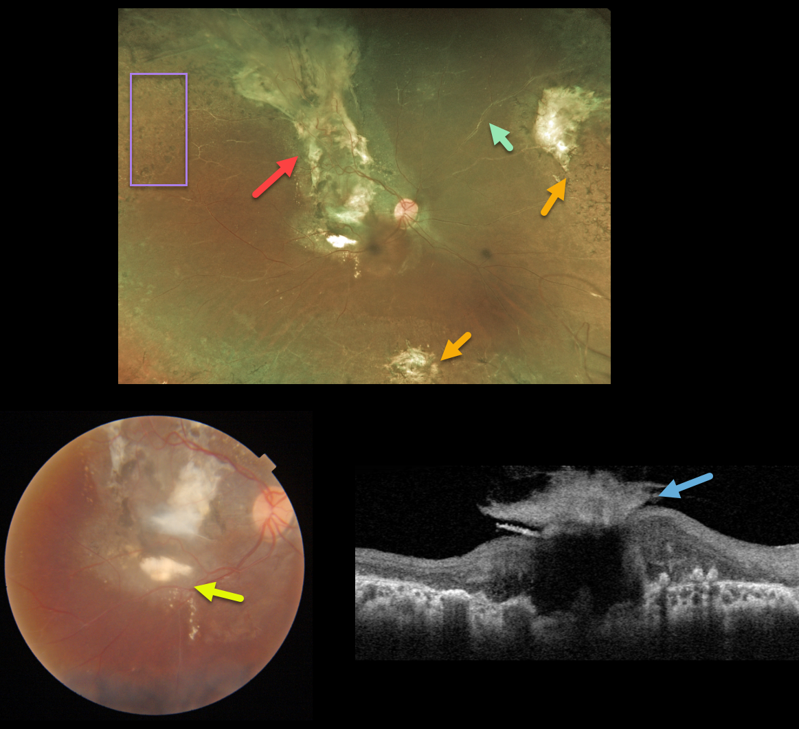
COATS DISEASE
Originally posted on @retina.rocks May 23, 2022
This 15YO female received prior thermal laser for Coats disease over 10 years earlier and was subsequently lost to follow-up. Right eye vision was counting fingers.
Optos color imaging shows subretinal scarring extending from the macula through the superior retinal periphery, nasal and inferior peripheral scarring, scattered peripheral retinal laser scarring, and sheathed vessels. There are no active Coats lesions.
Triton color imaging shows a magnified view of the macular scarring with some inferior macular subretinal lipid. Swept-source OCT shows a volcano-like vitreous component to the macular scar.
Learning Points:
Historically considered a unilateral disease, more recent ultra-widefield angiographic studies show subtle vascular changes, mostly in the temporal periphery (Brockmann et al., BJO 2021;105:1444-1453). Our patient’s left eye, however, was completely normal.
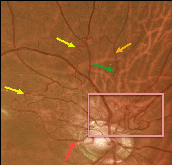
RETINAL ARTERIOVENOUS MALFORMATION
Originally posted on @retina.rocks May 3, 2022
This 61YO male presented with these asymptomatic blood vessels extending superiorly from the right nerve. Vision was 20/25 OU, and the left eye was normal.
The abnormal vessels emanate from a cilioretinal artery, extend superiorly into and then above the macula, then arch nasally and descend. Note that it crosses a vein, confirming that it is still arterial (remember that arteries cross veins, veins cross arteries, but arteries never cross arteries and veins never cross veins). It then descends towards the nerve in a tangle of vessels likely consisting of both arteries and veins.
We felt that these vessels represented a low-grade arteriovenous malformation (AVM).
Learning Points:
Since about 25% of patients with AVMs can have intracranial venous anomalies, a brain MRI is usually recommended.
These vessels somewhat resemble collateral vessels noted after retinal vein occlusions. Collaterals are dilated capillaries that result from increased hydrostatic pressure from an occlusion. They are almost always venous, and when seen, are a telltale sign for a prior occlusion. Unlike retinal neovascularization, they do not leak angiographically.
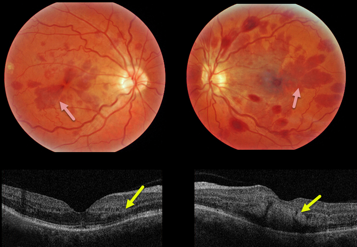
HENLE LAYER HEMORRHAGE
Originally posted on @retina.rocks April 18, 2022
This 47YO female presented with bilateral vision loss. She had a seizure 10 days prior, losing consciousness and hitting her head. Upon awakening, she had reduced vision in her right eye and complete central loss in her left. Vision was 20/60 OD and counting fingers at 3 feet OS. Head CT scanning was normal.
Triton color imaging shows deep radiating wedge-shaped retinal hemorrhages centered in each macula. Swept-source OCT shows that these hemorrhages are located primarily in the outer plexiform layer (OPL).
Learning Points:
Radiating deep macular hemorrhages are seen in multiple conditions, including Valsalva (likely this case) and trauma. Their unique radiating appearance is caused by blood accumulating in Henle’s layer.
Kon Graversen et al described hemorrhagic unilateral retinopathy, an idiopathic disorder with a similar appearance (Retina 2014;34:483-489).
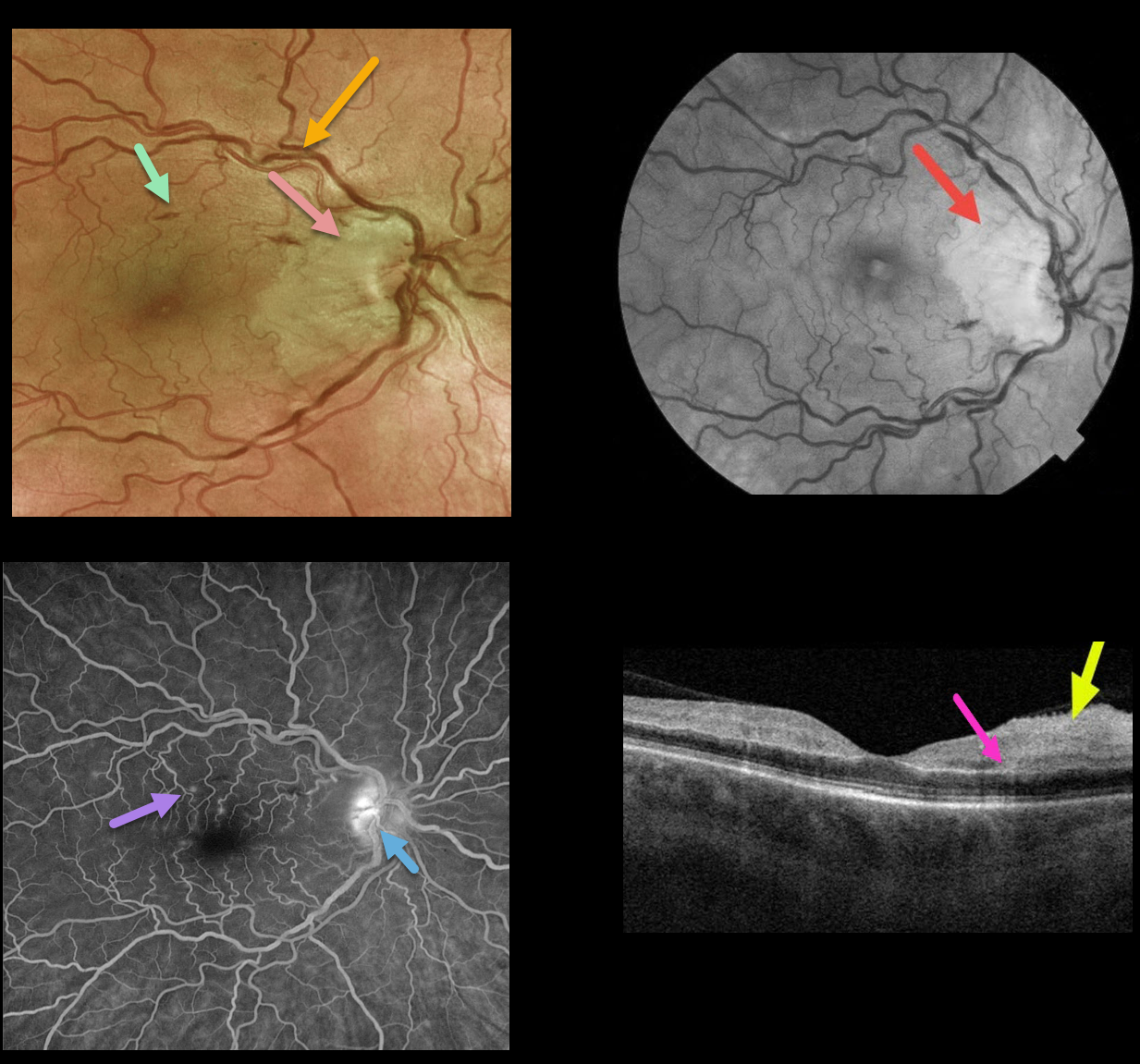
CRVO + RAO + PAMM
Originally posted on @retina.rocks March 4, 2022
This 50YO female presented with counting finger vision in her right eye due to an acute central retinal vein occlusion (CRVO) with a nasal macular retinal artery occlusion (RAO).
Optos fundus photography shows retinal vascular tortuosity, mild retinal hemorrhages, a mildly swollen nerve, and white ischemic nasal macular retina. The ischemic retinal changes are most apparent on the red-free photo.
Fluorescein angiography (FA) shows some mild focal areas of macular leakage with late staining of the temporal disc.
OCT shows areas of inner retinal edema and hyperreflectivity. There is also a small area of mid-retinal hyperreflectivity consistent with paracentral acute middle maculopathy (PAMM).
Learning Points:
Cilioretinal artery occlusion is usually non-embolic and seen in association with giant cell arteritis, secondary to a CRVO, or as an isolated event. The occlusion may be due in part to arterial compression from a swollen nerve.
Pichi et al felt that CRVO PAMM-type lesions, as seen in our patient, are due to hypoperfusion from increased downstream intraluminal pressure (Br J Ophthalmol 2019;103:1137-1145).
We couldn’t find a definite cilioretinal artery in our patient, although the ischemia was within the distribution of one if one were present. Interestingly, the nasal macular capillaries appeared perfused on FA. However, FA captures just the inner retinal capillary plexus.
Although the OCT shows evidence of acute inner capillary plexus ischemia, the angiographically perfused inner retina supports a more PAMM-type mechanism in our case.
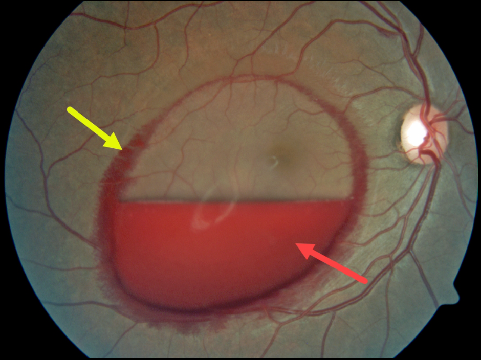
VALSALVA RETINOPATHY
Originally posted on @retina.rocks March 3, 2022
This patient presented with an acute layered sub-internal limiting membrane (ILM) hemorrhage due to a Valsalva maneuver. There is also a rim of blood along the circumference of the ILM detachment, which is most likely located in Henle’s layer, given its radiating deep retinal pattern.
Learning Points:
Sub-ILM hemorrhages most commonly follow a Valsalva maneuver or trauma, but can also happen spontaneously, as with anemia.
These hemorrhages virtually always resolve without sequelae, although the blood can be drained into the inferior vitreous by creating a small opening in the ILM using either a thermal or Nd:YAG laser.
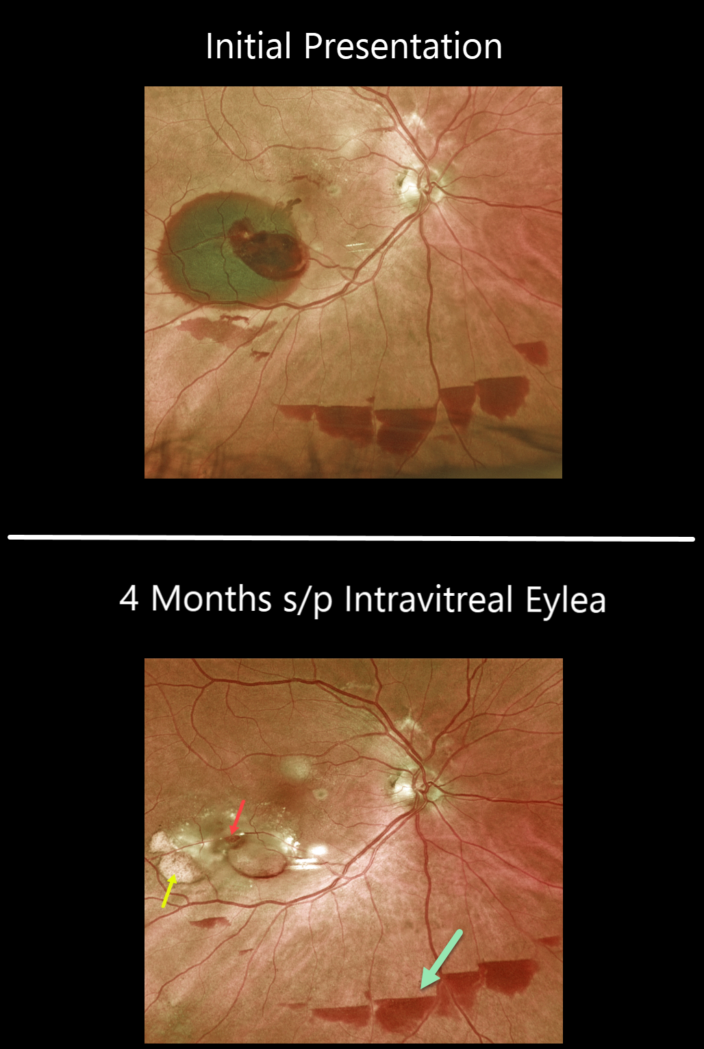
RETINAL ARTERIAL MACROANEURYSM (RAM)
Originally posted on @retina.rocks February 14, 2022
This 85 YO female with a retinal arterial macroaneurysm (RAM) continues to improve with 20/30 vision following a single Eylea injection four months earlier.
The macroaneurysm still appears patent. The old subretinal devitalized blood is devitalized and yellow. There are also multiple patches of resolving inferior subhyaloid blood.
Learning Points:
A RAM is thought to occur in a weakened arterial wall from arteriosclerosis and appears as a yellow-white dilated lesion along a retinal arteriole within the first 3 bifurcations from the optic nerve.
There are 2 distinct presentations: hemorrhagic or exudative. The hemorrhagic type often presents, as in this case, with pathognomonic preretinal, intraretinal, and subretinal blood. Acute macular hemorrhage can be treated with observation, anti-VEGF injections, or thermal laser.
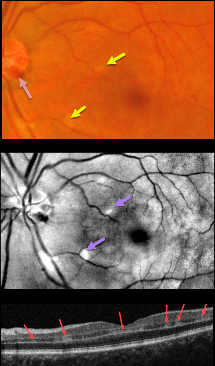
PARACENTRAL ACUTE MIDDLE MACULOPATHY (PAMM)
Gehad Hassan Youssef
Originally posted on @retina.rocks February 10, 2022
This 57YO female presented with sudden painless vision loss in her left eye. There was no significant past ocular or medical history. Vision was 20/20 in her normal right eye and 20/130 in her left eye.
Fundus photography shows several small hemorrhages along the temporal disc margin and faint patches of scattered whitish retinal opacification, which are much more pronounced in a perivenular distribution in the red-free image.
OCT B-scan shows multiple patches of focal hyperreflectivity involving the inner plexiform, inner nuclear, and outer plexiform layers.
Learning Points:
Paracentral acute middle maculopathy (PAMM) is a recently described phenotype that consists of acute mid-retinal ischemia, occurring as an idiopathic entity or in other ocular disorders, including Purtscher’s, diabetic retinopathy, retinal vascular occlusions, and acute macular neuroretinopathy.
For a great recent review, see Scharf et al, Progress in Retinal and Eye Research 2021;81;100884.
Perivenular PAMM may be the only presenting sign for an incomplete central retinal artery occlusion or mild central retinal vein occlusion (see Zhao et al, AJO 2022;234:15-19). Our patient may indeed have a very mild CRVO, given the optic nerve hemorrhages.
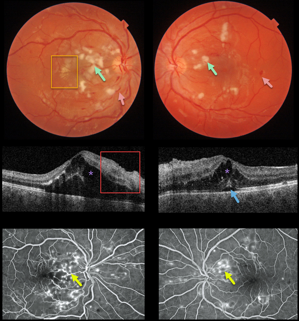
HYPERTENSIVE RETINOPATHY
Originally posted on @retina.rocks February 4, 2022
This 22YO male presented with subacute vision loss of 20/200 OD and 20/50 OS. Triton color imaging shows diffuse bilateral cotton wool spots with intraretinal hemorrhages, along with an early macular star temporal to the right fovea.
Swept-source OCT shows bilateral intraretinal thickening (mostly in the outer nuclear layer) and hyperreflective inner retinal layers, along with subfoveal fluid.
Optos fluorescein angiography shows bilateral telangiectasia and staining, mostly around the nerve fiber layer infarcts.
Blood pressure was 169/135. He was immediately sent to the nearest emergency room.
Learning Points:
Malignant hypertension, defined as blood pressure above 180 systolic and/or 120 diastolic, is a life-threatening medical emergency. Eye doctors are in a unique position to often diagnose this condition.
Patients will often present with bilateral optic nerve swelling, flame-shaped retinal hemorrhages, and, with more chronic disease, lipid precipitates in the nasal macular outer plexiform layer.
Since the macular fluid originates from the optic nerve, macular thickening is always worse nasally and is an important clinical clue for this diagnosis.
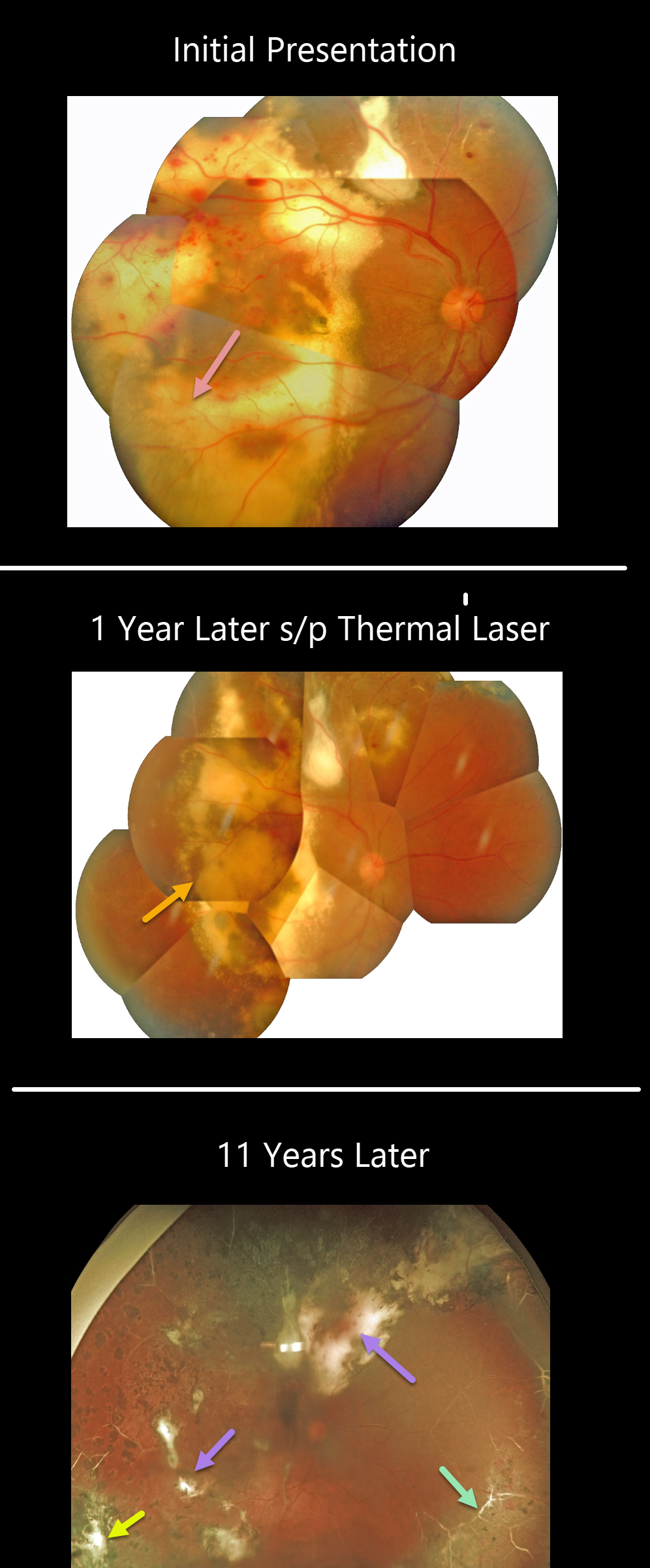
COATS DISEASE
Originally posted on @retina.rocks January 20, 2022
This patient was first seen in 2009 when he was 16 years old. Confluent subretinal lipid can be seen extending to the periphery. Several sessions of thermal laser were applied, and 1 year later, photos showed inactive Coats lesions with decreased subretinal lipid.
He was then lost to follow-up for 11 years when his vision was 20/400. Optos shows total resolution of all exudation and subretinal lipid with variable residual subretinal scarring. Peripheral scatter laser scarring is noted. The peripheral retinal vessels are white. There are no active Coats lesions.
Learning Points:
Historically considered a unilateral disease, more recent ultra-widefield angiographic studies show subtle vascular changes, mostly in the temporal periphery (Brockmann et al., BJO 2021;105:1444-1453).
Our patient’s left eye, however, was completely normal clinically and angiographically.
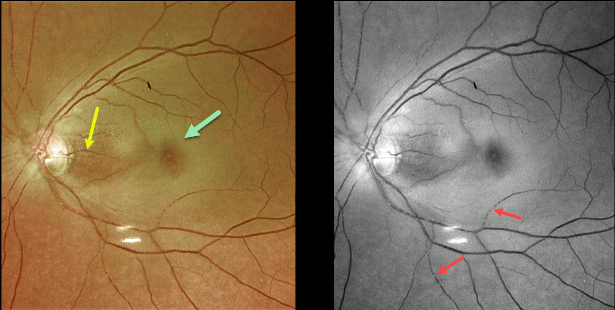
CRAO
Originally posted on @retina.rocks January 7, 2022
This 73YO female presented with counting-fingers vision secondary to an acute central retinal artery occlusion (CRAO).
The foveal retina is still perfused by the underlying choroid, resulting in the classic cherry red spot appearance. The nasal macula is also preserved due to the presence of a cilioretinal artery, which unfortunately does not supply the macular center. The Optos green channel best images the stagnant blood column.
Our patient’s MRI revealed several acute diffuse embolic strokes. Carotid duplex ultrasound showed severe bilateral carotid stenosis. In addition, her blood glucose was 357, and her erythrocyte sedimentation rate was 94. She was also started on systemic prednisone by the ER until giant cell arteritis could more definitively be ruled out.
Learning Points:
Patients with acute retinal ischemia (defined as transient monocular vision loss, acute BRAO, or acute CRAO) need to be emergently referred to a stroke center.
This is especially urgent with an acute CRAO since about 75% of patients have already developed a recent stroke.
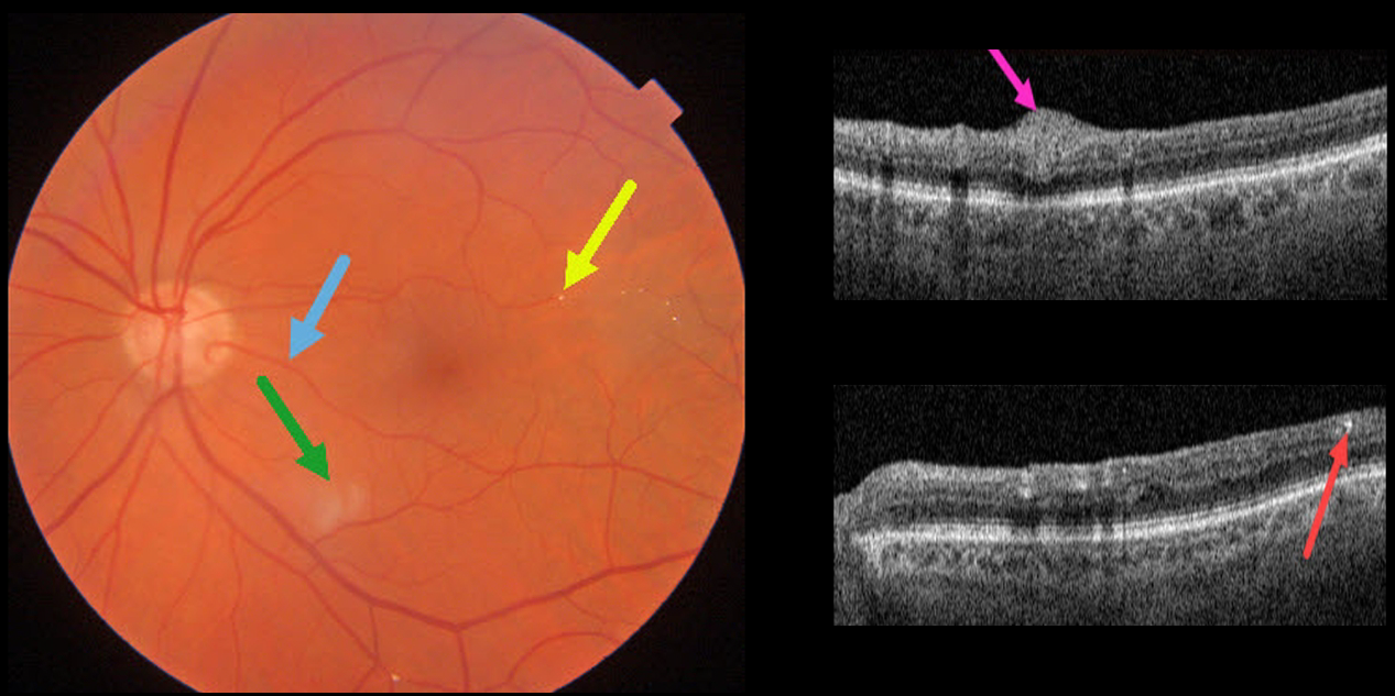
BRAO
Originally posted on @retina.rocks January 6, 2022
This 85YO male has 20/25 vision despite multiple retinal emboli and a resolving branch retinal artery occlusion (BRAO).
Triton swept-source OCT shows residual inner retinal edema and opacification. The embolus on Triton photography is also imaged in the OCT B-scan.
A cilioretinal artery may be responsible for the good vision.
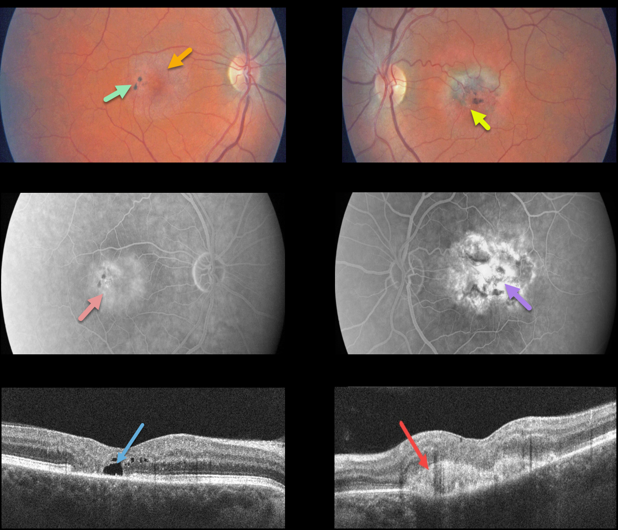
MACULAR TELANGIECTASIA
Originally posted on @retina.rocks December 27, 2021
This patient presented with classic findings of macular telangiectasia type 2 (MacTel2), including an opaque perifoveal retina, inner retinal crystal, and black clumps of intraretinal pigment migration.
Deep retinal angiographic leakage is noted in the right eye with more diffuse staining in the left eye. OCT scanning shows an outer retinal cavitation OD and type 2 macular neovascularization (MNV) OS.
Learning Points:
Originally described by Gass (Arch Ophthalmology 1982;100:769-780), MacTel2 is a neurodegenerative disorder, most likely originating from Müller cell dysfunction.
The term telangiectasia is misleading because the funduscopic findings are mostly nonvascular.
The refractile inner retinal ‘crystals’ are thought to be Müller cell footplates. The foveal retina shows a whitish discoloration, most likely due to retinal opacification resulting from Müller cell dysfunction.
Photoreceptor loss allows RPE cells to migrate along intraretinal capillaries, causing the coarse clinical pigment clumping.
The majority of patients will also have subclinical chorioretinal anastomosis associated with right-angle venules (Spaide et al., Retina 2018;38:1920-1929).
Secondary MNV can be successfully treated per age-related macular degeneration protocols.
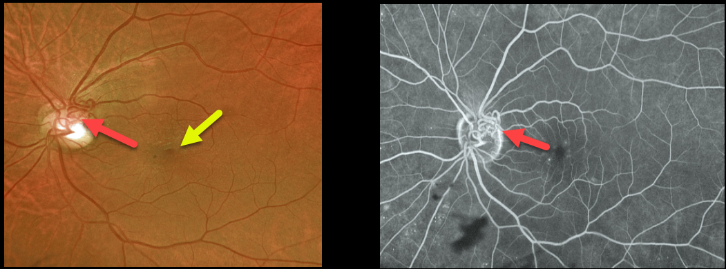
RETINAL ARTERIOVENOUS MALFORMATION
Originally posted on @retina.rocks December 9, 2021
This 67YO male with type 2 diabetes presented with these asymptomatic blood vessels overlying the left nerve, which did not leak on fluorescein angiography. Vision was 20/20 OU. Mild non-proliferative retinopathy was noted bilaterally, and there was a mild asymptomatic macular pucker in the left eye.
We felt that these vessels represented an arteriovenous malformation (AVM). The other possibility would be extremely large and complex collateral vessels, but we saw no evidence of retinal vein occlusion, clinically or angiographically.
Learning Points:
Since about 25% of patients with AVMs can have intracranial venous anomalies, a brain MRI is usually recommended. Our patient was asymptomatic, and his MRI and MRA were normal.
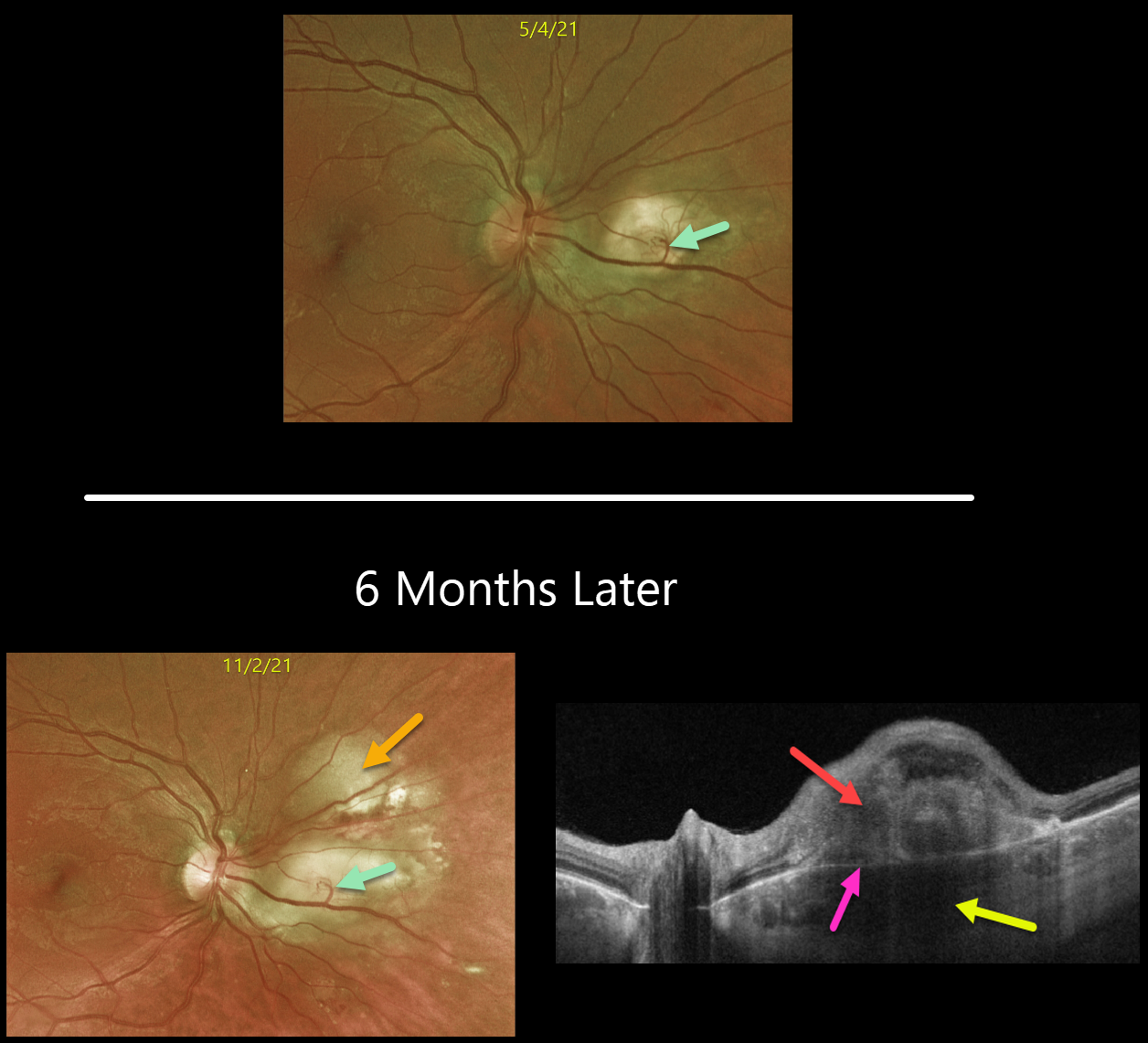
CHOROIDAL GRANULOMA
Originally posted on @retina.rocks December 8, 2021
This is a follow-up from our 6/7/21 post of a choroidal granuloma, likely representing the initial manifestation of systemic sarcoidosis. The patient was sent to pulmonology to confirm the diagnosis, but he did not keep the appointment.
Six months later, the choroidal granuloma had significantly enlarged.
A retinochoroidal anastomosis is noted between the overlying retinal vein and the lesion. Triton swept-source OCT shows a thickened, hyporeflective choroidal lesion, a hyperreflective outer retinal lesion with overlying inner retinal disorganization, and visible Bruch’s membrane separating the choroidal and retinal components.
Learning Points:
Choroidal granulomas are aggregations of inflammatory cells that appear clinically as a solitary, creamy-white, round subretinal lesion. Causes include sarcoidosis, tuberculosis, and VKH.
They are hyporeflective with increased transmission on OCT imaging (see Invernizzi et al, Retina 2015;35:525-531).
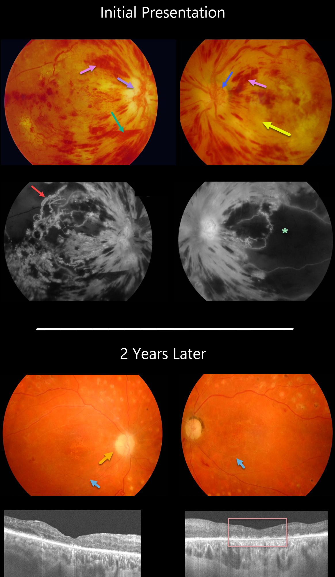
PDR + HYPERTENSIVE RETINOPATHY
Originally posted on @retina.rocks November 30, 2021
This 30YO female presented in 2008 with acute severe bilateral vision loss. She had a history of well-controlled type 1 diabetes.
There were severe bilateral retinal hemorrhages with white, ischemic maculas. A small preretinal hemorrhage was present just inferior to the right nerve. The right optic nerve was swollen. Disc neovascularization was noted in her left eye.
Fluorescein angiography showed severe macular ischemia, especially in her left eye, with near total obliteration of the normal macular vascular anatomy. Prominent staining venous beading was present in the right superotemporal macula.
Her blood pressure was severely elevated. Following emergent hypertensive control, bilateral anti-VEGF therapy was given, followed by panretinal photocoagulation.
Two years later, vision returned to 20/40 OD and 20/400 OS. The optic nerves were pale, the retinal vessels were narrowed and sheathed, and the foveas had dry pigmentary changes.
OCT scanning showed thinned, atrophic maculae with disorganized inner retinal layers (DRIL).
Learning Points:
Malignant hypertension, defined as blood pressure above 180 systolic and/or 120 diastolic, is a life-threatening medical emergency. Eye doctors are in a unique position to often diagnose this condition. Patients will often present with bilateral optic nerve swelling, flame-shaped retinal hemorrhages, and, with more chronic disease, lipid precipitates in the nasal macular outer plexiform layer.
Hypertensive retinopathy can have a similar appearance to diabetic retinopathy, and patients (like this one) can even present with signs of both.
Over time, patients with hypertensive retinopathy will develop near complete regression of all findings following systemic blood pressure treatment and control.
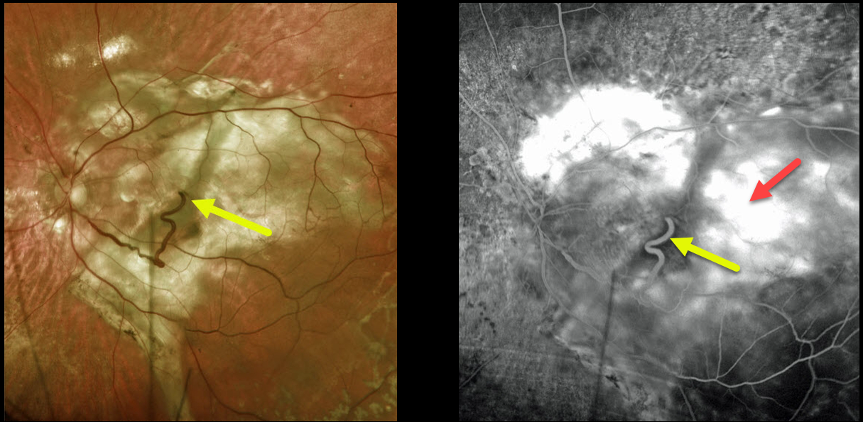
NEOVASCULAR AMD + RETINAL CHOROIDAL ANASTAMOSIS
Originally posted on @retina.rocks November 25, 2021
This 74YO female has a large complex disciform scar from end-stage age-related macular degeneration (AMD).
A very prominent retinal vein drains directly into the substance of the scar inferiorly, indicating a chorioretinal anastomosis. Fluorescein angiography shows diffuse staining of the subretinal scarring.
Learning Points:
Under normal conditions, the retinal and choroidal circulations remain separate and distinct. Rarely, the two circulations connect directly through a chorioretinal anastomosis.
These can occur in chorioretinal scars (typically from inactive toxoplasmosis lesions), in macular telangiectasia, and in disciform scars.
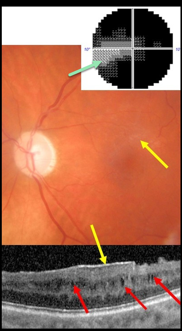
CYSTOID MACULAR EDEMA
Alex Hynes
Originally posted on @retina.rocks November 23, 2021
This 61YO male presented with end-stage glaucoma OS, 20/30 vision, and an IOP of 8 mmHg following successful trabeculectomy ten years earlier. He had prior cataract surgery in this eye 15 years ago with no documented history of pseudophakic cystoid macular edema.
There was a moderate macular pucker noted clinically and on OCT, along with multiple inner nuclear layer (INL) microcysts on OCT scanning. He has a tiny island remaining in his visual field.
Learning Points:
Microcystic macular changes have been associated with both advanced primary open-angle glaucoma and idiopathic macular pucker (see Govetto et al, AJO 2017;181:156-165).
Macular pucker-induced mechanical stress, coupled with retrograde trans-synaptic degeneration of bipolar cells secondary to retinal ganglion cell loss, likely predisposes towards the accumulation of fluid within the inner retinas of these patients.
Relative to non-glaucomatous eyes, microcystic macular changes in glaucomatous eyes tend to be more persistent following membrane peeling. This is likely due to retrograde trans-synaptic degeneration.
It is important that these microcystic spaces in eyes with optic neuropathy are not mistaken for inflammatory edema following membrane peeling, which could lead to unnecessary treatment.
Finally, disproportionate preservation of overall retinal thickness despite severe thinning of the ganglion cell and/or nerve fiber layers should not be surprising in glaucomatous eyes.
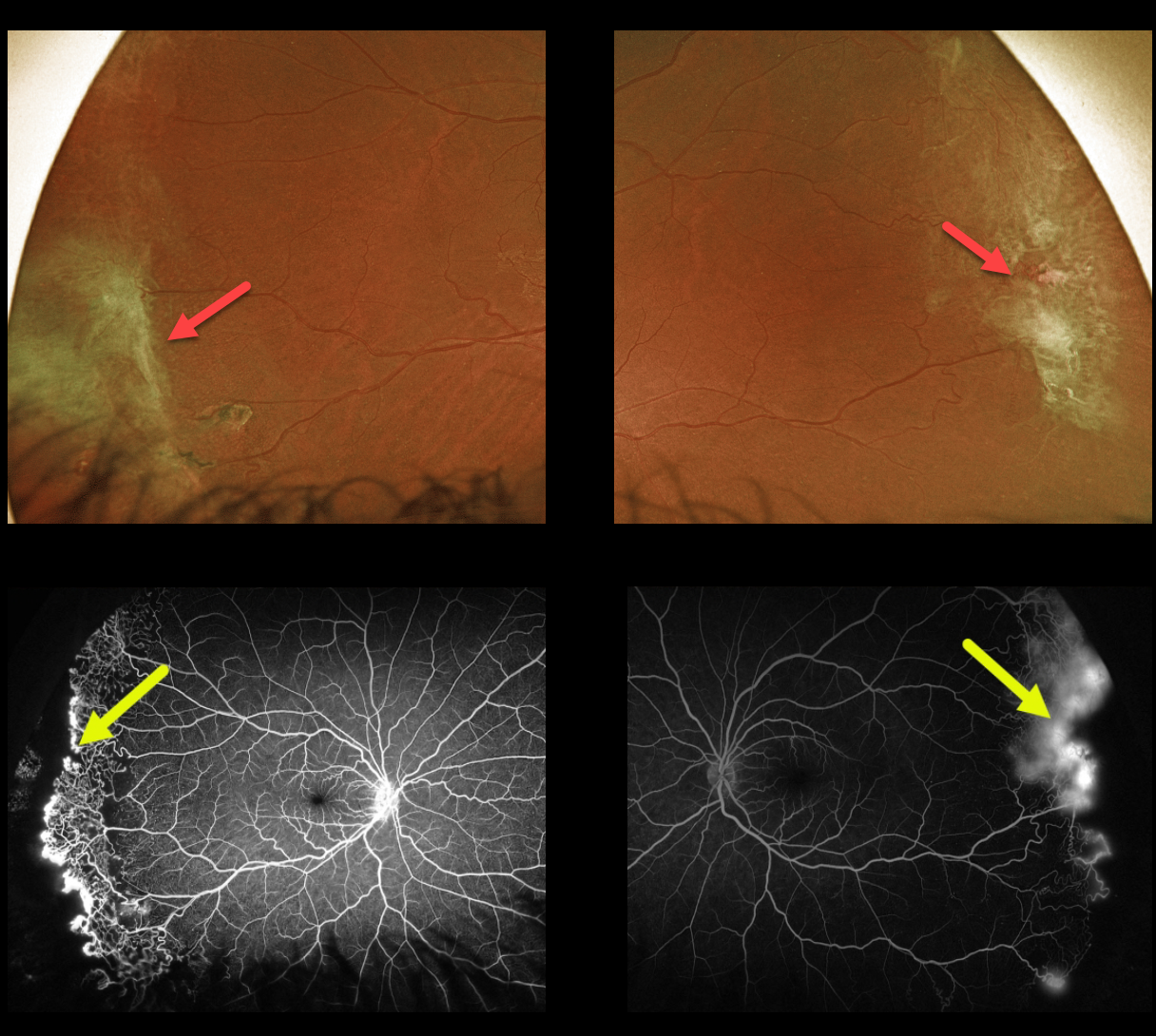
SICKLE CELL RETINOPATHY
Originally posted on @retina.rocks November 12, 2021
This 43YO female with a known history of SC disease presented with these asymptomatic fundus changes captured with Optos ultra-widefield imaging. Fibrosed areas of peripheral neovascularization are noted in each temporal periphery.
Fluorescein angiography shows a broad, well-demarcated zone of temporal peripheral retinal neovascularization at the junction of perfused and nonperfused retina.
Prophylactic scatter laser to the areas of capillary loss was recommended.
Learning Points:
Patients with sickle cell disease develop symptoms throughout their bodies as stiff, sickled red blood cells block capillary beds.
In the eye, this can cause proliferative sickle cell retinopathy (PSR), with sea-fan-shaped areas of peripheral retinal neovascularization developing at the junction of perfused and ischemic retina. Complications include vitreous hemorrhage and traction retinal detachment.
Although patients with SS disease tend to have more systemic complications, SC disease is usually associated with more severe ocular findings.
Treatment with peripheral scatter laser photocoagulation is somewhat controversial because, unlike more typical proliferative retinopathies (i.e., those from diabetes and branch retinal vein occlusions), these new vessels tend to auto-infarct.
For a great review article on sickle cell disease and the eye, see Elagouz et al, Surv Ophthalmology 2010;55:359-377.
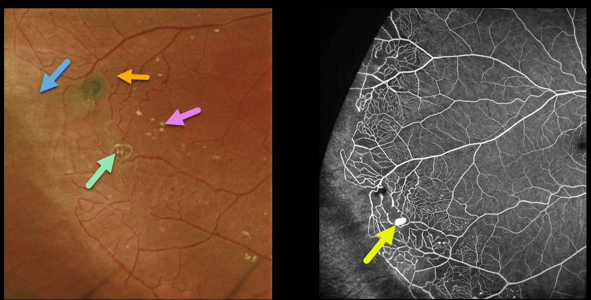
COATS DISEASE
Originally posted on @retina.rocks November 8, 2021
This healthy 13YO boy presented with these asymptomatic findings in his right eye. Vision was 20/20 OU.
Optos imaging shows classic findings for Coats disease in the temporal periphery, including telangiectatic vessels, a macroaneurysm, scattered mild lipid, and ischemic retina more distally. Fluorescein angiography more dramatically illustrates these vascular changes. The macroaneurysm stains.
Learning Points:
Historically considered a unilateral disease, more recent ultra-widefield angiographic studies show subtle vascular changes, mostly in the temporal periphery (Brockmann et al, BJO 2021;105:1444-1453). Our patient’s left eye, however, was completely normal clinically and angiographically.
Due to the lack of significant exudation, we are currently monitoring this patient twice yearly.
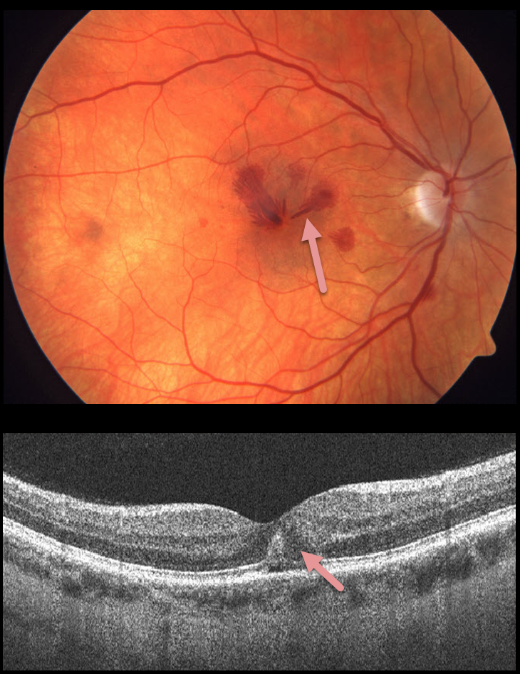
HEMORRHAGIC UNILATERAL RETINOPATHY
Originally posted on @retina.rocks October 26, 2021
This 41YO female presented with acute unilateral vision loss in her right eye. Vision was 20/50 OD and 20/20 OS. The exam revealed unilateral radiating deep Henle layer retinal hemorrhages with no identifiable systemic or ocular etiologies.
Learning Points:
Named by Kon Graversen et al (Retina 2014;34:483-489), hemorrhagic unilateral retinopathy presents with unilateral, deep intraretinal hemorrhages and sudden loss of vision ranging from 20/20 to hand motion.
There are no associated systemic conditions, nor a history of trauma or Valsalva maneuver. It is most common in females. Vision usually recovers completely within 6 months.
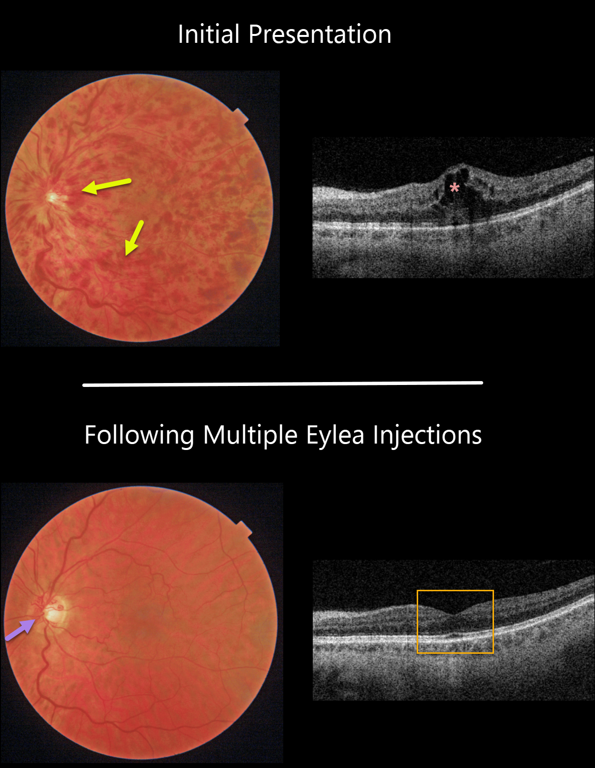
CRVO
Originally posted on @retina.rocks October 25, 2021
This 61YO male patient presented with a non-ischemic central retinal vein occlusion (CRVO), 20/30 vision, and macular edema.
With ongoing Eylea treat-extend protocol, the hemorrhages and macular edema completely resolved. Prominent optic nerve collaterals were noted.
Learning Points:
Before the introduction of intravitreal injections in the early 2000s, there were essentially no treatments for CRVO-related macular edema.
Anti-VEGF injections are now generally the initial treatment of choice, with about 50-60% of eyes gaining at least 3 lines of vision. Intravitreal steroids, usually Ozurdex, are more often used as secondary treatment.
Macular grid laser photocoagulation is not performed because it has been shown to confer no visual benefit in the Central Vein Occlusion Study Group (see Ophthalmology 1995;102;1425-1433).
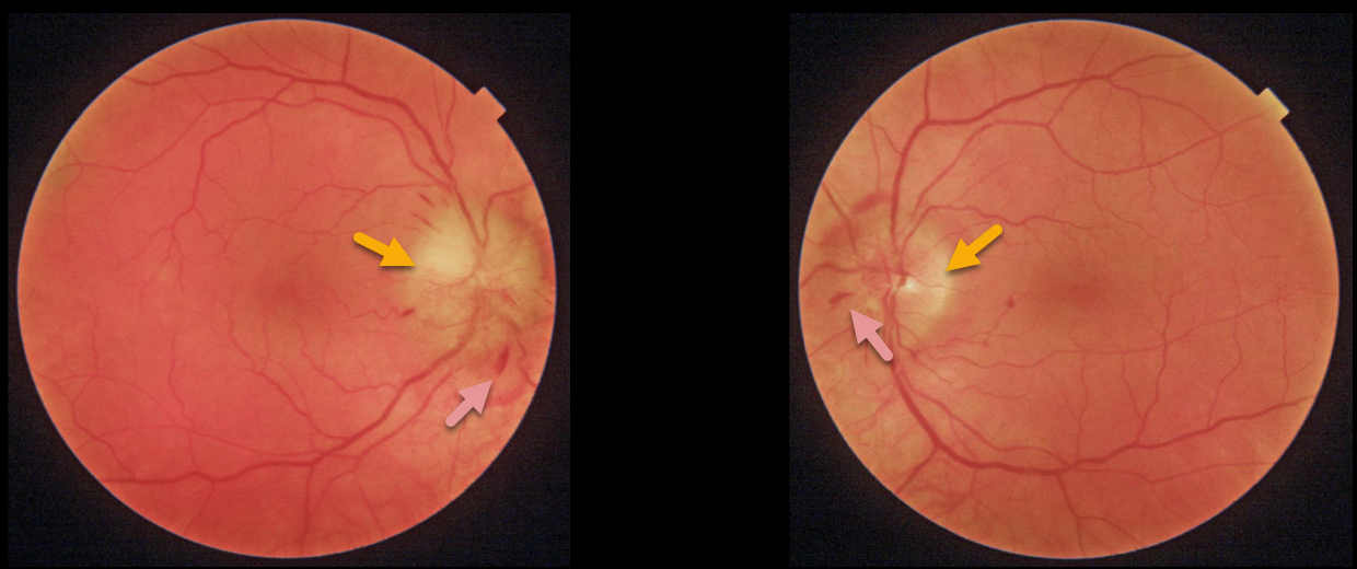
HYPERTENSIVE RETINOPATHY
Originally posted on @retina.rocks October 18, 2021
This 56YO male presented with two weeks of bilateral fuzzy vision. He gave a history of well-controlled hypertension and type 2 diabetes. Vision was 20/30 OD and 20/25 OS. Both optic nerves are swollen, and there are scattered intraretinal hemorrhages.
Blood pressure was 210/130. He was immediately sent to the nearest emergency room.
Learning Points:
Malignant hypertension, defined as blood pressure above 180 systolic and/or 120 diastolic, is a life-threatening medical emergency. Eye doctors are in a unique position to often diagnose this condition. Patients will often present with bilateral optic nerve swelling, flame-shaped retinal hemorrhages, and, with more chronic disease, lipid precipitated in the nasal macular outer plexiform layer.
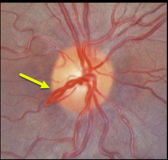
RETINAL VASCULAR LOOP
Originally posted on @retina.rocks October 13, 2021
This patient has benign congenital prepapillary vascular loops, which can be unilateral or bilateral.
Learning Points:
Although usually just an incidental finding, they can rarely cause a branch arterial or venous occlusion (see Retinal Cases Brief Rep 2014;8;124-126 and Ophthalmology 2020;127;124-126).
They should also not be confused with disc neovascularization, which consists of much smaller caliber vessels that leak on fluorescein angiography.
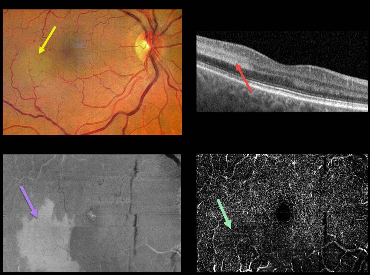
PARACENTRAL ACUTE MIDDLE MACULOPATHY (PAMM)
Lovro Soco
Originally posted on @retina.rocks October 1, 2021
This 63YO patient complained of a recent scotoma in the mid-periphery of her right eye. Vision was 20/20 OU. She gave a history of hypertension with a recent elevated BP of 200/110. She denied a history of diabetes.
Fundus photography shows a patch of deep retinal whitening in the temporal macula. There were a few scattered dot and blot retinal hemorrhages in the right eye, which may have been related to her recently elevated blood pressure.
OCT shows a well-demarcated hyperreflective region spanning the inner plexiform, inner nuclear, and outer plexiform layers. Wide-field en face OCT shows a placoid area of hyperreflectivity within this region. Wide-field OCT angiography of the deep capillary plexus shows a relative flow defect within this area.
Learning Points:
Paracentral acute middle maculopathy (PAMM) is a recently-described phenotype. It consists of acute mid-retinal ischemia found as an idiopathic entity or in other ocular disorders, including Purtscher’s, diabetic retinopathy, retinal artery and retinal vein occlusions, and acute macular neuroretinopathy.
For a great recent review, see Scharf et al, Progress in Retinal and Eye Research 2021;81;100884.
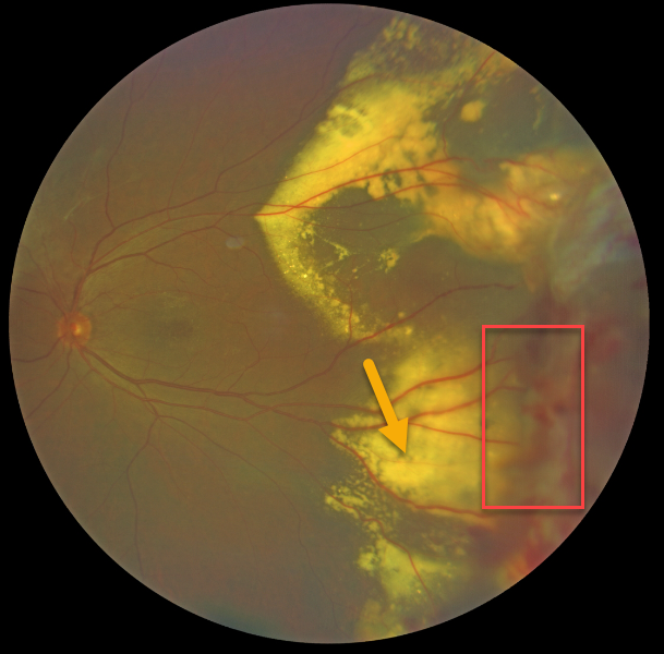
COATS DISEASE
Kumar Chugani
Originally posted on @retina.rocks September 16, 2021
This 66YO male presented with classic Coats disease findings. Vision was 20/30.
Confluent subretinal lipid extends into the peripheral temporal macula. The source for this lipid is noted in the temporal midperiphery, with dilated, irregular telangiectatic vessels with aneurysmal dilations.
Learning Points:
Laser photocoagulation can be used to treat leaking macroaneurysms and telangiectatic vessels in Coats disease. Anti-VEGF injections can also be used to stabilize vascular leakage.
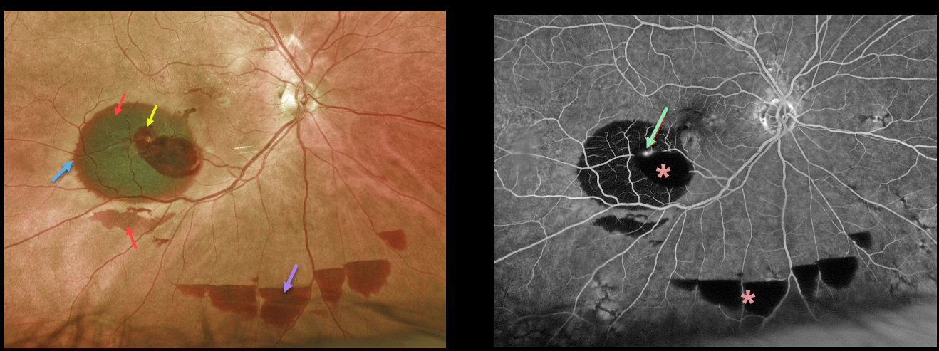
RETINAL ARTERIAL MACROANEURYSM (RAM)
Originally posted on @retina.rocks September 6, 2021
This 85YO female presented with some peripheral vision loss in her right eye. Vision was 20/40.
Optos color imaging shows areas of subretinal blood in the inferotemporal macula and below the inferotemporal arcade, intraretinal blood, and multiple areas of inferior preretinal blood. A rim of radiating outer plexiform blood surrounds the large subretinal blood.
Fluorescein angiography shows blockage from the various layers of blood, along with leakage from the causative macroaneurysm.
We recommended initial observation due to the excellent vision and ectopic blood.
Learning Points:
A retinal arterial macroaneurysm (RAM) is thought to result from a weakened arterial wall due to arteriosclerosis. A RAM appears as a yellow-white dilated lesion along a retinal arterial within the first 3 bifurcations from the optic nerve.
There are 2 distinct presentations, hemorrhagic or exudative. The hemorrhagic type often presents, as in this case, with pathognomonic preretinal, intraretinal, and subretinal blood.
Acute macular hemorrhage can be treated with observation, anti-VEGF injections, or thermal laser.
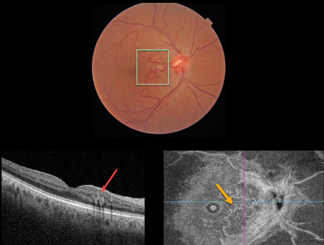
RETINAL ARTERIOVENOUS MALFORMATION
Originally posted on @retina.rocks September 3, 2021
This 66YO male presented with an asymptomatic macular arteriovenous malformation (AVM). Vision was 20/30.
Triton imaging shows an AVM involving the nasal macula. Swept-source OCT shows that the abnormal vessels lie within the deep vascular complex. En face OCT beautifully displays the abnormal vasculature.
Learning Points:
Since about 25% of patients with AVMs can have intracranial venous anomalies, a brain MRI is usually recommended.
Our patient was asymptomatic, and his MRI and MRA were normal.
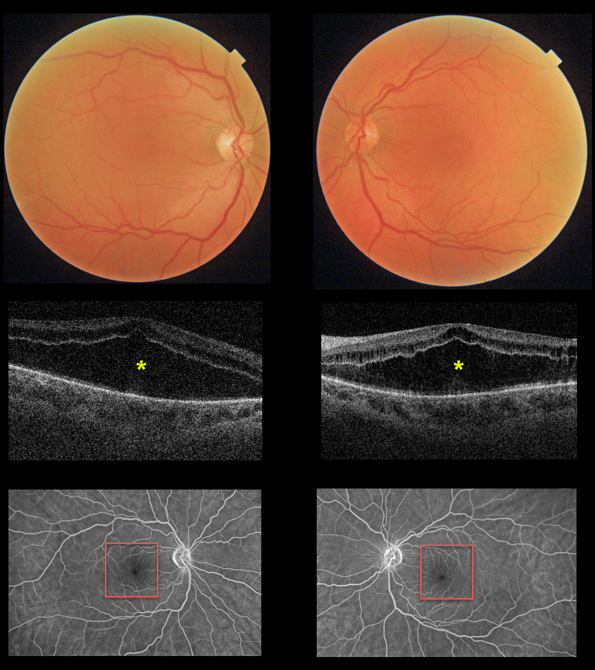
PACLITAXEL MACULOPATHY
Originally posted on @retina.rocks September 2, 2021
This 55YO female presented with an 8-month history of bilateral vision loss. She had been treated for metastatic breast cancer with Taxol (paclitaxel) for 1 year prior to this exam. Vision was 20/200 OU.
Triton imaging and swept-source OCT show severe symmetrical bilateral cystoid changes in each macula. The cystoid changes are most pronounced in the outer nuclear layer and less so in the inner nuclear layer. Fluorescein angiography shows no leakage.
We have consulted with her oncologist, and expect him to discontinue the paclitaxel. The macular changes and vision should improve within several months.
Learning Points:
Paclitaxel is a member of the taxane family of microtubule-stabilizing agents that is used to treat various solid malignant tumors. Their major adverse effects are bone marrow toxicity and peripheral neuropathy. Ophthalmic complications are rare, including taxane-related cystoid macular edema (CME).
Unlike more common exudative causes for CME, there is no angiographic leakage, and OCT shows intact and continuous inner and outer plexiform layers (see Perez et al, Graefe’s 2020;258:1607-1615).
The cause for CME is uncertain but may be due to Müller cell dysfunction (see Nakao et al, Ophthalmic Surg Lasers Retina 2016;47:81-84). There is no treatment for the CME other than stopping the causative drug.
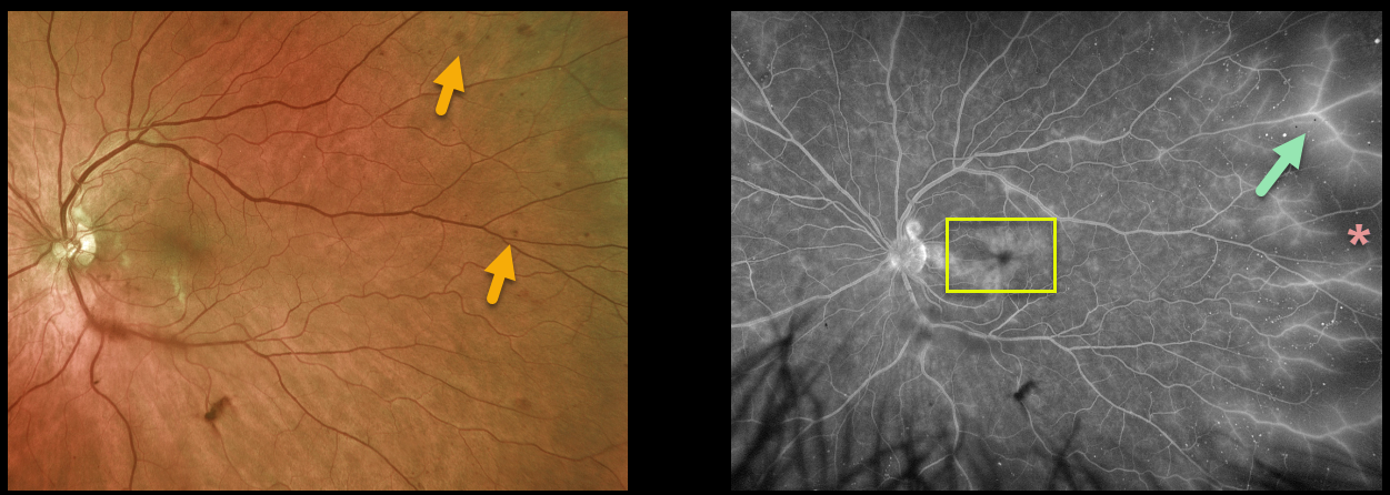
OCULAR ISCHEMIA
Originally posted on @retina.rocks August 17, 2021
This 62YO male presented with a few months of non-specific blurred vision in his left eye. Vision was 20/30 in his normal right eye and 20/40 in his left eye.
Scattered deep retinal hemorrhages were noted temporally OS. Optos fluorescein angiography showed temporal ischemia with diffuse vascular staining, along with cystoid macular edema.
On further questioning, he described a several-minute episode of transient vision loss in his left eye a few days earlier. An emergent carotid Doppler was performed, which revealed a critical stenosis of his left internal carotid artery. He was immediately referred to a vascular surgeon.
Learning Points:
The retinal hemorrhages in ocular ischemic syndrome (OIS) are unique. Retinal vascular disorders like diabetic retinopathy, retinal vein occlusions, and hypertensive retinopathy usually have flame- and dot-shaped inner retinal hemorrhages concentrated in the posterior pole.
Our patient’s findings are classic for OIS, with hemorrhages in the deeper peripheral retina. The retinal periphery is literally the end of the line for the retinal arterial circulation. In an eye receiving less blood supply due to carotid occlusive disease, the more proximal retina has first dibs on the available oxygen. There are penetrating capillaries that dive radially from the nerve fiber and ganglion cell layers into the deeper retina. The deep vascular complex that supplies the inner and outer plexiform layers is thus the furthest downstream, and these endothelial cells are likely damaged in OIS. Blood cells leaking into this space accumulate, causing the large outer blot to hemorrhage.
These patients need to have a carotid Doppler and be referred for medical or surgical intervention if they have a critical stenosis. They may also develop anterior or posterior segment neovascularization requiring photocoagulation and/or anti-VEGF injections. Iris neovascularization can develop, leading to neovascular glaucoma. Hypotony can also occur.
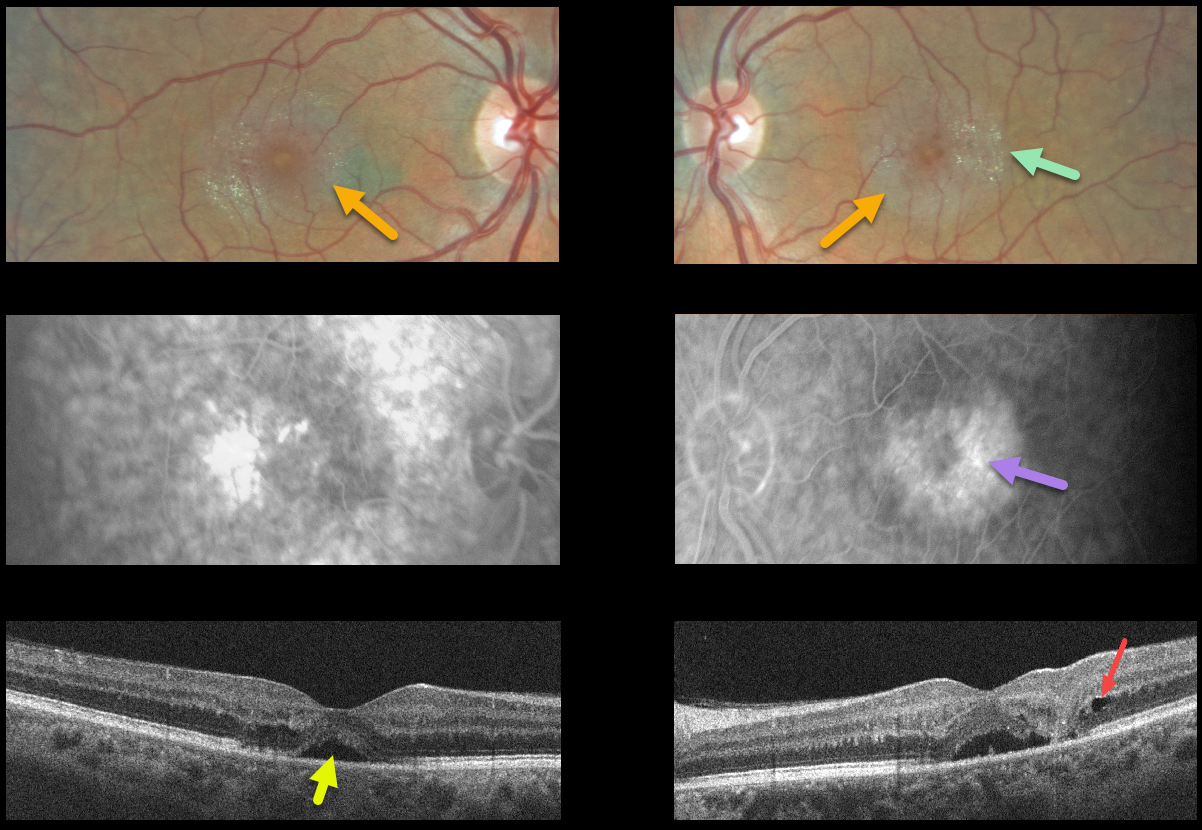
MACULAR TELANGIECTASIA
Originally posted on @retina.rocks July 30, 2021
This 52YO male presented with 20/70 vision OD, 20/100 vision OS, and many classic findings for macular telangiectasia type 2 (MacTel2).
The perifoveal retina is somewhat opaque with inner retinal crystals and deep retinal angiographic leakage.
OCT scanning of the right eye shows some temporal outer retinal atrophy with an atypical small serous detachment. OCT of the left eye shows somewhat similar findings, along with a small temporal outer nuclear layer cyst vs cavitation. The serous detachment in the left fovea may represent an atypical outer macular cavitation.
Learning Points:
Originally described by Gass (Arch Ophthalmology 1982;100:769-780), MacTel2 is a neurodegenerative disorder, most likely originating from Muller cell dysfunction.
The term telangiectasia is misleading because the funduscopic findings are mostly nonvascular. The refractile inner retinal ‘crystals’ are thought to be Muller cell footplates. The foveal retina shows a whitish discoloration, most likely due to retinal opacification resulting from Müller cell dysfunction. Photoreceptor loss allows RPE cells to migrate along intraretinal capillaries, causing the coarse clinical pigment clumping (not seen in our patient).
The majority of patients will also have subclinical chorioretinal anastomosis associated with right-angle venules (Spaide et al., Retina 2018;38:1920-1929). Secondary macular neovascularization can be successfully treated according to age-related macular degeneration protocols.
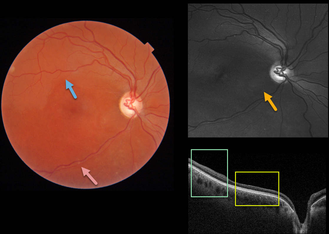
SUSAC SYNDROME
Originally posted on @retina.rocks July 20, 2021
This healthy 30YO female was referred for poor vision in her right eye. She mentioned losing vision in this eye during high school, but couldn’t provide any further details. She also gave a history of hearing loss, which had never been evaluated. Vision was HM OD and 20/30 in her normal left eye.
Triton imaging shows faint retinal whitening in the superior macula. The retinal arterioles are narrowed and somewhat sheathed inferiorly.
Swept-source OCT shows normal inner retinal architecture temporally, with inner retinal atrophy and disorganization of the normal architecture more nasally. Right inferotemporal nerve fiber loss is quite dramatic on Optos green imaging.
We diagnosed her with a resolved inferior branch retinal artery occlusion (BRAO), although this may represent Susac’s syndrome. The involved retina appeared redder than the uninvolved superior retina, accounting for the whitish superior retina noted above.
Subsequent work-up, including hypercoagulable labs and MRI, was negative. She denied prior intravenous drug use. To complete the workup, we will be obtaining carotid Doppler and cardiac ultrasonography.
Learning Points:
Susac’s syndrome, also known as retinocochleocerebral vasculopathy, is a rare microangiopathic disease characterized by a triad of encephalopathy, sensorineural hearing loss, and branch retinal artery occlusions with a predilection for younger females.
In the presence of Susac’s syndrome, MRI findings should show corpus callosum involvement. However, the full triad commonly takes months to develop, complicating diagnosis.
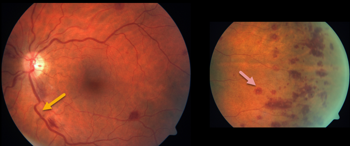
HYPERVISCOSITY WITH MONOCLONAL GAMMOPATHY
Originally posted on @retina.rocks July 8, 2021
This patient with known monoclonal gammopathy presented with bilateral engorged retinal vessels and scattered deep, white-centered retinal hemorrhages. These are most prominent in the temporal periphery.
Learning Points:
The term “Roth spot” was named after Dr. Moritz Roth, who, in 1872, noted white-centered hemorrhages in patients with bacterial endocarditis. He felt that these white spots were septic emboli that originated from an infected cardiac valve.
However, similar lesions can be seen in many other disorders, including leukemia, hyperviscosity, anemia, diabetes, and hypertension. The white material may represent a variety of causes, including fibrin.
So these lesions are best called white-centered hemorrhages, unless one is specifically referring to a lesion in a patient with bacterial endocarditis. See Duane et al Ophthalmology 1980;87;66-69.
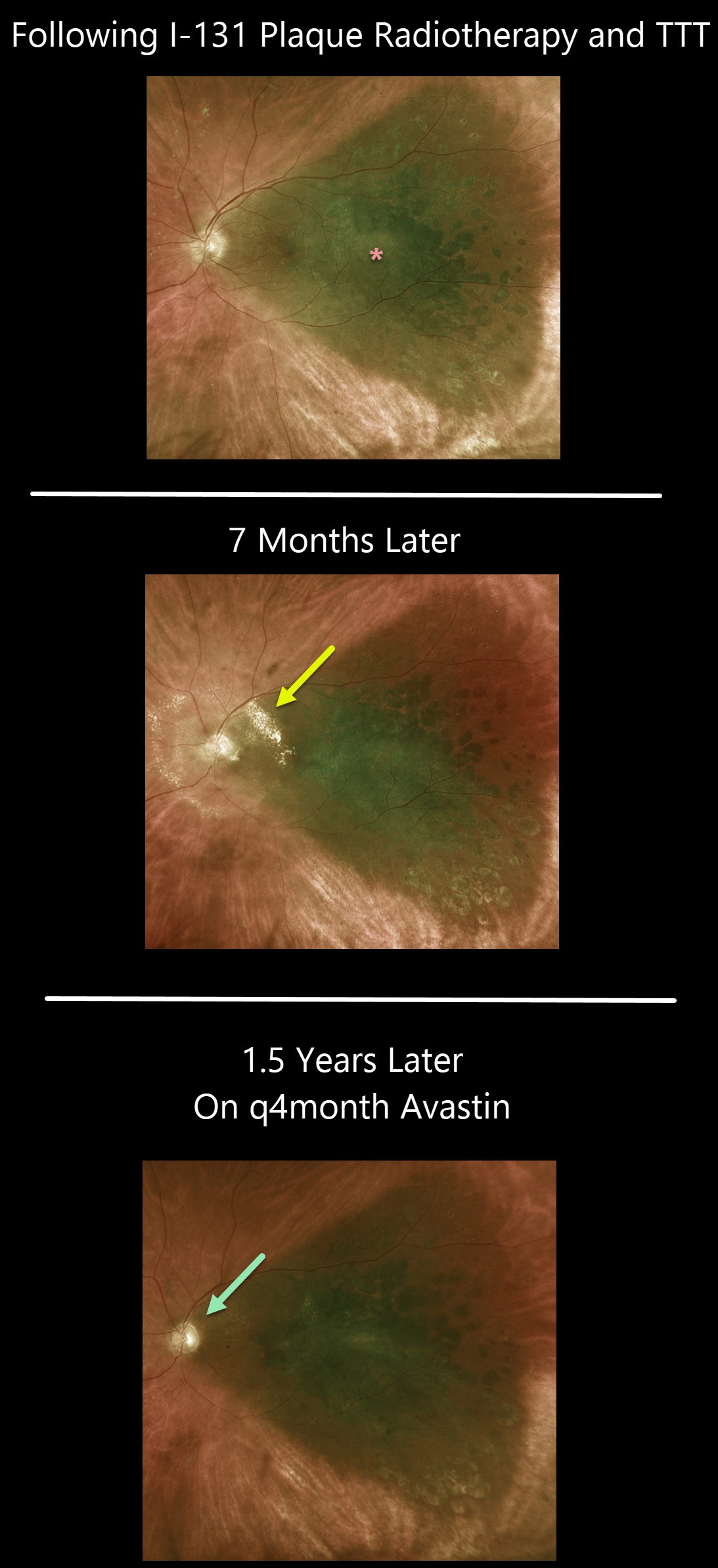
RADIATION PAPILLOPATHY
Originally posted on @retina.rocks July 2, 2021
This 66YO male was examined following I-131 plaque radiotherapy and transpupillary thermotherapy for a type 1A, PRAME+ uveal malignant melanoma.
Vision was 20/80, and the tumor was regressing compared to its preoperative appearance (not shown). He was getting intravitreal injections every 4 months as prophylaxis against radiation retinopathy.
The patient returned 8 months later for an overdue examination with new peripapillary lipid from radiation papillopathy. The intravitreal Avastin injections were restarted, and the papillopathy gradually resolved.
Learning Points:
The incidence of radiation retinopathy is relatively high following plaque therapy for uveal malignant melanoma. Ongoing intravitreal injections greatly reduce this complication (see Fallico et al, Surv Ophthalmology 2021;66;441-460).
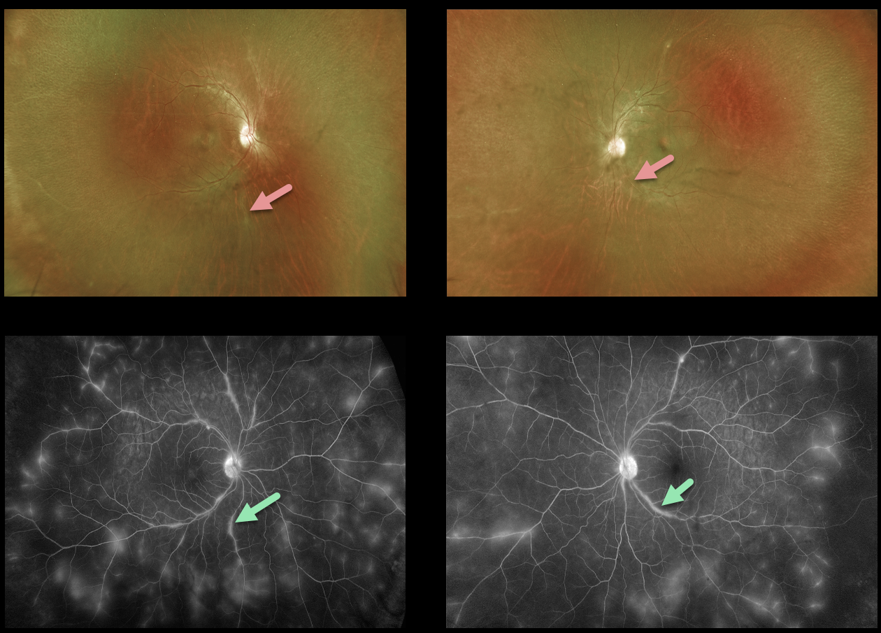
IDIOPATHIC RETINAL VASCULITIS ANEURYSMS AND NEURORETINITIS (IRVAN)
Originally posted on @retina.rocks July 1, 2021
This 41YO female presented with vision of 20/70 OD and 20/25 OS from bilateral retinal vasculitis, highlighted well on fluorescein angiogram.
Extensive workup was negative, including sarcoidosis (chest X-ray, ACE, lysozyme), ANCA, ANA, HLA-B27, Lyme, and RPR. Following the negative labs, she was started on oral prednisone 20mg/day.
Learning Points:
Idiopathic Retinitis, Vasculitis, Aneurysms, and Neuroretinitis (IRVAN) is a rare disease of unknown etiology characterized by retinal vasculitis, aneurysmal arterial dilations, and neuroretinitis. The diagnosis is clinical, as there are no systemic associations or diagnostic laboratory tests.
Retinal vasculitis can be seen in a number of conditions, including pars planitis, frosted branch angiitis, lupus vasculitis, Behcet’s, and idiopathic retinal vasculitis aneurysms and neuroretinitis (IRVAN).
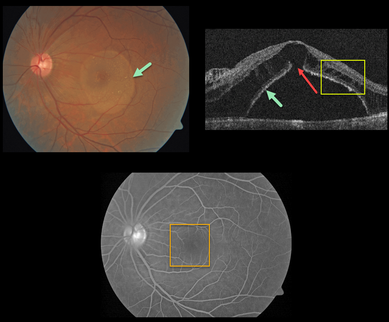
EXUDATIVE RETINAL DETACHMENT WITH OUTER MACULAR HOLE
Originally posted on @retina.rocks June 25, 2021
This patient presented with a sharply-circumscribed macular serous detachment, cystic outer retinal edema, and an outer macular hole. Fluorescein angiography is essentially normal without leakage.
An optic pit was absent clinically and on OCT imaging. Serum protein electrophoresis was normal, and a systemic cancer workup was negative.
Learning Points:
Serous detachment can be an uncommon ocular manifestation of paraproteinemia (see Mansour et al, Ophthalmology 2014;121;1925-1932). These detachments, which resemble those seen in optic pit maculopathy, are characterized by a well-defined serous detachment without angiographic leakage. A central outer macular hole is often present.
The fluid is likely due to an osmotic gradient generated by subretinal immunoglobulins that passively migrate into the subretinal space.
We do not know the cause of our patient’s findings, given his lack of an optic pit and a negative systemic workup.
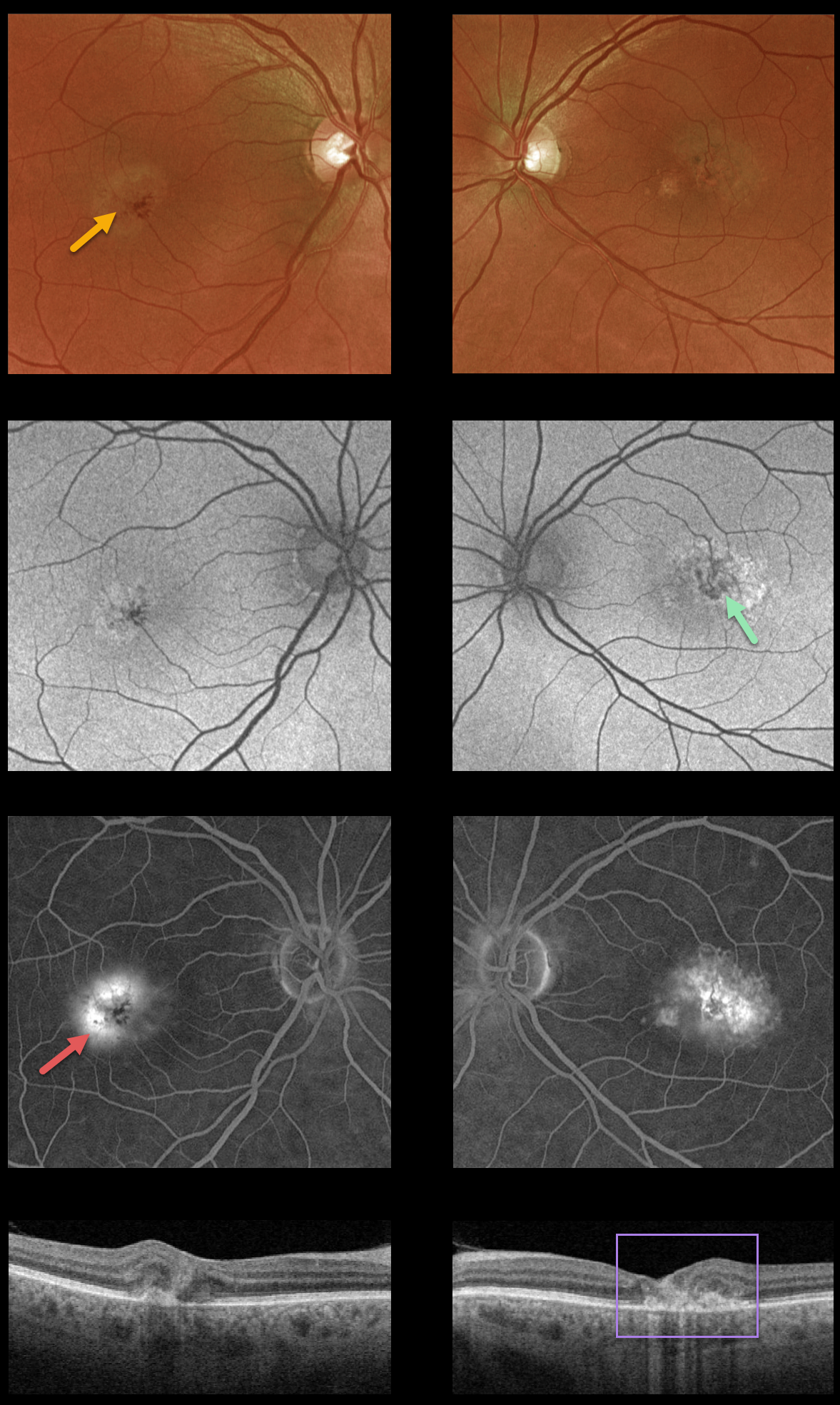
MACULAR TELANGIECTASIA
Originally posted on @retina.rocks May 14, 2021
This 55YO male presented with macular telangiectasia type 2 (MacTel2). He was visually asymptomatic, and vision was 20/40 OD and 20/50 OS.
This case highlights various multimodal imaging findings. The foveal retina shows a whitish discoloration, most likely due to retinal opacification resulting from Müller cell dysfunction.
There are variable macular hyper- and hypofAF abnormalities, with angiographic staining and leakage. OCT scanning shows mostly temporal foveal outer retinal atrophy and disorganization.
Characteristic MacTel2 features absent in our patient include refractile inner retinal ‘crystals’ (thought to be Muller cell footplates, similar to the refractile dots found in retinoschisis) and coarse black clumps of intraretinal pigment migration.
Learning Points:
Originally described by Gass (Arch Ophthalmology 1982;100:769-780), MacTel2 is a neurodegenerative disorder, most likely originating from Muller cell dysfunction. The term telangiectasia is misleading because the funduscopic findings are mostly nonvascular.
Although much remains to be learned about its pathophysiology and there is no treatment for the underlying disease, secondary macular neovascularization can be successfully treated according to age-related macular degeneration protocols.
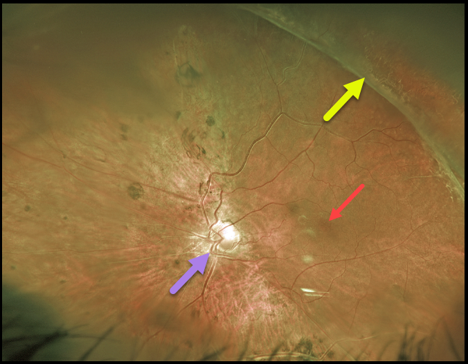
RETINOPATHY OF PREMATURITY
Originally posted on @retina.rocks May 13, 2021
For over 20 years, we’ve been following this 39YO male with stable regressed retinopathy of prematurity in his left eye. Vision is NLP OD and 20/80 OS.
Optos imaging shows that the macular center is dragged temporally due to vitreoretinal traction from regressed neovascularization.
The macular center is usually about 2 disc diameters temporal to the edge of the optic nerve, and our patient’s is about 3 disc diameters. This dragging is usually accompanied by a straightening of the temporal arcades.
However, in our patient, this is masked by situs inversus, a congenital abnormality in which the retinal vessels exit the optic nerve with a nasal bend before coursing temporally.
Learning Points:
These outcomes can be minimized through aggressive neonatal screening and treatment with laser photocoagulation and anti-VEGF agents.
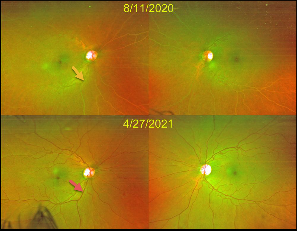
LIPEMIA RETINALIS
Grace Wong
Originally posted on @retina.rocks May 10, 2021
This 49YO female’s retinal vasculature was initially creamy white from very high triglycerides.
After 8 months of systemic treatment, her triglyceride levels normalized, her blood turned back to its normal red color, and her retinal vessels normalized in appearance.
Learning Points:
Lipemia retinalis is a rare retinal manifestation of severe hypertriglyceridemia, characterized by creamy white discoloration of the retinal vasculature. In severe cases, the fundus can turn salmon-colored.
Patients are asymptomatic, and vision is normal. However, they are at obvious cardiovascular risk without systemic treatment.
BABY YODA MACULOPATHY
Originally posted on @retina.rocks May 4, 2021
In a galaxy far, far away, this wife of Retina Rock’s founder had an unexpected visitor following her cataract surgery.
Grogu, otherwise known as Baby Yoda or The Child, decided to take refuge within her retina as cystoid macular edema (CME). Thankfully, for the mental health of both the patient and her retinal specialist husband, the CME resolved following topical steroids and non-steroidals.
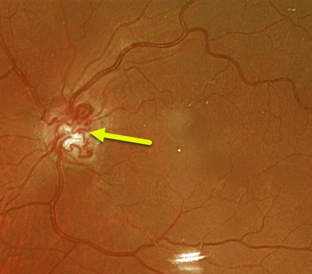
CRVO
Originally posted on @retina.rocks April 22, 2021
This 74YO female has large optic nerve collaterals from an old non-ischemic central retinal vein occlusion. Vision was 20/30.
Learning Points:
Collateral vessels are dilated, pre-existing vessels that bypass an occlusion. These are most commonly seen in the posterior pole, following retinal venous occlusions, often on the optic disc.
In collateral vessels, the endothelial tight junctions are intact, so they don’t leak angiographically. This helps differentiate them from neovascularization, which does leak.
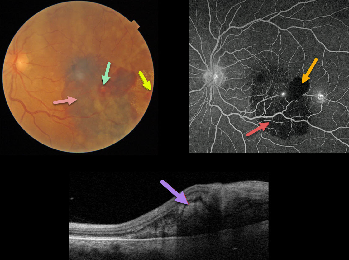
RETINAL ARTERIAL MACROANEURYSM (RAM)
Originally posted on @retina.rocks April 8, 2021
This 68YO female presented with acute vision loss in her left eye. Vision was hand motion from submacular blood.
A retinal arterial macroaneurysm (RAM) is noted overlying the blood, with an additional RAM with lipid more temporally.
Triton swept-source OCT scanning shows subretinal blood beneath the RAM. Fluorescein angiography shows blockage from the subretinal and intraretinal blood, with leakage from both lesions. These multiple layers of blood (subretinal and intraretinal) are classic for RAMs.
In this case, the patient elected for treatment with anti-VEGF injections.
Learning Points:
RAMs present with either acute blood or with chronic exudation, and this case is unique in that we can see both presentations in the same eye.
Acute macular hemorrhage can be treated with observation, anti-VEGF injections, or thermal laser. Macular edema with lipid exudate that threatens or involves the macular center usually requires either thermal laser treatment or anti-VEGF injections.
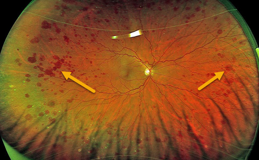
OCULAR ISCHEMIA
Originally posted on @retina.rocks April 7, 2021
This patient presented with these asymptomatic, unilateral, peripheral, large, outer retinal hemorrhages.
The patient was later diagnosed with a 100% internal carotid artery occlusion. Carotid surgery was not recommended, but we are monitoring the patient due to the risk for anterior or posterior segment neovascularization.
Learning Points:
The retinal hemorrhages in ocular ischemic syndrome (OIS) are somewhat unique. Retinal vascular disorders like diabetic retinopathy, retinal vein occlusions, and hypertensive retinopathy usually have flame- and dot-shaped inner retinal hemorrhages concentrated around the posterior pole.
Our patient’s findings are classic for OIS, with hemorrhages in the deeper peripheral retina. The retinal periphery is literally the end of the line for the retinal arterial circulation. In an eye receiving less blood supply due to carotid occlusive disease, the more proximal retina has first dibs on the available oxygen. There are penetrating capillaries that dive radially from the nerve fiber and ganglion cell layers into the deeper retina. The deep vascular complex that supplies the inner and outer plexiform layers is thus furthest downstream, and these endothelial cells are likely damaged in OIS. Blood cells leaking into this space accumulate, causing the large outer blot to hemorrhage.
These patients need to have a carotid Doppler and ultrasound and be referred for medical or surgical intervention if they have a critical stenosis. They may also develop anterior or posterior segment neovascularization requiring photocoagulation and/or anti-VEGF injections. Iris neovascularization can develop, leading to neovascular glaucoma. Hypotony can also occur.
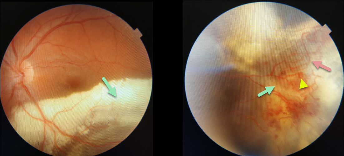
COATS DISEASE
Originally posted on @retina.rocks April 6, 2021
This 20YO male presented with classic Coats disease findings. Confluent subretinal lipid extends into the inferior macula, sparing the foveal center, allowing for 20/30 vision.
The source for this lipid is noted in the inferotemporal midperiphery with dilated, irregular vessels, aneurysmal dilations, and underlying subretinal fluid and lipid.
Learning Points:
Laser photocoagulation can be used to treat leaking macroaneurysms and telangiectatic vessels in Coats disease. Anti-VEGF injections can also be used to stabilize vascular leakage.
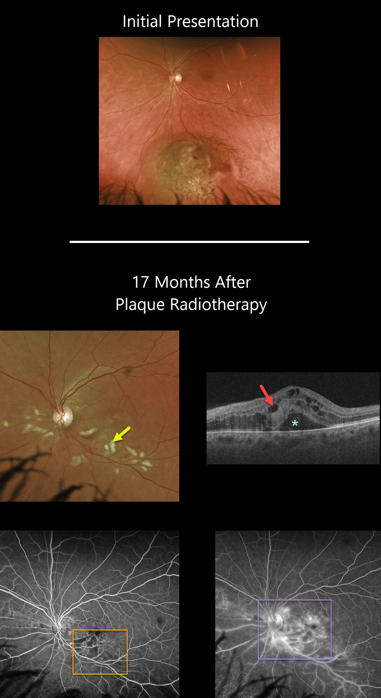
UVEAL MELANOMA
Originally posted on @retina.rocks March 29, 2021
Seventeen months following plaque radiotherapy for a uveal malignant melanoma, this patient developed radiation retinopathy with scattered inner retinal hemorrhages and nerve fiber layer infarcts (cotton wool spots). Vision was counting fingers, although it had been 20/40 ten days following plaque therapy a year and a half earlier.
Cystoid macular edema with subretinal fluid is noted on Triton swept-source OCT. Fluorescein angiography shows areas of capillary dropout, telangiectasias, and hyperfluorescent leakage secondary to cystoid macular edema.
Learning Points:
Radiation retinopathy usually occurs 3 months to 3 years after external beam radiation (e.g., for facial or orbital cancers) or plaque radiotherapy. The ocular findings closely resemble those seen with diabetic retinopathy, as in our patient, who is receiving Avastin injections for the macular edema.
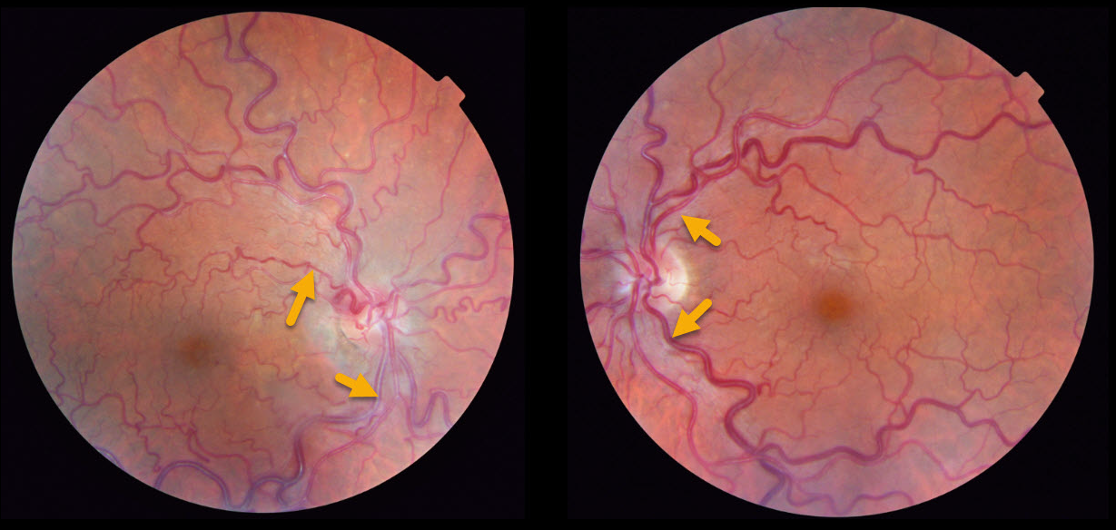
RETINAL VASCULAR TORTUOSITY
Originally posted on @retina.rocks March 18, 2021
This patient presented with asymptomatic bilateral retinal venous and arterial vascular tortuosity. There was no significant past medical or family history.
Learning Points:
This appearance must be differentiated from acquired retinal vascular tortuosity caused by retinal venous occlusive disease, hyperviscosity (polycythemia, dysproteinemia, leukemia etc.), Fabry’s disease, and fetal alcohol syndrome.
Familial retinal arteriolar tortuosity, a rare autosomal dominant disorder, affects the macular and peripapillary second and third order arterioles, and can rarely develop symptomatic vitreous hemorrhage (see Sutter and Helbig Surv Ophthalmology 2003;48:245-255).
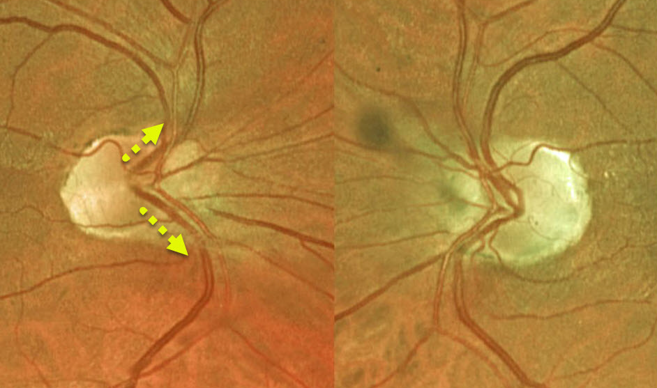
SITUS INVERSUS
Originally posted on @retina.rocks March 17, 2021
Situs inversus is a congenital abnormality where the retinal vessels exit the optic nerve with a nasal bend prior to heading temporally.
Learning Points:
Situs inversus occurs in 1 to 2% of the population and is sometimes associated with a tilted optic disc.
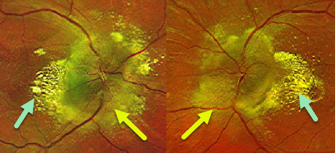
HYPERTENSIVE RETINOPATHY
Originally posted on @retina.rocks March 9, 2021
This 24YO male presented with these fundus findings. Both optic nerves were severely and diffusely swollen with a lipid star in the nasal macula. Blood pressure in the office was 224/145.
Learning Points:
The nasal macular lipid star originated from the optic nerve, where fluid and lipoproteins leaked into the surrounding tissues.
When the fluid is absorbed, the lipid precipitates out in Henle’s layer. This is also seen in other causes of optic nerve swelling or inflammation, including neuroretinitis.
The lipid supranasal to the right optic nerve is a bit unusual.
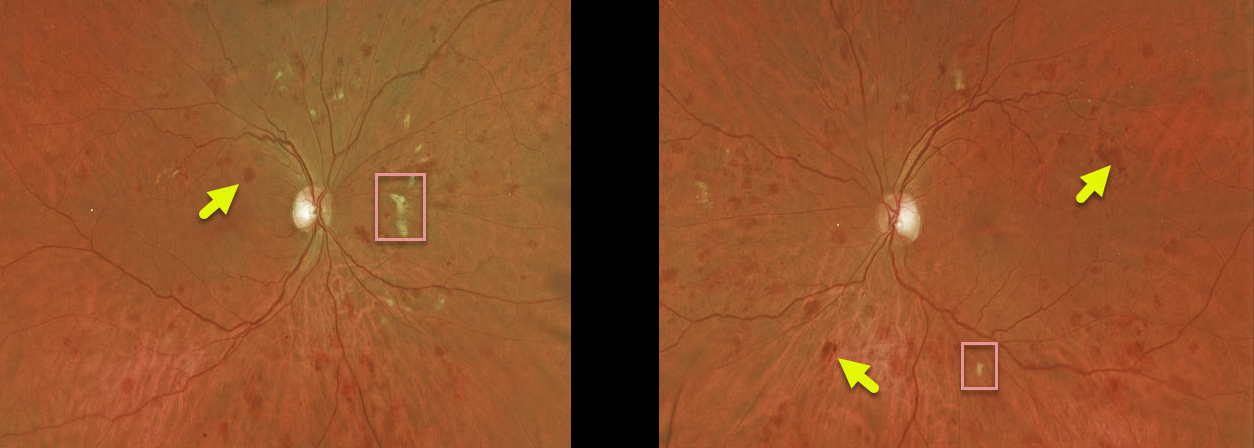
LEUKEMIA
Originally posted on @retina.rocks February 22, 2021
This patient with acute myeloid leukemia (AML) was referred by their oncologist for an eye examination following a failed allogeneic stem cell transplant due to graft versus host disease. Vision was 20/30 OD and 20/25 OS. His WBC was 14.7 (with 32% marrow myeloblasts), hemoglobin was 8.2, and platelet count was 23,000.
This patient has large, deep blot hemorrhages in the outer plexiform layer as well as scattered cotton-wool spots, indicative of leukemic retinopathy.
Learning Points:
These deeper hemorrhages would be unusual for the more common disorders seen in clinical practice (including diabetic retinopathy, retinal vein occlusions, and hypertensive retinopathy) and are usually indicative of blood dyscrasias, including anemia and hematologic malignancies. These deeper hemorrhages are also seen peripherally in ocular ischemia.
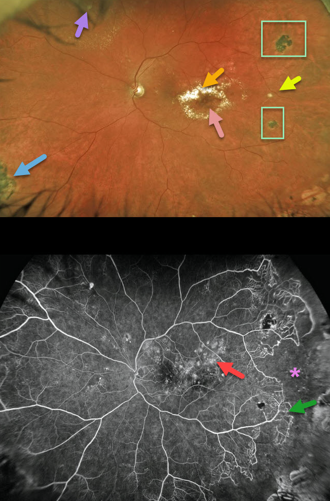
COATS DISEASE
Originally posted on @retina.rocks February 12, 2021
This 53YO has classic findings of Coats disease. A leaking macroaneurysm in the distal inferotemporal macula is causing exudation with lipid threatening the macular center. Prior macular laser scars are noted in the superior macula.
There is a fibrosed macroaneurysm in the temporal midperiphery, and some fibrosed telangiectatic lesions are visible in the superonasal periphery. Scattered peripheral hyperpigmented laser scars are also seen.
Fluorescein angiography nicely shows these findings and more, including macular telangiectasia with staining laser scars, some patches of mid-peripheral telangiectasia and capillary loss, and a large region of temporal capillary loss.
Learning Points:
Laser photocoagulation can be used to treat leaking macroaneurysms and telangiectatic vessels in Coats disease. Anti-VEGF injections can also be used to stabilize vascular leakage.
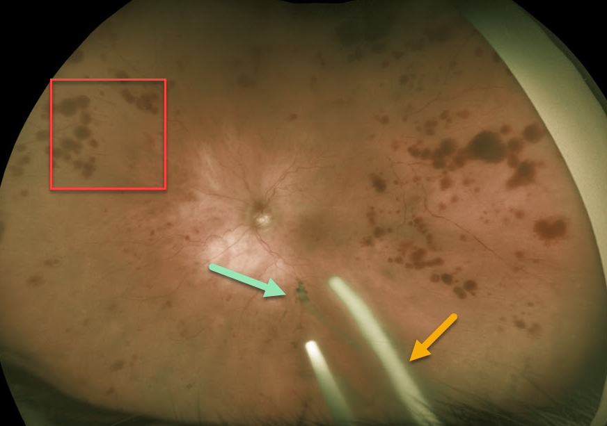
OCULAR ISCHEMIA WITH OZURDEX IMPLANTS
Originally posted on @retina.rocks February 4, 2021
This 86YO patient was receiving Ozurdex (dexamethasone 0.7%) injections every 8 weeks in her left eye for macular edema from an inferior hemiretinal vein occlusion.
She subsequently developed ocular ischemic syndrome (OIS) like changes with scattered peripheral large outer retinal hemorrhages. Inferiorly, more recent Ozurdex implants are noted, along with another ghost-like remnant of an older implant. Although the Ozurdex implants are supposed to completely degrade, these remnants may persist beyond 1 year (see Kim et al Retina 2020;40;2226-2231).
Learning Points:
The retinal hemorrhages in ocular ischemic syndrome (OIS) are unique. Retinal vascular disorders like diabetic retinopathy, retinal vein occlusions, and hypertensive retinopathy usually have flame- and dot-shaped inner retinal hemorrhages concentrated around the posterior pole.
Our patient’s findings are classic for OIS, with hemorrhages in the deeper peripheral retina. The retinal periphery is literally the end of the line for the retinal arterial circulation. In an eye receiving less blood supply due to carotid occlusive disease, the more proximal retina has first dibs on the available oxygen. There are penetrating capillaries that dive radially from the nerve fiber and ganglion cell layers into the deeper retina. The deep vascular complex that supplies the inner and outer plexiform layers is thus furthest downstream, and these endothelial cells are likely damaged in OIS. Blood cells leaking into this space accumulate, causing the large outer blot to hemorrhage.
These patients need to have a carotid Doppler and ultrasound and be referred for medical or surgical intervention if they have a critical stenosis. They may also develop anterior or posterior segment neovascularization requiring photocoagulation and/or anti-VEGF injections. Iris neovascularization can develop, leading to neovascular glaucoma. Hypotony can also occur.
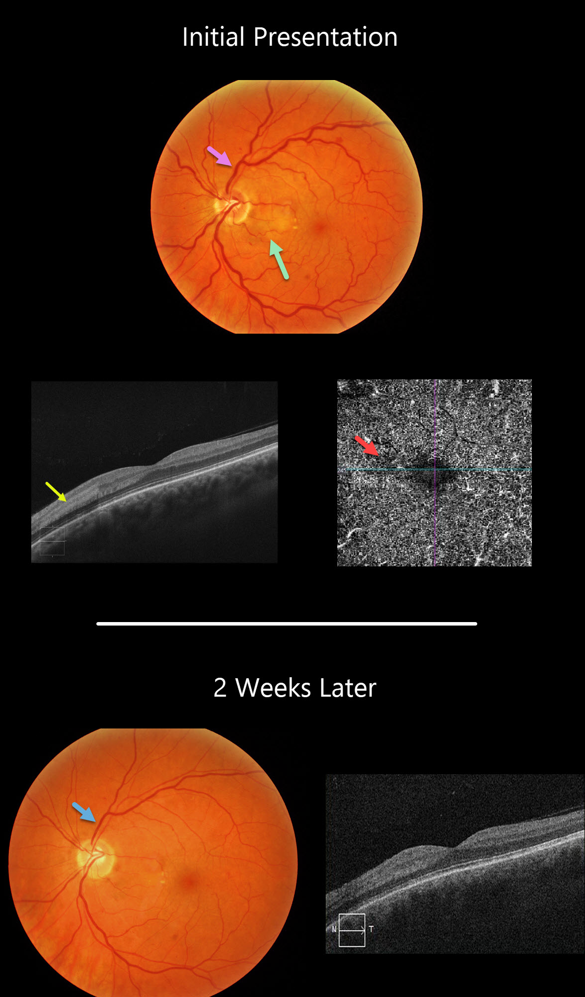
PARACENTRAL ACUTE MIDDLE MACULOPATHY (PAMM)
Originally posted on @retina.rocks February 3, 2021
This 45YO female presented with 20/40 vision in her left eye with an acute negative scotoma temporal to her visual axis. This corresponded with a focal area of retinal whitening in the nasal macula.
This appeared as hyperreflectivity of the inner plexiform, inner nuclear, and outer plexiform layers on OCT and a relative flow void on OCT angiography.
Two weeks later, the retina and OCT findings improved, and vision was 20/30. This may have represented a mild, spontaneously resolving central retinal vein occlusion, as the initial fundus photo shows very mild, scattered retinal hemorrhages with dilated veins, and the venous caliber normalized at her exam 2 weeks later.
Learning Points:
Paracentral acute maculopathy (PAMM) is a recently described phenotype.
It consists of acute mid-retinal ischemia found as an idiopathic entity or in other ocular disorders, including Purtscher’s, diabetic retinopathy, retinal artery and retinal vein occlusions, and acute macular neuroretinopathy.
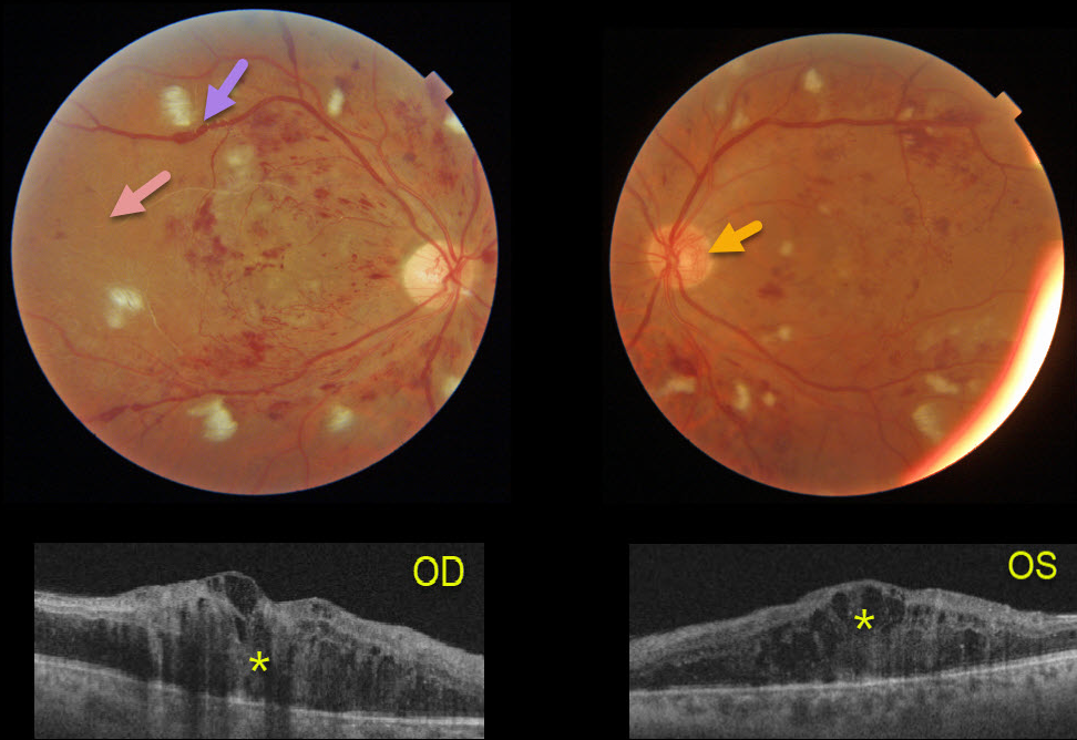
HYPERTENSION + PROLIFERATIVE DIABETIC RETINOPATHY (PDR)
Originally posted on @retina.rocks February 2, 2021
This 41YO male presented after a recent hospitalization for severe hypertension with vision of counting fingers OD and 20/60 OS.
The bilateral nerve fiber layer infarcts (cotton-wool spots) are more characteristic of hypertension than of diabetes.
Severe retinal hemorrhages and disc neovascularization are noted in each eye. The right eye, in particular, is severely ischemic, with Triton imaging showing a featureless, ischemic retina and white vessels, most pronounced in the temporal retina.
The venous sausaging is a clinical sign of severe ischemia, and when present, virtually always shortly precedes or is indicative of concurrent proliferative disease. Triton swept-source OCT shows severe bilateral center-involving diabetic macular edema.
He subsequently received multiple bilateral anti-VEGF injections followed by panretinal photocoagulation, with marked improvement of the central edema.
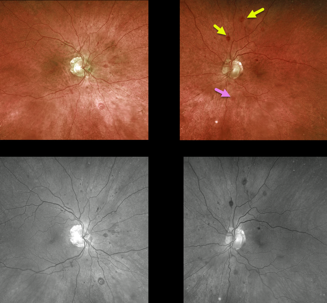
MULTIPLE MYELOMA
Originally posted on @retina.rocks January 26, 2021
This 76YO male presented with normal vision and asymptomatic bilateral retinal hemorrhages following a recent diagnosis of multiple myeloma.
There are a variety of retinal hemorrhages seen on Optos and green-channel images, including inner retinal white-centered hemorrhages and deep blot hemorrhages in the outer plexiform layer.
Learning Points:
White-centered hemorrhages or “roth spots” can be seen in many disorders, including leukemia, hyperviscosity, anemia, diabetes, and hypertension. The white material may represent a variety of causes, including fibrin.
The term “Roth spots” should thus be reserved only for patients with bacterial endocarditis. See Duane et al Ophthalmology 1980;87;66-69.
The deeper hemorrhages would be unusual for the more common disorders seen in clinical practice (including diabetic retinopathy, retinal vein occlusions, and hypertensive retinopathy), and are usually indicative of blood dyscrasias, including anemia and hematologic malignancies. These deeper hemorrhages are also seen peripherally in ocular ischemia.
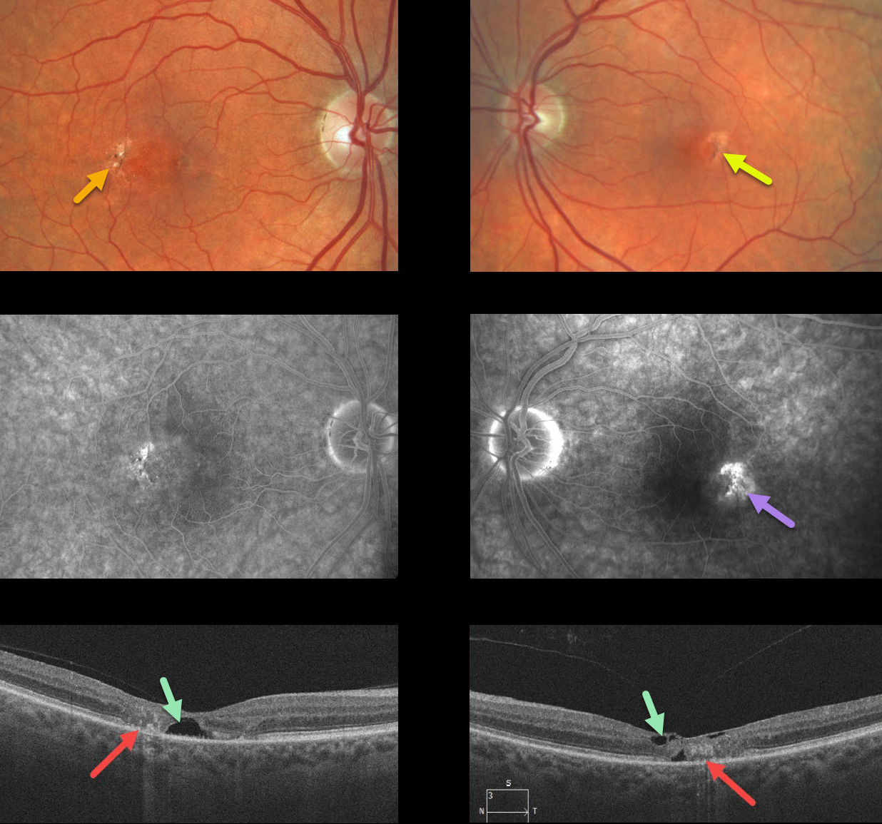
MACULAR TELANGIECTASIA
Originally posted on @retina.rocks January 22, 2021
Our patient shows classic bilateral findings of macular telangiectasia type 2 (MacTel2) with temporal foveal intraretinal pigment migration and inner retinal crystals, as well as angiographic staining and leakage.
Our patient’s optical coherence tomography scans also show classic findings of mostly temporal foveal retinal atrophy, disorganization, and hyporeflective tissue loss (cavitations).
Learning Points:
MacTel type 2 is a neurodegenerative disorder, most likely originating from Müller cell dysfunction. The term telangiectasia is misleading because the funduscopic findings are mostly nonvascular.
It is thought that photoreceptor loss allows RPE cells to migrate along intraretinal capillaries, creating the pigment clumping. This is a similar mechanism to that found in other causes of intraretinal pigment migration, including photoreceptor loss or damage, such as retinitis pigmentosa, chronic retinal detachment, and blunt trauma.
The refractile inner retinal ‘crystals’ are thought to be Muller cell footplates, similar to the refractile dots found in retinoschisis.
Although much remains to be learned about its pathophysiology and there is no treatment for the underlying disease, secondary macular neovascularization can be successfully treated according to age-related macular degeneration protocols.
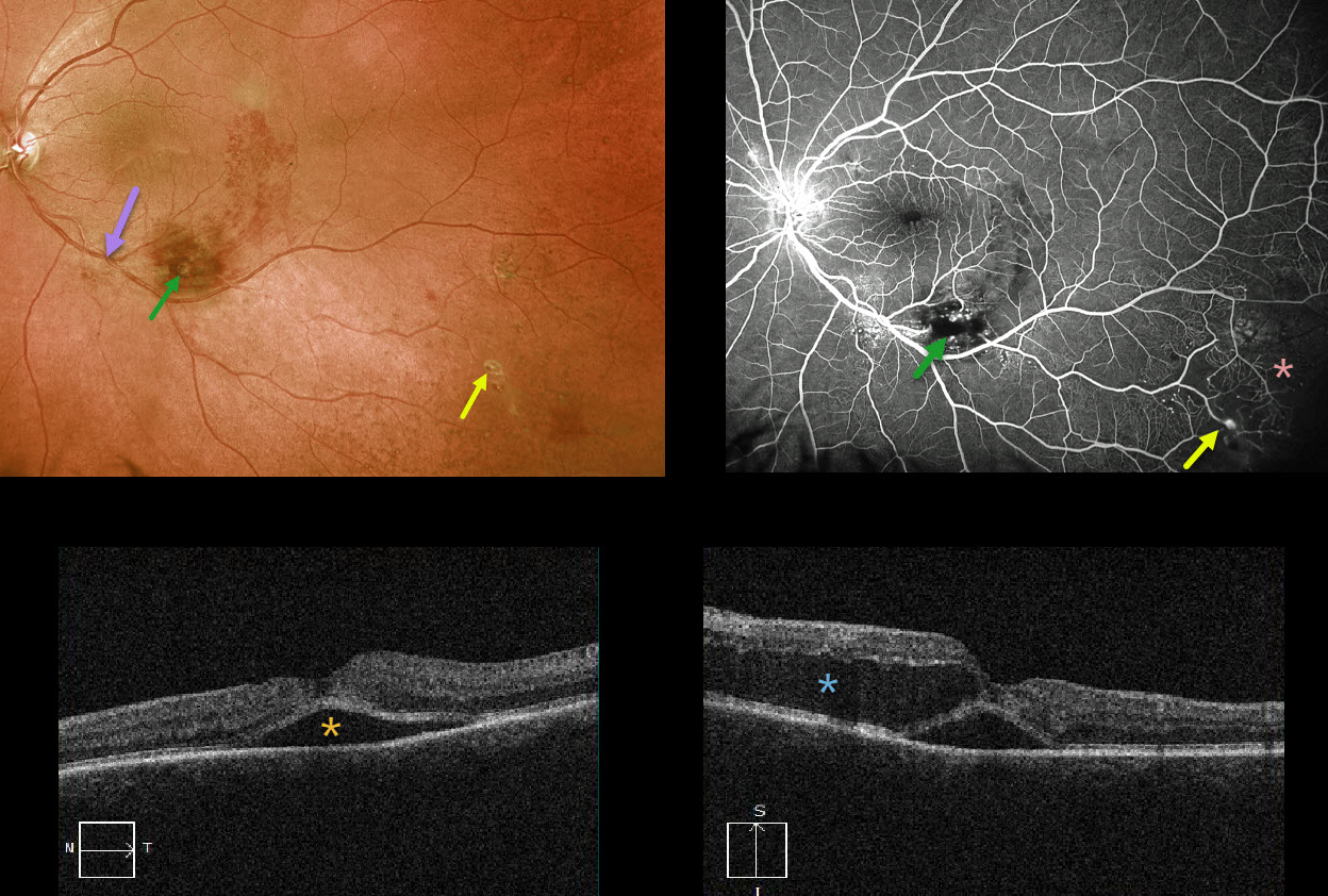
UNKNOWN DIAGNOSIS
Originally posted on @retina.rocks January 6, 2021
This 59YO female presented with acute vision loss of 20-100 in her left eye. At first glance, it looks like a macular branch retinal vein occlusion (BRVO) arising from an artery crossing over a vein more proximally. There is a likely macroaneurysm within the area of more confluent inferior macular blood.
But a more careful inspection shows some retinal telangiectasia more inferiorly and proximally, as well as more distal inferotemporal areas of retinal hemorrhages, telangiectasia, and an arterial macroaneurysm.
Fluorescein angiography better shows these vascular changes and broad peripheral areas of inferotemporal capillary loss.
OCT shows foveal subretinal fluid with a large pocket of outer intraretinal fluid.
Our best guess is that this patient has Coats disease, given the multiple macroaneurysms, multifocal areas of retinal vascular telangiectasia, and capillary loss. Although uncommon, Coats can be found in females.
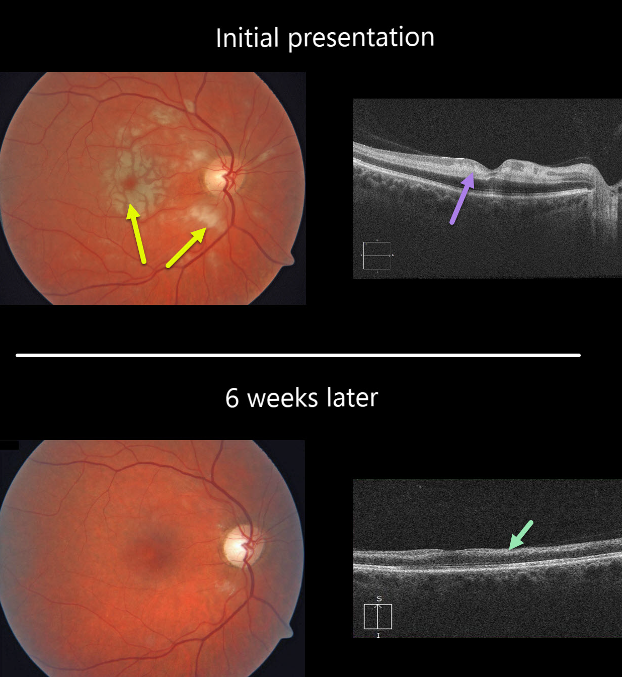
CRAO
Originally posted on @retina.rocks December 24, 2020
This healthy 49YO female presented with severe, sudden vision loss in her right eye.
There were multiple white macular patches similar to Purtscher’s retinopathy, but without retinal hemorrhages.
Optical coherence tomography (OCT) showed multiple mid-retinal hyperreflective paracentral acute macular maculopathy (PAMM) lesions. Since we felt this could represent an incomplete central retinal artery occlusion (CRAO), we sent her immediately to the nearest stroke center for further assessment.
At her six-week follow-up, the retinal appearance virtually normalized. However, OCT showed severe diffuse thinning of the inner non-photoreceptor retinal layers consistent with a resolved CRAO. Her stroke workup was unremarkable.
Learning Points:
Purtscher’s retinopathy was first described by Otmar Purtscher in 1910. Although originally described in a man who fell from a tree with cranial trauma, these white patches of retinal ischemia and hemorrhages are found in numerous other etiologies, including pancreatitis, chest trauma, and collagen vascular diseases.
The findings are called Purtscher’s retinopathy when due to trauma, and Purtscher ‘s-like retinopathy when seen from other causes.
The underlying cause of Purtscher’s is unknown, but is thought to involve leukoembolization due to complement activation. The areas of ischemia include more superficial nerve fiber layer infarcts (cotton wool spots) and deeper ischemia involving the middle retinal layers (PAMM).
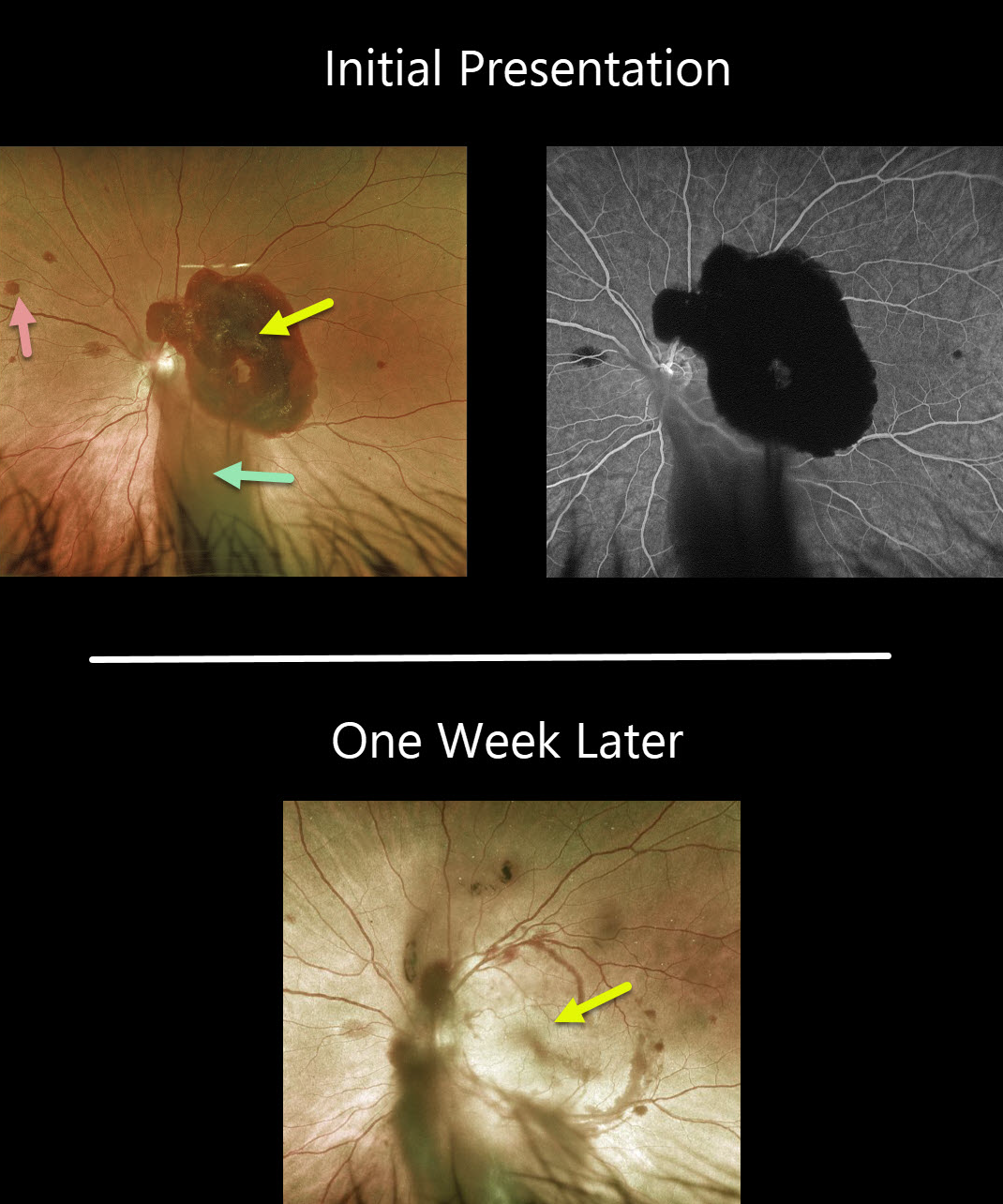
HYPERTENSIVE RETINOPATHY
Originally posted on @retina.rocks December 22, 2020
This 40YO male presented with sudden vision loss in his left eye. Vision was 20/200 OS and 20/20 in his normal right eye.
Fundus photo shows a large subhyaloid hemorrhage covering the macula, vitreous hemorrhage extending inferiorly, and some scattered deep retinal hemorrhages. Fluorescein angiography shows no leakage or source for the blood.
Blood pressure was 235/128. He was immediately sent to the ER for further management.
One week later, vision spontaneously improved to 20/30, the subhyaloid hemorrhage almost completely resolved, and the deep retinal hemorrhages were fading.
Learning Points:
The subhyaloid blood and deep retinal hemorrhages, along with the unilateral presentation, are all quite unusual for hypertensive retinopathy. We are not sure if these hemorrhages were related to his hypertension, but thankfully, they are resolving now that his blood pressure has normalized.
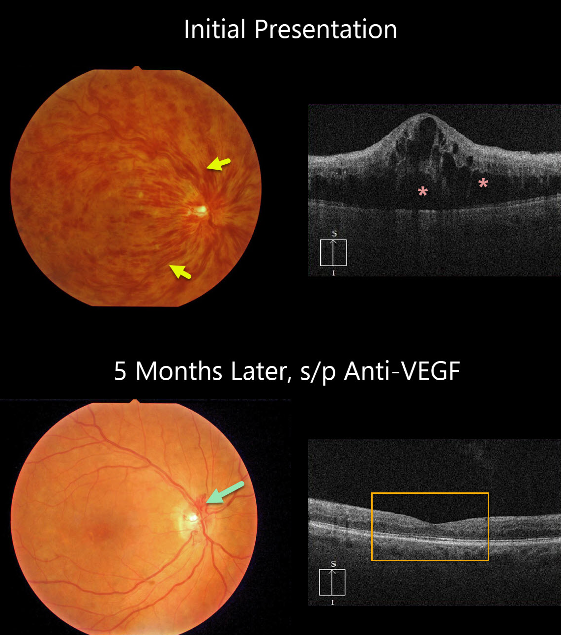
CRVO
Originally posted on @retina.rocks December 21, 2020
This patient presented with counting-finger vision from an acute hemorrhagic central retinal vein occlusion (CRVO) with marked macular edema. The poor vision and extensive retinal hemorrhages suggest that this is an ischemic CRVO.
After five monthly anti-VEGF injections, vision improved to 20/50, the retinal hemorrhages resolved dramatically, optic disc collaterals formed, and OCT showed complete resolution of all macular fluid. This degree of improvement is quite unusual.
Learning Points:
Patients with CRVO are at risk of vision loss due to macular edema or anterior segment neovascularization. Macular edema is common in both non-ischemic and ischemic occlusions, and this responds best to anti-VEGF therapy.
Rubeosis is found almost exclusively with ischemic occlusions, developing in about two-thirds of patients with untreated ischemic occlusions. This was common in the pre-anti-VEGF days.
However, since most patients with CRVO have macular edema and are receiving ongoing anti-VEGF therapy, neovascularization is now rarely seen unless patients present with untreated CRVO or therapy is discontinued.
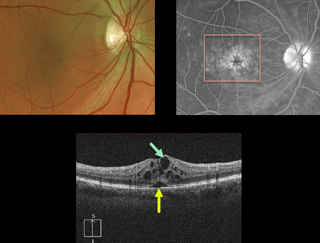
CYSTOID MACULAR EDEMA
Originally posted on @retina.rocks December 10, 2020
This is a classic case of cystoid macular edema (CME) following cataract surgery, otherwise known as the Irvine-Gass Syndrome.
The decompensated inner blood retinal barrier of the perifoveal capillaries allows fluid to leak into the surrounding outer plexiform (Henle’s) layer, causing the classic radiating petaloid pooling on fluorescein angiography.
OCT shows intraretinal edema with cystic spaces and some foveal subretinal fluid.
Learning Points:
Dr. Irvine was the first to describe the condition in 1953 (AJO 1953;36:599-619), and Dr. Gass expanded on this entity in his classic paper (Arch Ophthalmology 1966;76:646-661).
Clinically, at the slit lamp, the cystic changes are best visualized with a contact lens examination (this was how Drs. Irvine and Gass figured things out 60-70 years ago!), although this is rarely currently utilized due to the wide availability and superiority of OCT.
For a great explanation for differentiating exudative from tractional cystic spaces with multimodal imaging, see Govetto et al AJO 2020;212:43-56.
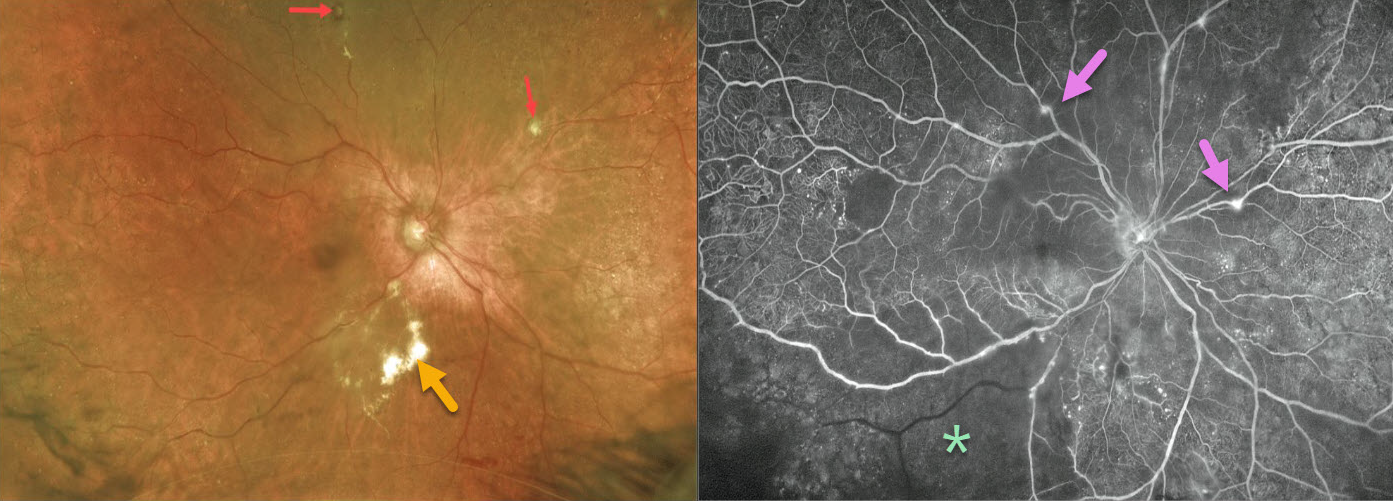
IDIOPATHIC RETINAL VASCULITIS ANEURYSMS AND NEURORETINITIS (IRVAN)
Originally posted on @retina.rocks December 1, 2020
This 77YO female initially presented with a vitreous hemorrhage, which obscured her fundus. The blood spontaneously cleared, allowing us to see multiple fibrosed arterial macroaneurysms and subretinal lipid.
Fluorescein angiography (FA) shows extensive marked inferotemporal retinal ischemia, scattered capillary dropout, and focal areas of mostly venous staining superiorly and nasally. Panretinal photocoagulation (PRP) was applied to all ischemic areas.
Learning Points:
Idiopathic Retinitis, Vasculitis, Aneurysms, and Neuroretinitis (IRVAN) is a rare disease of unknown etiology characterized by retinal vasculitis, aneurysmal arterial dilations, and neuroretinitis. The diagnosis is clinical, as there are no systemic associations or diagnostic laboratory tests.
Early PRP to areas of widespread retinal nonperfusion is recommended to prevent neovascular complications and vitreous hemorrhage (Samuel et al Ophthalmology 2007;114;1526-1529).
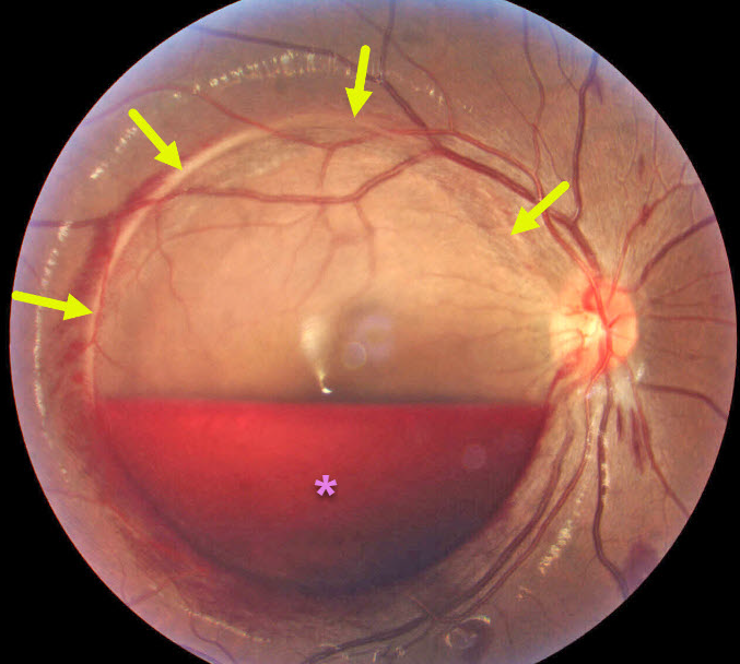
ANEMIA
Originally posted on @retina.rocks November 30, 2020
This 29YO 4-month pregnant female presented with acute vision loss. The internal limiting membrane (ILM) was detached throughout the entire macula, and an inferior-layered sub-ILM hemorrhage spared the macular center. Her hemoglobin was later found to be 11.
Learning Points:
Acute sub-ILM hemorrhage is most common following a Valsalva maneuver or trauma (which our patient denied), but can also happen spontaneously with anemia.
These hemorrhages virtually always resolve without sequelae, although the blood can be drained into the inferior vitreous by creating a small opening in the ILM using either a thermal or Nd:YAG laser.
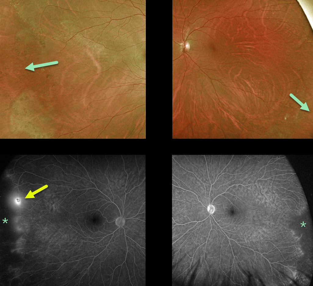
EALES DISEASE
Originally posted on @retina.rocks October 21, 2020
This healthy 50YO male presented with asymptomatic bilateral peripheral retinal ischemia and temporal neovascularization OD seen on fluorescein angiography.
Learning Points:
Eale’s disease is an idiopathic peripheral occlusive retinal vasculopathy that can later develop peripheral neovascularization.
It is more common in males in their 20’s to 30’s, and may be associated with prior tuberculosis exposure. Most patients are asymptomatic unless they develop a vitreous hemorrhage. There is no associated uveitis.
It is likely a heterogeneous disorder and is a diagnosis of exclusion, needing first to rule out the multitude of other disorders that can cause peripheral retinal ischemia and neovascularization.
We have seen many patients with these findings since integrating Optos ultrawidefield imaging into our practice, and therefore suspect that these peripheral retinal findings are much more common than previously described.
Peripheral retinal microvascular abnormalities have also recently been described with ultrawidefield imaging, and may represent a normal variant in this retinal vascular watershed zone (see Shah et al, Retina 2016;36;1087-1092).
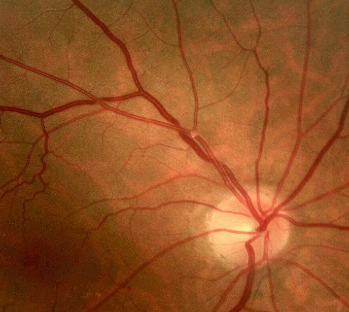
RETINAL EMBOLUS
KMKoptometrypro
Originally posted on @retina.rocks October 14, 2020
This patient had a small asymptomatic embolus. Elective carotid workup revealed a 95% ipsilateral carotid stenosis that was subsequently treated with carotid endarterectomy.
Learning Points:
Acute retinal ischemia, defined as transient monocular vision loss (or amaurosis fugax), acute branch retinal artery occlusion, or acute central retinal artery occlusion, requires an immediate referral to the nearest stroke center.
Asymptomatic emboli, as in this patient, can be more effectively evaluated for an embolic source, which should include carotid doppler and cardiac B-scan ultrasonography.
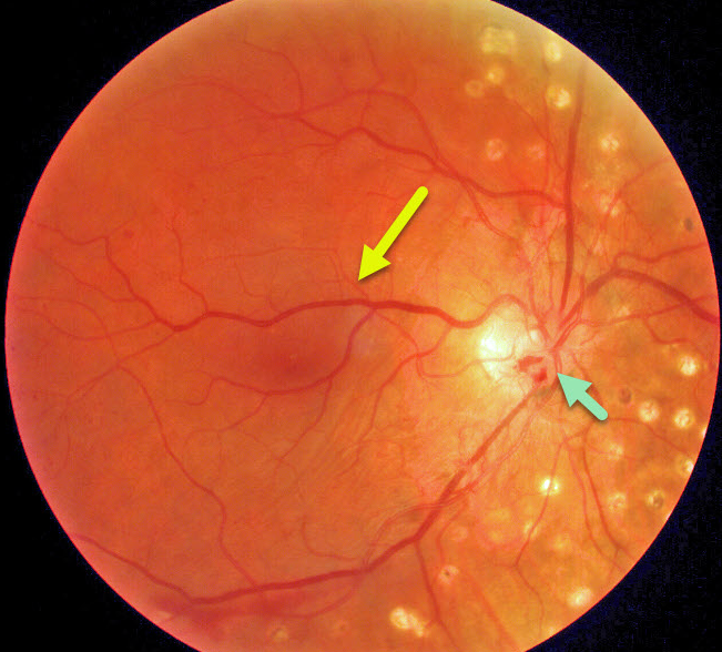
RETINAL VENOUS MALFORMATION
Originally posted on @retina.rocks October 12, 2020
This patient has a retinal venous malformation (formerly known as retinal macrovessel), in addition to full panretinal photocoagulation (PRP) for proliferative diabetic retinopathy. Residual flat disc neovascularization remains despite full PRP.
Learning Points:
A retinal venous malformation is an anomalous retinal vessel that crosses the horizontal raphe (remember that normal retinal vessels respect the horizontal meridian).
These vessels are technically arterio-venous malformations (AVM). Since patients with AVMs can have additional venous anomalies in the brain (24% compared to up to 6% of the population), a brain MRI is recommended to rule out any intracranial abnormalities.
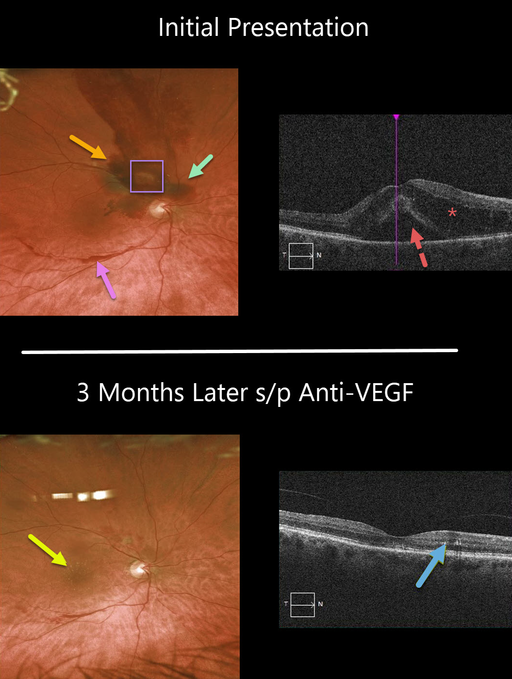
RETINAL ARTERIAL MACROANEURYSM (RAM)
Originally posted on @retina.rocks October 9, 2020
This 71YO female presented with counting-finger vision in her right eye. Clinically, there are multiple layers of blood, including preretinal, intraretinal, and subretinal. These multiple layers are virtually pathognomonic for a ruptured retinal arterial macroaneurysm (RAM).
Despite the causative RAM being just superotemporal to the optic nerve, significant intra- and subretinal fluid extends through the macular center, which is seen on OCT.
Following three monthly anti-VEGF injections, the fluid resolved completely, and vision returned to 20/30. Some residual precipitated lipid is seen in Henle’s layer as a faint lipid star in the color image and as hyperreflective dots in the outer plexiform layer.
Learning Points:
A retinal arterial macroaneurysm (RAM) is thought to result from a weakened arterial wall due to arteriosclerosis.
A RAM appears as a yellow-white dilated lesion along a retinal artery within the first 3 bifurcations from the optic nerve.
There are 2 distinct presentations, hemorrhagic or exudative. The hemorrhagic type often presents as a pathognomonic multilayer macular hemorrhage, with preretinal, intraretinal, and subretinal blood.
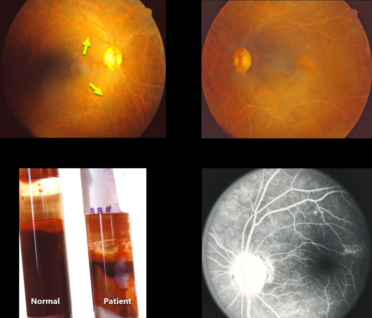
LIPEMIA RETINALIS
Originally posted on @retina.rocks September 10, 2020
This patient’s blood vessels appear milky and possibly occluded, although fluorescein angiography is normal.
A tube of blood drawn from both the patient and a much younger 25YO future founder of Retina Rocks (in his first year of ophthalmology residency) is displayed for comparison. The patient’s blood shows increased lipid content.
Learning Points:
Lipemia retinalis is a rare manifestation of severe hypertriglyceridemia. The blood vessels, and sometimes the entire posterior pole, may appear milky or even salmon-colored.
Patients are asymptomatic, and vision is normal. However, they are at obvious cardiovascular risk without systemic treatment.
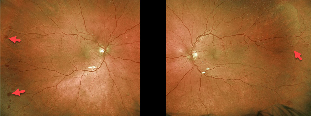
OCULAR ISCHEMIA
Originally posted on @retina.rocks September 8, 2020
This 71YO patient has bilateral, mostly temporal, peripheral deep blot retinal hemorrhages. He had a known history of bilateral carotid stenosis.
Our patient’s findings are classic for ocular ischemic syndrome (OIS), with hemorrhages in the deeper retina of the temporal periphery.
These patients need to have a carotid Doppler and ultrasound and be referred for medical or surgical intervention for critical stenosis. They may also develop anterior or posterior segment neovascularization requiring photocoagulation and/or anti-VEGF injections. Neovascular glaucoma or hypotony can also occur.
Learning Points:
The retinal hemorrhages in OIS are somewhat unique. Retinal vascular disorders such as diabetic retinopathy, central retinal vein occlusion, and hypertensive retinopathy typically present with flame- and dot-shaped retinal hemorrhages concentrated around the posterior pole, located in the inner retina.
The retinal periphery is literally the end of the line for the retinal arterial circulation. In an eye receiving less blood supply due to carotid occlusive disease, the more proximal retina has first dibs on the available oxygen. There are penetrating capillaries that dive radially from the nerve fiber and ganglion cell layers into the deeper retina. The deep vascular complex that supplies the inner and outer plexiform layers is thus furthest downstream, and these endothelial cells are likely damaged in OIS. Blood cells leaking into this space accumulate, causing the large outer blot to hemorrhage.
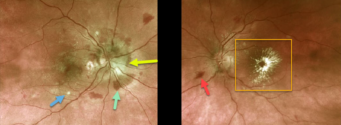
HYPERTENSIVE RETINOPATHY
Originally posted on @retina.rocks September 7, 2020
This previously healthy 36 YO female presented with recent vision loss of 20/200 bilaterally.
The right optic nerve is moderately swollen with surrounding mostly deep retinal hemorrhages, faint scattered macular lipid, and a few nerve fiber layer infarcts (cotton wool spots). The left nerve is flat with a few surrounding inner flame-shaped retinal hemorrhages and a prominent foveal lipid star.
Blood pressure was 239/159, and she was sent immediately to the emergency room for malignant hypertension.
Learning Points:
Her presentation was a bit unusual, in that systemic problems usually cause symmetric ocular findings. Her blood pressure was likely elevated for weeks to months since the macular lipid is a late finding.
We are not sure why the left nerve is not swollen, but it certainly was in the past. The lipid star is a telltale sign of prior optic nerve swelling, in which fluid and lipoproteins leaked into the surrounding tissues. When the fluid is absorbed, the lipid precipitates out in Henle’s layer. This is also seen in other causes of optic nerve swelling or inflammation, including neuroretinitis.
The deep retinal hemorrhages in her right eye are also unusual, since these are usually found in the nerve fiber layer with hypertension.
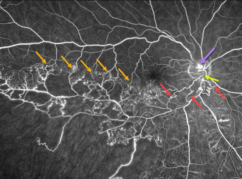
BRVO
Originally posted on @retina.rocks August 31, 2020
This patient has an ischemic branch retinal vein occlusion (BRVO) with extensive collateral vessels.
Occlusion occurs when an artery compresses the underlying vein where the vessels share a common adventitial sheath. Extensive collateral vessels cross the temporal horizontal raphe as well as around the occlusion more posteriorly. A small area of disc neovascularization leaks.
Learning Points:
Collateral vessels are dilated, pre-existing vessels that bypass an occlusion. These are most commonly seen in the posterior pole following retinal venous occlusions, but can also occur on the optic disc with optic nerve meningiomas.
The endothelial tight junctions are intact, so there’s no leakage on angiography. This helps differentiate them from neovascularization, which does leak.
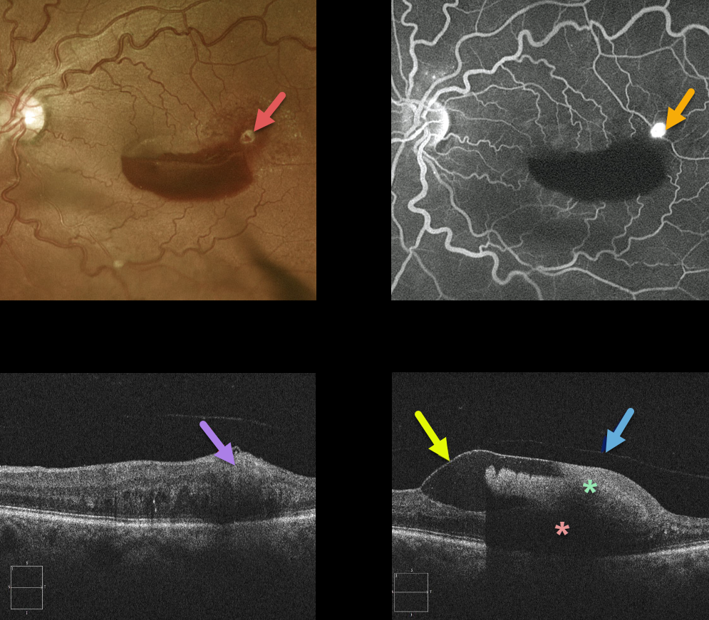
RETINAL ARTERIAL MACROANEURYSM (RAM)
Originally posted on @retina.rocks August 20, 2020
This retinal arterial macroaneurysm (RAM) appears as a yellow-white inner retinal lesion along the superotemporal edge of a macular hemorrhage. Angiography confirms the presence of the hyperfluorescent RAM.
An OCT line scan through the RAM shows its location in the inner retina.
OCT scanning shows that the blood is almost all above the retina and below the detached internal limiting membrane. The detached hyaloid can be seen anterior to the ILM. The dense sub-ILM blood blocks almost all retinal and subretinal detail more posteriorly.
Learning Points:
A retinal arterial macroaneurysm (RAM) is thought to result from a weakened arterial wall due to arteriosclerosis.
A RAM appears as a yellow-white dilated lesion along a retinal artery within the first 3 bifurcations from the optic nerve.
There are 2 distinct presentations, hemorrhagic or exudative. The hemorrhagic type often presents as a pathognomonic multilayer macular hemorrhage, with preretinal, intraretinal, and subretinal blood.
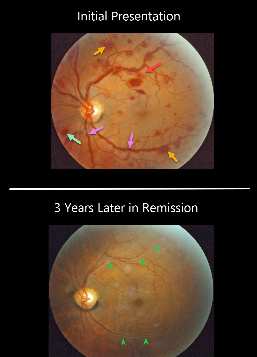
MULTIPLE MYELOMA
Originally posted on @retina.rocks August 6, 2020
This patient presented with bilateral multiple flame-shaped and white-centered retinal hemorrhages. There is also at least one deeper, round outer plexiform hemorrhage just inferonasal to the nerve. The retinal veins are somewhat dilated and tortuous, consistent with hyperviscosity. This patient was subsequently diagnosed with multiple myeloma.
Three years later, the patient was in remission after receiving treatment. Although the vascular caliber has normalized, vascular sheathing remained due to prior damage to the vessel walls.
Learning Points:
White-centered hemorrhages or “roth spots” can be seen in many disorders, including leukemia, hyperviscosity, anemia, diabetes, and hypertension. The white material may represent a variety of causes, including fibrin.
The term “Roth spot” was originally named after Dr. Moritz Roth, who, in 1872, noted white-centered hemorrhages in patients with bacterial endocarditis. He felt that these white spots were septic emboli that originated from an infected cardiac valve.
This term should thus be reserved only for patients with bacterial endocarditis. See Duane et al Ophthalmology 1980;87;66-69.
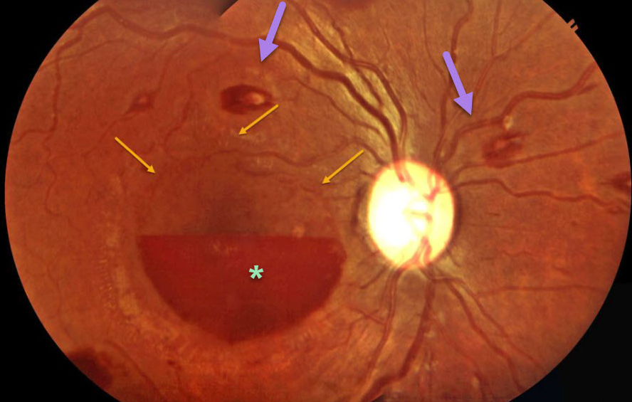
ANEMIA
Originally posted on @retina.rocks August 5, 2020
This patient presented with multiple white-centered flame-shaped retinal hemorrhages and sub-ILM hemorrhages consistent with anemic retinopathy. ILM detachment can be seen superior to the hemorrhage anterior to the fovea on the right eye.
Learning Points:
The term “Roth spot” was named after Dr. Moritz Roth, who, in 1872, noted white-centered hemorrhages in patients with bacterial endocarditis. He felt that these white spots were septic emboli that originated from an infected cardiac valve. However, similar lesions can be seen in many other disorders, including leukemia, anemia, diabetes, and hypertension.
The white material may represent a variety of causes, including fibrin. So these lesions are best called white-centered hemorrhages, unless one is specifically referring to a lesion in a patient with bacterial endocarditis. See Duane et al Ophthalmology 1980;87;66-69.
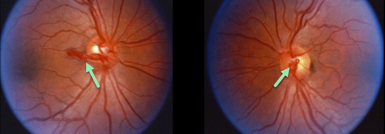
RETINAL VASCULAR LOOP
Originally posted on @retina.rocks August 4, 2020
This patient has benign bilateral congenital prepapillary vascular loops. These can be either unilateral or bilateral.
Learning Points:
Although usually just an incidental finding, they can rarely cause a branch retinal arterial or venous occlusion (see Retinal Cases Brief Rep 2014;8;124-126 and Ophthalmology 2020;127;124-126).
They should also not be confused with disc neovascularization, which consists of much smaller caliber vessels that leak on fluorescein angiography.
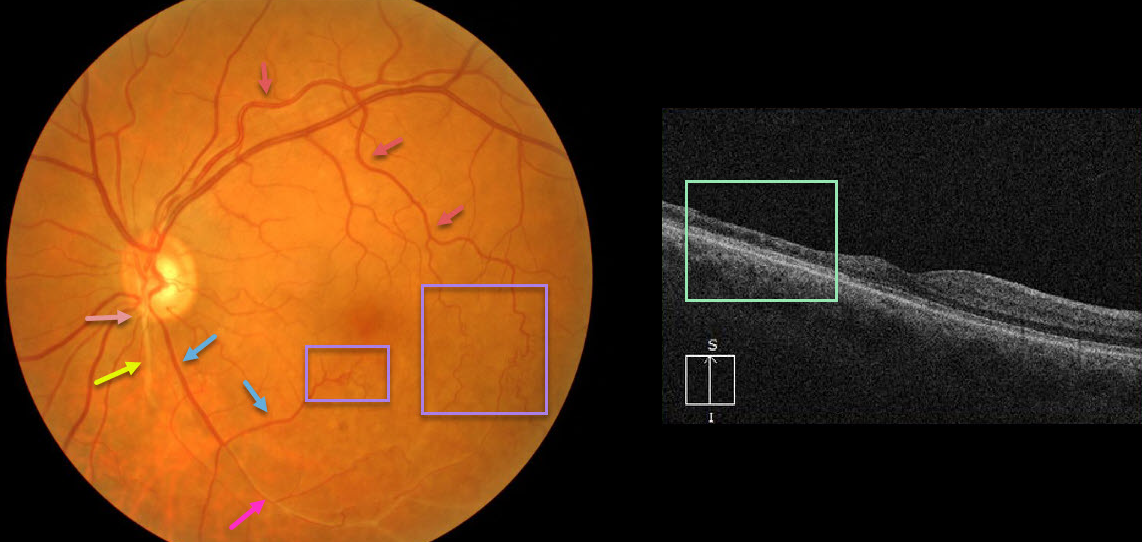
RETINAL ARTERIOVENOUS MALFORMATION
Originally posted on @retina.rocks July 28, 2020
At first or quick glance, this looks like an old inferotemporal branch retinal vein occlusion (BRVO).
On closer inspection, there is a white-sheathed vessel arising from the nerve inferiorly. Numerous collateral vessels are noted inferior and temporal to the fovea, and there is a sheathed vessel in the inferior arcade.
OCT shows marked inner retinal atrophy with preservation of the photoreceptor layers, consistent with a prior branch retinal artery occlusion. So the collateral vessels (virtually always a sign of a prior venous occlusion) indicate an old BRVO, and the atrophic and sheathed vessel indicates an old BRAO, both in the same quadrant. Right?
Tracing the large branch of the superotemporal arcade inferotemporally, we see that the collaterals appear to be arterial. Inferior to the nerve, we see an arteriole that crosses the sheathed vessel, indicating that the sheathed vessel is not an artery but a vein. Moreover, the large vessel off the nerve, inferotemporal in location, appears to be a vein that eventually connects to the collateral vessels we previously identified as arterial.
Our best guess is that this may be a low-grade arteriovenous malformation, possibly with a prior secondary BRAO/BRVO. Unfortunately, we don’t have a fluorescein angiogram, which may help better define the vascularization.
Learning Points:
Remember that arteries don’t cross arteries and veins don’t cross veins. Tracing vessels can be a helpful key to identifying retinal vascular disorders.
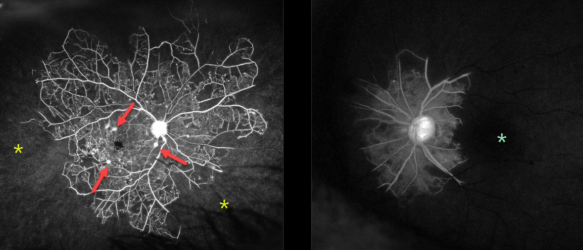
IDIOPATHIC RETINAL VASCULITIS ANEURYSMS AND NEURORETINITIS (IRVAN)
Originally posted on @retina.rocks July 6, 2020
This 64YO female presented with 20/30 vision OD and no light perception OS. Her blood pressure was elevated at 147/101, and she had a history of type 2 diabetes. She had a prior macular laser for macular edema.
Although clinically the peripheral retina appeared ischemic, fluorescein angiography showed dramatic, extensive, and total peripheral ischemia OD, with even worse near total loss of retinal perfusion OS. Multiple aneurysmal dilations are seen in the right posterior pole.
We injected both eyes with Avastin that day and applied full panretinal photocoagulation bilaterally over the next 2 weeks. Carotid dopplers showed less than 50% bilateral internal carotid stenosis.
Learning Points:
Idiopathic Retinitis, Vasculitis, Aneurysms, and Neuroretinitis (IRVAN) is a rare disease of unknown etiology characterized by retinal vasculitis, aneurysmal arterial dilations and neuroretinitis. The diagnosis is clinical, as there are no systemic associations or diagnostic laboratory tests.
Early PRP to areas of widespread retinal nonperfusion is recommended to prevent neovascular complications and vitreous hemorrhage (Samuel et al Ophthalmology 2007;114;1526-1529).
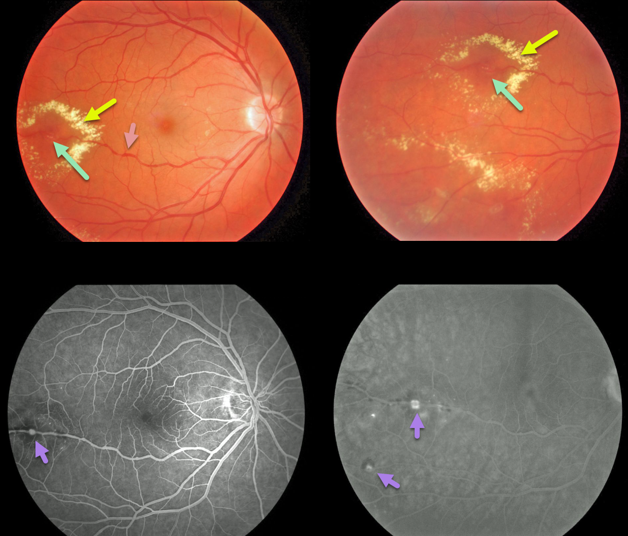
COATS DISEASE
Originally posted on @retina.rocks June 24, 2020
This is a classic case of Coats disease, with unilateral retinal vascular leakage from both the capillary bed and larger vessels.
Fundus images show dilated capillaries and retinal vessels. In addition, there are multiple arterial macroaneurysms with surrounding exudate.
Fluorescein angiography shows the leaking telangiectatic vessels.
Learning Points:
Laser photocoagulation can be used to treat leaking macroaneurysms and telangiectatic vessels in Coats disease. Anti-VEGF injections can also be used to stabilize vascular leakage. Patients will usually require multiple rounds of treatment.
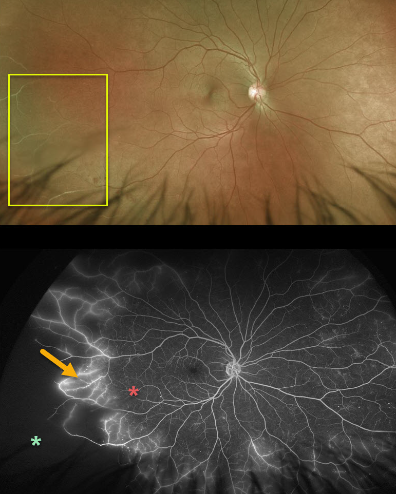
EALES DISEASE
Originally posted on @retina.rocks June 17, 2020
Eale’s disease is our best guess for this 44YO male patient. Eale’s disease is characterized by peripheral, sharply circumscribed regions of ischemia. Fluorescein angiogram demonstrates leakage of retinal vessels at the junction of perfused and ischemic retina.
For now, we are carefully observing the ischemic retina, but we will treat this region with scatter laser if neovascularization develops.
Learning Points:
Eale’s disease is an idiopathic peripheral occlusive retinal vasculopathy that can later develop peripheral neovascularization. It is more common in males in their 20’s to 30’s, and may be associated with prior tuberculosis exposure. Most patients are asymptomatic unless they develop a vitreous hemorrhage.
It is likely a heterogeneous disorder and is a diagnosis of exclusion, needing first to rule out the multitude of other disorders that can cause peripheral retinal ischemia and neovascularization.
We have seen many patients with these findings since integrating Optos ultrawidefield imaging into our practice, and therefore suspect that these peripheral retinal findings are much more common than previously described.
Peripheral retinal microvascular abnormalities have also recently been described with ultrawidefield imaging, and may represent a normal variant in this retinal vascular watershed zone (see Shah et al, Retina 2016;36;1087-1092).
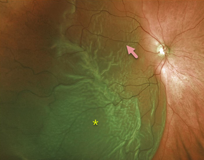
RHEGMATOGENOUS RETINAL DETACHMENT
Originally posted on @retina.rocks June 10, 2020
This patient has an acute, macula-off retinal detachment with a large cilioretinal artery.
Learning Points:
Cilioretinal arteries arise from the choroidal circulation. This results in cilioretinal sparing with central retinal artery occlusion.
Unfortunately, this vessel offers no visual protection against retinal detachment!
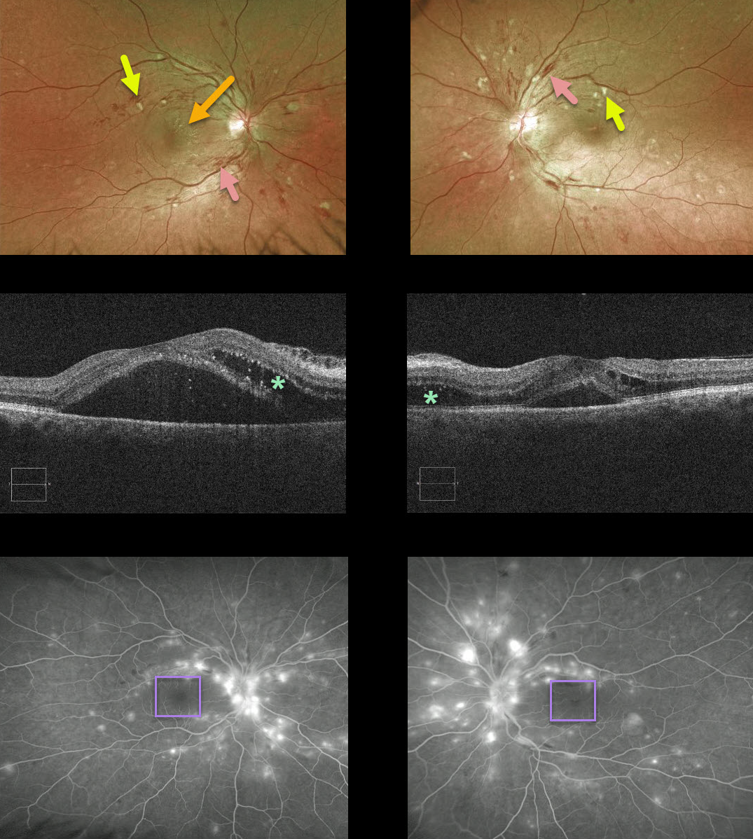
HYPERTENSIVE RETINOPATHY
Originally posted on @retina.rocks April 10, 2020
This patient presented with bilateral inner-retinal flame-shaped hemorrhages and nerve fiber layer infarcts. There was no past ocular or past medical history. In the office, her blood sugar was normal but her blood pressure was 160/92.
In this patient’s OCT, the macular fluid is coming mostly from the nerves; note the early nasal macular star OD and the bilateral outer retinal fluid on OCT that is most prominent nasally.
Angiography also shows a striking absence of macular leakage that should have been present if diabetes were the cause.
Although her blood pressure was not that severely elevated, we suspected it was likely much higher in the near past. She had no health coverage and no primary care doctor, so we sent her to the ER for further treatment and management.
Learning Points:
Hypertensive retinopathy can have a similar appearance to diabetic retinopathy. Clues to hypertensive cause include clinical, angiographic, and OCT signs.
Over time, patients with hypertensive retinopathy will develop near complete regression of all findings following systemic blood pressure treatment and control.
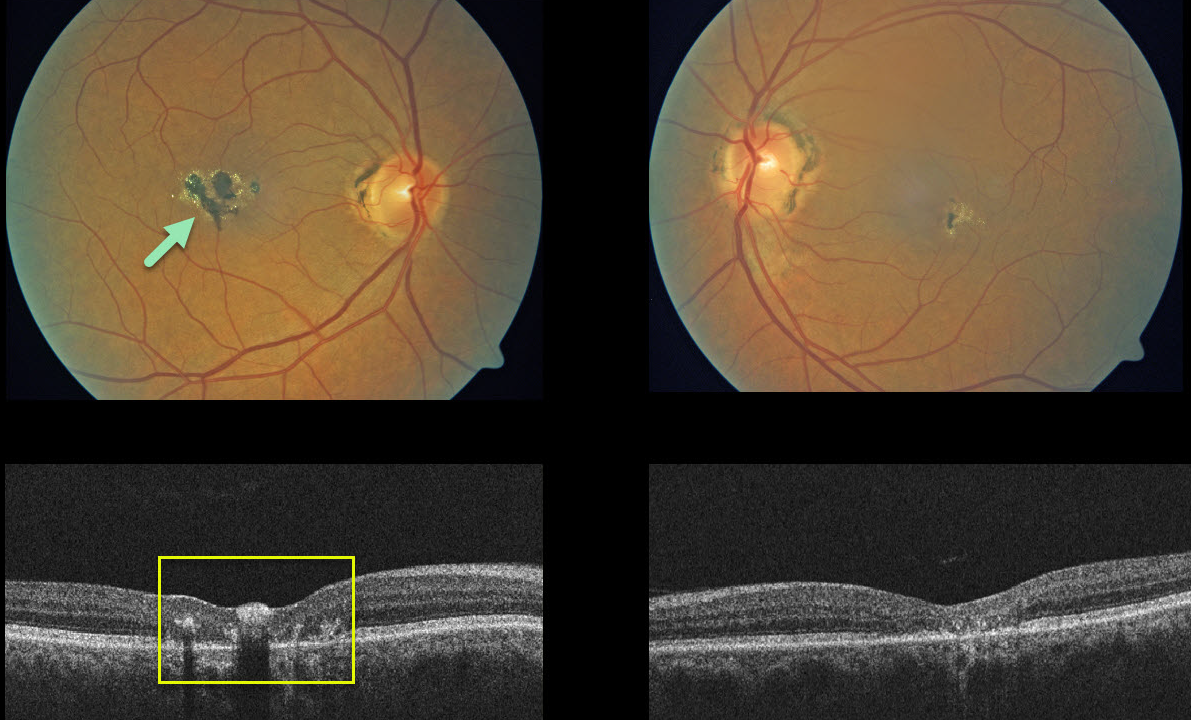
MACULAR TELANGIECTASIA
Originally posted on @retina.rocks April 9, 2020
Our patient shows classic bilateral findings for macular telangiecasia (MacTel) type 2 with coarse pigment clumps.
The OCTs show central/temporal retinal atrophy with disorganized retinal layers. Absent in our patient, the OCT will also often display hyporeflective cavitations of tissue loss.
The majority of patients will also have subclinical chorioretinal anastomosis in association with right-angle venules.
Learning Points:
MacTel type 2 is a neurodegenerative disorder, most likely originating from Muller cell dysfunction. The term, telangiectasia, is misleading in that the funduscopic findings are mostly non-vascular.
It is thought that photoreceptor loss allows RPE cells to migrate along intraretinal capillaries causing the clinical pigment clumping. There are also numerous refractile inner retinal ‘crystals,’ thought to be Muller cell footplates.
Although there is much to be learned about its pathophysiology and there is no treatment for the underlying disease, secondary macular neovascularization can be successfully treated per AMD protocols.
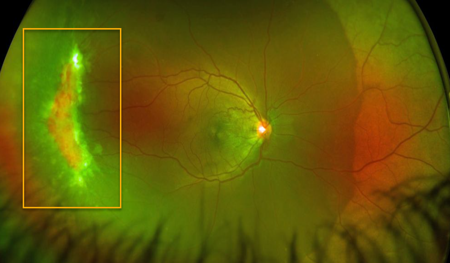
SICKLE CELL RETINOPATHY
Originally posted on @retina.rocks April 2, 2020
This patient, courtesy of Southern College of Optometry, presented with a large temporal peripheral area of active neovascularization from proliferative sickle cell retinopathy in the right eye. The left fundus was normal.
Learning Points:
Patients with sickle cell disease develop symptoms throughout their body from stiffened sickled red blood cells blocking capillary beds.
In the eye, this manifests as proliferative sickle cell retinopathy (PSR) with sea-fan shaped areas of peripheral retinal neovascularization developing at the junction of perfused and ischemic retina.
Complications include vitreous hemorrhage and traction retinal detachment.
Treatment with peripheral scatter laser is somewhat controversial since, unlike more typical proliferative retinopathies (ie from diabetes and branch retinal vein occlusions), these new vessels tend to auto-infarct.
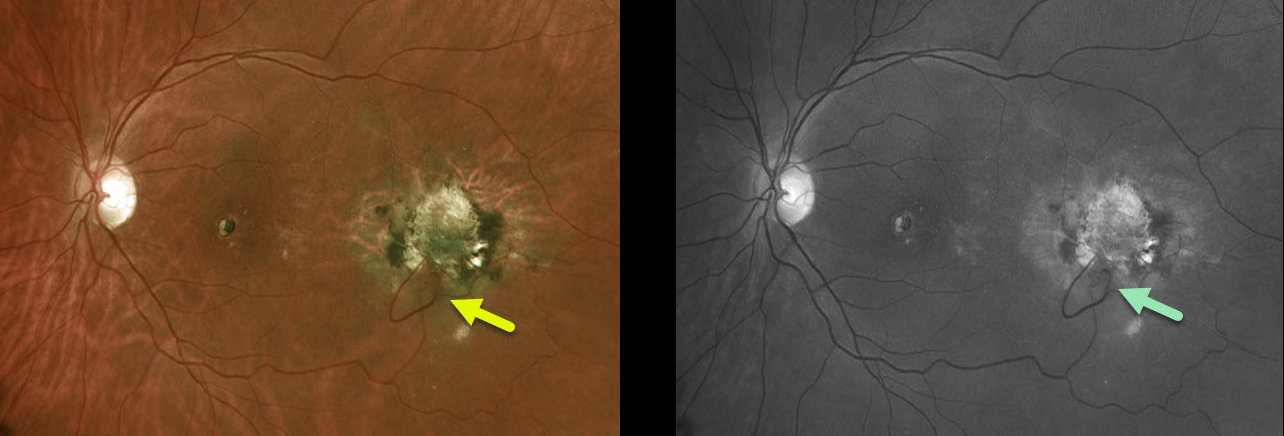
RETINAL CHOROIDAL ANASTOMOSIS
Originally posted on @retina.rocks March 24, 2020
Our patient has a very prominent chorioretinal anastomosis from an idiopathic ectopic disciform scar. It is best seen in the Optos color and green-channel images.
Learning Points:
Under normal conditions the retinal and choroidal circulations remain separate and distinct. Rarely, the two circulations directly connect with each other thru a chorioretinal anastomosis.
These can occur in chorioretinal scars (typically from inactive toxoplasmosis lesions), in macular telangiectasia, and in disciform scars. Laser photocoagulation can create an iatrogenic retinal venous-choroidal anastomosis to treat macular edema in non-ischemic CRVOs, although its use has not been widely adopted.
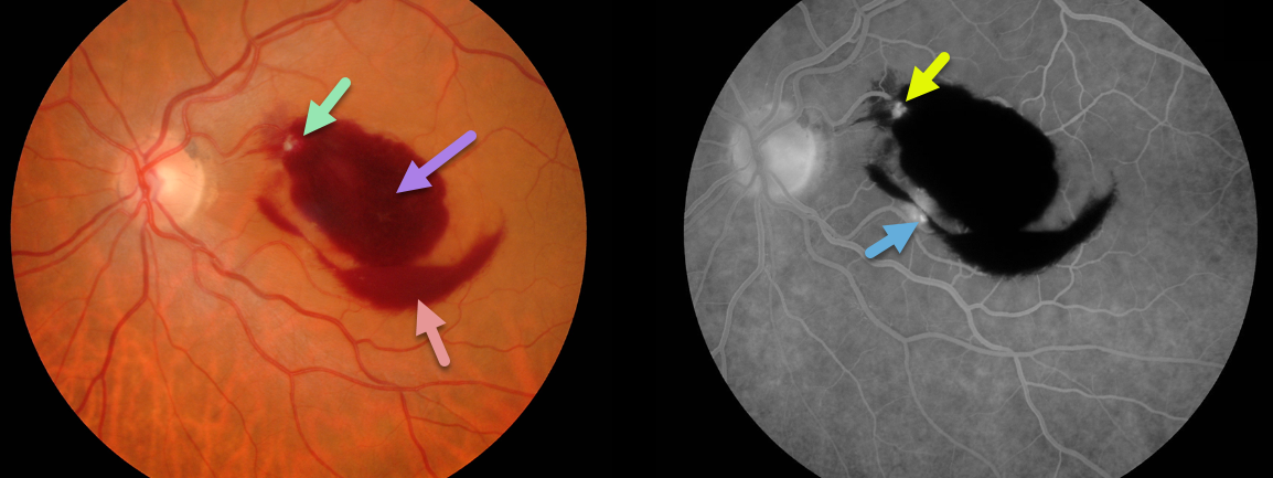
Retinal Arterial Macroaneurysm (RAM)
Originally posted on @retina.rocks March 10, 2020
Our patient’s retinal arterial macroaneurysm (RAM) is seen within the superonasal portion of the macular blood. Preretinal and dense intraretinal blood are the clues that a RAM is the cause.
The RAM is confirmed on fluorescein angiography. Although usually singular, our patient may also have a second RAM inferiorly.
Learning Points:
A retinal arterial macroaneurysm (RAM) is thought to occur in a weakened arterial wall from arteriosclerosis.
A RAM appears as a yellow-white dilated lesion along a retinal arterial within the first 3 bifurcations from the optic nerve.
There are 2 distinct presentations, hemorrhagic or exudative. The hemorrhagic type often presents as a pathognomonic multilayer macular hemorrhage, with preretinal, intraretinal and subretinal blood.
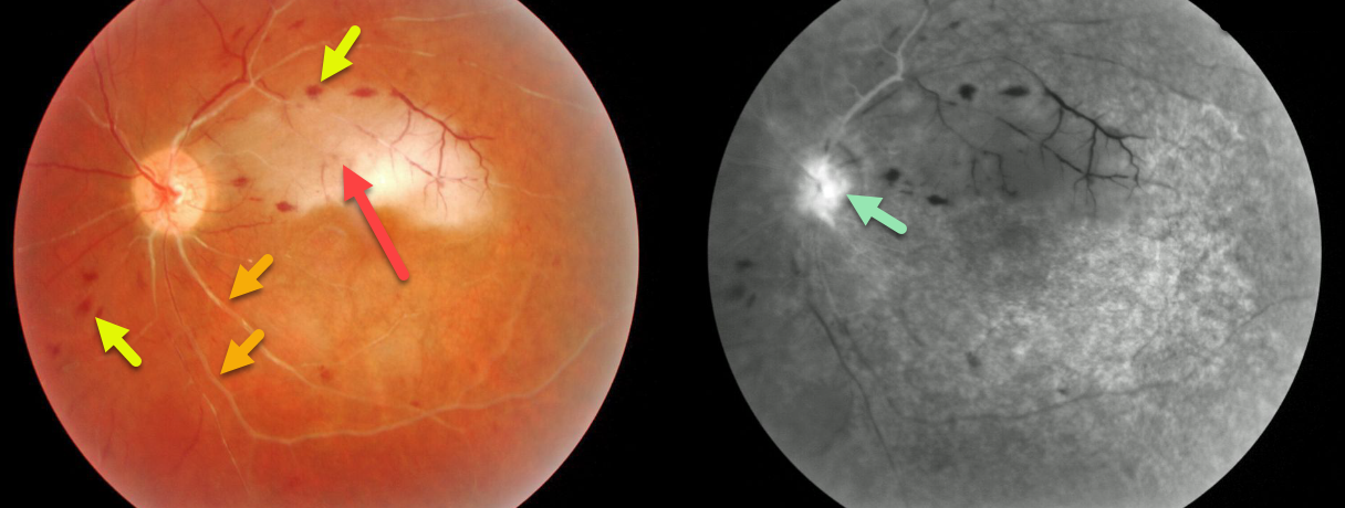
ANTIPHOSPHOLIPID ANTIBODY SYNDROME
Originally posted on @retina.rocks February 18, 2020
This young woman presented with severe vision loss of the left eye and an unusual constellation of ischemic fundus findings, including both acute and chronic arterial and venous occlusions. She was subsequently diagnosed with the antiphospholipid antibody syndrome.
Most obvious is an acute superior macular branch retinal arterial occlusion. Scattered retinal hemorrhages in all quadrants are consistent with a prior central retinal vein occlusion. Both the major arteries and veins are sclerosed and there is disc neovascularization indicating chronic ischemia.
Despite anti-VEGF injections and panretinal photocoagulation she eventually went to no light perception vision. Remarkably the other eye remains normal.
Learning Points:
Antiphospholipid antibody syndrome, also known as anticardiolipin antibody syndrome or lupus anticoagulant syndrome, is caused by autoantibodies directed against phospholipid-bound proteins.
These predispose patients to venous and arterial thrombi and increase the risk for pregnancy complications and miscarriage.
The syndrome is more common in women and in those with other autoimmune disorders including systemic lupus.
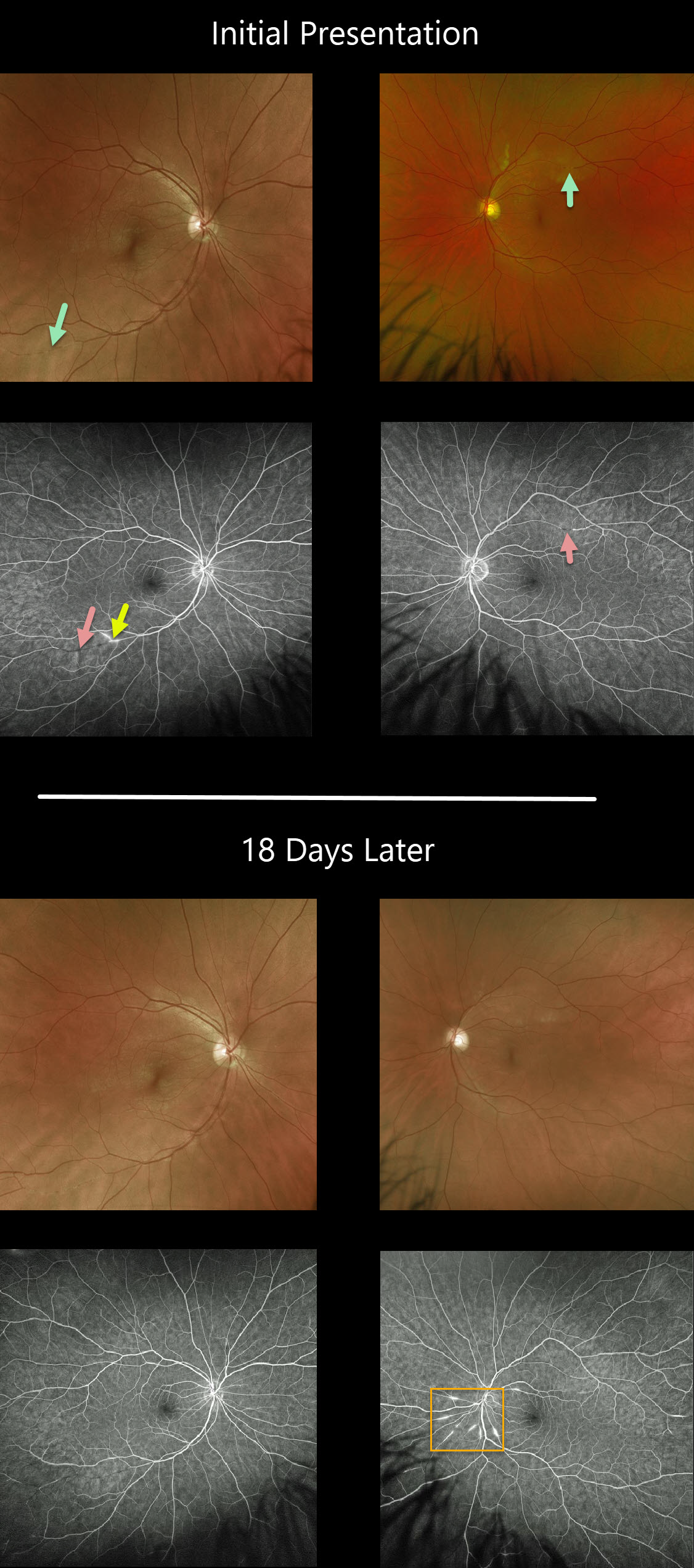
SUSAC SYNDROME
Originally posted on @retina.rocks January 29, 2020
Our patient presented with silent ischemia inferotemporally in the right eye and new lesions superotemporally in the left. Note the areas of non-perfusion and leakage.
18 days later new multi-focal lesions OS are visualized dramatically on fluorescein angiography.
Fortunately, the patient has maintained excellent VA
Learning Points:
Retinocochleocerebral Vasculopathy, more commonly known as Susac Syndrome, is a rare, microangiopathic disease of debated pathophysiology characterized by the classic triad of encephalopathy, sensorineural hearing loss and branch retinal artery occlusions. The full triad may take months to evolve, frustrating the diagnosis.
Retinal arterial wall atheromatous plaques, also known as Gass plaques, are seen often in later stages. They are a result of the occlusion, not the cause.
The development of wide-field angiography has occasionally allowed for early detection which can be critical in order to minimize the often long-term, debilitating effects of the disease.
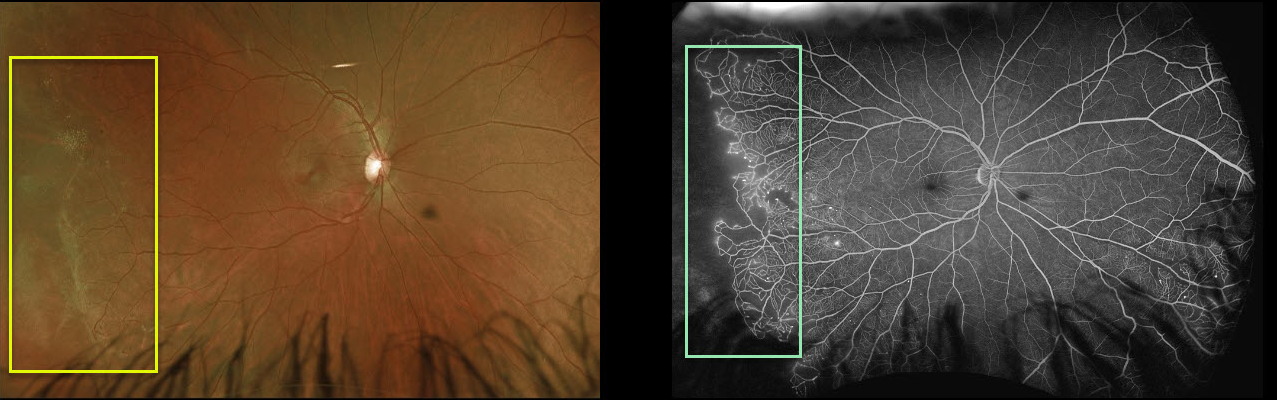
EALES DISEASE
Originally posted on @retina.rocks January 6, 2020
In this patient’s right eye, there is a classic sharp margin between perfused and nonperfused retina. The fellow eye (not pictured) is relatively normal.
Angiography shows some vascular leakage at the margin of the perfused and nonperfused retina.
Ultra-widefield imaging in our practice has dramatically increased the detection of Eale’s disease.
Learning Points:
Eales’ disease is an idiopathic peripheral occlusive vasculopathy that can cause retinal neovascularization and vitreous hemorrhage.
It is characterized by peripheral sharply circumscribed regions of ischemia, often with retinal neovascularization at the junction of perfused and ischemic retina.
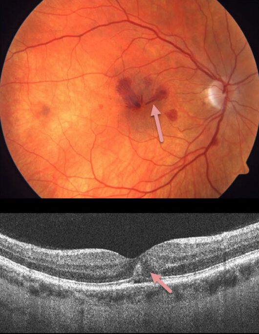
HEMORRHAGIC UNILATERAL RETINOPATHY
Originally posted on @retina.rocks December 21, 2019
This 41YO female presented with acute unilateral vision loss in her right eye. Vision was 20/50 OD and 20/20 OS. Exam revealed unilateral radiating deep Henle layer retinal hemorrhages with no identifiable systemic or ocular etiologies.
Learning Points:
Named by Kon Graversen et al (Retina 2014;34:483-489), hemorrhagic unilateral retinopathy presents with unilateral, deep, intraretinal hemorrhages with sudden reduced vision ranging from 20/20 to hand motion.
There are no associated systemic conditions, nor history of trauma or Valsalva maneuver. It is most common in females. Vision usually recovers completely within 6 months.
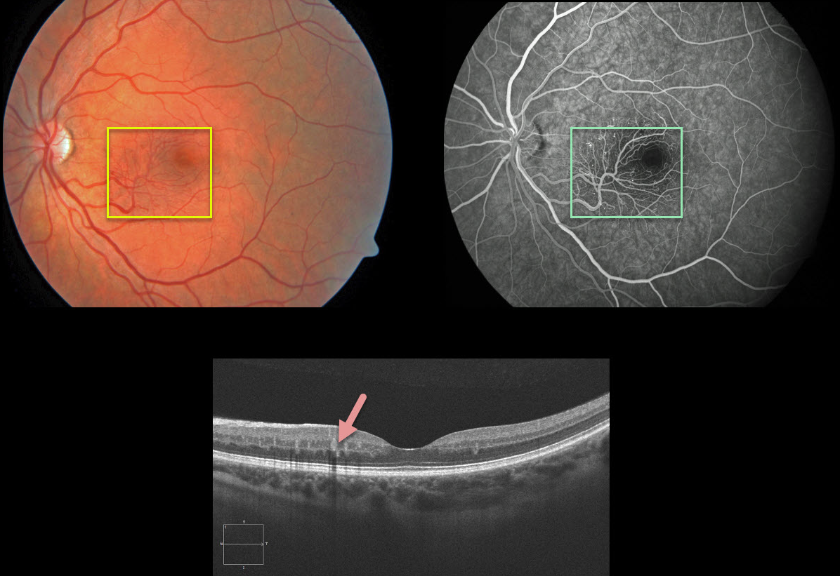
RETINAL ARTERIOVENOUS MALFORMATION
Originally posted on @retina.rocks December 19, 2019
This patient was asymptomatic and found to have a macular arteriovenous malformation (AVM). OCT B-scan shows the vascular abnormality mostly involves the deep vascular complex. Fluorescein angiography demonstrates no leakage.
The patient was asymptomatic with a normal MRI and MRA of the brain/orbits.
Learning Points:
Reinal arteriovenous malformations (AVM) are typically unilateral and sporadic.
Our patient had an isolated AVM. Wyburn-Mason Syndrome is characterized by the presence of retinal AVMs, as well as AVMs of the cerebral nervous system (CNS) and/or orbit.


