

Asteroid Hyalosis
<< Back to Cases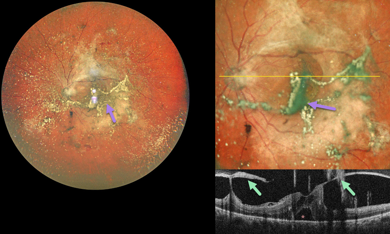
PROLIFERATIVE DIABETIC RETINOPATHY WITH ASTEROID HYALOSIS
The European VitreoRetinal Society (EVRS), Akansha Sharma, Manish Nagpal, and Vaidehi Sathaye
Originally posted on @retina.rocks April 27, 2026
This 55YO diabetic male presented with 2 months of bilateral vision loss. Vision was 20/80 OD and 20/120 OS.
Pseudocolor SLO ultrawidefield imaging of his left eye shows moderate asteroid hyalosis, with larger condensed stands of material obscuring the underlying macula. Spectral domain OCT scanning shows complex variable vitreous traction on the disc and macula with a small foveal detachment. The asteroid particles appear as hyperreflective vertical lines resembling falling rain.
Learning Points:
Examining the fundus in a patient with dense asteroid, as in this diabetic patient, can be extremely challenging. Fluorescein angiography and OCT are great modalities because they can visualize the retina when the clinical exam can’t. In our patient’s case, the OCT revealed foveal traction, indicating the need for vitrectomy surgery.
PROLIFERATIVE DIABETIC RETINOPATHY (PDR) WITH ASTEROID HYALOSIS
Originally posted on @retina.rocks October 16, 2024
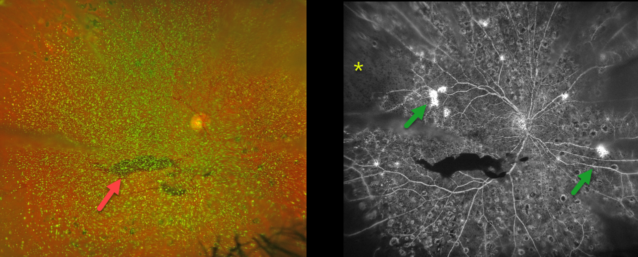
This 50YO female with a history of type 2 diabetes was referred for new floaters. Several years earlier, we began panretinal photocoagulation (PRP) for proliferative diabetic retinopathy (PDR), but she had been lost to follow-up.
Optos color RG imaging shows dense asteroid hyalosis with a hazy inferior preretinal hemorrhage. Fluorescein angiography (FA) shows significant retinal ischemia, scattered retinal neovascularization, and partial PRP scarring.
Examining the fundus with dense asteroid, as in this diabetic patient, can be extremely challenging. FA and OCT are great modalities because they can visualize the retina when the clinical exam can’t. Optos FA was critical in confirming proliferative retinopathy with peripheral ischemia in our patient.
We were fortunately able to complete the PRP despite the vitreous opacities.
ASTEROID HYALOSIS
Originally posted on @retina.rocks December 25, 2023
A holiday gift for our awesome viewers!
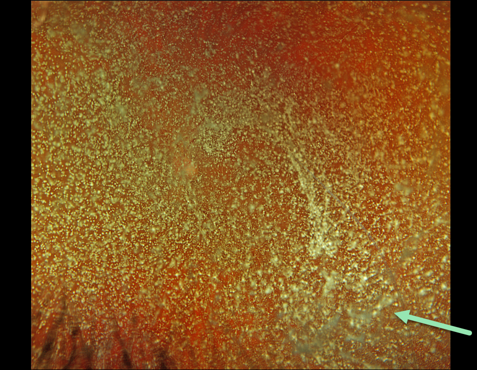
Here’s a gorgeous Optos color RGB image of asteroid hyalosis in a healthy 51YO man. Vision was 20/25. And he was completely visually asymptomatic.
As we exit 2023, enter 2024, and celebrate Retina Rock’s 4th birthday, we are so grateful that people across the globe enjoy our images and discover what they teach us about the retina. Thank you for giving us a reason to exist and keep our mission alive: to help educate the world’s eye care providers about why the retina rocks!
ASTEROID HYALOSIS
Originally posted on @retina.rocks June 29, 2022
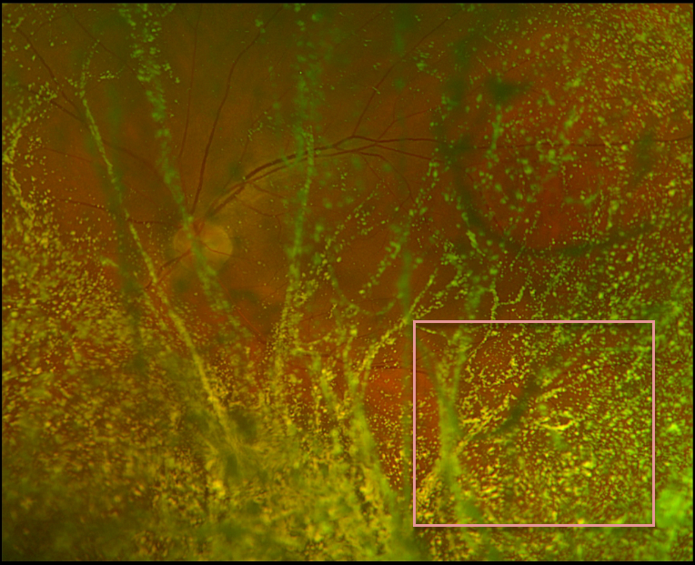
This 72YO male presented with unilateral asteroid hyalosis in his left eye. Vision was 20/40 OS with mildly symptomatic floaters. Retinal examination was extremely difficult due to the dense asteroid, although the fundus appeared normal.
Learning Points:
Asteroid hyalosis is caused by calcium-lipid complexes suspended throughout the vitreous. The condition gets its name from the white opacities resembling stars on a night sky.
Even with dense opacities, patients are rarely symptomatic, although retinal examination can be difficult. If a patient has vision loss with asteroid, the cause is almost always due to another condition.
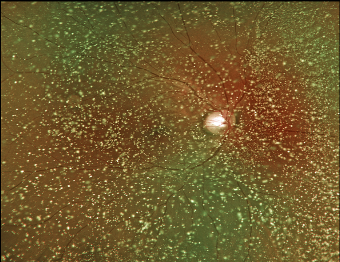
ASTEROID HYALOSIS
Originally posted on @retina.rocks March 7, 2022
This 22YO male presented with bilateral asteroid hyalosis. Vision was 20/20 OU with mildly symptomatic floaters.
Retinal examinations were extremely difficult due to the dense asteroid, although the fundi appeared normal.
Learning Points:
Asteroid hyalosis is caused by calcium-lipid complexes suspended throughout the vitreous. The condition gets its name from the white opacities resembling stars on a night sky.
Even with dense opacities, patients are rarely symptomatic, although retinal examination can be difficult. If a patient has vision loss with asteroid, the cause is almost always due to another condition.
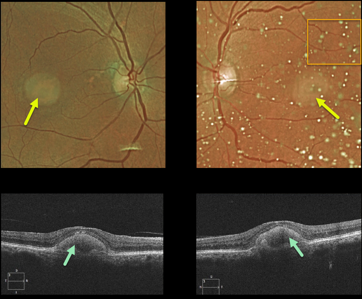
ADULT-ONSET FOVEOMACULAR VITELLIFORM DYSTROPHY
Originally posted on @retina.rocks January 18, 2022
This 87YO female shows classic findings for adult-onset foveomacular vitelliform dystrophy (Adult Best). Vision was 20/60 OD and 20/80 OS Optos imaging shows small, round, symmetrical subfoveal yellow lesions. Moderate asteroid hyalosis is noted as an incidental finding on the OS.
Triton swept-source OCT shows subretinal hyperreflective material above the RPE that extends into and indents the overlying retina.
Learning Points:
Adult Best is associated with a mutation in the BEST1 gene, which encodes the bestrophin-1 protein. Bestrophin-1, a calcium-activated chloride channel, is primarily found in the basolateral plasma membrane of the RPE.
BEST1 mutations cause a variety of phenotypes, including autosomal recessive bestrophinopathy, best vitelliform macular dystrophy, and autosomal dominant vitreoretinochoroidopathy.
These patients tend to maintain fairly good vision. Outer retinal and RPE atrophy can cause central vision loss, as can the rare development of macular neovascularization. Patients should therefore regularly monitor their vision with an Amsler grid.
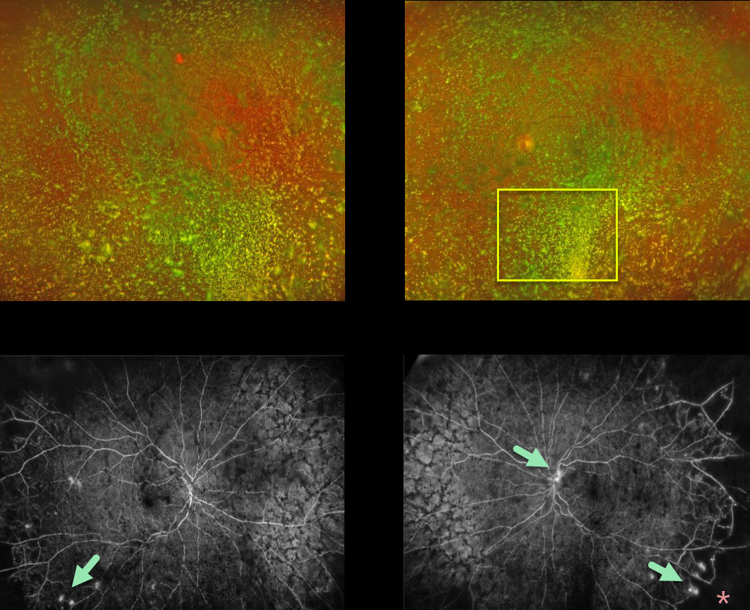
ASTEROID HYALOSIS
Originally posted on @retina.rocks July 29, 2020
Although this diabetic patient had normal vision with absolutely no symptoms, what we saw on clinical examination and Optos color imaging was nothing but asteroid hyalosis.
Fluorescein angiography (FA) showed bilateral proliferative diabetic retinopathy with areas of leakage and peripheral capillary dropout. In-office panretinal photocoagulation (PRP) was not possible due to the dense asteroid.
We discussed ongoing anti-VEGF injections versus vitrectomy + PRP to clear the visual axis. She decided to proceed with vitrectomy in both eyes.
Learning Points:
Asteroid hyalosis is caused by the accumulation of benign calcium-lipid particles suspended in the vitreous.
Fluoroscein angiography and OCT can image through asteroid hyalosis.
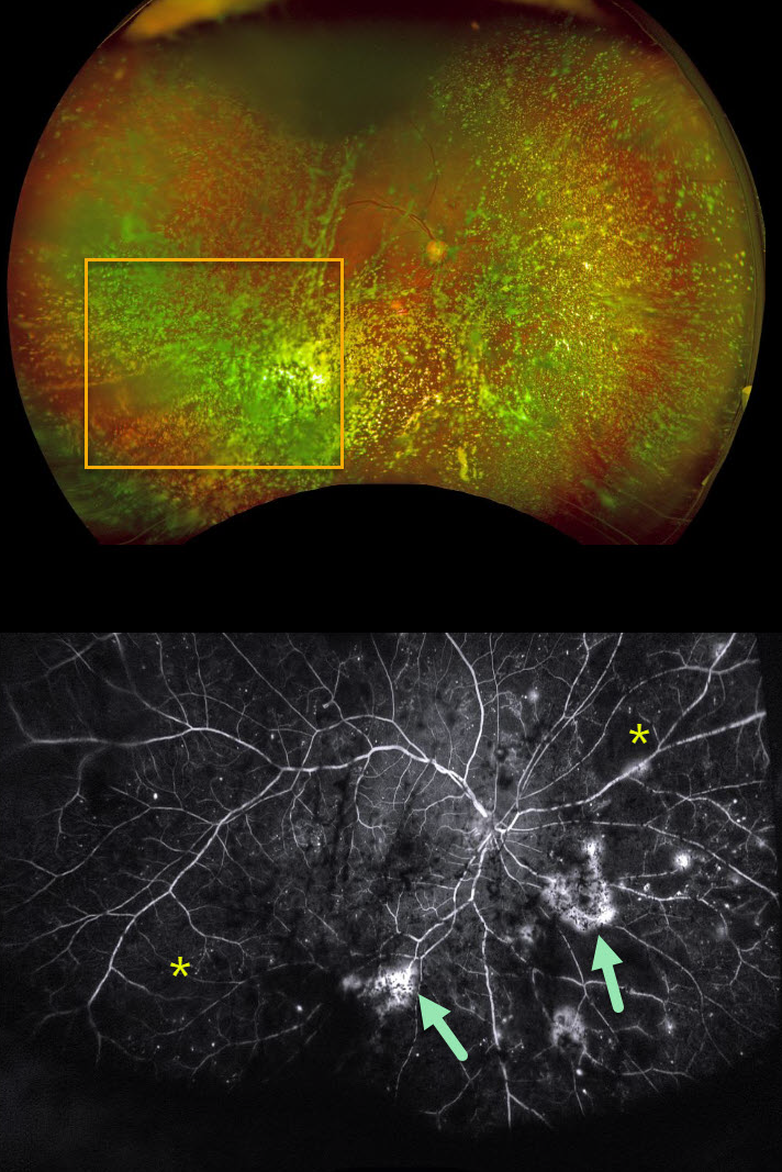
ASTEROID HYALOSIS OBSCURING OCCULT PROLIFERATIVE DIABETIC RETINOPATHY (PDR)
Originally posted on @retina.rocks April 17, 2020
Examining the fundus with dense asteroid hyalosis, as in this diabetic patient, can be extremely challenging.
Fluorescein angiography (FA) and OCT are great modalities since they’re both able to visualize the retina when the clinical exam can’t.
FA was critical in diagnosing proliferative retinopathy with peripheral ischemia in this patient.
Learning Point:
Asteroid hyalosis is caused by calcium-lipid complexes suspended throughout the vitreous. These white opacities resemble stars on a night sky – hence the name.


