

06 Vitreous, Vitreoretinal Interface Abnormalities, and Peripheral Retina
<< Back to Cases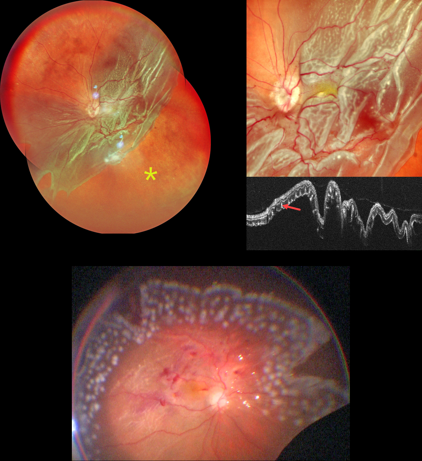
GIANT RETINAL TEAR
Shraddha Raj Shrivastava and Manish Nagpal
Originally posted on @retina.rocks September 1, 2026
This 62YO male presented with 3 months of vision loss in his left eye. Vision was counting fingers.
Pseudocolor SLO imaging shows a giant retinal tear extending from the 2 to 8 o’clock meridians with bare RPE distal to the posterior retina. There are extensive outer retinal corrugations within the posteriorly detached retina. OCT dramatically shows the detached retina, which is redundantly folded onto itself. The outer retinal corrugations appear as numerous hyperreflective outer retinal pillars.
The detachment was emergently repaired with vitrectomy, membrane peeling, endolaser photocoagulation, and silicone oil. The retina was completely attached at the end of the procedure.
Learning Points:
Outer retinal corrugations, also known as hydration outer retinal folds, are one of the key findings that help differentiate a rhegmatogenous retinal detachment from exudative and traction retinal detachments. The cause of these folds remains uncertain but may result from the hydration of glycosaminoglycans in the interphotoreceptor matrix (Dalvin et al, Retinal Cases & Brief Reports 2020;14:355-359).
DEGENERATIVE RETINOSCHISIS
Malvika Singh and Manish Nagpal
Originally posted on @retina.rocks August 25, 2026
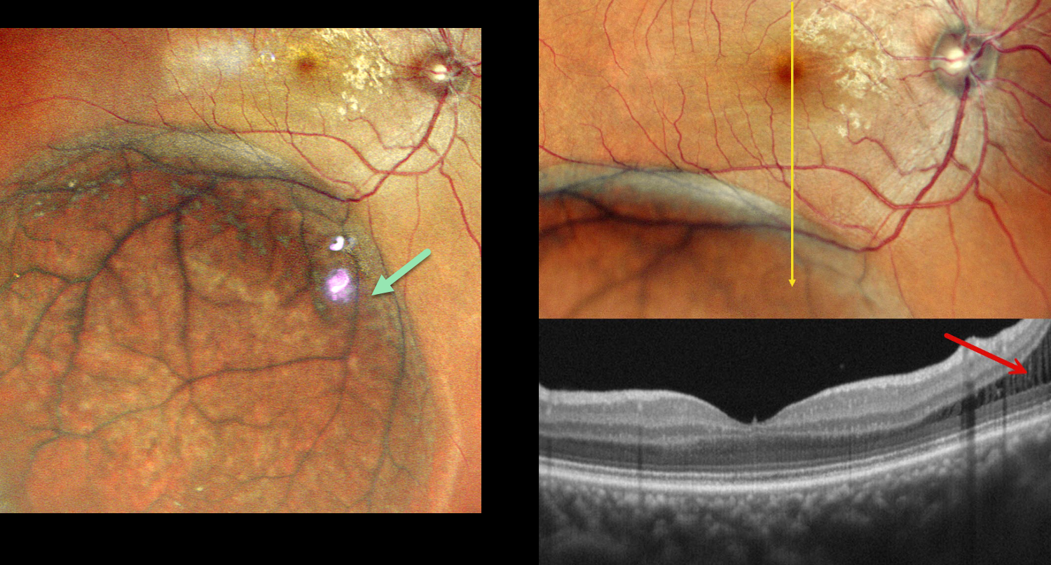
This asymptomatic 40YO female was referred for a retinal detachment in her right eye. Vision was 20/20 OD and 20/20 OS in her normal OS.
Pseudocolor SLO imaging shows a large area of inferotemporal degenerative retinoschisis (DRS) extending just into the distal macula. OCT scanning confirms schisis without retinal detachment. Observation was recommended.
Learning Points:
Uncomplicated DRS typically follows a stable or slowly evolving course that should not be treated. The development of inner-layer and outer-layer holes essentially creates a full-thickness retinal break. Progression of subretinal fluid beyond the DRS margins (combined schisis-rhegmatogenous retinal detachment, RRD), in the presence of both inner- and outer-layer holes, is the only DRS scenario requiring intervention.
It is often clinically difficult to differentiate pure DRS from a combined schisis-RRD or from RRD. OCT scanning through the area of interest, as in our patient, often provides definitive evidence for the proper diagnosis and management (Paris et al, Survey Ophthalmology 2026;71:71-80).
PNEUMATIC RETINOPEXY
Originally posted on @retina.rocks August 13, 2026
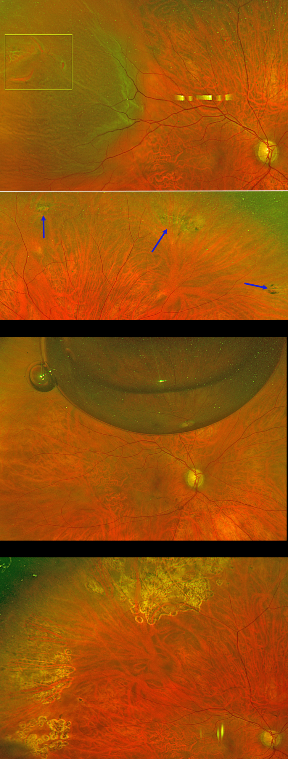
This 56YO phakic male presented with floaters and a football-shaped nasal shadow in his right eye for one week. Vision was 20/20 bilaterally.
Optos color RG imaging shows a superotemporal macula-on rhegmatogenous retinal detachment (RRD) from several tiny adjacent breaks within lattice. There were other areas of lattice degeneration superiorly, inferiorly, and inferotemporally within the attached retina. Options for retinal detachment repair were discussed, including pneumatic retinopexy, scleral buckle, and vitrectomy.
The patient chose to undergo pneumatic retinopexy (PnRP) with 0.3cc of pure C3F8. The areas of lattice within the attached retina were lasered just prior to performing the procedure. The retina was completely attached the following day. The causative break was lasered four days later once the detachment flattened. Two months postoperatively, vision was 20/20, and the retina remains attached. Given the diffuse areas of lattice and absence of posterior vitreous detachment in the fellow eye, we performed prophylactic laser retinopexy to the left eye (not shown).
Learning Points:
PnRP was introduced by Hilton and Grizzard in 1986 as an in-office alternative to traditional retinal reattachment surgery (Ophthalmology 1986;93:626-651). It was initially recommended for detachments with one or more breaks within a single clock hour, located within the superior eight clock hours of the fundus.
Forty years after its introduction, the role of PnRP, compared with scleral buckling and vitrectomy, remains controversial. In Canada, where access to operating rooms is more limited than in the US, PnRP is often the initial treatment of choice for appropriate detachments. The Canadian PIVOT trial reported that PnRP offered better final vision, less vertical metamorphopsia, and reduced morbidity compared to vitrectomy (Ophthalmology 2019;126:531-539).
However, in the US, PnRP is not as popular except on a late Friday afternoon (we’re only partially kidding). A recent critical review (Chronopoulos et al, Surv Ophthalmology 2021;66:585-593) argued against PnRP for initial treatment due to the need for “multiple procedures that largely negate its potential cost savings and subjects the patient to prolonged stress and disability…” A recent cost-utility analysis surprisingly found PPV to be the most cost-effective primary procedure (Teja et al, AJO 2023;255:141-154).
Although extensive lattice degeneration is generally considered a contraindication for pneumatic retinopexy, we decided to perform prophylactic treatment. Our patient did well after lasering the areas of lattice prior to performing the pneumatic procedure and was followed closely with scleral depression exams.
COMBINED RETINOSCHISIS-RHEGMATOGENOUS RETINAL DETACHMENT
Originally posted on @retina.rocks August 12, 2026
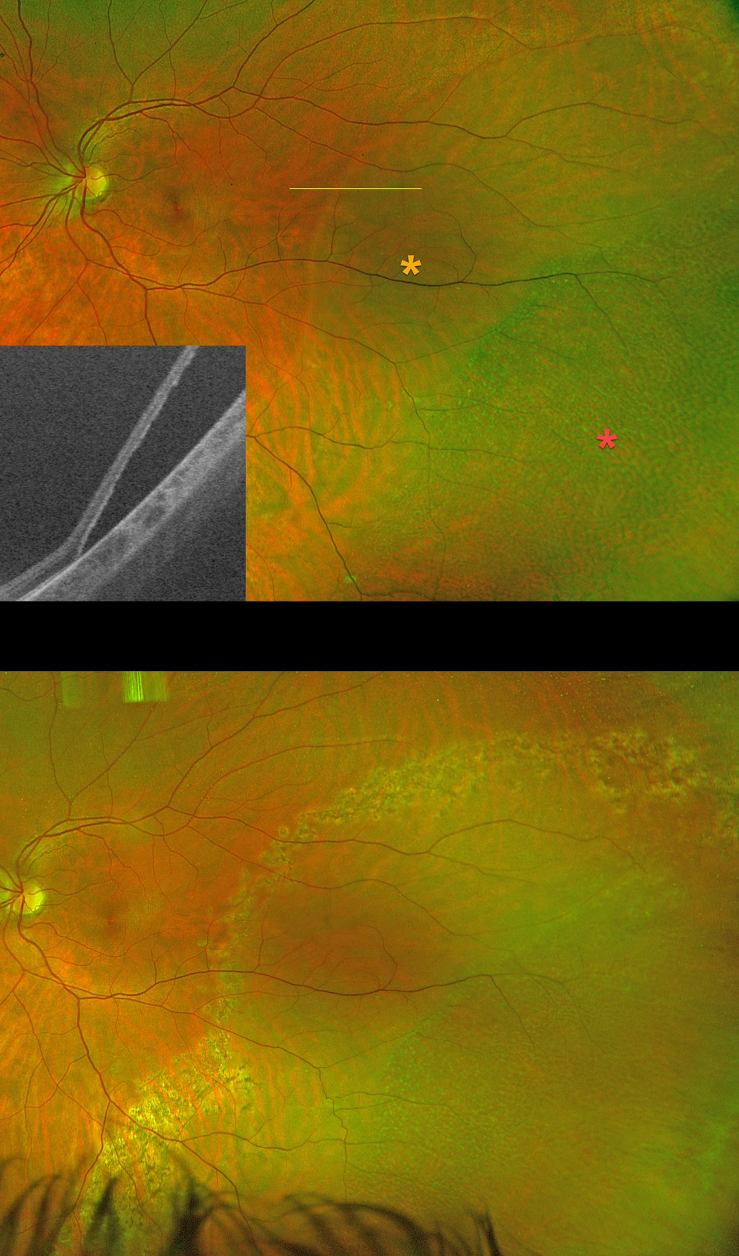
This 35YO pregnant female presented with 3 days of flashes in her left eye. She had a family history of retinal tears in her father and rhegmatogenous retinal detachment (RRD) in her maternal uncle. Vision was 20/20 OS.
Silverstone color RG imaging shows a macula-on combined retinoschisis-RRD. There was no posterior vitreous detachment. Schisis is noted more peripherally, with the rhegmatogenous fluid noted more posteriorly. Swept-source OCT confirms the presence of a full-thickness retinal detachment. Treatment options were discussed, including laser demarcation, primary scleral buckle with cryotherapy, and pars plana vitrectomy. She elected for laser retinopexy, which was performed that day.
She delivered a healthy baby, and her retina has remained stable over the past 9 months as of her last visit.
Learning Points:
Isolated degenerative retinoschisis should not be treated. However, it can progress to a combined schisis-RRD in the presence of breaks in both the inner and outer layers. Asymptomatic combined detachments can be demarcated, as in our patient. Symptomatic detachments are usually treated with pars plana vitrectomy.
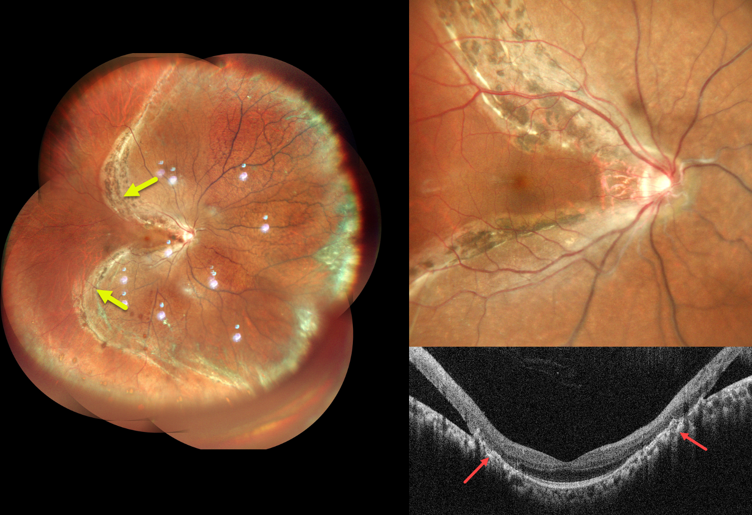
SELF-SEALING RHEGMATOGENOUS RD WITH HEAVY ENCIRCLING DEMARCATION LINE
Anand Gandhi and Manish Nagpal
Originally posted on @retina.rocks August 11, 2026
This healthy 31YO male presented with blurred vision in his right eye for 2 years. Best-corrected visual acuity was 20/20 OU.
Pseudocolor SLO imaging shows a chronic rhegmatogenous retinal detachment (RRD) involving the superior, nasal, and inferior quadrants, sparing the central and temporal macula. A prominent pigmented demarcation encircles the RRD. Vertical OCT scanning shows a normal macular center with broad chorioretinal scarring superiorly and inferiorly bordering the attached and detached retina.
Learning Points:
Acute RRDs are virtually always symptomatic, requiring emergent surgery. However, asymptomatic detachments often have a low risk of progression and can usually be safely observed (Cohen et al, AJO 2005;139;777-779; Sengillo et al, Ophthalmology Retina 2023;7:215-220) or demarcated (Vrabec and Baumal, Ophthalmology 2000;107:1063-1067).
In the present case, although the retinal detachment was symptomatic at onset, it was not diagnosed until several years later. The heavy demarcation line indicates chronicity with likely stability. Given the preserved visual acuity, macular sparing, and chronic nature of the detachment, close observation was recommended.
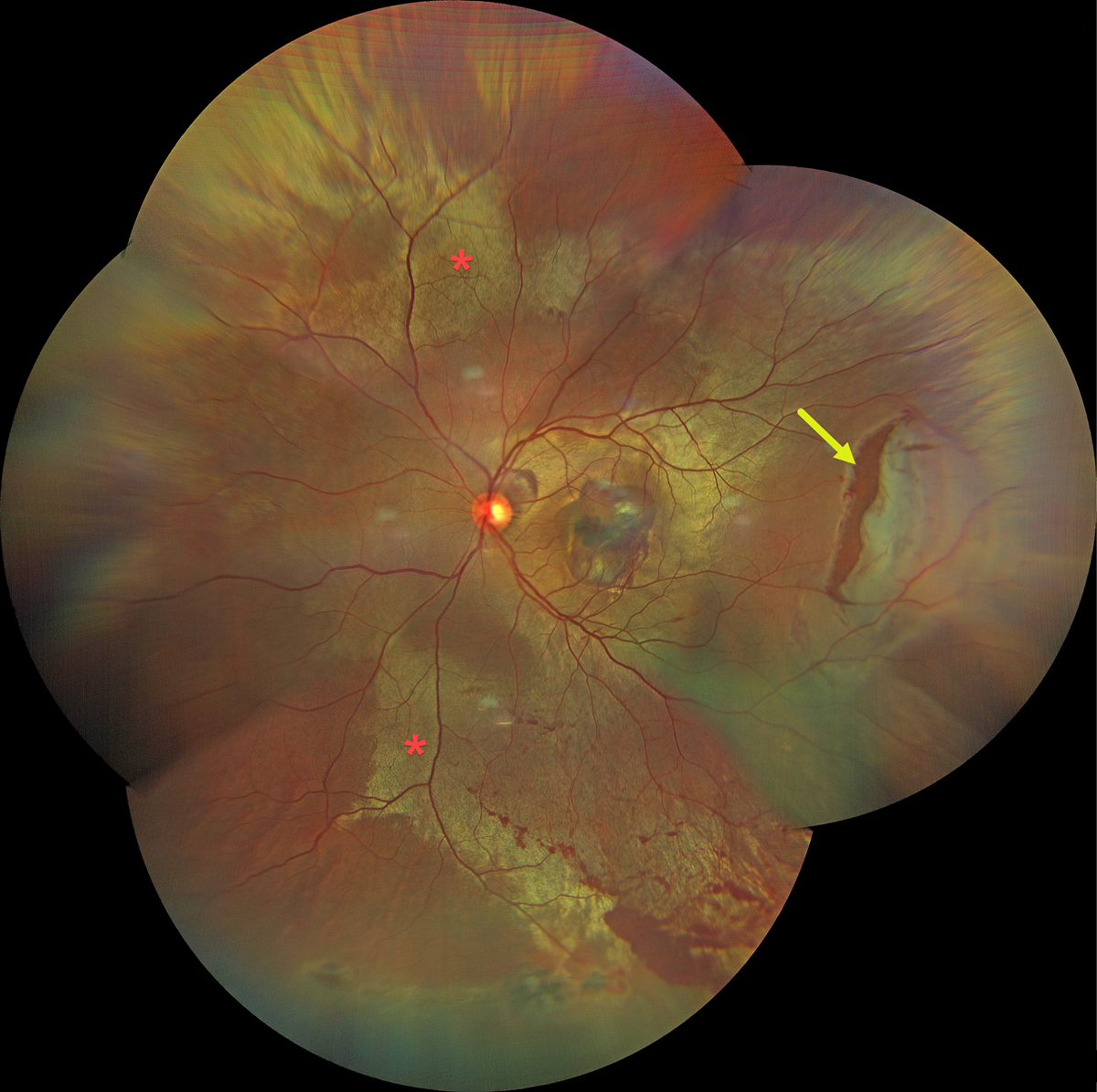
COMMOTIO RETINAE WITH TRAUMATIC RETINAL BREAK
Dibya Prabha
Originally posted on @retina.rocks August 7, 2026
This 38YO male presented with sudden vision loss in his left eye immediately following blunt trauma from a cricket bat 2 days earlier. Vision was 20/200.
Color fundus photography shows extensive commotio retinae throughout the posterior pole, along with a large retinal tear in the temporal midperiphery. Oral and topical steroids were prescribed, and the retinal break was surrounded with laser photocoagulation.
Learning Points:
Commotio retinae is caused by direct, blunt eye trauma. The outer retinal whitening is due to shearing of the photoreceptor outer segments. The term “Berlin’s edema” is a misnomer and should not be used, as the retina is not edematous. The acute white retinal changes usually resolve within several weeks. With resolution, the fundus appearance can normalize. With more severe injury, outer retinal loss can allow RPE cells to migrate into the retina, giving a pseudo-retinitis pigmentosa appearance. Patients can also develop retinal breaks and detachment.
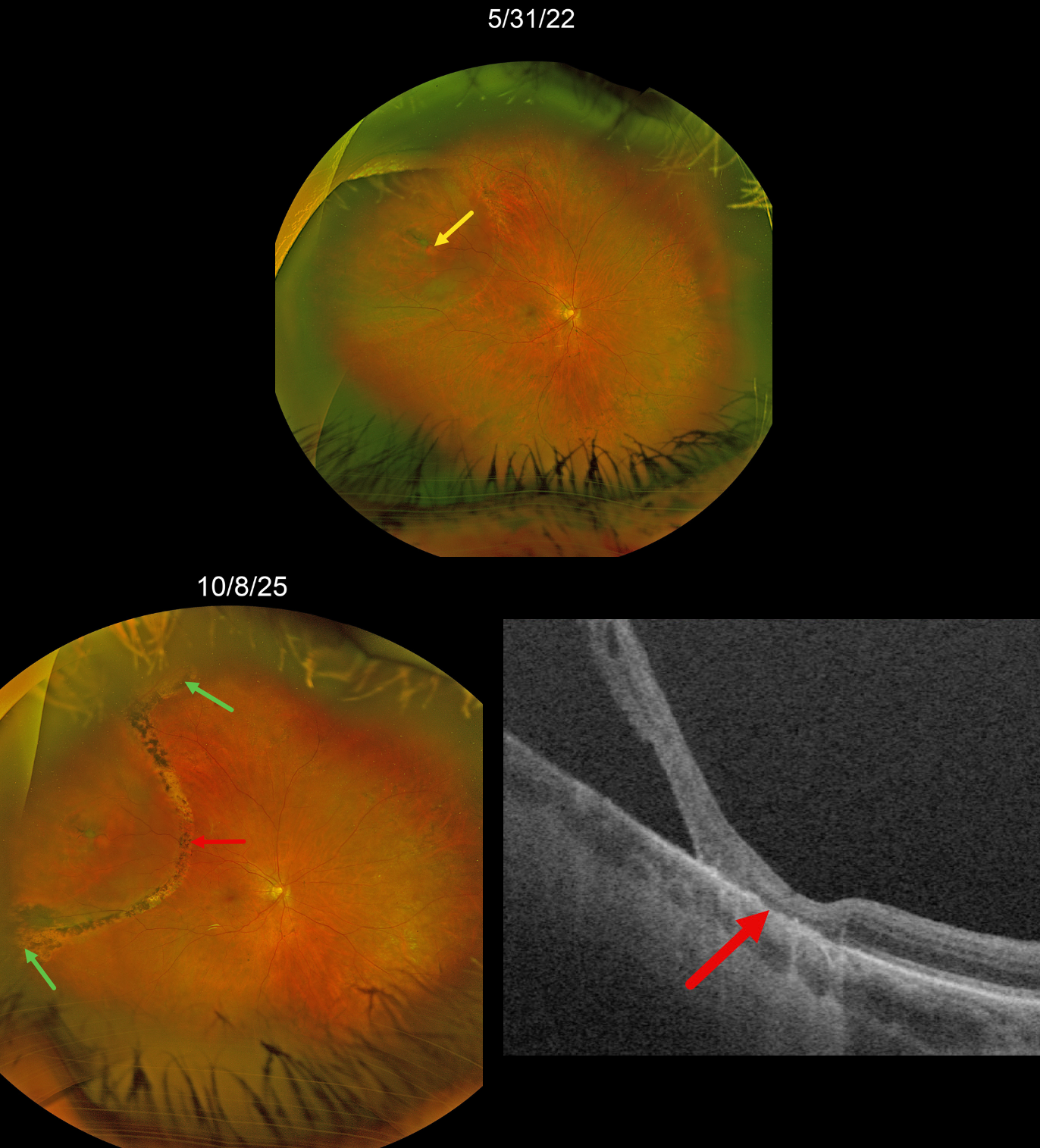
RHEGMATOGENOUS RD WITH LASER DEMARCATION
Originally posted on @retina.rocks July 29, 2026
This 74YO female was referred on 5/31/22 for an asymptomatic rhegmatogenous retinal detachment (RRD) in her right eye.
Optos color RG imaging shows a chronic appearing superotemporal macula-on RRD with a single retinal break. A faint demarcation line is noted along the detachment’s posterior edge. The detachment was subsequently demarcated with thermal laser photocoagulation posteriorly and cryotherapy to its anterior edges.
We have been examining her twice yearly since. When last examined on 10/8/25, the detachment remained well-demarcated and without spread, with scarring from laser and cryotherapy. Optos Silverstone RGB swept-source OCT shows a broad area of outer retinal atrophy within the area of retinopexy.
Learning Points:
Demarcation, rather than reattachment, of the retina is a valid option for select cases of RRD. When demarcating these detachments, it is critical that the retinopexy completely surrounds the subretinal fluid and that treatment extends to the ora; otherwise, the detachment can spread through the untreated retina. Patients need to be followed postoperatively, as detachment can occasionally progress despite prior retinopexy.
See Vrabec and Baumal, Ophthalmology 2000;107:1063-1067 for an excellent review regarding demarcation laser photocoagulation for selected macula-sparing retinal detachments.
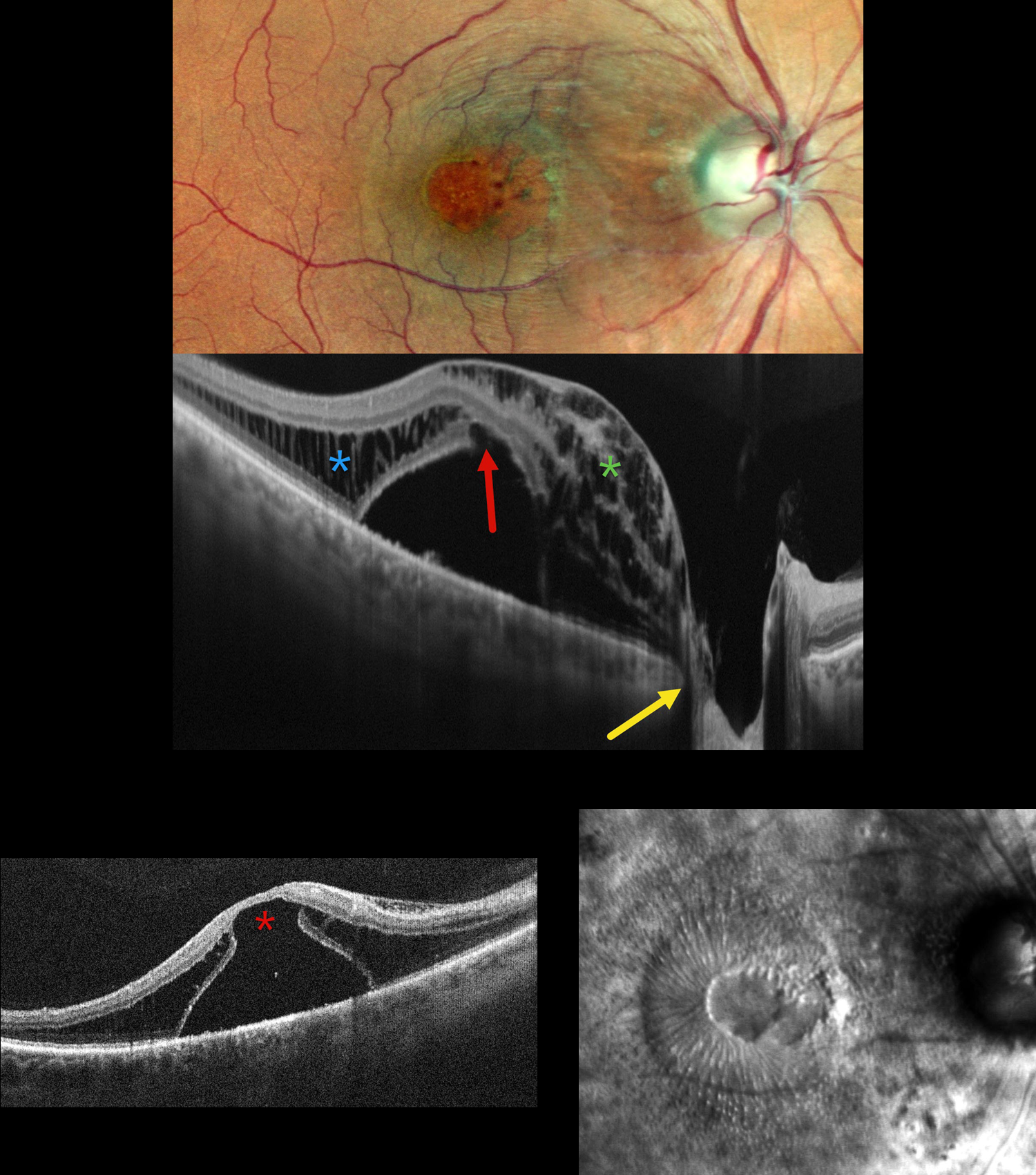
OPTIC NERVE PIT
Shraddha Raj Shrivastava and Manish Nagpal
Originally posted on @retina.rocks July 24, 2026
This 50YO male presented with 1 year of decreased vision in his right eye. He had been treated elsewhere with intravitreal injections for cystoid macular edema. Vision was 20/200 OD and 20/20 in his normal OS.
Pseudocolor SLO imaging shows a possible serous macular detachment with a large macular pseudohole. A possible pit is noted along the temporal aspect of the optic nerve. OCT scanning shows an elevated serous detachment. An outer-layer macular hole is noted along the posterior aspect of the fovea, along with full-thickness cystic retinal edema nasally and outer nuclear layer cystic edema temporally. The subretinal fluid directly communicates with the optic nerve pit. Retro mode reveals a unique view of the macular pathology, with the central outer macular hole and radiating fluid.
Learning Points:
An optic nerve pit is a rare (about 0.1% prevalence) congenital anomaly thought to be caused by incomplete closure of the optic fissure during gestation. Pits are most often located within or along the inferior-temporal disc margin and usually appear as a gray depression. While most optic pits are asymptomatic, they can cause vision loss by forcing fluid into the inner retina, outer retina, and, ultimately, the subretinal space (optic pit maculopathy). The source of the fluid remains a subject of ongoing debate, with opinions ranging from the vitreous to the subarachnoid space.
Vitrectomy surgery was discussed, and the patient elected for observation unless his symptoms worsened.
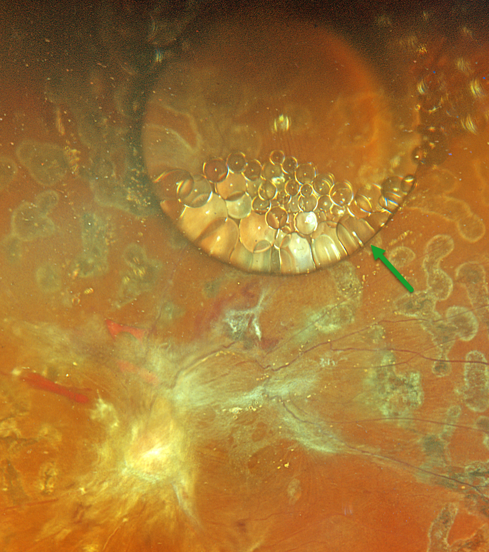
SILICONE FISH EGGS
Originally posted on @retina.rocks July 21, 2026
This 43YO male underwent a vitrectomy elsewhere for a diabetic traction retinal detachment about 5 months earlier. Vision was 20/400.
Optos color RGB imaging shows a large, round silicone oil droplet in the superior midperiphery. This bubble acts like a convex lens, magnifying the details of the underlying retina. Suspended within and along the edges of this droplet are numerous fish egg droplets. Residual fibrotic membranes extend from the nerve into the macula and along the superotemporal arcade. Repeat vitrectomy was scheduled.
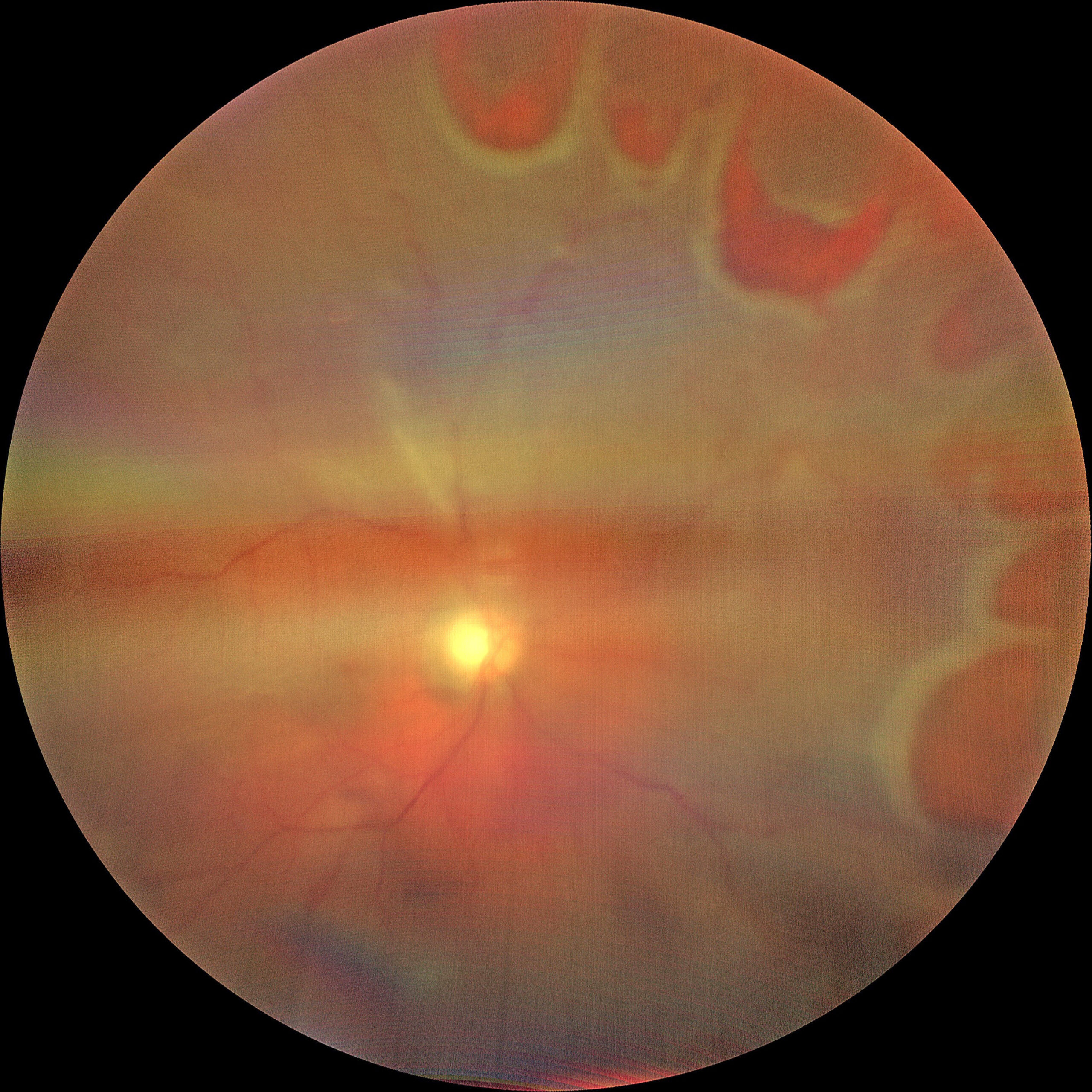
RHEGMATOGENOUS RETINAL DETACHMENT
Abhishek Karra and Dinesh Garg
Originally posted on @retina.rocks July 1, 2026
This 53YO male presented with 1 week of sudden vision loss in his right eye. Vision was counting fingers.
Fundus photography shows multiple retinal tears extending from 12 to 4 o’clock. There is a macula-off superior and nasal rhegmatogenous retinal detachment (RRD). The image is hazy due to an overlying vitreous hemorrhage. Emergency vitrectomy was scheduled.
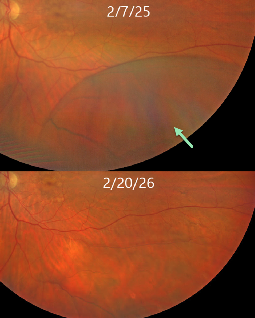
SPONTANEOUSLY FLATTENED DEGENERATIVE RETINOSCHISIS
Kris Garbig and Doug Garbig
Originally posted on @retina.rocks June 10, 2026
This 77YO female presented for routine examination on 2/7/25 with asymptomatic degenerative retinoschisis (DRS) inferotemporally in her left eye. When she returned one year later, the schisis had completely spontaneously flattened.
Learning Points:
The dynamics of DRS cavities were noted by Byer in 1972 when he reported the first case of spontaneous regression (Arch Ophthalmology 1972;88:207-209). Since adopting UWF imaging in our practice, we have found that schisis is often not static, with DRS enlarging or flattening. Inner- and outer-layer breaks also appear over time, often independent of changes in the schisis cavity.
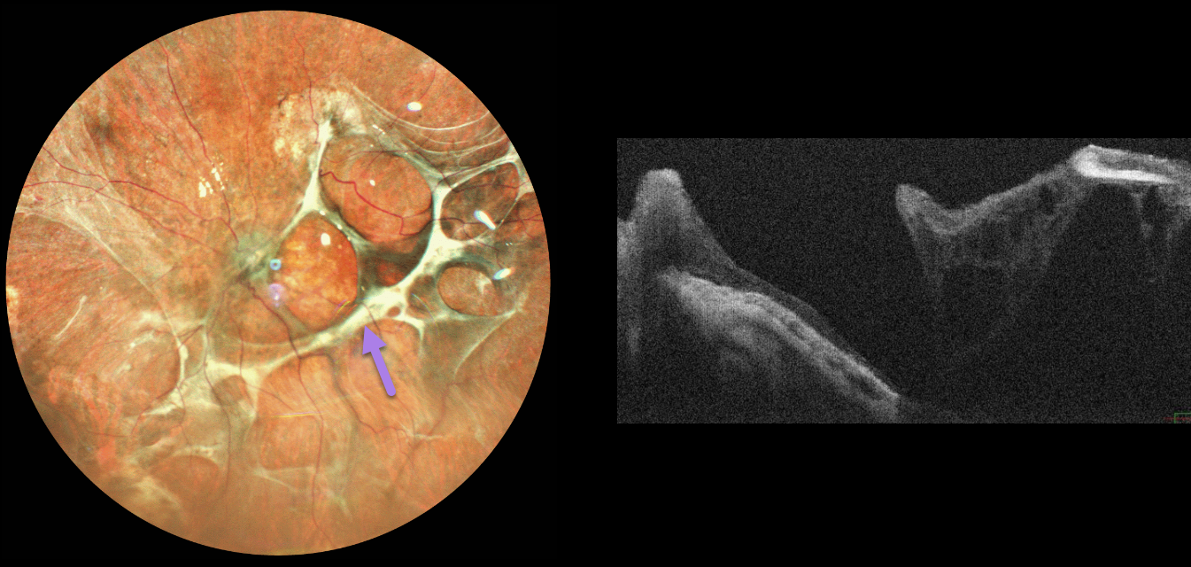
SUBRETINAL AND PRERETINAL PROLIFERATIVE VITREORETIONPATHY
The European VitreoRetinal Society (EVRS), Akansha Sharma, Manish Nagpal and Navneet Mehrotra
Originally posted on @retina.rocks May 29, 2026
This 71YO male presented with one year of poor vision in his left eye following vitrectomy with silicone oil elsewhere for a rhegmatogenous retinal detachment. Vision was light perception.
Pseudocolor SLO imaging shows a complex pattern of interlacing subretinal fibrosis bands. OCT scanning shows areas of disorganized retinal thinning, thickened disorganized retina nasally, and retinal detachment more temporally. Observation was recommended given the poor visual prognosis.
Learning Points:
Proliferative vitreoretinopathy (PVR), which consists of cellular membranes growing on either retinal surface, develops in about 5-10% of RRD. The membranes contract, foreshorten the retina, and likely represent an aberrant wound-healing response. There are no proven pharmacologic therapies to prevent or treat PVR. These complex detachments can be managed with vitrectomy and membrane peeling, retinectomy, scleral buckling, and silicone oil. However, PVR remains the main cause for ultimate retinal reattachment surgical failure.
See Garweg et al Surv Ophthalmol 2013;48:321-329 for a good review of PVR pathophysiology.
GIANT RETINAL TEAR
Originally posted on @retina.rocks May 27, 2026
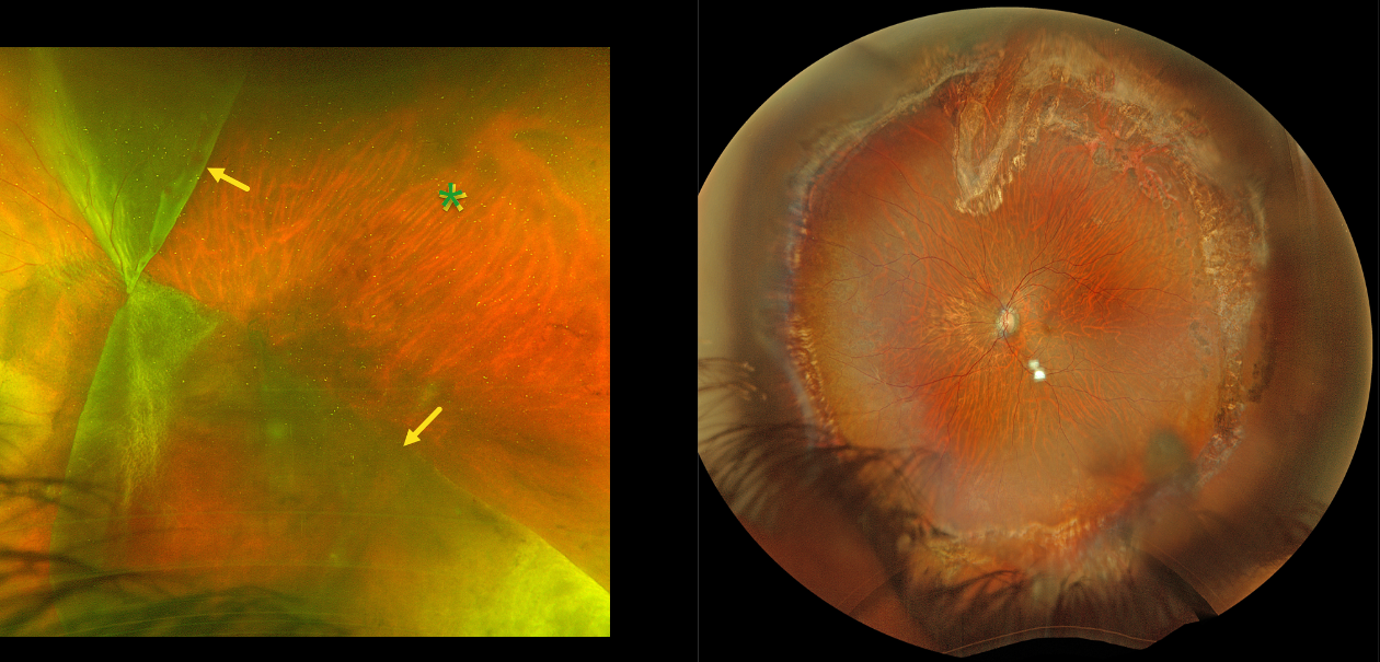
This 44YO male presented with hand motion vision OS from an acute giant tear (GRT) retinal detachment. Optos color RG imaging shows a complex GRT with one flap folded superonasally and the other inferotemporally, creating a sharply defined square region of temporal bare RPE. The optic disc is covered by the giant tear. Emergent vitrectomy was scheduled.
Six months postoperatively, the retina remained completely attached. Vision was 20/80.
LATTICE DEGENERATION WITH SYMPTOMATIC RETINAL TEARS
Originally posted on @retina.rocks May 14, 2026
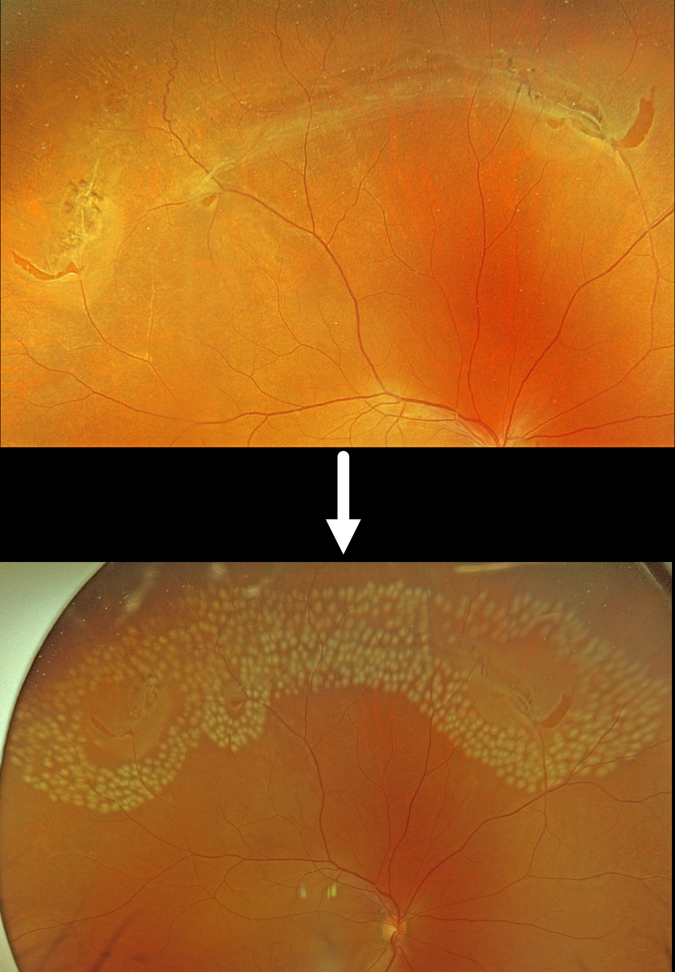
This 55YO male was referred for an acute posterior vitreous detachment with symptomatic retinal tears. Vision was 20/25.
Optos color RGB imaging shows several patches of lattice degeneration extending from the 10 to 1:30 o’clock meridians associated with multiple retinal tears. Some inferior vitreous hemorrhage was noted (not shown). Immediate prophylactic laser photocoagulation was applied to all surrounding the lattice and breaks.
Lattice lesions have a pocket of liquefied vitreous overlying thinned retina with a firm area of surrounding vitreoretinal adhesion. White, fibrosed vessels often crisscross the lesions, giving them their name, “lattice.” This firm vitreoretinal adhesion, along with the often-present atrophic holes, is what can predispose these eyes to retinal breaks and detachment. Although prophylactic treatment is rarely needed, symptomatic breaks, as in our patient, need prompt retinopexy.
PROLIFERATIVE DIABETIC RETINOPATHY WITH ASTEROID HYALOSIS
The European VitreoRetinal Society (EVRS), Akansha Sharma, Manish Nagpal, and Vaidehi Sathaye
Originally posted on @retina.rocks April 27, 2026
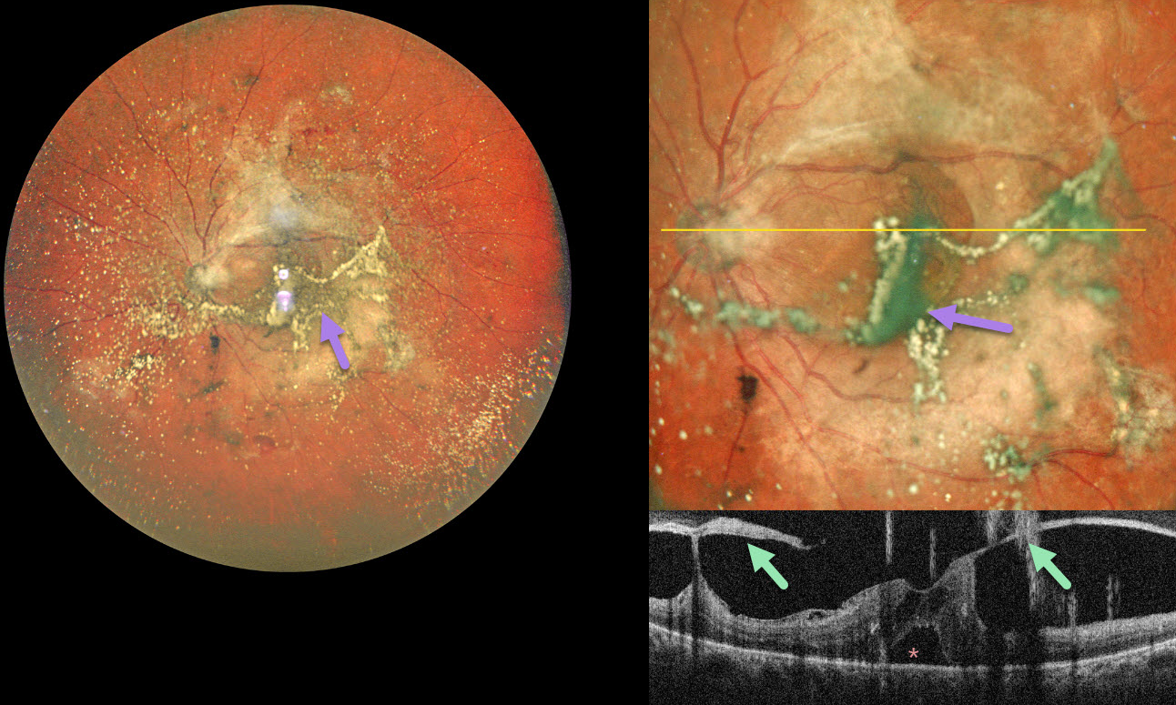
This 55YO diabetic male presented with 2 months of bilateral vision loss. Vision was 20/80 OD and 20/120 OS.
Pseudocolor SLO ultrawidefield imaging of his left eye shows moderate asteroid hyalosis, with larger condensed stands of material obscuring the underlying macula. Spectral domain OCT scanning shows complex variable vitreous traction on the disc and macula with a small foveal detachment. The asteroid particles appear as hyperreflective vertical lines resembling falling rain.
Learning Points:
Examining the fundus in a patient with dense asteroid, as in this diabetic patient, can be extremely challenging. Fluorescein angiography and OCT are great modalities because they can visualize the retina when the clinical exam can’t. In our patient’s case, the OCT revealed foveal traction, indicating the need for vitrectomy surgery.
VITREOPAPILLARY TRACTION WITH CHRONIC MACULAR HOLE
Originally posted on @retina.rocks April 15, 2026
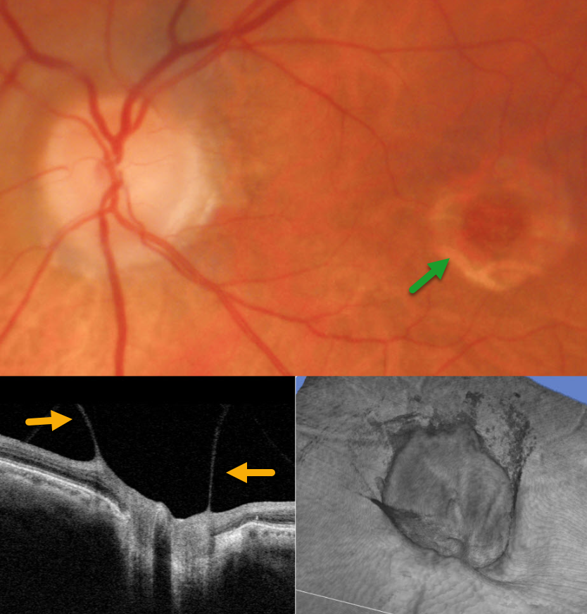
This 81YO male presented for his yearly examination without new complaints. Vision in his left eye was stable at 20/200 due to a chronic macular hole.
Triton color photography shows a full-thickness macular hole with a surrounding large rim of pigment loss. On slit lamp biomicroscopy, a partial PVD extended to the nerve with an elevated, partially attached Weiss ring. Vitreopapillary traction was confirmed on B-scan and 3D OCT. Continued observation was recommended.
Learning Points:
The normal vitreous undergoes a decades-long process of orchestrated degeneration, including liquefaction and gradual separation from the retinal surface. The posterior vitreous detachment (PVD) begins in the perifoveal retina early in life, followed by a foveal PVD, then a peripheral PVD, and then finally a complete PVD when the vitreous separates from the optic nerve (Johnson AJO 2010;149:371-382). The integrity of the internal limiting membrane (ILM) is integral to normal vitreoretinal adhesion, where a thinner ILM has a firmer adhesion than a thicker ILM. The ILM represents the basement membrane of the Müller cell. Hence, an area with fewer Müller cells reaching the inner retina will naturally have a firmer vitreoretinal adhesion. Since the optic nerve is completely devoid of Müller cells, this is the area of the posterior pole that has the firmest vitreoretinal adhesion, and this is the reason why the vitreous tends to remain so firmly attached to it.
Persistent vitreopapillary traction is a rarely reported phenomenon (Gabriel et al, Neuro-ophthalmology 2020;44:213-218), probably due to its lack of symptoms and often subclinical findings. In our patient’s case, the traction caused retinal thickening and hemorrhage, somewhat mimicking a peripapillary macular neovascularization. In our experience, these eyes usually remain asymptomatic and can be observed, although vitrectomy is very successful in relieving the traction and improving vision.
GIANT RETINAL TEAR
The European VitreoRetinal Society (EVRS), Shilpi Narnaware and Prashant Bawankule
Originally posted on @retina.rocks April 10, 2026
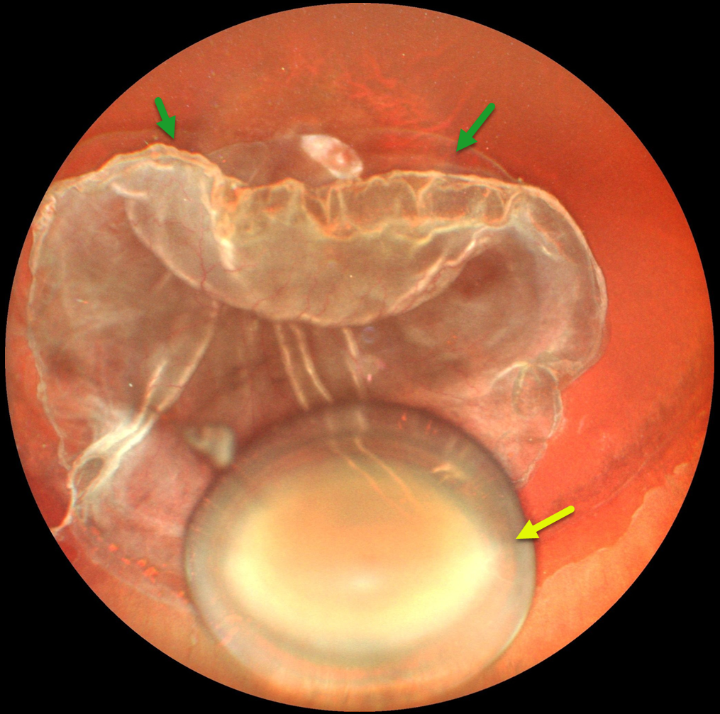
This 32YO male with known Marfan’s syndrome presented with one month of vision loss in his left eye. Examination revealed pathological myopia with bilateral spontaneously dislocated cataracts and a total rhegmatogenous retinal detachment (RRD) OS. Best-corrected vision was 20/80 OD and light perception OS.
Ultrawidefield photography shows a superior >270-degree giant retinal tear, total RRD, rolled edges, and a dislocated cataractous crystalline lens in the inferior vitreous. Emergent pars plana vitrectomy/lensectomy was planned, but unfortunately, he did not show for his surgery and was subsequently lost to follow-up.
PROLIFERATIVE VITREORETINOPATHY
Originally posted on @retina.rocks April 8, 2026
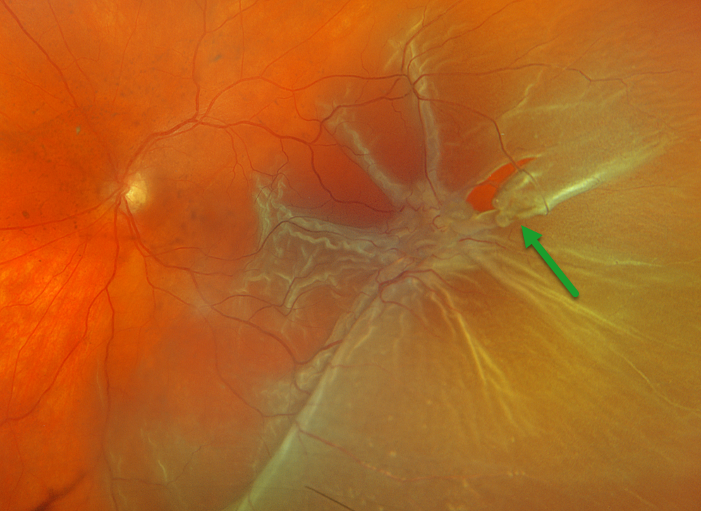
This 63YOF presented with a week of vision loss in her right eye. Optos color RGB imaging shows a temporal macula-off rhegmatogenous retinal detachment (RRD) complicated by proliferative vitreoretinopathy (PVR) with a large star fold just posterior to a retinal tear. She was scheduled for emergent vitrectomy.
Learning Points:
PVR, which consists of cellular membranes growing on either retinal surface, develops in about 5-10% of retinal detachments. RRD with a concurrent non-causal macular hole is found in about 2% detachments (Iros et al, Retina 2023;43:581-584). Although most eyes achieve anatomic success, final vision is usually poor. In our patient, unfortunately, there is no further clinical information, so it is unclear whether the macular hole caused the detachment or was secondary to traction from the star fold.
GIANT RETINAL TEAR
The European VitreoRetinal Society (EVRS), Akansha Sharma, Manish Nagpal, Vaidehi Sathaye, and Navneet Mehrotra
Originally posted on @retina.rocks March 27, 2026
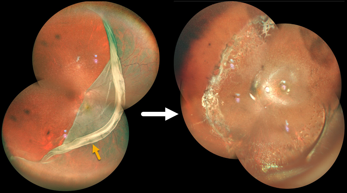
This 15YO male presented with sudden vision loss in his right eye. Vision was hand motion. Pseudocolor SLO imaging shows a giant retinal tear extending from 7:30 to 12 o’clock, with the macular retina folded onto itself nasal to the nerve. Emergent pars plana vitrectomy with silicone oil, followed by removal of the oil 7 months later, successfully repaired the detachment with vision improving to 20/200.
PNEUMATIC RETINOPEXY
Originally posted on @retina.rocks March 25, 2026
This 76YO female presented with several days of painless vision loss in her right eye. Vision was 20/30.
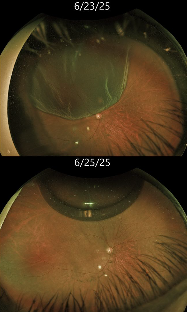
Optos color RG imaging shows a bullous superotemporal rhegmatogenous retinal detachment (RRD). The macular center was shallowly detached. Scleral depression revealed a single tiny retinal tear anterior to the 11:30 equator. Cryotherapy for this tear, followed by pneumatic retinopexy with 0.2 cc C3F8, was performed. Two days later, vision was 20/30. A single gas bubble is noted superiorly, and the retina is completely attached.
Learning Points:
Pneumatic retinopexy (PnRP) was introduced by Hilton and Grizzard in 1986 as an in-office alternative to traditional retinal reattachment surgery (Ophthalmology 1986;93:626-651). It was initially recommended for detachments with one or more breaks within a single clock hour, located within the superior eight clock hours of the fundus.
Nearly 40 years after its introduction, the role of RRD repair in RRD repair, compared with scleral buckling and vitrectomy, remains controversial. In Canada, where access to operating rooms is more limited than in the US, PnRP is often the initial treatment of choice for appropriate detachments. The Canadian PIVOT trial reported that PnRP offered better final vision, less vertical metamorphopsia, and reduced morbidity compared to vitrectomy (Ophthalmology 2019;126:531-539). However, in the US, PnRP is not as popular except on a late Friday afternoon (we’re only partially kidding). A recent critical review (Chronopoulos et al, Surv Ophthalmology 2021;66:585-593) argued against PnRP for initial treatment due to the need for “multiple procedures that largely negates its potential cost savings and subjects the patient to prolonged stress and disability…” A recent cost-utility analysis surprisingly found PPV to be the most cost-effective primary procedure (Teja et al, AJO 2023;255:141-154).
We personally recommend PnRP for patients with mobile superior RRD with superior breaks. Patients must be able to properly position their head. We must be able to visualize the retinal periphery well to ensure there are no inferior breaks, and ideally, we prefer that the macula be detached in case a failed PnRP converts a macula-on RRD to a macula-off RRD.
We therefore felt that our patient was a perfect candidate for PnRP. The PIVOT trial also supported our decision, with a recent post hoc analysis showing similar single-surgery success with PnRP (88%) vs PPV (91%) for single-break primary RRD (Pecaku et al., Br J Ophthalmol 2025;109:113-118).
POSTERIOR VITREOUS DETACHMENT
Originally posted on @retina.rocks March 24, 2026
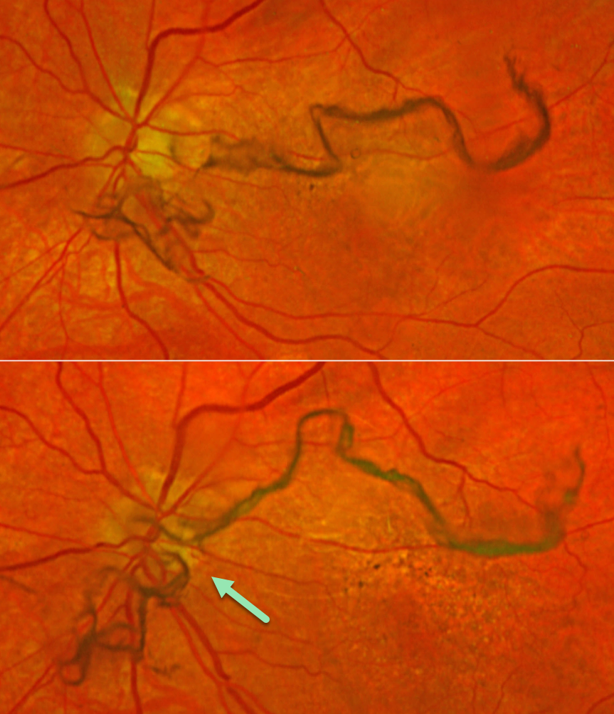
This 73YO female presented with several days of new floaters in her left eye. Vision was 20/15 bilaterally. Optos color RGB imaging shows extremely prominent mobile string-like posterior hyaloid condensations floating over the disc and macula. There were no retinal breaks or detachment.
Learning Points:
The clinical diagnosis for a complete posterior vitreous detachment (PVD) remains challenging and can be established with biomicroscopy when a Weiss ring, representing avulsed peripapillary glial tissue, is visualized. OCT can be helpful in supplementing the clinical exam (Wagley et al, Retina 2021;41:2296-2300).
An acute PVD is the moment of truth for when a retinal break or detachment develops. The historical risk of a break is 10-15%, although a large retrospective study from Kaiser Permanente found this risk to be significantly lower at 5% for a retinal break and 4% for retinal detachment (Ophthalmology 2022;129:67-72). The presence of vitreous pigment or blood and lattice, and a history of a retinal break or detachment in the fellow eye, increases this risk.
SHOULD THIS LESION BE EMERGENTLY LASERED???
Originally posted on @retina.rocks March 11, 2026
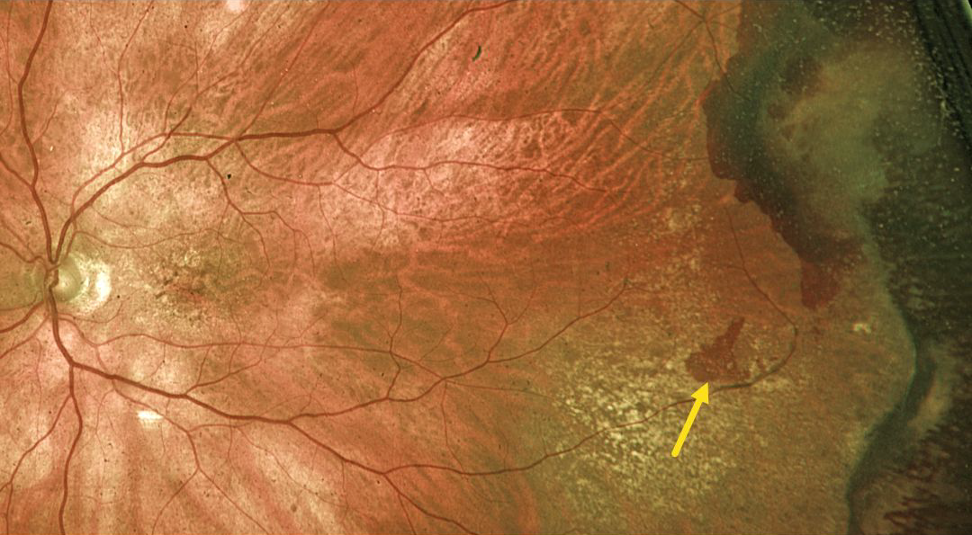
This 89YO male was referred for an asymptomatic retinal tear with possible retinal detachment in his left eye. Vision was 20/60.
Optos color RG imaging shows a red horseshoe-shaped lesion in the temporal midperiphery. Would you recommend immediate thermal laser demarcation of this retinal tear?
We hope not! This patient has asymptomatic and chronic ectopic choroidal neovascularization (CNV), which is evidenced by the more peripheral subretinal scarring and superotemporal dark subretinal blood. Ectopic CNV can often masquerade as a choroidal mass or uveal melanoma. It can also cause extensive choroidal/suprachoroidal, subretinal and vitreous hemorrhage, more commonly in patients who are on blood thinners. The subretinal blood often spontaneously regresses without treatment, although anti-VEGF injections can be used for symptomatic or increasing exudation.
We recommended observation and expect this lesion to remain relatively stable.
LATTICE DEGENERATION WITH SUBCLINICAL DEGENERATIVE RETINOSCHISIS
Originally posted on @retina.rocks March 5, 2026
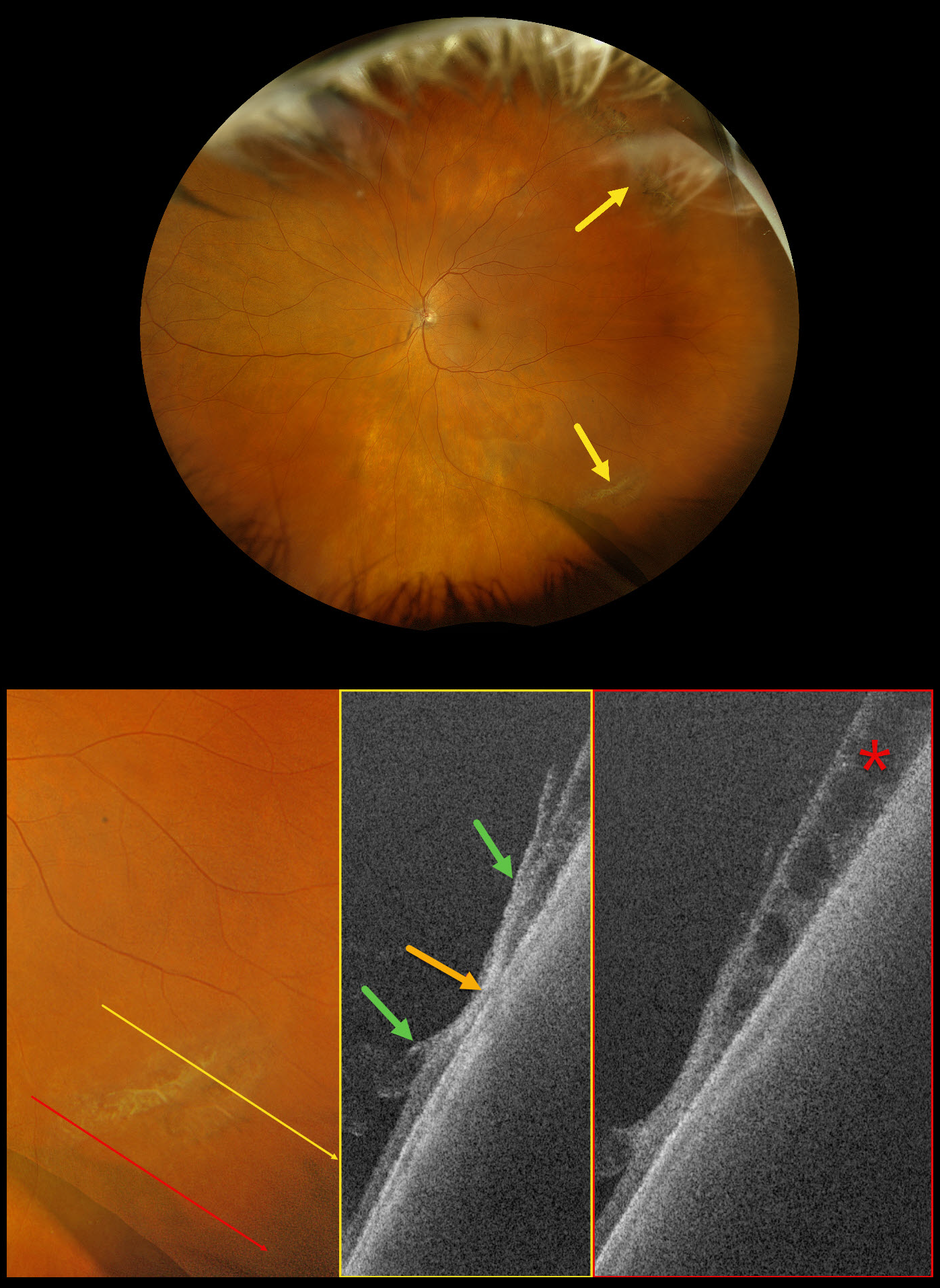
This 73-year-old female was referred for a symptomatic posterior vitreous detachment (PVD) with a retinal break in her left eye. Vision was 20/20.
Optos Silverstone RGB imaging shows supero- and inferotemporal lattice degeneration with some atrophic retinal holes. Swept-source OCT through the lattice shows a thinned, disorganized retina with areas of vitreoretinal adhesion along its margins. A more inferior line scan shows degenerative retinoschisis, which could not be visualized clinically or on the Optos image.
Our patient showed no retinal breaks from her symptomatic PVD, and observation was recommended.
Learning Points:
Ultrawidefield imaging has revolutionized our specialty, making many of us realize how limited our clinical examinations are compared with our imaging devices. In our patient’s case, the RGB image captured the fundus nearly from ora to ora. Swept-source OCT provided a near histologic view of the lattice pathology, including atrophic retina centrally, with a firm area of vitreoretinal adhesion along its margins. And finally, it revealed subclinical degenerative retinoschisis.
ANTERIORLY MIGRATED ILUVIEN IMPLANT
Fatma Shakarchi and Ahmed Sallam
Originally posted on @retina.rocks March 2, 2026
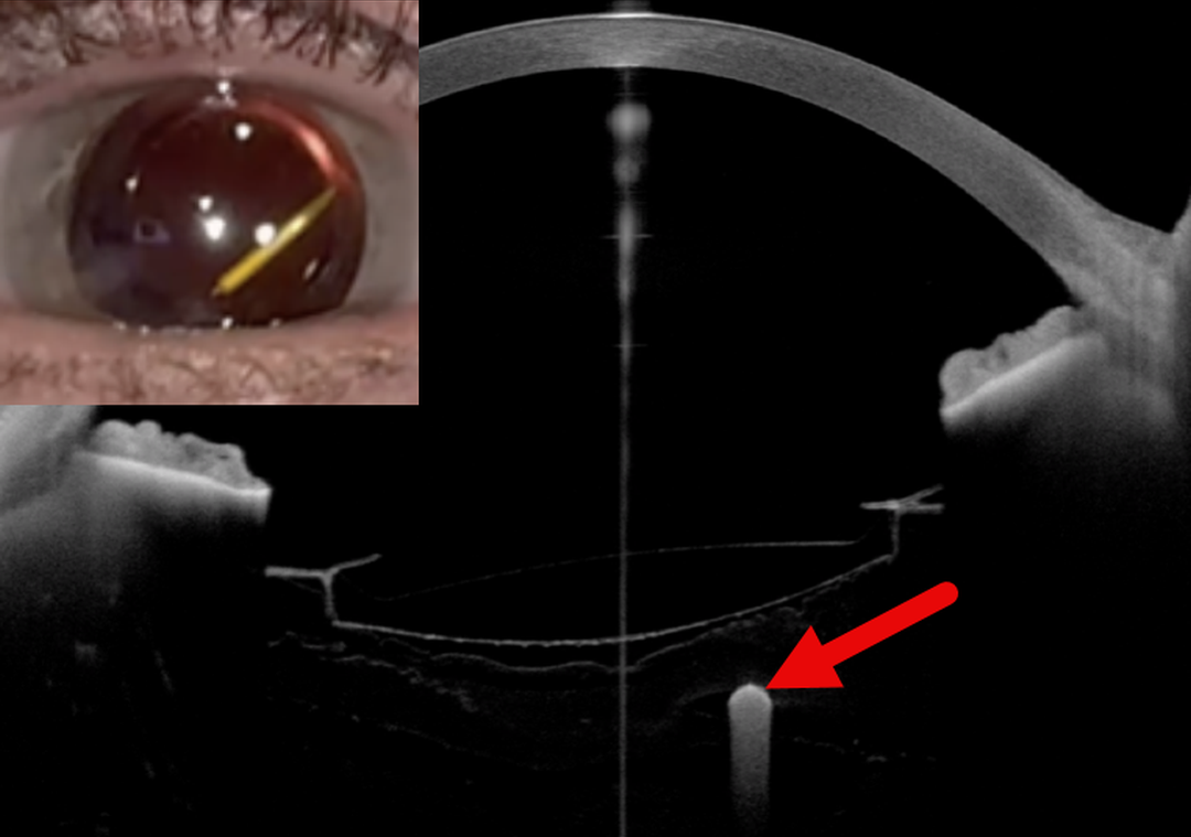
This 58YO female with panuveitis treated with bilateral fluocinolone acetonide intravitreal implants (Iluvien) and systemic adalimumab presented with an intermittent, moving floater near the visual axis in her right eye. She was pseudophakic in both eyes with posterior chamber intraocular lenses (IOLs) positioned within intact capsular bags.
External slit-lamp photography and anterior segment OCT show an Iluvien implant in the anterior vitreous cavity just behind the IOL.
Learning Points:
Migration of Iluvien implants into the visual axis has been reported and may cause visually disturbing symptoms, particularly when the implant assumes a vertical orientation within the vitreous cavity. In selected cases, Nd:YAG laser disruption of anchoring vitreous fibers (Moisseiev et al JAMA Ophthalmol 2016:134:1057-1058) or vitrectomy (Knight et al Retinal Cases & Brief Reports 2023;17:26-28) have been described as potential management strategies.
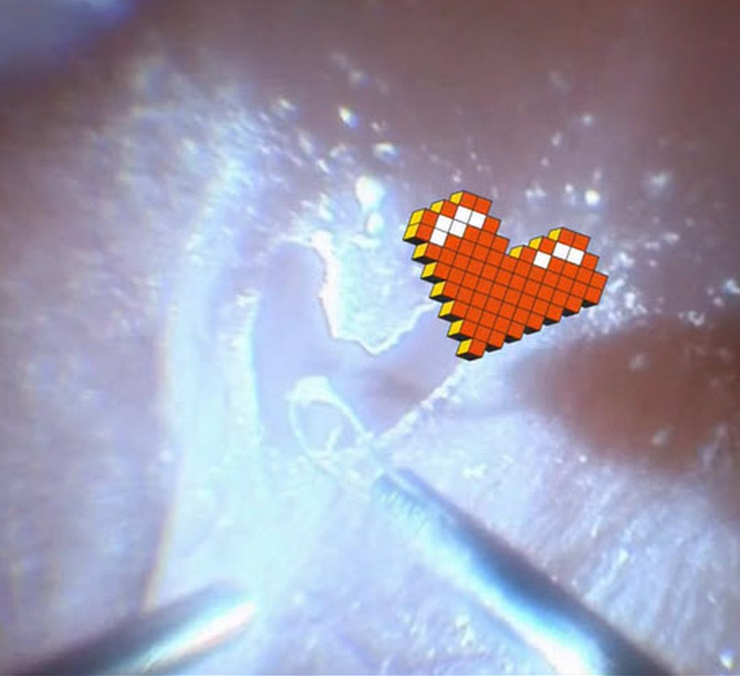
INTRAOPERATIVE HEART DURING TRIAMCINOLONE-ASSISTED MEMBRANE PEELING
The European VitreoRetinal Society (EVRS), Claudio Brancato and Gregorio Lo Giudice
Originally posted on @retina.rocks February 13, 2026
This 75YO male presented with hand motion vision in his right eye from a rhegmatogenous retinal detachment (RRD). Intraoperatively, triamcinolone was injected to stain the posterior hyaloid. During FINESSE Flex Loop-assisted membrane peeling, some love came back at us from the patient’s retina. The retina remained attached postoperatively, and vision improved to 20/50.
Learning Points:
Valentine’s Day traces its origins to a blend of ancient Roman customs, early Christian tradition, and the imaginative influence of medieval literature. What began as mid-February festivals gradually became associated with figures named Valentine, remembered for acts of compassion and steadfast conviction. Over time, poets such as Chaucer helped link the date with courtly love, giving rise to the exchange of affectionate notes and tokens. By the 18th and 19th centuries, these gestures evolved into the expensive cards, flowers, and symbols of romance that define the holiday today, transforming a complex historical tapestry into a widely celebrated expression of affection.
So, for you retinal surgeons out there, what could be a more romantic Valentine’s Day gift than offering to vitrectomize your loved one?
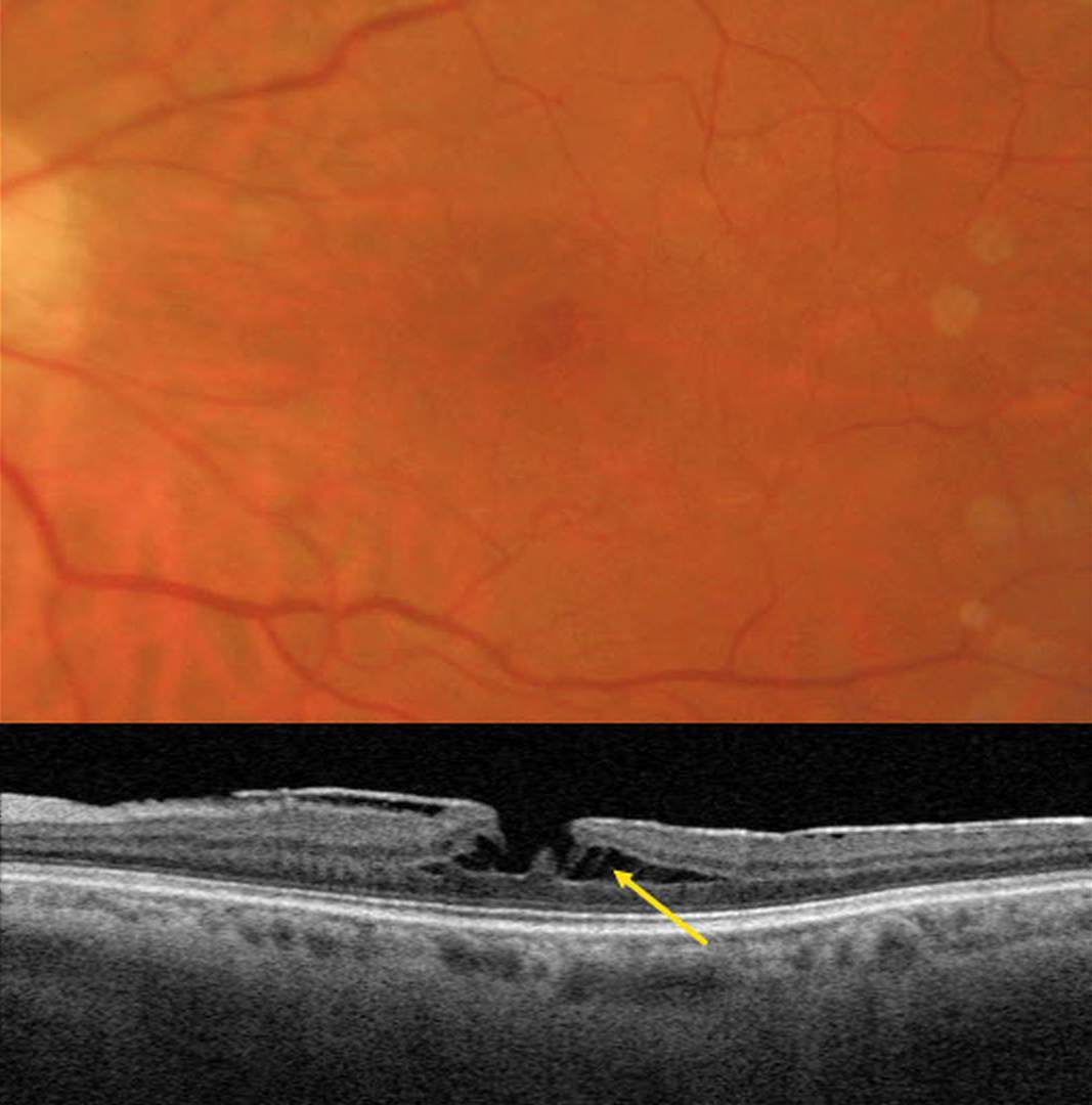
TRACTIONAL LAMELLAR MACULAR HOLE
Originally posted on @retina.rocks February 10, 2026
This 62YO female has a known history of an asymptomatic macular pucker. Vision was 20/70.
Triton color imaging shows a faint, irregular macular pseudohole. Swept-source OCT shows a variably adherent hyperreflective macular pucker. A tractional lamellar macular hole (LMH) is noted centrally, with pillars of Müller cells spanning the split outer plexiform layer (OPL).
Learning Points:
LMH can be defined as tractional or degenerative (Govetto et al, AJO 2016;164:99-109). Tractional LMH shows OPL splitting, an intact ellipsoid zone, and is associated with vitreomacular traction and tractional epiretinal membranes. OCT features of a degenerative LMH include loss of outer retinal tissue below the OPL and an outer retinal ‘bump’. Non-tractional epimacular proliferation is often present and will often extend around the posterior edge of the degenerative LMH.
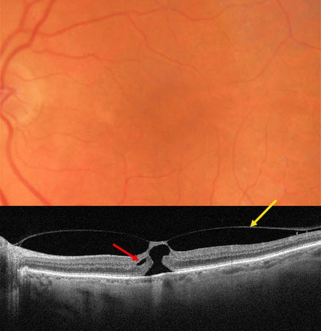
FOCAL VITREOMACULAR TRACTION
Originally posted on @retina.rocks February 9, 2026
This 82YO female was referred for an asymptomatic macular finding noted on screening OCT scanning. Vision was 20/200.
Triton color imaging shows a poorly defined foveal reflex. Swept-source OCT shows a partial macular posterior vitreous detachment (PVD) that is adherent to the foveal center. A thin strip of intact inner tissue is adherent to the overlying vitreous with an underlying outer macular hole. A tiny area of split outer plexiform layer is noted nasally.
Learning Points:
The International Vitreomacular Traction Study Group classified vitreomacular traction by size (focal <= 1500 microns or broad >1500 microns) and etiology (isolated or concurrent, Ophthalmology 2013;120:2611-2619). The prevalence of vitreomacular traction (VMT) increases with age and is present in about 1% of people over 40YO. The natural history is still being defined, but most patients have stable traction and vision. VMT can also resolve spontaneously in about 20% (Errera et al, Ophthalmology 2018;125:701-707).
Management includes observation, pneumatic vitreolysis, or vitrectomy. In our experience, most patients with VMT remain completely asymptomatic despite impressive OCT findings. We therefore almost always observe VMT unless the patient has significant symptoms. Our patient elected to continue observation since she was asymptomatic bilaterally.
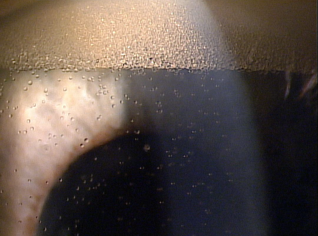
EMULSIFIED SILICONE OIL WITH REVERSE PSEUDOHYPOPYON
Originally posted on @retina.rocks February 3, 2026
This patient underwent prior successful retinal reattachment surgery with silicone oil and presented with these asymptomatic findings.
Slit lamp photography shows a reverse pseudohypopyon of emulsified silicone oil droplets floating in the anterior chamber. Suspended droplets of oil are noted throughout the aqueous more inferiorly.
Learning Points:
Silicone oil as an adjunct for repairing complex retinal detachments was introduced by Paul Cibis in the early 1960s (Cibis et al, Arch Ophthalmol 1962;68:590-599). It is used primarily for complex detachments at high risk for failure, including severe proliferative vitreoretinopathy and viral retinitis. Although silicone oil is generally well tolerated as a long-term tamponade, it can rarely emulsify, resulting in numerous tiny, opaque droplets. The development of emulsification is multifactorial, including the properties of the oil, the surgical procedure, and postoperative factors.
When severe, the emulsified droplets can cause significant vision loss, requiring their removal. The droplets can also migrate into the anterior chamber, causing a reverse ‘hypopyon’ with secondary elevated intraocular pressure or corneal decompensation.

BRILLIANT BLUE DYE G-STAINED PERFLUOROCARBON LIQUID BUBBLES
Shishir Verghese
Originally posted on @retina.rocks January 28, 2026
This intraoperative image was obtained from a 55YO female undergoing pars plana vitrectomy for a total rhegmatogenous retinal detachment. Brilliant Blue G (BBG) dye was injected to facilitate membrane peeling, followed by the injection of perfluorocarbon liquid (PFCL) to flatten the posterior retina.
The PFCL was introduced while residual BBG dye was still present within the vitreous cavity. During the injection, a transient increase in infusion pressure caused mild intraoperative turbulence, resulting in fragmentation of the PFCL into multiple microbubbles that unexpectedly became infused with BBG dye.
Intraoperative photography captured this striking appearance of mid-vitreous BBG-stained PFCL microbubbles. The posterior retina is attached beneath a larger, unstained PFCL bubble, while the retina remains detached in the more anterior regions.
TRACTIONAL RETINAL DETACHMENT WITH PSEUDO-GIANT MACULAR HOLE
The European VitreoRetinal Society (EVRS) and Shishir Verghese
Originally posted on @retina.rocks January 16, 2026
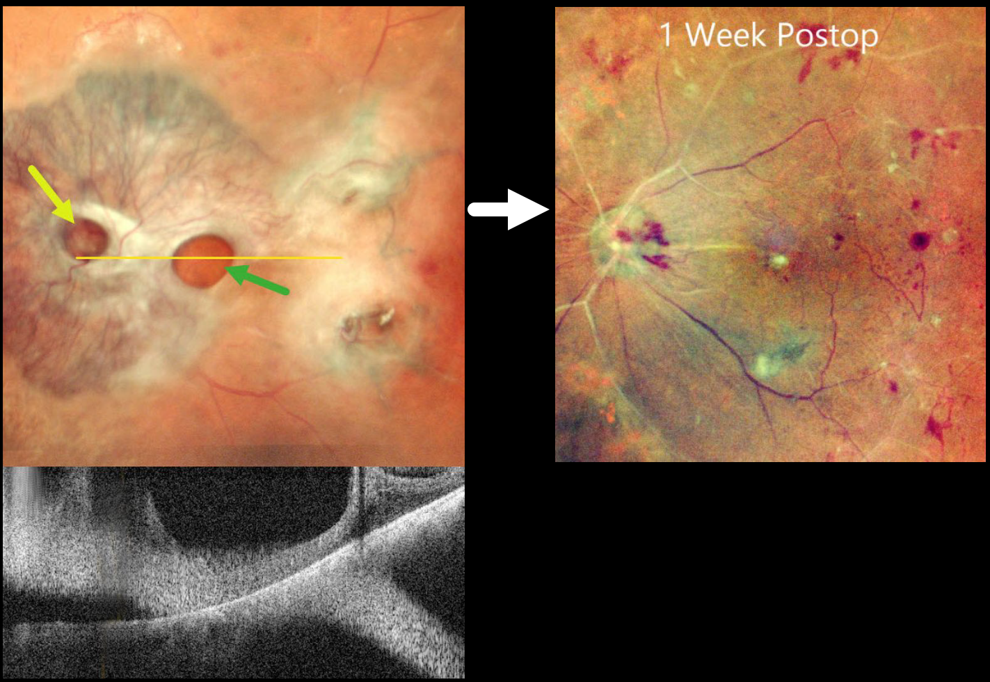
This 65YO diabetic female presented with counting fingers bilaterally from proliferative diabetic retinopathy with macular tractional retinal detachments.
Preoperative pseudocolor SLO imaging of her left eye shows a complex fibrovascular membrane covering the posterior pole. Pseudo giant-macular holes are noted overlying the disc and macula. OCT scanning confirms the lack of a true macular hole. One week following vitrectomy, membrane peeling, and air injection, vision improved to 20/200, and the retina was completely attached with no residual traction.
Learning Points:
A giant macular hole, also reported in Alport syndrome (Shah and Weinberg Ophthalmic Genetics 2010;31(2):93-97), is an exceedingly rare traumatic event (Hernandez-Da Mota Case Rep Ophthalmol 2011;2:283-286). The unusual anatomy of our patient’s fibrovascular diabetic membrane simulated a giant macular hole.
PRE-ANTERIOR HYALOID GAS FOLLOWING FAILED PNEUMATIC RETINOPEXY
Arthur Zupelli, Bruno Felipe Oliveira Silva, Leonardo Luis Cassoni Marcelo Brandão Guimarães Rego and Rodrigo Jorge
Originally posted on @retina.rocks January 13, 2026
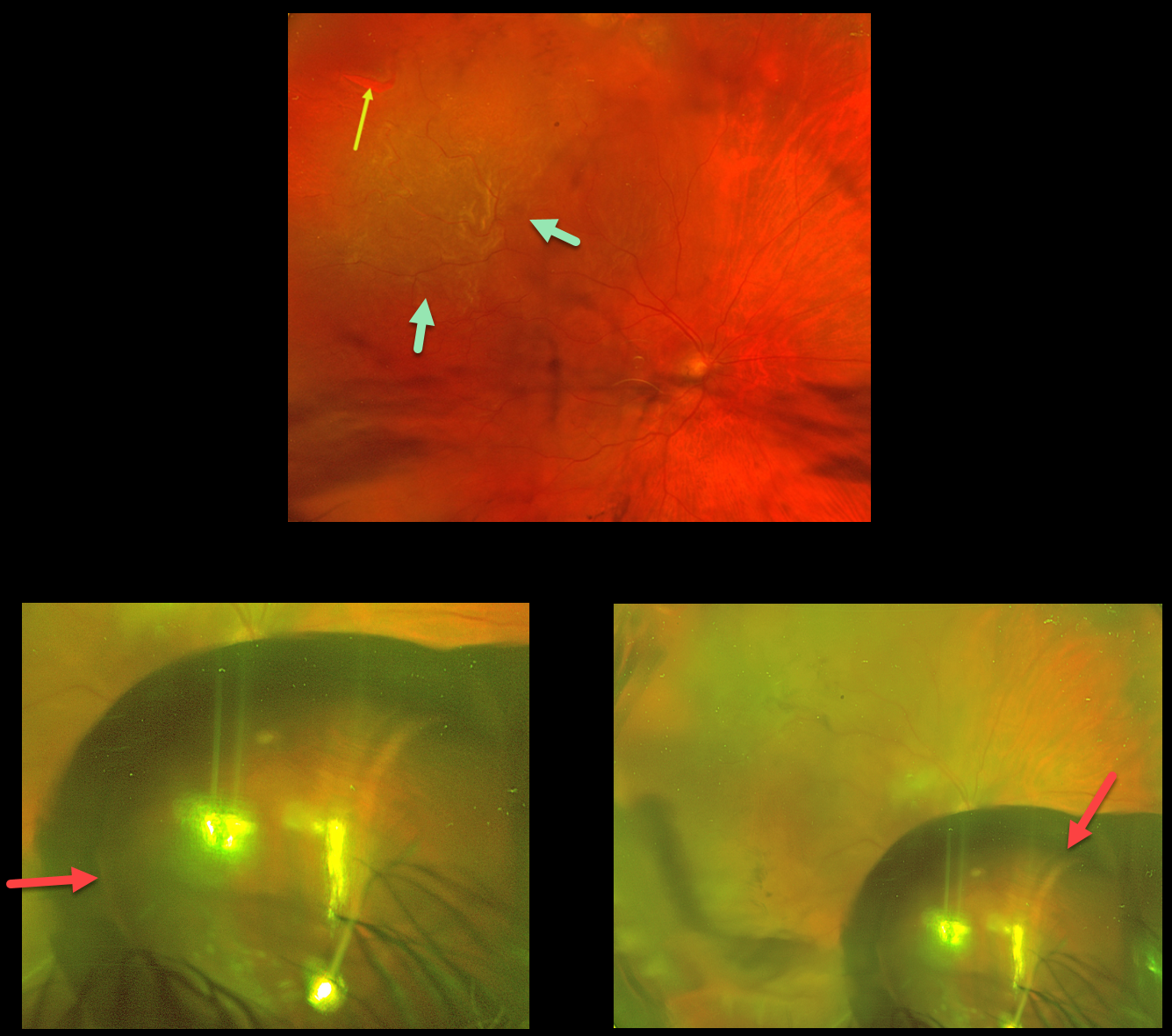
This 71YO male presented with decreased vision in his right eye one month following cataract surgery. Vision was hand motion. Slit lamp examination revealed a well-centered posterior chamber IOL.
Optos color RG imaging shows a superotemporal macula-off rhegmatogenous retinal detachment (RRD) caused by a single break. A pneumatic retinopexy using 0.35cc of pure perfluoropropane gas (C3F8) was performed, followed by intravenous mannitol. One day later, vision was hand motion, and a large gas bubble was trapped in the pre-hyaloid space. This configuration reflected gas sequestration within the potential space bordered by the posterior lens capsule, the anterior hyaloid membrane, and the ciliary body. The appearance was unchanged 2 days later.
We attempted to remove the gas in the office, using a plungerless syringe with the needle inserted into the bubble. This maneuver was unsuccessful, necessitating pars plana vitrectomy. The retina remained attached postoperatively with a final vision of 20/100.
Learning Points:
Gas entrapment in the pre-hyaloid space is an exceedingly rare but visually striking complication of pneumatic retinopexy (Chan et al, Surv Ophthalmol 2008;53:443-478), and most likely is caused by a too shallow needle entry into the vitreous. On slit-lamp or fundus examination, the bubble may appear as a donut- shaped or sausage-like lucency located immediately behind the lens.
MACULAR PUCKER WITH RNFL SCHISIS
Originally posted on @retina.rocks January 5, 2026
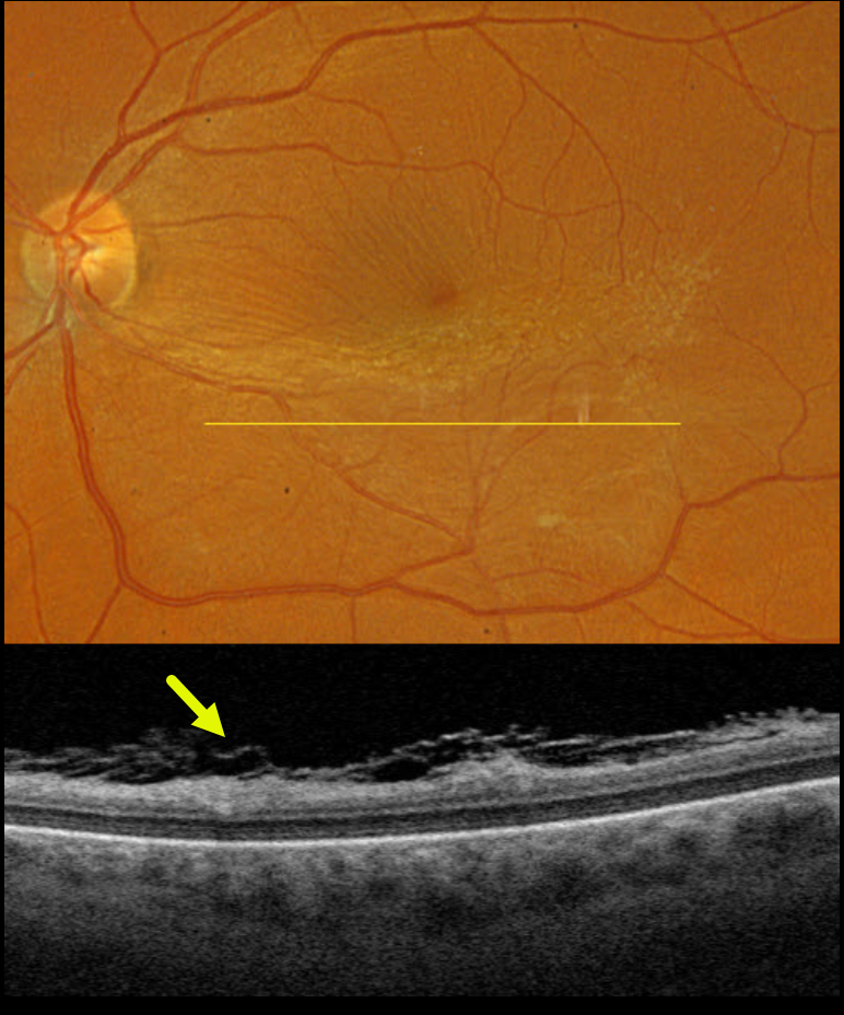
This 58YO female was referred for an asymptomatic macular pucker in her left eye. Vision was 20/25.
Optos color RGB imaging shows a moderate macular pucker centered in the inferior macula. Fine inner retinal striae are noted superiorly. Triton swept-source OCT shows a shredded appearance of multi-layered hyperreflective tissue overlying the retinal surface (the ‘spaghetti sign’).
Learning Points:
Fibrillary changes overlying an epiretinal membrane (ERM) were initially noted by Kim et al (AJO 2012;153:692-697), and termed schisis of the retinal nerve fiber layer by Hussnain et al (AJO 2019;207:304-3120). Despite rarely being described in the medical literature, it is present in over 50% of ERMs (Russell et al, Ophthalmology Retina 2023;7:325-332). Felt to arise from tangential tractional forces from the overlying epimacular tissue, it can appear clinically as whitish retinal areas overlying a pucker. During intraoperative membrane peeling, this tissue can be mistaken for residual epimacular proliferation and should not be removed to avoid iatrogenic retinal damage.
VITREOUS CYST
The European VitreoRetina Society (EVRS) and Prithvi Chandrakanth
Originally posted on @retina.rocks November 21, 2025
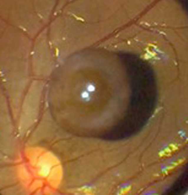
This healthy 20YO female presented with chronic floaters in her left eye. Vision was 20/20 in her healthy OD and 20/20 OS.
Funduscopy shows a large pigmented vitreous cyst floating over the superior macula. Observation was recommended.
Learning Points:
Vitreous cysts are extremely rare, idiopathic, and usually diagnosed incidentally. Likely originating from a ciliary body cyst (Dhull et al, Ophthalmology Retina 2020;4:1208), they can be either congenital or acquired. Acquired cysts have been associated with numerous ocular disorders, including trauma and retinitis pigmentosa.
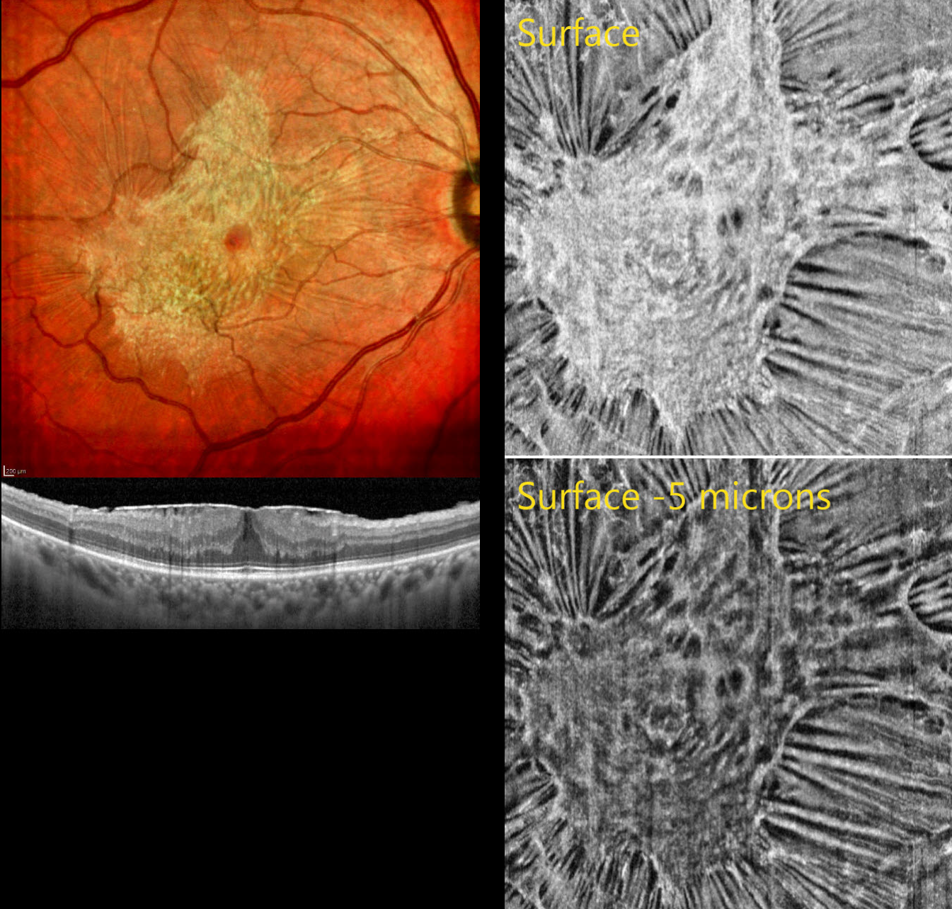
MACULAR PUCKER
The European VitreoRetina Society (EVRS), Mohamed Tawfik and Norhan Lotfy
Originally posted on @retina.rocks November 14, 2025
This 60YO male presented with gradual blurred vision in his right eye. Vision was 20/30.
Color SLO imaging shows a variably dense and glistening macular pucker with radiating retinal striae extending outwards from multiple areas of tangential traction. OCT shows the hyperreflective epiretinal membrane causing diffuse retinal thickening. The retinal bands are fairly well-preserved. En face imaging at the membrane’s surface reveals better detail for the irregular pucker and surface traction. Imaging 5 microns below the surface reveals dimpling of the underlying tissue.
Surgery will be recommended if our patient develops increased symptoms.
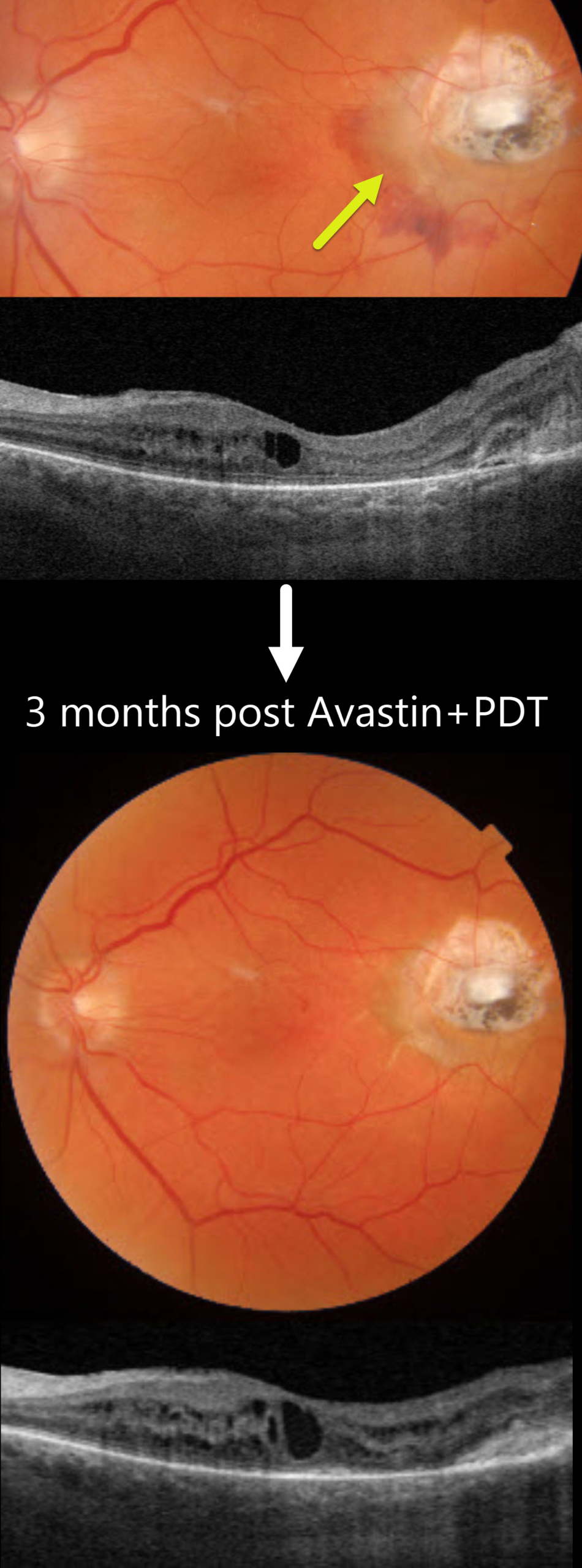
CNV AT INTERNAL DRAINAGE SITE FOLLOWING VITRECTOMY FOR RHEGMATOGENOUS RD
Originally posted on @retina.rocks November 10, 2025
This 75YO male underwent successful vitrectomy for a macula-off rhegmatogenous retinal detachment (RRD). Vision improved from a preoperative hand motion vision to counting vision 7 months following surgery. However, he returned 4 months later with new distortion and blurriness. Vision was 20/400.
Triton color imaging shows an active choroidal neovascularization (CNV) with associated subretinal blood along the temporal and inferior edge of the drainage retinotomy scar. Swept-source OCT shows mild macular cystic edema arising from the temporal type 2 (above the RPE) CNV. The peripheral retina remained attached.
The lesion failed to respond to several intravitreal Avastin injections, necessitating combined Avastin and photodynamic therapy. Exudation completely resolved 3 months following this treatment.
Learning Points:
Invented by Steve Charles in the early days of vitrectomy, the drainage retinotomy has become an integral part of retinal reattachment surgery (Ramamurthy et al, Surv Ophthalmology 2023;68:1038-1049). Following the adage that ‘every time a hole is made in the retina, some vision leaks out,’ it is always preferable to drain subretinal fluid through a pre-existing break whenever possible. When required, the site should ideally be located superiorly and well away from the macula. Drainage retinotomies are associated with an increased risk of macular pucker and cystoid macular edema (McKay et al, AJO 2022;241:227-237). CNV is an exceedingly rare complication, which may be caused by inadvertent RPE-choroidal touch by the drainage needle or thermal-induced damage.
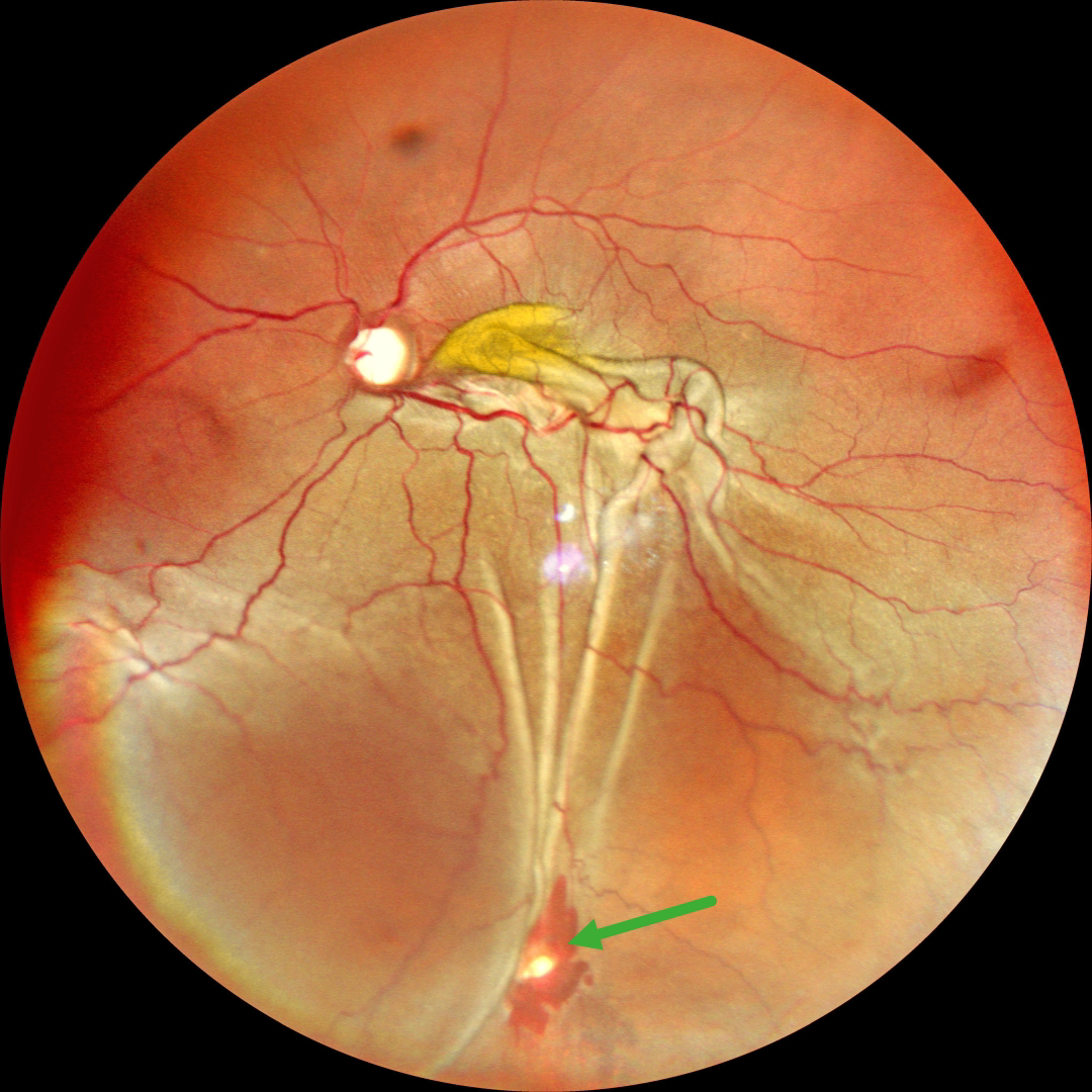
RHEGMATOGENOUS RD WITH RETINAL INCARCERATION FOLLOWING FAILED SCLERAL BUCKLING
Malvika Singh and Manish Nagpal
Originally posted on @retina.rocks November 4, 2025
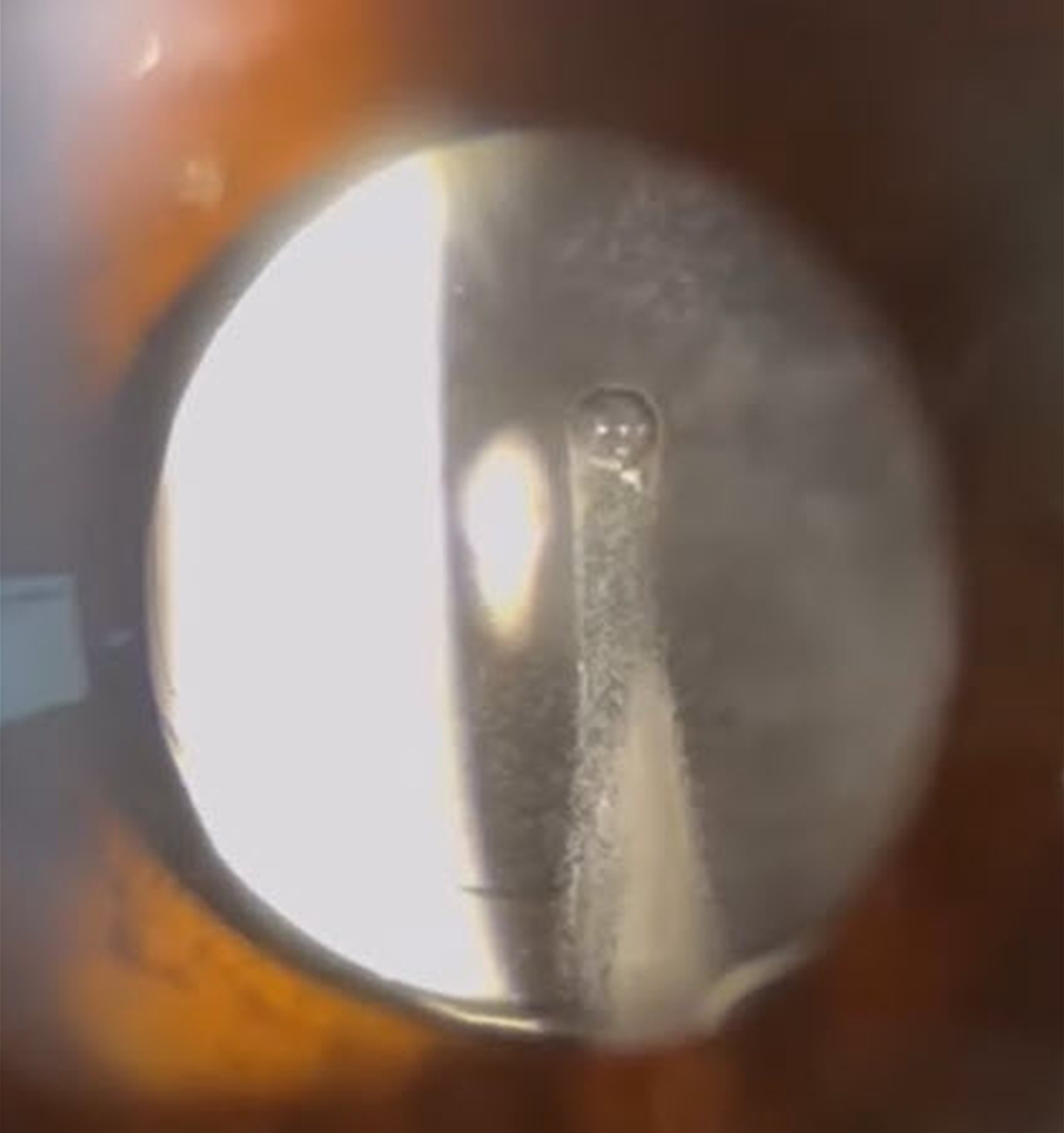
TRIAMCINOLONE-MOXIFLOXACIN (TriMoxi)
Adam Mueller, Benjamin Conkright, and Andrew Steele
Originally posted on @retina.rocks October 31, 2025
This 64YO female presented for her one-day post-dropless cataract surgery with some minor irritation and floaters. Vision without correction was 20/25+. Unbeknownst to our happy patient, a sinister ghoul was lurking behind her perfectly centered intraocular implant.
Our practice, along with many others, offers ‘no drop’ cataract surgery as a convenience to our patients. Instead of needing to take postoperative antibiotic and steroid drops, 0.2cc of TriMoxi (triamcinolone acetate 15mg/cc with moxifloxacin 1mg/cc) is injected into the anterior inferior vitreous at the end of the cataract surgery. Although this liquid usually remains well outside the visual axis, patients will often experience mild floaters perioperatively. Rarely, as in this patient, the liquid disperses throughout the vitreous cavity, causing significant transient floaters.
PRIOR CHRONIC ICSC WITH NEW TRACTIONAL MACULAR HOLE-RRD WITH POSTOPERATIVE MENISCUS MICROPYON
Originally posted on @retina.rocks October 29, 2025
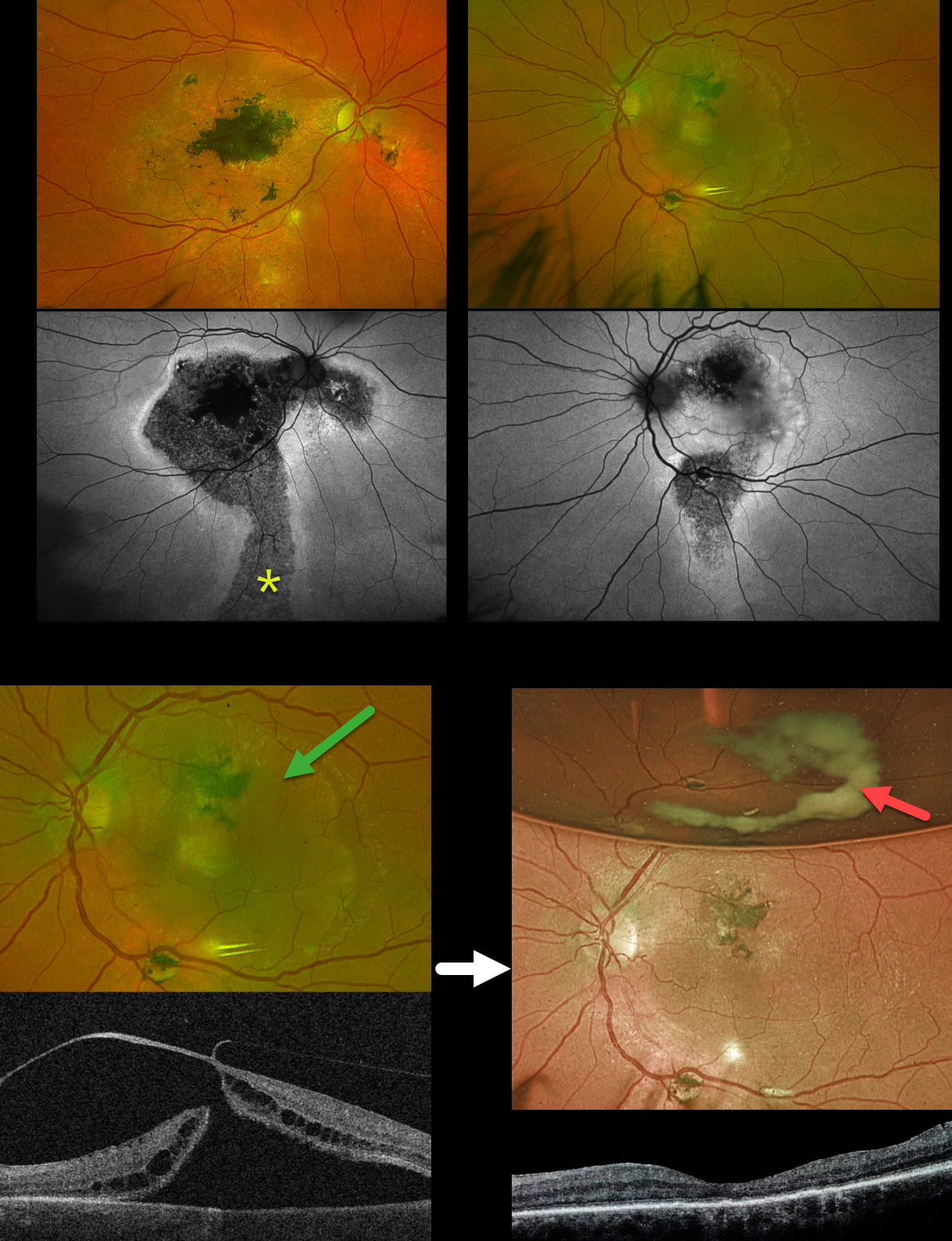
This 58YO male was referred for a retinal detachment in his left eye. He has a long history of idiopathic central serous chorioretinopathy (ICSC). He was previously treated elsewhere for neovascular AMD OD. For many years, he has been receiving intravitreal Avastin OS every 1-2 months. Vision was counting fingers OD and 20/400 OS.
Optos color RG imaging shows bilateral macular scarring with confluent hyperpigmented foveal changes and intraretinal pigment migration, especially in his right eye. Inferior hypopigmented gutters are most obvious on fundus autofluorescence (FAF) as hypo-FAF with surrounding hyper-FAF margins. A rhegmatogenous retinal detachment (RRD) occupies the entire left macula. Triton swept-source OCT shows a full-thickness macula hole, active vitreoretinal traction on the temporal edge of the hole, and a macular detachment.
The tractional macular hole with secondary RRD in our patient’s left eye is completely unrelated to his poorly treated ICSC. One month following vitrectomy, vision improved to 20/100. The retina was completely attached, and the macular hole was closed. A meniscus micropyon is adherent to the inferior meniscus of the gas bubble.
Learning Points:
Photodynamic therapy (PDT) is currently the standard of care for ICSC with symptomatic acute or chronic subretinal fluid. Unfortunately, Visudyne is rarely available outside the US, which has led to the adoption of unproven therapies, including mineralocorticoid receptor antagonists (eplerenone), beta blockers, and intravitreal anti-VEGF agents. Recent meta-analyses and systematic reviews confirm that these alternative therapies are ineffective (Felipe et al Int J Retina Vitreous 2022;8;34 and Kim et al Ophthalmology 2025;132:343-353). Despite the clear lack of efficacy in the literature, specialists both within and outside the US continue to use these drugs (Vankatesh et al Int J Retina Vitreous 2023;9;61). Our patient’s macular findings are a testament to what can happen when proven therapies are ignored.
Russell et al recently reported a unique ophthalmoscopic sign, a meniscus micropyon, that follows PPV with gas tamponade for all causes (Retina 2024;44:1766-1776). Features they noted that distinguish this sterile micropyon from post-PPV fibrin include a delayed appearance, hyperautofluorescence, and absence of anterior chamber or vitreous fibrinous strands/sheets. Vitreous biopsy in two of their cases revealed predominantly white blood cells. The micropyon appearance was associated with epiretinal proliferation (80%). Recurrent RRD from proliferative vitreoretinopathy (PVR) developed 33% of eyes, 12% re-detached without frank PVR, and 18% developed worsening epiretinal membranes.
Our patient’s meniscus micropyon disappeared as the bubble resolved. Three months postoperatively, vision was 20/60, and the retina remained completely reattached without PVR.
RETINAL TEAR
Originally posted on @retina.rocks October 22, 2025
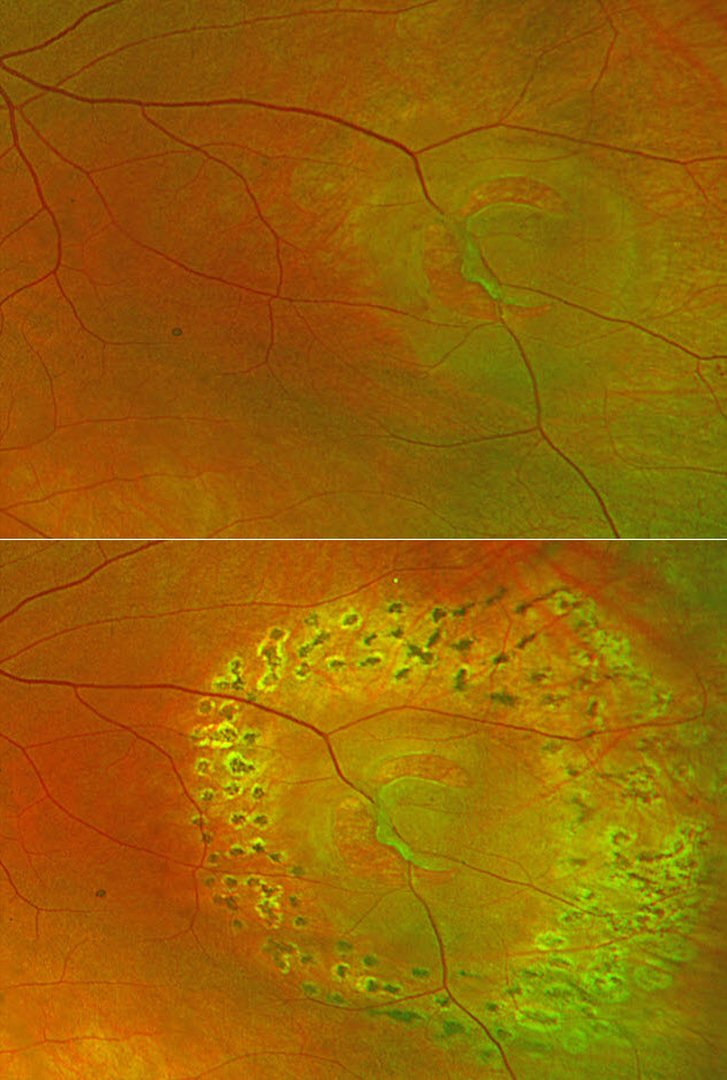
This 60YO female presented with a symptomatic retinal tear in her left eye. Optos color RG imaging shows the break with localized surrounding subretinal fluid and a bridging vessel. The break was immediately surrounded by laser. When examined 1 month later, the tear and fluid were well demarcated, and no new breaks were found.
Learning Points:
Symptomatic retinal tears in the presence of an acute posterior vitreous detachment have a high risk of causing a rhegmatogenous retinal detachment. Prophylactic retinopexy is therefore indicated. Treatment must completely surround the tear and, ideally, extend to the ora serrata. Both thermal laser photocoagulation and cryotherapy are equally effective in creating a vitreoretinal adhesion, which takes at least several weeks to form. The treatment must also be inspected during the postoperative period to ensure adequate scarring surrounds the break.
LOCULATED FLUID AFTER SUCCESSFUL PNEUMATIC RETINOPEXY
Originally posted on @retina.rocks October 21, 2025
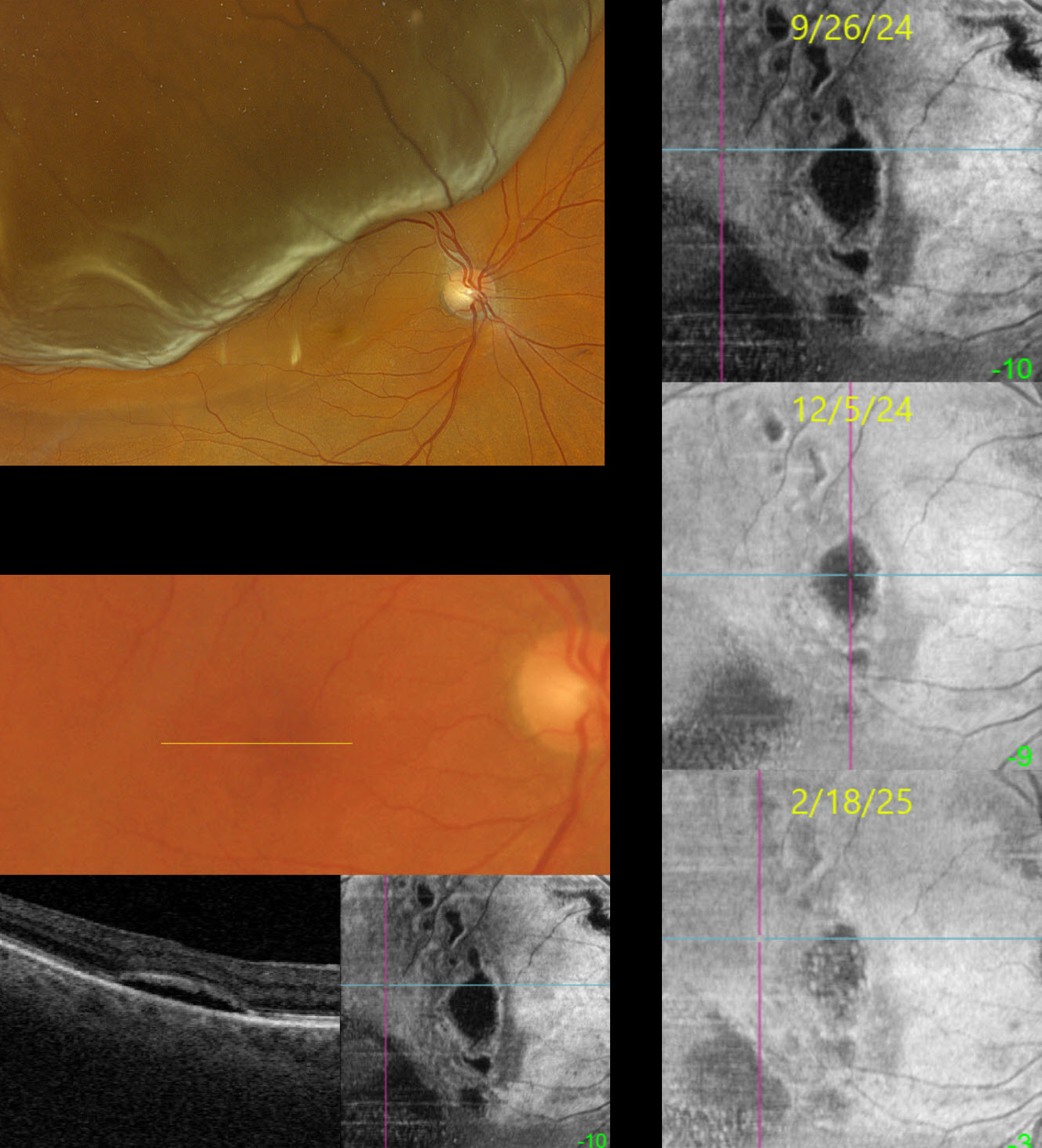
This 69YO female presented on 4/17/24 with a superior macula-off rhegmatogenous retinal detachment (RRD) and counting fingers vision. Following successful pneumatic retinopexy, the retina reattached, and vision returned to 20/25 one month later.
When examined on 9/26/24, she was subjectively doing well with 20/50 vision. Although the macula looked clinically normal, Triton swept-source OCT B-scan and en face imaging show multifocal pockets of loculated subretinal fluid (SRF). With observation, these lesions continue to shrink and flatten. When last examined on 2/18/25, vision was 20/30, and continued observation was recommended.
Learning Points:
Loculated fluid represents an area of persistent subclinical SRF in the presence of a clinically reattached retina and is seen postoperatively in up to 15% of RRD following vitrectomy (Retina 2020;40:1153-1159) and eventually resolves after many months of observation. In a post hoc analysis of the PIVOT trial, there were no differences in the incidence of loculated fluid between vitrectomy and pneumatic retinopexy (Bansal et al BJO 2023;107:1693-1697). Although subfoveal loculated fluid was associated with reduced visual acuity at 3 months, vision eventually was similar to that of those without fluid at 1 year.
Bansal et al recently described the en face findings for loculated fluid (Ophthalmology Retina 2023;7:496-502). We continue to find that en face imaging often provides a unique and compelling perspective on a variety of pathologies, and we hope other clinicians will add it to their daily clinical routine.
TRAUMATIC RETINAL HOLES
Fatma Shakarchi and Ahmed Sallam
Originally posted on @retina.rocks October 6, 2025
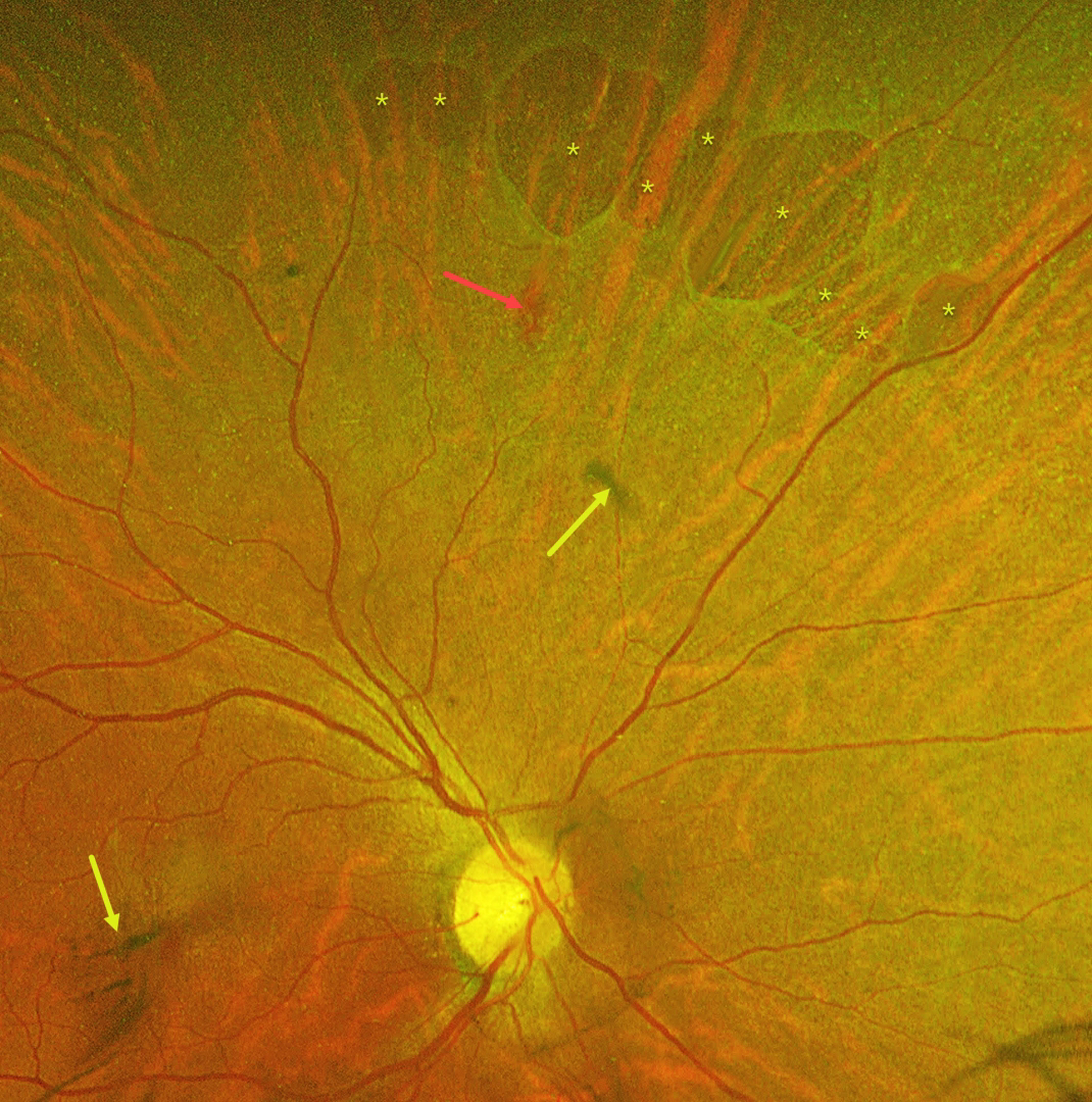
This 55YO female was referred with mildly decreased vision in her right eye two days following blunt ocular trauma. Vision was 20/40 OD and 20/20 in her normal OS.
Optos color RG imaging reveals numerous irregularly oval superonasal retinal holes, a retinal hemorrhage, and some clumps of vitreous blood. There was no vitreous detachment clinically.
Learning Points:
Retinal breaks following blunt trauma include retinal dialysis, horseshoe tears, and giant retinal tears. Breaks can also develop from concussive injury at the impact site or from a coup-contrecoup injury, so-called impact or necrotic breaks, which may be associated with varying degrees of commotio retinae acutely (Cox et al, Arch Ophthalmol 1966;76:678-685) or following its resolution (Longstaff et al, Br J Ophthalmol 1987;71:375-376).
In this case, the breaks identified 2 days after the injury were not associated with commotio. However, their appearance and the adjacent hemorrhage support an acute traumatic etiology. Therefore, prophylactic laser retinopexy was promptly applied around all breaks.
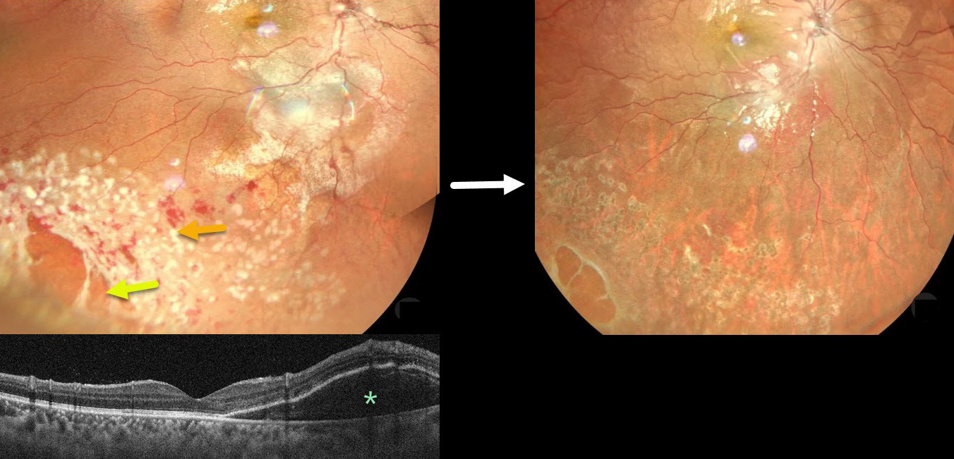
COMMOTIO RETINAE WITH RETINAL BREAKS AND CONTUSIVE SEROUS MACULAR DETACHMENT
Tejaswita Verma and Manish Nagpal
Originally posted on @retina.rocks September 24, 2025
This 13YO male was hit in his right eye with a cricket ball. When he presented later that day vision was 20/30.
Color photography shows a large area of commotio retinae with scattered retinal hemorrhages involving the inferotemporal fundus. There is a poor foveal reflex with possible nasal macular subretinal fluid, which is confirmed on OCT. Several atrophic retinal breaks are noted peripherally. Immediate prophylactic laser was applied around all breaks. Seven weeks later, the commotio and hemorrhages are resolved, and all breaks are well-surrounded with laser scarring. Vision was 20/20.
Although blunt trauma is a common cause of retinal breaks, acute or subsequent dissolution of the retina within areas of commotio with secondary holes is quite rare in our experience (Cox et al Arch Ophthalmol 1966;76:678-685 and Longstaff et al Br J Ophthalmology 1987;71:375-376). The acute white retinal changes usually resolve within several weeks. With resolution, the fundus appearance can normalize. With more severe injury, outer retinal loss can allow RPE cells to migrate into the retina, giving a pseudo-retinitis pigmentosa appearance.
Transient subretinal fluid is an uncommon finding with blunt trauma. Injury to the choroid and RPE allows fluid to accumulate in the subretinal space and outer retina (Mishra et al, Journal of Vitreoretinal Diseases 2021;5:165-169).
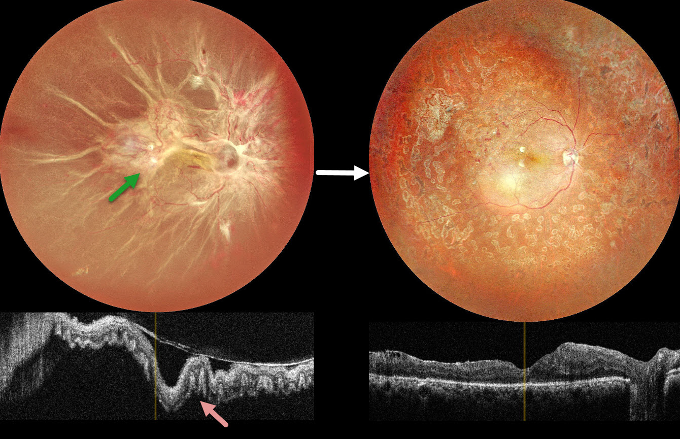
COMBINED TRACTIONAL-RHEGMATOGENOUS RETINAL DETACHMENT
The European VitreoRetina Society (EVRS) and Shishir Verghese
Originally posted on @retina.rocks September 19, 2025
This 40YO female presented with vision loss in her right eye. There was a 15-year history of type 1 diabetes. Vision was hand motion OD.
Color photography shows a total combined traction-rhegmatogenous retinal detachment (RRD). There is a complex pattern of fibrovascular tissue covering the posterior pole with radiating retinal folds extending peripherally. OCT scanning shows a variably undulated detached retina with outer retinal corrugations.
Five days following an intravitreal anti-VEGF injection, she underwent pars plana vitrectomy with membrane peeling, endolaser, and silicone oil injection. Three months later, the silicone oil was removed. Postoperatively, vision improved to 20/80 with complete retinal reattachment.
Learning Points:
Outer retinal corrugations, also known as hydration outer retinal folds, are one of the key findings that help differentiate a RRD from exudative and traction retinal detachments. The cause of these folds remains uncertain but may result from the hydration of glycosaminoglycans in the interphotoreceptor matrix (Dalvin et al, Retinal Cases & Brief Reports 2020;14:355-359). This OCT biomarker confirms the rhegmatogenous component of our patient’s detachment.
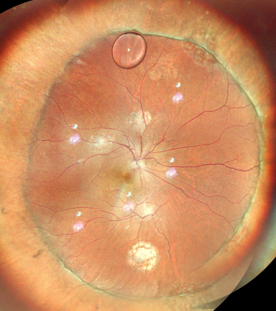
SILICONE OIL DROPLET
Ankit Jain and Manish Nagpal
Originally posted on @retina.rocks September 17, 2025
This 56YO male underwent successful retinal reattachment with vitrectomy, scleral buckling, and silicone oil. One month following silicone oil removal, he presented complaining of an inferior floater. Vision was 20/40.
Color SLO imaging shows an attached retina with an encircling buckle. A residual silicone oil droplet is noted superiorly along the buckle edge. Observation was recommended.
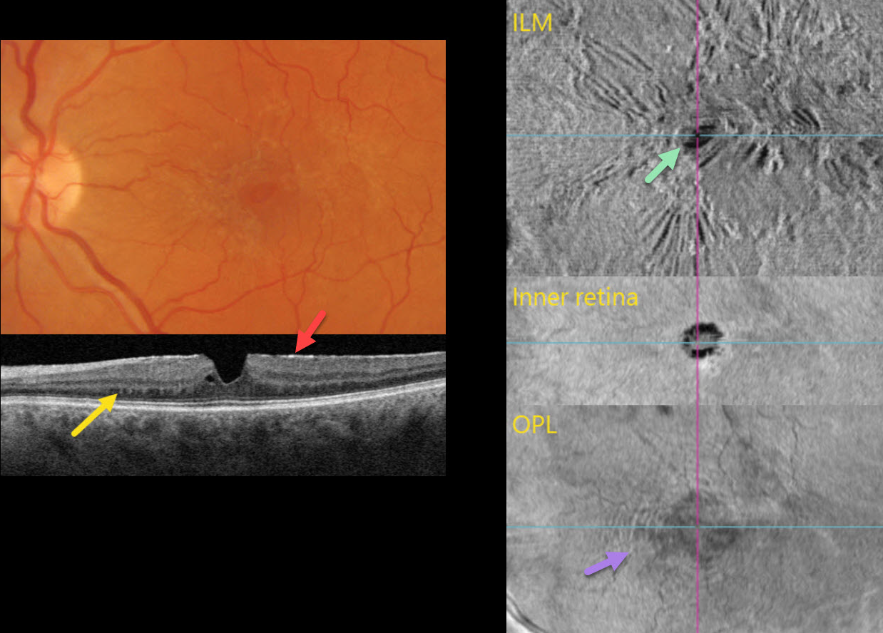
MACULAR PUCKER
Originally posted on @retina.rocks September 8, 2025
This 56YO female was referred for asymptomatic retinal findings. Vision was 20/20.
Multimodal imaging shows numerous pucker-related findings.
Triton color photography shows a diffuse macular pucker with a central teardrop-shaped pseudohole.
Triton swept-source OCT B-scanning shows an adherent hyperreflective epiretinal membrane, a vertical foveal contour, and trace adjacent outer plexiform splitting. The nasal outer plexiform layer (OPL) contains numerous peg-like hyperreflective lesions that extend into the outer nuclear layer.
En face OCT of the retinal surface shows variable striae and distortion from the pucker, along with the hyporeflective pseudohole. En face OCT through the inner retina shows the irregular pseudohole. En face of the OPL, through the peg-like lesions noted on the B-scan, shows a hyperreflective fingerprint.
Learning Points:
The ‘fingerprint sign’ is a unique OCT en face finding, described by Griffin et al (Retina 2021;41:381-386), appearing as concentric, fingerprint-like waves of Henle’s layer underlying epiretinal membranes. The authors felt that the undulating OPL creates hyperreflective lines when oriented perpendicular to the incident OCT beam and hyporeflective lines when oriented parallel to it.
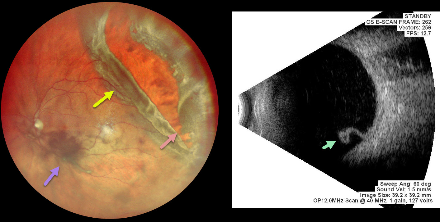
GIANT RETINAL TEAR
Malvika Singh and Manish Nagpal
Originally posted on @retina.rocks August 27, 2025
This 56 YO male presented with 4 days of vision loss in his left eye. He gave a history of multiple prior retinal reattachment surgeries in his right eye. Vision was 20/400 in his OD and 20/20 in OS.
Pseudocolor SLO imaging shows a superotemporal rhegmatogenous retinal detachment (RRD) from a giant retinal tear (extends over 3 clock hours) with mild vitreous hemorrhage. The anterior portion of the tear has multiple jagged edges, and the posterior flap has folded over onto itself. The macula is attached clinically and on B-scan ultrasonography, which shows the mobile posterior flap folded over onto itself. Emergency vitrectomy was recommended, but unfortunately, he was immediately lost to follow-up.
Learning Points:
A vitreous hemorrhage will often prevent visualization of an acute RRD. Ultrasonography is often utilized to confirm the presence of a detachment. In our patient, the B-scan also beautifully imaged the mobile posterior flap of the giant tear. This pathognomonic appearance would alter the surgical planning in the case of opaque media.
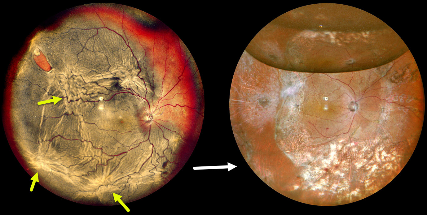
PROLIFERATIVE VITREORETINOPATHY
The European VitreoRetina Society (EVRS) and Shishir Verghese
Originally posted on @retina.rocks August 8, 2025
This 75YO male presented with decreased vision in the right eye for four months. Vision was hand motion OD and 20/25 in his normal OS.
Pseudocolor SLO imaging shows a complex total rhegmatogenous retinal detachment with a supertemporal tear and multiple proliferative vitreoretinopathy (PVR) star folds. Following three vitrectomy surgeries, including a relaxing retinectomy, the retina is attached. Vision was 20/120.
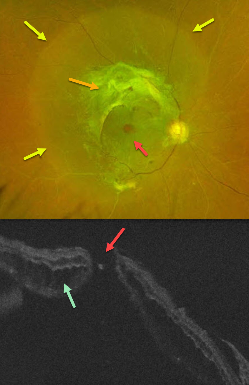
COMBINED TRACTIONAL-RHEGMATOGENOUS RETINAL DETACHMENT WITH MACULAR HOLE
Sucheta Kulkarani, Anita Gaikwad and Anjana Mirajkar
Originally posted on @retina.rocks July 24, 2025
This 49YO male with type 2 diabetes noticed decreased vision in his right eye for 4 months. A single anti-VEGF injection was given elsewhere in his right eye around the time of his vision loss. Vision was 20/400 OD and 20/30 OS.
Optos color RG imaging OD shows a combined tractional-rhegmatogenous retinal detachment from dense fibrovascular proliferation and a full-thickness macular hole. OCT shows diffuse cystic edema within the detached retina, predominantly in the outer nuclear layer, and a macular hole.
Learning Points:
Tractional macular holes as a cause of combined tractional-rhegmatogenous diabetic retinal detachments are a rare event in our experience. Unfortunately, our patient was lost to follow-up immediately. We expect him to lose central vision in his left eye soon if he does not seek care.
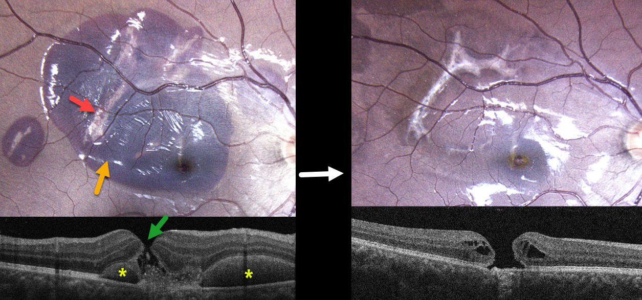
ACUTE CHOROIDAL RUPTURES WITH SUBRETINAL BLOOD + TRAUMATIC MACULAR HOLE
Originally posted on @retina.rocks July 16, 2025
This healthy 17YO male was in a motor vehicle accident 2 weeks earlier. He was not wearing a seat belt and suffered a right upper lid laceration and closed head injury. He noticed immediate vision loss in his right eye. Vision was 20/200 OD and 20/20 in his normal OS.
Color photography shows a large macular and a smaller distal temporal submacular hemorrhage. Multiple choroidal ruptures are visible beneath this blood. OCT scanning shows a central full-thickness macular hole with speckled hyperreflective subretinal material. The submacular blood is hyperreflective with posterior shadowing on either side of the macular hole.
Learning Points:
An acute submacular hemorrhage following blunt trauma always indicates an underlying choroidal rupture as its source. The final vision is generally good unless the rupture extends through the macular center. These patients need long-term follow-up due to the increased risk of macular neovascularization.
Since fresh traumatic holes can spontaneously flatten, especially in pediatric cases (Liu et al, AJO 2020;210:174-183), an initial period of observation is recommended before considering vitrectomy. When examined about 6 months following his injury, all blood had resolved, and the temporal foveal choroidal rupture appears to spare the macular center. We will likely recommend vitrectomy if the hole fails to close with several more months of observation.
MACULAR PUCKER WITH LAMELLAR DEFECTS
The European VitreoRetina Society (EVRS) and Shishir Verghese
Originally posted on @retina.rocks July 11, 2025
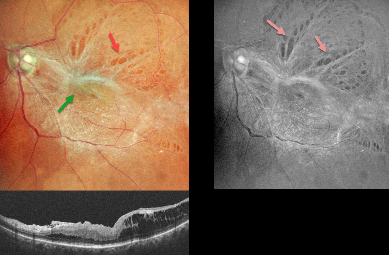
This 58YO female presented with 6 months of vision loss in her left eye. Vision was 20/60 OS.
Color SLO imaging shows a taut epiretinal membrane (ERM) with multiple paravascular lamellar defects, which are better visualized on red-free imaging. OCT scanning shows a stage 3 macular pucker with ectopic inner foveal layer (EIFL) and temporal inner layer schisis.
Learning Points:
An ERM is a thin, fibrous membrane that forms on the retinal surface, often causing central vision loss with metamorphopsia. It can be classified into 4 OCT-based stages (Gonzalez-Saldivar et al, Retina 2020;40:710-717): Stage 1 ERM with preserved foveal contour, Stage 2 Loss of foveal depression with thickened outer nuclear layer, Stage 3 EIFL, and Stage 4 EIFL with complete loss of inner retinal anatomy.
ERMs can cause paravascular lamellar defects due to shearing forces at the vessel borders, and these defects can extend towards the fovea. Our patient’s highly contractile membrane caused multiple striking lamellar defects visualized on fundus photography.
RHEGMATOGENOUS RD WITH INADEQUATE LASER DEMARCATION
Originally posted on @retina.rocks July 9, 2025
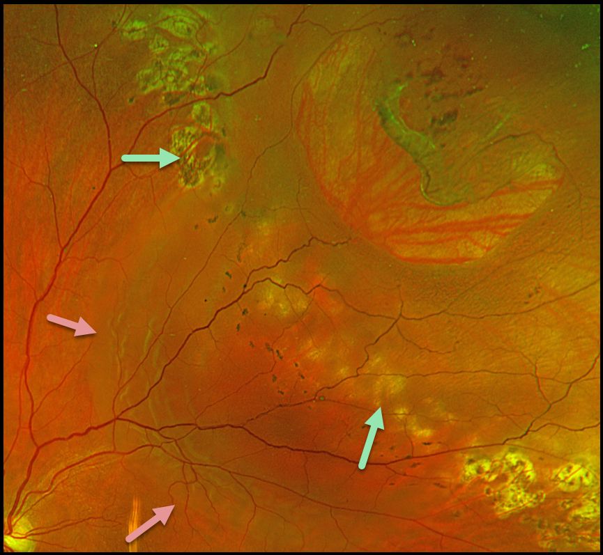
This 56YO male presented with several days of decreased vision in his left eye. He received prior treatment 1.5 years ago elsewhere for a retinal detachment. Vision was 20/30.
Optos color RG imaging shows a large lattice tear superotemporally with a surrounding rhegmatogenous retinal detachment (RRD). Several rows of appropriate and nearly confluent laser photocoagulation scarring are noted along the superonasal and inferotemporal edges of the detachment. However, barely visible depigmented laser scarring is noted more posteriorly, along with multiple tiny, 50-150 micron hyperpigmented laser scars. The detachment has broken through this region of inadequate laser.
Learning Points:
Demarcating rather than reattaching the retina is a valid option in select cases of RRD. When demarcating these detachments, it is critical that the retinopexy completely surrounds the subretinal fluid and extends to the ora; otherwise, the detachment can spread through the untreated retina, as occurred in this patient. Treatment should consist of several rows of nearly confluent, 300-500 micron photocoagulation burns. Cryotherapy or treatment with the laser indirect ophthalmoscope is often needed for the anterior treatment.
Assuming appropriate initial treatment, patients need to be followed postoperatively since the detachment can occasionally progress through the prior retinopexy. See Vrabec and Baumal, Ophthalmology 2000;107:1063-1067 for an excellent review regarding demarcation laser photocoagulation for selected macula-sparing retinal detachments.
LINCOFF RULES TO LOCATE BREAKS IN RHEGMATOGENOUS RETINAL DETACHMENT
Nilesh Kumar
Originally posted on @retina.rocks July 1, 2025
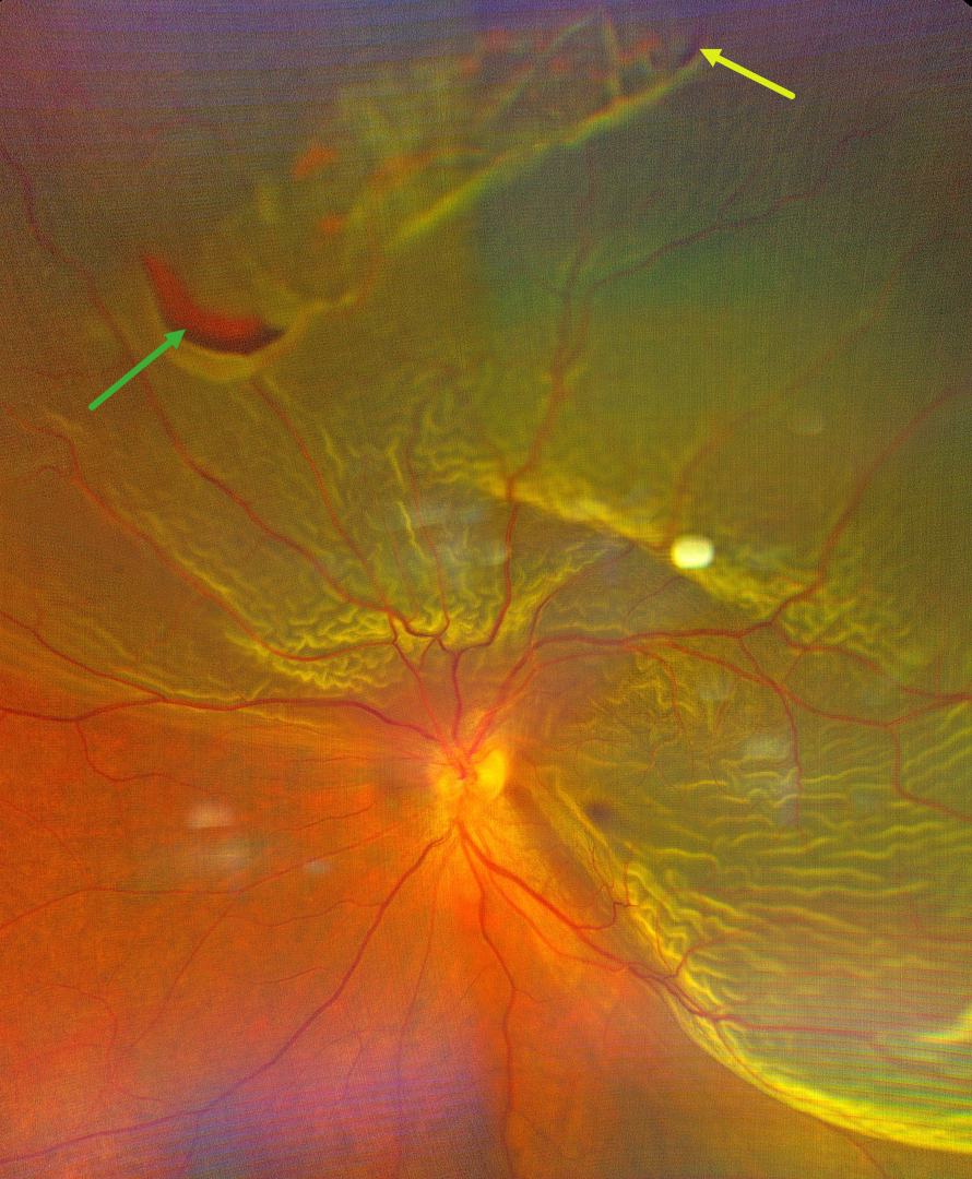
This 44YO male presented with sudden, painless, and progressive central and inferior peripheral vision loss. Color photography shows a superior and temporal macula-off rhegmatogenous retinal detachment (RRD). There is a superior patch of lattice degeneration with retinal tears at either edge of the lattice at 10:30 and 12:30.
Learning Points:
RRD occurs when liquid vitreous accesses the subretinal space through at least one full-thickness break in the neurosensory retina. A key principle for surgical success is identifying and closing all breaks. In 1971, Harvey Lincoff, in his classic paper ‘Finding the Retinal Hole,’ defined 4 findings based on RRD morphology that guide clinicians in determining where the causative breaks should be found (Lincoff and Gieser, Arch Ophthalmology 1971;85:565-569). These have since become known as the ‘Lincoff Rules.’ Simply put, gravity helps explain a detachment’s configuration and the location of the causative breaks.
Lincoff Rule 2 specifically addresses superior detachments crossing the 12 o’clock midline. It states that the primary break lies within a triangular zone with an apex at 12 o’clock, spanning 1.5 clock hours on either side (10:30-1:30 positions). This rule applies to 93% of such cases, as gravity causes subretinal fluid to spread symmetrically downward from the break.
Our patient’s detachment extends more inferiorly on the temporal edge. The two identified breaks in our image probably do not fully explain our RRD’s configuration, since the inferior edge of the detachment should fall on the nasal, not the temporal edge. At surgery, we would therefore search extra hard for at least one additional break along the temporal side.
TRAUMATIC MACULAR SCARRING AND MACULAR HOLE
Liubov Sitaruk and Ophthalmocoach
Originally posted on @retina.rocks June 10, 2025
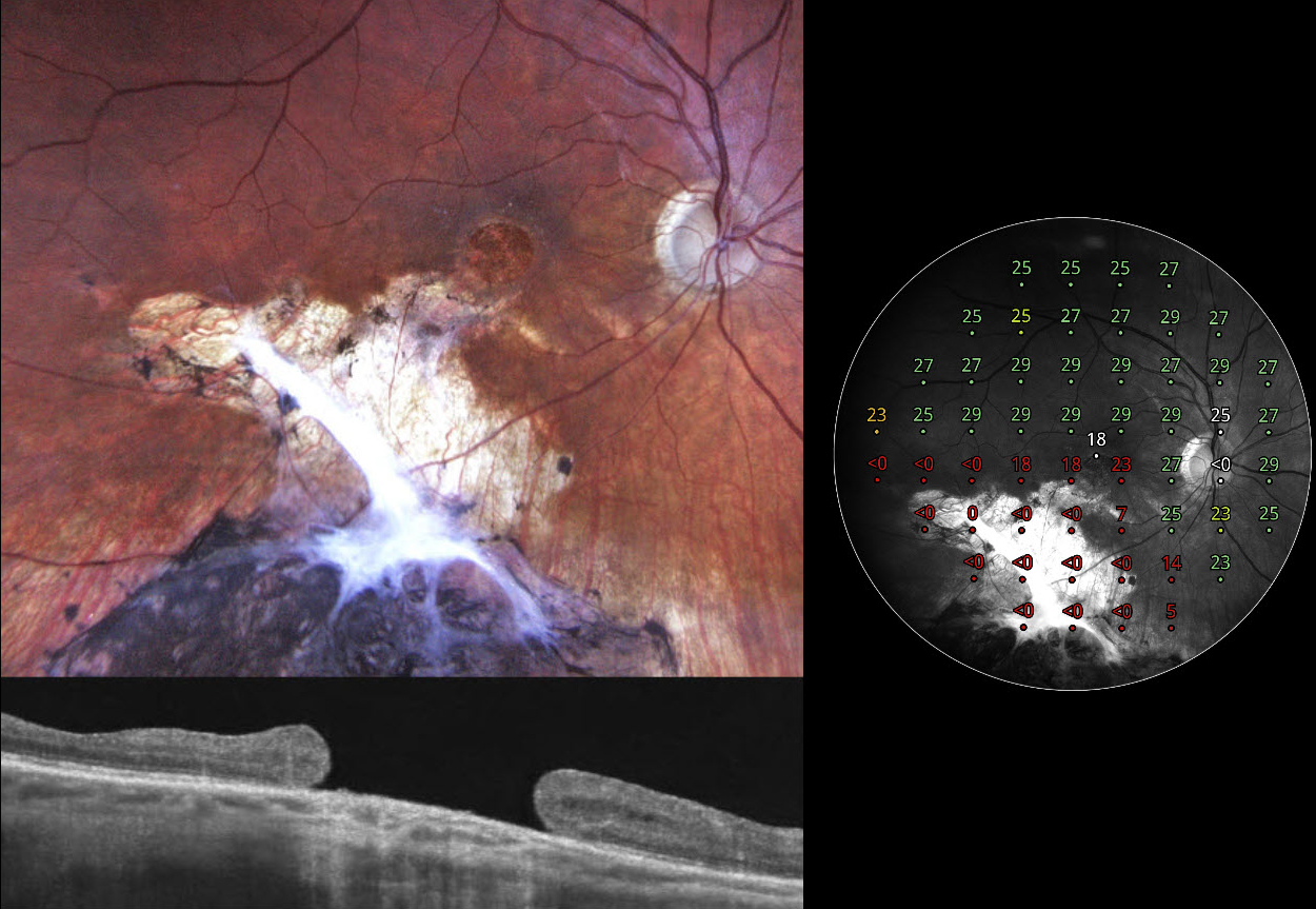
This 36YO Ukrainian male sustained a firearm-related facial injury during military combat in 2014 while in Eastern Ukraine. At the time, he was treated in a frontline hospital, where the left eye underwent primary enucleation due to a non-reconstructible globe rupture. His right eye sustained a penetrating injury with scleral rupture, which was primarily repaired. Over the following months, he underwent pars plana vitrectomy with endolaser and silicone oil due to complications, likely including retinal detachment and/or vitreous hemorrhage. Given the complexity of the trauma, the monocular status, and probable limited access to regular follow-up during wartime, silicone oil was likely retained for an extended period, potentially several years, until its removal at an undetermined later date. On our 2024 examination, he had no new visual complaints, and vision was 20/100.
Fundus photography shows atrophic inferior macular chorioretinal scarring, which becomes hyperpigmented as it extends into the inferior midperiphery. A large macular hole is noted funduscopically and on OCT. Macular perimetry shows marked depression within the area of scarring with preservation of some central sensitivity. Observation was recommended.
ECTOPIC MACULAR PUCKER
Shelby Culver
Originally posted on @retina.rocks May 15, 2025
This 51YO male was referred for a minimally symptomatic, unusual-appearing macular pucker. Vision was 20/20.
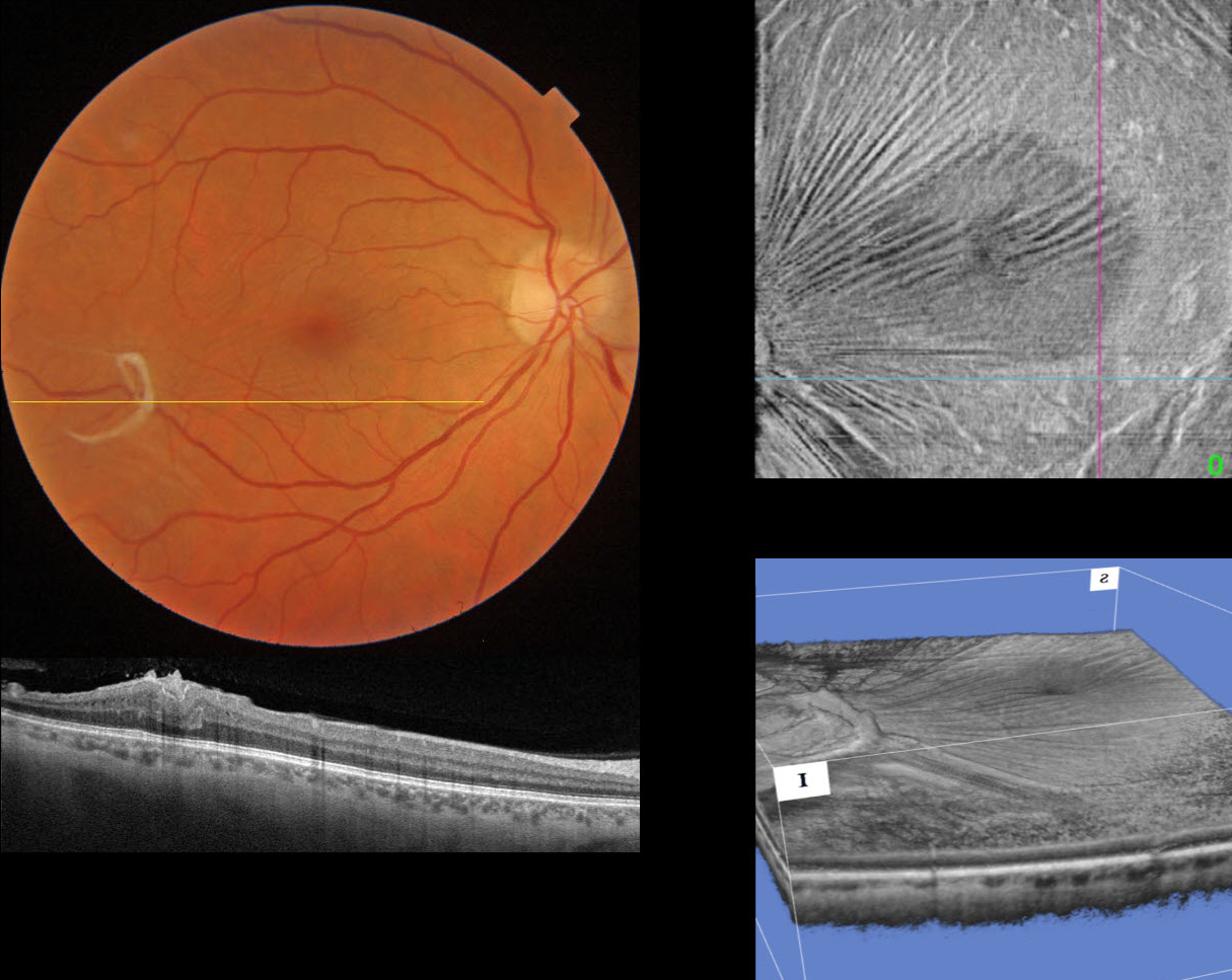
Triton color imaging and swept-source OCT show a semicircular area of epiretinal proliferation and traction in the distal inferotemporal macula. Radiating inner retinal striae are drawn towards this area of traction, which are best seen on en face OCT of the inner retina. A 3D reconstruction shows another view of this traction.
Since our patient was virtually asymptomatic, observation was recommended.
MENISCUS MICROPYON
Mattie Adams
Originally posted on @retina.rocks April 3, 2025
This 72YO female presented with one day of peripheral vision loss in her right eye. Vision was 20/25 OD and 20/25 in her healthy OS.
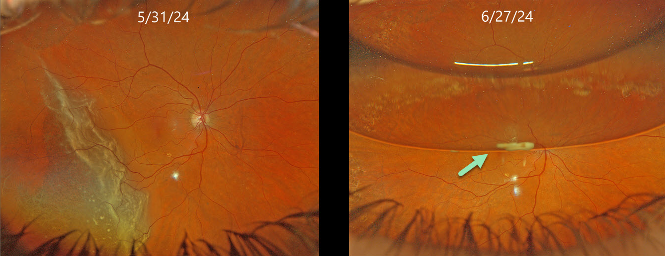
Optos color RG imaging (image 2, top) shows a temporal macula-on rhegmatogenous retinal detachment (RRD). Pars plana vitrectomy (PPV) with a C3F8 bubble was performed later that day. On her 1-month postoperative visit, vision was 20/50 with about a 50% gas bubble. A greyish globular deposit was adherent to the inferior edge of the gas meniscus.
Learning Points:
Russell et al recently reported a unique ophthalmoscopic sign, a meniscus micropyon, that follows PPV with gas tamponade for all causes (Retina 2024;44:1766-1776). Features they noted that distinguish this sterile micropyon from post-PPV fibrin include delayed appearance, hyperautofluorescence, and absence of fibrinous strands or sheets in the anterior chamber or vitreous. Vitreous biopsy in two of their cases revealed predominantly white blood cells. The micropyon appearance was associated with epiretinal proliferation (80%). Recurrent RRD from proliferative vitreoretinopathy (PVR) developed in 33% of eyes, 12% re-detached without frank PVR, and 18% developed worsening epiretinal membranes.
Fortunately, our patient’s postoperative course was uneventful. The micropyon resolved at the following visit and 4 months postoperatively, vision was 20/20 and the retina remained completely attached without epiretinal proliferation (not shown). Since the publication of Russell’s article, we have seen several patients with this biomarker, so we suspect it to be a not uncommon finding.
SUBRETINAL PERFLUORON
Originally posted on @retina.rocks March 10, 2025
This 33YO male presented with stable vision following prior vitrectomy for a rhegmatogenous retinal detachment (RRD) in his right eye. Vision was 20/400.
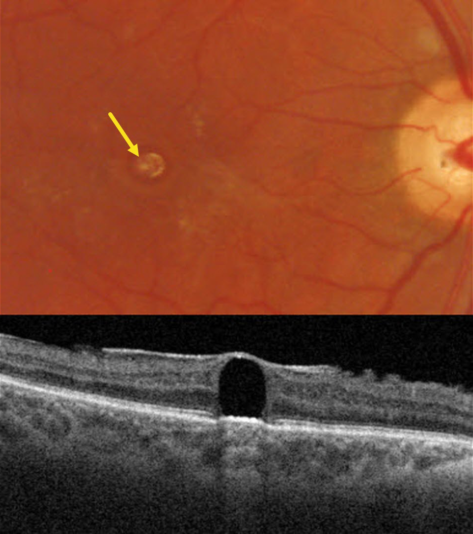
Triton color imaging shows a round yellowish subfoveal lesion. Swept-source OCT shows a hyporeflective subretinal lesion compressing the overlying neurosensory retina into a thin inner retinal hyperreflective line. The underlying RPE band is slightly anteriorly displaced, with hyperreflective choroidal shadowing. A mild epiretinal membrane is also noted.
Learning Points:
Perfluorooctane (PFO) is a liquid heavier than water used intraoperatively for complex retinal detachments, including proliferative vitreoretinopathy and giant retinal tears. The PFO is removed during the fluid-air exchange. Rarely, PFO droplets can develop intraoperatively and migrate subretinally. These were much more common from intraocular turbulence prior to the introduction of modern valved cannulas.
Subretinal PFO bubbles have a characteristic appearance on OCT, including an acute angle between the bubble and the RPE, thinned, ill-defined overlying retinal layers, elevation of the underlying RPE band, and hyperreflective choroidal shadowing (Figueroa and Contreras, Retina 2012;32:2177-2178). The subretinal PFO can be removed using a variety of surgical maneuvers, although we recommend observation given its chronicity and absence of new visual complaints.
DEGENERATIVE RETINOSCHISIS + SITUS INVERSUS
Sayena Jabbehdari and Ahmed Sallam
Originally posted on @retina.rocks March 5, 2025
This 64YO female was referred for a possible asymptomatic rhegmatogenous retinal detachment (RRD) in her left eye. Vision was 20/20 in her normal OD and 20/20 OS.
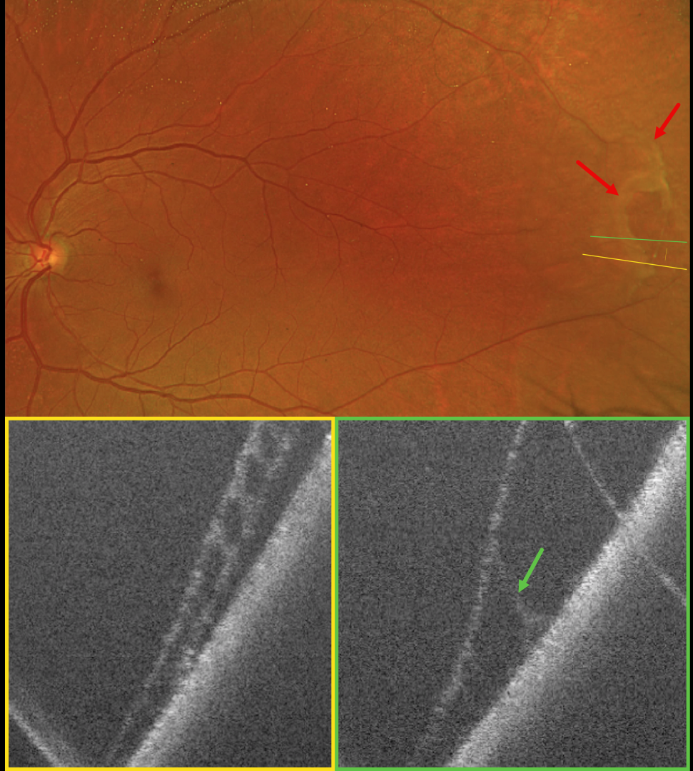
Optos color RG imaging shows superotemporal degenerative retinoschisis with a few outer-layer holes (OLH). OCT confirms the schisis as well as the outer layer hole.
Learning Points:
Clinically, degenerative retinoschisis usually appears as a smooth, dome-shaped peripheral retinal elevation without the outer retinal corrugations classically seen in RRD (Oquendo et al, AJO 2024;268:212-221). Degenerative retinoschisis is virtually never prophylactically treated (Byer 1986;93:1127-1137). Although the schisis cavities can enlarge, prophylactic treatment, including thermal laser, does not prevent further spread and should not be performed (Ness et al, Surv Ophthalmology 2022;67:892-907).
Situs inversus of the optic disc was identified as a benign incidental finding in our patient. This occurs in 5% of normal eyes and 70% of eyes with tilted discs (Witmer et al, Surv Ophthalmology 2010;55:403-428). It can be mistaken for other congenital disc anomalies.
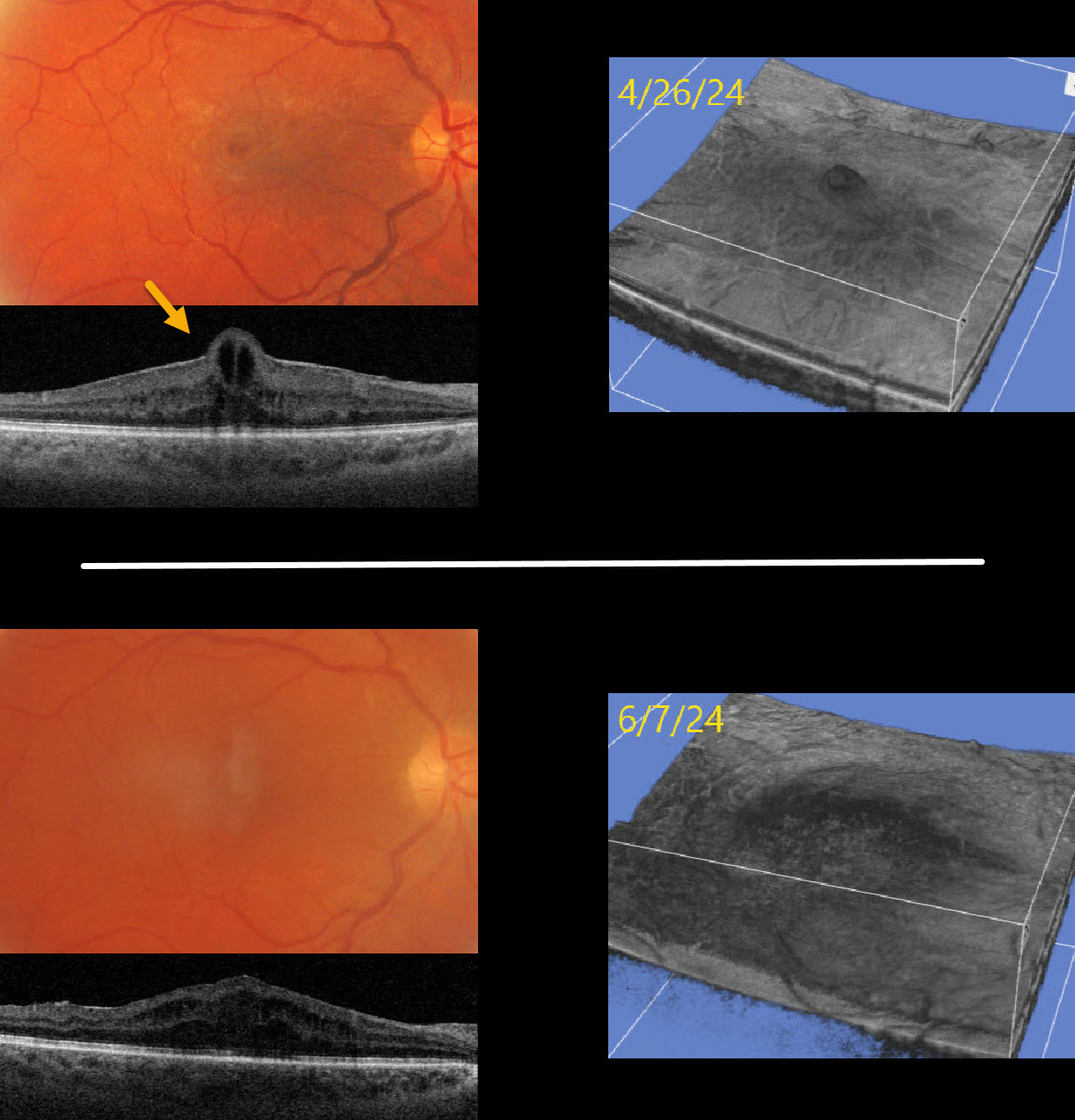
MACULAR PUCKER WITH FOVEAL HERNIATION
Originally posted on @retina.rocks February 27, 2025
This 53YO male presented with counting-fingers vision in his right eye due to a symptomatic macular pucker.
Optos color RG imaging shows a macular pucker with a foveal cyst. Triton swept-source OCT shows an adherent pucker with central foveal herniation. Vitrectomy surgery was performed, and one month later, vision improved to 20/100. OCT scanning shows no pucker, moderate diffuse cystic retinal thickening, and resolution of the foveal herniation. The pre- and postoperative appearances are also well visualized with 3D OCT reconstruction.
Learning Points:
Foveal herniation is found in about 1% of eyes with idiopathic epiretinal membranes (Uzel et al, Eye 2023;37:1357-1360). Epiretinal membranes not uncommonly surround the foveal center, and central contraction can result in uninvolved inner foveal tissue being squeezed through the central membrane defect, resulting in this unique appearance. Vitrectomy surgery with membrane peeling usually results in si
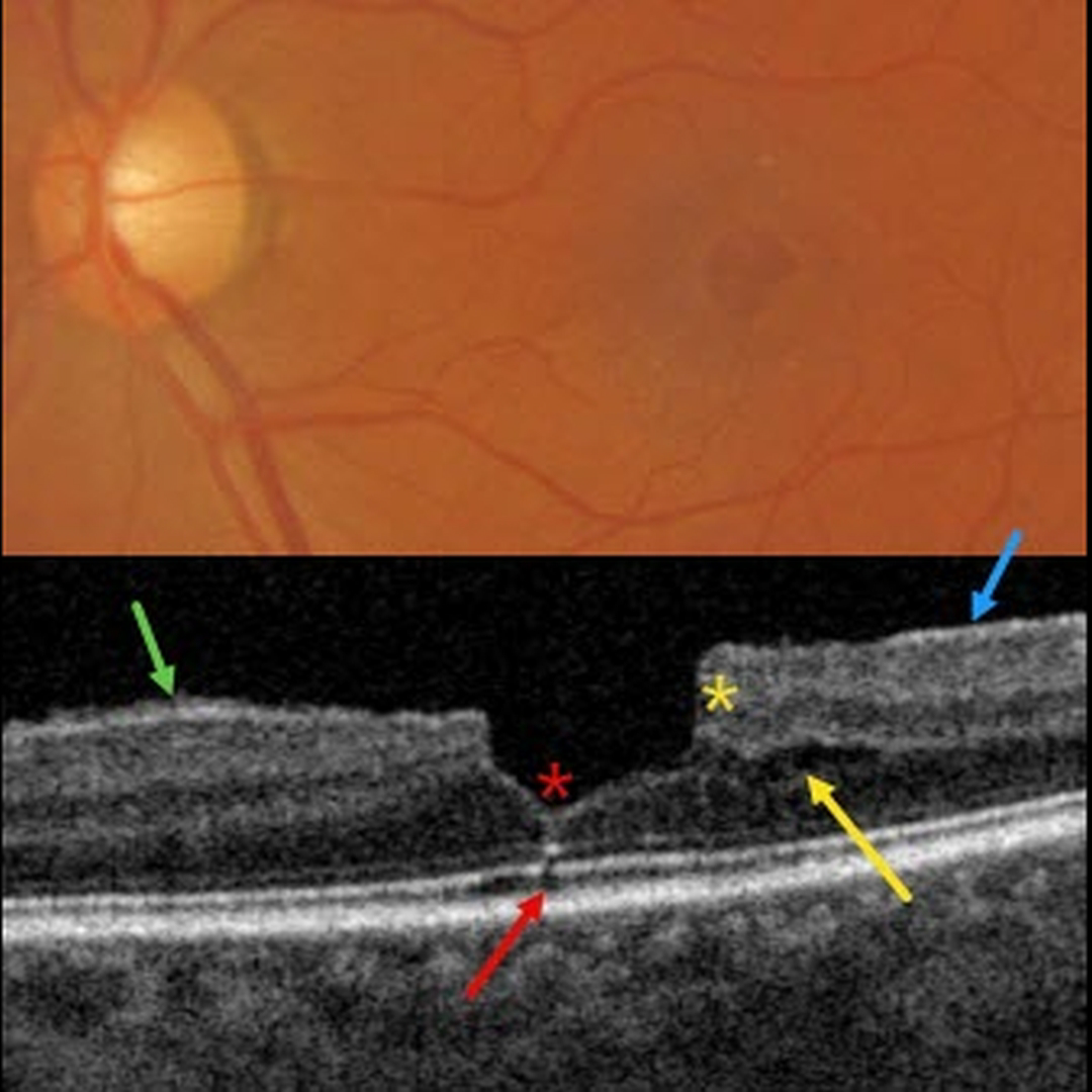
MACULAR PUCKER
Originally posted on @retina.rocks February 19, 2025
This patient presented with 20/30 vision due to a mildly symptomatic clinical macular pucker with a pseudohole; observation was recommended. Multimodal imaging reveals extensive pucker-related findings.
Triton color imaging shows an irregular macular pseudohole with a faint surrounding epiretinal membrane (ERM). True full-thickness macular holes are virtually always perfectly round, and these pucker-related pseudoholes are often irregularly round, as in this eye.
Swept-source OCT shows an adherent ERM with some shaggy-appearing adherent vitreous cortex, favoring epimacular proliferation. The fovea has a vertical contour, often present with ERM, due to the encircling membrane contracting towards the foveal center, dragging the inner retina centrally.
There are features of both a tractional lamellar macular hole (LMH) with splitting of the outer plexiform layer, along with a degenerative LMH with descending loss of tissue into the outer nuclear layer (Govetto et al, AJO 2016;164:99-109).
Finally, a tiny area of shadowing vs an outer foveal microdefect (Cohen et al, Ophthalmology Retina 2021;5:553-561) is seen.
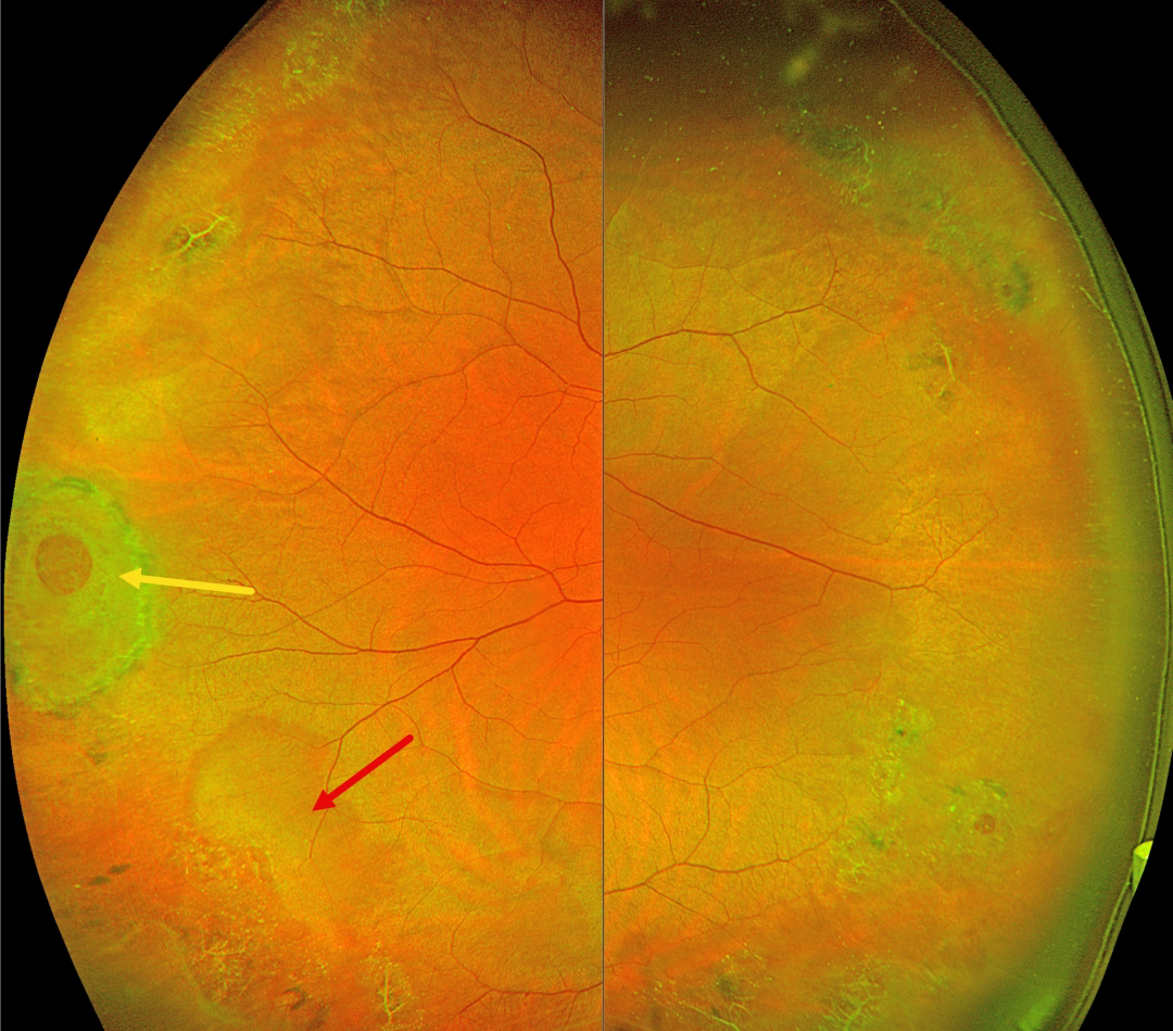
ASYMPTOMATIC LATTICE DEGENERATION WITH HOLES
Lattice degeneration SLF-
Originally posted on @retina.rocks February 18, 2025
This 42YO female was referred for asymptomatic peripheral retinal findings. Vision was 20/20 OU.
Optos color RG imaging shows extensive bilateral temporal lattice lesions with atrophic holes. A larger hole with surrounding pigment is seen in the right eye, along with a nearby area of white without pressure.
Learning Points:
Lattice lesions have a pocket of vitreous liquefaction overlying the thinned retina with a firm vitreo-retinal adhesion along their borders. White, fibrosed vessels often crisscross the lesions, which leads to the name, lattice. This firm vitreoretinal adhesion, along with the often-present atrophic holes, is what can predispose these eyes to rhegmatogenous retinal detachment (RRD).
Retinal detachment occurs in 1 in 10,000 people per year. Lattice is found in 10% of the general population, especially in high myopes. Since about one-third of patients with retinal detachment have lattice as the cause, the question always arises: Should prophylactic retinopexy be performed? The short answer is that asymptomatic lattice should generally not be treated.
Byer, in a Long-term natural history study of 423 untreated eyes, concluded that prophylactic treatment with or without holes in phakic non-fellow eyes should not be performed (Ophthalmology 1989;96:1396-1402). Folk et al (Ophthalmology 1989;96:72-79, Retina 1990;10:165-169) found no compelling evidence for prophylactic treatment of phakic fellow higher-risk eyes with more extensive lattice or higher myopia. They also found that some eyes developed subsequent non-lattice breaks outside the prophylactic treatment. A recent Cochrane systematic review by Wilkinson concluded that asymptomatic breaks and lattice should not be treated (Cochrane Database of Systematic Reviews 2014;9;CD003170) and the most recent 2019 American Academy of Ophthalmology Preferred Practice Pattern also cautions against prophylactic treatment.
A recent study by Curran et al recommended prophylactic treatment of lattice in fellow retinal detachment eyes, but this was in a retrospective, uncontrolled case series (Retina 2024;44:63-70). Another recent report by Kazan et al (Journal of VitreoRetinal Diseases 2024;8:381-387), in an uncontrolled retrospective series of eyes with “high-risk” lattice, found few post-laser detachments and concluded that treatment “may be effective in decreasing progression to RD…” But 3% of their treated eyes developed RRD over a mean 3- to 4-year follow-up period, which is higher than expected from the natural history of untreated lattice. So, our interpretation of their results is that prophylactic treatment may have actually increased the risk for detachment.
Stickler syndrome is a usually autosomal-dominant genetic disorder characterized by defective vitreous collagen (types II, IX, and XI). It is the most common cause for inherited rhegmatogenous retinal detachment and is the exception to the rule against prophylactic treatment. Prophylactic encircling laser extending from the pre-equator to the ora should be strongly considered since it lowers the risk for retinal detachment and giant retinal tears (Linton et al, Retina 2023;43:88-93).
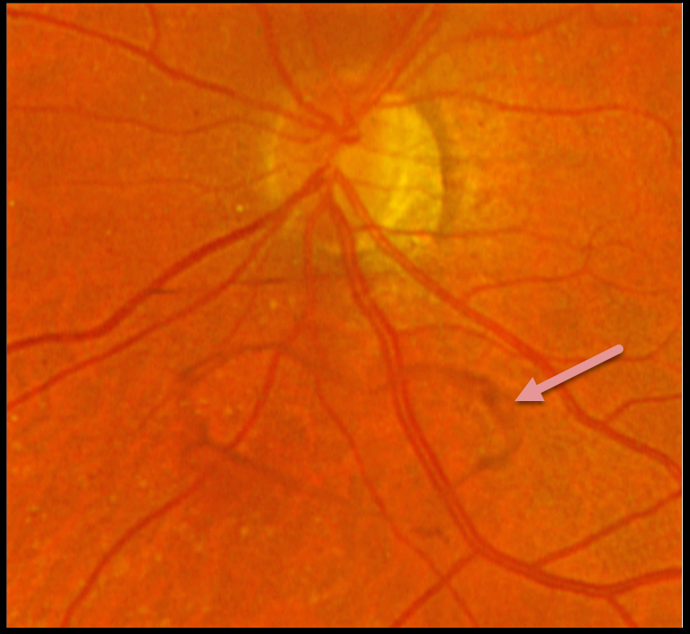
POSTERIOR VITREOUS DETACHMENT
Originally posted on @retina.rocks February 14, 2025
This 61YOF put a smile on our face by presenting with this asymptomatic heart-shaped Weiss ring just inferior to her left nerve.
HAPPY VALENTINE’S DAY!
RHEGMATOGENOUS RETINAL DETACHMENT WITH SUB-ILM HEMORRHAGE
Originally posted on @retina.rocks February 12, 2025
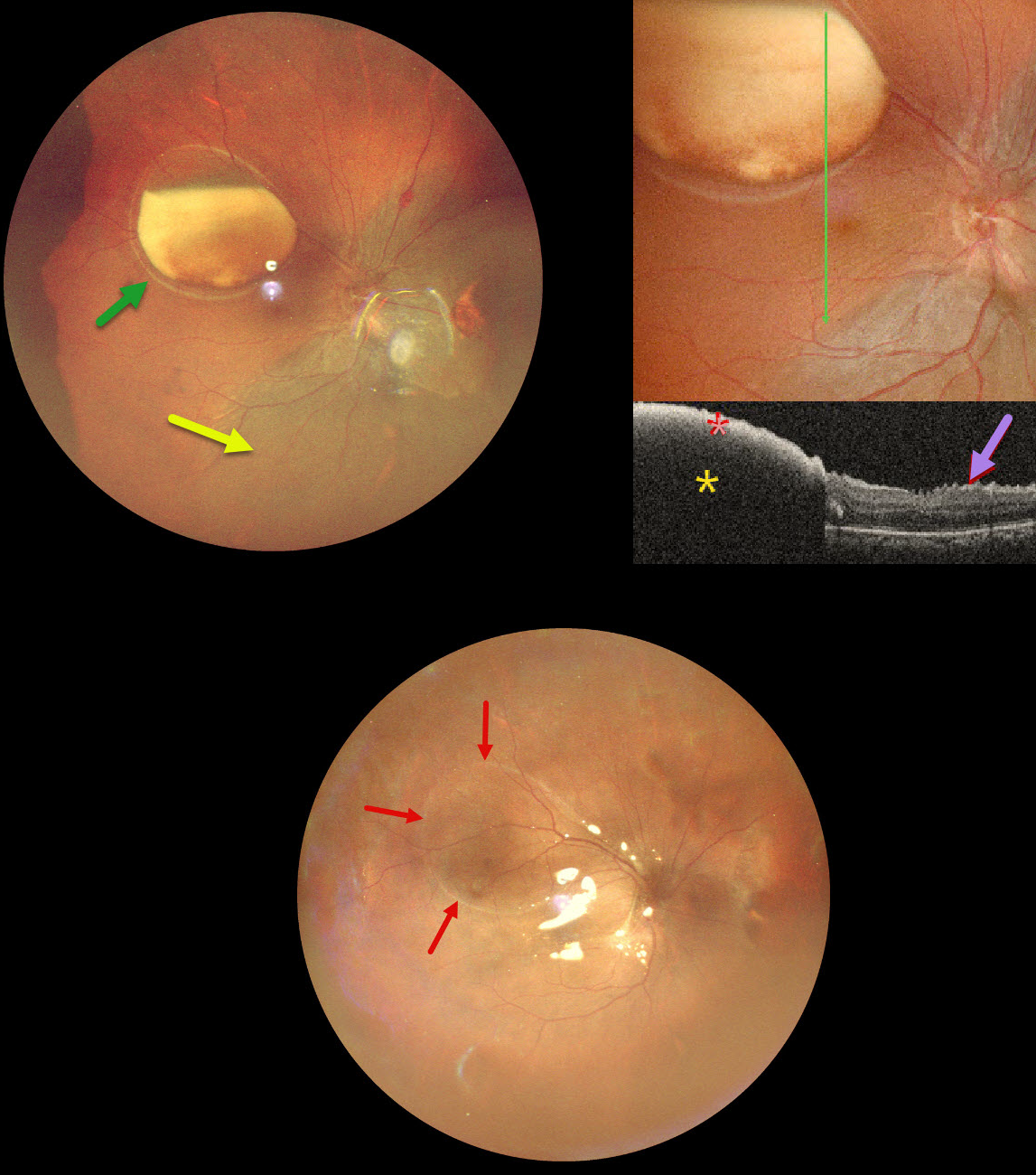
This 39YO male underwent vitrectomy OD elsewhere for a vitreous hemorrhage one month earlier. The treating surgeon noticed a sub-internal limiting membrane (ILM) hemorrhage at a postoperative visit and referred him to us for further evaluation. Vision was 20/30 OD and 20/20 in his normal OS.
Ultrawidefield pseudocolor SLO imaging shows a large partially layered sub-ILM hemorrhage with devitalized yellow blood in the superotemporal midperiphery. A retinal detachment is noted inferiorly and nasally. Inner retinal folds are visible at higher magnification. The blood is highly reflective on OCT scanning with total posterior shadowing. The inner retinal folds are noted inferiorly.
We don’t have a good explanation for our patient’s sub-ILM hemorrhage following vitrectomy for a vitreous hemorrhage. The yellow devitalized blood must have been present preoperatively, even though the initial surgeon told us he just noticed it one month postoperatively. Possibly our patient developed a breakthrough vitreous hemorrhage from a Valsalva maneuver, but without further history, this is just speculative.
We repaired the detachment with vitrectomy and silicone oil, and drained the sub-ILM blood intraoperatively. The retina was completely attached 2 weeks later. A high-water mark defines the margins of the prior ILM detachment.
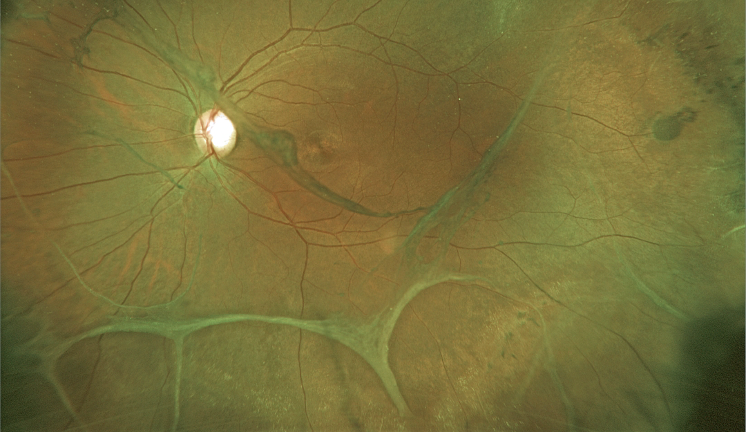
VITREOSCHISIS WITH MACULAR PUCKER
Originally posted on @retina.rocks February 11, 2025
This 28YO 8-diopter myopic female presented with recent floaters in her left eye. Vision was 20/30 OD and 20/40 OS.
Optos color RG imaging shows extensive bands of variably adherent vitreoschisis and epiretinal membranes. A prominent vitreous membrane vs pucker is noted in the inferonasal macula. There was bilateral variably pigmented paravenous lattice (not shown).
We recommended twice-yearly examinations, but unfortunately, the patient was immediately lost to follow-up.
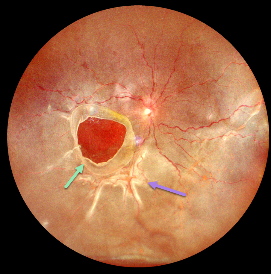
GIANT MACULAR HOLE WITH TOTAL RETINAL DETACHMENT
Anand Temkar and Manish Nagpal
Originally posted on @retina.rocks February 10, 2025
This 10YO boy presented with 1 week of vision loss in his left eye. The parents gave a vague history of trauma with a tennis ball 2 years earlier. Vision was counting fingers OD and 20/20 in his normal OS.
Ultrawidefield imaging shows a giant macular hole occupying nearly the entire macula. The hole’s edges are rolled, and proliferative vitreoretinopathy with star folds is present within the total rhegmatogenous retinal detachment (RRD).
Emergent vitrectomy surgery with silicone oil was scheduled.
Learning Points:
Blunt ocular trauma can result in numerous sequelae, including commotio retina, retinal breaks and detachment, choroidal rupture, and macular hole. A giant macular hole, also reported in Alport syndrome (Shah and Weinberg Ophthalmic Genetics 2010;31(2):93-97), is an exceedingly rare traumatic event (Hernandez-Da Mota Case Rep Ophthalmol 2011;2:283-286).
Traumatic macular holes are thought to result from a vitreous coup contrecoup injury or a jet stream of anterior chamber aqueous humor shooting through Cloquet canal into the premacular bursa.
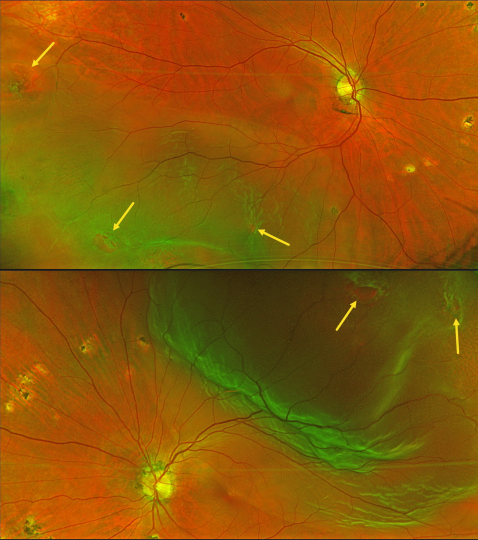
RHEGMATOGENOUS RETINAL DETACHMENT
Fraser McKay
Originally posted on @retina.rocks February 3, 2025
This 60YO male presented with 2 weeks of floaters in his left eye. Vision was 20/25 OD and 20/30 OS. Optos color RG imaging shows bilateral rhegmatogenous retinal detachments (RRD) with numerous breaks. The asymptomatic right detachment extended into the inferotemporal macula and split the left fovea. Scattered ocular histoplasmosis scars are present bilaterally.
Learning Points:
Patients with a RRD have a lifelong 10% risk of detachment in their fellow eye. Only a very small percentage will present with simultaneous detachments (Singh et al, Retina 2019;39:1504-1509).
Although the surgical success for each eye should be no different from that in patients presenting with unilateral detachments, treating the patient (not the eye) is more complicated. If one chooses pars plana vitrectomy (PPV), the postoperative gas bubble can cause severe, temporary vision loss. If both eyes receive PPV close together in time, the patient will be functionally blind for several weeks to months. We therefore find it helpful in these cases to treat the first eye with either silicone oil (with postoperative glasses) or perfluoro-octane liquid (followed by surgical removal a few weeks later). We can then repair the second eye within a few weeks of the initial eye. If possible, scleral buckling is also a good choice since no gas bubble is needed.
Emergent pars plana vitrectomy surgery was scheduled for our patient’s left detachment. We will follow the right eye closely perioperatively and will be forced to operate sooner if the detachment spreads towards the macular center. Otherwise, the right detachment will be repaired once the intraocular bubble is less than 50% in his left eye.
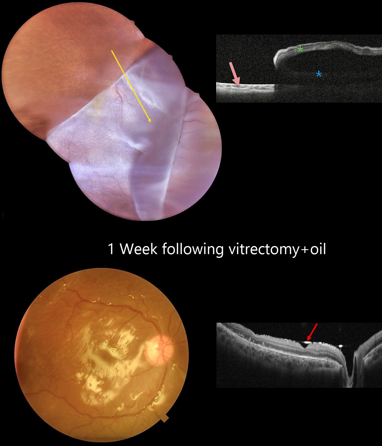
GIANT RETINAL TEAR
Seif Anwar
Originally posted on @retina.rocks January 30, 2025
This 12YO boy presented with 3 weeks of vision loss in his right eye immediately following blunt trauma. Vision was hand motion OD and 20/20 in his normal OS.
Color photography shows a superotemporal giant retinal tear that has folded over the optic nerve and macula inferonasally. The yellow macular xanthophyll is visible just inferotemporal to the nerve. OCT scanning shows bare RPE superotemporally, the underlying detachment, and the overlying inverted retina.
Emergent surgical repair with pars plana vitrectomy was scheduled. One week postoperatively, the retina is completely attached. The hyperreflective oil-retinal interface is visible overlying the fovea and optic nerve.
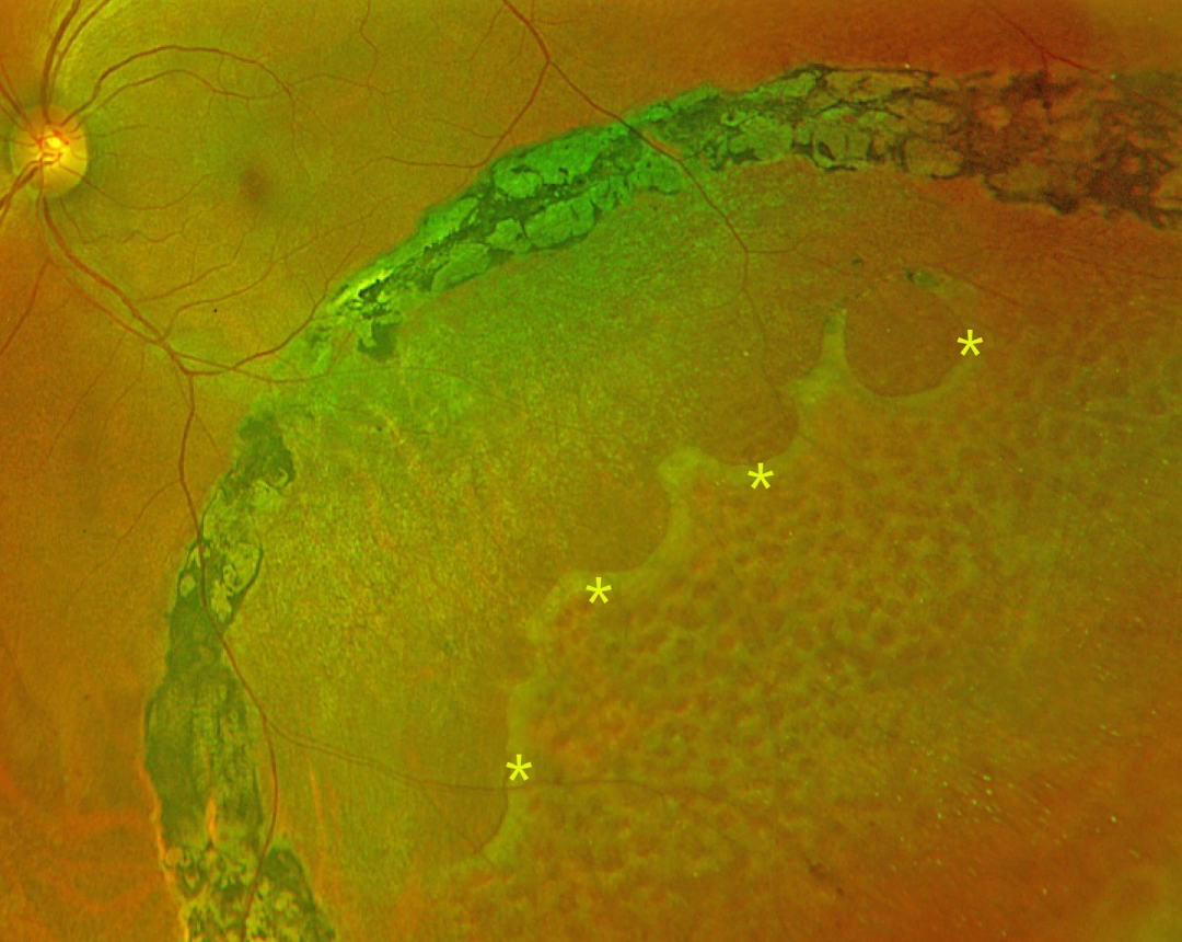
DEGENERATIVE RETINOSCHISIS
Vaibhav Sethi
Originally posted on @retina.rocks January 21, 2025
This 47YO female presented with 4 years of flashes in her left eye that began immediately following laser photocoagulation elsewhere. Vision was 20/20 bilaterally.
Optos color RG imaging shows inferotemporal degenerative retinoschisis, demarcated by heavy, confluent laser scarring along its posterior extent. A row of outer-layer holes is noted within the schisis cavity, with innumerable inner-layer holes more anteriorly.
Learning Points:
Degenerative retinoschisis is virtually never prophylactically treated (Byer 1986;93:1127-1137). Although the schisis cavities can enlarge, prophylactic treatment including thermal laser does not prevent further spread, should not be performed (Ness et al, Surv Ophthalmology 2022;67:892-907), and in our patient’s case, caused persistent symptomatic flashes. Vitrectomy surgery can successfully repair combined schisis rhegmatogenous retinal detachment (RRD) when liquid vitreous enters the subretinal space through holes in both the inner and outer layers.
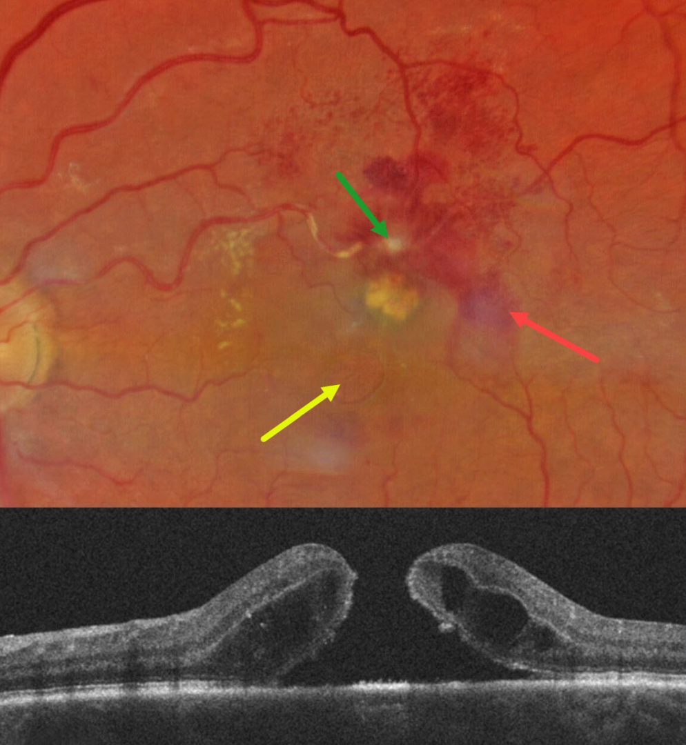
RETINAL ARTERIAL MACROANEURYSM (RAM) WITH MACULAR HOLE
Will Gibson
Originally posted on @retina.rocks January 13, 2025
This 67YO white female presented for her six-month diabetic follow-up with a few weeks of vision loss in her left eye. Vision was 20/80 OS and 20/20 in her normal OD.
Color photography shows a fibrosed retinal arterial macroaneurysm (RAM), in the superior macula. Faint subretinal blood is noted temporally, with mostly inner retinal blood superotemporally. The artery is sheathed just proximal to the RAM. A large macular hole with surrounding subretinal fluid is present, accompanied by a lipid clump between the hole and the RAM. Faint lipid is also noted in the nasal macula. She was referred to a retinal specialist for further management.
Learning Points:
Macular hole is a rare finding with retinal arterial macroaneurysms (Sagara et al, Br J Ophthalmol 2009;93:1337-1340). Subretinal and sub-internal limiting membrane blood appears to increase the risk of macular hole.
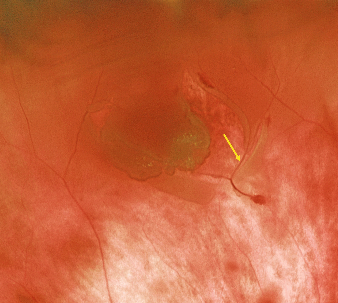
RETINAL TEAR WITH BRIDGING VESSEL
Originally posted on @retina.rocks January 9, 2025
This patient presented with a macula-on rhegmatogenous retinal detachment (RRD). Optos color RG imaging shows the causative retinal tear with a bridging vessel. The RRD was successfully repaired with vitrectomy.
Learning Points:
The overall risk of an acute PVD causing a retinal break or detachment is about 5% (Seider et al, Ophthalmology 2022;129:67-72), and this risk is increased in the presence of vitreous pigment or blood. The reason for this is that, as in our patient, the retina often tears along a blood vessel. An occult retinal break is often the cause of an unexplained vitreous hemorrhage, which is why early vitrectomy is often recommended for these eyes, even when a break cannot be found on office examination (Flores-Sanchez et al, Eye 2023;37:3191-3196).
The bridging vessels in retinal breaks can also cause recurrent vitreous hemorrhage, a condition known as the avulsed retinal vessel syndrome (Robertson et al, Arch Ophthalmology 1971;85:669-672). The traction can spontaneously release, or vitrectomy may be required for sputtering hemorrhages or non-clearing blood.
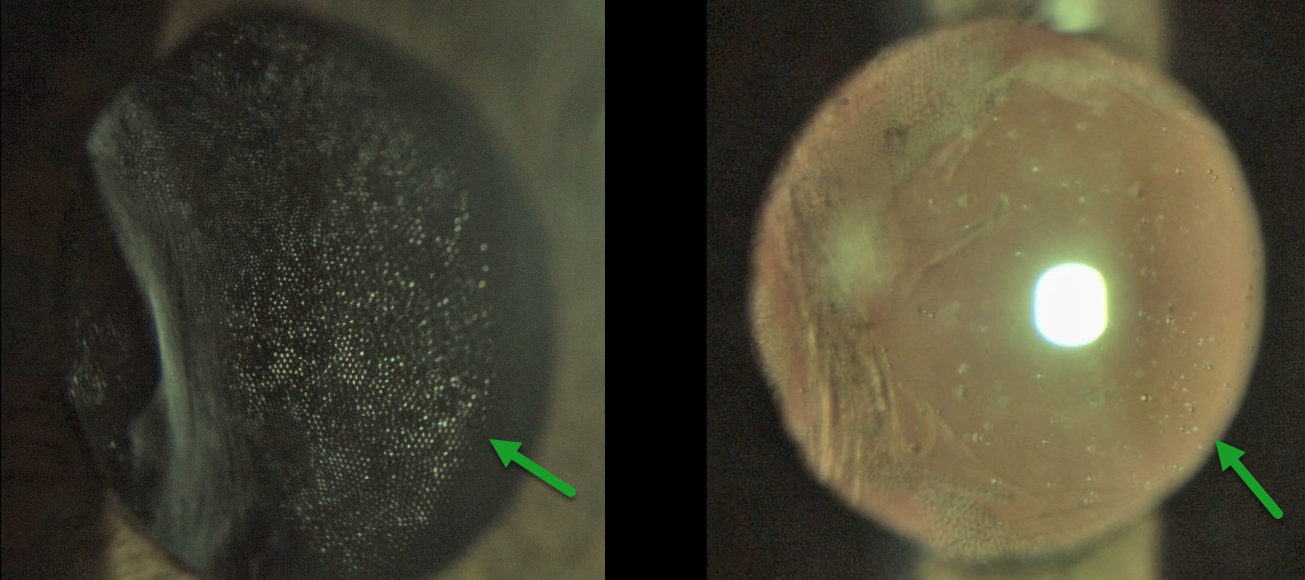
SILICONE OIL ON CAPSULE
Sharat Hegde
Originally posted on @retina.rocks December 30, 2024
This 70 YO male presented with 2 months of blurred vision in his right eye. Vision was counting fingers OD and 20/20 in his normal OS. There was a history of a prior retinal detachment that was repaired with vitrectomy and silicone oil. Removal of the silicone oil, along with combined cataract surgery with a hydrophilic intraocular lens (IOL), was performed 6 months earlier.
Slit lamp photography shows a honeycomb pattern of adherent silicone oil bubbles along the intact posterior capsule.
Learning Points:
Intravitreal silicone oil droplets can irreversibly adhere to the surface of IOLs, particularly silicone lenses, where they can cause significant glare and vision loss (Apple et al, Ophthalmology 1996;103:1555-1562). Silicone oil can also rarely cause IOL opacification (Oner et al, Ophthalmic Lasers Imaging Retina 2021;52:37-43). The adherent oil cannot be removed, and these lenses often require explantation.
Our case is somewhat unique in that the oil droplets adhered to the posterior capsule rather than the IOL. YAG capsulotomy displaced the oil bubbles, and vision improved to 20/60.
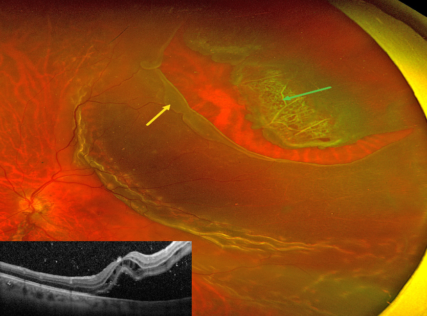
RHEGMATOGENOUS RETINAL DETACHMENT
Yuenpang Cheung
Originally posted on @retina.rocks December 19, 2024
This 46YO male presented with recent vision loss in his left eye. Vision was counting fingers.
Optos color RG imaging shows a superotemporal macula-off rhegmatogenous retinal detachment (RRD). The anterior flap of the causative tear encompasses an entire lattice lesion. The posterior edge of the tear has a posteriorly-rolled edge, indicating early Grade B proliferative vitreoretinopathy (The Retina Society Terminology Committee, Ophthalmology 1983;90:121-125).
OCT shows cystic retinal edema mostly in the outer nuclear layer, along with hyperreflective vitreous and subretinal cells. The patient was scheduled for emergent vitrectomy.
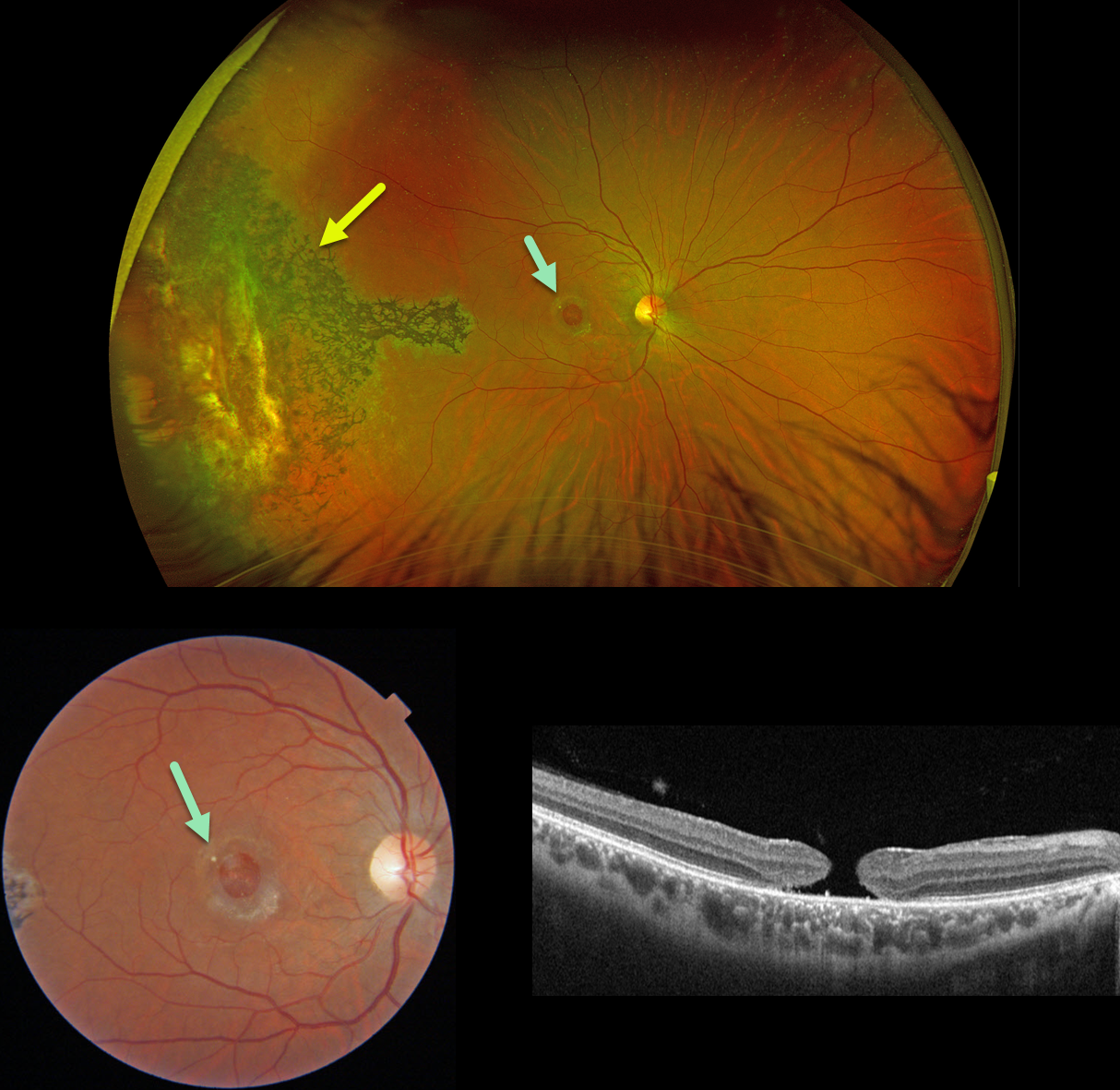
TRAUMATIC MACULAR HOLE
Originally posted on @retina.rocks December 18, 2024
This 31YO male suffered a paint gun injury to his right eye when he was 12YO. Vision has since been stable and poor in this eye. Vision was 20/200.
Optos color RG imaging shows temporal peripheral scarring with intraretinal pigment migration. A large macular hole with surrounding pigmentary changes is noted on Triton color imaging and swept-source OCT.
Learning Points:
Since fresh traumatic holes can spontaneously flatten, especially in pediatric cases (Liu et al, AJO 2020;210:174-183), an initial period of observation is recommended before considering vitrectomy. Given the size and chronicity of our patient’s hole, surgery was not considered.
Photoreceptor loss from numerous causes, including blunt trauma, retinal detachment, macular telangiectasia, and retinitis pigmentosa (RP), can lead to intraretinal RPE migration, a shared phenotype. Unilateral RP is exceedingly rare and requires normal electrophysiologic testing with long-term observation to ensure that delayed findings are present in the fellow eye.
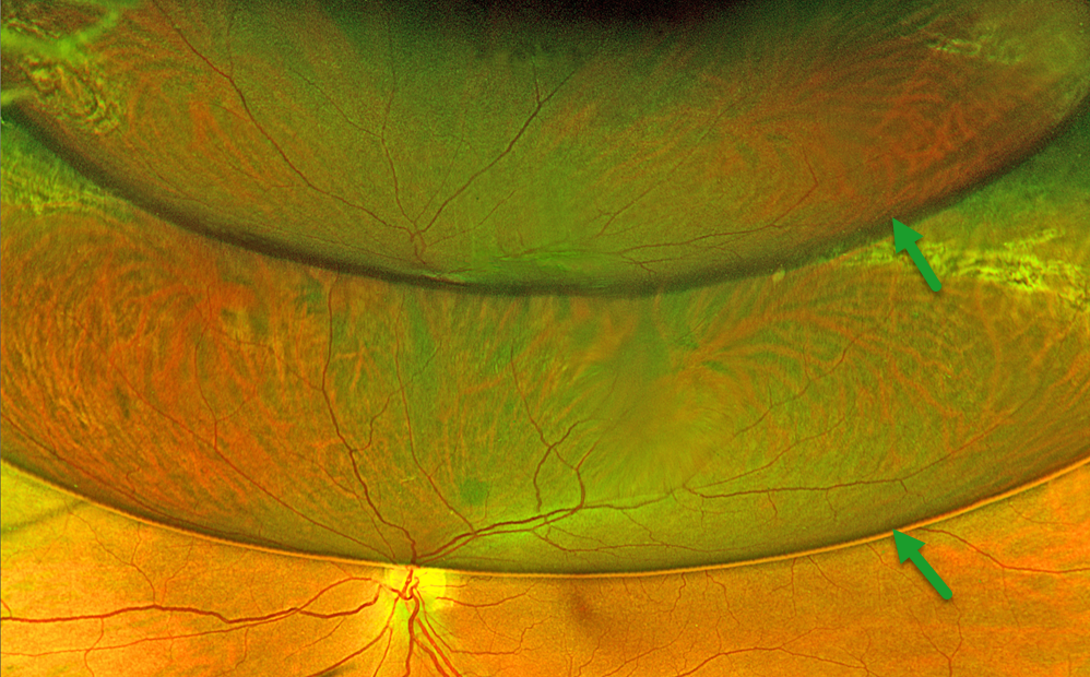
POST-OPERATIVE RD REPAIR WITH GAS
Originally posted on @retina.rocks December 16, 2024
This 56YO female underwent successful vitrectomy surgery for a rhegmatogenous retinal detachment (RRD) 1 month earlier. Optos color RG imaging shows an unusual double-bubble with different reflections of the attached inferior retina.
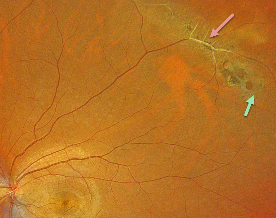
LATTICE DEGENERATION
Originally posted on @retina.rocks December 5, 2024
This healthy 33YO male was referred for asymptomatic lattice degeneration in his left eye. Optos color RGB imaging shows a circumferential patch of midperipheral superotemporal paravenous lattice. The large retinal vessels within the lesion are fibrosed. A small atrophic retinal hole is noted along its inferior edge. Observation was recommended.
Learning Points:
Lattice lesions have a pocket of vitreous liquefaction overlying the thinned retina with a firm vitreo-retinal adhesion along their borders. White, fibrosed vessels often crisscross the lesions, hence the name, lattice. This firm vitreoretinal adhesion, along with the often-present atrophic holes, is what can predispose these eyes to retinal detachment.
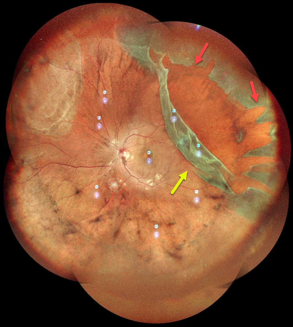
GIANT RETINAL TEAR
Anjana Mirajkar and Manish Nagpal
Originally posted on @retina.rocks November 26, 2024
This healthy 24YO male presented with several days of floaters in his left eye. Vision was 20/20 OU.
Pseudocolor SLO imaging shows a superotemporal giant retinal tear (extending over 3 clock hours) and detachment. The anterior portion of the tear has multiple jagged edges, and the posterior flap has folded over onto itself. The macula is attached. The detachment was repaired with emergent pars plana vitrectomy.
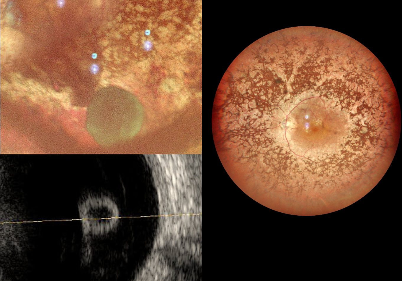
VITREOUS CYST
Anand Temkar and Manish Nagpal
Originally posted on @retina.rocks November 11, 2024
This 31YOF with a known history of retinitis pigmentosa (RP) presented with 2 days of decreased vision OD. There was a history of trauma to her right eye at 2 years of age. Vision was counting fingers OD and 20/120 OS. Slit lamp examination OD revealed a non-central corneal scar and anterior synechia consistent with the prior trauma.
MultiColor imaging shows bilateral RP intraretinal pigment migration and narrowed arterioles. A free-floating vitreous cyst is noted OD inferiorly.
Learning Points:
Vitreous cysts are extremely rare, idiopathic, and usually diagnosed incidentally. Likely originating from a ciliary body cyst (Dhull et al, Ophthalmology Retina 2020;4:1208), they can be either congenital or acquired. Acquired cysts have been associated with numerous ocular disorders, including trauma and retinitis pigmentosa, as in our patient.
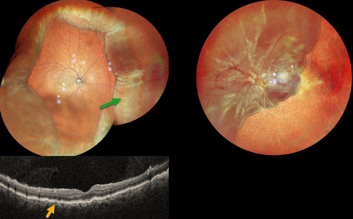
CHOROIDAL DETACHMENT
Anand Temkar and Manish Nagpal
Originally posted on @retina.rocks October 31, 2024
This 52YO male presented with 2 weeks of vision loss OD. He had a complicated ocular history of multiple prior surgeries elsewhere, including cataract surgery OD 2 months earlier and multiple retinal detachment surgeries OS. Vision was 20/30 OD and LP OS. Pressure was 8mmHg with no wound leaks on slit lamp OD.
MultiColor imaging shows encircling peripheral choroidal detachments OD, and OCT shows shallow macular chorioretinal folds. A total retinal detachment is noted OS with bare choroid nasally and inferiorly.
Learning Points:
A choroidal detachment is defined by the abnormal presence of fluid or blood in the suprachoroidal space. Serous choroidal detachments, also known as choroidal effusions, are a frequent complication of glaucoma surgery or following any intraocular procedure. Frank wound leaks usually require surgical repair. Otherwise, conservative management, often with topical/systemic steroids, is recommended.
Since our patient’s surgical wound was secure and the anterior chamber was formed, topical and oral steroids were prescribed.
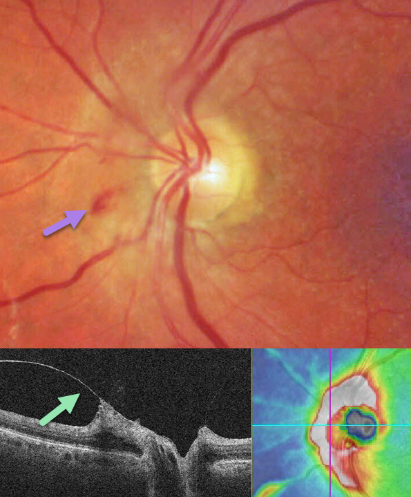
VITREOPAPILLARY TRACTION
Originally posted on @retina.rocks October 29, 2024
This 76YO female presented with asymptomatic optic nerve findings in her left eye. Color imaging shows somewhat opaque retina with some inner retinal hemorrhages along the nasal aspect of the nerve. OCT shows a partial PVD extending to the nerve with vitreopapillary traction and secondary thickening of the peripapillary nerve fiber layer. Observation was recommended.
Learning Points:
The normal vitreous goes through a decades long process of an orchestrated degeneration, including liquefaction and gradual separation from the retinal surface. The posterior vitreous detachment (PVD) begins in the perifoveal retina early in life, followed by a foveal PVD, then a peripheral PVD, and then finally a complete PVD when the vitreous separates from the optic nerve (Johnson AJO 2010;149:371-382). The integrity of the internal limiting membrane (ILM) is integral to normal vitreoretinal adhesion, where a thinner ILM has a firmer adhesion than a thicker ILM. The ILM represents the basement membrane of Muller cell. Hence an area that has a lower population of Muller cells reaching the inner retina will naturally have a firmer vitreoretinal adhesion. Since the optic nerve is completely devoid of Muller cells, this is the area of the posterior pole that has the firmest vitreoretinal adhesion, and this is the reason why the vitreous tends to remain so firmly attached to it.
Persistent vitreopapillary traction is a rarely reported phenomenon (Gabriel et al, Neuro-ophthalmology 2020;44:213-218), probably due to its lack of symptoms and often subclinical findings. In our patient’s case, the traction caused retinal thickening and hemorrhage, somewhat mimicking a peripapillary macular neovascularization. In our experience, these eyes usually remain asymptomatic and can be observed, although vitrectomy is very successful in relieving the traction and improving vision.
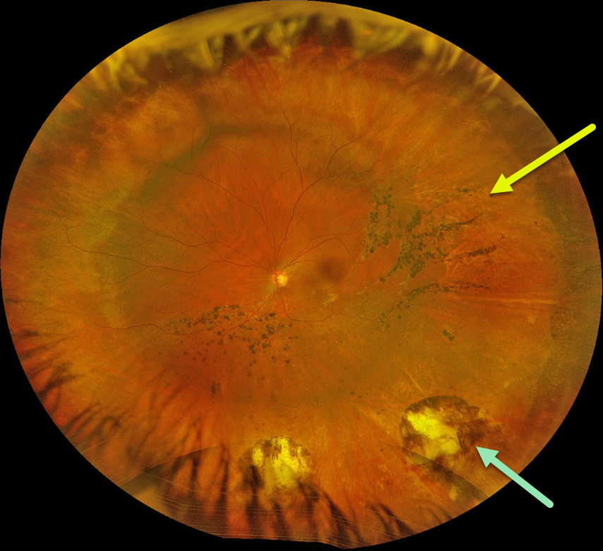
SCLERAL BUCKLE
Originally posted on @retina.rocks October 28, 2024
This 31YO female underwent successful scleral buckling surgery about 7 years earlier. Optos color RGB imaging shows a broad encircling buckle. Cryotherapy scarring is noted inferior to the buckle. Multiple demarcation lines with some intraretinal pigment migration are noted within the region of the prior detachment.
Learning Points:
The normal neurosensory retina remains attached to the RPE due to numerous physiologic mechanisms, including the inner (retinal vascular endothelium) and outer (tight junctions at the apex of the RPE cells) blood retinal barriers, hyperosmotic choroid passively drawing fluid out of the subretinal space, RPE pump, interphotoreceptor matrix, and friction between the RPE microvilli and the photoreceptor outer segments. Rhegmatogenous retinal detachment (RRD) occurs when liquid vitreous, entering the subretinal space through a retinal break, overwhelms these forces, favoring retinal detachment.
Repairing RRD involves finding and closing all breaks, essentially denying the liquid vitreous access to the subretinal space. The break(s) can be closed internally with a gas bubble with pneumatic retinopexy or vitrectomy surgery, or externally by sewing a piece of silicone material to the eye wall (scleral buckle). This indents, or buckles, the RPE and sclera up against the retinal break and formed vitreous.
The choice of procedure to reattach the retina is complex, depending on each patient’s unique anatomy and the surgeon’s preference. Historically, scleral buckling was the only method until vitrectomy surgery appeared in the 1970’s. There are advantages and disadvantages to all techniques. With one or more surgeries, there is a high anatomic success rate regardless of the method of retinal reattachment.
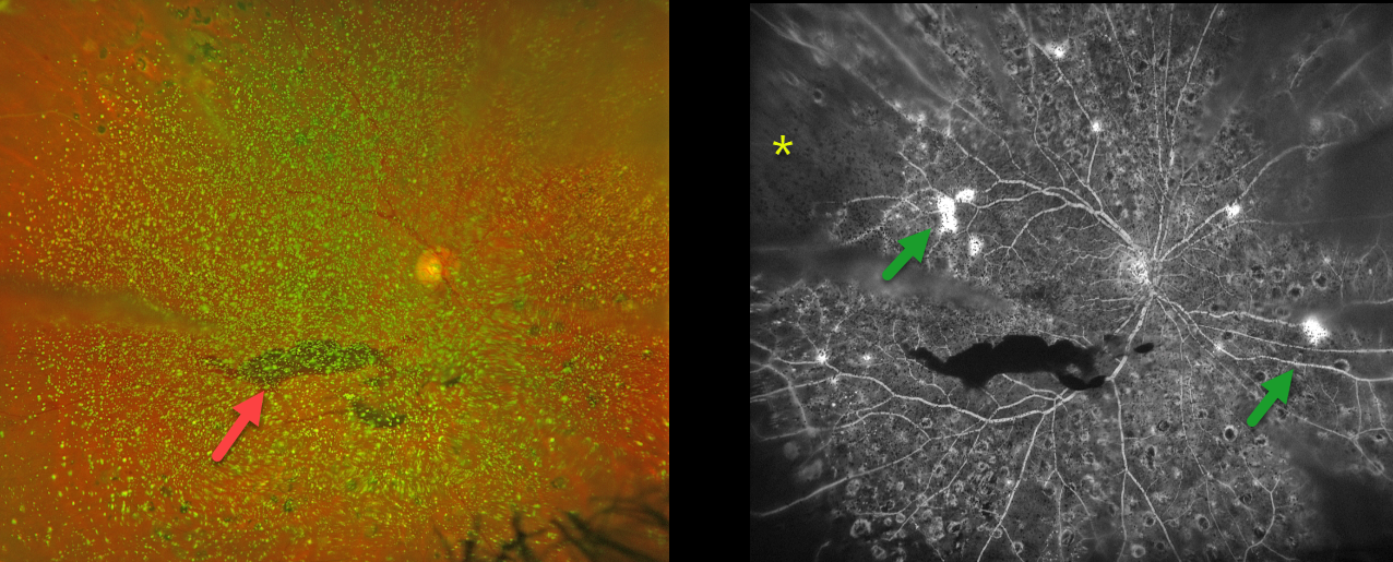
PROLIFERATIVE DIABETIC RETINOPATHY (PDR) WITH ASTEROID HYALOSIS
Originally posted on @retina.rocks October 16, 2024
This 50YO female with a history of type 2 diabetes was referred for new floaters. Several years earlier, we began panretinal photocoagulation (PRP) for proliferative diabetic retinopathy (PDR), but she had been lost to follow-up.
Optos color RG imaging shows dense asteroid hyalosis with a hazy inferior preretinal hemorrhage. Fluorescein angiography (FA) shows significant retinal ischemia, scattered retinal neovascularization, and partial PRP scarring.
Examining the fundus with dense asteroid, as in this diabetic patient, can be extremely challenging. FA and OCT are great modalities because they can visualize the retina when the clinical exam can’t. Optos FA was critical in confirming proliferative retinopathy with peripheral ischemia in our patient.
We were fortunately able to complete the PRP despite the vitreous opacities.
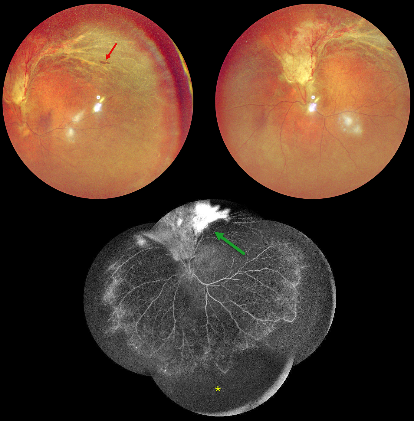
CMV RETINITIS WITH RETINAL DETACHMENT
Originally posted on @retina.rocks September 2, 2024
This 48YO HIV positive male on highly active antiretroviral therapy (HAART) presented with 2 months of vision loss in his left eye. His last CD4 count was 81. Vision was 20/20 OD and counting fingers OS.
Multicolor imaging shows active cytomegalovirus (CMV) retinitis with retinal detachment extending superiorly from the optic nerve into the superotemporal midperiphery. A few atrophic retinal breaks are noted. Fluorescein angiography of his left eye shows variable leakage within the area of retinitis superiorly and severe encircling peripheral ischemia. Vitrectomy with silicone oil was scheduled to repair the detachment in his left eye.
Learning Points:
CMV retinitis develops as a reactivation of latent CMV in immunosuppressed individuals. Before effective antiretroviral treatment emerged in the mid to late 1990’s, CMV retinitis developed in up to 40% of HIV/AIDS patients, often within the last 6 months of life. Treatment includes a combination of intravenous and intravitreal medications, including ganciclovir, foscarnet and cidofovir. Rhegmatogenous retinal detachment is a common late sequela and requires vitrectomy with silicone oil.
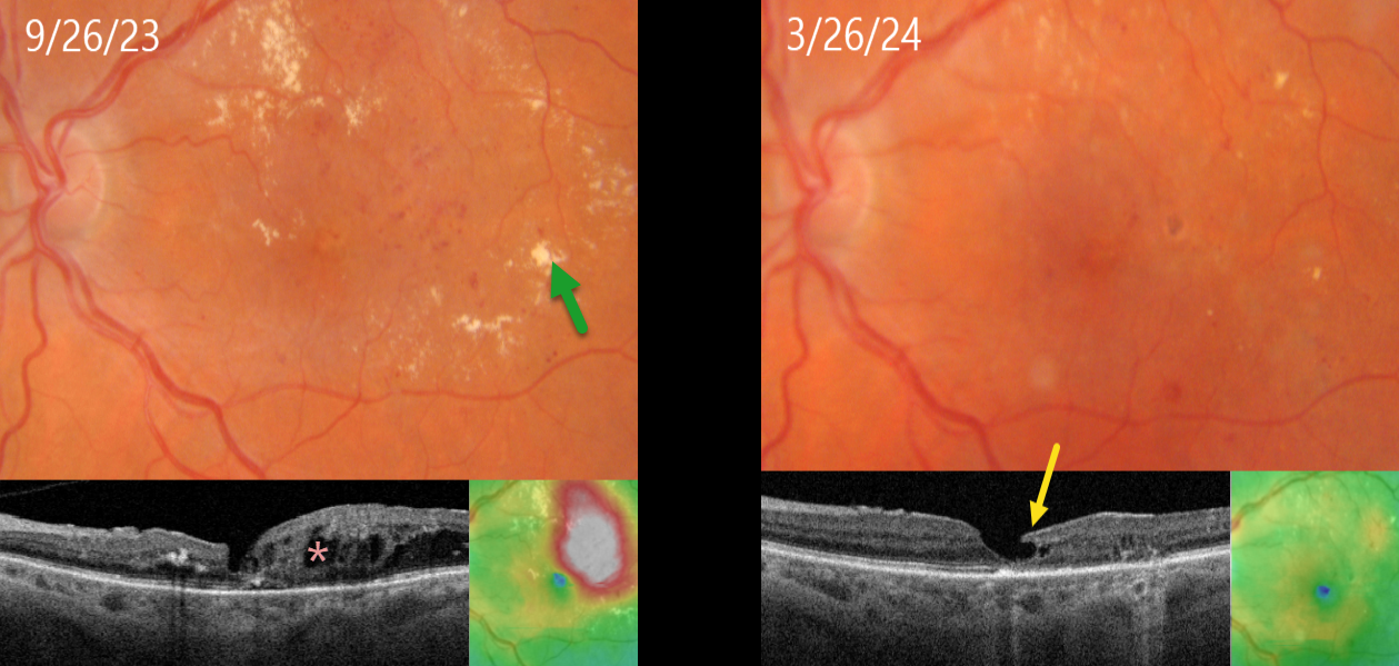
FOCAL LASER FOR DIABETIC MACULAR EDEMA (DME) WITH SUBSEQUENT DEGENERATIVE LAMELLAR MACULAR HOLE
Originally posted on @retina.rocks August 15, 2024
This 71YO female with type 2 diabetes presented diabetic macular edema (DME) and lipid exudates. Triton swept-source OCT shows central foveal thinning with surrounding cystic edema. Vision was 20/60. Thermal macular laser was applied.
Six months later, the edema completely resolved following this single treatment, with near total resolution of the macular lipid. A partial degenerative lamellar macular hole (LMH) is noted. Vision improved to 20/50.
Learning Points:
Our patient exemplifies why macular laser still has a place in any retinal surgeon’s toolbox and should be considered a viable option for treating non-central involved diabetic macular edema (NCI-DME) and even center-involved edema (CI-DME) when the leaking microaneurysms are outside the foveal avascular zone (FAZ). In our practice, macular laser remains the initial treatment for eyes with NCI-DME. When applied gently with small, low-power, and short-duration applications well outside the FAZ, patients rarely, if ever, experience scotoma. Both the treatment burden and the financial costs to society are dramatically less. And there is no risk for endophthalmitis.
Although the role of thermal laser for treating DME is currently a source of debate (see Eye 2022;36:485-486 and Eye 2022;36:483-484 for great pro-laser and anti-laser editorials) we are not sure what all the fuss is about. Old-fashioned lasers work and, in our opinion, are incredibly under-utilized.
LMH can be defined as tractional or degenerative (Govetto et al, AJO 2016;164:99-109). Our patient developed a partial degenerative LMH following likely degeneration or rupture of a foveal cyst following closure of the leaking microaneurysms. There is some debate regarding the benefits of surgery for symptomatic LMH, with some suggesting visual improvement (Morescalchi et al, Retina, 2020;40:1087-1093) and others noting little visual benefit (Mohammed and Thompson, J Vitreoretinal Diseases 2024;8:125-130). Since our patient was visually happy, observation was recommended.
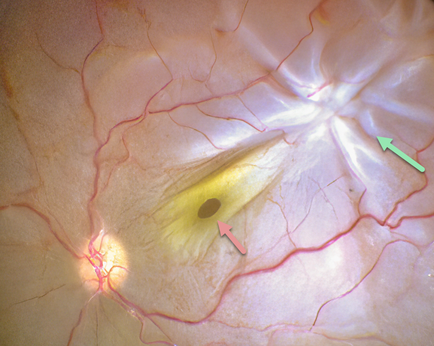
PROLIFERATIVE VITREORETINOPATHY (PVR) WITH MACULAR HOLE
Anjana Mirajkar and Manish Nagpal
Originally posted on @retina.rocks August 14, 2024
This intraoperative photograph shows a rhegmatogenous retinal detachment (RRD) complicated by proliferative vitreoretinopathy (PVR) with a star fold superotemporally. A full-thickness macular hole surrounded by its luteal pigment is dragged towards the PVR. The macular hole and detachment were repaired with vitrectomy.
Learning Points:
PVR, which consists of cellular membranes growing on either retinal surface, develops in about 5-10% of retinal detachments. RRD with a concurrent non-causal macular hole is found in about 2% detachments (Iros et al, Retina 2023;43:581-584). Although most eyes achieve anatomic success, final vision is usually poor. In our patient, unfortunately, there is no further clinical information, so it is unclear whether the macular hole caused the detachment or was secondary to traction from the star fold.
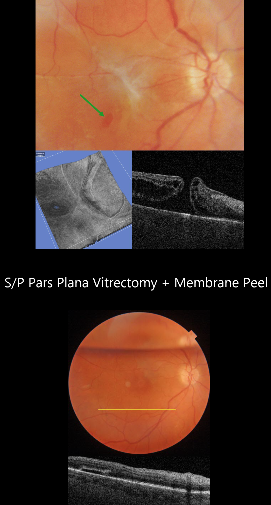
MACULAR PUCKER WITH ECTOPIC MACULAR HOLE
Originally posted on @retina.rocks July 25, 2024
This 52YO female underwent vitrectomy for a hand motion, macula-off rhegmatogenous retinal detachment on 1/25/21. Despite successful reattachment, vision was counting fingers postoperatively, in part due to a severe macular pucker identified on 8/4/21. An ectopic secondary macular hole was present in the inferotemporal macula.
Vitrectomy surgery with membrane peeling was performed on 10/14/21. One week postoperatively, the ectopic hole was closed, and the macula was completely free of epimacular tissue. When last examined on 3/24/23, vision was 20/70.
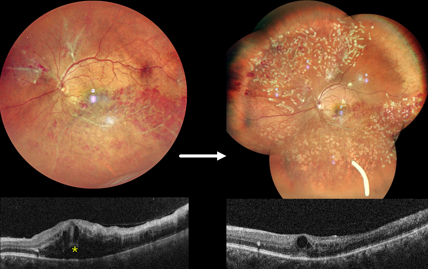
OCCLUSIVE RETINAL VASCULITIS
Anjana Mirajkar and Manish Nagpal
Originally posted on @retina.rocks July 23, 2024
This healthy 32YO male presented on 4/23/24 with 2 months of decreased vision in his left eye. Family history was negative. A prior workup by a prior doctor was negative, including protein electrophoresis and homocysteine levels. He failed a short trial of oral steroids. Vision was 20/20 in his healthy right eye and counting fingers in his left eye.
Fundus photography shows an ischemic inferotemporal branch retinal vein occlusion with moderate, mostly peripheral retinal hemorrhages. The superotemporal retinal vein is diffusely beaded, and an apparent independent ischemic BRVO is seen superonasally, with probable preretinal blood. Marked macular edema is seen on OCT. Unfortunately, fluorescein angiography is not available.
An Ozurdex injection was given for macular edema, and two sessions of panretinal photocoagulation were performed for proliferative disease. On 6/3/24, vision improved to 20/120, and the macular edema was improving.
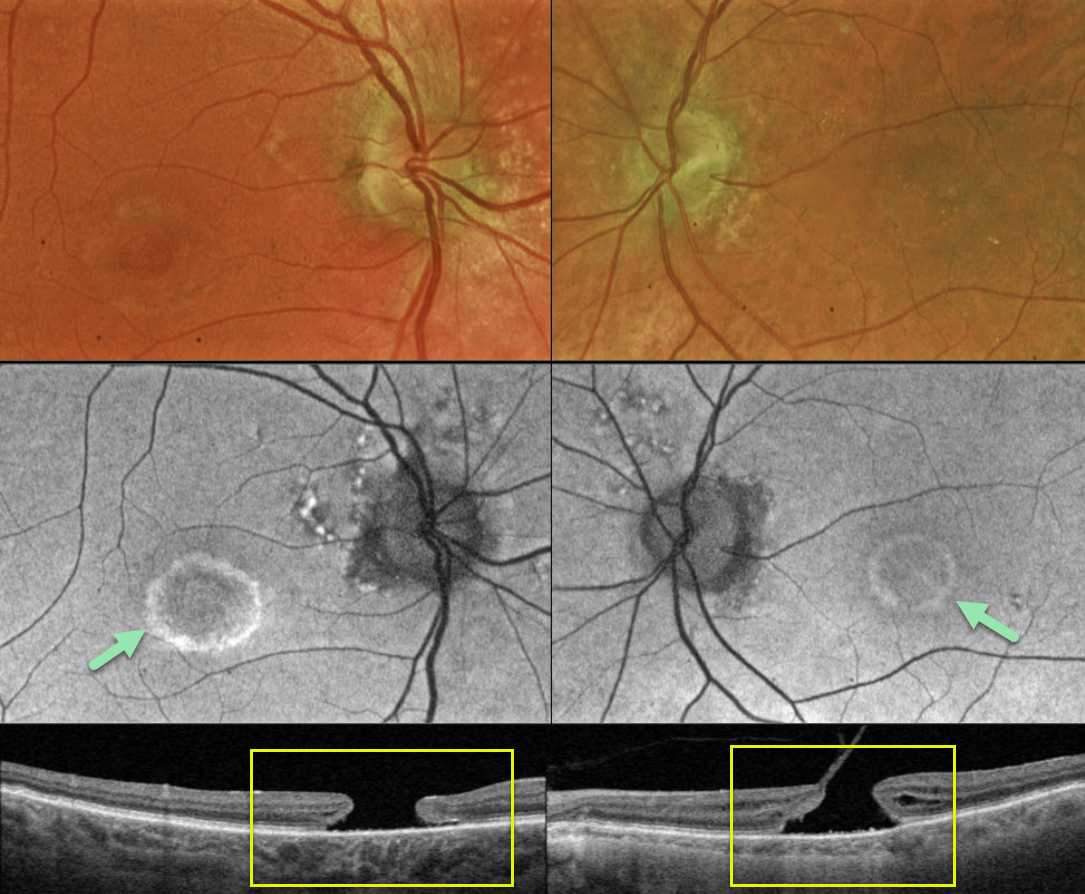
MACULAR HOLE WITH PSEUDO BULLS-EYE MACULOPATHY
Originally posted on @retina.rocks June 24, 2024
This 83YO male presented with a history of chronic bilateral vision loss. Vision was 20/150 OD and 20/200 OS.
Optos color RG imaging shows a bull’s eye pattern of bilateral pigmentary changes along with a large macular hole OU. Fundus autofluorescence (FAF) shows the bull’s-eye lesions as hyper-FAF. Triton swept-source OCT confirms bilateral macular holes.
Our patient has bilateral chronic macular holes with secondary RPE depigmentation, resulting in a bull’s-eye appearance. Due to the chronicity of the holes, observation was recommended.
Learning Points:
Bull’s eye maculopathy is characterized by a rim of subretinal pigment loss and outer retinal atrophy, most commonly found in inherited retinal diseases (usually ABCA4 disorders) and hydroxychloroquine/chloroquine toxicity.
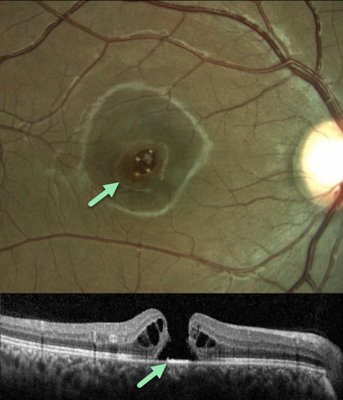
LASER-INDUCED MACULAR HOLE
Tareq Alsulami and Naser Alsaedi
Originally posted on @retina.rocks June 17, 2024
This 14YO boy presented with vision loss in his right eye following self-directed exposure to a blue wavelength laser pointer. Vision was 20/200 in his right eye and 20/20 in his normal left eye.
Color imaging and OCT scanning show an irregular full-thickness macula hole. The hole closed following vitrectomy surgery, but vision failed to improve.
Learning Points:
Laser pointers can cause significant macular injury, including solar maculopathy-like outer foveal microdefects, hemorrhage, pseudo macular dystrophy, vitelliform lesions, and macular scarring (Bhavsar et al, Surv Ophthalmology 2021;66:231-260). There are also numerous reports of a secondary full-thickness macular hole, as developed in our patient.
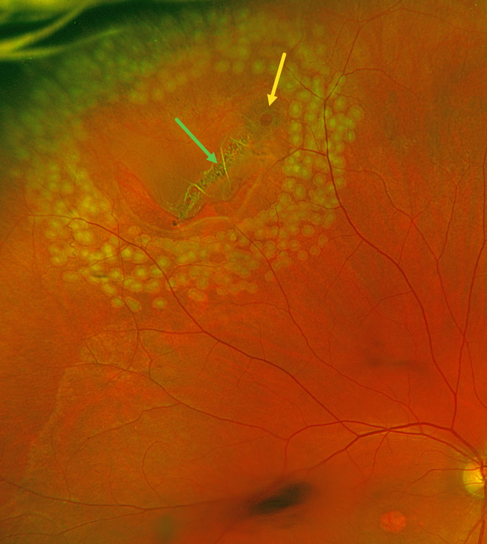
RETINAL BREAK
Justin Grassmeyer, Ambar Faridi, and Brittany Heckerman
Originally posted on @retina.rocks June 5, 2024
This 63YO male with history of idiopathic central serous chorioretinopathy (ICSC) with secondary macular neovascularization OD and lattice degeneration OU presented with new floaters in his right eye. He was scheduled for an anti-VEGF injection for the right eye on a treat-and-extend protocol the same day. Vision was stable at 20/100 OD and 20/20 OS. An acute, related retinal tear was found and immediately demarcated with thermal laser photocoagulation.
Optos color RG imaging immediately following the laser procedure shows the retinal break well surrounded by multiple nearly confluent rows of laser burns. The flap of the tear consists of an entire lattice lesion along with a chronic atrophic hole with surrounding pigment. There is an area of central macular atrophy from the ICSC.
Learning Points:
Lattice lesions have a pocket of liquefied vitreous overlying thinned retina with a firm area of surrounding vitreoretinal adhesion. White, fibrosed vessels often crisscross the lesions, which gives the lesions their name, “lattice.” This firm vitreoretinal adhesion, along with the often-present atrophic holes, is what can predispose these eyes to retinal breaks and detachment. Although prophylactic treatment is rarely needed, symptomatic breaks, as in our patient, need prompt retinopexy.
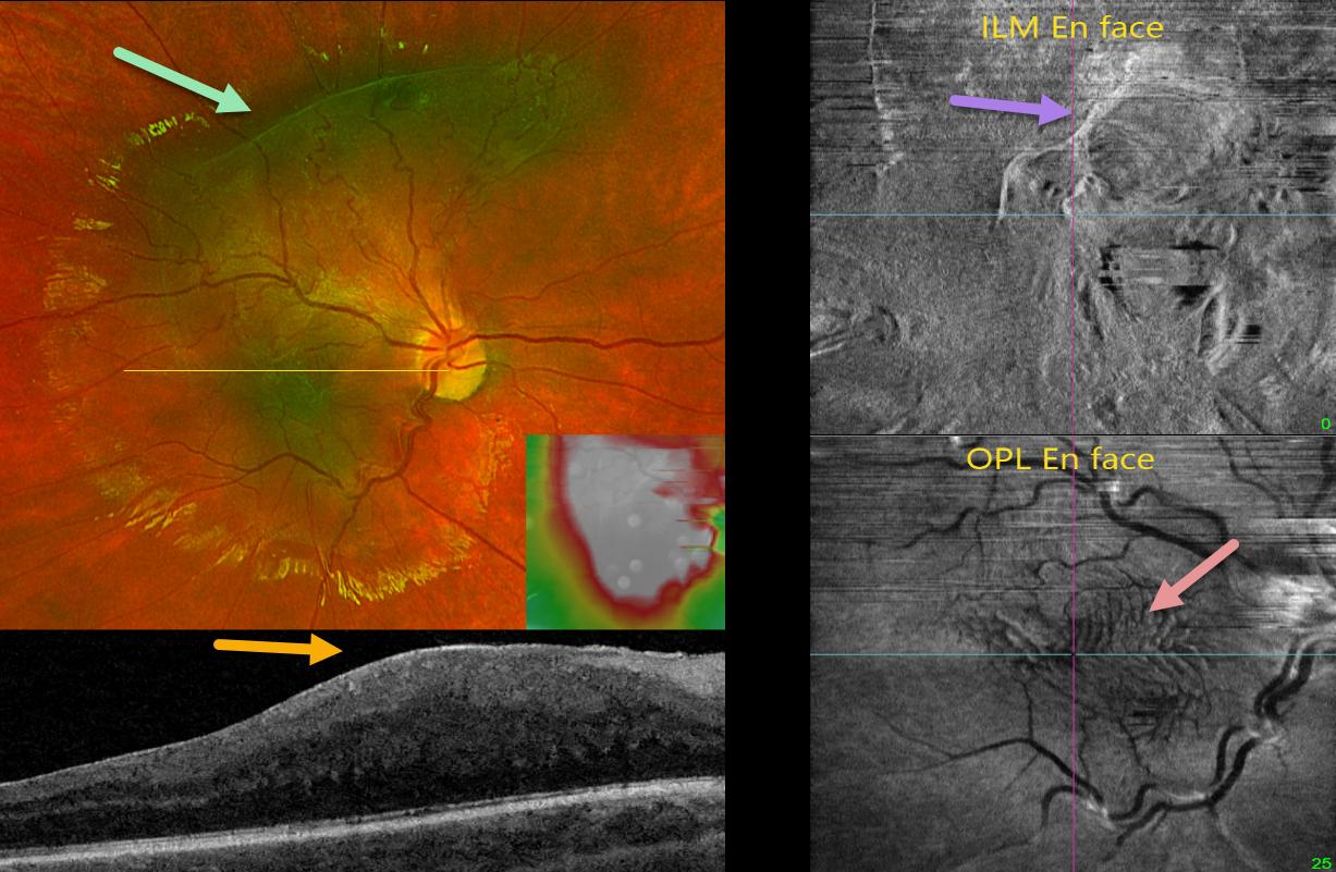
MACULAR PUCKER
Originally posted on @retina.rocks May 29, 2024
This healthy 9YO boy was referred for asymptomatic retinal changes in his right eye. Vision was 20/25 OU. The left eye was normal.
Optos color RG imaging shows a greenish hyperpigmented lesion in the macula extending into the superior midperiphery. Diffuse macular thickening is noted on swept-source OCT. En face imaging offers another multimodal perspective. En face of the internal limiting membrane/vitreous interface shows the epimacular fibrosis, while en face of the outer plexiform layer shows variable fingerprint-like whorls centrally.
Learning Points:
In 2017, Govetto et al described an OCT finding of ectopic inner foveal layers (EIFL) associated with epiretinal membranes (AJO 2017;175:99-113). The findings range from mild underlying retinal changes (stage 1, thin ERM with foveal depression), widening of the ONL and loss of foveal depression (stage 2), and ERM continuous with EIFL crossing the entire foveal area (stage 3). All retinal layers are clearly visualized in stages 1 through 3. In stage 4, the ERM is thick, and all retinal layers are disrupted. Vision is worse with increasing stage severity. It is also associated with worse postoperative visual recovery (Yang et al, Retina 2022;42:1472-1478).
The ‘fingerprint sign’ is a unique en face OCT finding described by Griffin et al (Retina 2021;41:381-386) that appears as concentric, fingerprint-like waves of Henle’s layer underlying epiretinal membranes. The authors found that the undulating OPL creates hyperreflective lines when oriented perpendicular to the incident OCT beam and hyporeflective lines when parallel to it.
Combined hamartomas are classically described as benign congenital lesions composed of glial cells, vascular tissue, and pigmented retinal pigment epithelial cells. However, more recent data indicate that these lesions primarily arise from the inner retinal layers (Chawla et al, AJO 2017;181:88-96).
Hamartomas and severe macular puckers can look very similar, even to some of the most seasoned retina specialists. OCT may be helpful in differentiating these entities (Ophthalmic Surg Lasers Imaging Retina 2017;48;122-125). In our experience, we feel that many ‘combined hamartomas’ described in the literature are most likely severe macular pucker with ectopic inner retina layers as in our case.
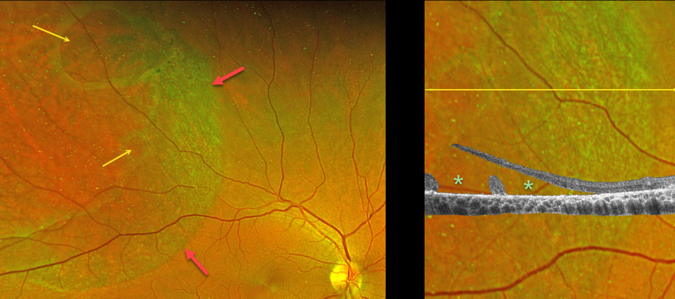
DEGENERATIVE RETINOSCHISIS
Janelle Adeniran
Originally posted on @retina.rocks May 16, 2024
This 55YO male was referred for an asymptomatic possible retinal detachment in his right eye. Vision was 20/30.
Optos color RG imaging shows superotemporal degenerative retinoschisis with two outer-layer holes (OLHs). A broad band of pigmented chorioretinal scarring is noted along the posterior edge of the schisis.
OCT scanning through the smaller outer layer hole inferiorly shows the elevated edges of the OLH with the overlying inner schisis cavity. Despite the posterior pigment suggesting a demarcation line, no detachment of the outer layer was noted, and observation was recommended.
Learning Points:
A combined schisis-retinal detachment occurs when degenerative schisis contains both inner- and outer-layer breaks, essentially forming a full-thickness break that allows liquid vitreous to reach the subretinal space.
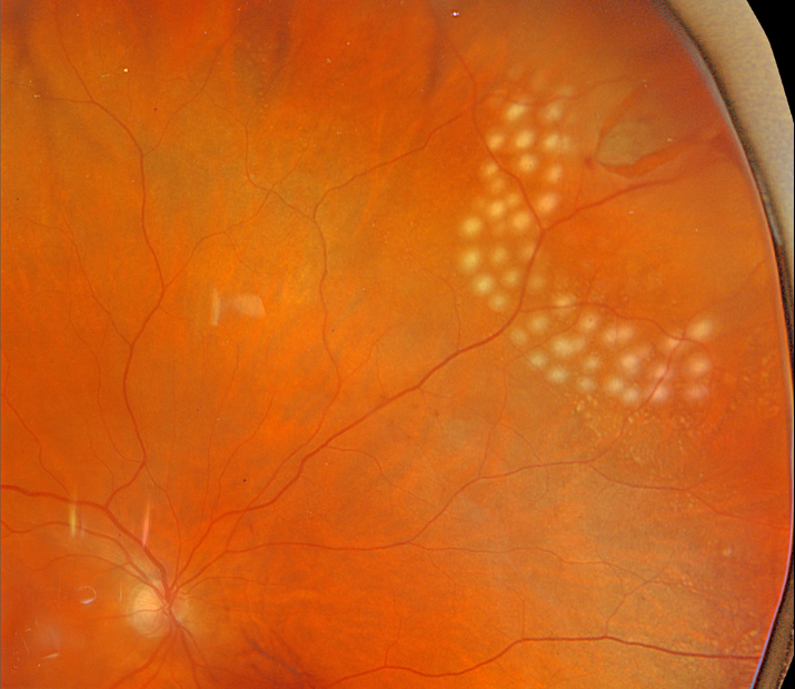
RETINAL TEAR
Originally posted on @retina.rocks May 6, 2024
This 70YO female was examined for new flashes and floaters in her right eye and was found to have a retinal tear with localized subretinal fluid at the superonasal equator.
Several nearly confluent rows of laser photocoagulation were applied along its posterior and lateral edges, but we could not extend treatment around its anterior margins with the available slit lamp delivery.
The patient returned the following day to another office where we have our cryotherapy device, where the remainder of the break and fluid were administered.
Learning Points:
Symptomatic retinal tears in the presence of an acute posterior vitreous detachment have a high risk of causing a rhegmatogenous retinal detachment. Prophylactic retinopexy is therefore indicated. Treatment must completely surround the tear and, ideally, extend to the ora serrata. Both thermal laser photocoagulation and cryotherapy are equally effective in creating a vitreoretinal adhesion, which takes at least several weeks to form.
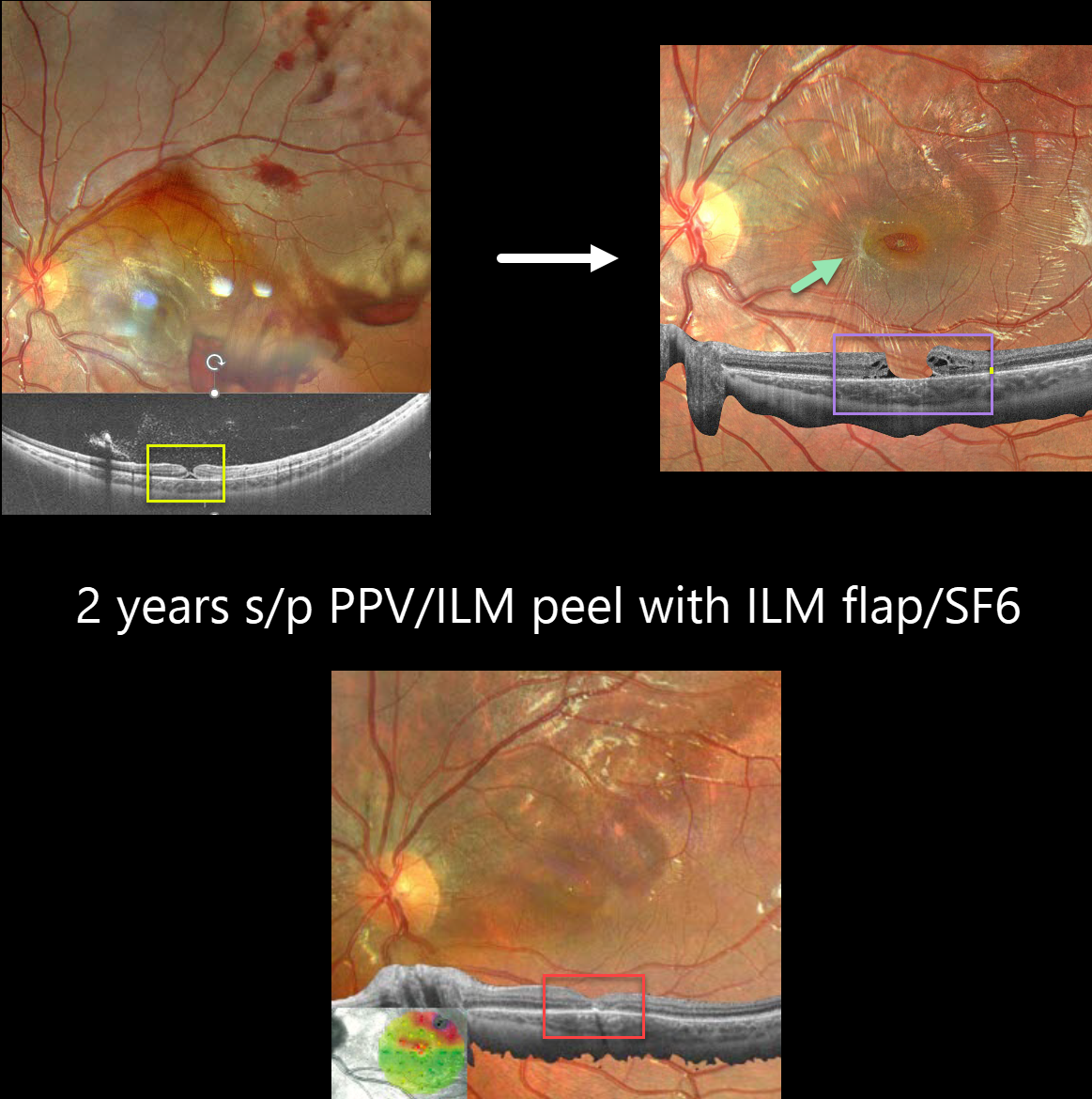
TRAUMATIC MACULAR HOLE
Barbara Parolini, Veronika Matello, Giulia Freschi, and Roberta Penzani
Originally posted on @retina.rocks April 9, 2024
This 10YO boy was referred for vision loss immediately after getting hit in his left eye by a soccer ball 7 days earlier. Vision was 20/70.
Fundus photography shows scattered intra- and preretinal hemorrhages with a large confluent area of commotio retina superiorly and temporally.
Widefield OCT shows a partial-thickness macular hole with the external retina intact but elevated. Three months later, vision was 20/100, and an irregular, full-thickness macular hole with surrounding macular pucker was seen.
25-gauge vitrectomy with ILM peeling, ILM flap, SF6 gas tamponade, and 3 days face-down positioning was performed. Two months later, vision improved to 20/40 with complete closure of the hole. Two years later, vision was 20/25.
Learning Points:
This patient illustrates several sequelae of blunt trauma, including retinal/preretinal blood, commotio retinae, and macular hole formation. Since fresh traumatic holes can spontaneously flatten, especially in pediatric cases (Liu et al, AJO 2020;210:174-183), an initial period of observation is recommended before considering vitrectomy.
In our patient’s case, the hole did not resolve, and surgery provided good anatomic and long-lasting visual outcomes.
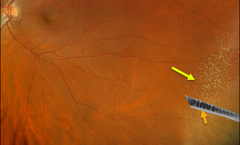
DEGENERATIVE RETINOSCHISIS
Originally posted on @retina.rocks April 4, 2024
This 52YO female was referred for a possible retinal detachment in her left eye. There were no visual symptoms.
Optos color RGB imaging shows inferotemporal retinoschisis with numerous inner retinal refractile dots. Optos California swept-source OCT confirms schisis with splitting of the outer plexiform layer.
Learning Points:
Degenerative retinoschisis is an age-related splitting of the outer plexiform layer, most commonly located inferotemporally.
It is sometimes difficult to differentiate a schisis from a rhegmatogenous detachment, and ultrawidefield OCT imaging, as in this case, is an amazing diagnostic tool.
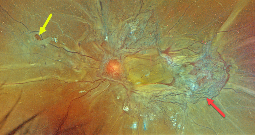
COMBINED TRACTIONAL-RHEGMATOGENOUS RETINAL DETACHMENT
Originally posted on @retina.rocks March 27, 2024
This 46YO female with type 2 diabetes presented with recent bilateral vision loss. Vision was 20/200 OD and hand motion OS.
Optos color RGB imaging of her left eye shows a combined tractional-rhegmatogenous total retinal detachment with fibrovascular tissue encircling the macula. Temporally radial outer retinal folds are dragged toward the more succulent, red neovascularization. A retinal tear is noted temporally.
Vitrectomy surgery was scheduled.
Proliferative diabetic retinopathy with disc and peripheral neovascularization was noted in her right eye (not shown). Panretinal photocoagulation was performed in her left eye and surgery was scheduled for her left eye.
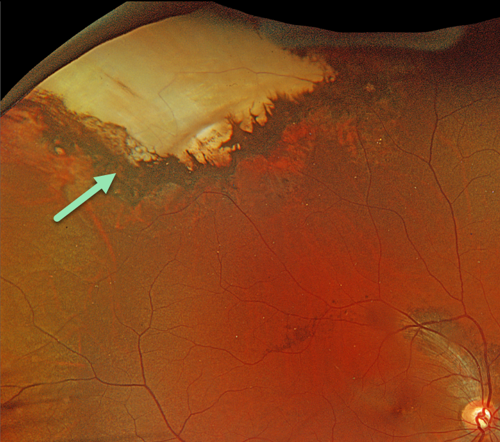
SCLERAL BUCKLE
Originally posted on @retina.rocks March 21, 2024
This 31YO male gave a history of scleral buckling surgery in Cuba 14 years earlier. There were no visual complaints, and vision was 20/70.
Optos color RGB imaging shows an intruding scleral buckle in the superotemporal periphery. The buckle is covered by bare neurosensory retina with some surrounding hyperpigmented chorioretinal scarring. Externally, the eye was quiet, and the conjunctiva completely covered the buckle.
Learning Points:
Intrusion of a scleral buckle is now a rare event with modern surgical techniques and buckling materials. This was most common following MIRAgel explants, which have since been removed from the market (Davuluri et al, JAMA Ophthalmology 2023;141:1081-1082).
An intruding buckle is usually asymptomatic but can cause vitreous hemorrhage (Zarei et al, Int J Retina Vitreous 2020;6;7). Observation for our patient was recommended.
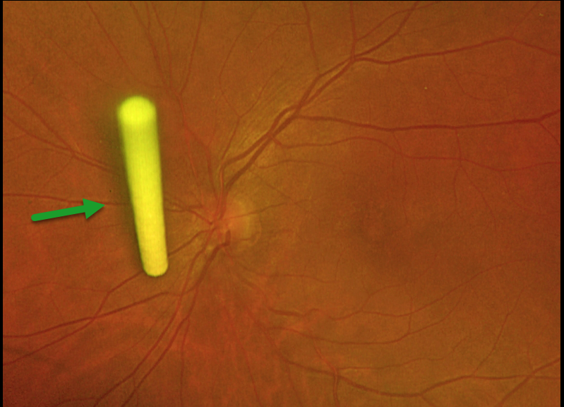
OZURDEX
S. Nazem Ibrahim, Joseph Touma, and Ahmed Sallam
Originally posted on @retina.rocks February 15, 2024
This 44YO female with a diagnosis of chronic anterior uveitis in the left eye presented with visual acuity of 20/60 and cystoid macular edema recalcitrant to topical and periocular steroids. We treated her with an intravitreal dexamethasone implant (Ozurdex).
Optos imaging one week later revealed an unusual vertical orientation of the implant, ‘standing proud’ near the optic disc within the posterior vitreous. There was no prior pars plana vitrectomy surgery.
Even in non-vitrectomized eyes, an Ozurdex implant may take a vertical position within the vitreous and not attach to the peripheral retina as it usually does. The patient was asymptomatic, and observation was recommended.
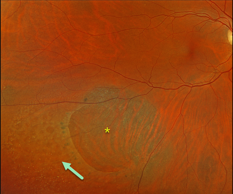
DEGENERATIVE RETINOSCHISIS
Originally posted on @retina.rocks February 14, 2024
This 61YO female has been followed for several years for asymptomatic unilateral degenerative retinoschisis in her right eye. Vision is 20/30.
Optos color RGB imaging shows a large posterior outer layer hole with surrounding pigment. Innumerable small inner-layer holes are noted more peripherally within the inferotemporal schisis cavity.
Learning Points:
Degenerative retinoschisis is virtually never prophylactically treated (see Byer 1986;93:1127-1137). Patients can rarely develop a combined schisis rhegmatogenous retinal detachment (RRD) when liquid vitreous accesses the subretinal space through both an inner-layer and an outer-layer hole.
We will therefore continue to follow her twice yearly.
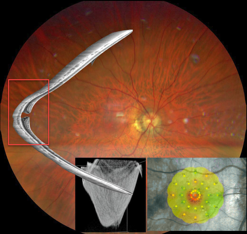
STAPHYLOMA + MACULAR HOLE
Barbara Parolini and Veronika Matello
Originally posted on @retina.rocks February 13, 2024
This Eyecare Clinic (in Brescia, Italy) case was submitted by Barbara Parolini, Veronika Matello, Giulia Freschi, and Roberta Penzani.
This 48YO highly myopic male presented for a sudden drop in vision. Vision was 20/100 OD and 20/20 in OS.
Color photography shows a blunted macular reflex. Widefield OCT shows a conical-shaped staphyloma of the entire posterior pole with a full-thickness macular hole. Microperimetry shows a decreased but not absent foveal sensitivity.
According to the Myopic Traction Maculopathy Staging System (Parolini et al, Eur J Ophthalmol 2021;31:1299-1312), this eye is in Stage 1c. The left eye showed a similar staphyloma but without a macular hole (not shown). The patient declined surgery.
Learning Points:
A macular staphyloma consists of a concave posterior bowing of the eye wall in patients with high myopia. These myopic patients also have a markedly thinned choroid throughout the posterior pole.
Due to stretching of the posterior retinal structures, these eyes are at increased risk of numerous findings, including macular schisis/traction and macular neovascularization.
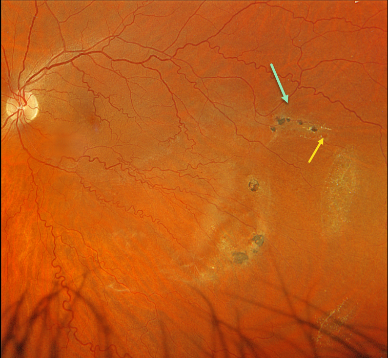
LATTICE DEGENERATION
Originally posted on @retina.rocks February 6, 2024
This 42YO male was referred for asymptomatic findings on retinal examination in his left eye. Vision was 20/20 in his normal OD and 20/20 OS.
Optos color RGB imaging shows patches of variably pigmented lattice degeneration in the inferotemporal midperiphery. At least one atrophic retinal hole is noted in the superior-most lesion.
The retinal veins are also diffusely tortuous except superotemporally, likely indicating a prior spontaneously resolved retinal vein occlusion.
Observation was recommended.
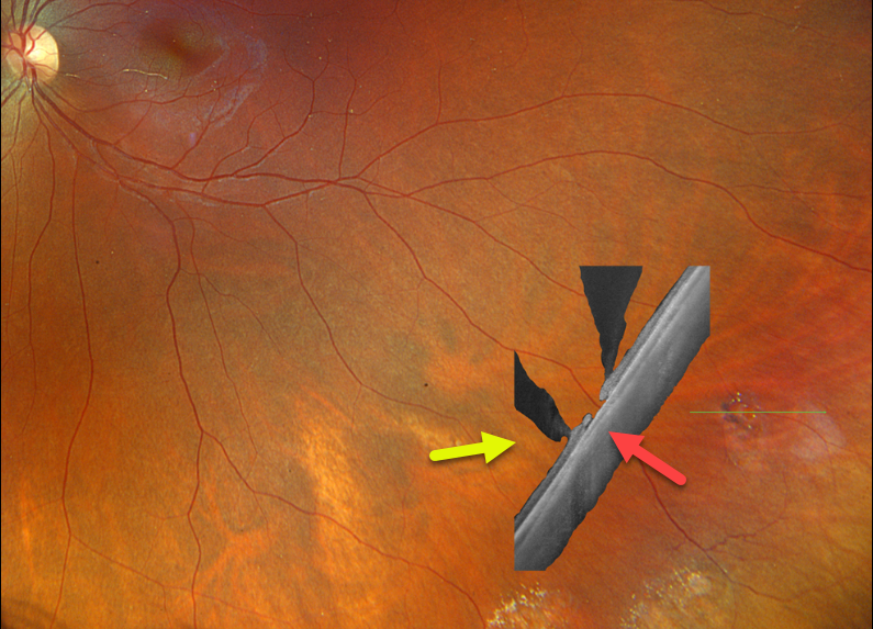
LATTICE DEGENERATION
Originally posted on @retina.rocks January 30, 2024
This healthy 27YO female has a known history of lattice degeneration with atrophic holes. She is completely asymptomatic, and vision is 20/20 OU.
Optos color RGB imaging of her left eye shows patches of inferotemporal lattice degeneration. Optos California Silverstone swept-source OCT through a lesion containing an atrophic retinal hole reveals classic findings, including retinal thinning within the lattice, liquified vitreous overlying the lattice lesion, and adherent vitreous along the edges of the lattice.
Learning Points:
Lattice lesions have a pocket of vitreous liquefaction overlying the thinned retina with a firm vitreo-retinal adhesion along their borders. White, fibrosed vessels often crisscross the lesions, which leads to the name “lattice”. This firm vitreoretinal adhesion, along with the often present atrophic holes, is what can predispose these eyes to retinal detachment.
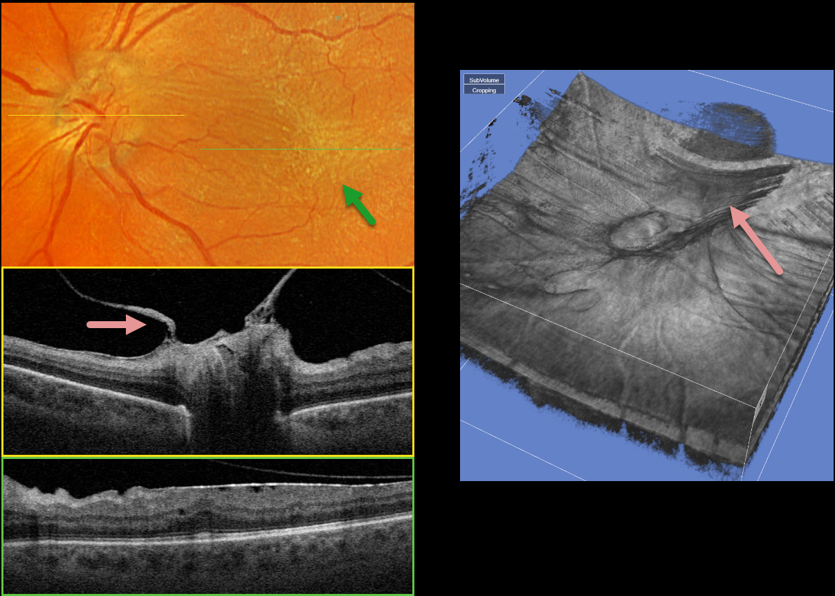
PROLIFERATIVE DIABETIC RETINOPATHY (PDR)
Originally posted on @retina.rocks January 29, 2024
This 58YO male with type 2 diabetes has long-involuted proliferative diabetic retinopathy (PDR) following full panretinal photocoagulation (PRP). He has no visual complaints, and vision is 20/50 OS.
Optos color RGB imaging shows a diffuse macular pucker with striae extending nasally towards the nerve and temporally into the distal macula.
A condensation of elevated fibrosed tissue, representing a partially detached Weiss’ ring, surrounds the optic nerve. This traction is clearly evident in Triton swept-source OCT (image 1, middle) and in the 3D reconstruction (image 2). A moderate epiretinal membrane extends throughout the macula (image 1, bottom).
Observation was recommended, and vitrectomy with membrane peeling will be performed if the patient becomes more symptomatic.
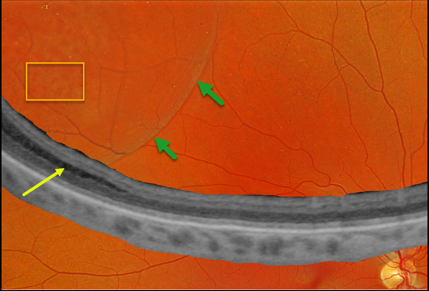
DEGENERATIVE RETINOSCHISIS
Originally posted on @retina.rocks January 24, 2024
This 46YO female was referred for a possible retinal detachment. She had no visual complaints. Vision was 20/20 OD and 20/20 in her normal OS.
Optos color RGB imaging shows a bullous superotemporal retinoschisis. Numerous inner-layer holes are seen. Optos Silverstone ultrawidefield OCT shows splitting of the outer plexiform layer, confirming a schisis.
Learning Points:
Degenerative retinoschisis is an age-related splitting of the outer plexiform layer, most commonly located inferotemporally. It is sometimes difficult to differentiate a schisis from a rhegmatogenous detachment, and ultrawidefield OCT imaging, as in this case, is an amazing diagnostic tool.
Patients can rarely develop a combined schisis rhegmatogenous retinal detachment (RRD) when liquid vitreous accesses the subretinal space through both an inner-layer and an outer-layer hole. We will follow up with our patient yearly.
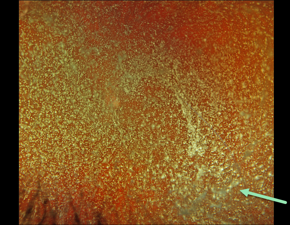
ASTEROID HYALOSIS
Originally posted on @retina.rocks December 25, 2023
A holiday gift for our awesome viewers!
Here’s a gorgeous Optos color RGB image of asteroid hyalosis in a healthy 51YO man. Vision was 20/25. And he was completely visually asymptomatic.
As we exit 2023, enter 2024, and celebrate Retina Rock’s 4th birthday, we are so grateful that people across the globe enjoy our images and discover what they teach us about the retina. Thank you for giving us a reason to exist and keep our mission alive: to help educate the world’s eye care providers about why the retina rocks!
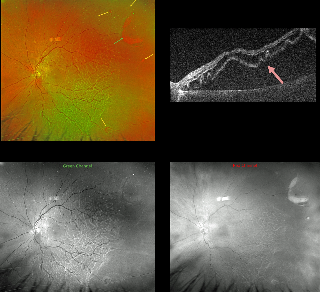
OUTER RETINAL FOLDS/CORRUGATIONS
Originally posted on @retina.rocks December 20, 2023
This 61YO male presented with a several-week history of severe vision loss in his left eye. Vision was hand motion.
Optos color RG imaging shows a rhegmatogenous retinal detachment (RRD) with corrugated outer retinal folds. A large horseshoe retinal tear is seen superotemporally, with numerous smaller breaks noted.
The outer retinal folds are best seen with the green channel compared with the red channel. Triton swept-source OCT shows how these findings involve just the outer retina.
Vitrectomy surgery was scheduled.
Learning Points:
Hydration of outer retinal folds is one of the key findings to help differentiate a RRD from exudative and traction retinal detachments. The cause for these folds remains uncertain, but they may form as the result of the hydration of glycosaminoglycans in the interphotoreceptor matrix (Dalvin et al, Retinal Cases & Brief Reports 2020;14:355-359).
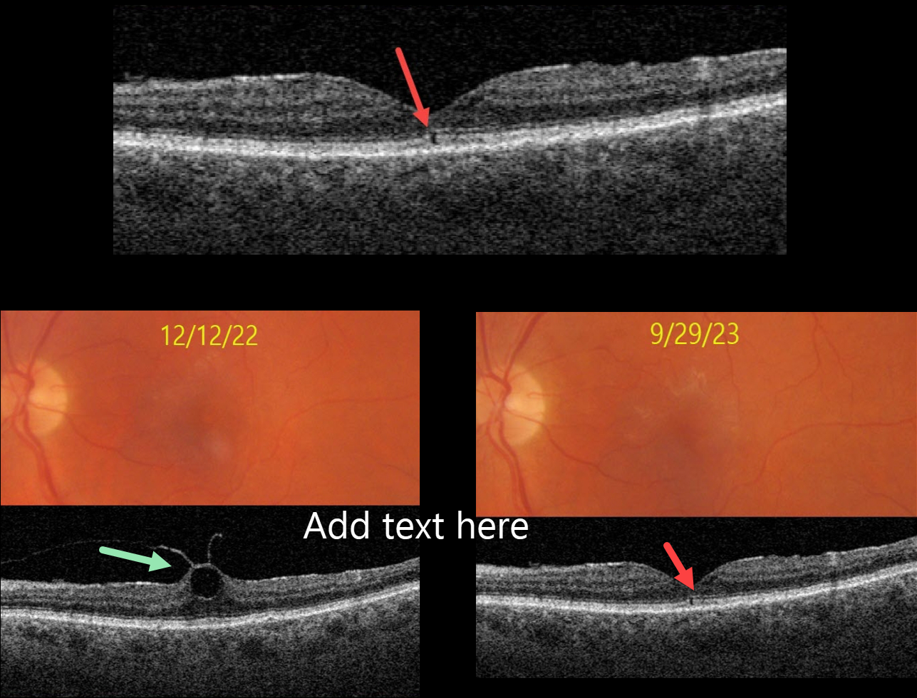
OUTER FOVEAL MICRODEFECT
Originally posted on @retina.rocks December 13, 2023
This 76YO female presented with no visual symptoms, 20/40 vision, and an abnormal OCT in her left eye. What caused this subtle OCT finding?
She was previously being followed for asymptomatic focal vitreomacular traction (VMT) in this eye. On 12/12/22, Triton color imaging showed a subtle foveal cyst with focal VMT on swept-source OCT.
We recommended observation due to the absence of symptoms, and when she returned on 9/29/23, these findings spontaneously resolved as the vitreous separated, leaving a tiny outer foveal microdefect.
Learning Points:
Outer foveal microdefects (Cohen et al, Ophthalmology Retina 2021;5:553-561) have been described in numerous conditions, including macular telangiectasia, tamoxifen use, ABCA4 disorders (cone-rod dystrophies, Stargardt disease, and fundus flavimaculatus), phototoxicity, trauma, and vitreomacular traction disorders.
The prevalence of VMT increases with age, and is present in about 1% of people over 40YO. The natural history is still being defined, but most patients have stable traction and vision. VMT can also resolve spontaneously in about 20% (Errera et al, Ophthalmology 2018;125:701-707).
Many patients with VMT remain completely asymptomatic despite impressive OCT findings. We therefore almost always observe VMT, as in this case, unless the patient has significant symptoms.
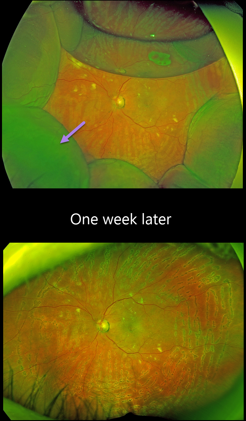
CHOROIDAL DETACHMENT
Originally posted on @retina.rocks December 11, 2023
This 66YO female presented for her postoperative exam following an uneventful pars plana vitrectomy one day earlier for a diabetic traction retinal detachment.
Optos RG imaging shows 360 degrees of peripheral choroidal detachments with a 30% air bubble. Fresh panretinal photocoagulation burns are noted, and the retina is attached. All wounds were secure, and the intraocular pressure was 20mmHG.
We are not sure why our patient presented with these choroidals 1 day postoperatively, since her pressure was normal and there were no wound leaks.
Uveal effusion is a rare transient complication following heavy panretinal photocoagulation (Gentile et al, Ophthalmology 1996;103:827-832), although our patient’s endolaser did not appear overly intense.
When examined one week later, vision was 20/400, and the IOP was 17. The choroidals had spontaneously and completely resolved.
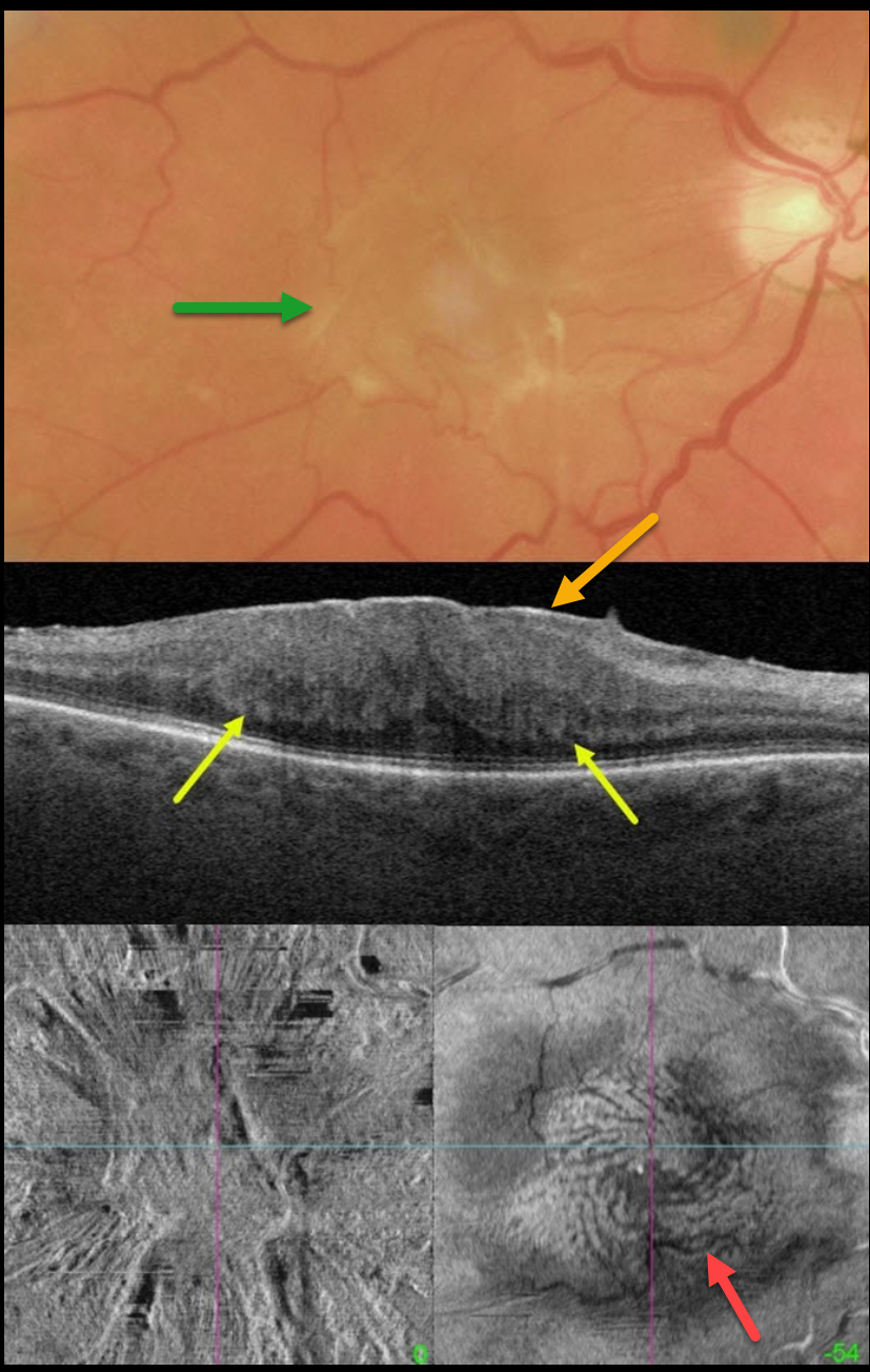
MACULAR PUCKER WITH FINGERPRINT SIGN
Matt Meredith
Originally posted on @retina.rocks December 4, 2023
This 67YO male presented with 20/400 vision in his right eye from a symptomatic macular pucker.
Color imaging shows a severe central macular pucker. Triton swept-source OCT shows a variably adherent epiretinal membrane (ERM) with inner retinal hyperreflectivity and thickening, and complete loss of the normal inner retinal structures. The outer plexiform layer (OPL) is scalloped along its margin with the outer nuclear layer.
En face OCT of the inner retina shows variable surface distortion. En face imaging through the OPL shows a fingerprint-like whorl of hyperreflective lines.
Vitrectomy surgery was scheduled.
Learning Points:
The ‘fingerprint sign’ is a unique OCT en face finding, described by Griffin et al (Retina 2021;41:381-386), appearing as concentric, fingerprint-like waves of Henle’s layer underlying epiretinal membranes.
The authors found that the undulating OPL creates hyperreflective lines when oriented perpendicular to the incident OCT beam and hyporeflective lines when parallel to it.
In 2017, Govetto et al described an OCT finding of ectopic inner foveal layers (EIFL) associated with epiretinal membranes (AJO 2017;175:99-113). The findings range from mild underlying retinal changes (stage 1, thin ERM with foveal depression), widening of the ONL and loss of foveal depression (stage 2), and ERM continuous with EIFL crossing the entire foveal area (stage 3). All retinal layers are clearly visualized in stages 1 through 3. In stage 4, the ERM is thick, and all retinal layers are disrupted. Vision is worse with increasing stage severity. It is also associated with worse postoperative visual recovery (Yang et al, Retina 2022;42:1472-1478).
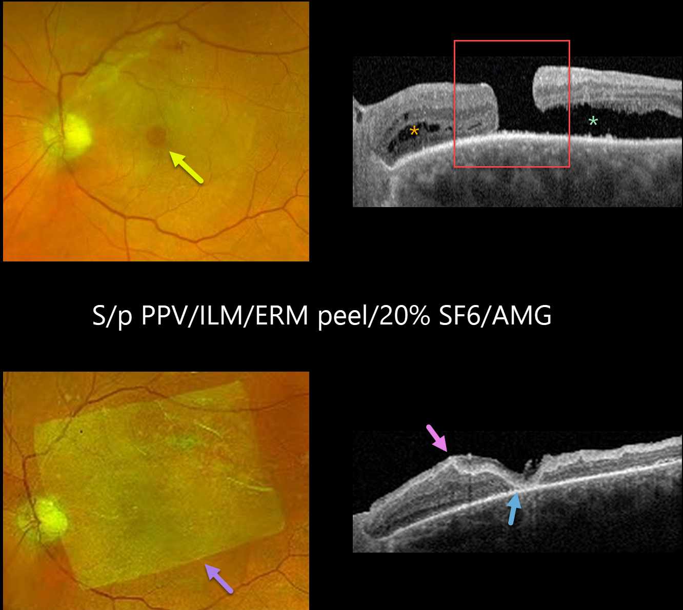
MACULAR HOLE
Yuenpang Cheung, Stephanie Choi and Tongalp Tezel
Originally posted on @retina.rocks November 28, 2023
This 77YO male had a history of failed vitrectomy with internal limiting membrane (ILM) peeling and SF6 gas for a long-standing full-thickness macular hole, and presented for a second opinion. Vision was 20/125.
Optos color RG imaging shows a persistent macular hole with some underlying subretinal fluid. There is a residual epiretinal membrane (ERM) along the superior arcade.
OCT shows the full-thickness macular hole with outer retinal cysts nasally and subretinal fluid temporally. Some ellipsoid zone (EZ) and outer retinal changes are visible.
The patient was eager to try additional surgery and underwent a vitrectomy with ERM/ILM peel, endolaser, 20% SF6, and amniotic membrane graft (AMG) placement. At 9 weeks post-op, the patient’s vision had improved to 20/80.
Fundus imaging shows a sheet of AMG over the macular hole and several superior laser scars. OCT scanning shows the macula hole to be closed under the AMG with EZ and outer retinal changes, but resolved temporal subretinal fluid.
Learning Points:
Approximately 90% of macular holes close with a single surgery. However, larger holes over 400 microns and more chronic holes can be challenging to close. Several techniques have been described to treat these refractory holes, including a broader ILM peel, instruments such as a Flex Loop to loosen up the edges of the hole, amniotic membrane grafts, or various variations of inverted ILM flaps to drape the hole, and even autologous full-thickness retinal tissue transplantation.
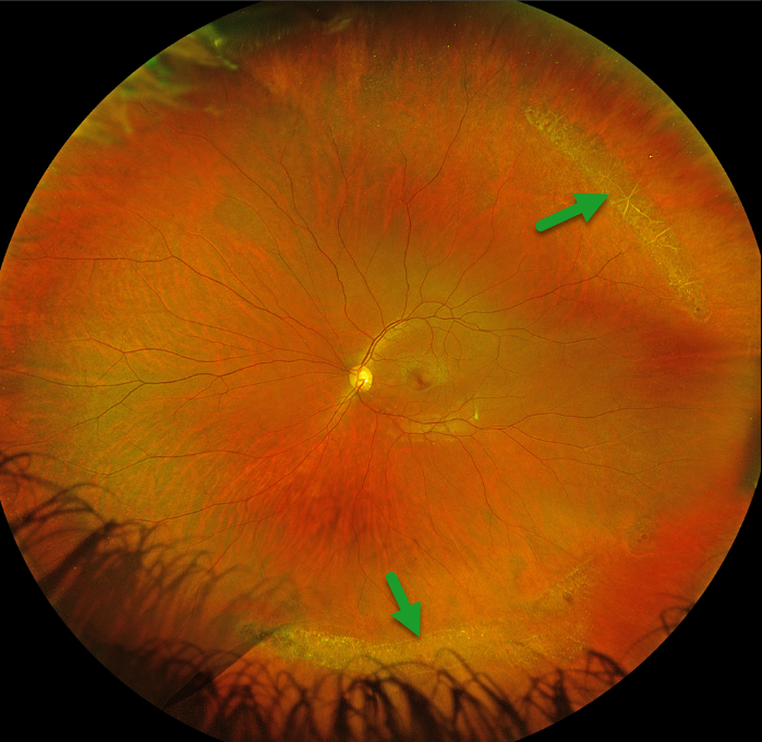
LATTICE DEGENERATION
Originally posted on @retina.rocks November 9, 2023
This 35YO female was referred for asymptomatic lattice degeneration. Vision was 20/20 OU.
Optos color RGB imaging of her left eye shows prominent temporal and inferior lattice lesions, some of which are associated with atrophic retinal holes. Similar findings were noted in her right eye (not shown).
Learning Points:
Lattice lesions have a pocket of vitreous liquefaction overlying the thinned retina with a firm vitreo-retinal adhesion along their borders. White, fibrosed vessels often crisscross the lesions, which leads to the name “lattice”. These are particularly well seen in our patient’s superotemporal lesion. This firm vitreoretinal adhesion, along with the often present atrophic holes, is what can predispose these eyes to retinal detachment.
Retinal detachment occurs in 1 in 10,000 people per year. Lattice is found in 10% of the general population, especially in high myopes. Since about one-third of patients with retinal detachment have lattice as the cause, the question always arises: Should prophylactic retinopexy be performed?
The short answer is that asymptomatic lattices generally should not be treated. To put things into perspective, imagine a sports stadium of 30,000 people. Three people will develop a retinal detachment in the coming year. Only one of the 3,000 people in the stadium with a lattice would develop one of those 3 detachments. Assuming prophylactic treatment would be 100% effective and complication-free, 2,999 eyes would be unnecessarily treated to prevent one detachment.
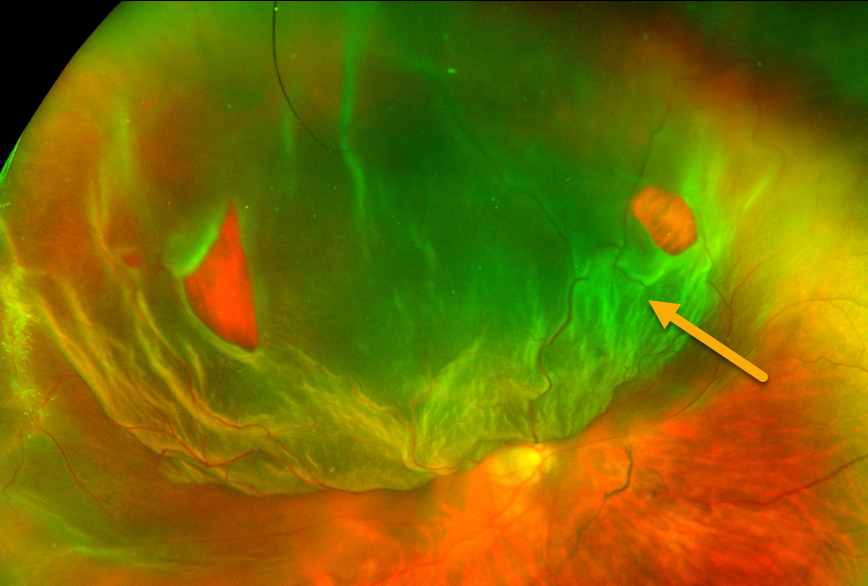
JACK O- LANTERN DETACHMENT
Sandeep Kumar
Originally posted on @retina.rocks October 31, 2023
This 45YO female presented with a superior Jack-O-Lantern macula-off retinal detachment in her right eye. The detachment was successfully repaired with vitrectomy and C3F8 tamponade.
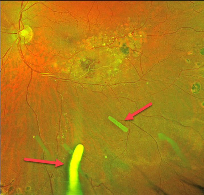
TRIAMCINOLONE-MOXIFLOXACIN (TriMoxi)
Originally posted on @retina.rocks October 30, 2023
This 41YO male has been followed for years with Coats disease in his left eye. Numerous treatment modalities have been used, including thermal laser, intravitreal anti-VEGF, and Ozurdex (dexamethasone 0.7mg) injections. Most recently, he has received Ozurdex every 3-4 months for the past 2 years. Vision is stable at 20/30.
Optos color RG imaging shows multiple Ozurdex ghosts in variable stages of absorption.
Learning Points:
The Ozurdex implant slowly dissolves while releasing its dexamethasone. Although the clinical effect usually lasts about 2 to 3 months, remnants can remain in the eye for over a year (Kim et al, Retina 2020;40:2226-2231).
Approved for diabetic and retinal vein occlusion macular edema and posterior uveitis, it is also often used off-label for other causes of macular edema, including uveitic and pseudophakic CME. For our patient, Ozurdex has been the most successful modality at keeping his central macula dry.
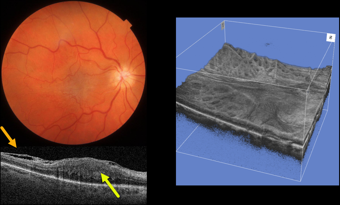
MACULAR PUCKER
Originally posted on @retina.rocks October 23, 2023
This 64YO female was referred for an asymptomatic macular pucker in her right eye. Vision was 20/200 OD and 20/20 OS.
Color imaging shows a severe central macular pucker. Triton swept-source OCT shows a variably adherent epiretinal membrane (ERM) with inner retinal hyperreflectivity and thickening, and complete loss of the normal inner retinal structures.
A 3D OCT reconstruction offers another view of the inner macular appearance, with unusual radiating dimples more peripherally.
Learning Points:
In 2017, Govetto et al described an OCT finding of ectopic inner foveal layers (EIFL) associated with epiretinal membranes (AJO 2017;175:99-113). The findings range from mild underlying retinal changes (stage 1, thin ERM with foveal depression), widening of the ONL and loss of foveal depression (stage 2), and ERM continuous with EIFL crossing the entire foveal area (stage 3). All retinal layers are clearly visualized in stages 1 through 3.
In stage 4, the ERM is thick, and all retinal layers are disrupted. Vision is worse with increasing stage severity. It is also associated with worse postoperative visual recovery (Yang et al, Retina 2022;42:1472-1478).
Combined hamartomas are classically described as benign congenital lesions composed of glial cells, vascular tissue, and pigmented retinal pigment epithelial cells. However, more recent data indicate that these lesions primarily arise from the inner retinal layers (see Chawla et al AJO 2017;181:88-96).
Combined hamartomas and severe macular puckers can look very similar, even to some of the most seasoned retina specialists. In our opinion, many cases of combined hamartomas in the literature most likely represent severe ERMs with EIFL.
Our patient has stage 4 EIFL. We recommended observation since she is binocularly asymptomatic.
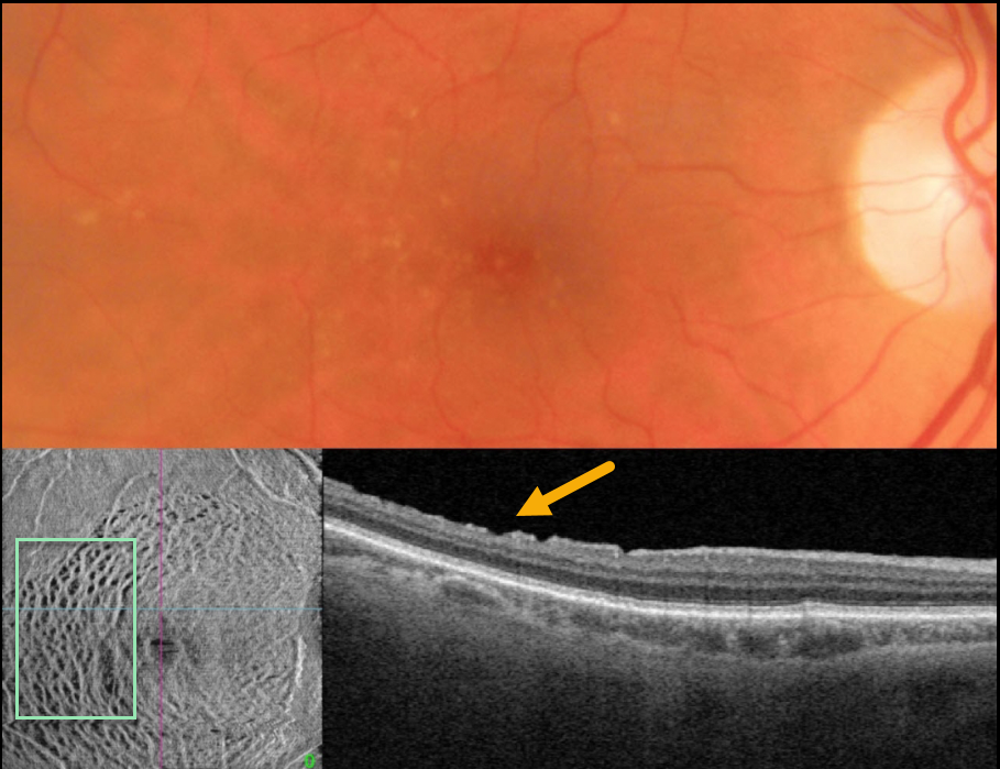
DISSOCIATED RETINAL NERVE FIBER LAYER
Originally posted on @retina.rocks October 17, 2023
This 75YO male underwent successful macular hole surgery 5 years earlier. Vision was 20/40.
Color photography shows scattered mixed drusen. B-scan OCT shows small irregular defects in the inner temporal macula. En face OCT shows extensive dimpling that parallels the nerve fiber layer.
Learning Points:
Pars plana vitrectomy with membrane peeling is frequently performed for the management of many vitreomacular interface disorders, including vitreomacular traction, macular hole, and epiretinal membrane.
While ILM peeling decreases epiretinal membrane recurrence and enhances macular hole closure, it also causes mechanical trauma to the inner retina, as evidenced by dissociated optic nerve fiber layer.
Many mechanisms have been proposed, including Müller cell damage from direct contact with surgical instruments, damage to the deep inner retinal layers, damage from visualization dye, or tractional thinning of the temporal retina.
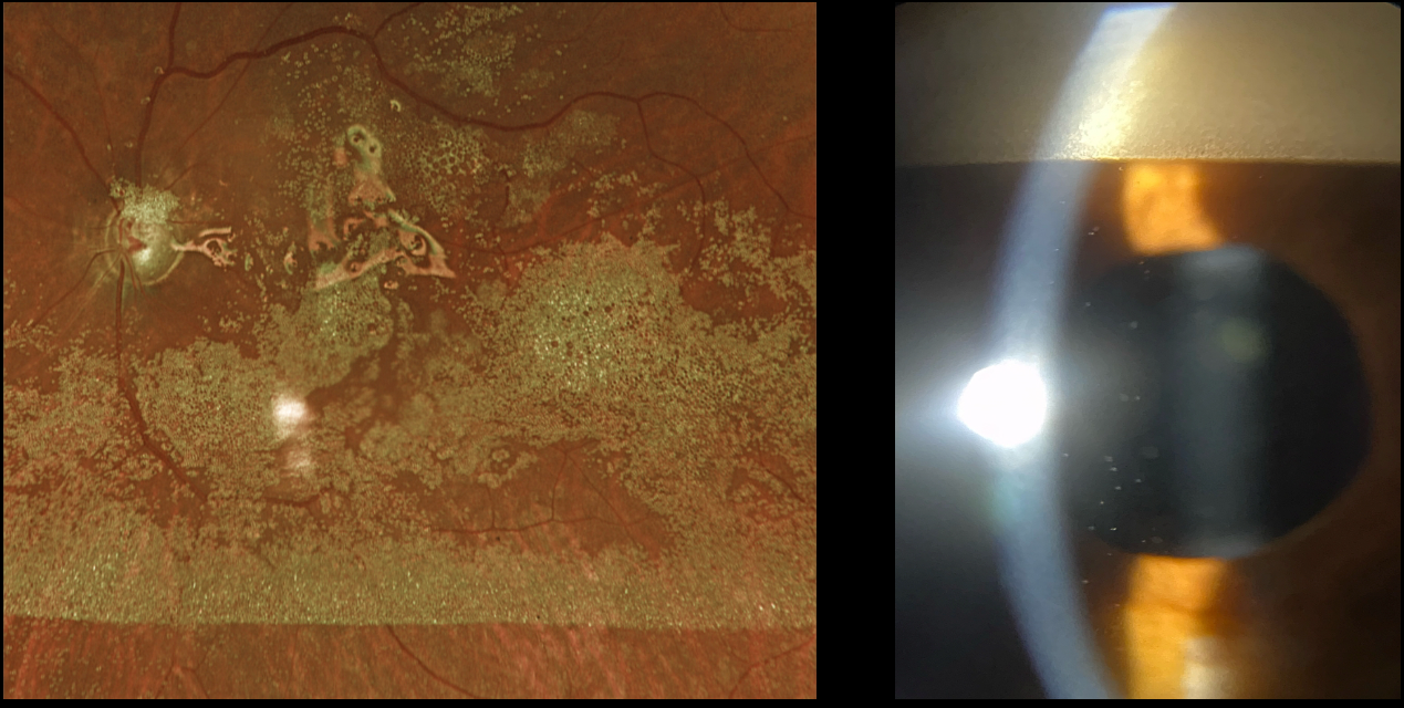
EMULSIFIED SILICONE OIL
Originally posted on @retina.rocks September 25, 2023
This 65YO male underwent vitrectomy surgery with silicone oil 5 years earlier for a complex rhegmatogenous retinal detachment with proliferative vitreoretinopathy. Vision has remained at counting fingers since the surgical repair, and he presented for his annual visit without new complaints.
Optos RG imaging shows innumerable tiny droplets of emulsified silicone oil coating the posterior pole. Slit lamp photography shows a reverse ‘hypopyon’ of emulsified oil superiorly. Suspended oil droplets are also noted throughout the anterior chamber.
Learning Points:
Silicone oil as an adjunct for repairing complex retinal detachments was introduced by Paul Cibis in the early 1960s (Cibis et al, Arch Ophthalmol 1962;68:590-599). It is used primarily for complex detachments at high risk for failure, including severe proliferative vitreoretinopathy and viral retinitis.
Although silicone oil is generally well-tolerated as a long-term tamponade, it can occasionally emulsify, resulting in numerous tiny, opaque droplets. Emulsification is multifactorial, involving the properties of the oil, the surgical procedure, and postoperative factors.
When severe, emulsified droplets can cause significant vision loss and require removal. The droplets can also migrate into the anterior chamber, causing a reverse ‘hypopyon’ with secondary elevated intraocular pressure or corneal decompensation.
Our patient’s increased intraocular pressure of 32 mmHg, which was elevated for the first time, was attributed to emulsified oil clogging the trabecular meshwork. Topical glaucoma drops were started. If the pressure cannot be adequately controlled medically, then vitrectomy with removal of the emulsified oil and likely exchange for new silicone oil will be recommended.
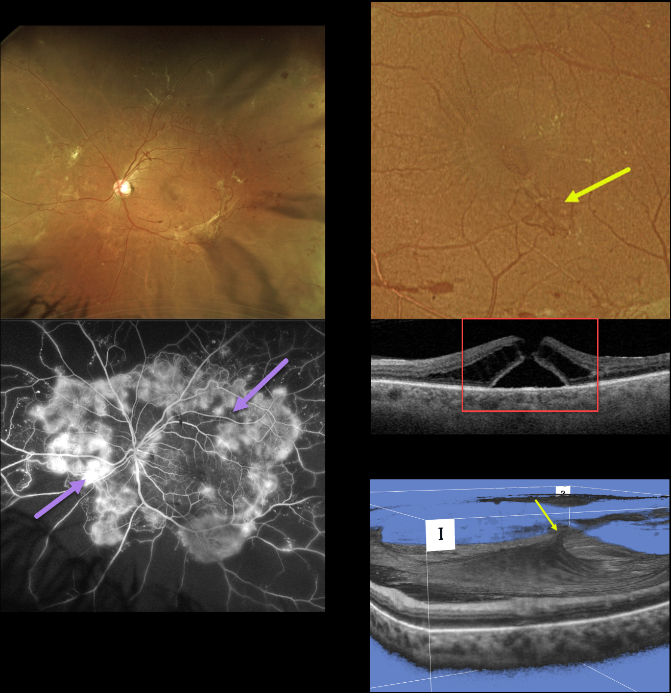
MACULAR HOLE DUE TO DIABETIC TRACTION
Originally posted on @retina.rocks September 21, 2023
This 49YO female with type 2 diabetes presented with a several-year history of blurred vision in her left eye. Vision was 20/40 OD and 20/60 OS.
Optos RG imaging shows variably fibrosed retinal neovascularization encircling the posterior pole. Fluorescein angiography shows active leakage from these vessels.
An area of small neovascularization is present in the inferior temporal macula, which caused a tractional full-thickness macular hole. A 3D OCT view best shows this diabetic traction.
Vitrectomy surgery was scheduled for her left eye. Primary vitrectomy will be performed for her right eye as well once the left eye has stabilized.
Learning Points:
In 1988, Dr. Gass described his concept for the pathogenesis of idiopathic macular holes (Arch Ophthalmol 1988;106:629-639). His classification system, from stage 1 for an impending macular hole through stage 4 for a fully developed macular hole with complete posterior vitreous detachment, was based on his concept of localized, mostly tangential traction caused by the shrinking foveal vitreous cortex. This classic paper heralded modern macular hole surgery for a previously untreatable condition.
The advent of OCT technology, however, has shown that macular hole development is more often due to focal vitreomacular traction, as our case beautifully illustrates.
The current classification system, described by the International Vitreomacular Traction Study Group, emphasizes the characteristics that determine treatment choice and visual prognosis (Ophthalmology 2013;120:2611-2619). This includes size (small <= 250 microns, medium >250 microns and <= 400 microns, large >400 microns), presence or absence of VMT, and cause (primary from VMT or secondary).
Our patient emphasizes why this new classification system was implemented, since it provides information that guides our treatment. Our patient’s hole is secondary to diabetic traction, which obviously requires a very different vitrectomy procedure compared to a primary hole.
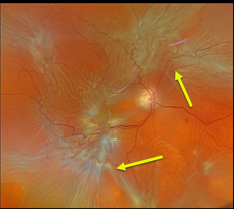
PROLIFERATIVE VITREORETINOPATHY (PVR)
Originally posted on @retina.rocks August 22, 2023
This 76YO male presented with a vague history of recent vision loss in his right eye. Vision was counting fingers.
Optos color RGB imaging shows a total rhegmatogenous retinal detachment (RRD) with diffuse posterior proliferative vitreoretinopathy (PVR). The detachment was successfully repaired with vitrectomy and silicone oil.
Learning Points:
PVR, which consists of cellular membranes growing on either retinal surface, develops in about 5-10% of RRD. The membranes contract and foreshorten the retina, likely as an aberrant wound-healing response.
There are no proven pharmacologic therapies to prevent or treat PVR. These complex detachments can be managed with vitrectomy and membrane peeling, retinectomy, scleral buckling, and silicone oil. However, PVR remains the main cause for ultimate retinal reattachment surgical failure.
See Garweg et al Surv Ophthalmol 2013;48:321-329 for a good review of PVR pathophysiology.
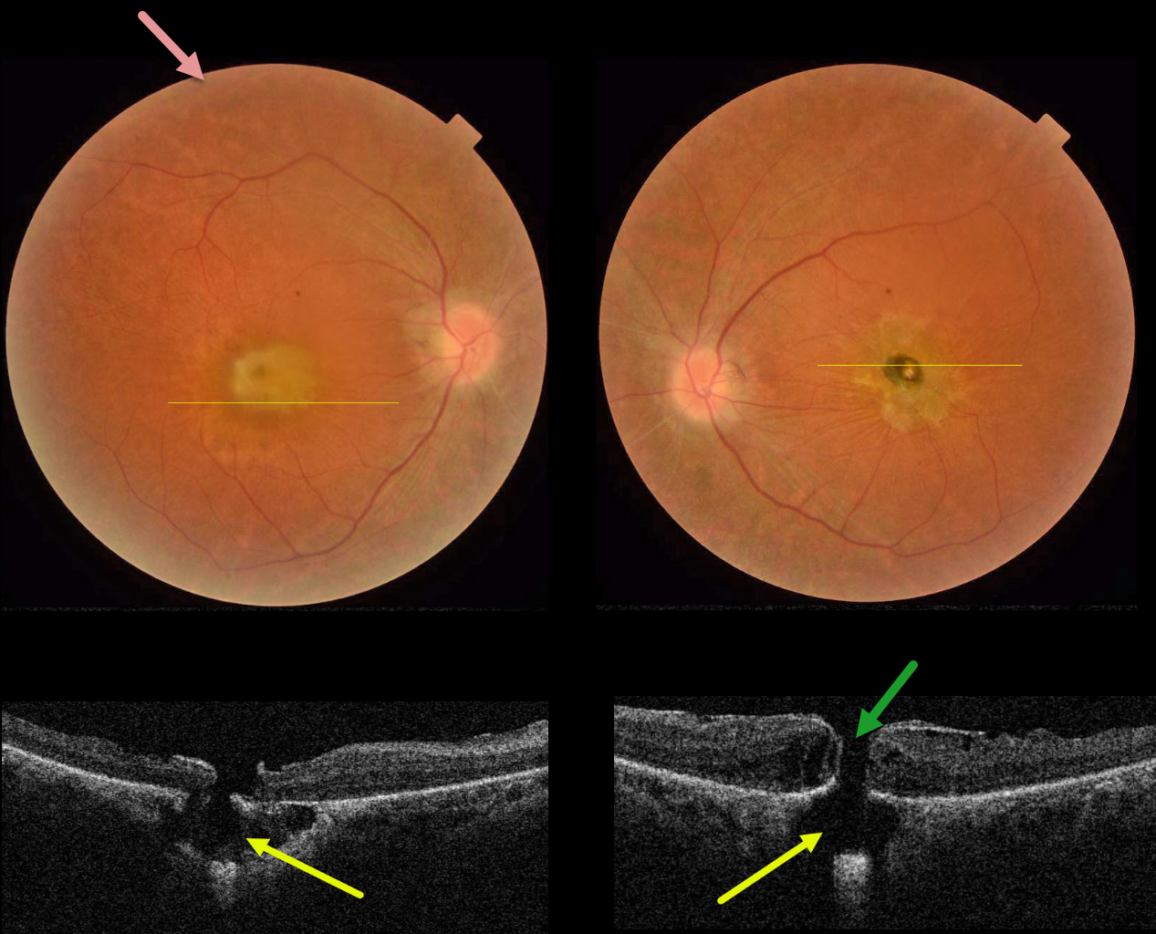
RETINITIS PIGMENTOSA WITH CHOROIDAL CAVITATIONS
Omar Mulki and Faisal Fayyad
Originally posted on @retina.rocks August 16, 2023
This 32YO female presented with a known history of severe vision loss from retinitis pigmentosa (RP). Her parents are first-degree cousins, and there was no family history of RP. Vision was hand motion OD and counting fingers OS.
Fundus photography shows bilateral irregular yellow central macular pigmentary changes, with a central hyperpigmented spot OS.
OCT scanning through each macula shows a full-thickness macular hole with an underlying defect in the RPE. Each macular hole is suspended over a choroidal cavitation.
Surgical repair was offered with appropriate counseling regarding the poor visual prognosis, and the patient elected to be observed.
Learning Points:
Initially described by Freund et al (Arch Ophthalmol 2003;121:197-204), intrachoroidal cavitations are most commonly found in high myopes as a peripapillary yellow-orange subretinal lesion.
Ornek and Ornek reported a patient with pathologic myopia and a unilateral macular hole communicating with a choroidal cavitation (Retinal Cases 2020;14:328-330).
Although macular holes can rarely be associated with RP (Jin et al, Retina 2008;28:610-614), to the best of our knowledge, ours is the first occurrence of RP with bilateral macular holes and underlying choroidal cavitations.
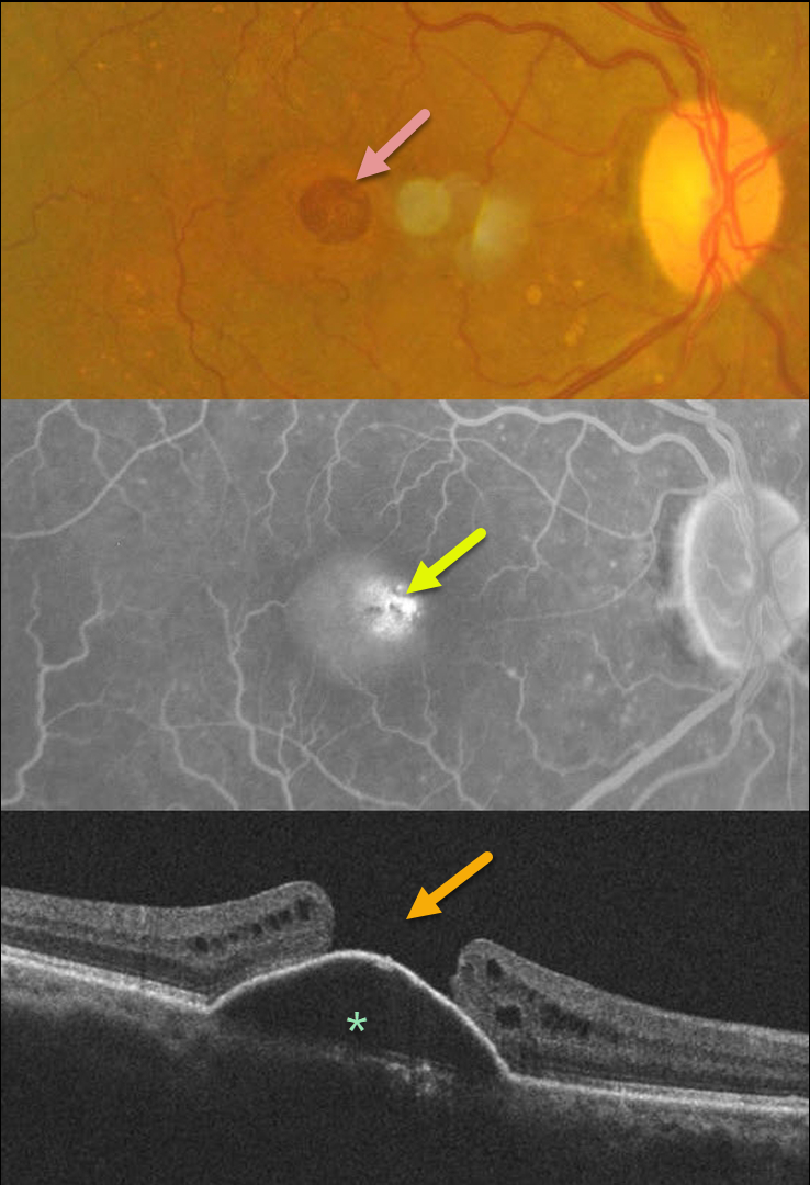
MACULAR HOLE
Shilpi Narnaware and Prashant Bawankule
Originally posted on @retina.rocks August 8, 2023
This 60YO female presented with 1 month of vision loss in her right eye. Vision was 20/100 OD and 20/40 OS.
Fundus photography of her right eye shows a large macular hole with some nasal pachydrusen. There was an old branch retinal vein occlusion without macular drusen in her left eye (image not shown).
Fluorescein angiography shows central pooling beneath a retinal pigment epithelial detachment (PED) with increased transmission through the macular hole. OCT scanning shows a PED with an overlying large macular hole.
This case presents a unique and interesting challenge in that macular hole surgery likely would not close the hole, given the underlying PED. She received 3 monthly Lucentis injections in her right eye, with no change in the PED.
Our thought would be to try a different anti-VEGF agent, such as Eylea, or consider adding photodynamic therapy for a possible pachychoroid-related avascular PED.
A similar case showed spontaneous closure of a macular hole following anti-VEGF therapy (see Chakraborty and Sheth, Indian J Ophthalmol Case Rep 2023;3:439-440).
If our patient’s macular hole remained open once the PED flattened, then macular hole surgery would be performed.
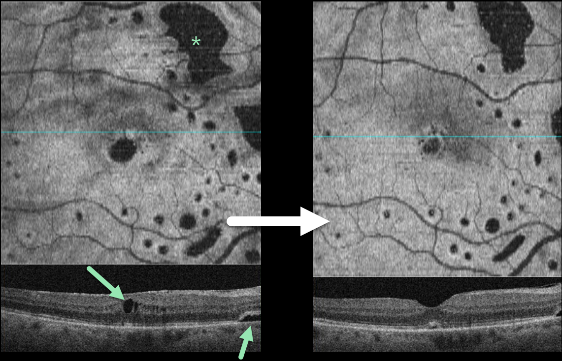
LOCULATED FLUID AFTER RETINAL DETACHMENT REPAIR
Mattie Adams
Originally posted on @retina.rocks July 26, 2023
This 66YO female was examined following successful vitrectomy three months earlier for a macula-off, rhegmatogenous retinal detachment (RRD). Vision improved from a preoperative 20/200 to 20/40 postoperatively.
OCT scanning shows multifocal areas of intraretinal and subretinal fluid (SRF). Four months later, vision remains at 20/40, and all areas of the SRF are shrinking.
Learning Points:
Loculated fluid, which represents an area of persistent subclinical SRF in the presence of a clinically reattached retina, is seen postoperatively in up to 15% of RRD following vitrectomy (Retina 2020;40:1153-1159), and eventually resolves after many months of observation.
In a post hoc analysis of the PIVOT trial, there were no differences in the incidence of loculated fluid between vitrectomy and pneumatic retinopexy (BJO 2021-320981, ahead of print). Although subfoveal loculated fluid was associated with reduced visual acuity at 3 months, vision eventually was similar to that of those without fluid at 1 year.
Bansal et al described the en face findings for loculated fluid (Ophthalmology Retina 2023;7:496-502), which prompted us to perform en face imaging in this patient.
In our practice, we continue to find that en face imaging often provides a unique and compelling perspective on a variety of pathologies, and we hope other clinicians will add it to their daily clinical routine.
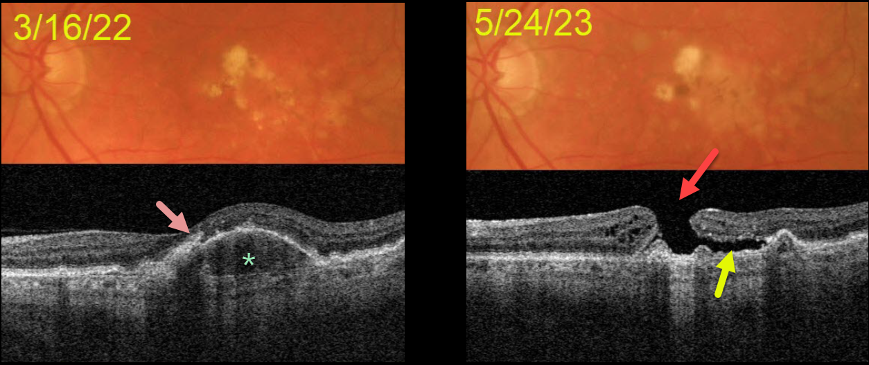
MACULAR HOLE AFTER COLLAPSE OF PED
Originally posted on @retina.rocks July 25, 2023
This 72YOM was examined with asymptomatic intermediate AMD bilaterally. Vision was 20/40 OD and 20/50 OS.
Color imaging of his left eye initially shows mostly large, soft drusen and pigment clumps. Swept source OCT shows a RPE detachment (PED) with overlying hyperreflective foci.
He returned over a year later without new symptoms, despite vision dropping to counting fingers. OCT shows a large macular hole overlying the collapsed PED. Shallow subretinal fluid lies below the temporal aspect of the hole.
The development of a macular hole following spontaneous collapse of a PED is quite unusual. Fortunately, vision in his right eye remains at 20/40 with intermediate dry AMD.
Since our patient was completely asymptomatic, he did not want macular hole surgery, and we will monitor him twice yearly.
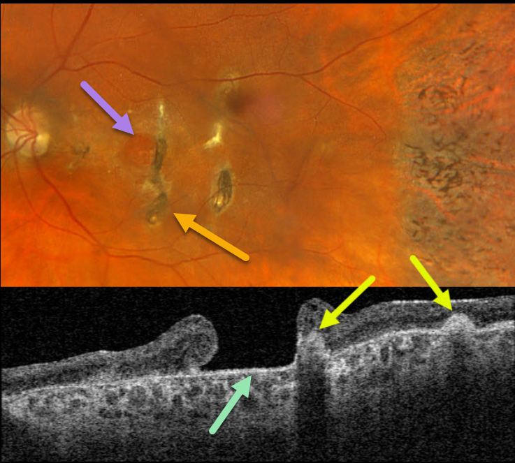
TRAUMATIC MACULAR HOLE + CHOROIDAL RUPTURE
Originally posted on @retina.rocks July 11, 2023
This 57YO female has a long history of vision loss in her left eye following blunt trauma years earlier. Vision was hand motion OS.
Optos color RGB imaging shows a large chronic macular hole with surrounding pigmentary changes. There are several hyperpigmented choroidal ruptures just temporal to the hole. Chorioretinal scarring with intraretinal pigment migration is noted in the temporal periphery.
Swept-source OCT shows a large macular hole. The choroidal ruptures appear as elevated hyperreflective subretinal mounds with posterior shadowing.
Learning Points:
This patient illustrates several sequelae of blunt trauma, including macular hole formation, choroidal rupture, and intraretinal pigment migration with a pseudo-sector retinitis pigmentosa appearance due to commotio-induced photoreceptor loss.
Since fresh traumatic holes can spontaneously flatten, especially in pediatric cases (Liu et al, AJO 2020;210:174-183), an initial period of observation is recommended before considering vitrectomy.
Given the chronicity of our patient’s hole, surgery was not considered.
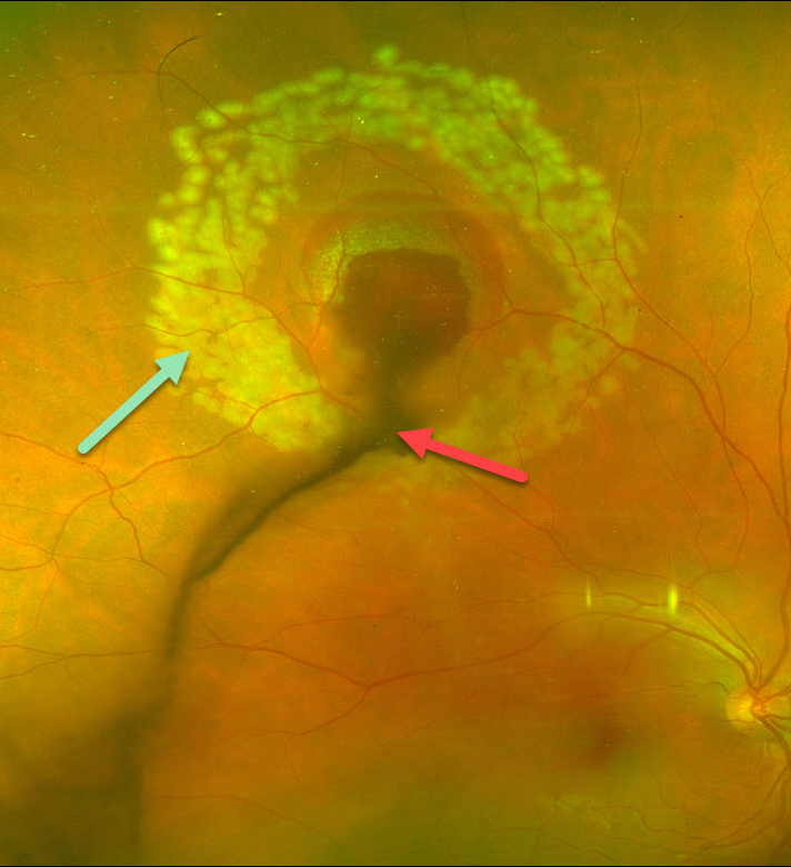
RETINAL BREAK
Mohammad Abbas
Originally posted on @retina.rocks July 10, 2023
This 27 YO technician working without eye protection in a dermatology laser clinic presented with sudden, painless vision loss and floaters in his right eye. Vision was 20/40.
Optos color imaging shows vitreous blood emanating from the site of laser injury in the super midperiphery. A rim of subretinal blood surrounds the lesion. Fresh laser retinopexy encircles the hemorrhage.
A retinal break is likely obscured by the blood, and this laser should hopefully reduce the risk of a retinal detachment.
Learning Points:
Ocular damage from these lasers depends highly on the laser wavelength, power, and duration. There are several types of lasers used in dermatology, and eye protection for both patients and clinic staff should be used.
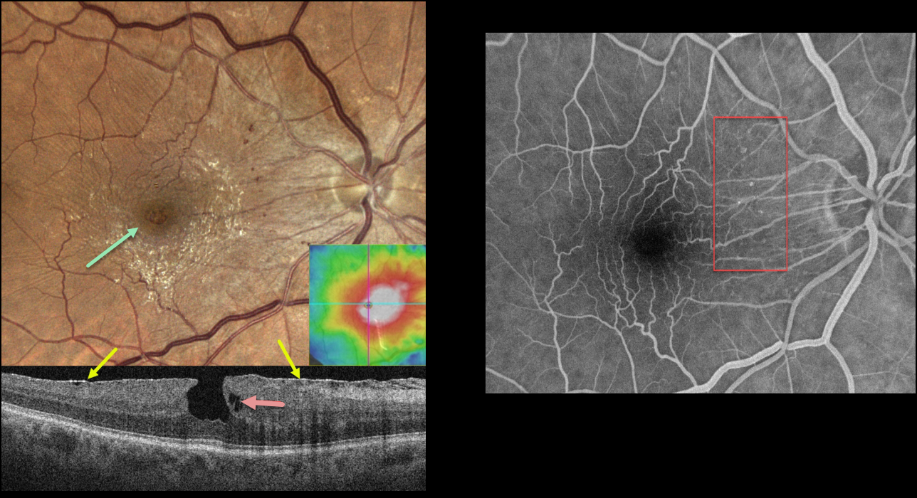
MACULAR PUCKER
Originally posted on @retina.rocks July 4, 2023
This 79YO male was referred for asymptomatic macular findings in his right eye. Vision was 20/20.
Color imaging shows a shiny, mostly nasal and inferior, macular pucker with a small, irregular macular pseudohole. Small dots of retinal blood vs telangiectasia are seen nasally.
OCT scanning shows an adherent macular pucker, a vertical foveal contour, and a small area of outer plexiform layer (OPL) schisis. The macula was diffusely thickened.
Fluorescein angiography shows no macular leakage, with filling of the nasal telangiectatic vessels.
Learning Points:
This case nicely illustrates many clinical and multimodal imaging findings for macular pucker. Clinically, true macular holes are virtually always perfectly round, whereas these pseudoholes are usually somewhat oval. The adherent epimacular tissue usually spares the foveal center. The adherent surrounding tissue contracts, drawing the surrounding tissue towards the macular center, giving the fovea a more vertical contour.
The Müller cell spans nearly the full thickness of the retina and helps mechanically hold the 3 layers of retinal cells together. Henle’s layer (the macular OPL) is inherently weakened since the photoreceptor axons and Muller cells are splayed laterally. Vitreomacular traction or traction from an overlying epimacular membrane will often therefore split Henle’s causing non-exudative schisis or cysts.
Finally, the secondary telangiectatic vascular changes are caused by mechanical damage from the overlying membrane. In severe cases, a pucker with the vascular changes can simulate a combined hamartoma.
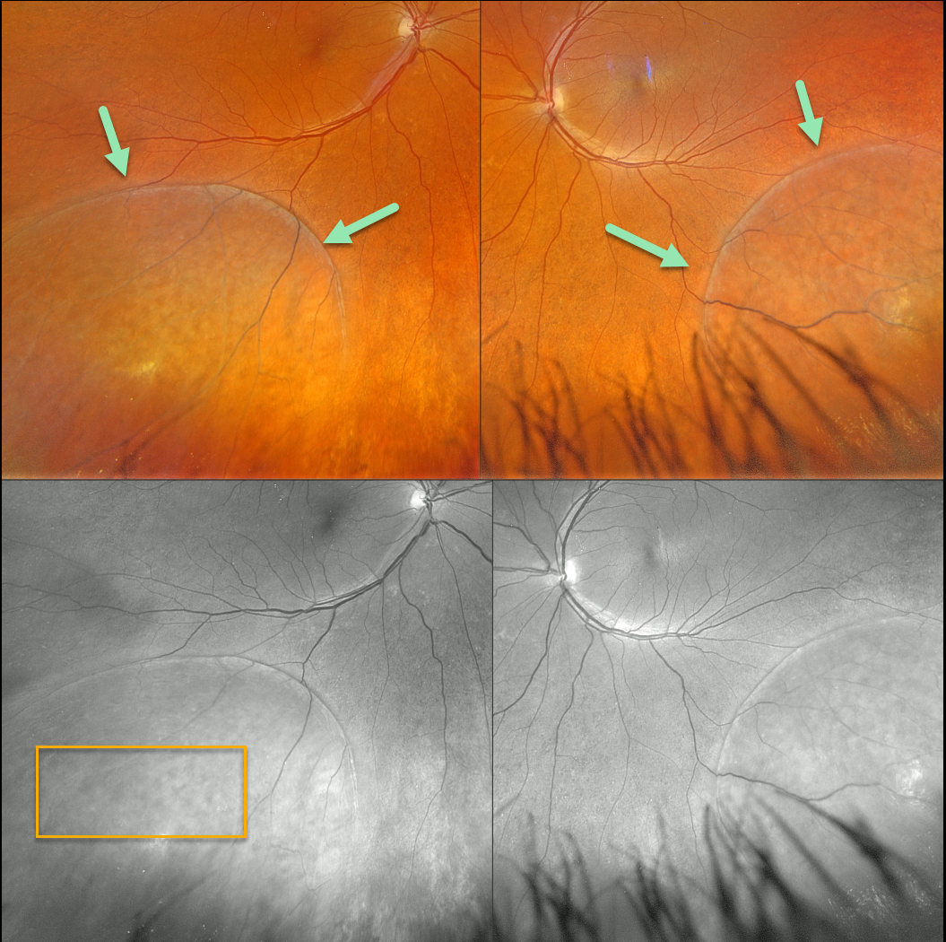
DEGENERATIVE RETINOSCHSIS
Originally posted on @retina.rocks June 27, 2023
This 50YO female was referred for possible bilateral retinal detachments. Vision was 20/30 OD and 20/40 OS.
Optos color RGB imaging shows bullous, symmetrical inferotemporal retinoschisis. The innumerable inner-layer holes are best seen in the green channel.
Learning Points:
Degenerative retinoschisis is virtually never prophylactically treated (see Byer 1986;93:1127-1137). Patients can rarely develop a combined schisis rhegmatogenous retinal detachment (RRD) when liquid vitreous accesses the subretinal space through both inner and outer retinal holes.
Our patient is at low risk for developing a combined schisis RRD due to the lack of outer-layer breaks. We will follow her yearly.
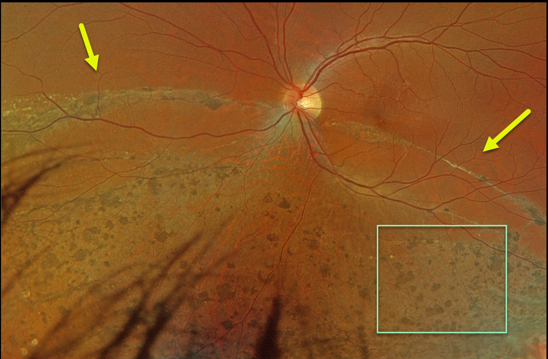
RHEGMATOGENOUS RETINAL DETACHMENT
Originally posted on @retina.rocks June 26, 2023
This 36YO male was referred for asymptomatic unilateral fundus findings. He had a history of retinal reattachment surgery 13 years earlier. Vision was 20/40.
Optos color RGB imaging shows chorioretinal scarring with diffuse intraretinal pigment migration within the area of prior detachment, giving the appearance of pseudo-sector retinitis pigmentosa (RP). A demarcation line courses through the inferior macula. This just spares the central macula, thankfully leaving him with good central acuity.
Learning Points:
Photoreceptor loss from numerous causes, including blunt trauma, retinal detachment, macular telangiectasia, and RP, can allow for intraretinal RPE migration with this shared phenotype.
Unilateral RP is exceedingly rare and requires normal electrophysiologic findings and long-term observation to ensure delayed findings in the fellow eye.
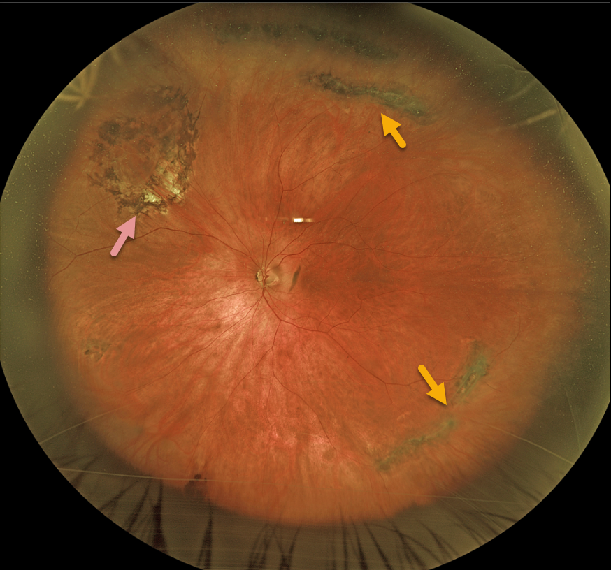
LATTICE DEGENERATION
Originally posted on @retina.rocks June 12, 2023
This 68YO male presented with asymptomatic lattice degeneration bilaterally. He remembered getting a laser to one of his eyes many years earlier at an outside practice.
Optos imaging of his left eye shows a near ora to ora view of scattered circumferential pigmented lattice lesions. Laser scarring surrounds an area of lattice with atrophic holes superonasally.
Learning Points:
Lattice lesions have a pocket of vitreous liquefaction overlying the thinned retina with a firm vitreo-retinal adhesion along their borders. White, fibrosed vessels often criss-cross the lesions, which leads to the name, lattice. This firm vitreoretinal adhesion, along with the often-present atrophic holes, can predispose these eyes to retinal detachment.
Retinal detachment occurs in 1 in 10,000 people per year. Lattice is found in 10% of the general population, especially in high myopes.
Since about one-third of patients with retinal detachment have lattice as the cause, the question always arises, especially in patients like this: Should prophylactic retinopexy be performed? The short answer is that asymptomatic lattices generally should not be treated.
To put things into perspective, imagine a sports stadium of 30,000 people. Three people will develop a retinal detachment in the coming year. Only one of the 3,000 people in the stadium with lattice would develop one of those 3 detachments. Assuming prophylactic treatment is 100% effective with no complications, 2,999 eyes would be unnecessarily treated to prevent one detachment.
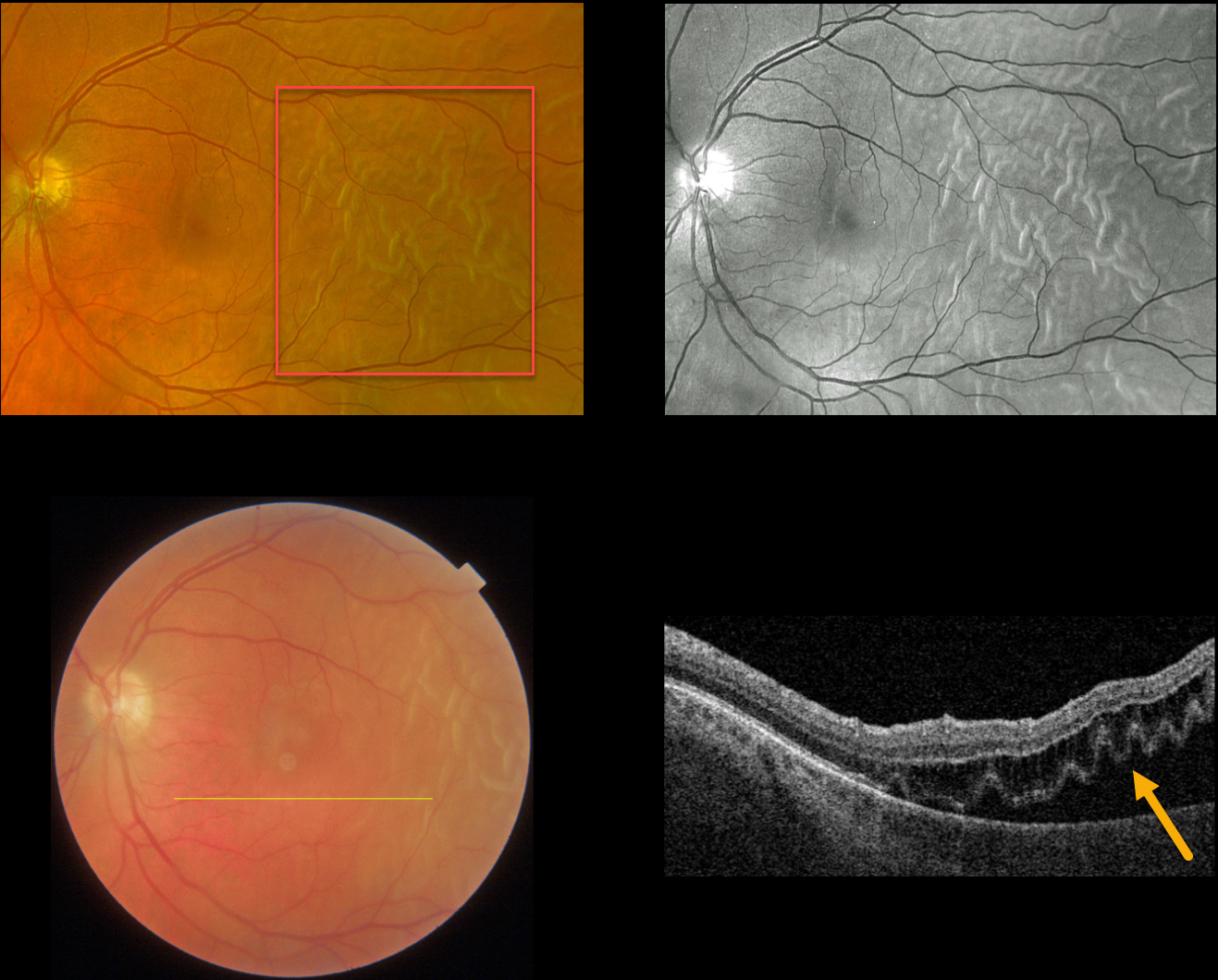
OUTER RETINAL FOLDS/CORRUGATIONS
Originally posted on @retina.rocks June 7, 2023
This 51YO male presented with this acute temporal macula-off rhegmatogenous retinal detachment (RRD). Vision was 20/200.
Optos color RGB imaging shows an RRD with corrugated outer retinal folds. These folds are best seen with the green channel. Swept-source OCT shows how these findings involve just the outer retina. The detachment was repaired with vitrectomy the following day.
Learning Points:
Hydration of outer retinal folds are one of the key findings to help differentiate a RRD from exudative and traction retinal detachments.
The cause for these folds remains uncertain, but they may form as the result of hydration of glycosaminoglycans in the interphotoreceptor matrix (Dalvin et al, Retinal Cases & Brief Reports 2020;14;355-359).
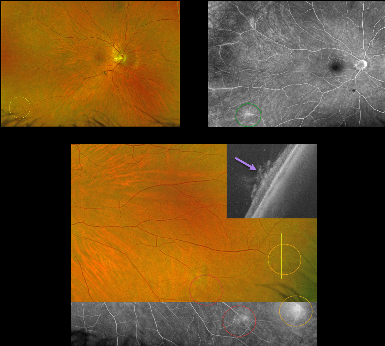
POSSIBLE CELIAC RETINAL NEOVASCULARIZATION
Paulo Eduardo Stanga, Javier Valentin Bravo, and Sebastian Stangam
Originally posted on @retina.rocks June 6, 2023
This 55YO female presented with asymptomatic peripheral retinal vascular findings. She has a history of high myopia and celiac disease. Vision was 20/20 OU.
Optos imaging of the OD shows telangiectatic vessels in the inferotemporal periphery with angiographic leakage in this region and superotemporally. Two similar areas are noted inferotemporally in the OS. Vertical UWF-OCT scanning through the most temporal lesion confirms that this represents retinal neovascularization growing into the vitreous.
Learning Points:
In 2016, Shah et al described common peripheral retinal vascular findings on Optos UWF FA in otherwise healthy adults (58 eyes, 31 patients) undergoing imaging for unrelated macular pucker or choroidal nevi (Retina 2016;36:1087-1092). Findings included vessels crossing the horizontal raphe (45%), right-angle vessels (71%), terminal networks (78%), absence of capillary detail (98%), ground glass hyperfluorescence (88%), drusen (34%), and microaneurysms (41%). One or more of these findings were present in most eyes.
Another study by Lu et al. found that normal peripheral retinas typically exhibit granular background fluorescence, with or without a mottled fluorescent band (AJO 2017;173:84-90).
Since integrating Optos UWF imaging into our practice a few years ago, we have observed similar findings in many of our patients undergoing FA for unrelated diagnoses. In the beginning, we often checked carotid doppler and ultrasonography, which were invariably normal. We now also consider these normal peripheral findings and no longer routinely work up these patients. Retinal neovascularization has not been described in this entity.
Celiac disease is associated with uveitis, subclinical macular microvascular changes, central retinal vein occlusion, and retinal findings from vitamin A deficiency (Fousekis et al, Int Ophthalmol 2020;40:1049-1054). Peripheral retinal ischemia with neovascularization has not been described. It is therefore unclear whether or not our patient’s Celiac disease contributed to these findings.
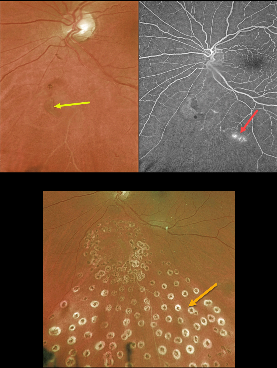
RETINAL TEAR + BRVO
Originally posted on @retina.rocks June 5, 2023
This 63YO female presented with symptomatic flashes and floaters in her left eye.
Optos color imaging shows a retinal tear just inferior to the nerve. An ischemic branch retinal vein occlusion (BRVO) is noted inferiorly. A posterior vitreous detachment (PVD) was noted clinically.
Fluorescein angiography confirmed the inferior ischemic BRVO with some small areas of retinal neovascularization. The tear appeared to be completely independent of the venous occlusion.
The retinal tear was surrounded by a few nearly confluent rows of laser photocoagulation burns, and inferior scatter laser was applied into the distribution of the BRVO. Fundus photography, a few months later, shows the laser scarring.
Learning Points:
Retinal tears are virtually always seen in the retinal periphery and are caused by an adherent vitreous pulling the retina anteriorly towards the vitreous base, most commonly during an acute PVD.
Our patient’s tear is unusual due to its quite posterior location and the flap occurring along its posterior edge. This implies that the vitreous was tugging towards the optic nerve. The optic nerve is usually the last site for the vitreous to release from during an acute PVD (Johnson, AJO 2010;149:371-382).
We hypothesize that the vitreous was completely detached except at the optic nerve and just inferiorly, and the tear was created from anterior to posterior traction when the vitreous separated from the nerve.
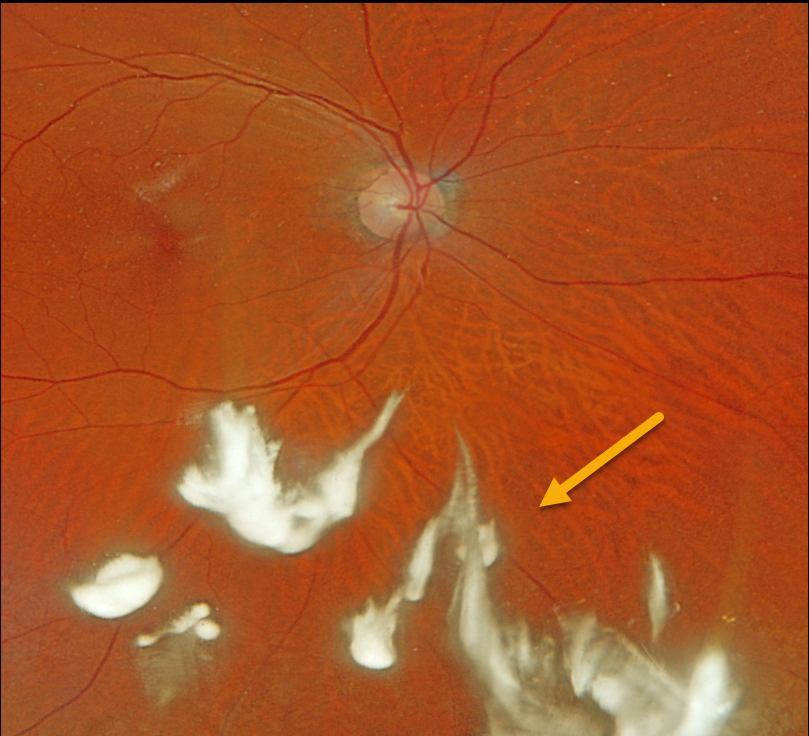
TRIAMCINOLONE-MOXIFLOXACIN (TriMoxi)
Originally posted on @retina.rocks June 1, 2023
This 68YO male presented with symptomatic floaters 10 days following uneventful ‘no-drop’ cataract surgery with Trimoxi.
Optos color RGB imaging shows clumps of white triamcinolone suspended in the inferior vitreous.
Learning Points:
Our practice, along with many others, offers ‘no drop’ cataract surgery as a convenience to our patients. Instead of needing to take postoperative antibiotic and steroid drops, 0.2cc of Trimoxi (triamcinolone acetate 15mg/cc with moxifloxacin 1mg/cc) is injected into the anterior inferior vitreous at the end of the cataract surgery.
Although this liquid usually remains well outside the visual axis, patients will often experience mild floaters perioperatively. Rarely, as in this patient, the liquid disperses throughout the vitreous, causing significant transient floaters.
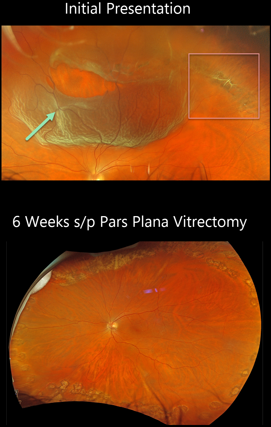
RHEGMATOGENOUS RETINAL DETACHMENT
Originally posted on @retina.rocks May 29, 2023
This 55YO male presented with this acute superior macula-on rhegmatogenous retinal detachment (RRD). Vision was 20/30.
Optos color RGB imaging shows a large patch of equatorial lattice degeneration extending from 11 to 2 o’clock. Nasally, the lattice lesion has torn along its lateral and posterior edges, causing the detachment. Several intact bridging retinal vessels are noted within the tear.
The detachment was repaired with vitrectomy the following day and remained attached 6 weeks later, following complete absorption of the intraocular gas bubble.
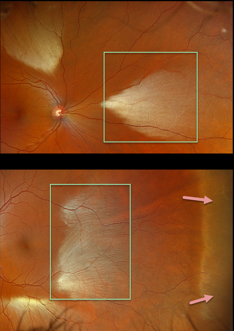
MYELINATED NERVE FIBER LAYER + WWP
Originally posted on @retina.rocks May 18, 2023
This healthy 35YO male was referred for asymptomatic fundoscopic changes. Vision was 20/20 OU.
Opto imaging shows large multifocal patches of myelinated nerve fiber layer (NFL) in both eyes. Temporal white without pressure (WWP) is also noted in his left eye.
Learning Points:
Myelinated NFL is a relatively uncommon, unilateral or bilateral, usually isolated finding present at birth. The myelinated NFL is often connected to the optic nerve, but not necessarily as in this case. Most patients are asymptomatic, although some may have a corresponding relative scotoma. The myelination can progress in about 10% of cases and can also resolve following retinal ischemic events.
Myelinated NFL occurs when retinal nerve fibers develop a myelin sheath, which usually stops posterior to the lamina cribrosa. The myelination may represent an oligodendrocytic choristoma (see Rao et al Retina 2019;39:1125-1132).
WWP is a relatively common benign peripheral finding, most often seen in darkly pigmented individuals. Typically, there are bilateral peripheral geographic areas of whitish retinal discoloration. WWP gets its name because the area appears white without indentation, unlike scleral depression (white with pressure). It can sometimes be confused with retinal breaks or detachment.
The exact cause of this phenomenon is unknown. Historically, it was felt to be due to a benign vitreoretinal interface change, but more recent OCT findings show increased reflectivity in the outer retina (see Diaz et al, Retina 2014;34:1020-1021).
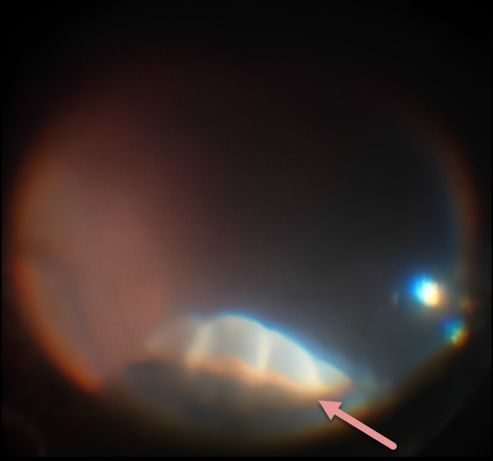
PARS PLANA CYST
Niki Gupta
Originally posted on @retina.rocks May 9, 2023
During vitrectomy for a macular pucker, this intraoperative photo shows an incidental finding of 3-4 pars plana cysts in the superior quadrant found on scleral depression. The cysts were oval, and the inner cyst walls appeared smooth and transparent. Anteriorly, they extended variably over about one-half of the pars plana towards the ciliary processes.
Learning Points:
Pars plana cysts are found in up to 18% of eyes and are more prevalent in the older population. They typically appear as ovoid elevations with a smooth, taut surface.
Pars plana cysts are usually acquired and represent a normal ocular aging change. While the cysts themselves are benign, they may occur secondary to ocular or systemic pathology, including retinal detachment, posterior uveitis, and multiple myeloma.
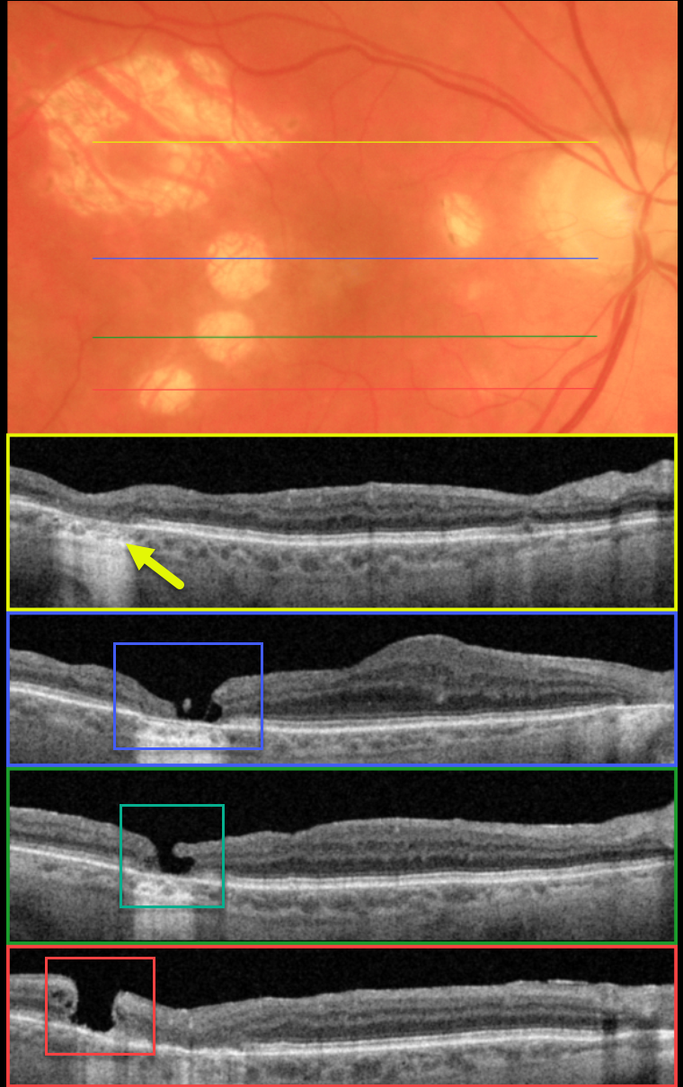
IATROGENIC MACULAR HOLE
Originally posted on @retina.rocks May 3, 2023
This 79YO female presented for a routine examination. ‘Successful’ macular hole surgery was performed elsewhere at least several years earlier.
Optos color imaging shows multiple atrophic scars in the temporal macula with a smaller scar nasally. Swept-source OCT scanning through the most superior, larger scar shows outer retinal atrophy.
OCT scans through each of the 3 smaller temporal scars show independent macular holes. Scanning of the central macula shows a thickened retina just nasal to fixation.
According to our patient, the single vitrectomy was uneventful, but her imaging suggests otherwise. It seems the surgeon made at least 5 deep retinal touches with the intraocular forceps while attempting to peel the internal limiting membrane. The larger superotemporal scar was probably a touch that was surrounded by endolaser.
The 3 ectopic macular holes may have developed intra- or post-operatively. Fortunately, her central macular hole closed, her vision was 20/40, and she remains thrilled with her surgical results.
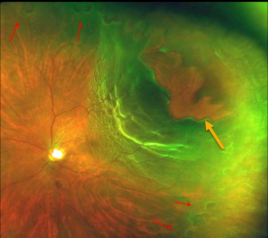
RHEGMATOGENOUS RETINAL DETACHMENT
Priyanak Gupta
Originally posted on @retina.rocks April 11, 2023
This patient presented with sudden vision loss from this rhegmatogenous retinal detachment (RRD).
Optos imaging shows a superotemporal macula-off RRD with an equatorial ring of numerous small retinal tears, along with a large ragged tear simulating stalactites. The patient subsequently underwent successful retinal reattachment surgery with vitrectomy.
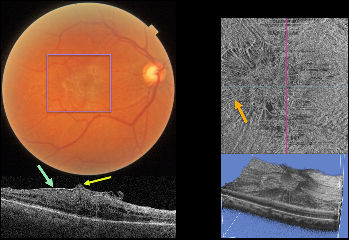
MACULAR PUCKER
Originally posted on @retina.rocks March 27, 2023
This 61YO female presented with 20/40 vision in her right eye from a mildly symptomatic macular pucker.
Color imaging shows a moderate macular pucker. Swept source OCT shows a variably adherent hyperreflective pucker, moderate retinal thickening with loss of the inner retinal layer detail, and a small foveal prolapse. En face imaging shows variable inner retinal striae from the pucker, and 3D reconstruction shows another unique perspective.
Learning Points:
Herniation of the central fovea is a rare finding with macular pucker. A recent case series (Shah et al, Retina 2023;43:182-190) found that larger foveal herniation height was associated with greater preoperative retinal thickness and worse pre- and postoperative vision.
However, vitrectomy with membrane peeling still results in significant anatomic and visual improvement. We have been observing our patient since she has been completely asymptomatic.
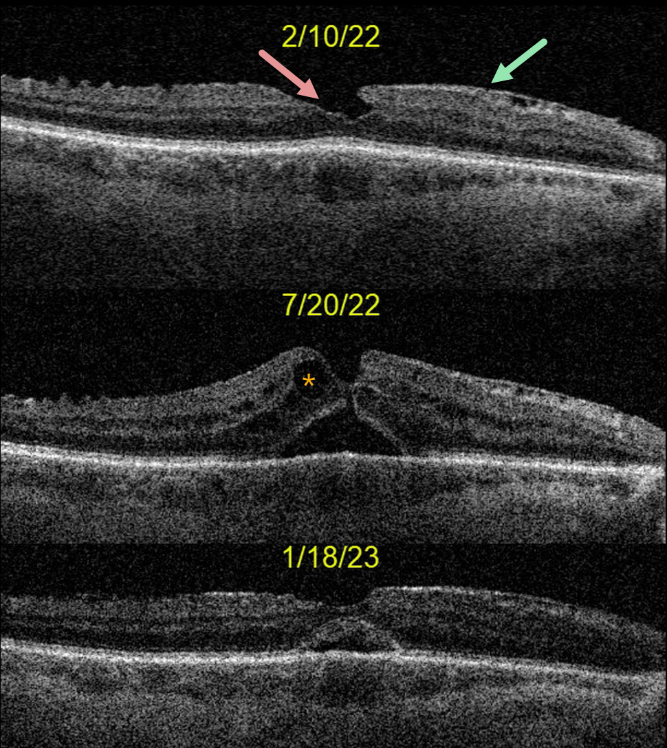
LAMELLAR HOLE
Originally posted on @retina.rocks February 23, 2023
This 73YO male was seen on 2/10/22 with 20/40 vision and an asymptomatic partial lamellar macular hole from an epiretinal membrane. Cataract surgery was performed on 5/25/22, and vision improved to 20/50 postoperatively.
However, on 7/20/22, vision decreased to 20/200 due to a new, extremely tiny full-thickness macular hole and pseudophakic cystoid macular edema (CME). Topical prednisolone acetate 1% and diclofenac QID were prescribed.
The macular hole partially closed one month later with 20/80 vision, and he was tapered off of topical therapy. When last examined on 1/18/23, vision had improved to 20/70.
OCT shows a stable appearance with a persistent outer retinal defect. We recommended continued observation.
Learning Points:
Topical steroid and non-steroidal therapy can sometimes close macular holes without traction, especially those that are small and associated with significant CME (see Ophthalmology Retina 2020;4:695-699). We therefore usually recommend topical therapy for selected symptomatic patients before proceeding to surgery.
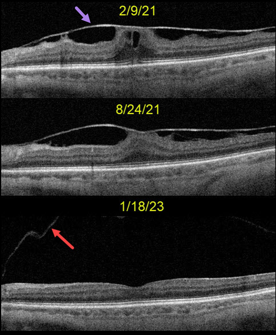
MACULAR PUCKER
Originally posted on @retina.rocks February 13, 2023
This 71YO female was seen on 2/9/21 with a completely asymptomatic variably adherent macular pucker. Vision was 20/80, and observation was recommended. On 8/24/21, vision improved to 20/40, and the macular traction had decreased.
When last examined on 1/18/23, the membrane had completely and spontaneously peeled itself as part of a macular posterior vitreous detachment. The back surface of the posterior hyaloid is seen nasally. Vision was 20/50.
Learning Points:
Spontaneous macular pucker separation is a known but rare event, occurring in about 2% of eyes with pre-existing posterior vitreous detachment (PVD) and 13% of eyes without PVD (Yang et al., Retina 2014;34:2079-2087).
We generally avoid macular pucker, vitreomacular traction, or macular hole surgery in visually happy patients. This patient also reminds us that these conditions can sometimes resolve on their own.
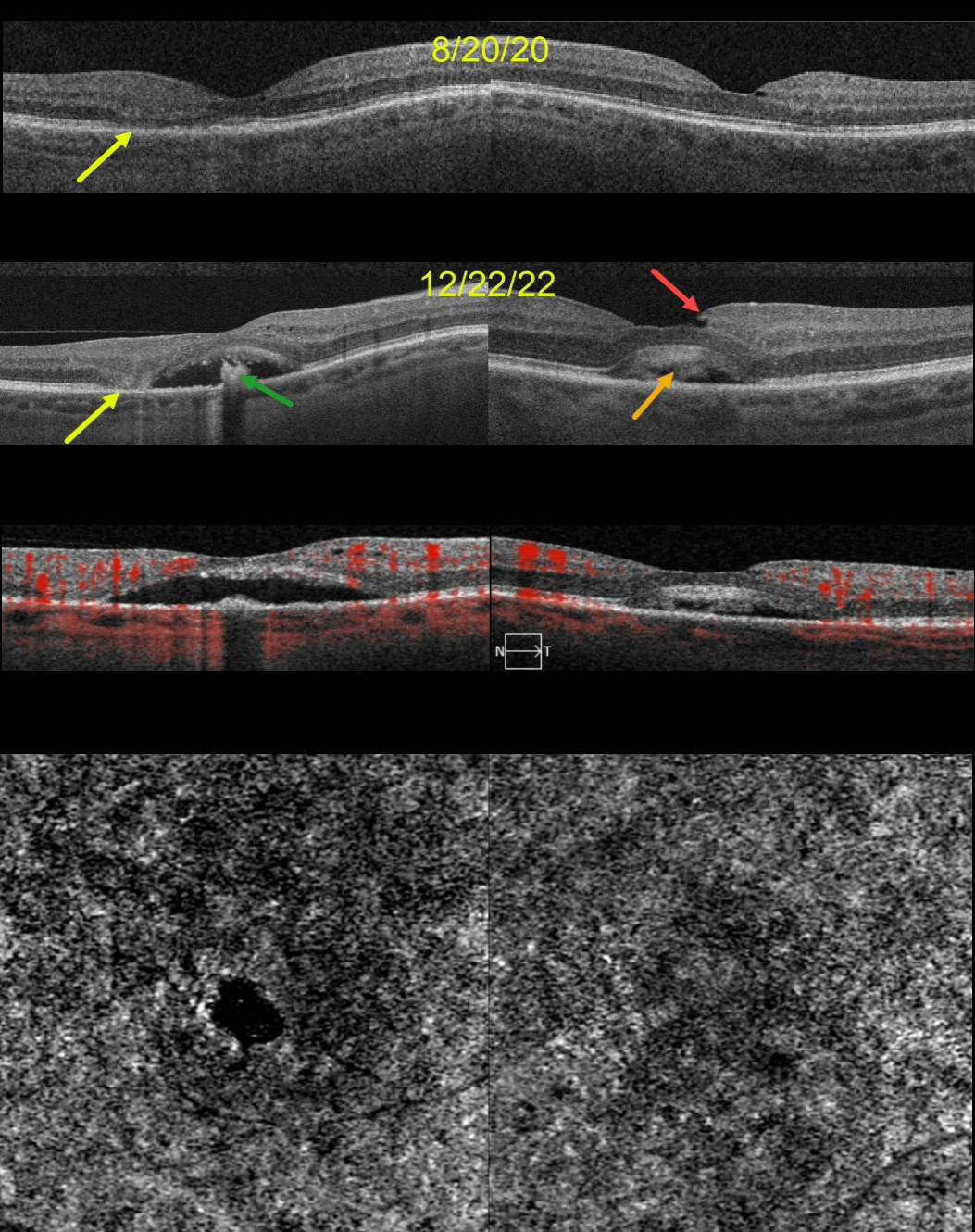
ACQUIRED VITELLIFORM LESION
Originally posted on @retina.rocks January 30, 2023
This 68YO female was seen on 8/20/20. Vision was 20/70 OD and 20/40 OS. Although there were no visual complaints, OCT scanning showed bilateral defects in the EZ bands, along with some temporal outer retinal atrophy OD.
She returned two years later, complaining of gradual vision loss OU, although vision was stable at 20/70 OD and 20/30 OS. OCT scanning shows bilateral acquired vitelliform lesions (AVL). The lesion OD is mostly scrambled with a hyporeflective space between the RPE and outer retina, along with a central hyperreflective clump on top of the RPE. There is now complete outer retinal atrophy temporally.
The AVL in her left macula shows variable hyperreflective material. An unrelated small partial lamellar macular hole is noted temporally. OCT angiography (OCTA) shows no flow signals from either AVL.
Learning Points:
Vision loss from AVLs most commonly results from lesions collapsing due to secondary atrophy or macular neovascularization, which can develop in about 8% of eyes (Balaratnasingam et al., AJO 2016;172:28-38).
The absence of OCTA flow signals in either eye confirmed that these lesions were avascular, and we continue to follow her.
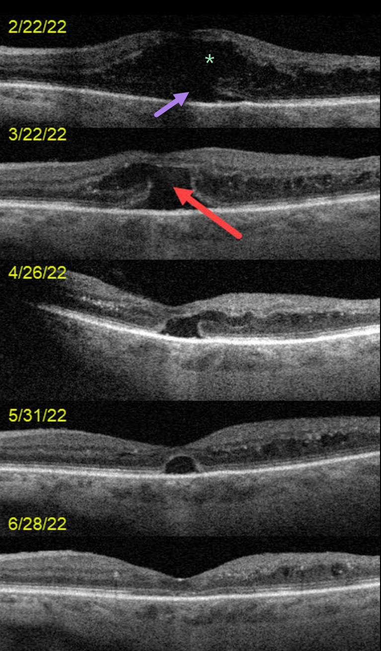
DIABETIC MACULAR EDEMA (DME)
Originally posted on @retina.rocks January 26, 2023
This 72YO female presented on 2/22/22 with counting-finger vision in her left eye.
OCT scanning at the initial visit shows severe center-involved diabetic macular edema (CI-DME) with shallow subretinal fluid. Anti-VEGF therapy was started.
One month later, vision remained at counting fingers. The retinal thickness had improved, but a new outer macular hole was noted.
Following 3 additional monthly injections, the edema continued to resolve with spontaneous closure of the outer macular hole. Vision was 20/70 at the 6/28/22 visit.
Learning Points:
Traditional primary full-thickness macular holes are caused by vitreomacular traction. Outer macular holes, however, have a much broader range of etiologies.
In our practice, we most commonly see these lesions in optic pit maculopathy and paraproteinemia (see Mansour et al, Ophthalmology 2014;121:1925-1932).
Our patient’s outer macular hole was likely caused by inner retinal exudation that extended through the outer retina. The return of central vision and closure of the defect, with relatively intact outer macular architecture, likely indicates that this was an outer macular dehiscence rather than a true loss of tissue. See Kumawat et al for a great review of atypical macular holes (Retina 2019;39:1236-1264).
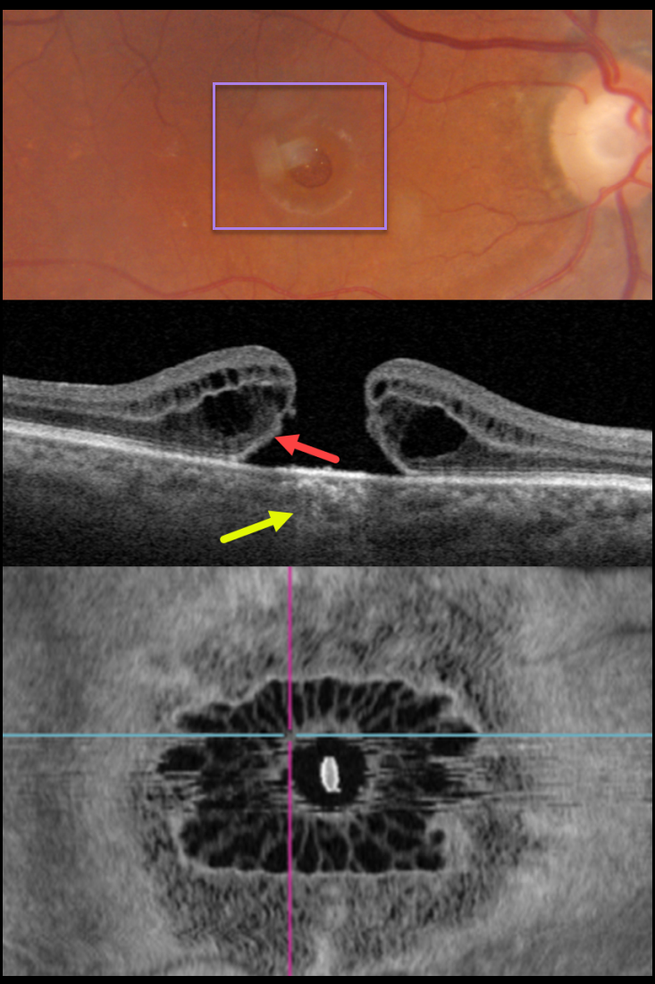
MACULAR HOLE
Originally posted on @retina.rocks January 12, 2023
This 81YO female presented with 20/200 vision in her right eye from a large full-thickness macular hole.
Swept-source OCT shows the classic appearance of a hole from a foveal dehiscence with cystic thickening. Outer retinal bands are noted along the posterior aspect of the hole’s elevated edges, indicating the lack of true tissue loss. This retinal defect results in increased light transmission into the choroid. En face imaging shows a unique and beautiful perspective of the hole.
Vitrectomy surgery was recommended.
Learning Points:
As described by Govetto et al (AJO 2020;212:43-56), idiopathic macular holes have a sunflower appearance showing features of both exudative and tractional cystoid spaces. The macular hole is hyporeflective centrally with multiple hyporeflective cystoid spaces radiating from the center.
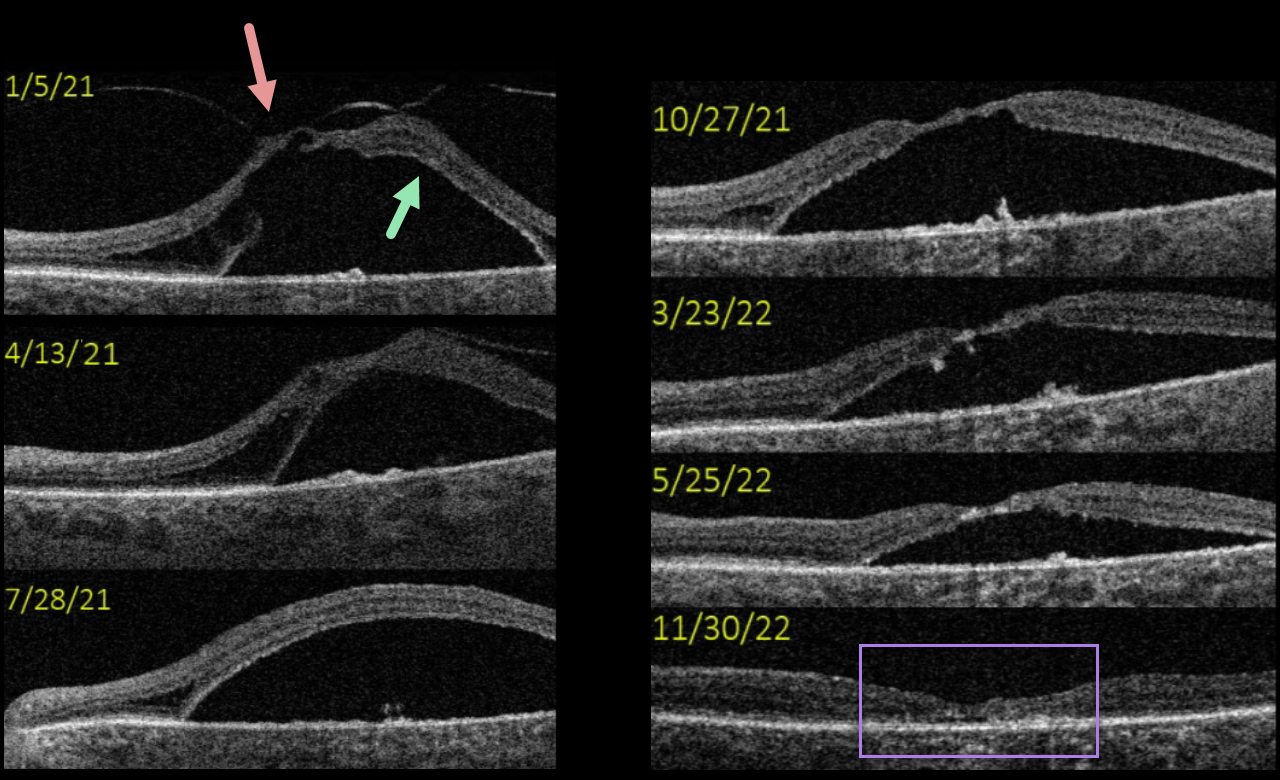
VITREOMACULAR TRACTION
Originally posted on @retina.rocks January 5, 2023
This 77YO female presented with vision of 20/400 in her left eye from severe vitreomacular traction with a secondary tractional macular detachment. The patient underwent pars plana vitrectomy in January 2021.
A few months post-vitrectomy, all traction was relieved, and the detachment was slightly decreased.
The detachment gradually resolved over the next year and a half, ultimately flattening completely with secondary foveal atrophy.
Although her final vision remained at 20/400, she was subjectively happy with her surgical results.
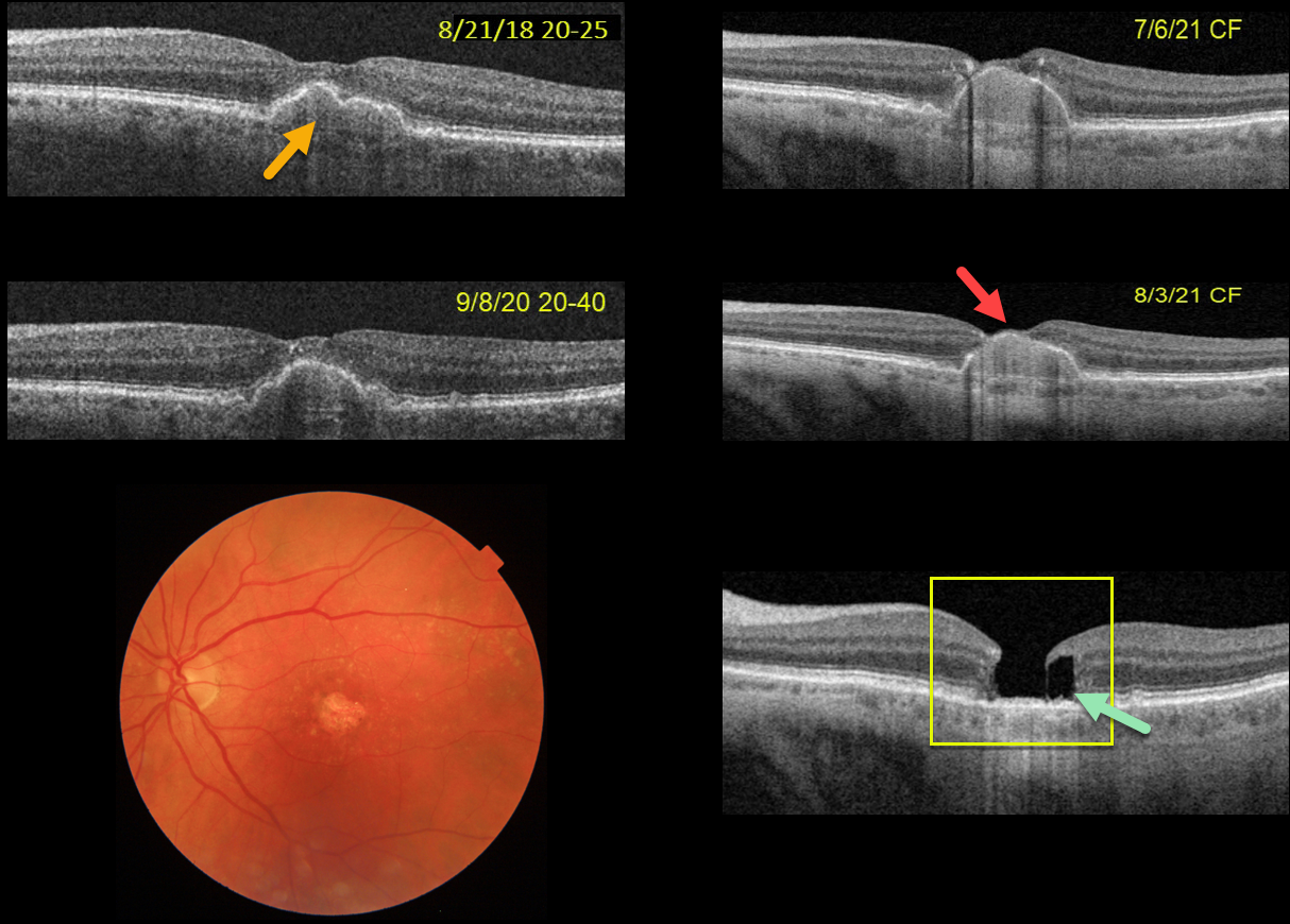
LAMELLAR HOLE AFTER AMD PIGMENT EPITHELIAL DETACHMENT (PED) COLLAPSE
Originally posted on @retina.rocks December 30, 2022
This 75YOF was seen in 2018 with non-neovascular AMD and a central pigment epithelial detachment (PED). Vision was 20/25. She returned on 9/8/20 with 20/40 vision with mild enlargement of the PED.
She complained of decreased vision when seen nearly one year later. Vision had dropped to 20/80. The PED had enlarged to involve full-thickness retina. She elected for an Eylea injection in hopes of possibly treating an occult macular neovascularization vs an avascular PED.
On 7/6/21, vision decreased to counting fingers although the OCT looked fairly stable. An additional Eylea injection followed by half-fluence PDT was given. On 8/3/21, the dome of the PED now extended through the inner retina and vision remained at counting fingers.
On 1/4/22, the PED had completely collapsed, with secondary retinal atrophy and a degenerative lamellar macular hole (LMH).
The progression of her PED through full-thickness retina and subsequent collapse with secondary retinal/RPE atrophy and a LMH are all quite unusual. Fortunately her right eye is 20/20 with intermediate dry AMD.
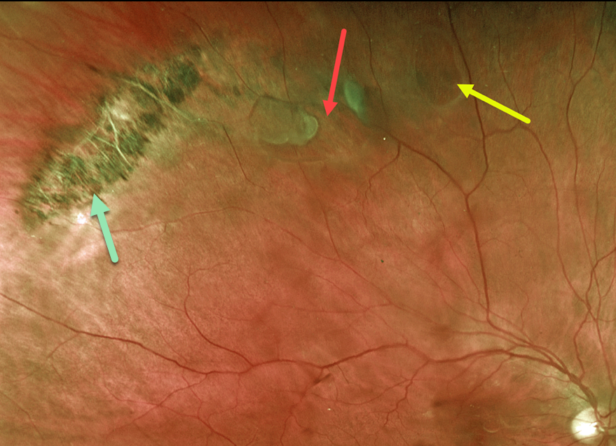
LATTICE DEGENERATION WITH RETINAL TEAR
Originally posted on @retina.rocks December 13, 2022
This 64YO male presented with acute flashes and floaters in his right eye. Optos imaging shows a patch of pigmented lattice superotemporally, associated with a retinal tear and atrophic hole. The breaks were surrounded with laser photocoagulation.
Learning Points:
Retinal detachment occurs in 1 in 10,000 people per year. Lattice is found in 10% of the general population, especially in high myopes. Since about one-third of patients with retinal detachment have lattice as the cause, the question always arises: Should prophylactic retinopexy be performed?
The short answer is that asymptomatic lattice generally should not be treated. However, an eye with symptomatic lattice breaks, as in our patient, is at high risk for developing rhegmatogenous detachment and should always be treated.
Excellent references on this controversial topic include: Byer Ophthalmology 1989;96:1396-1402, Folk et al Ophthalmology 1989;96:72-79 and Folk et al Retina 1990;10:165-169.
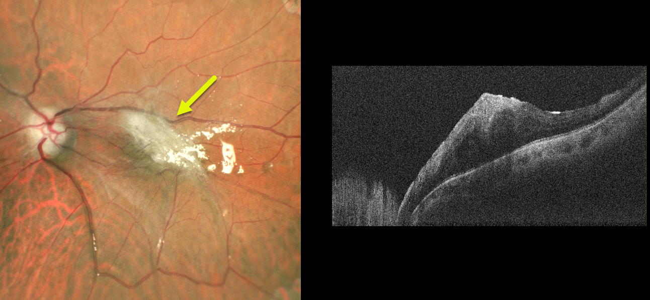
MACULAR PUCKER
Vaidehi Sathaye and Manish Nagpal
Originally posted on @retina.rocks December 9, 2022
This 45YO male underwent vitrectomy with silicone oil for a recurrent rhegmatogenous retinal detachment (RRD).
Although the retina remained attached, he developed a severe macular pucker postoperatively. Removal of the silicone oil with membrane peeling is planned in the near future.
Learning Points:
Macular pucker following vitrectomy for RRD develops clinically in about 10% of patients (Katira et al, Retina 2008;28:744-748), with the incidence likely being higher than this using OCT.
Half of the eyes developing macular pucker can occur after the first postoperative year (Szigiato et al, International Journal of Retina and Vitreous 2022;8:70).
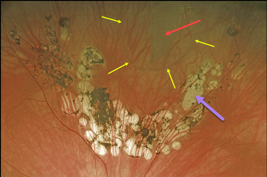
RETINAL BREAK
Originally posted on @retina.rocks December 7, 2022
This 67YO female presented with a symptomatic vitreous floater that started about 6 months earlier when she developed a symptomatic retinal tear.
The break was treated elsewhere with laser photocoagulation, and on subsequent visits, she was told the treatment was successful and looked perfect.
However, Optos imaging in our office shows otherwise. A large tear is visualized superiorly. Laser scars are seen posterior to the tear, but barely extend up its lateral margins and completely miss the anterior edge. Even where present, the scarring doesn’t extend to the edge of the break.
Elective vitrectomy surgery will be performed in the near future for her symptomatic vitreous floaters.
We will complete the prophylactic retinopexy at the time of surgery.
Learning Points:
Symptomatic retinal tears in the presence of an acute posterior vitreous detachment have a high risk of causing a rhegmatogenous retinal detachment. Prophylactic retinopexy is therefore indicated.
Treatment must completely surround the tear and, ideally, extend to the ora serrata. Both thermal laser photocoagulation and cryotherapy are equally effective in creating a vitreoretinal adhesion, which takes at least several weeks to form.
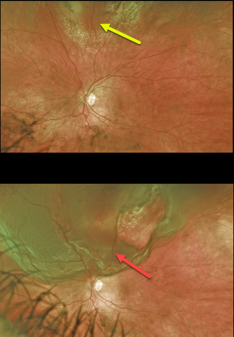
RETINAL TEAR
Originally posted on @retina.rocks November 17, 2022
This 37YO female presented with a symptomatic superior retinal tear in her left eye. The posterior and lateral edges of the break were surrounded with thermal laser. The anterior edges could not be reached with the laser, so these were surrounded with cryotherapy. One week later, pigmented scarring surrounds the treated tear.
Three weeks later, she presented with a few days of inferior visual field loss. She now has a superior macula-on rhegmatogenous retinal detachment (RRD) due to lifting off the originally treated tear. Vitrectomy surgery was performed the following day.
Learning Points:
Untreated symptomatic retinal tears have a high risk for RRD. Retinopexy, either with thermal laser or cryotherapy, must completely surround the break and, for more anterior lesions, ideally extend to the ora.
Patients must be followed closely, especially during the first month, due to the risk of additional breaks or retinal detachment. A firm chorioretinal adhesion takes several weeks to form (Yoon and Marmor, Ophthalmology 1988;95:1385-1388). Patients must always be cautioned to return immediately for new flashes, floaters, or vision loss.
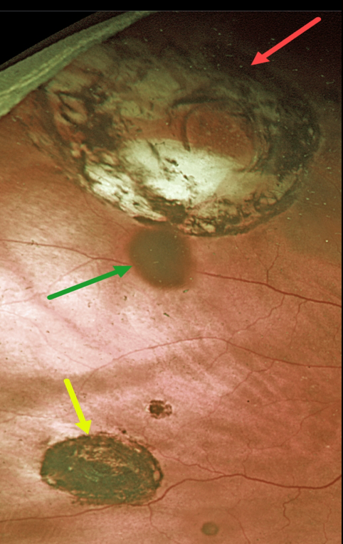
CHRPE + RETINAL BREAK
Originally posted on @retina.rocks November 15, 2022
This 87YOM received thermal laser around a retinal break 23 years earlier. He presented to us without symptoms and 20/30 vision.
Optos imaging shows a small inferior congenital hypertrophy of the RPE (CHRPE) with a few tiny additional CHRPE lesions.
Confluent variably pigmented laser scarring around a superotemporal retinal break simulates an atrophic CHRPE lesion.
The operculum casts a shadow on the underlying retina, somewhat giving the impression of yet another pigmented lesion.
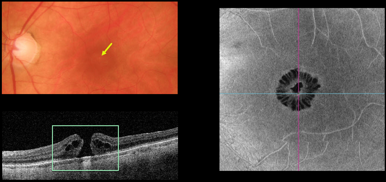
MACULAR HOLE
Originally posted on @retina.rocks November 11, 2022
This 65YO female presented with 20/200 vision in her left eye from a symptomatic full-thickness macular hole. The hole is barely visible funduscopically.
Swept-source OCT shows the classic appearance of a hole from a foveal dehiscence with cystic thickening. En face imaging shows a unique and beautiful perspective of the hole.
Learning Points:
As described by Govetto et al (AJO 2020;212:43-56), idiopathic macular holes have a sunflower appearance showing features of both exudative and tractional cystoid spaces.
The macular hole is hyporeflective centrally with multiple hyporeflective cystoid spaces radiating from the center.
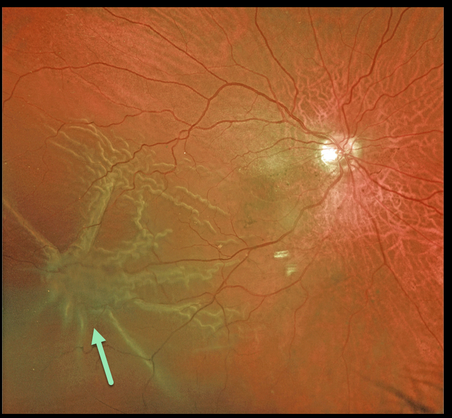
PROLIFERATIVE VITREORETINOPATHY (PVR)
Originally posted on @retina.rocks November 1, 2022
This 72YO male presented with this macula-off rhegmatogenous retinal detachment (RRD).
Optos color imaging shows a prominent star fold inferotemporally. This detachment was repaired with vitrectomy and membrane peeling.
Learning Points:
Proliferative vitreoretinopathy (PVR), which consists of cellular membranes growing on either retinal surface, develops in about 5-10% of retinal detachments.
The membranes contract and foreshorten the retina, likely as an aberrant wound-healing response.
There are no proven pharmacologic therapies to prevent or treat PVR. These complex detachments can be managed with vitrectomy and membrane peeling, retinectomy, scleral buckling, and silicone oil. However, PVR remains the main cause for ultimate retinal reattachment surgical failure.
See Garweg et al Surv Ophthalmol 2013;48:321-329 for a good review of PVR pathophysiology.
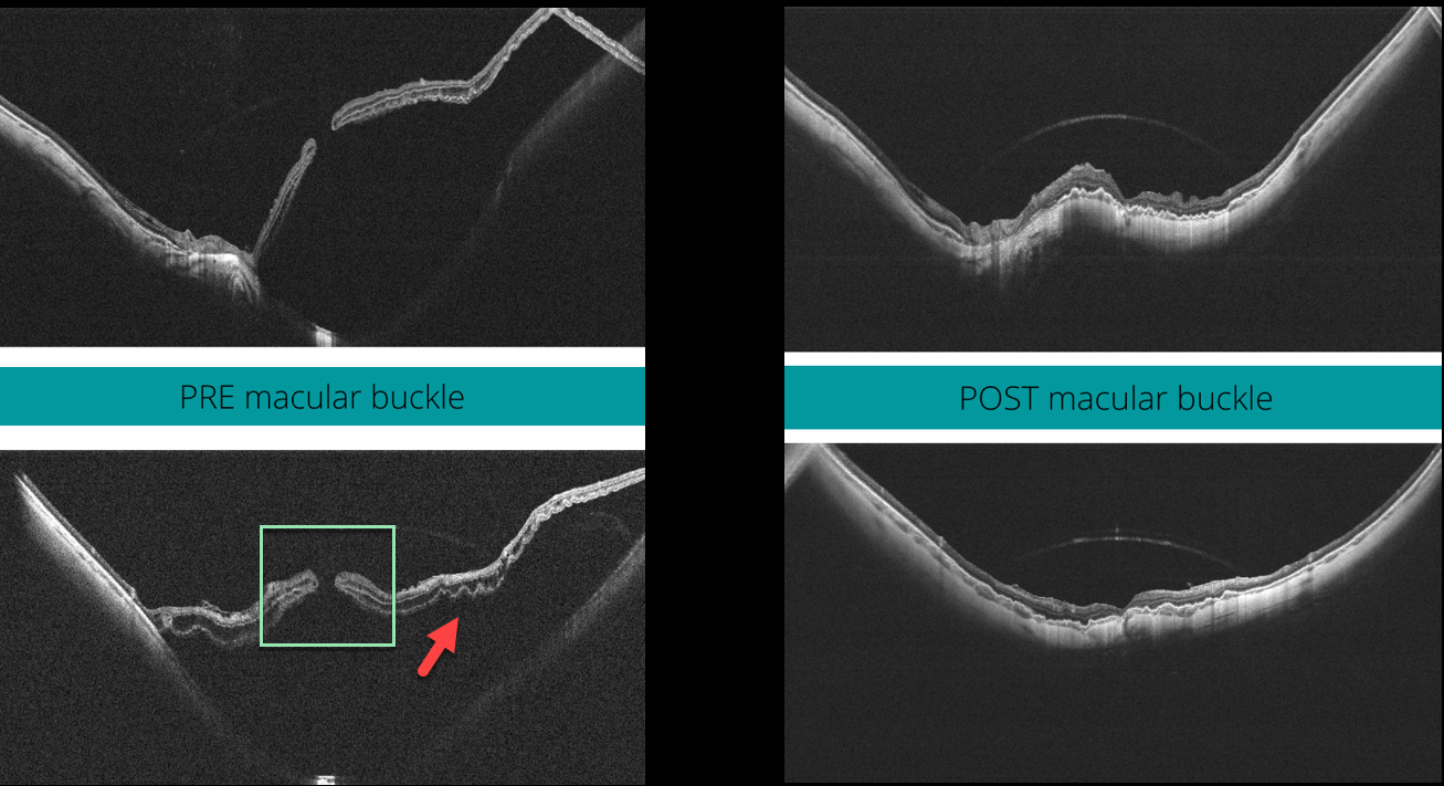
MACULAR HOLE
Veronika Matello and Barbara Parolini
Originally posted on @retina.rocks October 25, 2022
This 58YO highly myopic female presented with 20/400 vision in her left eye. The axial length was 32.45 mm, and her refractive error prior to LASIK surgery was 13D.
Widefield OCT scanning (Canon Xephilio SQ) shows a large macular retinal detachment with (or associated to) a full-thickness macular hole, defined as stage 4c according to the Myopic Traction Maculopathy Staging System (Parolini et al, Eur J Ophthalmol 2021;31:1299-1312).
Pars plana vitrectomy with an ILM flap and macular buckling was performed. One month postoperatively, the retina was flat, and the macular hole closed. Vision was 20/80.
Learning Points:
In our experience, these detachments are totally tractional, with the macular hole developing before or after the retina detaches. The macular buckle treats both the macular schisis and detachment, whereas the vitrectomy with ILM peeling closes the macular hole.
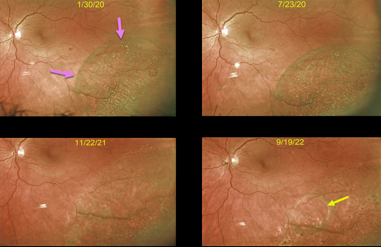
DEGENERATIVE RETINOSCHISIS
Originally posted on @retina.rocks October 19, 2022
This 59YO female was initially seen with asymptomatic retinoschisis in her left eye. When examined six months later, the schisis appeared stable.
16 months later, the schisis cavity was slightly enlarged in all dimensions with the formation of an early outer layer hole. Most recently, the outer layer hole enlarged, and the schisis spontaneously mostly collapsed, essentially resolving itself. Continued observation was recommended.
Learning Points:
Degenerative schisis cavities can enlarge, and in these cases, prophylactic treatment including thermal laser does not prevent further spread and is therefore not recommended (Ness et al, Surv Ophthalmology 2022;67;892-907).
Schisis very rarely progresses into the fovea (Byer Ophthalmology 1986;93:1127-1137) and even then is compatible with preserved central vision as long as the outer layer remains attached (Watzke et al 2013;33:606-612).
Our patient shows how the life cycle of a schisis cavity is often dynamic, including enlargement, development of inner or outer layer breaks, and resolution.
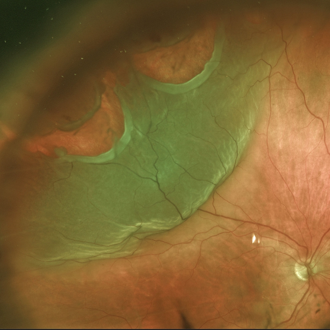
RHEGMATOGENOUS RETINAL DETACHMENT
Originally posted on @retina.rocks October 13, 2022
This 60YO female was referred for a symptomatic superotemporal rhegmatogenous retinal detachment (RRD) in her right eye.
Two large causative retinal tears are seen, both with posteriorly rolled edges.
The patient underwent emergent successful pars plana vitrectomy.
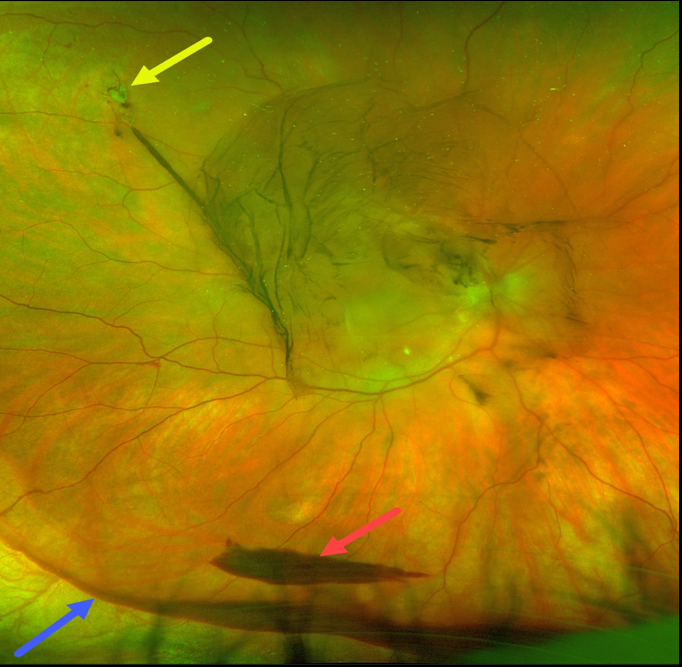
RETINAL TEAR
Originally posted on @retina.rocks October 10, 2022
This 59YO male presented with sudden flashes and large floaters in his right eye. Optos imaging shows a small, superotemporal causative retinal tear. There is a moderate central vitreous hemorrhage, a layered inferior pre-retinal hemorrhage, and more inferior blood collecting along the attachment of the vitreous base.
The vitreous has multiple attachments, the strongest of which is within the vitreous base. The sharp inferior border of this patient’s inferior vitreous blood beautifully highlights this anatomy.
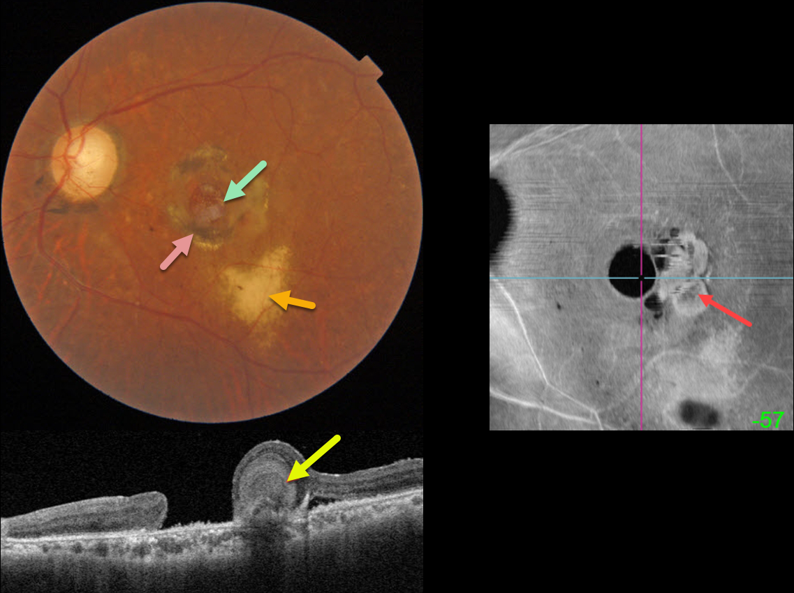
CHRONIC MACULAR HOLE
Originally posted on @retina.rocks September 19, 2022
This 59YO male has a complicated ocular and medical history, including HIV/AIDS, prior cytomegalovirus (CMV) retinitis that was treated with a ganciclovir implant in the 1990s, multiple retinal detachment surgeries in his right eye, eventually leading to evisceration for a blind and painful eye, and a chronic macular hole in his left eye.
Color imaging shows a large macular hole surrounded by a rim of variable subretinal hyperpigmentation. Yellowish subretinal scarring from prior CMV retinitis is noted more inferiorly.
Swept-source OCT shows an unusual ball of deep retinal hyperreflectivity. En face OCT shows a unique perspective for this macular hole, including the hyperreflective deep retinal material.
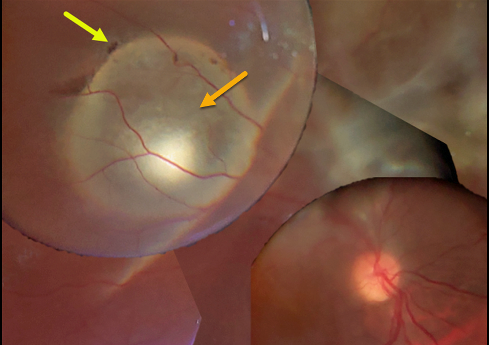
CYSTICERCOSIS WITH RHEGMATOGENOUS RETINAL DETACHMENT
Chhaya Bharti
Originally posted on @retina.rocks September 14, 2022
This 15YO boy presented with 20/200 vision. A large subretinal cysticercosis cyst is noted in the right distal superotemporal macula.
There is an overlying rhegmatogenous retinal detachment caused by a small retinal break. Unfortunately, the patient was subsequently lost to follow-up.
Learning Points:
Ocular cysticercosis is a rare parasitic eye infection caused by a pork tapeworm (larval form of the cestode, Taenia solium).
The ocular adnexa are most commonly affected, but the encysted organism can appear within the eye, including the anterior segment, vitreous cavity, and the subretinal space, as in this patient.
Vitrectomy with complete removal of the subretinal cyst is required since the dead organism can cause a severe inflammatory response.
For a great recent cysticercosis in ophthalmology review article, see Pujari et al, Surv Ophthalmology 2022;67:544-569.
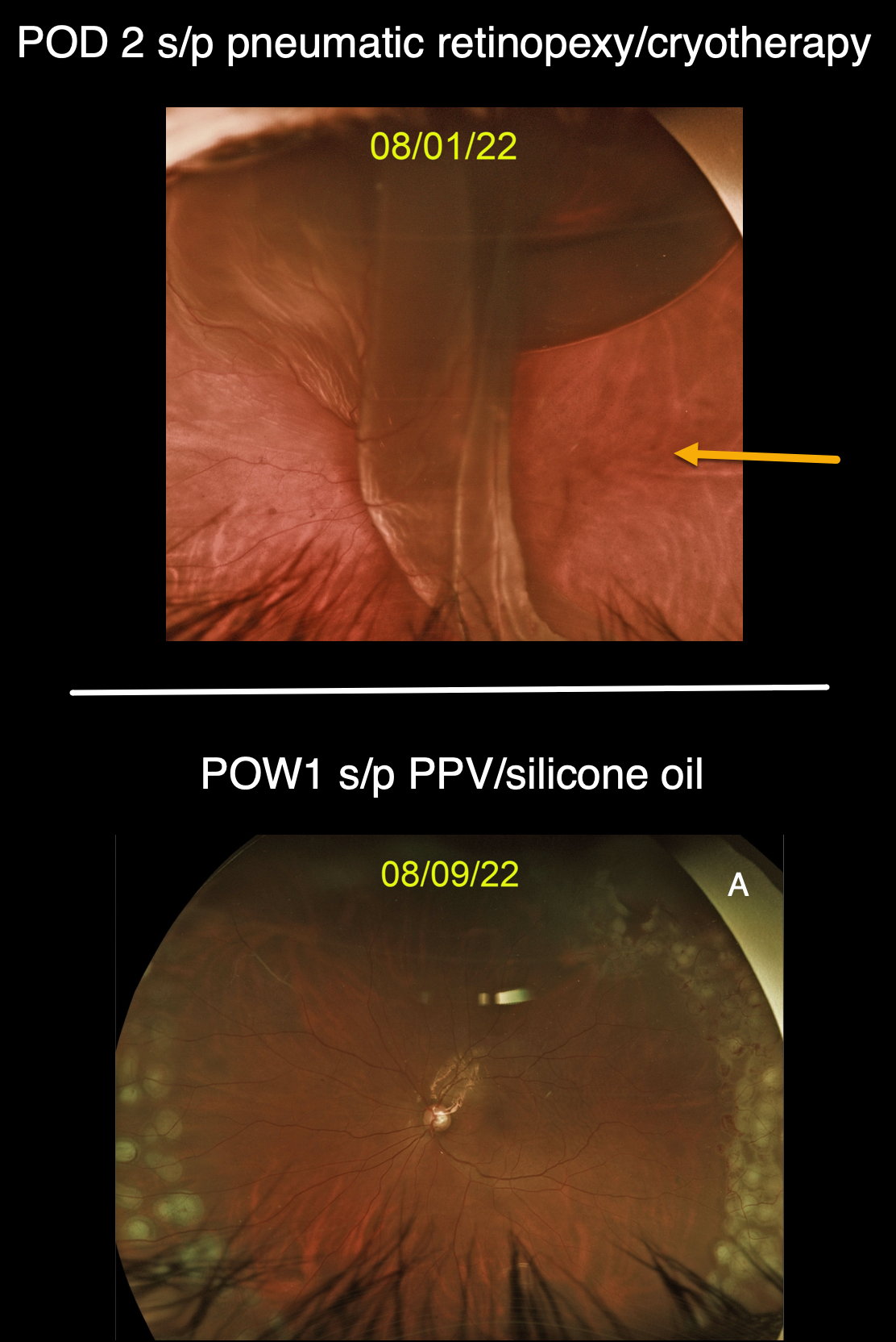
GIANT RETINAL TEAR
Originally posted on @retina.rocks September 9, 2022
This 50YO male presented with a superior macula-off retinal detachment from a single retinal tear at the 1 o’clock periphery (image not shown), which was treated with a pneumatic retinopexy. Cryotherapy was applied before injecting the gas bubble, confirming this single break.
However, when he returned 2 days later, a new giant retinal tear extended from 12 to 5 o’clock, with subretinal gas migrating beneath the superior retina.
The detachment was repaired with vitrectomy and silicone oil, and the second image shows the 1-week postoperative visit.
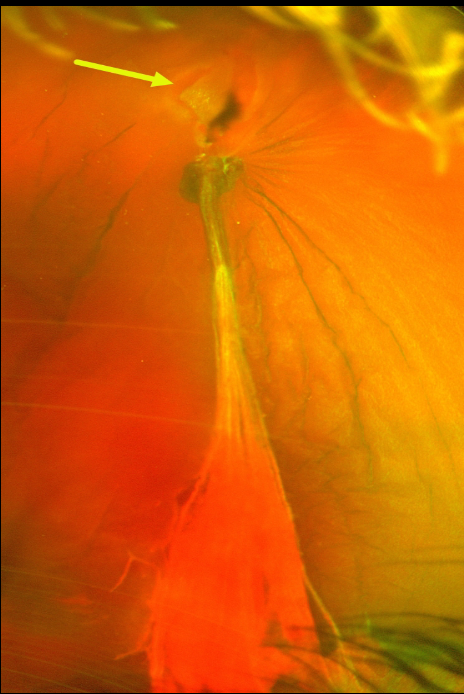
RETINAL BREAK
Originally posted on @retina.rocks August 22, 2022
This 50YO female presented with sudden vision loss and floaters in her left eye. Despite the presence of the vitreous hemorrhage, we were able to adequately visualize the causative break and surround it with cryotherapy (not pictured).
A week later, the vitreous hemorrhage increased, and vitrectomy was performed. At the time of surgery, the vitreoretinal traction on the causative tear and blood vessel was relieved. The retina was attached without other breaks. Vision improved to 20/20 five days postoperatively.
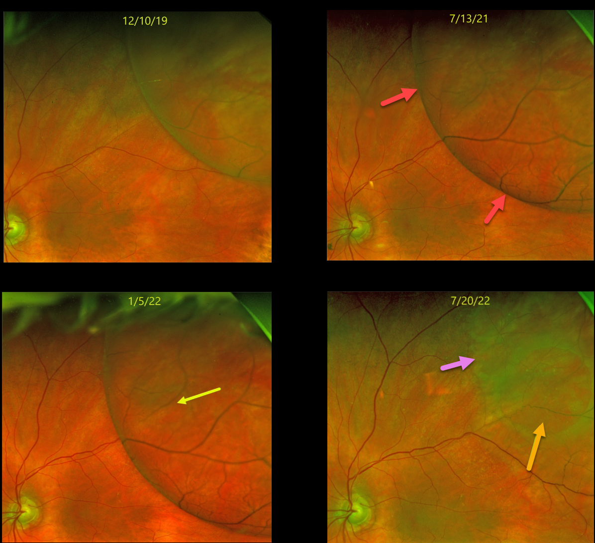
DEGENERATIVE RETINOSCHISIS
Originally posted on @retina.rocks August 9, 2022
This 80YOF was initially seen with asymptomatic retinoschisis in her left eye. When examined 1.5 years later on 7/13/21, she remained asymptomatic, but the schisis cavity had enlarged in all dimensions.
Another six months later (1/5/22), the schisis cavity continued to enlarge in all dimensions with the formation of an early outer layer hole. Most recently (7/20/22), the outer-layer hole enlarged, and the schisis spontaneously collapsed, essentially resolving itself. Continued observation was recommended.
Learning Points:
Schisis cavities can enlarge, and in these cases, prophylactic treatment, including thermal laser, does not prevent further spread. Schisis very rarely progresses into the fovea (Byer Ophthalmology 1986;93:1127-1137) and even then is compatible with preserved central vision as long as the outer layer remains attached (Watzke et al 2013;33:606-612).
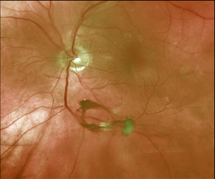
POSTERIOR VITREOUS DETACHMENT (PVD)
Originally posted on @retina.rocks August 5, 2022
This 67YO male presented with a symptomatic posterior vitreous detachment (PVD). A prominent Weiss ring is beautifully captured in this Optos image.
His peripheral retina was normal, and he was asked to return in a few months unless he developed new flashes, floaters, or vision loss.
Learning Points:
The clinical diagnosis for a complete PVD can be established with biomicroscopy when a Weiss ring, representing avulsed peripapillary glial tissue, is visualized. OCT can be helpful in supplementing the clinical exam (see Wagley et al, Retina 2021;41:2296-2300).
An acute PVD is the moment of truth for when a retinal break or detachment develops. The historical risk for a break is at least 10-15%, although a recent large retrospective study from Kaiser Permanente found this risk to be significantly lower at 5% for a retinal break and 4% for retinal detachment (Ophthalmology 2022;129;67-72).
The presence of vitreous pigment or blood and lattice, and a history of a retinal break or detachment in the fellow eye, increases this risk.
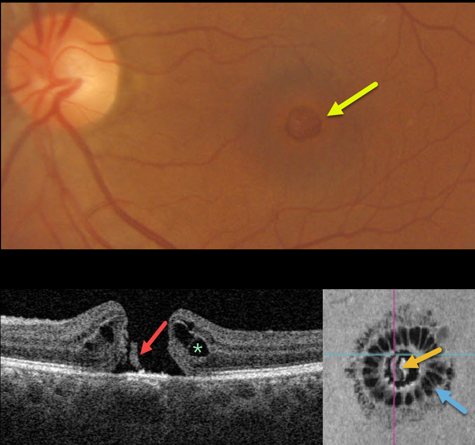
MACULAR HOLE
Originally posted on @retina.rocks August 4, 2022
This 51YO presented with counting-fingers vision secondary to a large (600-micron), full-thickness macular hole.
Swept-source OCT shows the classic appearance of a hole from a foveal dehiscence, with cystic thickening. A small remnant of the outer retina is adherent to the underlying RPE.
Vitrectomy surgery was recommended.
Learning Points:
En face imaging shows a unique and beautiful perspective of the hole. As described by Govetto et al (AJO 2020;212:43-56), idiopathic macular holes have a sunflower appearance showing features of both exudative and tractional cystoid spaces.
The macular hole is hyporeflective centrally with multiple hyporeflective cystoid spaces radiating from the center.
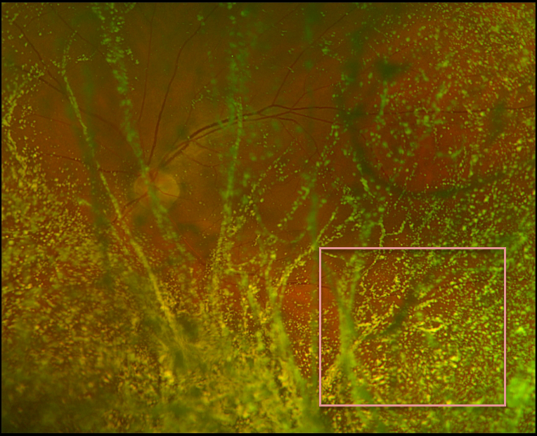
ASTEROID HYALOSIS
Originally posted on @retina.rocks June 29, 2022
This 72YO male presented with unilateral asteroid hyalosis in his left eye. Vision was 20/40 OS with mildly symptomatic floaters. Retinal examination was extremely difficult due to the dense asteroid, although the fundus appeared normal.
Learning Points:
Asteroid hyalosis is caused by calcium-lipid complexes suspended throughout the vitreous. The condition gets its name from the white opacities resembling stars on a night sky.
Even with dense opacities, patients are rarely symptomatic, although retinal examination can be difficult. If a patient has vision loss with asteroid, the cause is almost always due to another condition.
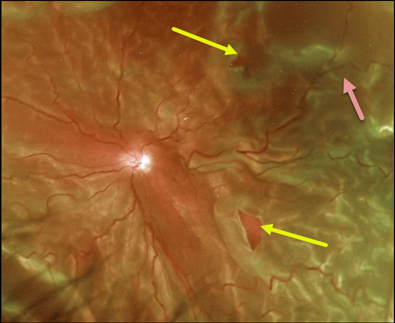
PROLIFERATIVE VITREORETINOPATHY (PVR)
Originally posted on @retina.rocks June 17, 2022
This 65YO male presented with light perception in his left eye secondary to a very chronic total rhegmatogenous retinal detachment (RRD).
Optos color imaging captured the detachment, which was fairly stiff due to diffuse proliferative vitreoretinopathy (PVR). Two posterior retinal breaks are seen.
The retina was successfully reattached with vitrectomy, inferior retinectomy, and silicone oil.
Learning Points:
PVR, which consists of cellular membranes growing on either retinal surface, develops in about 5-10% of retinal detachments. The membranes contract, foreshorten the retina, and likely proliferate as an aberrant wound-healing response. There are no proven pharmacologic therapies to prevent or treat PVR.
These complex detachments can be managed with vitrectomy with membrane peeling, retinectomy, scleral buckling, and silicone oil. However, PVR remains the main cause for ultimate retinal reattachment surgical failure.
See Garweg et al Surv Ophthalmol 2013;48:321-329 for a good review of PVR pathophysiology.
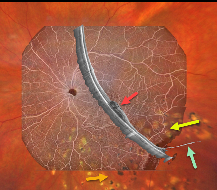
BRVO WITH DEGENERATIVE RETINOSCHISIS
Barbara Parolini and Veronika Matello
Originally posted on @retina.rocks June 13, 2022
This 67YO female has a history of branch retinal vein occlusion (BRVO) in her left eye. Asymptomatic degenerative retinoschisis was also present bilaterally. Vision was 20/33. She had received prior scatter laser and intravitreal injections about 3 years earlier.
Fortunately, all findings remained stable despite loss to follow-up due to the COVID pandemic.
Clarus 500 wide field imaging shows mild inferotemporal tortuosity with collateral vessels within the involved quadrant. Prior scatter laser scarring for secondary retinal neovascularization, along with prior barrier laser around the posterior edge of the schisis cavity, is noted.
Canon Xephilio S1 OCT angiography highlights the collateral vessels and more peripheral retinal nonperfusion. Xephilio B-scan shows some inferotemporal macular edema, and just catches the more distal inferotemporal schisis.
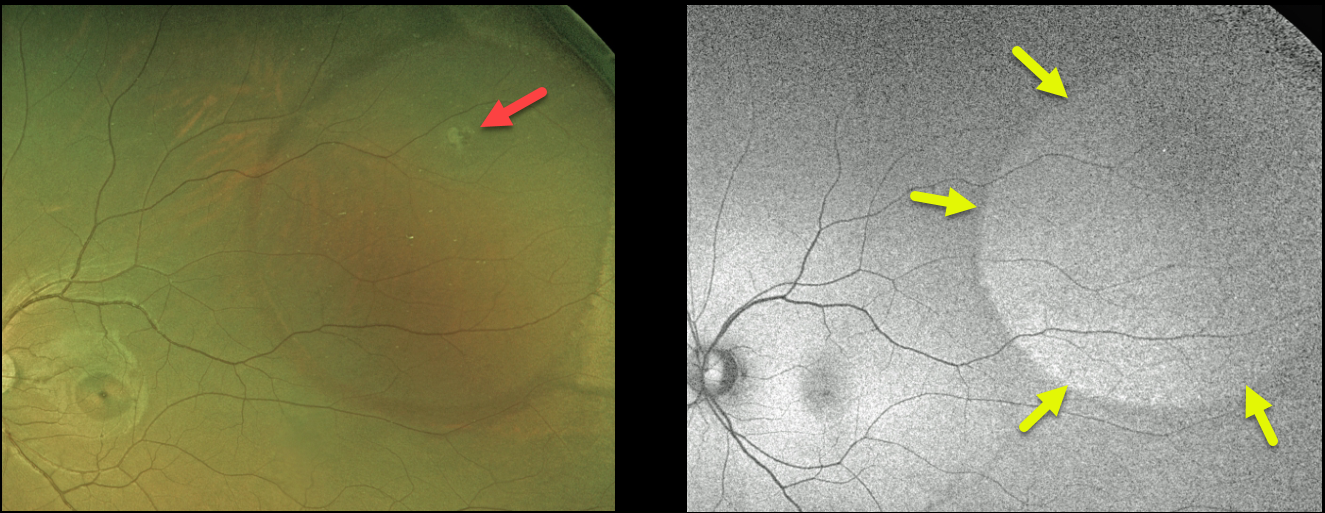
RHEGMATOGENOUS RETINAL DETACHMENT
Originally posted on @retina.rocks June 8, 2022
This 25YO female was referred for an asymptomatic rhegmatogenous retinal detachment (RRD) in her left eye. The detachment had an unusual oval shape that did not reach the ora, with a small causative break.
Fundus autofluorescence (FAF) shows variable hyper-FAF throughout the detachment, likely due to unmasking of the underlying RPE by dysfunctional photoreceptors.
Various treatment options were discussed, and the patient opted for thermal laser demarcation.
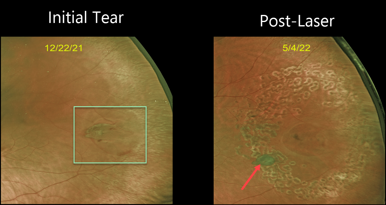
RETINAL TEAR
Originally posted on @retina.rocks May 19, 2022
This patient presented with new flashes and floaters in their left eye. Optos imaging shows a tractional retinal tear with a few dots of blood and localized subretinal fluid.
Barrier laser was performed to reduce the risk of retinal detachment. Six months later, good laser scarring surrounds the break, which has operculated.
In our experience, many of these tears will operculate following laser photocoagulation.
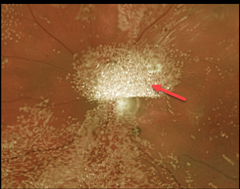
EMULSIFIED SILICONE OIL
Originally posted on @retina.rocks May 12, 2022
Eight years earlier, this 42YO female underwent successful retinal reattachment surgery elsewhere with scleral buckling, vitrectomy, and silicone oil. Vision was light perception.
Optos imaging shows extensive droplets of emulsified silicone oil.
We decided to observe our patient due to the long history of profound vision loss in a pain-free eye.
Learning Points:
Silicone oil as an adjunct for repairing complex retinal detachments was introduced by Paul Cibis in the early 1960s (Cibis et al, Arch Ophthalmol 1962;68:590-599). It is used primarily for complex detachments at high risk of failure, including severe proliferative vitreoretinopathy and viral retinitis.
Although silicone oil is generally well-tolerated as a long-term tamponade, it can rarely emulsify, resulting in numerous tiny opaque droplets.
The development of emulsification is multifactorial, including the properties of the oil, the surgical procedure, and postoperative factors.
When severe, the emulsified droplets can cause significant vision loss and may require removal. The droplets can also migrate into the anterior chamber, causing a ‘reverse hypopyon’ with secondary elevated intraocular pressure or corneal decompensation.
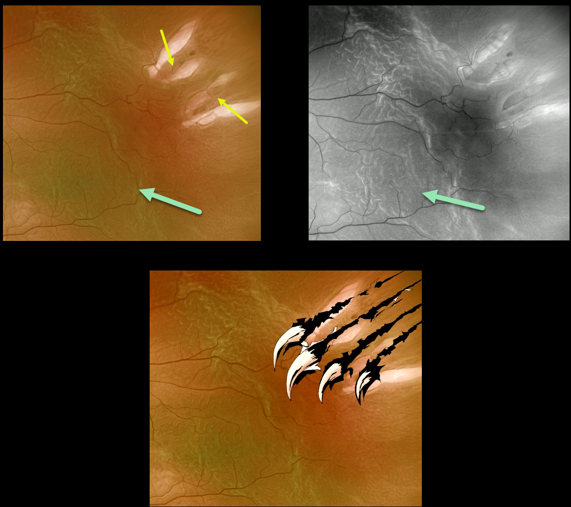
WOLVERINE RETINOPATHY
Originally posted on @retina.rocks May 10, 2022
This 72YO male presented with a macula-off rhegmatogenous retinal detachment (RRD).
Optos imaging gives the illusion of four breaks, although in actuality, there are only two tractional tears. The hydrated outer retinal folds are best visualized on green channel imaging.
Vision was 20/60, and the retina was successfully reattached with emergent pars plana vitrectomy.
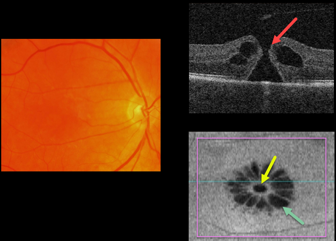
MACULAR HOLE
Originally posted on @retina.rocks May 5, 2022
This 63YO male patient has a full-thickness macular hole (MH) with 20/80 vision.
The hole is not well imaged in the color photograph.
OCT B-scan shows a small MH without traction.
The en face image beautifully depicts a unique perspective on an otherwise unremarkable case. The macular hole is hyporeflective centrally with multiple hyporeflective cystoid spaces radiating from the center.
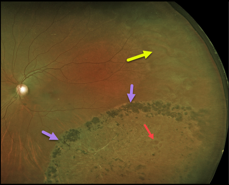
RHEGMATOGENOUS RETINAL DETACHMENT
Originally posted on @retina.rocks April 15, 2022
Years earlier, this 44YO female underwent laser demarcation of an asymptomatic inferotemporal rhegmatogenous retinal detachment (RRD). The prior detachment remains well demarcated, but the detachment itself has completely flattened.
Some of the causative lattice-related atrophic holes are seen within the area of prior detachment. White without pressure is also noted in the temporal periphery.
Learning Points:
Demarcation, rather than reattachment, of the retina is a valid option for select cases of RRD. When demarcating these detachments, it is critical that the retinopexy completely surrounds the subretinal fluid and that treatment extends to the ora; otherwise, the detachment can spread through the untreated retina.
Patients need to be followed postoperatively for life since the detachment can occasionally progress through the prior retinopexy.
Interestingly, we have seen that some of these detachments, as occurred in this case, spontaneously flatten following demarcation.
See Vrabec and Baumal, Ophthalmology 2000;107:1063-1067 for an excellent review regarding demarcation laser photocoagulation for selected macula-sparing retinal detachments.
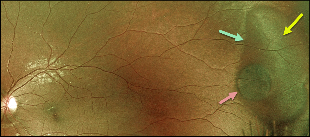
DARK WITHOUT PRESSURE + CHRPE
Originally posted on @retina.rocks April 7, 2022
This 22YO female presented with a large temporal patch of dark without pressure (DWP), white without pressure (WWP), and a small round patch of congenital hypertrophy of the RPE (CHRPE). None of these findings is of concern, but we thought it was cool that all of them are associated with each other at this single location.
Learning Points:
WWP and DWP are relatively common benign peripheral findings most commonly found in darkly pigmented individuals. Typically, there are bilateral peripheral geographic areas of whitish (WWP) or dark (DWP) retinal discoloration.
WWP gets its name because the area is white without indentation, such as during scleral depression (white with pressure). It can sometimes be confused with retinal breaks or detachment.
The exact cause of this phenomenon is unknown. Historically, it was thought to be due to a benign vitreoretinal interface change, but more recent OCT findings show increased (WWP) or decreased (DWP) reflectivity in the outer retina (see Diaz et al., Retina 2014;34:1020-1021).
Although CHRPE lesions can enlarge with time (see Shields et al, Ophthalmology 2003;110:1968-1973), they carry virtually no malignant potential. Histologic RPE hypertrophy presents in several ways.
The typical isolated CHPRE lesions (this lesion) and bear tracks are not associated with intestinal polyps or cancer, and thus require no systemic evaluation. Only the multiple, small comet-shaped lesions are part of the familial polyposis spectrum.
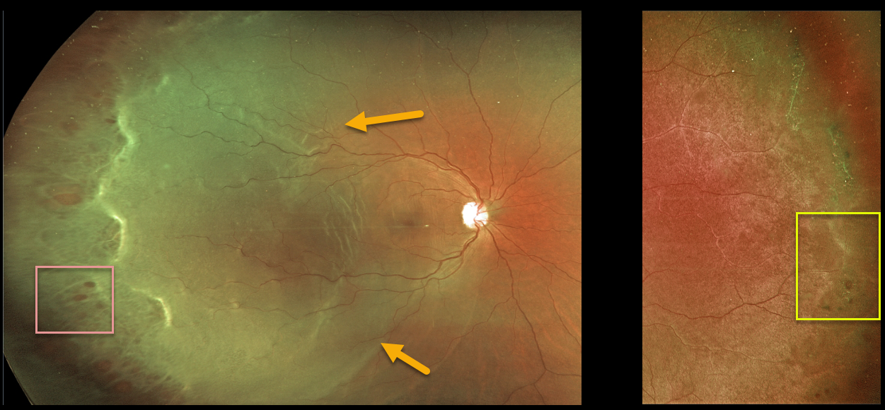
RHEGMATOGENOUS RETINAL DETACHMENT
Originally posted on @retina.rocks April 1, 2022
This 32YO female presented with a macula-off rhegmatogenous retinal detachment (RRD) in her right eye. There were innumerable temporal retinal holes.
The detachment was successfully repaired with vitrectomy surgery.
Her asymptomatic left eye showed extensive temporal lattice lesions with atrophic holes, and prophylactic laser was recommended.
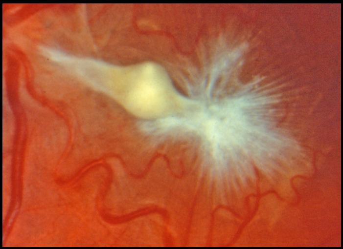
MACULAR PUCKER FOLLOWING ENDOPHTHALMITIS
Originally posted on @retina.rocks March 31, 2022
This eye shows a severely contracted and complex epimacular membrane in a patient who recovered from prior endophthalmitis. Unfortunately, we don’t have any further clinical information.
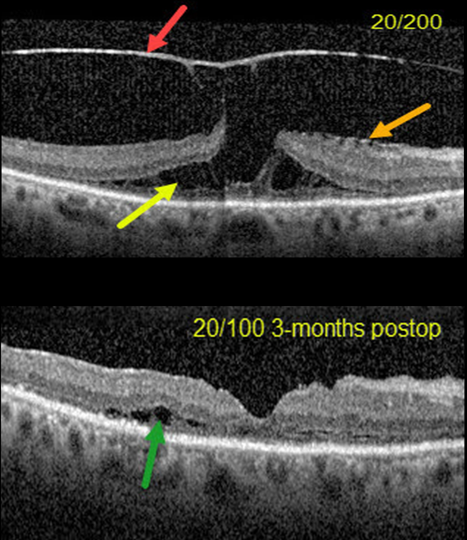
VITREOMACULAR TRACTION + LAMELLAR MACULAR HOLE
Erdem Dinç
Originally posted on @retina.rocks March 29, 2022
This 70YO female presented with 20/200 vision in her right eye from severe vitreomacular traction with a tractional lamellar macular hole (LMH).
OCT scanning shows a highly elevated posterior hyaloid adherent to the underlying fovea, with tractional splitting of the outer plexiform layer (OPL). There is also a faint nasal epiretinal membrane.
Combined cataract surgery, 23-gauge pars plana vitrectomy, membrane peeling, temporal half-moon inverted ILM flap, and fluid-air-C3F8 exchange was performed.
Three months postoperatively, the tractional LMH was closed with 20/100 acuity. There is still some temporal OPL splitting along with disorganization and thinning of the outer macular layers.
Learning Points:
LMH can be defined as tractional or degenerative (see Govetto et al, AJO 2016;164:99-109). Tractional LMH shows OPL splitting, an intact ellipsoid zone, and is associated with vitreomacular traction and tractional epiretinal membranes.
OCT features of a degenerative LMH include loss of outer retinal tissue below the OPL and an outer retinal ‘bump’. Non-tractional epimacular proliferation is often present and will often extend around the posterior edge of the degenerative LMH.
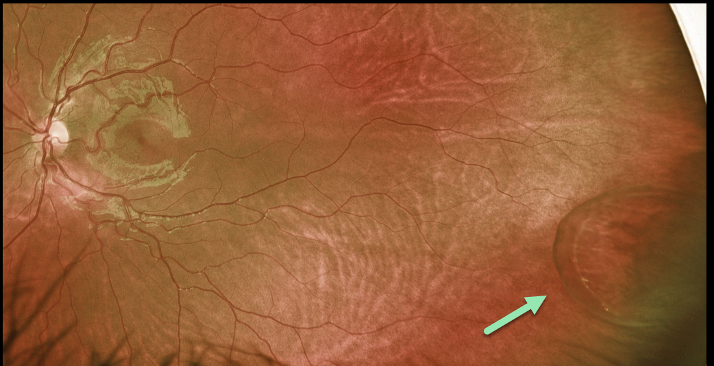
PARS PLANA CYST
Originally posted on @retina.rocks March 15, 2022
This 9YO female was referred for an asymptomatic lesion in her left eye. She was found to have a large pars plana cyst in the temporal periphery.
Learning Points:
Pars plana cysts are localized to the pars plana region, commonly along a single ora bay. They form when the epithelial cell layers separate between the non-pigmented ciliary epithelium and pigmented epithelium, or solely within the pigmented epithelium.
Although they are benign findings, they need to be differentiated from retinal detachment or retinoschisis.
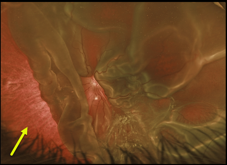
GIANT RETINAL TEAR
Originally posted on @retina.rocks March 9, 2022
This 6YO boy was found to have this asymptomatic chronic total rhegmatogenous retinal detachment (RRD) in his left eye during a routine eye examination.
There is a large, giant tear nasally from about 6 to 11 o’clock, with an additional giant tear temporally (not shown). There were numerous areas of lattice degeneration with atrophic holes in his right eye (not shown). There was a strong family history of RRD.
He was referred for surgical repair of the left detachment and prophylactic laser in his right eye.
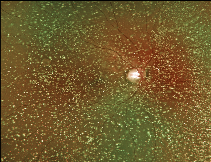
ASTEROID HYALOSIS
Originally posted on @retina.rocks March 7, 2022
This 22YO male presented with bilateral asteroid hyalosis. Vision was 20/20 OU with mildly symptomatic floaters.
Retinal examinations were extremely difficult due to the dense asteroid, although the fundi appeared normal.
Learning Points:
Asteroid hyalosis is caused by calcium-lipid complexes suspended throughout the vitreous. The condition gets its name from the white opacities resembling stars on a night sky.
Even with dense opacities, patients are rarely symptomatic, although retinal examination can be difficult. If a patient has vision loss with asteroid, the cause is almost always due to another condition.
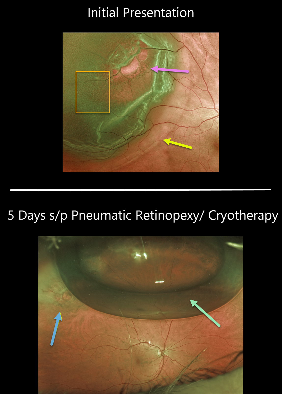
RHEGMATOGENOUS RETINAL DETACHMENT
Originally posted on @retina.rocks February 25, 2022
This 63YO female presented with 20/25 vision and a superotemporal retinal detachment. The posterior edge of the detachment was just outside the macular center. There was diffuse fine microcystic retinal edema within the area of detachment, except for outer retinal hydration folds more posteriorly.
The retina was completely attached 5 days later following successful cryotherapy with pneumatic retinopexy. Early cryotherapy scarring is already visible.
Learning Points:
This detachment could have been repaired with any technique, including vitrectomy, scleral buckling, or pneumatic retinopexy. All techniques yield similar final anatomic success, although the debate continues about the relative merit of one over the other.
See the ___References folder in the Retinal detachment (RD) rhegmatogenous (RRD) folder in the Image Library at www.retinarocks.org for a summary of the key journal articles.
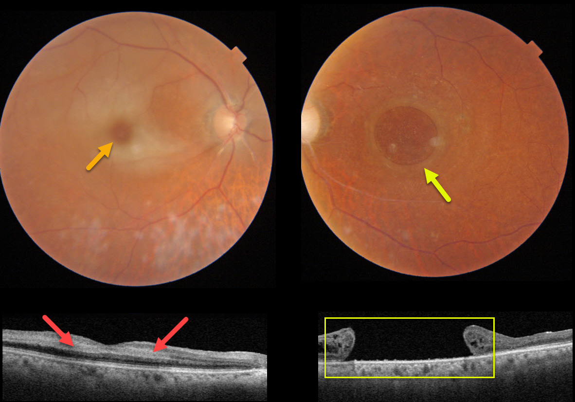
CRAO + MACULAR HOLE
Originally posted on @retina.rocks February 21, 2022
This 82YO female presented with counting fingers vision in her previously healthy right eye. Vision in her left eye had been counting fingers for many years due to a large chronic macular hole measuring 2,870 microns.
An acute central retinal artery occlusion (CRAO) was noted in her right eye. The foveal retina is still perfused by the underlying choroid, resulting in the classic cherry red spot.
Swept-source OCT shows hyperreflectivity from the opaque, ischemic inner retinal layers.
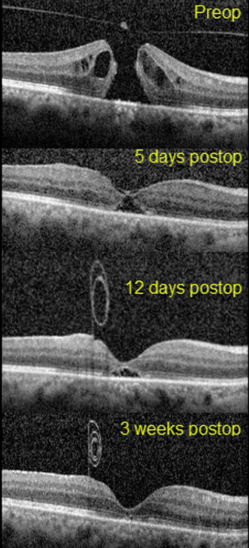
MACULAR HOLE
Erdem Dinç
Originally posted on @retina.rocks February 15, 2022
This 51yo presented with 20/1250 vision in her right eye from a primary full-thickness macular hole without traction measuring 407 microns. There was also a moderately severe nuclear sclerotic cataract.
Combined cataract surgery with 23-gauge pars plana vitrectomy, temporal half-moon inverted ILM flap, and fluid-air-SF6 exchange was performed, followed by prone positioning.
On the 5th postoperative day, the flap extended over the closed hole, but one week later, the ILM flap retracted and was rolled inward, suspended anterior to the macula.
Three weeks following vitrectomy, the hole was completely flattened with partial restoration of the outer retinal layers. The ILM flap continued to contract and scroll onto itself. Vision was 20/40.
Learning Points:
Introduced by Kelly and Wendel (Arch Ophthalmol 1991;109:654-659), macular hole surgery is one of the modern success stories in ophthalmology. ILM flaps appear to improve the surgical success for large (>400 microns) macular holes.
In this case, the ILM flap inadvertently scrolled onto itself and away from the macular hole by post-op day 12. Some authors have suggested that the ILM flap provides a scaffold for cells to grow over and help heal the hole.
However, most macular holes are actually foveal dehiscences without tissue loss, where the foveal retina splits and elevates like a drawbridge (Tornambe, Retina 2003;23:421-424). For successful surgery, there is thus no need or significant evidence for glial or neurosensory retina regrowth, just for the “drawbridge” to close.
As this case exemplifies, the ILM flap most likely serves the same purpose as the gas bubble, helping to sequester the macular hole from the overlying vitreous. This allows the RPE to actively pump (and the choroid to passively absorb) the subretinal fluid, allowing the foveal tissue to flatten into its normal position. Once the hole closes, as long as the vitreous and epimacular traction are relieved, there is likely no need for the ILM flap.
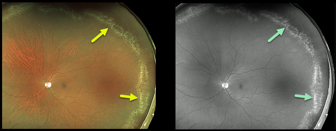
LATTICE DEGENERATION
Originally posted on @retina.rocks February 8, 2022
This 36YO male presented with extensive asymptomatic lattice degeneration seen on Optos color photography and Optos green channel.
The circumferential glistening lattice lesions are also called snail tracks. Treatment was not recommended.
Learning Points:
White, fibrosed vessels often crisscross the lesions, which gives rise to the term “lattice.” There is vitreous liquefaction overlying the thinned retina with a firm vitreo-retinal adhesion along the borders of the lesions. This firm vitreoretinal adhesion, along with the often-present atrophic holes, can predispose these eyes to retinal detachment.
Retinal detachment occurs in 1 in 10,000 people per year. Lattice is found in 10% of the general population, especially in high myopes. Since about one-third of patients with retinal detachment have lattice as the cause, the question always arises, especially in patients like this: Should prophylactic retinopexy be performed? The short answer is that asymptomatic lattice generally should not be treated.
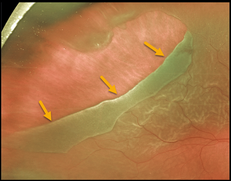
GIANT RETINAL TEAR
Originally posted on @retina.rocks January 27, 2022
This 57YO male presented with 20/400 vision from a macula-off retinal detachment. The causative giant retinal tear extended from 9 to 12 o’clock.
He underwent successful pars plana vitrectomy surgery using intraoperative perfluorooctane (PFO) liquid to flatten the retina, followed by gas exchange with long-acting C3F8 (perfluoropropane) gas tamponade.
Learning Points:
By definition, giant retinal tears extend at least 3 clock hours. Surgical repair can be greatly facilitated by using Perfluoron (PFO), a heavier-than-water liquid that helps unroll the giant tear and prevents posterior retinal slippage.
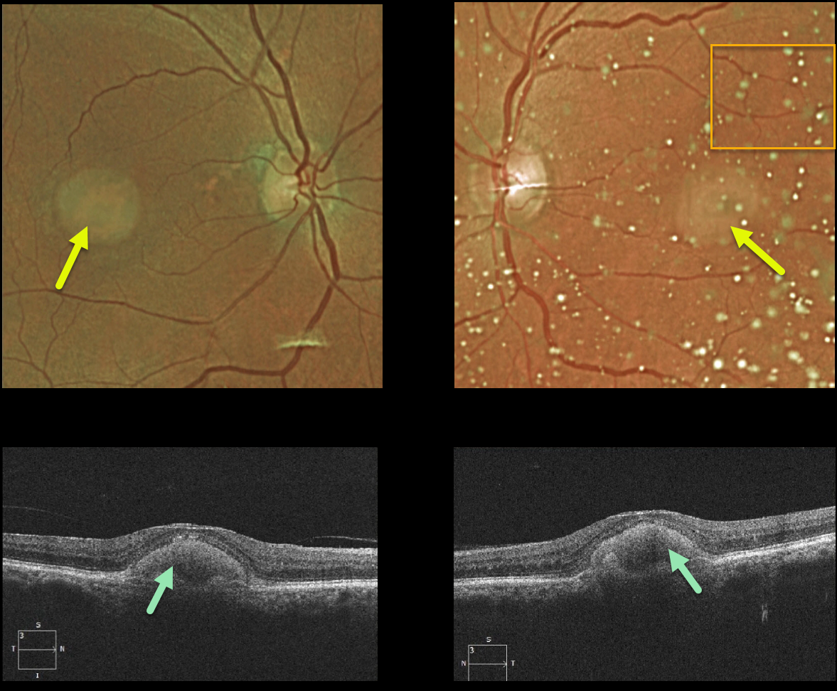
ADULT-ONSET FOVEOMACULAR VITELLIFORM DYSTROPHY
Originally posted on @retina.rocks January 18, 2022
This 87YO female shows classic findings for adult-onset foveomacular vitelliform dystrophy (Adult Best). Vision was 20/60 OD and 20/80 OS Optos imaging shows small, round, symmetrical subfoveal yellow lesions. Moderate asteroid hyalosis is noted as an incidental finding on the OS.
Triton swept-source OCT shows subretinal hyperreflective material above the RPE that extends into and indents the overlying retina.
Learning Points:
Adult Best is associated with a mutation in the BEST1 gene, which encodes the bestrophin-1 protein. Bestrophin-1, a calcium-activated chloride channel, is primarily found in the basolateral plasma membrane of the RPE.
BEST1 mutations cause a variety of phenotypes, including autosomal recessive bestrophinopathy, best vitelliform macular dystrophy, and autosomal dominant vitreoretinochoroidopathy.
These patients tend to maintain fairly good vision. Outer retinal and RPE atrophy can cause central vision loss, as can the rare development of macular neovascularization. Patients should therefore regularly monitor their vision with an Amsler grid.
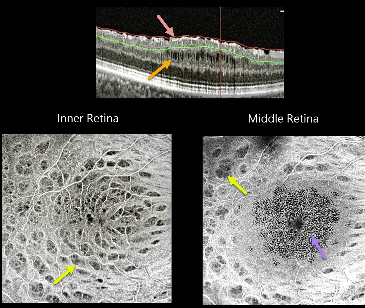
MACULAR PUCKER
Seif Anwar
Originally posted on @retina.rocks January 4, 2022
This 52YO pseudophakic female with an idiopathic macular pucker and 20/60 vision underwent pars plana vitrectomy (PPV) with internal limiting membrane (ILM) peeling. Vision improved to 20/30 after surgery, followed 6 months later by a gradual decrease to 20/50.
SOLIX OCT B-scan imaging shows multiple inner surface irregularities and thickening, with middle-layer cavitations.
En face OCT of the inner retina shows multiple concentric dark spots (dimples), characteristic of dissociated optic nerve fiber layer (DONFL). En face imaging through the mid-retinal region shows a central pattern of tiny hyporeflective cysts, with still-visible dimples more peripherally.
Learning Points:
PPV with membrane peeling is frequently performed to manage many vitreomacular interface disorders, including vitreomacular traction, macular hole, and epiretinal membrane.
While ILM peeling decreases epiretinal membrane recurrence and enhances macular hole closure, it also causes mechanical trauma to the inner retina, as evidenced by DONFL.
Many mechanisms have been proposed, including Müller cell damage from direct contact with surgical instruments, damage to the deep inner retinal layer, damage from the visualization dye, or tractional thinning of the temporal retina.
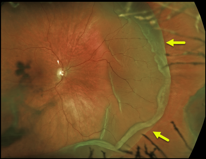
GIANT RETINAL TEAR
Originally posted on @retina.rocks December 24, 2021
This 54YO female presented with a giant retinal tear with macula-on rhegmatogenous retinal detachment.
At vitrectomy several hours later, the tear was found to extend for 190 degrees.
Learning Points:
By definition, giant retinal tears extend at least 3 clock hours. Surgical repair can be greatly facilitated by using Perfluoron (PFO), a heavier-than-water liquid that helps unroll the giant tear and prevents posterior retinal slippage.
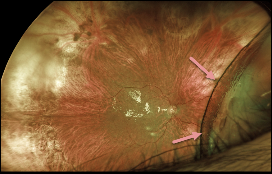
RHEGMATOGENOUS RETINAL DETACHMENT
Originally posted on @retina.rocks December 17, 2021
This patient presented with light perception vision following a 1-year-old total retinal detachment with proliferative vitreoretinopathy. At vitrectomy, all membranes were peeled, and the anterior loop traction was relieved (or so we thought). The retina was mobile and completely flattened with fluid-air exchange. 1000 centistoke silicone oil was infused.
At the one-day and one-week postoperative visits, vision remained light perception. The retina was completely attached except for the nasal periphery, which was locally detached with subretinal silicone oil.
Since the surgical goal was to prevent total blindness and possible phthisis, we have decided to observe for now as long as the residual detachment remains localized.
What would your management be at this point?
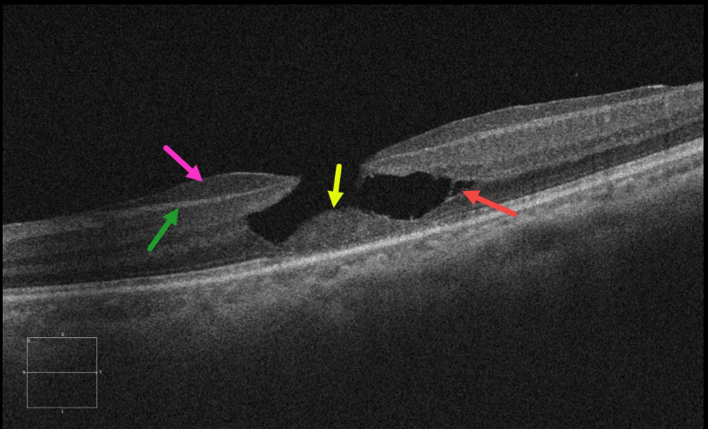
LAMELLAR HOLE
Originally posted on @retina.rocks December 15, 2021
This 62YO female presented with 20/80 vision in her left eye from an asymptomatic lamellar macular hole (LMH). Epiretinal proliferation is adherent to the underlying nerve fiber layer. A localized splitting of the outer plexiform layer (OPL) is seen temporally. There is a loss of tissue below the OPL with a central mound of remaining featureless outer retina.
Learning Points:
LMH can be defined as tractional or degenerative (see Govetto et al, AJO 2016;164:99-109).
Our case has all the classic findings of a degenerative LMH, which Govetto et al. described as resembling the brim of a top hat. The epimacular proliferation often extends around the posterior edge of the LMH, but this was not seen in our case.
Although surgery is usually observed, it can close these holes with some visual improvement (see Morescalchi et al, Retina, 2020;40:1087-1093).
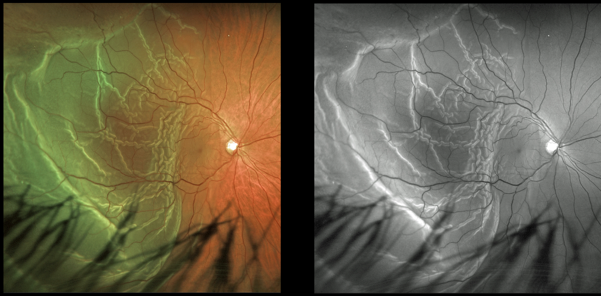
RHEGMATOGENOUS RETINAL DETACHMENT
Originally posted on @retina.rocks December 3, 2021
This 47YO male presented with hand motion vision from a macula-off rhegmatogenous retinal detachment (RRD). The outer retinal folds are dramatically imaged in both the Optos color and green channel images.
Learning Points:
Hydration outer retinal folds are one of the key findings to help differentiate a RRD from exudative and traction retinal detachments.
The cause of these folds remains uncertain but may result from the hydration of glycosaminoglycans in the interphotoreceptor matrix (Dalvin et al, Retinal Cases & Brief Reports 2020;14:355-359).
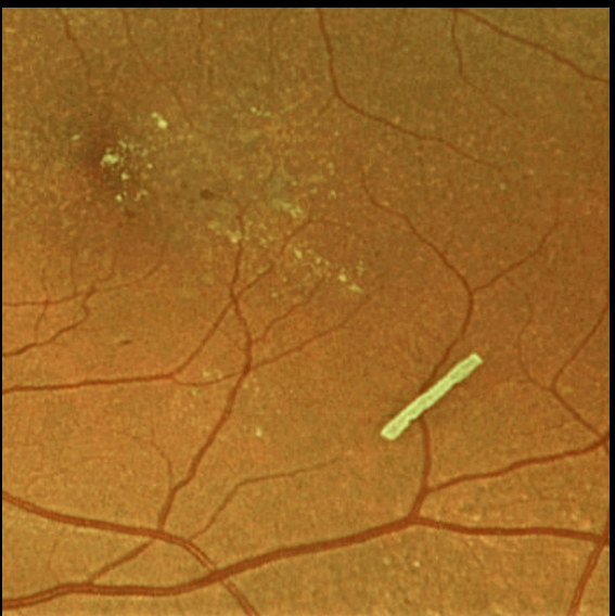
OZURDEX GHOST
Originally posted on @retina.rocks November 26, 2021
This 71YO female is getting Ozurdex intravitreal injections every 2 months to control her macular edema.
Learning Points:
The Ozurdex implant slowly releases the steroid and eventually totally absorbs, with the clinical effect usually lasting about 2 to 4 months.
The ghost-like remnants can persist for many months (see Kim et al, Retina 2020;40;2226-2231).
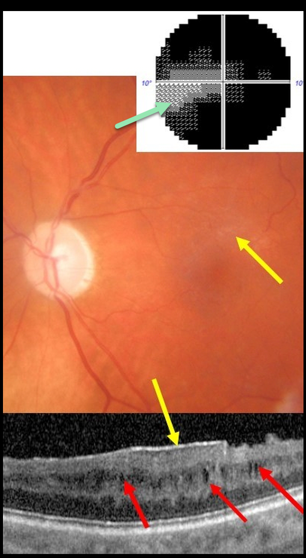
CYSTOID MACULAR EDEMA
Alex Hynes
Originally posted on @retina.rocks November 23, 2021
This 61YO male presented with end-stage glaucoma OS, 20/30 vision, and an IOP of 8 mmHg following successful trabeculectomy ten years earlier. He had prior cataract surgery in this eye 15 years ago with no documented history of pseudophakic cystoid macular edema.
There was a moderate macular pucker noted clinically and on OCT, along with multiple inner nuclear layer (INL) microcysts on OCT scanning. He has a tiny island remaining in his visual field.
Learning Points:
Microcystic macular changes have been associated with both advanced primary open-angle glaucoma and idiopathic macular pucker (see Govetto et al, AJO 2017;181:156-165).
Macular pucker-induced mechanical stress, coupled with retrograde trans-synaptic degeneration of bipolar cells secondary to retinal ganglion cell loss, likely predisposes towards the accumulation of fluid within the inner retinas of these patients.
Relative to non-glaucomatous eyes, microcystic macular changes in glaucomatous eyes tend to be more persistent following membrane peeling. This is likely due to retrograde trans-synaptic degeneration.
It is important that these microcystic spaces in eyes with optic neuropathy are not mistaken for inflammatory edema following membrane peeling, which could lead to unnecessary treatment.
Finally, disproportionate preservation of overall retinal thickness despite severe thinning of the ganglion cell and/or nerve fiber layers should not be surprising in glaucomatous eyes.
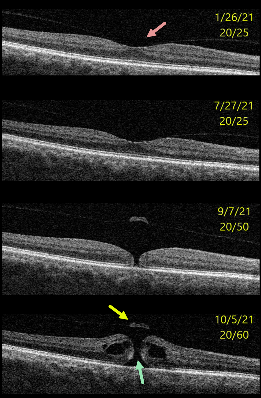
MACULAR HOLE
Originally posted on @retina.rocks November 9, 2021
This 64YO male presented with 20/25 vision and asymptomatic vitreomacular adhesion (VMA) in his right eye.
Over the next 8 months, this progressed to subtle focal vitreomacular traction (VMT) and eventually a small full-thickness macular hole without traction. Vitrectomy surgery was recommended.
Learning Points:
In 1988, Dr. Gass described his concept for the pathogenesis of idiopathic macular holes (Arch Ophthalmol 1988;106:629-639).
His classification system, from stage 1 for an impending macular hole through stage 4 for a fully developed macular hole with complete posterior vitreous detachment, was based on his concept of localized, mostly tangential traction caused by shrinking foveal vitreous cortex. This classic paper heralded the advent of modern macular hole surgery for a previously untreatable condition.
The advent of OCT technology, however, showed that macular hole development is driven more by focal vitreomacular traction, as our case beautifully illustrates.
The current classification system, described by the International Vitreomacular Traction Study Group, emphasizes the various characteristics that determine choice of treatment and visual prognosis (Ophthalmology 2013;120:2611-2619). This includes size (small <= 250 microns, medium >250 microns and <= 400 microns, large >400 microns), presence or absence of VMT, and cause (primary from VMT or secondary).
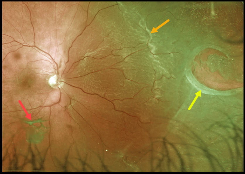
RHEGMATOGENOUS RETINAL DETACHMENT
Originally posted on @retina.rocks November 2, 2021
This patient presented with a macula-on rhegmatogenous retinal detachment (RRD) from a large nasal retinal tear. The tear has a rolled posterior edge, indicating possible early proliferative vitreoretinopathy.
The hydration outer retinal folds at the posterior edge of the detachment are one of the key clinical findings that help differentiate a rhegmatogenous from exudative and traction retinal detachments.
The causative posterior vitreous detachment is seen floating above the retina just inferior to the macula.
Learning Points:
The normal neurosensory retina remains attached to the RPE due to numerous physiological mechanisms, including the inner (retinal vascular endothelium) and outer (tight junctions at the apex of the RPE cells) blood retinal barriers, hyperosmotic choroid passively drawing fluid into the subretinal space, RPE pump, interphotoreceptor matrix, and friction between the RPE microvilli and the photoreceptor outer segments.
Rhegmatogenous retinal detachment (RRD) occurs when liquefied vitreous, entering the subretinal space through a retinal break, overwhelms the forces that favor retinal detachment.
Repairing RRD involves finding and closing all breaks, essentially denying the liquid vitreous access to the subretinal space. The break(s) can be closed internally with a gas bubble with pneumatic retinopexy or vitrectomy surgery, or externally by sewing a piece of silicone material to the eye wall (scleral buckle). This indents, or buckles, the sclera up against the retina break.
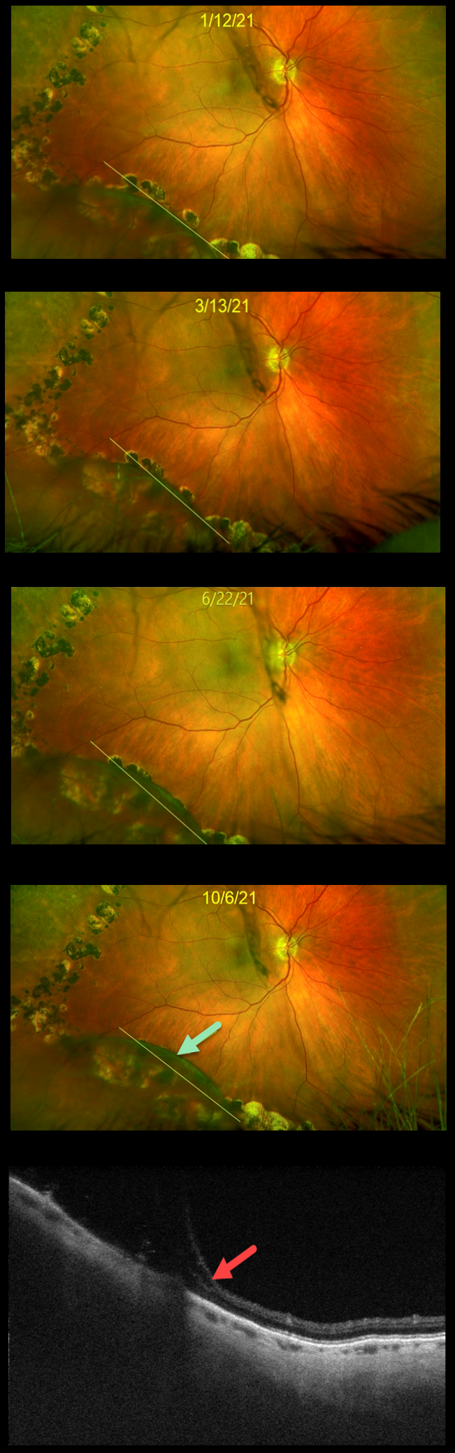
DEGENERATIVE RETINOSCHISIS
Originally posted on @retina.rocks October 27, 2021
This 71YO female was treated elsewhere with barrier laser around degenerative schisis in her right eye. Over the past 9 months, the inferotemporal schisis cavity has gradually enlarged. The yellow line indicates the original location of schisis. At the most recent visit on 10/6/21, the schisis cavity extended through the barrier laser.
Triton swept-source OCT scanning through this area confirmed pure schisis with no retinal detachment. She remains completely asymptomatic, and we continue to recommend observation.
Learning Points:
This patient is at virtually no risk of a combined schisis-rhegmatogenous detachment due to the absence of inner and outer layer breaks.
Schisis cavities can enlarge, and in these cases, prophylactic treatment, including thermal laser, does not prevent further spread.
Schisis very rarely progresses into the fovea (Byer Ophthalmology 1986;93:1127-1137) and even then is compatible with preserved central vision as long as the outer layer remains attached (Watzke et al 2013;33:606-612).
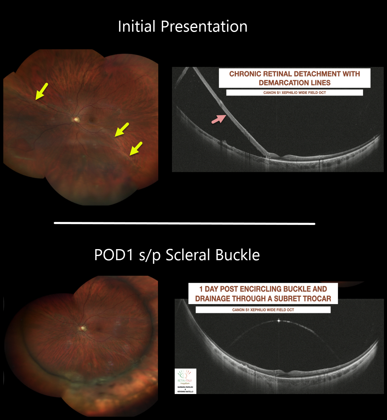
SCLERAL BUCKLE
Barbara Parolini and Veronika Matello
Originally posted on @retina.rocks September 30, 2021
This 36YO male presented with 20/20 vision despite a chronic rhegmatogenous retinal detachment with a demarcation line of his left eye. The wide-field OCT shows the chronic retinal detachment.
The patient underwent scleral buckling with drainage of subretinal fluid through a trocar. The retina is completely attached on post-operative day one.
Learning Points:
The normal neurosensory retina remains attached to the RPE due to numerous physiologic mechanisms, including the inner and outer blood-retinal barriers, the hyperosmotic choroid passively drawing fluid into the subretinal space, the RPE pump, the interphotoreceptor matrix, and friction between the RPE microvilli and the photoreceptor outer segments.
Rhegmatogenous retinal detachment (RRD) occurs when liquid vitreous, entering the subretinal space through a retinal break, overwhelms these forces, favoring retinal detachment.
Repairing RRD involves finding and closing all breaks, essentially denying the liquid vitreous access to the subretinal space. The break(s) can be closed internally with a gas bubble via pneumatic retinopexy or vitrectomy surgery, or externally by securing a piece of silicone material to the sclera (scleral buckle). This indents, or buckles, the RPE against the retinal break.
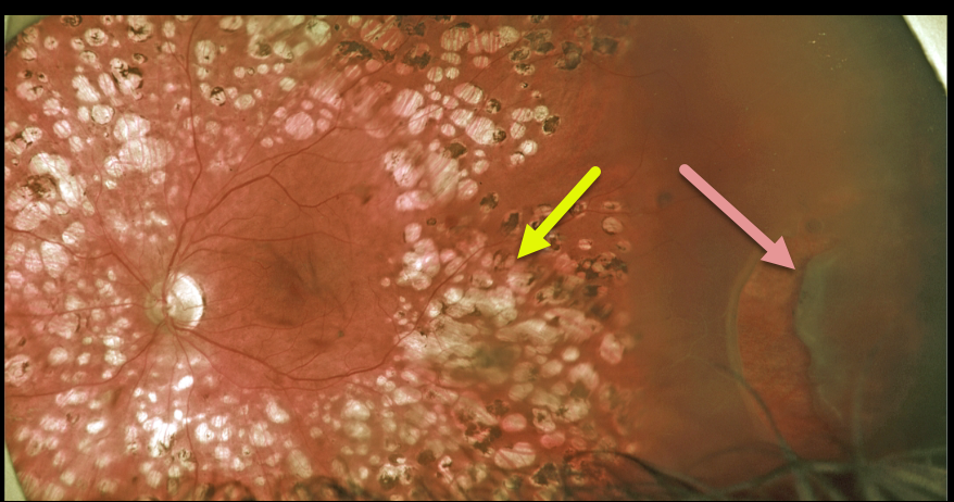
RHEGMATOGENOUS RETINAL DETACHMENT
Originally posted on @retina.rocks September 22, 2021
This 50YO male presented for a routine diabetic follow-up. A new asymptomatic peripheral retinal tear causing a rhegmatogenous retinal detachment was noted anterior to full panretinal photocoagulation (PRP) scarring.
We will often demarcate asymptomatic retinal detachments with laser photocoagulation (Vrabec and Baumel, Ophthalmology 2000;107:1063-1067). This patient essentially had laser demarcation before his retinal detachment, but the end result was the same, with his detachment being contained by the laser scarring.
Learning Points:
We opted to treat this eye with vitrectomy, but one could argue that this detachment could also be closely followed since it was contained by the prior laser.
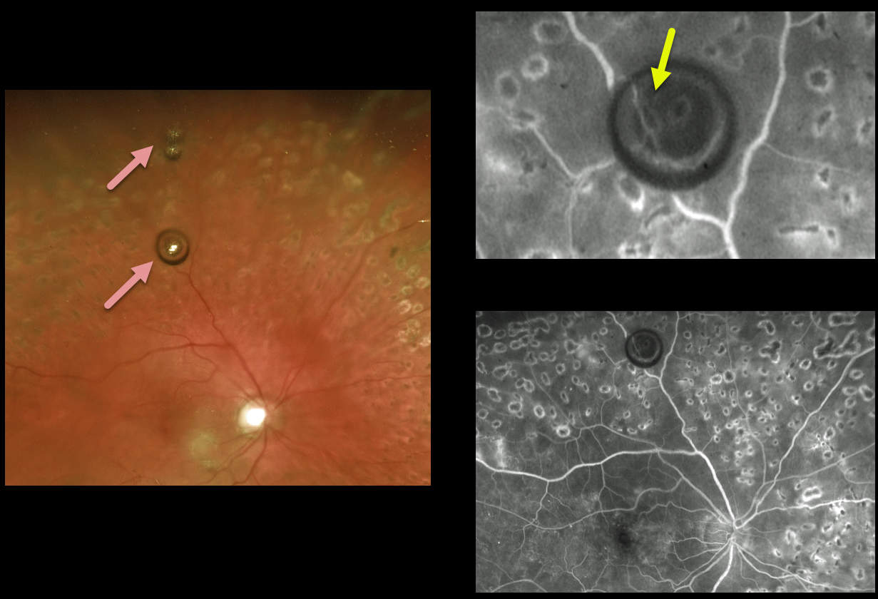
SILICONE OIL DROPLET
Originally posted on @retina.rocks September 20, 2021
This patient with involuted proliferative diabetic retinopathy received prior intravitreal Avastin injections for diabetic macular edema.
On routine examination, a few asymptomatic silicone oil bubbles were suspended in the superior vitreous.
Fluorescein angiography best shows how these bubbles distort and minify the underlying retinal detail.
Learning Points:
The insulin syringe needles provided by compounding pharmacies for Avastin injections are lubricated with silicone.
Many eyes receiving ongoing intravitreal Avastin developed these vitreous silicone droplets, most of which were completely asymptomatic.
Once knowledge of these silicone droplets became available, use of these insulin syringes was discontinued.
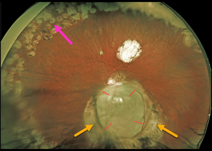
CHOROIDAL COLOBOMA
Originally posted on @retina.rocks September 10, 2021
This 57YO male underwent successful vitrectomy surgery 8 years earlier for rhegmatogenous retinal detachment (RRD). Vision is now 20/40.
The detachment was caused by a retinal break associated with an inferior choroidal coloboma. The margins of the coloboma are surrounded by confluent depigmented scarring from prior laser photocoagulation. Prophylactic peripheral laser was also applied intraoperatively.
Learning Points:
Choroidal colobomas arise from incomplete closure of the embryonic fissure during the second month of embryonic development.
Patients are at lifelong risk of rhegmatogenous retinal detachment due to breaks in the overlying thinned retina.
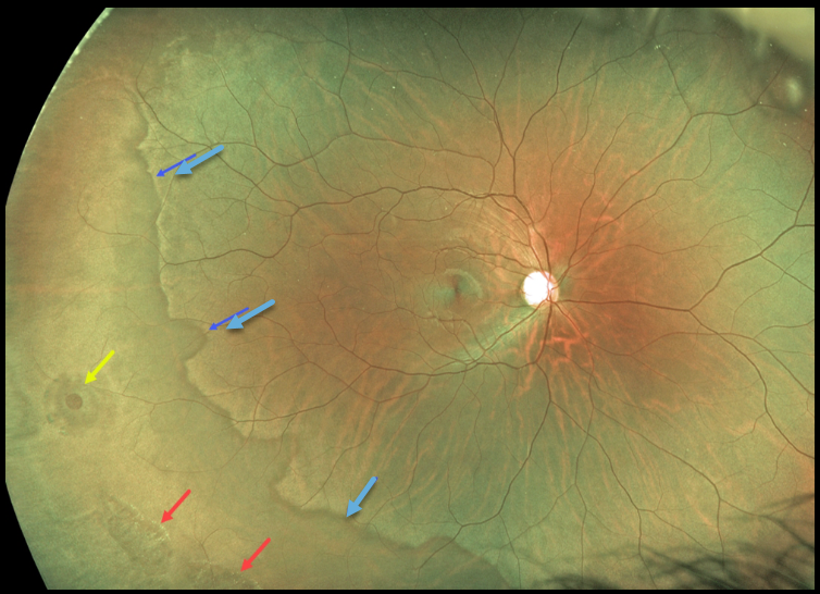
DARK WITHOUT PRESSURE
Originally posted on @retina.rocks August 31, 2021
This 22YO male presented with bilateral dark without pressure, lattice lesions, and atrophic holes. He is asymptomatic, and prophylactic treatment was not recommended.
Learning Points:
White without pressure (WWP) and dark without pressure (DWP) are relatively common benign peripheral findings most commonly found in darkly pigmented individuals. Typically, there are bilateral peripheral geographic areas of whitish (WWP) or dark (DWP) retinal discoloration.
WWP gets its name because the area appears white without indentation, unlike scleral depression (white with pressure). It can sometimes be confused with retinal breaks or detachment. The exact cause of this phenomenon is unknown.
Historically, it was felt to be due to a benign vitreoretinal interface change, but more recent OCT findings show increased (WWP) or decreased (DWP) reflectivity in the outer retina (see Diaz et al, Retina 2014;34:1020-1021).
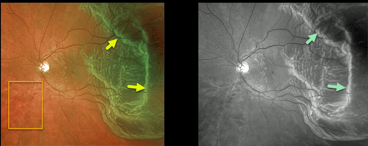
RHEGMATOGENOUS RETINAL DETACHMENT
Originally posted on @retina.rocks August 30, 2021
This 67YO male presented with hand motion vision from a macula-off rhegmatogenous retinal detachment (RRD).
The outer retinal folds are dramatically imaged in both the Optos color and green channel images. An incidental, old asymptomatic branch retinal vein occlusion is also noted inferonasally.
Learning Points:
Hydration outer retinal folds are one of the key findings to help differentiate a RRD from exudative and traction retinal detachments.
The cause of these folds remains uncertain, but they may form as a result of hydration of glycosaminoglycans in the interphotoreceptor matrix (Dalvin et al., Retinal Cases & Brief Reports 2020;14;355-359).
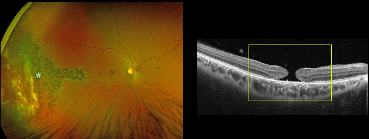
MACULAR HOLE
Originally posted on @retina.rocks August 24, 2021
This 31YO male was hit in his right eye by a paintball when he was 12 years old. His vision was 20/200 due to a large, chronic 1100-micron full-thickness macular hole.
Optos imaging shows chorioretinal scarring with intraretinal pigment migration extending from the distal macula into the temporal periphery, underlying the likely impact site.
Learning Points:
Traumatic macular holes are more common in younger patients, often from recreational or sports injury, and tend to be more eccentric in shape.
The mechanism could be sudden vitreous separation or stretching of the posterior pole resulting from anteroposterior ocular compression. The holes can spontaneously close.
Unfortunately for our patient, the hole remained open, and at this point, we saw little possible surgical benefit given its chronicity, size, and secondary pigmentary changes.
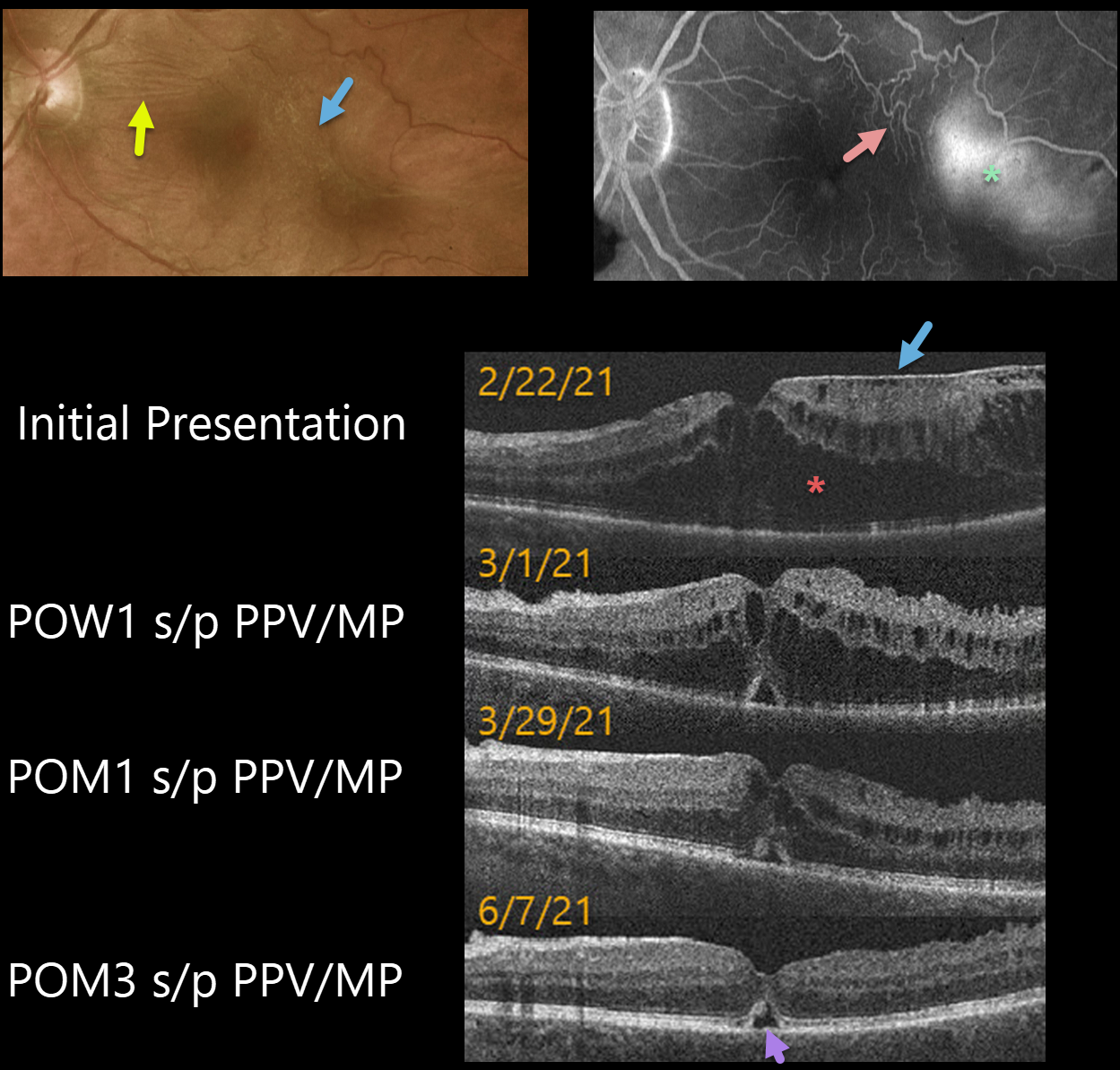
MACULAR PUCKER
Originally posted on @retina.rocks August 5, 2021
This 62YO male presented with 20/200 vision in his left eye. An epiretinal membrane (ERM) was noted in the temporal macula, associated with more nasal horizontal retinal striae.
Optos fluorescein angiography shows distortion of the temporal macular vessels with late leakage.
Vitrectomy with membrane peeling was performed the following day, and one week later, on 3/1/21, vision improved to 20/70 with a significant decrease in macular thickening.
When last examined on 6/7/21, about 3 months post-op, vision was still 20/70, but the macular thickening and contour were almost normal except for a small blister of foveal subretinal fluid.
Learning Points:
OCT scanning confirmed the temporal ERM, but the severe cystic macular thickening with possible tractional schisis that extended into the nasal macula seemed disproportionate to the imaged temporal ERM. Fortunately, both the vision and macular thickening significantly improved following vitrectomy with membrane peeling.
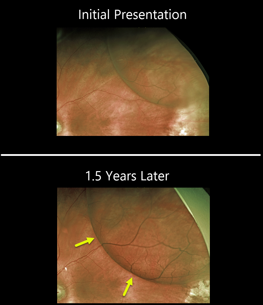
DEGENERATIVE RETINOSCHISIS
Originally posted on @retina.rocks July 27, 2021
This 80YOF was initially seen with asymptomatic retinoschisis in her left eye. When examined approximately a year and a half later, she remained asymptomatic, but the schisis cavity had enlarged in all dimensions.
Observation was recommended. This patient is at virtually no risk of a combined schisis-rhegmatogenous detachment due to the absence of breaks in the inner and outer layers.
Learning Points:
Retinochisis cavities can enlarge. In these cases, prophylactic treatment, including thermal laser, does not prevent the schisis from enlarging. Schisis very rarely progresses into the fovea (Byer Ophthalmology 1986;93:1127-1137) and even then is compatible with preserved central vision as long as the outer layer remains attached (Watzke et al 2013;33:606-612).
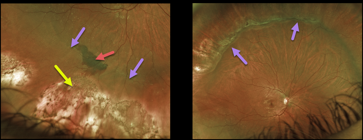
SCLERAL BUCKLE
Originally posted on @retina.rocks July 23, 2021
This 23YO female underwent successful retinal reattachment surgery with drainage of subretinal fluid and an encircling buckle. The drainage was uncomplicated, but at the completion of surgery, indirect ophthalmoscopy revealed a localized subretinal hemorrhage posterior to the drainage site.
One week later, Optos imaging shows resolving subretinal blood. Cryotherapy scarring of the causative lattice and breaks is noted with a depigmented drainage site. A shallow buckling effect is seen.
Learning Points:
Drainage of subretinal fluid at the time of scleral buckling is usually performed if the break cannot be opposed to the RPE with buckling alone.
Subretinal bleeding sometimes follows puncture of the choroid with either a needle or a laser. This is usually self-limited and resolves postoperatively without treatment.
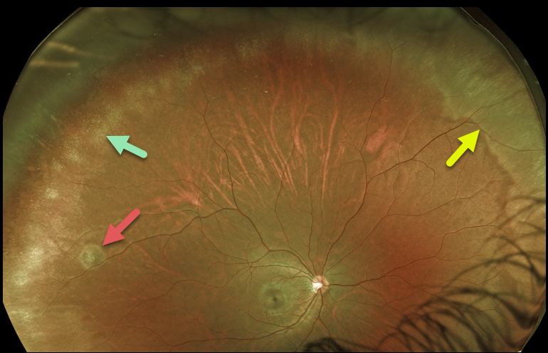
SNOWFLAKE DEGENERATION
Originally posted on @retina.rocks July 21, 2021
This 25YO highly myopic female presented with bilateral encircling areas of snowflake degeneration and lattice degeneration vs a retinal tuft with white without pressure in her right eye. There was no family history of retinal detachment.
Learning Points:
Snowflake degeneration is a rare, autosomal dominant disorder localized to chromosome 2q36 (Jiao et al, Invest Ophthalmol Vis Sci 2004;45:4498-4503).
Originally described by Hirose et al in a family of 15 members (AJO 1974;77:143-153), discrete, small refractile deposits form in the peripheral fundi. This is accompanied by fibrillar vitreous degeneration, WWP, lattice degeneration, and retinal detachment. There are no associated systemic abnormalities.
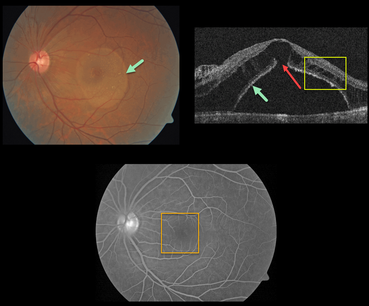
EXUDATIVE RETINAL DETACHMENT WITH OUTER MACULAR HOLE
Originally posted on @retina.rocks June 25, 2021
This patient presented with a sharply-circumscribed macular serous detachment, cystic outer retinal edema, and an outer macular hole. Fluorescein angiography is essentially normal without leakage.
An optic pit was absent clinically and on OCT imaging. Serum protein electrophoresis was normal, and a systemic cancer workup was negative.
Learning Points:
Serous detachment can be an uncommon ocular manifestation of paraproteinemia (see Mansour et al, Ophthalmology 2014;121;1925-1932). These detachments, which resemble those seen in optic pit maculopathy, are characterized by a well-defined serous detachment without angiographic leakage. A central outer macular hole is often present.
The fluid is likely due to an osmotic gradient generated by subretinal immunoglobulins that passively migrate into the subretinal space.
We do not know the cause of our patient’s findings, given his lack of an optic pit and a negative systemic workup.
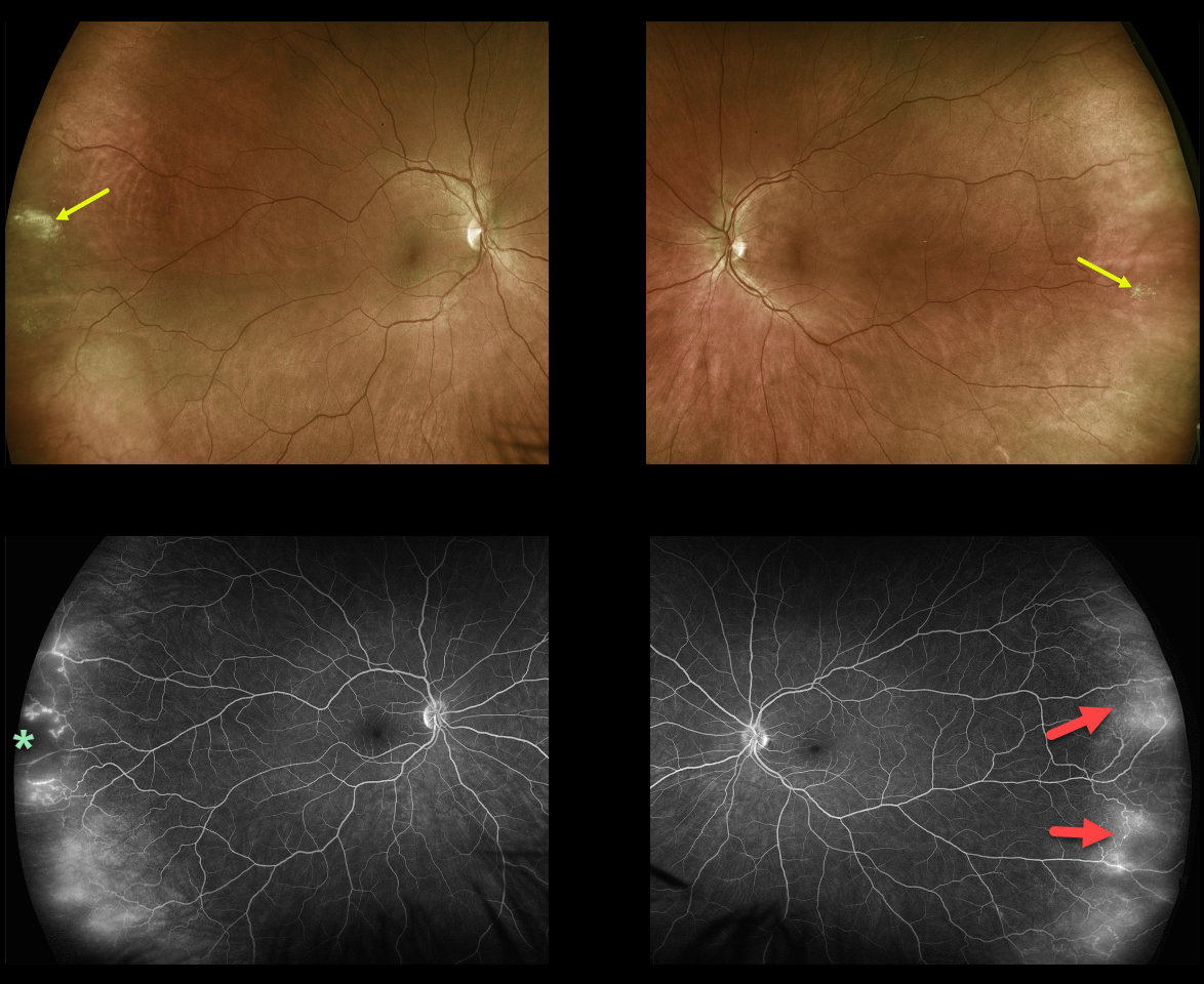
PERIPHERAL MICROVASCULAR ABNORMALITIES
Originally posted on @retina.rocks June 16, 2021
This healthy, asymptomatic 40YO female presented with 20/25 vision bilaterally.
Optos ultra-widefield (UWF) imaging showed far temporal peripheral probable lattice lesions and subtle telangiectasia. The vascular changes, including capillary dropout and telangiectasia, were more evident on fluorescein angiography (FA).
Learning Points:
In 2016, Shah et al described common peripheral retinal vascular findings on Optos UWF FA in otherwise healthy adults (58 eyes, 31 patients) undergoing imaging for unrelated macular pucker or choroidal nevi (Retina 2016;36:1087-1092).
Findings included vessels crossing the horizontal raphe (45%), right-angle vessels (71%), terminal networks (78%), absence of capillary detail (98%), ground glass hyperfluorescence (88%), drusen (34%), and microaneurysms (41%). One or more of these findings were present in most eyes.
Another study by Lu et al. found that normal peripheral retinas typically exhibit granular background fluorescence, with or without a mottled fluorescent band (AJO 2017;173:84-90).
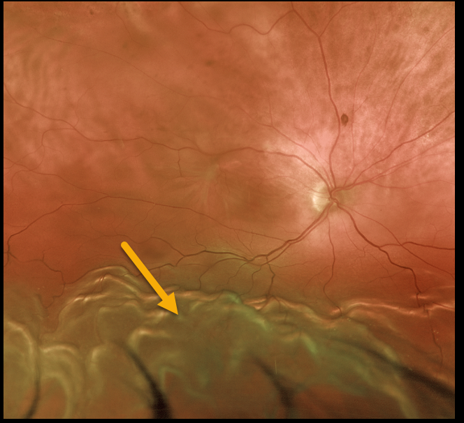
RHEGMATOGENOUS RETINAL DETACHMENT
Originally posted on @retina.rocks June 10, 2021
This 77YO male presented with 20/400 vision from a chronic macula-off rhegmatogenous retinal detachment (RRD).
Demarcation lines are common in these cases (not present here). The billowing, corrugated convex appearance of the inferior retina is typical of RRD. The retina is mobile without proliferative vitreoretinopathy.
Learning Points:
The normal neurosensory retina remains attached to the RPE due to numerous physiologic mechanisms, including the inner (retinal vascular endothelium) and outer (tight junctions at the apex of the RPE cells) blood retinal barriers, hyperosmotic choroid passively drawing fluid into the subretinal space, RPE pump, interphotoreceptor matrix, and friction between the RPE microvilli and the photoreceptor outer segments.
Rhegmatogenous retinal detachment (RRD) occurs when liquid vitreous, entering the subretinal space through a retinal break, overwhelms these forces, favoring retinal detachment.
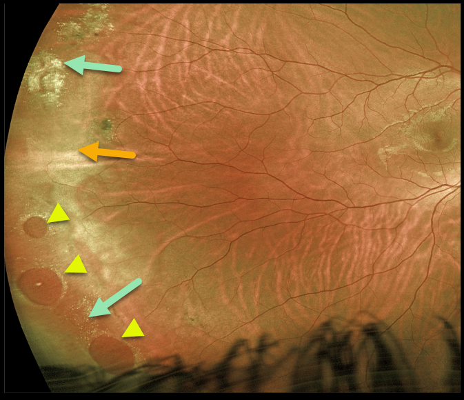
LATTICE DEGENERATION
Originally posted on @retina.rocks June 9, 2021
This 24YO male presented with asymptomatic but somewhat confusing clinical findings beautifully captured by this Optos image.
Three large atrophic retinal holes within an area of lattice are found in the inferotemporal periphery, along with a few areas of lattice and scarring superotemporally. There is also associated white without pressure, giving the appearance of a retinal detachment (remember the normally transparent retina turns whitish when it detaches). However, the retina was completely attached, and we are treating him with observation.
Learning Points:
Retinal detachment occurs in 1 in 10,000 people per year. Lattice is found in 10% of the general population, especially in high myopes. Since about one-third of patients with retinal detachment have lattice as the cause, the question always arises, especially in patients like this: Should prophylactic retinopexy be performed?
The short answer is that asymptomatic lattice generally should not be treated. To put things into perspective, consider a pre-COVID, non-social-distancing football stadium with 30,000 people. Three people will develop retinal detachment in the coming year. Only one of the 3,000 people in the stadium with lattice would develop one of those 3 detachments.
Excellent references on this controversial topic include: Byer Ophthalmology 1989;96;1396-1402, Folk et al Ophthalmology 1989;96;72-79, and Folk et al Retina 1990;10:165-169.
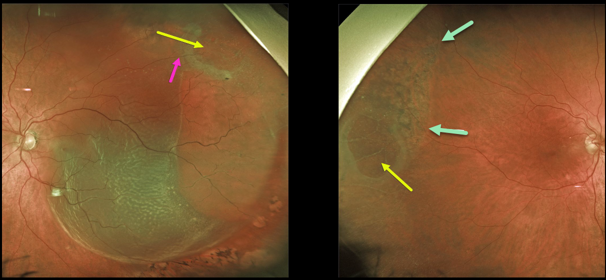
DEGENERATIVE RETINOSCHISIS
Originally posted on @retina.rocks May 27, 2021
This 72YO male presented with an acute macula-off retinal detachment (RD) in his left eye.
Optos imaging shows that this is not a typical rhegmatogenous RD since an outer layer hole (OLH) is noted superotemporally. A retinal vessel can be traced over the OLH, indicating that this is not a full-thickness defect.
The patient also has degenerative retinoschisis with an OLH in his right eye. A shallow subclinical schisis detachment is likely present due to the pigmentary changes throughout the schisis cavity.
Prophylactic treatment is not recommended for this eye, but we are carefully following him, as he does have a small risk of developing an RD here as well.
Learning Points:
This detachment in the left eye does not have a typical full-thickness break, but is caused by degenerative retinoschisis.
Numerous tiny inner-layer holes, along with the visible large OLH, essentially created a full-thickness retinal defect, giving the liquid vitreous access to the subretinal space. The detachment was successfully repaired with vitrectomy surgery.
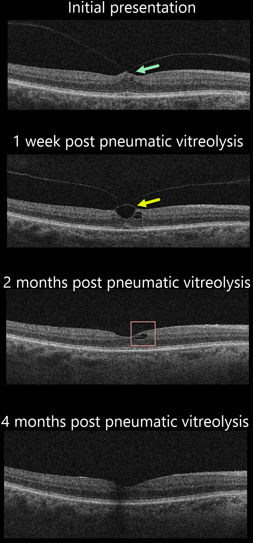
VITREOMACULAR TRACTION
Originally posted on @retina.rocks May 26, 2021
This 72YO male presented with symptomatic vitreomacular traction (VMT) and 20/40 vision. Various treatment options were discussed, including observation, ocriplasmin (Jetrea), pneumatic vitreolysis, and vitrectomy. He decided to proceed with pneumatic vitreolysis.
One week later, OCT showed that the vitreous remained focally attached to the central fovea, with increased traction.
Two months following the procedure, there was a complete vitreous detachment with normalization of the foveal contour and a small residual splitting of the outer plexiform layer nasally.
Four months following the procedure, OCT was virtually normal, and vision was 20/30. The patient was completely symptom-free.
Learning Points:
The prevalence of VMT increases with age and is present in about 1% of people over 40. The natural history is still being defined, but most patients have stable traction and vision. VMT can also resolve spontaneously in about 20% (Errera et al, Ophthalmology 2018;125:701-707).
Many patients with VMT remain completely asymptomatic despite impressive OCT findings. We therefore almost always observe VMT unless the patient has significant symptoms, as in this case.
Shortly after we treated this patient, the results of the DRCR Retina Network Protocol AG, which evaluated pneumatic vitreolysis with C3F8 for VMT, were published ahead of print (https://doi.org/10.1016/j.ophtha.2021.05.005). Gas successfully created a complete PVD in 78% of eyes. However, the study was terminated early due to safety concerns related to retinal detachment (8% of eyes). Based on these safety issues, we personally would no longer recommend this as a viable option unless medical issues make vitrectomy impossible.
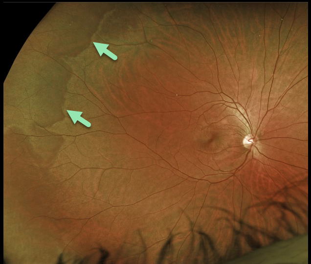
WHITE WITHOUT PRESSURE
Originally posted on @retina.rocks May 21, 2021
This patient’s fundus shows areas of white without pressure. Typically, there are bilateral peripheral geographic areas of whitish retinal discoloration.
Learning Points:
White without pressure (WWP) is a common benign peripheral finding, most often seen in darkly pigmented individuals. WWP gets its name because the area appears white without indentation, unlike scleral depression (white with pressure).
It can sometimes be confused with retinal breaks or detachment. The exact cause of this phenomenon is unknown. Historically, it was thought to be due to a change at the vitreoretinal interface, but more recent OCT findings show increased reflectivity in the outer retina (see Diaz et al, Retina 2014;34:1020-1021).
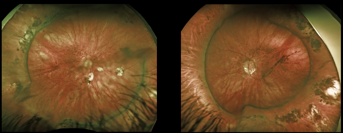
SCLERAL BUCKLE
Originally posted on @retina.rocks May 12, 2021
This is a beautiful Optos image of bilateral scleral buckles in a 51YO female myopic patient who underwent successful retinal reattachment surgery years ago.
Learning Points:
The normal neurosensory retina remains attached to the RPE due to numerous physiologic mechanisms, including the inner and outer blood-retinal barriers, the hyperosmotic choroid passively drawing fluid into the subretinal space, the RPE pump, the interphotoreceptor matrix, and friction between the RPE microvilli and the photoreceptor outer segments.
Rhegmatogenous retinal detachment (RRD) occurs when liquid vitreous, entering the subretinal space through a retinal break, overwhelms these forces, favoring retinal detachment.
Repairing RRD involves finding and closing all breaks, essentially denying the liquid vitreous access to the subretinal space. The break(s) can be closed internally with a gas bubble via pneumatic retinopexy or vitrectomy surgery, or externally by securing a piece of silicone material to the sclera (scleral buckle). This indents, or buckles, the RPE against the retinal break.
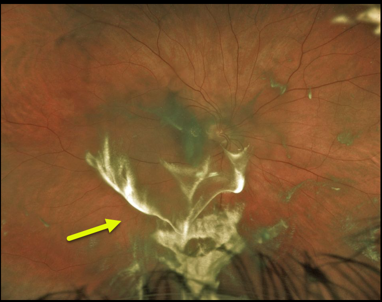
TRIAMCINOLONE-MOXIFLOXACIN (TriMoxi)
Originally posted on @retina.rocks May 11, 2021
This patient experienced some superior visual field loss following cataract surgery. A stagnant blob of triamcinolone-moxifloxacin accumulated in the vitreous and subhyaloid space in the inferior macula.
The medication eventually dissolved as the patient’s symptoms resolved.
Learning Points:
TriMoxi is commonly used in dropless cataract surgery, so patients don’t need to use postoperative antibiotic and steroid drops. At the end of the procedure, 0.2cc of TriMoxi is injected into the anterior inferior vitreous. Usually, patients notice some large black floaters that subside within a few weeks.
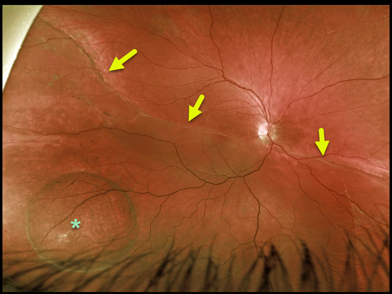
RHEGMATOGENOUS RETINAL DETACHMENT
Originally posted on @retina.rocks May 3, 2021
This 34YO male presented with 20/80 vision from a chronic macula-off rhegmatogenous retinal detachment (RRD).
A demarcation line extends along the entire posterior extent of the detachment. There is a retinal macrocyst within the detachment inferotemporally.
Learning Points:
Chronic retinal detachments rarely develop these macrocysts, which are located within the outer plexiform layer (see Marcus and Aaberg, Arch Ophthalmology 1979;97;1275-1279).
They do not need to be treated during RRD surgery unless they prevent closure of the retinal break(s). They promptly flatten following retinal reattachment.
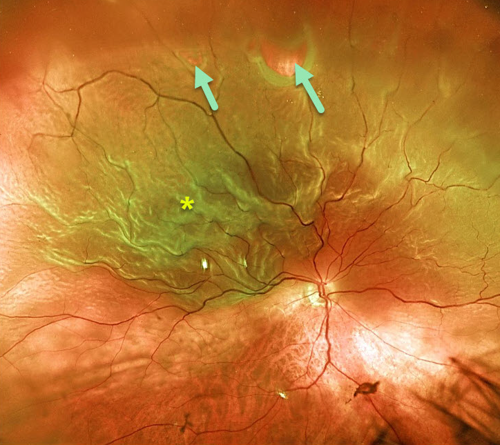
RHEGMATOGENOUS RETINAL DETACHMENT
Originally posted on @retina.rocks April 19, 2021
This 77YO patient presented with 20/200 vision from a superior macula-off retinal detachment. Optos imaging beautifully captures the detachment that was caused by several superior retinal tears.
Learning Points:
The normal neurosensory retina remains attached to the RPE due to numerous physiologic mechanisms, including the inner (retinal vascular endothelium) and outer (tight junctions at the apex of the RPE cells) blood retinal barriers, hyperosmotic choroid passively drawing fluid into the subretinal space, RPE pump, interphotoreceptor matrix, and friction between the RPE microvilli and the photoreceptor outer segments.
Rhegmatogenous retinal detachment (RRD) occurs when liquid vitreous, entering the subretinal space through a retinal break, overwhelms these forces, favoring retinal detachment.
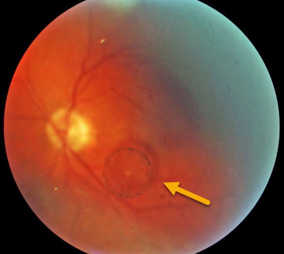
VITREOUS CYST
Originally posted on @retina.rocks April 13, 2021
This patient was noted to have an asymptomatic vitreous cyst in the left eye.
Learning Points:
Vitreous cysts are extremely rare, idiopathic, and usually diagnosed as an incidental finding, as in this patient. If symptomatic, the cyst can literally be popped with a YAG laser.
These cysts likely originate from a ciliary body cyst (see Dhull et al, Ophthalmology Retina 2020;4:1208).
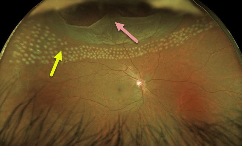
RHEGMATOGENOUS RETINAL DETACHMENT
Originally posted on @retina.rocks April 9, 2021
This 51YO male presented with a completely asymptomatic superior rhegmatogenous retinal detachment. There was a history of blunt trauma 1 year earlier.
We discussed the various surgical options, and he decided to proceed with laser demarcation due to the lack of symptoms and remote history of the causative injury. The immediate post-laser Optos image shows the barrier laser treatment.
We were unable to reach the ora with the slit lamp delivery system, so the anterior edges were subsequently treated with cryotherapy.
Learning Points:
When demarcating these detachments, it is critical that the retinopexy completely surrounds the subretinal fluid and that treatment extends to the ora; otherwise, the detachment can spread through the untreated retina.
Patients need to be followed postoperatively, as detachment can occasionally progress despite prior retinopexy.
See Vrabec and Baumal, Ophthalmology 2000;107:1063-1067 for an excellent review regarding demarcation laser photocoagulation for selected macula-sparing retinal detachments.
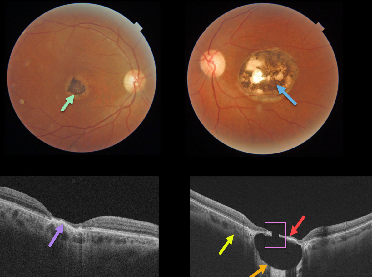
TOXOPLASMOSIS
Originally posted on @retina.rocks March 26, 2021
This 57YO male gave a history of lifelong poor vision in his left eye. Vision was 20/40 OD and 20/200 OS.
A small hyperpigmented foveal scar is noted in his right eye with neurosensory retinal atrophy and irregular elevation of the RPE.
A larger, round, variably pigmented, excavated macular scar is noted in his left eye. Triton swept-source OCT reveals a thinned atrophic retina and intact RPE suspended over an optically empty hyporeflective excavated colobomatous type lesion. The plane of the intact surrounding sclera is indicated by the yellow arrow. A small full-thickness retinal and RPE defect is noted centrally.
Learning Points:
For a great discussion on how to differentiate these infectious macular colobomatous lesions (congenital toxoplasmosis, Zika, and cytomegalovirus) from inherited dystrophies like North Carolina Macular Dystrophy (NCMD), see Kumar and Mahalingam AJO 2019;200;47-56.
In case you’re thinking our patient has NCMD, also see the first OCT description of congenital toxoplasmosis macular lesions (Garg et al, Retina 2009;29;631-637). Their Figures 4 and 6 look eerily similar to our patient’s right and left eyes!
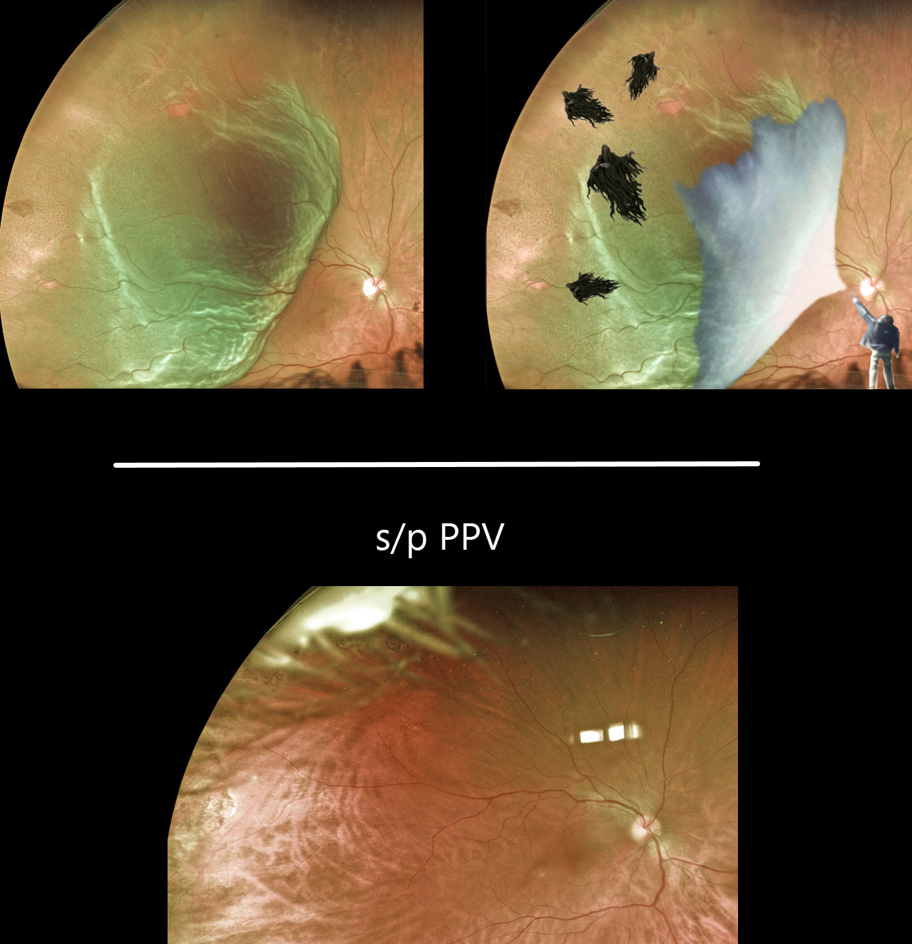
DEMENTOR DETACHMENT
Originally posted on @retina.rocks March 22, 2021
Multiple temporal retinal tears are visible in this superotemporal macula-on rhegmatogenous retinal detachment. Yet it appears to be held off like Harry Potter’s Dementors.
We happen to have a powerful wizard, too.
Just look at the beautifully attached retina after a successful pars plana vitrectomy!
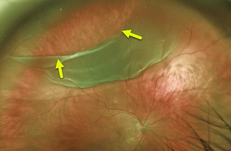
GIANT RETINAL TEAR
Originally posted on @retina.rocks March 1, 2021
Here is a preoperative Optos photo of a giant retinal tear extending from about 9:30 to 1 o’clock.
This patient underwent successful pars plana vitrectomy surgery with C3F8 (perflouropropane) gas tamponade.
Learning Points:
By definition, giant retinal tears extend at least 3 clock hours.
Surgical repair is greatly facilitated by the use of Perfluoron liquid (PFO, perfluoron octane), which is heavier than water and, intraoperatively, helps unroll the posterior retina and prevent posterior retinal slippage.
The PFO can either be removed at the initial repair or left in place for a week or two before being removed with a secondary procedure.
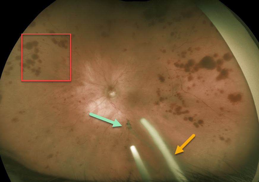
OCULAR ISCHEMIA WITH OZURDEX IMPLANTS
Originally posted on @retina.rocks February 4, 2021
This 86YO patient was receiving Ozurdex (dexamethasone 0.7%) injections every 8 weeks in her left eye for macular edema from an inferior hemiretinal vein occlusion.
She subsequently developed ocular ischemic syndrome (OIS) like changes with scattered peripheral large outer retinal hemorrhages. Inferiorly, more recent Ozurdex implants are noted, along with another ghost-like remnant of an older implant. Although the Ozurdex implants are supposed to completely degrade, these remnants may persist beyond 1 year (see Kim et al Retina 2020;40;2226-2231).
Learning Points:
The retinal hemorrhages in ocular ischemic syndrome (OIS) are unique. Retinal vascular disorders like diabetic retinopathy, retinal vein occlusions, and hypertensive retinopathy usually have flame- and dot-shaped inner retinal hemorrhages concentrated around the posterior pole.
Our patient’s findings are classic for OIS, with hemorrhages in the deeper peripheral retina. The retinal periphery is literally the end of the line for the retinal arterial circulation. In an eye receiving less blood supply due to carotid occlusive disease, the more proximal retina has first dibs on the available oxygen. There are penetrating capillaries that dive radially from the nerve fiber and ganglion cell layers into the deeper retina. The deep vascular complex that supplies the inner and outer plexiform layers is thus furthest downstream, and these endothelial cells are likely damaged in OIS. Blood cells leaking into this space accumulate, causing the large outer blot to hemorrhage.
These patients need to have a carotid Doppler and ultrasound and be referred for medical or surgical intervention if they have a critical stenosis. They may also develop anterior or posterior segment neovascularization requiring photocoagulation and/or anti-VEGF injections. Iris neovascularization can develop, leading to neovascular glaucoma. Hypotony can also occur.
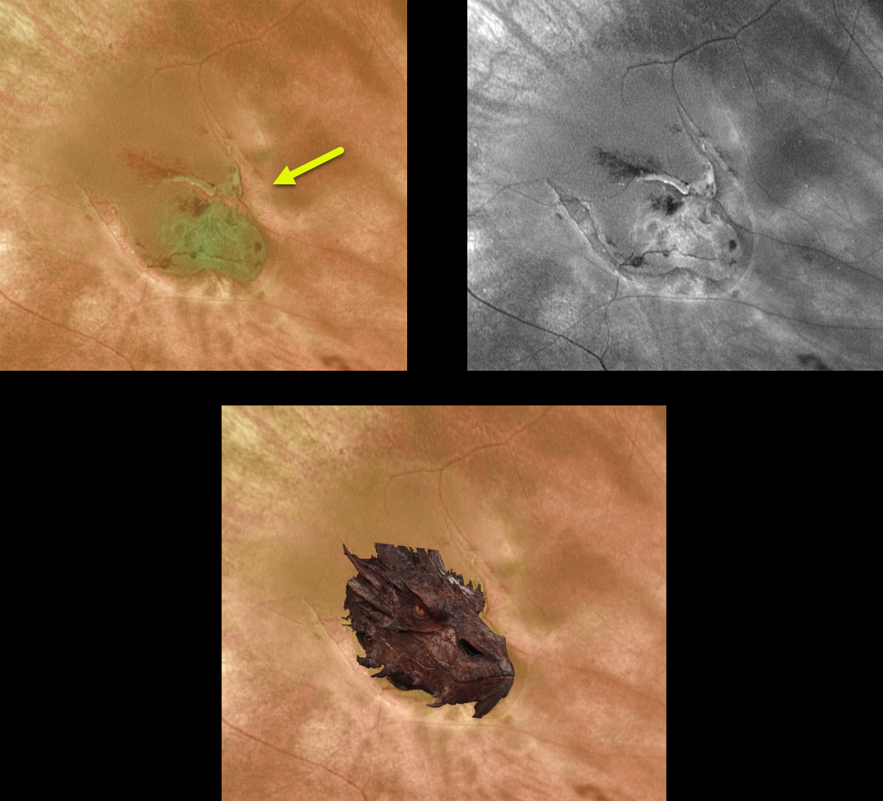
DRAGON RETINOPATHY
Originally posted on @retina.rocks February 1, 2021
The fiery dragon decided to make an appearance in the back of this patient’s eye. This dangerous predator caused its human prey to experience large black floaters and flashing lights.
Fortunately, the beast, aka retinal tear, was vanquished with an in-office barrier laser.
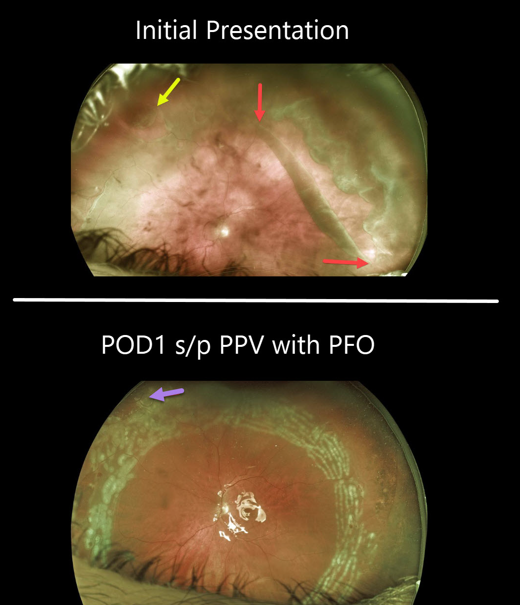
RHEGMATOGENOUS RETINAL DETACHMENT
Originally posted on @retina.rocks January 25, 2021
This patient presented with a symptomatic macula-on giant-tear retinal detachment (RD) in his left eye. The giant tear extends from about 12 to 4 o’clock with an additional tear noted superonasally.
One day after vitrectomy with Perfluoron (PFO, perfluoro octane) injection, the retina is completely attached, with fresh white encircling laser scarring. Some PFO bubbles can be seen superonasally.
The PFO liquid was removed two weeks later, and his retina has remained attached.
Learning Points:
By definition, giant retinal tears extend at least 3 clock hours. Surgical repair is greatly facilitated by using PFO liquids, which are heavier than water and help unroll the posterior retina and prevent posterior slippage of the detachment. The PFO can either be removed at the initial repair or left in place for a week or two before being removed with a secondary procedure.
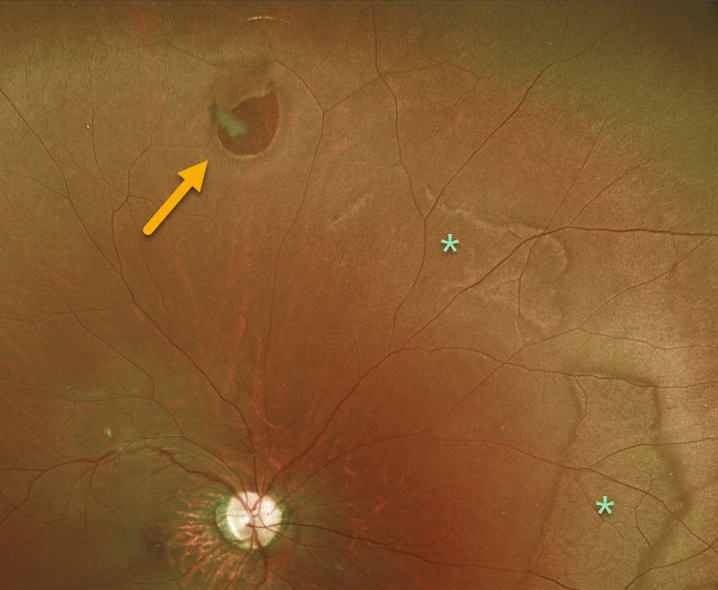
RETINAL BREAK
Originally posted on @retina.rocks January 13, 2021
This is a 19YO patient who presented with an asymptomatic retinal break with prominent posterior white without pressure.
Learning Points:
A retinal break is a full-thickness retinal defect. Breaks are classified as atrophic holes, operculated holes, tractional (horseshoe) tears, and retinal dialysis
(break at the ora serrata).
In general, asymptomatic retinal breaks are observed since they are at low risk of causing retinal detachment. Symptomatic retinal tears and retinal dialysis are at the highest risk for causing a detachment and are virtually always treated.
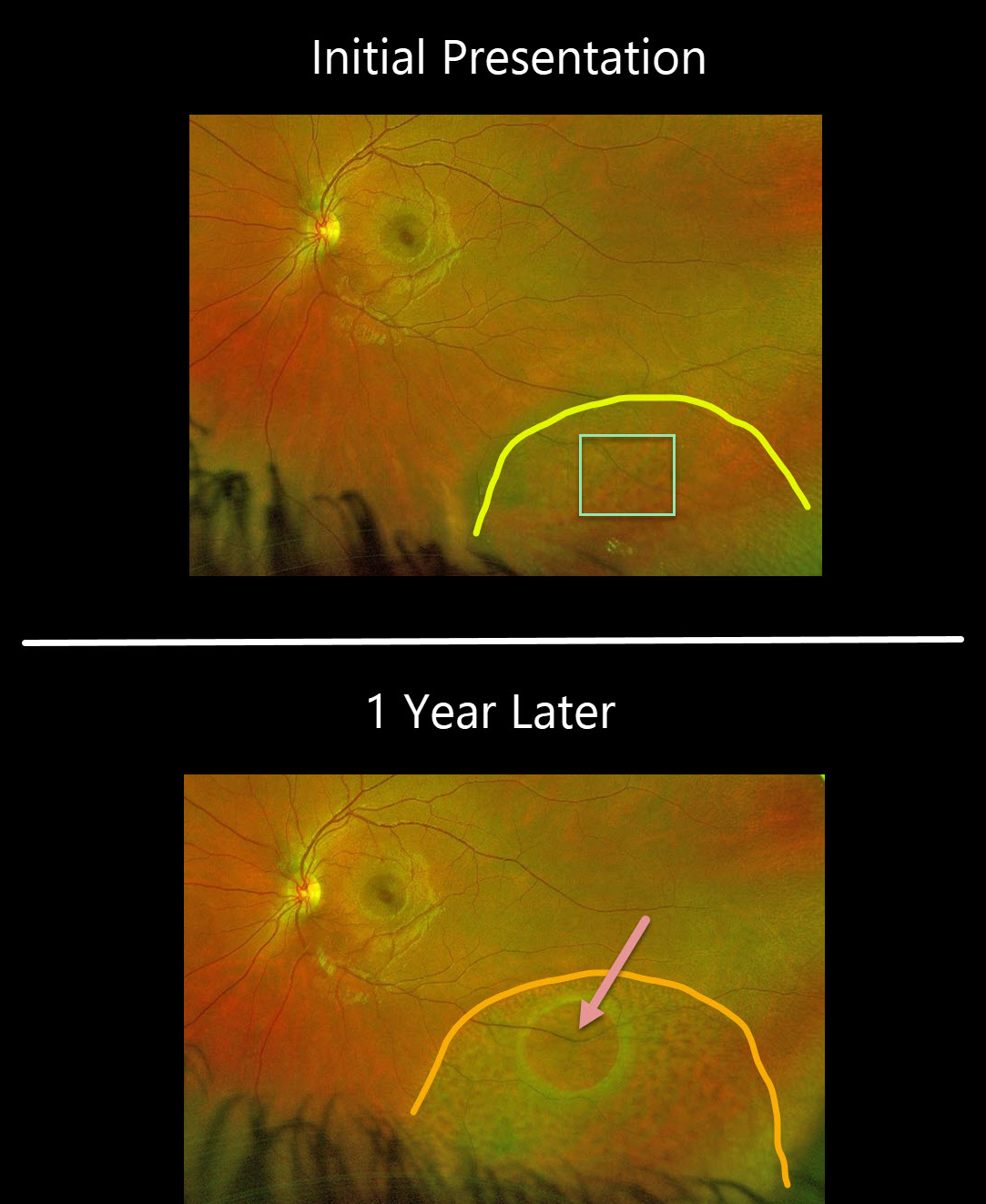
DEGENERATIVE RETINOSCHISIS
Originally posted on @retina.rocks December 25, 2020
This 22YO patient, courtesy of Dr. Brooke Spivey, presented with degenerative retinoschisis with numerous inner-layer holes.
One year later, the schisis cavity had enlarged, with a new posterior outer layer hole. There were no visual symptoms.
Learning Points:
This patient is at a low risk of developing a combined rhegmatogenous schisis detachment due to the presence of both inner and outer layer breaks.
The outer layer remains attached, and no prophylactic treatment was recommended since thermal laser does not prevent schisis from enlarging, schisis very rarely progresses into the fovea (Byer Ophthalmology 1986;93:1127-1137), and even then is compatible with central vision as long as the outer layer remains attached (Watzke et al 2013;33:606-612), and asymptomatic holes are generally not treated.
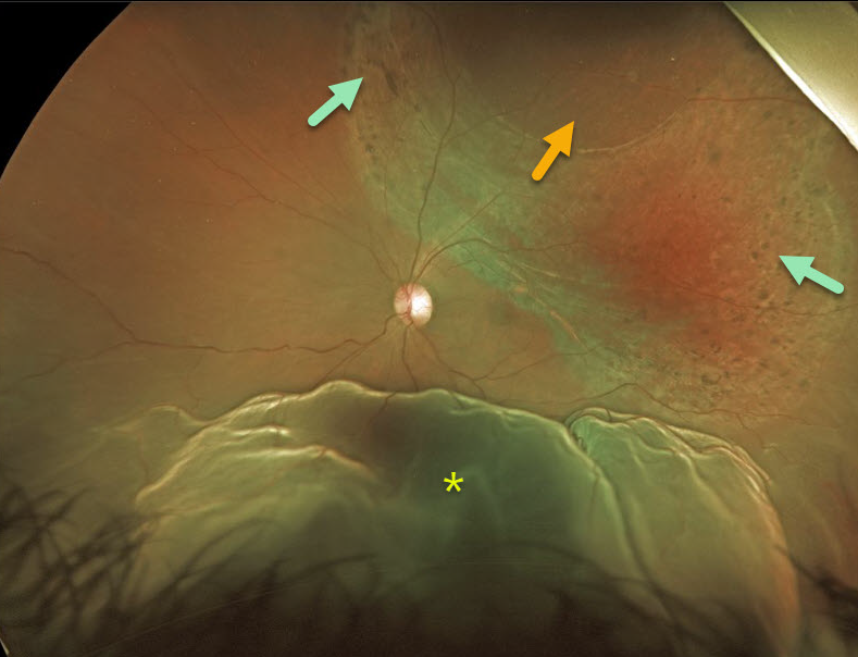
COMBINED RETINOSCHISIS-RHEGMATOGENOUS RETINAL DETACHMENT
Originally posted on @retina.rocks December 7, 2020
This 33YO patient presented with a prominent inferior retinal detachment associated with superotemporal retinoschisis with a large outer layer hole (OLH). The OLH was surrounded by laser-like chorioretinal scarring, although the patient denied prior laser surgery.
At vitrectomy, a superonasal gutter of peripheral detachment extended to an equatorial tear at 10:30 (not visualized in these images). The OLH and schisis were not detached.
Learning Points:
Patients with degenerative retinoschisis and associated inner- and outer-layer holes are at risk of rhegmatogenous retinal detachment.
Despite our patient probably undergoing prior prophylactic laser photocoagulation around the outer layer hole, he still developed a retinal detachment from a presumed new retinal tear outside the area of schisis.
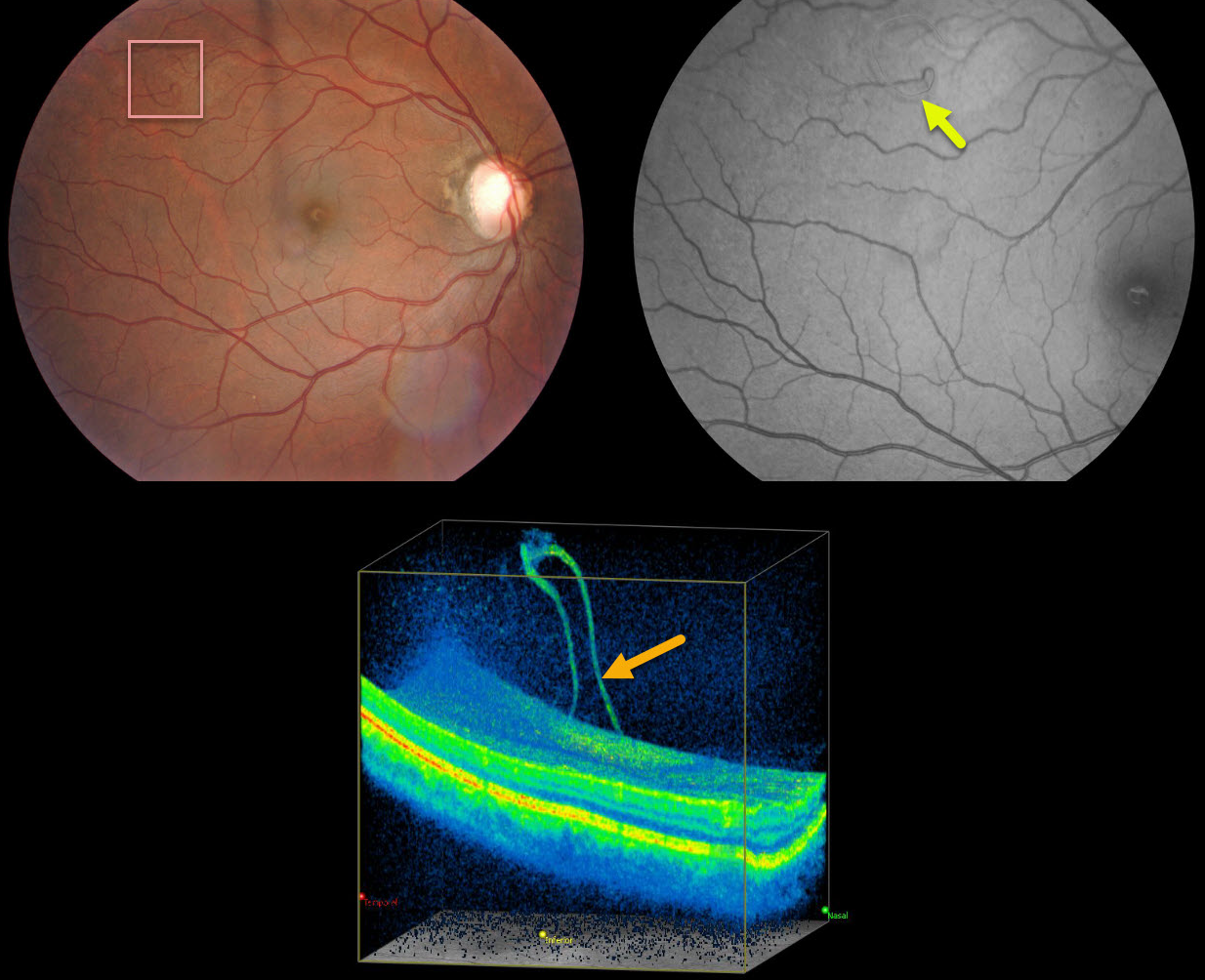
AVULSED RETINAL VESSEL
Originally posted on @retina.rocks December 3, 2020
This patient presented with an asymptomatic, superotemporal elevation of the retinal vessel, best seen on 3D OCT and red-free fundus photograph. This patient has done well with observation.
Learning Points:
Originally described by Robertson et al. (Arch Ophthalmology 1971;85:669-672), avulsed retinal vessels result from focal vitreous traction and can cause recurrent vitreous hemorrhage. The traction may or may not be associated with retinal breaks.
The traction can persist, can spontaneously resolve, or may require vitrectomy for recurrent or non-clearing vitreous hemorrhage. Of course, any acutely symptomatic associated retinal breaks should also be surrounded with retinopexy.
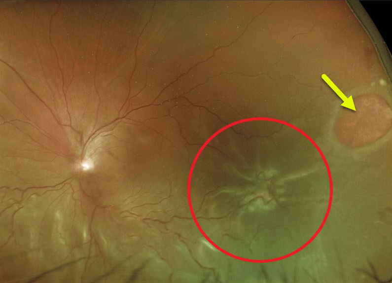
PROLIFERATIVE VITREORETINOPATHY (PVR)
Originally posted on @retina.rocks November 24, 2020
This patient’s Optos image demonstrates a large retinal hole and retinal detachment with proliferative vitreoretinopathy (PVR).
Learning Points:
Proliferative vitreoretinopathy (PVR), consisting of growth of cellular membranes on either retinal surface, develops in about 5-10% of retinal detachments. The membranes contract and foreshorten the retina, likely resulting from an aberrant wound-healing response.
There are no pharmacologic therapies to prevent or treat PVR. These complex detachments can be managed with vitrectomy with membrane peeling, retinectomy, scleral buckling, and silicone oil. However, PVR remains the main cause for ultimate retinal reattachment surgical failure.
See Garweg et al Surv Ophthalmol 2013;48:321-329 for a good review of PVR pathophysiology.
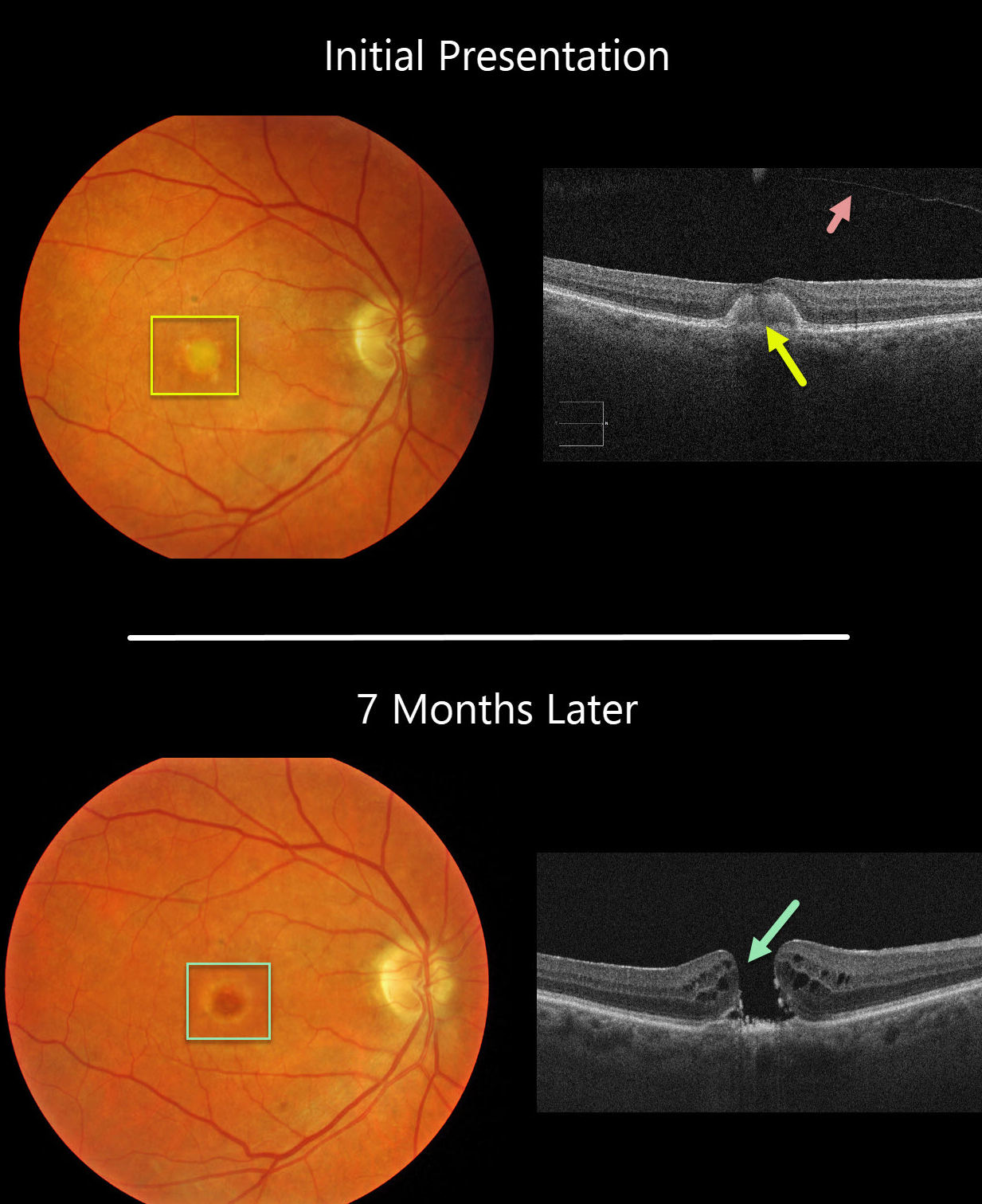
ADULT-ONSET FOVEOMACULAR VITELLIFORM DYSTROPHY
Originally posted on @retina.rocks November 18, 2020
This 77YO female was initially examined with 20/60 vision OD, 20/30 vision OS, and bilateral vitelliform lesions (only right eye pictured) consistent with Adult Best (adult-onset foveomacular vitelliform dystrophy). A complete posterior vitreous detachment (PVD) was noted in her right eye.
Seven months later, vision decreased to counting fingers OD due to a new full-thickness macular hole. This patient was asymptomatic due to good vision in her left eye and has opted against surgery for now.
Learning Points:
Macular holes are most commonly caused by a partial PVD with active vitreomacular traction. However, they can occasionally develop without traction following a prior PVD or vitrectomy surgery. Macular holes have also been reported as a rare complication of adult Best’s disease.
Adult-onset foveomacular vitelliform dystrophy is associated with a mutation in the BEST1 gene, which encodes the bestrophin-1 protein.
Bestrophin-1, a calcium-activated chloride channel, is primarily found in the basolateral plasma membrane of the RPE. BEST1 mutations cause a variety of phenotypes, including autosomal recessive bestrophinopathy, best vitelliform macular dystrophy, and autosomal dominant vitreoretinochoroidopathy.
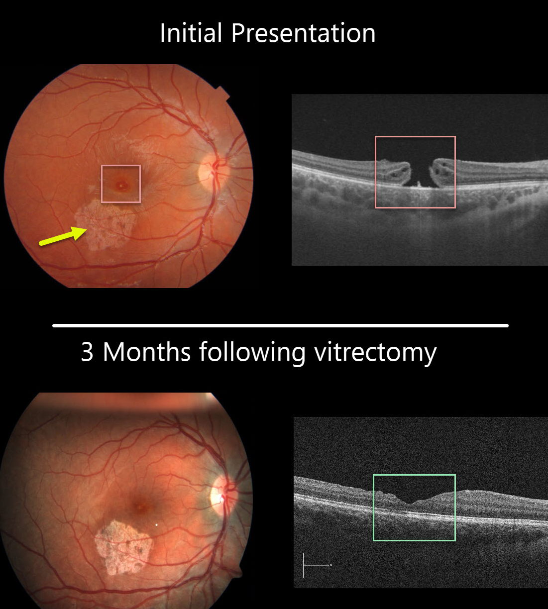
MACULAR HOLE
Originally posted on @retina.rocks November 6, 2020
This 13YO girl was hit in her right eye while playing volleyball. She presented to us several months later with 20/200 vision, an inferotemporal macular scar from resolved commotio retina, and a full-thickness macular hole.
The macular hole was closed following successful vitrectomy, and she was 20/100 one month postoperatively. Vision improved to 20/40 three months following surgery. Since the scotoma from the macular scar was superior, she is now virtually completely visually asymptomatic and extremely lucky!
Learning Points:
Traumatic macular holes are more common in younger patients, often from recreational or sports injury, and tend to be more eccentric in shape.
The mechanism could be due to sudden vitreous separation or to stretching of the posterior pole resulting from anteroposterior ocular compression. The holes can spontaneously close and have a high surgical success rate, as in our patient (see Liu et al AJO 2020;210:174-183).
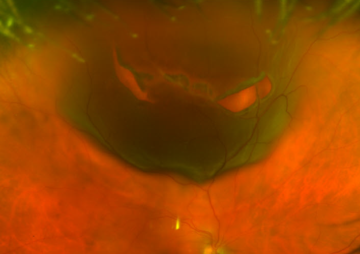
HALLOWEEN RETINOPATHY
Originally posted on @retina.rocks October 30, 2020
This patient presented with a jack-o’-lantern retinal detachment 2 weeks before Halloween.
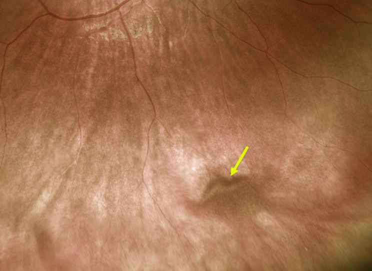
CHOROIDAL VARIX
Originally posted on @retina.rocks October 27, 2020
This patient has a vortex vein varix, a benign dilatation that can simulate a choroidal tumor.
The diagnosis is clinched when gentle digital pressure on the globe collapses the varix, making it temporarily disappear.
Learning Points:
The vortex veins provide the major venous outflow for the choroid, subsequently draining into the superior and inferior ophthalmic veins. The vortex ampullae are visible at the equator, usually in each quadrant.
The ampullae can sometimes dilate into a nodular, orange-brown varicosity that can simulate a choroidal tumor. They are usually isolated lesions, but can be multiple or bilateral.
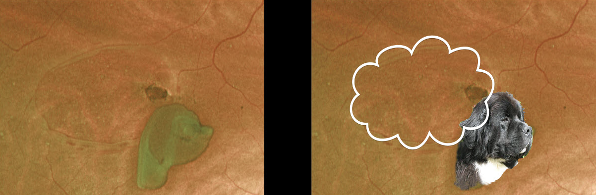
DOGGONE HOLE
Originally posted on @retina.rocks September 19, 2020
What an interesting operculated retinal break. What do you think this dog is thinking?
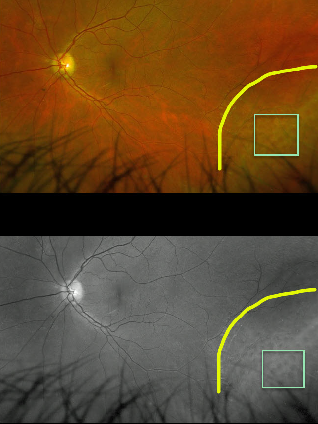
DEGENERATIVE RETINOSCHISIS
Originally posted on @retina.rocks September 4, 2020
This patient has degenerative retinoschisis with numerous inner-layer holes, best seen on the Optos green channel image.
Learning Points:
This patient just needs observation due to the small risk of developing a rhegmatogenous detachment.
This risk would increase if an outer-layer hole were present, since the inner- and outer-layer holes would essentially represent a full-thickness break, thereby giving the liquid vitreous access to the subretinal space.
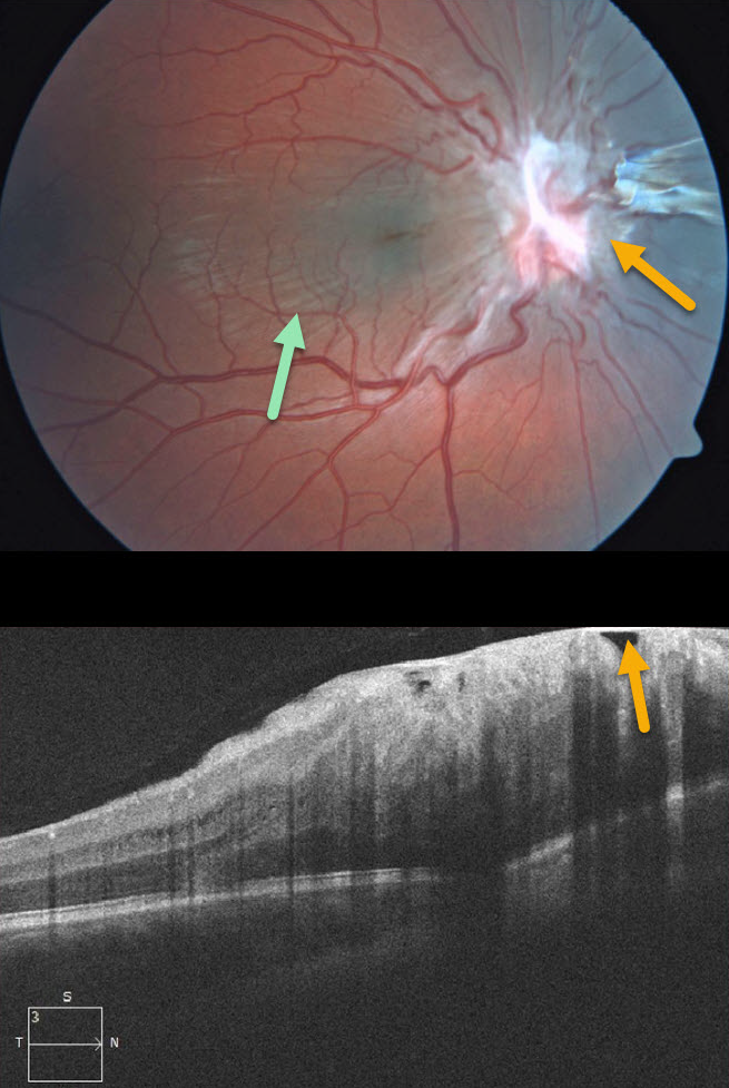
COMBINED HAMARTOMA OF THE RETINA AND RPE
Originally posted on @retina.rocks August 25, 2020
This 7YO boy presented without visual symptoms and 20/50 vision OD. The left eye was normal.
Contracted fibrotic tissue overlying the optic nerve is causing distortion of the surrounding retina, with striae visible through the macula. Some of this fibrosis may also be pigmented nasal to the nerve.
This case represents either a combined hamartoma of the retina and RPE or an unusual pucker centered over the optic nerve.
Learning Points:
Combined hamartomas are classically described as benign congenital lesions composed of glial cells, vascular tissue, and pigmented retinal pigment epithelial cells. However, more recent data indicate that these lesions primarily arise from the inner retinal layers.
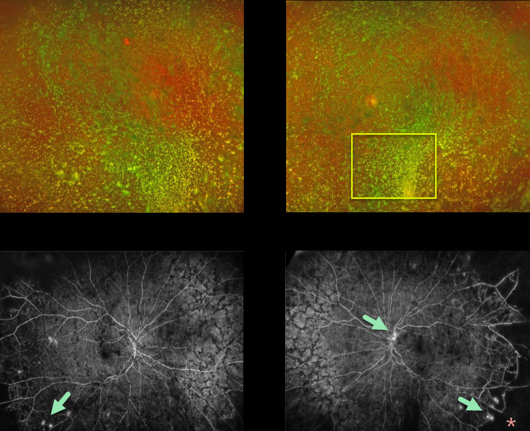
ASTEROID HYALOSIS
Originally posted on @retina.rocks July 29, 2020
Although this diabetic patient had normal vision with absolutely no symptoms, what we saw on clinical examination and Optos color imaging was nothing but asteroid hyalosis.
Fluorescein angiography (FA) showed bilateral proliferative diabetic retinopathy with areas of leakage and peripheral capillary dropout. In-office panretinal photocoagulation (PRP) was not possible due to the dense asteroid.
We discussed ongoing anti-VEGF injections versus vitrectomy + PRP to clear the visual axis. She decided to proceed with vitrectomy in both eyes.
Learning Points:
Asteroid hyalosis is caused by the accumulation of benign calcium-lipid particles suspended in the vitreous.
Fluoroscein angiography and OCT can image through asteroid hyalosis.
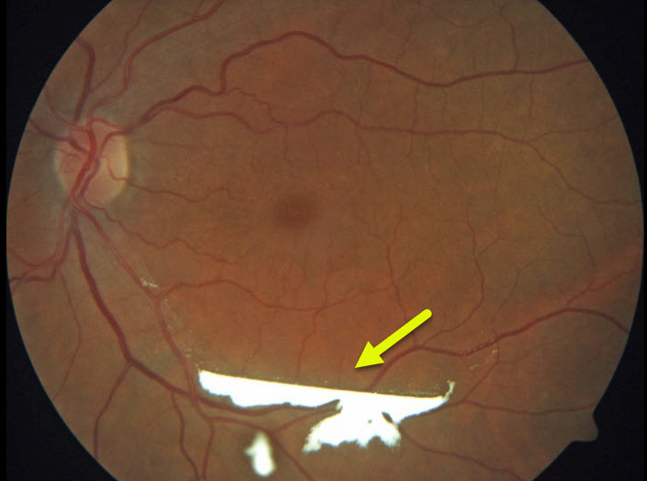
TRIAMCINOLONE-MOXIFLOXACIN (TriMoxi)
Originally posted on @retina.rocks July 21, 2020
This patient experienced some superior visual field loss following cataract surgery. A stagnant blob of triamcinolone-moxifloxacin accumulated in the subhyaloid space in the inferior macula.
The medication eventually dissolved as the patient’s symptoms resolved.
Learning Points:
TriMoxi is commonly used in dropless cataract surgery, so patients don’t need to use postoperative antibiotic and steroid drops. At the end of the procedure, 0.2cc of TriMoxi is injected into the anterior inferior vitreous. Usually, patients notice large black floaters that subside within a few weeks.
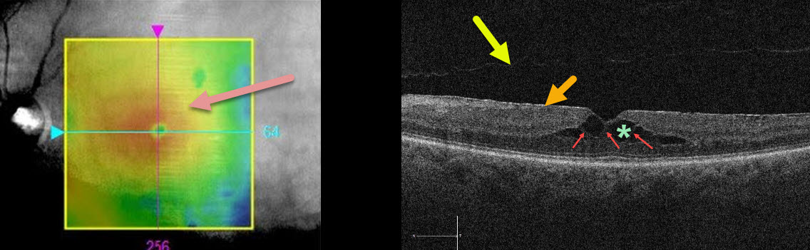
MACULAR PUCKER
Originally posted on @retina.rocks July 3, 2020
This patient was sent in for treatment for possible diabetic macular edema. The retinal thickness map shows central macular thickening.
The OCT B-scan shows an adherent, hyperreflective epiretinal membrane (ERM), a faint posterior vitreous detachment overlying the macula, and central cystic-like inner retinal spaces.
Muller cells, which extend vertically from the internal limiting membrane to the outer retina (external limiting membrane), essentially hold the 3 layers of retinal cells together.
In the central retina, the photoreceptor axons and Muller cells (Henle’s layer, which is the central OPL) are splayed outwards. This creates an intrinsically vertically weakened central macula. The central thin hyperreflective columns are Muller cells that span the region of split OPL.
Unlike exudative intraretinal cysts caused by leaking retinal blood vessels, these hyporeflective spaces are caused by tractional splitting of the outer plexiform layer (OPL) from either aborted vitreomacular traction and/or the ERM.
Learning Points:
When interpreting OCT scans, it is important to consider all the information in the OCT report, ideally by scrolling through the entire macular cube data in the review software.
The macula here is not truly edematous. It is thickened due to a tractional splitting of the retina. For a great discussion of tractional vs exudative cystoid spaces, see Govetto et al AJO 2020;212;43-56.
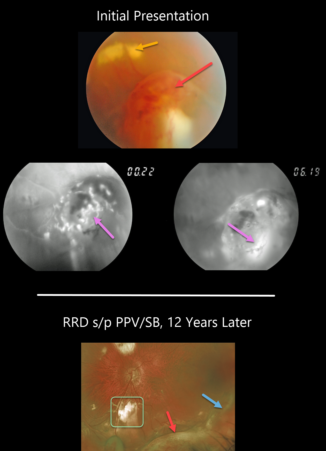
VASOPROLIFERATIVE TUMOR
Originally posted on @retina.rocks June 29, 2020
This 46YO presented with an asymptomatic vasoproliferative tumor (VPT). The VPT initially appeared as a tongue-like, reddish, elevated, peripheral mass with more posterior subretinal lipid. Fluorescein angiography (FA) revealed the vascular nature of the lesion.
He then developed a rhegmatogenous retinal detachment that was repaired with a combined vitrectomy and scleral buckle. The indentation from the buckle is visible more temporally and distinct from the complex enlarged VPT more inferiorly. The subretinal lipid resolved with more extensive subretinal scarring.
Learning Points:
Vasoproliferative tumors are benign globular, dome-shaped lesions arising in the peripheral retina. They can be yellow or red and cause subretinal exudation and detachment, anterior or posterior segment neovascularization, and vitreous hemorrhage.
They can be primary idiopathic lesions or a vascular response to various entities, including intermediate uveitis, retinitis pigmentosa, Coats disease, and prior retinal detachment.
For lesions causing symptomatic exudation or neovascularization, treatment with thermal laser, cryotherapy, and/or anti-VEGF injections is indicated.
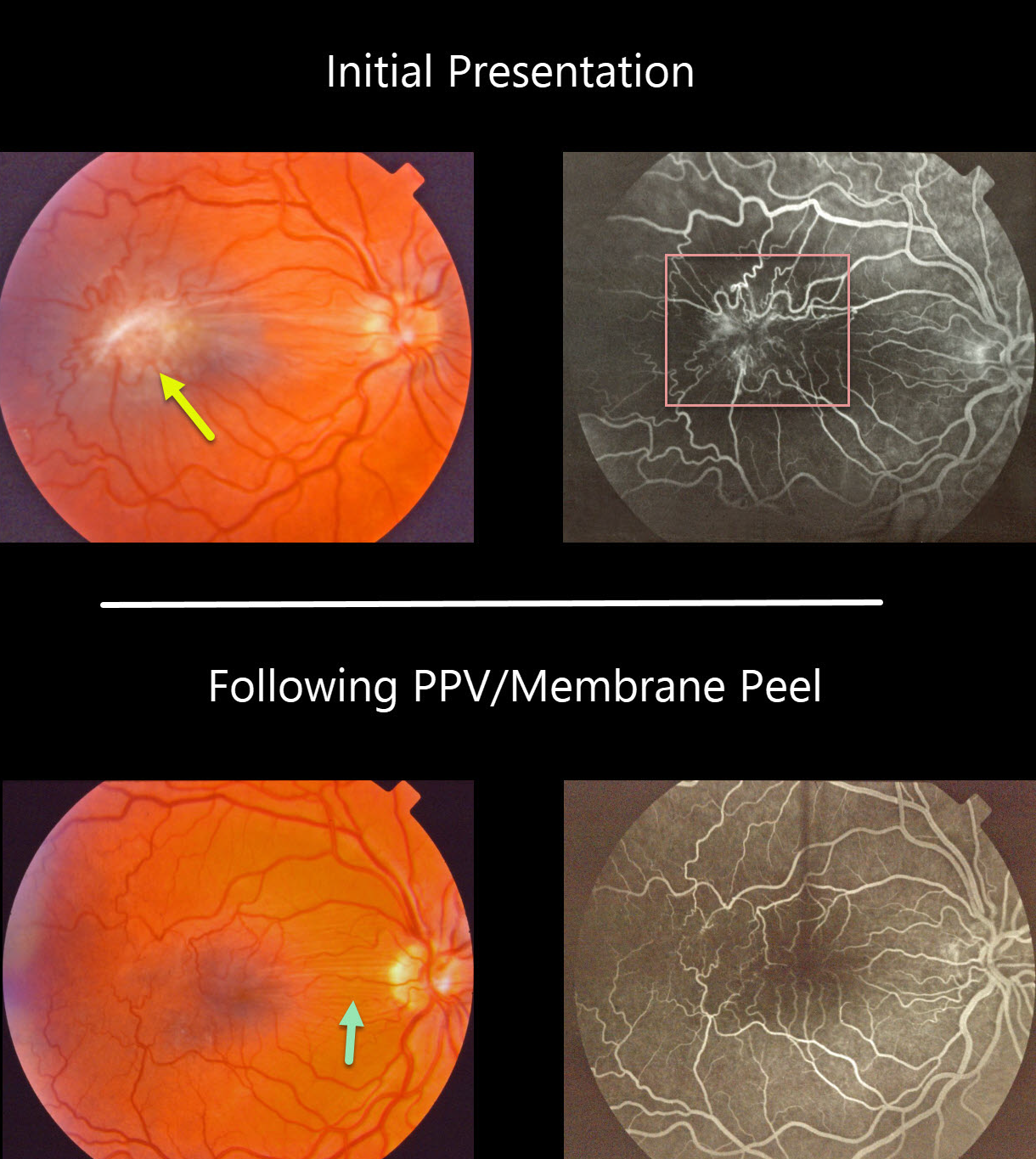
MACULAR PUCKER (PSEUDO-COMBINED HAMARTOMA)
Originally posted on @retina.rocks June 25, 2020
This patient had an isolated severe macular pucker. The fluorescein angiogram shows a vascular hamartomatous component due to severe retinal vascular distortion and leakage. This vascular component, along with the pigmented appearance of the pucker, helps give the false appearance that this was a combined hamartoma of the RPE.
The macular pucker was treated with vitrectomy and membrane peeling. Postoperatively, the retina appears nearly normal, with some residual peripapillary retinal folds and no vascular leakage.
Learning Points:
Hamartomas and severe macular puckers can look very similar, even to some of the most seasoned retina specialists. OCT may be helpful in differentiating these entities (see Ophthalmic Surg Lasers Imaging Retina 2017;48;122-125).
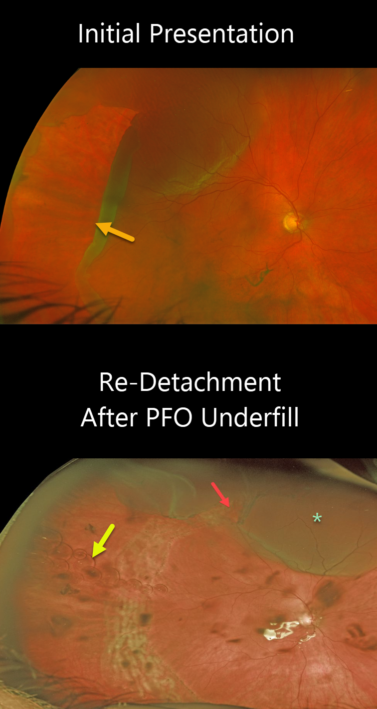
RHEGMATOGENOUS RETINAL DETACHMENT
Originally posted on @retina.rocks June 22, 2020
This patient presented with a retinal detachment caused by a giant retinal tear extending from 5 to 11 o’clock.
Unfortunately, the retina re-detached since the eye was inadvertently underfilled with perfluorocarbon (PFO) at the time of vitrectomy. The superior edge of the giant tear is open, resulting in a superior detachment. There are numerous small PFO bubbles floating along the interface between the PFO meniscus below and the liquid vitreous above.
The patient underwent subsequent surgery to remove the Perfluoron and successfully repair the superior detachment.
Learning Points:
PFO is a transparent, heavier-than-water liquid, often used during vitrectomy for these types of detachments. The PFO liquid is injected over the posterior pole, and as it fills the eye, it simultaneously unrolls the giant tear and flattens the retina. Traditionally, the PFO is exchanged for air, then for a long-acting gas. However, this often results in posterior retinal slippage.
Although PFO is not meant as a long-term vitreous substitute, it can be left in the eye for a week or two. Postoperatively, the patient sleeps on their back, allowing the PFO to tamponade the retina while the retinopexy begins to scar. Patients are then taken back to the operating room, where the PFO can be removed and replaced with a gas or silicone oil, without concern about the retina slipping intraoperatively.
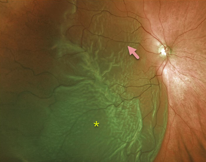
RHEGMATOGENOUS RETINAL DETACHMENT
Originally posted on @retina.rocks June 10, 2020
This patient has an acute, macula-off retinal detachment with a large cilioretinal artery.
Learning Points:
Cilioretinal arteries arise from the choroidal circulation. This results in cilioretinal sparing with central retinal artery occlusion.
Unfortunately, this vessel offers no visual protection against retinal detachment!
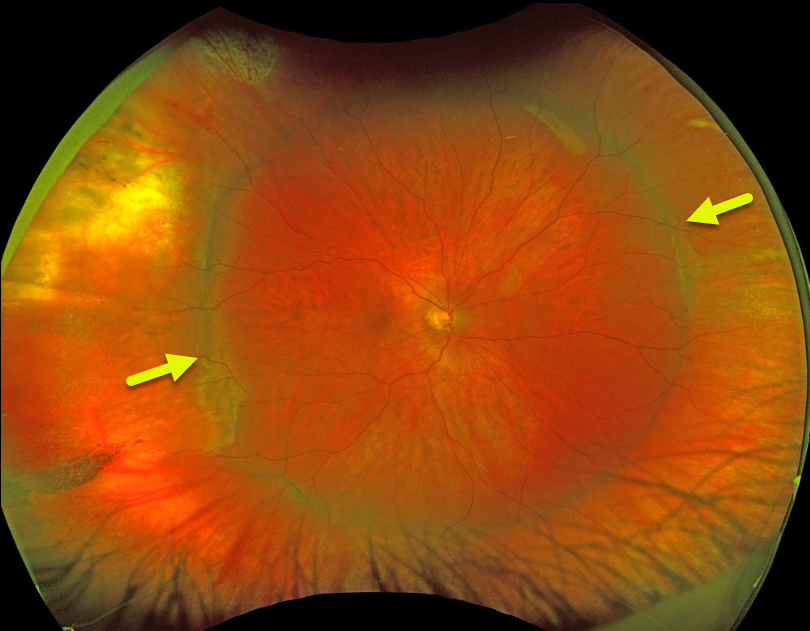
SCLERAL BUCKLE
Originally posted on @retina.rocks June 1, 2020
This patient has a broad encircling scleral buckle that’s beautifully imaged in this Optos photograph.
Learning Points:
The normal neurosensory retina remains attached to the RPE due to numerous physiologic mechanisms, including the inner (retinal vascular endothelium) and outer (tight junctions at the apex of the RPE cells) blood retinal barriers, hyperosmotic choroid passively drawing fluid into the subretinal space, RPE pump, interphotoreceptor matrix, and friction between the RPE microvilli and the photoreceptor outer segments.
Rhegmatogenous retinal detachment (RRD) occurs when liquid vitreous, entering the subretinal space through a retinal break, overwhelms these forces, favoring retinal detachment.
Repairing RRD involves finding and closing all breaks, essentially denying the liquid vitreous access to the subretinal space. The break(s) can be closed internally with a gas bubble via pneumatic retinopexy or vitrectomy surgery, or externally by securing a piece of silicone material to the sclera (scleral buckle). This indents, or buckles, the sclera against the retinal break.
The choice for what procedure to use to reattach the retina is complex, depending on each patient’s unique anatomy and on the surgeon’s preference. Historically, scleral buckling was the only method before vitrectomy surgery was introduced by Machemer in the early 1970’s.
There are advantages and disadvantages to all techniques. With one or more surgeries, there is a high anatomic success rate regardless of how the retina is reattached.
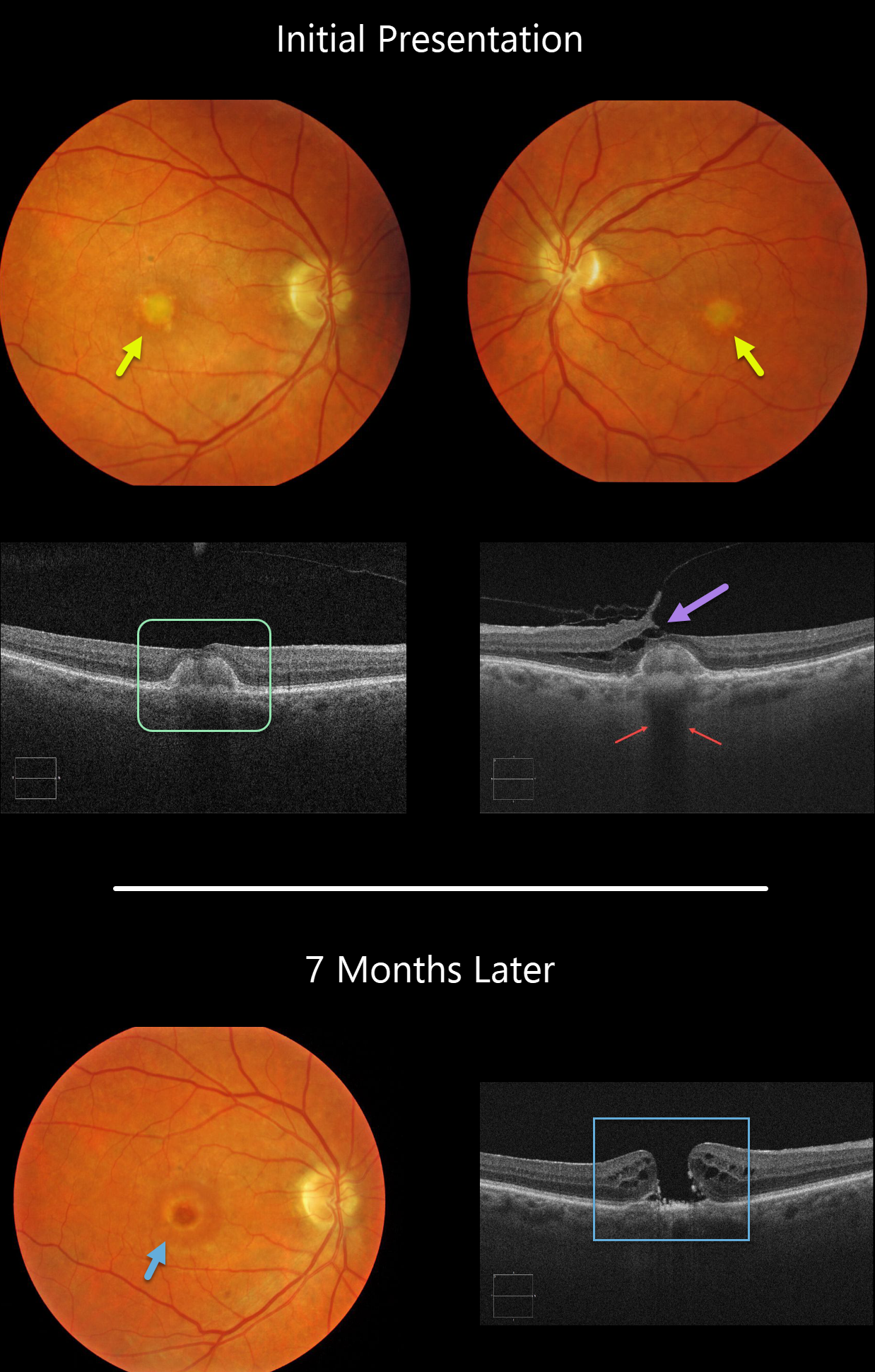
ADULT-ONSET FOVEOMACULAR VITELLIFORM DYSTROPHY WITH SECONDARY MACULAR HOLE
Originally posted on @retina.rocks May 28, 2020
Our patient shows classic findings for adult-onset foveomacular vitelliform dystrophy (Adult Best’s) including small, round, symmetrical subfoveal yellow spots, which is seen on OCT as subretinal hyperreflective material between the RPE and outer retina.
Additionally, the vitelliform lesions provide a great example of shadowing of the underlying choroid. Shadowing occurs when a hyperreflective structure prevents the OCT laser from penetrating to illuminate deeper structures.
A bonus finding in the left eye is focal nasal vitreofoveal traction lifting the retina, splitting the outer plexiform layer and forming a partial tractional lamellar macular hole.
Learning Points:
There are numerous causes for the subfoveal accumulation of yellow material (acquired vitelliform lesions. Adult-onset foveomacular vitelliform dystrophy (Adult Best’s) is associated with a mutation in the BEST1 gene, which encodes for the bestrophin-1 protein.
Bestrophin-1, a calcium-activated chloride channel, is primarily found in the basolateral plasma membrane of the RPE. BEST1 mutations cause a variety of varied phenotypes, also including autosomal recessive bestrophinopathy, best vitelliform macular dystrophy, and autosomal dominant vitreoretinochoroidopathy.
These patients tend to maintain fairly good vision. Outer retinal and RPE atrophy can cause central vision loss, as can the rare development of macular neovascularization. Patients should therefore regularly monitor their vision with an Amsler grid.
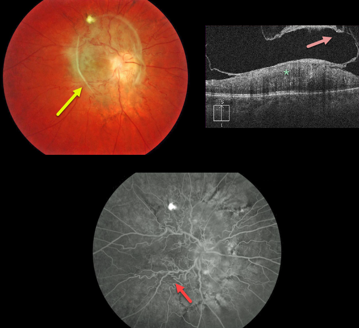
MACULAR PUCKER VS COMBINED HAMARTOMA
Originally posted on @retina.rocks May 25, 2020
The fundus photo shows an unusual peripapillary pigmented epiretinal membrane distorting the underlying retina.
The OCT shows a partially detached epiretinal membrane and marked hyperreflectivity of the inner retina, with loss of normal retinal layer detail.
Fluorescein angiography shows a distorted retinal vasculature with late leakage.
Occasionally, vitrectomy with membrane peeling can be offered, but we are electing to observe this eye since vision has been poor since birth.
Learning Points:
A combined hamartoma of the RPE is a benign congenital lesion composed of glial cells, vascular tissue, and pigmented epithelial cells.
Even though we’re calling this a combined hamartoma, we’re not positive that this may not just be an unusual peripapillary/macular epiretinal membrane.
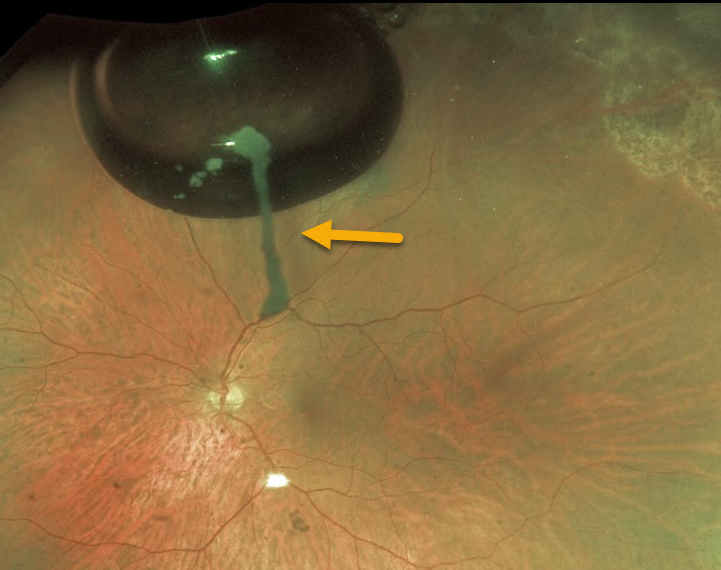
TRIAMCINOLONE-MOXIFLOXACIN (TriMoxi)
Originally posted on @retina.rocks May 22, 2020
Six weeks prior to this retinal detachment repair, the patient underwent uncomplicated cataract surgery. This image shows TriMoxi (triamcinolone acetonide/moxifloxacin) medication literally “hanging on” the absorbing gas bubble!
Learning Points:
During cataract extraction, a commonly used combination of triamcinolone (15 mg/mL) and moxifloxacin (1mg/mL) is injected intracamerally. The medication eventually absorbs completely.
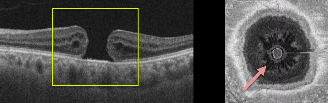
MACULAR HOLE
Originally posted on @retina.rocks May 8, 2020
This patient has a full thickness macular hole without traction. The en face image offers a unique perspective on an otherwise unremarkable case.
Learning Points:
The radiating cysts, as described in Govetto et al AJO 2020;212;43-56, have a sunflower appearance showing features of both exudative and tractional cystoid spaces.
The macular hole should be hyporeflective centrally. It is hyperreflective in this image due to the OCT software mis-identifying the RPE as being within the mid-retina.
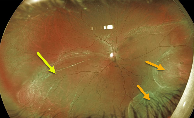
RHEGMATOGENOUS RETINAL DETACHMENT
Originally posted on @retina.rocks May 6, 2020
This 16YO presented with a chronic inferior macula-off rhegmatogenous retinal detachment (RRD). Vision was 20/200.
What’s most unique about this detachment is the rare appearance of intraretinal macrocysts inferonasally.
These cysts are a sign of very long-standing retinal detachments. The multiple demarcation lines also indicate chronicity.
Learning Points:
During RRD repair, the cysts require no special modifications and resolve following retinal reattachment.
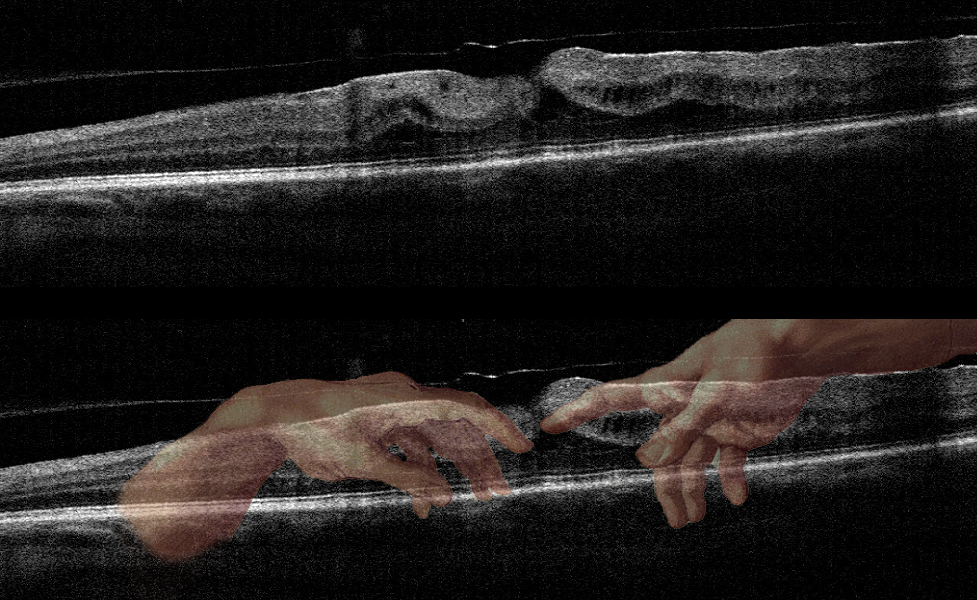
MICHELANGELO MACULOPATHY
Originally posted on @retina.rocks May 1, 2020
This patient has an epiretinal membrane with some vitreomacular traction.
This case was submitted by Dr. Jennifer Reynolds. The “artistic” mind of the submitting doctor, noticed the hand of the Creator in this scan!
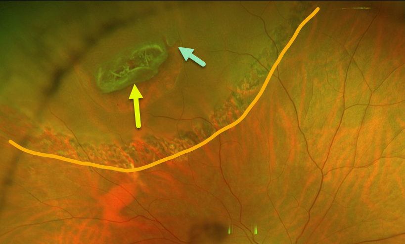
LATTICE DEGENERATION
Originally posted on @retina.rocks April 23, 2020
This patient had an asymptomatic peripheral lattice-related retinal detachment. Note that the entire lattice lesion represents the flap of the tear, with an adjacent secondary smaller tear.
The detachment was demarcated with laser photocoagulation and has remained stable since.
Learning Points:
Retinal detachment occurs in 1 in 10,000 people per year. Lattice is found in 10% of the general population, especially in high myopes.
Since about one-third of patients with retinal detachment have lattice as the cause, the question always arises, especially in patients like this: should prophylactic retinopexy be performed? The short answer is that asymptomatic lattice generally should not be treated.
However, this patient had lattice-related retinal detachment and thus underwent laser demarcation.
An especially relevant article for this topic is by Vrabec and Baumal, Ophthalmology 2000;107;1063-1067.
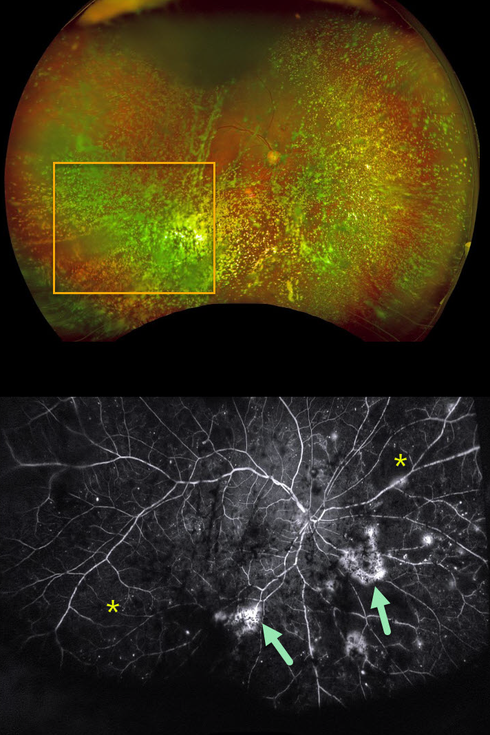
ASTEROID HYALOSIS OBSCURING OCCULT PROLIFERATIVE DIABETIC RETINOPATHY (PDR)
Originally posted on @retina.rocks April 17, 2020
Examining the fundus with dense asteroid hyalosis, as in this diabetic patient, can be extremely challenging.
Fluorescein angiography (FA) and OCT are great modalities since they’re both able to visualize the retina when the clinical exam can’t.
FA was critical in diagnosing proliferative retinopathy with peripheral ischemia in this patient.
Learning Point:
Asteroid hyalosis is caused by calcium-lipid complexes suspended throughout the vitreous. These white opacities resemble stars on a night sky – hence the name.
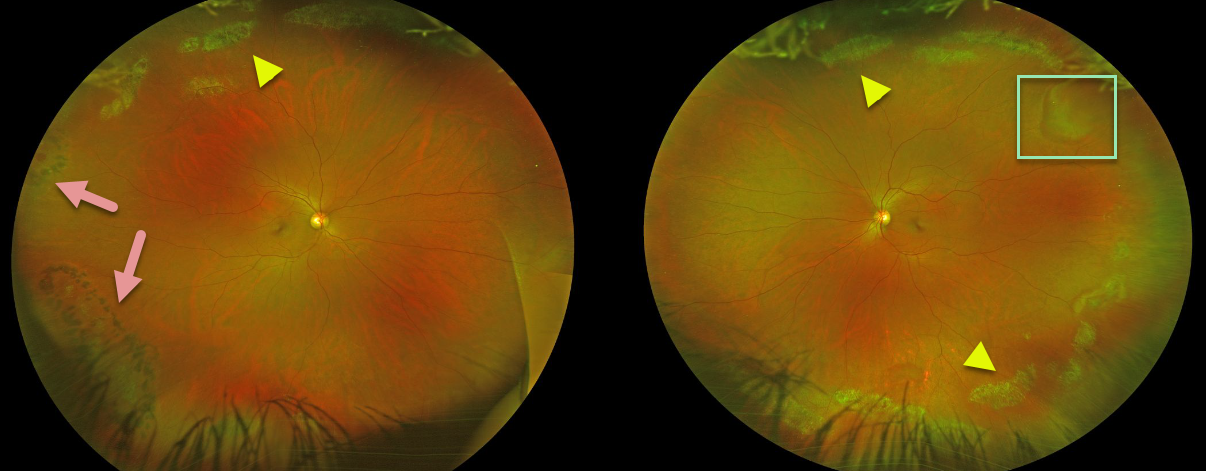
LATTICE DEGENERATION
Originally posted on @retina.rocks April 14, 2020
Beautiful Optos images of this patient demonstrate extensive bilateral lattice degeneration.
Prophylactic laser was placed at an outside institution around some of the lattice lesions in the right eye inferotemporally and around an atrophic hole temporally.
There is a pseudo-retinal tear superotemporally in the left eye from a patch of white without pressure.
Learning Points:
White, fibrosed vessels often criss-cross the lesions, which lead to the name, lattice. There is vitreous liquefaction overlying the thinned retina with a firm vitreo-retinal adhesion along the borders of the lesions. This firm vitreoretinal adhesion, along with the often present atrophic holes, is what can predispose these eyes to retinal detachment.
Retinal detachment occurs in 1 in 10,000 people per year. Lattice is found in 10% of the general population, especially in high myopes. Since about one-third of patients with retinal detachment have lattice as the cause, the question always arises, especially in patients like this: should prophylactic retinopexy be performed? The short answer is that asymptomatic lattice generally should not be treated.
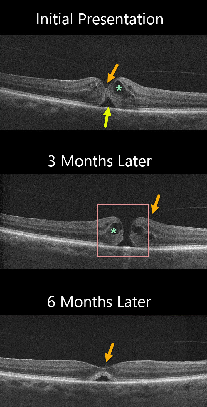
MACULAR HOLE
Originally posted on @retina.rocks April 3, 2020
The patient was initially diagnosed with idiopathic cystoid macular edema (CME) and a small foveal detachment with vitreomacular adhesion (VMA). Vision was 20/50. Although the vitreous was attached centrally, a foveal depression remained which would be unusual if vitreomacular traction (VMT) were the cause.
The patient was observed without treatment, and 3 months later vision decreased to 20/60. There was a small full-thickness macular hole with an attached hyaloid. CME was still present. The patient elected to observe the hole.
Another three months later vision improved to 20/40 and the hole spontaneously closed with a normal foveal contour and resolved CME.
The central vitreous remained attached.
Learning Points:
The differential diagnosis for a localized serous foveal detachment include central serous chorioretinopathy, VMT, prior macular hole surgery, and resolved acquired vitelliform lesions.
This patient has a faintly visualized posterior hyaloid that is still attached centrally making VMT unlikely. The choroid probably has a normal thickness which makes central serous less likely, and moreover, the fellow eye is normal.
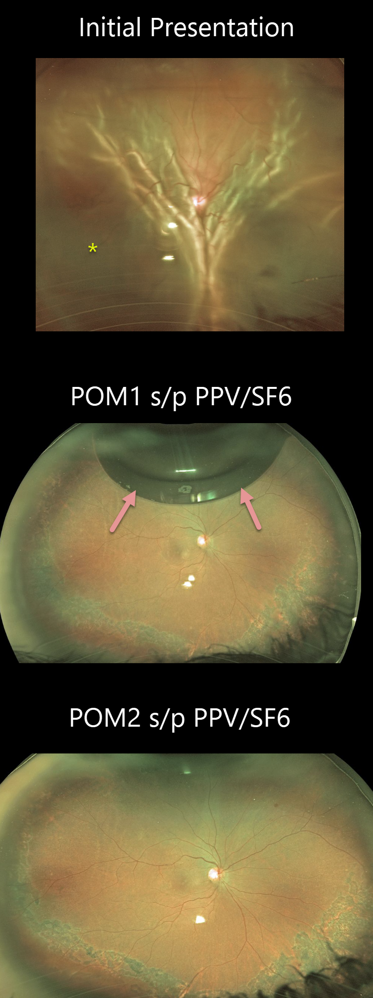
RHEGMATOGENOUS RETINAL DETACHMENT
Originally posted on @retina.rocks March 25, 2020
This patient presented with a total rhegmatogenous retinal detachment in the right eye.
The patient underwent repair with pars plana vitrectomy and SF6 gas. The Optos images show a small residual gas bubble at 1-month post-vitrectomy. Two months post-op, the patient’s vision is 20/200.
Learning Points:
Long-acting intraocular gas is routinely used in vitrectomy for rhegmatogenous retinal detachment repairs. C3F8 and sulfur hexafluoride (SF6) are used due to greater longevity compared to air. C3F8 lasts in the vitreous for 2-3 months, and SF6 lasts for several weeks.
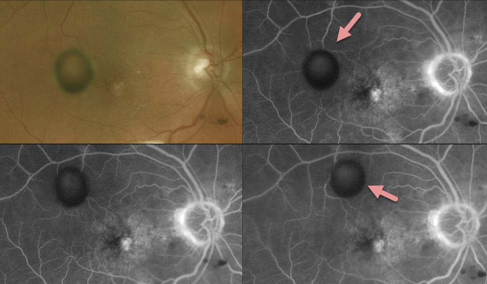
VITREOUS CYST
Originally posted on @retina.rocks March 20, 2020
This patient has a vitreous cyst in the right eye, which can be seen moving around on fluoroscein angiography (FA) as it blocks different areas of the retina.
Learning Points:
Vitreous cysts are extremely rare, idiopathic asymptomatic and usually diagnosed as an incidental finding as in this patient.
If symptomatic, the cyst can be popped with a YAG laser.
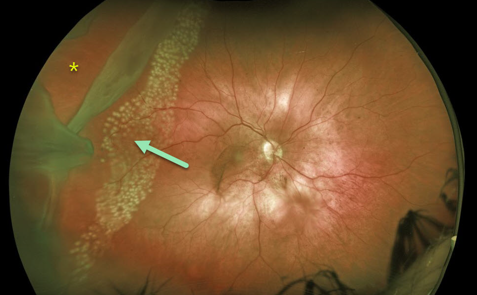
GIANT RETINAL TEAR
Originally posted on @retina.rocks March 19, 2020
This patient presented with a giant retinal tear. Against medical advice, the patient adamantly refused vitrectomy on multiple occasions, so we decided to demarcate it with laser photocoagulation. As expected, the detachment progressed through the laser scarring with the development of proliferative vitreoretinopathy. He subsequently needed several vitrectomy procedures, including retinectomy and silicone oil to obtain final retinal reattachment.
Learning Points:
Untreated retinal tears have a very high risk for progressing to retinal detachment. Urgent laser photocoagulation or cryotherapy prevents liquid vitreous from leaking thru the break. Patients need to be followed closely postoperatively to be sure no new breaks develop and that chorioretinal scarring surrounds the break.
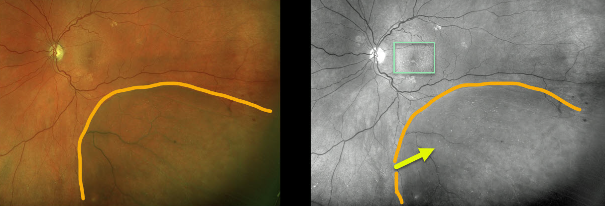
DEGENERATIVE RETINOSCHISIS
Originally posted on @retina.rocks March 11, 2020
This patient with degenerative retinoschisis has extensive refractile dots, thought to be Muller cell footplates, which are well-visualized in both the Optos pseudocolor and green channel images.
This diabetic patient also has subretinal drusenoid deposits that are best seen with the green channel.
Learning Points:
Degenerative retinoschisis is virtually never prophylactically treated (see Byer 1986;93;1127-1137).
Patients can rarely develop a rhegmatogenous retinal detachment when liquid vitreous accesses the subretinal space thru both an inner and outer layer hole.
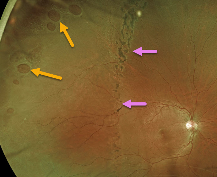
RHEGMATOGENOUS RETINAL DETACHMENT
Originally posted on @retina.rocks March 4, 2020
This patient with multiple retinal holes has a well demarcated retinal detachment.
Learning Points:
The normal neurosensory retina remains attached to the RPE due to numerous physiologic mechanisms including the inner (retinal vascular endothelium) and outer (tight junctions at the apex of the RPE cells) blood retinal barriers, hyperosmotic choroid passively drawing fluid into the subretinal space, RPE pump, interphotoreceptor matrix, and friction between the RPE microvilli and the photoreceptor outer segments.
Rhegmatogenous retinal detachment (RRD) occurs when liquid vitreous, entering the subretinal space through a retinal break, overwhelms these forces favoring retinal detachment.
Pigmented demarcation lines can form in a chronic RRD and limit the spread of subretinal fluid.
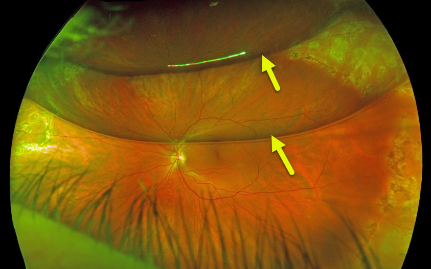
RHEGMATOGENOUS RETINAL DETACHMENT
Originally posted on @retina.rocks February 25, 2020
This patient had a rhegmatogenous retinal detachment that was repaired with pars plana vitrectomy and C3F8 gas.
The patient’s bubble has broken into 2 large bubbles, each with its own beautiful retinal reflection.
Learning Points:
Long-acting intraocular gas is routinely used in vitrectomy for rhegmatogenous retinal detachment repairs. C3F8 and sulfur hexafluoride (SF6) are used due to greater longevity compared to air. C3F8 lasts in the vitreous for 2-3 months, and SF6 lasts for several weeks.
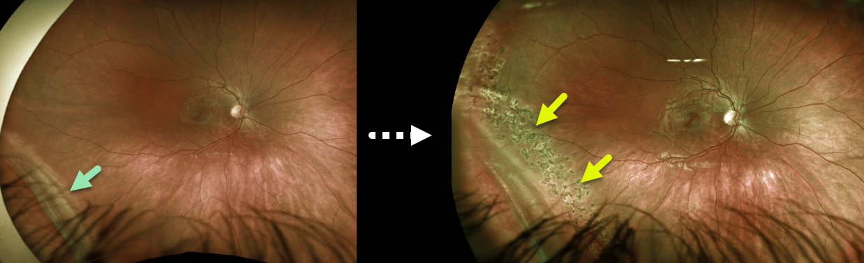
RETINAL DIALYSIS
Originally posted on @retina.rocks February 17, 2020
This healthy 9YO girl presented with an asymptomatic retinal dialysis. The dialysis was demarcated with laser to prevent her from developing a retinal detachment.
Learning Points:
A retinal dialysis is a retinal break at the ora serrata, is usually inferotemporal and can either develop spontaneously (usually in young men) or following trauma.
Dialyses are virtually always treated due to the risk of retinal detachment.
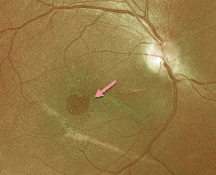
VALENTINE RETINOPATHY
Originally posted on @retina.rocks February 14, 2020
This patient has a heart-shaped macular hole just in time for Valentine’s Day!
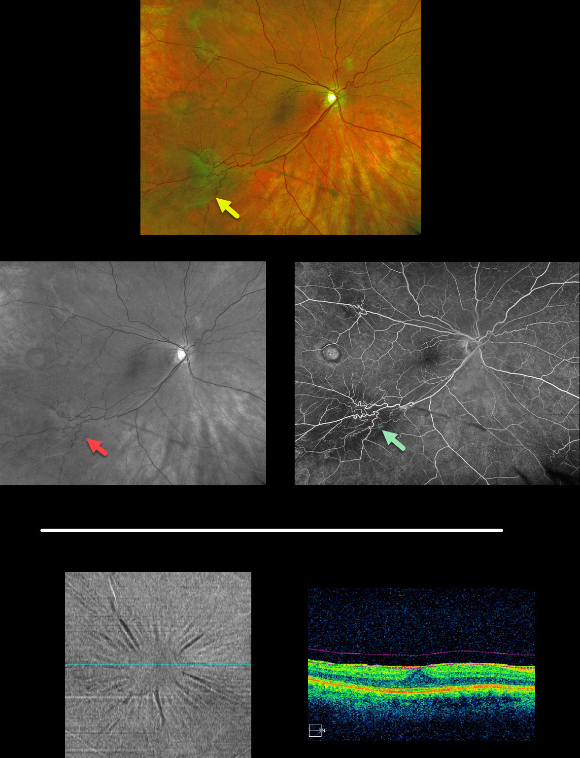
MACULAR PUCKER
Originally posted on @retina.rocks February 12, 2020
Multifocal images of an extramacular epiretinal membranes with an unrelated temporal retinal pigmented epithelial detachment are displayed here (Optos, red-free, and fluoroscein angiography).
BONUS: The second set of images is a different patient with a macular pucker that is well-visualized using OCT en face analysis, which provides transverse views of the retinal and choroidal layers using user-specified depths and thickness. This is an underutilized yet fantastic tool that gives another perspective for many chorioretinal disorders.
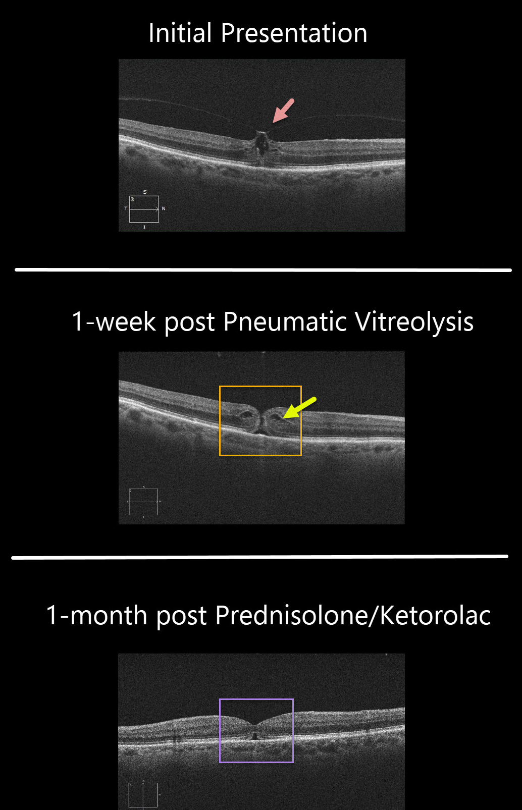
VITREOMACULAR TRACTION + MACULAR HOLE
Originally posted on @retina.rocks February 11, 2020
This patient’s symptomatic 20/80 vitreomacular traction was treated with pneumatic vitreolysis, where a small gas bubble was injected into the vitreous cavity to induce a posterior vitreous detachment. The patient wished to try this less invasive option instead of vitrectomy.
Although the traction successfully released 1 week postop, the patient developed an immediate macular hole with 20/100 vision.
Since the hole was tiny with some cystoid edema (CME), a trial of topical prednisolone acetate 1% and ketorolac was given.
One month later, the CME resolved and the hole closed with improvement to 20/60.
Learning Points:
Topical prednisolone acetate 1% and ketorolac is a newly described treatment that can occasionally be tried for macular holes without traction.
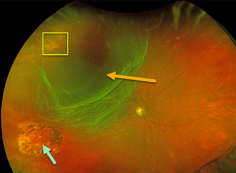
RHEGMATOGENOUS RETINAL DETACHMENT
Originally posted on @retina.rocks February 4, 2020
This patient presented with a new retinal tear causing retinal detachment in an eye with previously lasered operculated hole.
Learning Points:
The normal neurosensory retina remains attached to the RPE due to numerous physiologic mechanisms including the the inner (retinal vascular endothelium) and outer (tight junctions at the apex of the RPE cells) blood retinal barriers, hyperosmotic choroid passively drawing fluid into the subretinal space, RPE pump, interphotoreceptor matrix, and friction between the RPE microvilli and the photoreceptor outer segments.
Rhegmatogenous retinal detachment (RRD) occurs when liquid vitreous, entering the subretinal space through a retinal break, overwhelms these forces favoring retinal detachment.
Repairing RRD involves finding and closing all breaks, essentially denying the liquid vitreous access to the subretinal space. The break(s) can be closed internally with a gas bubble with pneumatic retinopexy or vitrectomy surgery, or externally by sewing a piece of silicone material to the eye wall (scleral buckle). This indents, or buckles, the sclera up against the retinal break.
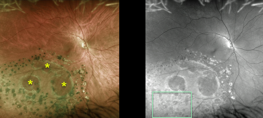
DEGENERATIVE RETINOSCHISIS
Originally posted on @retina.rocks January 23, 2020
This patient has degenerative retinoschisis. A doctor at an outside practice placed prophylactic barrier laser around the outer layer holes to prevent retinal detachment.
There are also extensive smaller inner layer holes that are best seen in the Optos green-channel image.
Learning Points:
Degenerative retinoschisis is virtually never prophylactically treated (see Byer 1986;93;1127-1137). Patients can rarely develop a rhegmatogenous retinal detachment when liquid vitreous accesses the subretinal space thru both an inner and outer layer hole.
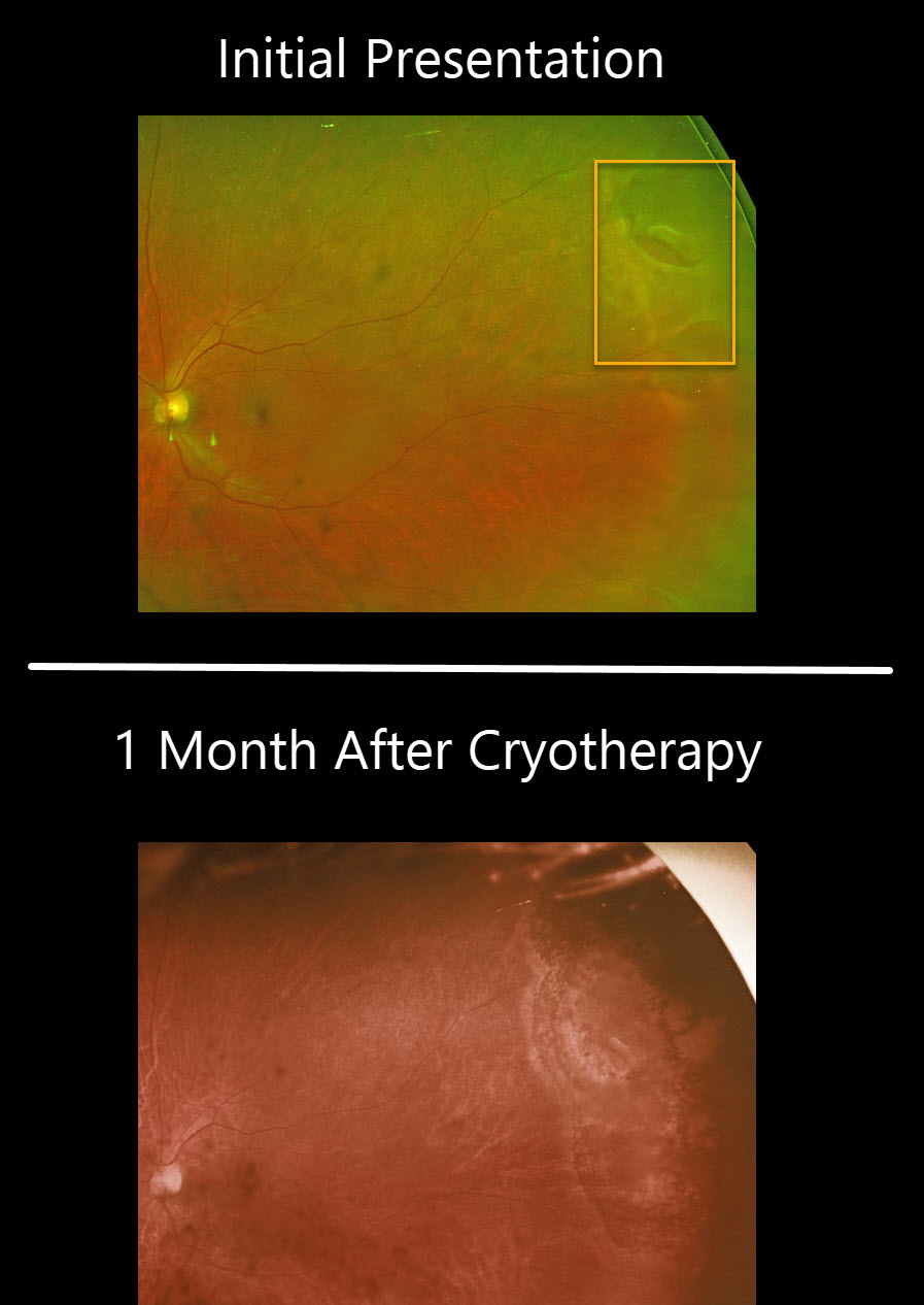
RETINAL BREAK
Originally posted on @retina.rocks January 14, 2020
This patient had a symptomatic retinal tear superotemporally in the left eye. The tear was treat with cryotherapy and fundus image shows the appearance of the retina one month later.
Learning Points:
Untreated retinal tears have a very high risk for progressing to retinal detachment. Urgent laser photocoagulation or cryotherapy prevents liquid vitreous from leaking thru the break. Patients need to be followed closely postoperatively to be sure no new breaks develop and that chorioretinal scarring surrounds the break. Unlike thermal laser demarcation, the cryotherapy applicatiohns need to surround the break with confluent scarring in order to adequately prophylax against retinal detachment.
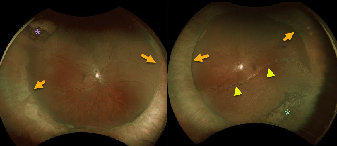
RHEGMATOGENOUS RETINAL DETACHMENT
Originally posted on @retina.rocks January 8, 2020
This patient has bilateral scleral buckles following prior rhegmatogenous retinal detachment surgeries.
Cryotherapy scarring is present superotemporally OD and laser scarring is present inferotemporally OS around a few tiny retinal holes that caused the initial detachment.
There is also a large demarcation line OS indicating that the left detachment was present for some time before being repaired.
Learning Points:
Primary scleral buckle can be used to repair certain types of rhegmatogenous retinal detachments.
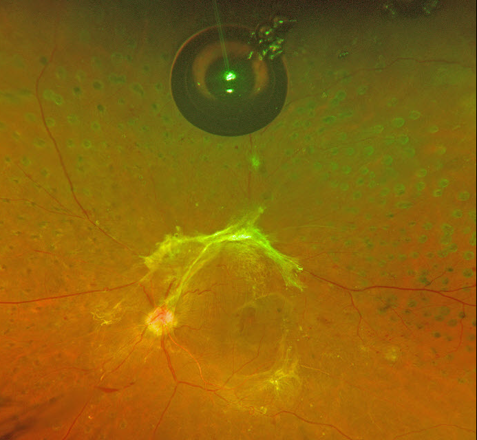
SILICONE OIL DROPLET
Originally posted on @retina.rocks January 2, 2020
This patient with proliferative diabetic retinopathy with tractional detachment was noted to have a large asymptomatic silicone oil bubble following multiple prior Avastin injections.
Learning Points:
The older insulin syringe needles provided by compounding pharmacies for the Avastin (bevacizumab) injections were lubricated with silicone.
Many eyes receiving intravitreal Avastin developed small vitreous silicone droplets, although the droplet is unusually large in this patient. Since then, these insulin syringe needles have been discontinued.
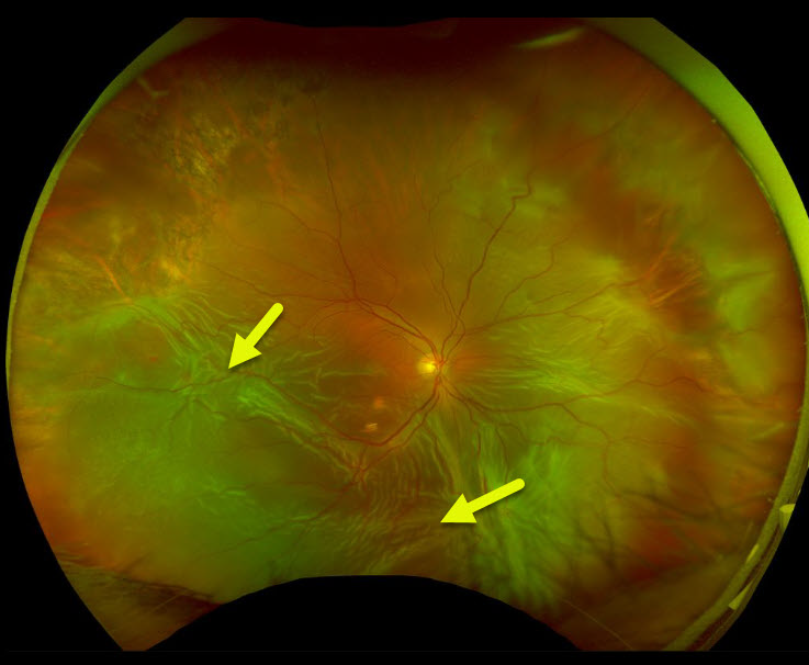
PROLIFERATIVE VITREORETINOPATHY
Originally posted on @retina.rocks December 31, 2019
This eye has a total retinal detachment, with numerous foci of fibrotic retina in the inferior hemisphere.
Learning Points:
PVR, or proliferative vitreoretinopathy, consists of fibrotic membranes growing on the retina surface.
It is the primary cause of retinal reattachment surgical failure and occurs in 5-10% of detachments.


