

07 Trauma
<< Back to Cases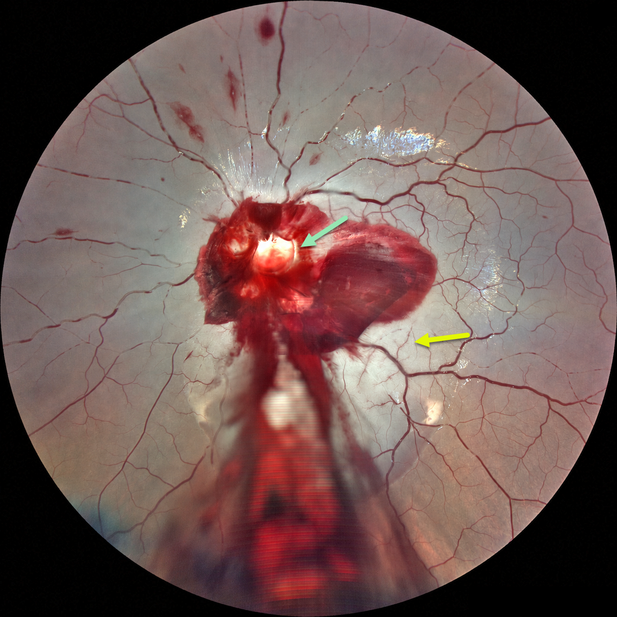
ACUTE OPTIC NERVE AVULSION WITH SECONDARY CRAO
Ayushi Gupta and Vishal Agrawal
Originally posted on @retina.rocks September 8, 2026
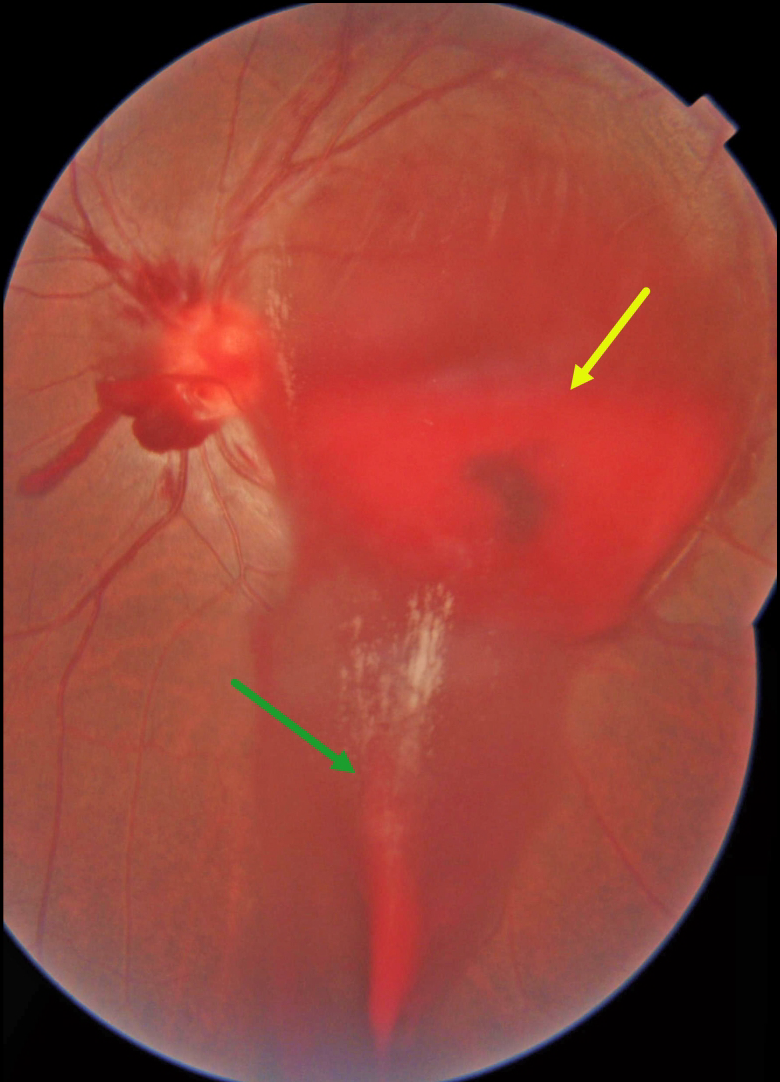
This 6YO boy developed sudden and complete loss of vision in his left eye immediately following blunt trauma. He was riding a bicycle as a pillion passenger when the bike stopped suddenly, throwing his face against the handlebar. Vision was no light perception. Anterior segments were normal.
Fundus photography shows preretinal blood surrounding the nerve, extending through the macula and into the inferior periphery. A pale-yellow cavity is noted where the nerve head should be. A fresh central retinal artery occlusion (CRAO) with a stagnant blood column is noted, along with some scattered inner retinal hemorrhages. There were no other physical or neurologic injuries.
Learning Points:
Traumatic optic nerve head avulsion is a rare and visually devastating traumatic event in which the optic nerve is forcibly separated from the globe at its junction with the sclera, typically resulting in immediate, severe, and irreversible vision loss. The mechanism involves sudden forceful rotation or anterior displacement of the globe following blunt periorbital trauma, including projectiles, falls, or finger-poke injuries, whereby the globe moves anteriorly or rotationally while the optic nerve remains relatively fixed, causing violent separation. Fundoscopic examination may reveal a partial or complete tear of the optic nerve head from the sclera (Foster et al, Arch Ophthalmol 1997;115:623-630). Avulsion can be partial or complete, and may be associated with a CRAO, as was noted in our patient.
COMMOTIO RETINAE WITH TRAUMATIC RETINAL BREAK
Dibya Prabha
Originally posted on @retina.rocks August 7, 2026
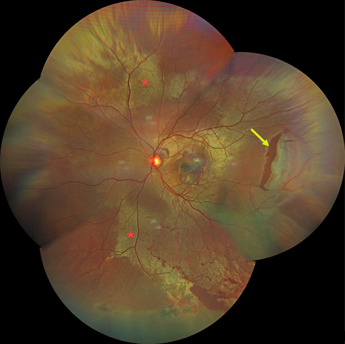
This 38YO male presented with sudden vision loss in his left eye immediately following blunt trauma from a cricket bat 2 days earlier. Vision was 20/200.
Color fundus photography shows extensive commotio retinae throughout the posterior pole, along with a large retinal tear in the temporal midperiphery. Oral and topical steroids were prescribed, and the retinal break was surrounded with laser photocoagulation.
Learning Points:
Commotio retinae is caused by direct, blunt eye trauma. The outer retinal whitening is due to shearing of the photoreceptor outer segments. The term “Berlin’s edema” is a misnomer and should not be used, as the retina is not edematous. The acute white retinal changes usually resolve within several weeks. With resolution, the fundus appearance can normalize. With more severe injury, outer retinal loss can allow RPE cells to migrate into the retina, giving a pseudo-retinitis pigmentosa appearance. Patients can also develop retinal breaks and detachment.
CHOROIDAL RUPTURE
Shraddha Raj Shrivastava and Manish Nagpal
Originally posted on @retina.rocks August 4, 2026
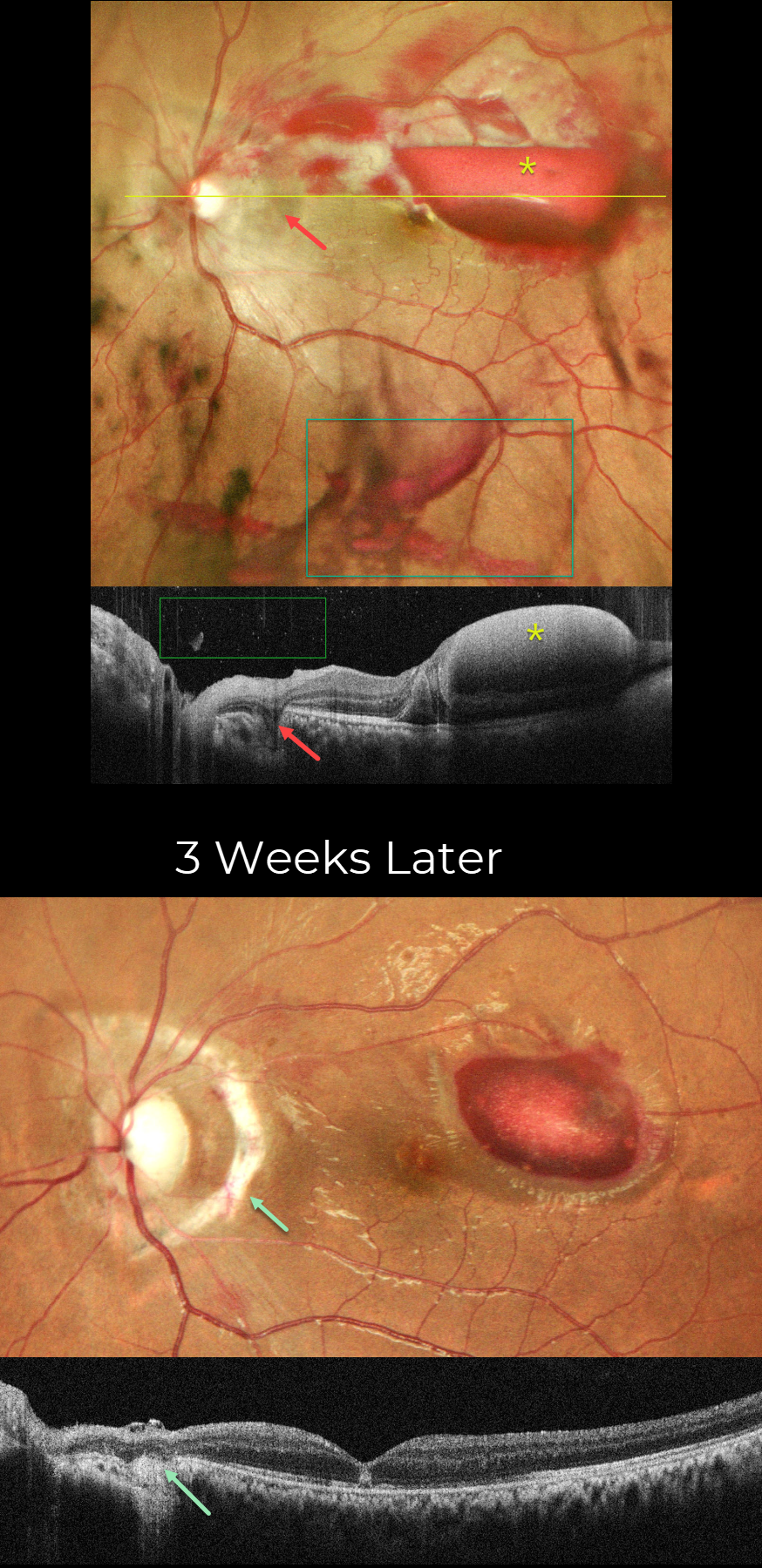
This 19YO male presented with vision loss in his left eye 2 days following blunt trauma from a tennis ball. Vision was counting fingers. Slit lamp examination revealed anterior chamber inflammation, traumatic mydriasis, and nasal subluxation of the crystalline lens.
Pseudocolor SLO imaging shows scattered retinal, sub-internal limiting membrane (ILM), and vitreous blood. The acute choroidal rupture appears as a crescent of increased subretinal pigmentation that arches around the nerve temporally and superiorly. Superior and superotemporal macular commotio retina is faintly visible. On OCT scanning, the vitreous and sub-ILM blood appear hyperreflective. The choroidal rupture appears as a hyporeflective defect extending from the outer retina to the sclera.
Topical steroids, atropine, and a short course of oral steroids were prescribed. Three weeks later, vision improved to 20/200. A prominent depigmented choroidal rupture with bare sclera surrounds the nerve nasally 180 degrees. The vitreous and retinal hemorrhages are virtually completely resolved, and the sub-ILM is decreasing. On OCT, the choroidal rupture shows loss of outer retinal and RPE band detail, with diffuse underlying choroidal hyperreflectivity. There is variable outer retinal band loss in the macula with a central hyperreflective lesion in the outer nuclear layer.
Learning Points:
An acute submacular hemorrhage following blunt trauma almost always indicates an underlying choroidal rupture. As the blood clears, the rupture appears as a curvilinear or crescent-shaped yellowish-white lesion. It occurs due to rapid compression-expansion stress on Bruch’s membrane, which is less elastic and has less tensile strength than the sclera, leading to its fracture. The final vision is usually good unless the rupture extends through the macular center. These patients need to be followed long-term due to the increased risk of macular neovascularization.
PURTSCHER RETINOPATHY
Abhishek Karra and Ravindra Karra
Originally posted on @retina.rocks July 28, 2026
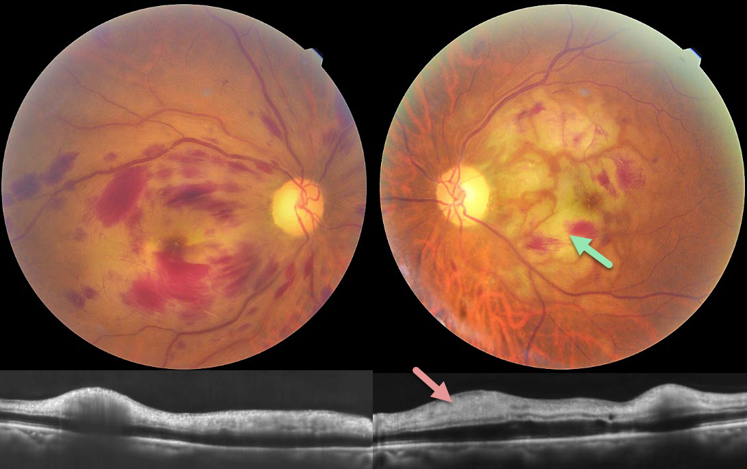
This 85YO male lost vision in each eye following a fall around 1 week back with multiple rib fractures. Vision was hand motion OU.
Color photography shows bilateral macular retinal hemorrhages with patches of white ischemic retina, most prominent in the left macula. OCT scanning shows a thickened hyperreflective inner retina bilaterally.
Learning Points:
Purtscher retinopathy was first described by Otmar Purtscher in 1910. Although originally described in a man who fell from a tree with cranial trauma, these white patches of retinal ischemia and hemorrhages are found in numerous other etiologies, including pancreatitis, chest trauma, and collagen vascular diseases. The findings are called Purtscher retinopathy when due to trauma (as in our patient), and Purtscher-like retinopathy when seen from other causes.
The underlying cause is unknown, but the findings are thought to result from leukoembolization due to complement activation. The areas of ischemia include more superficial nerve fiber layer infarcts (cotton wool spots) and deeper ischemia involving the middle retinal layers.

COMMOTIO RETINAE WITH CONTUSIVE RPE FLUID AND CHOROIDAL RUPTURE
Brahim Alfonso Khouri Lopez, Julian Villarreal, Alexis Ramirez, and Angie Maldonado
Originally posted on @retina.rocks July 22, 2026
This 13YO boy presented 2 days following a slingshot injury to his right eye. Vision was counting fingers.
Color photography shows an opaque white discoloration below the retina throughout the posterior pole. Diffuse faint commotio retinae is also noted in the superior retinal periphery. There is a large choroidal rupture inferior to the macula, along with an inner retinal hemorrhage inferonasal to the nerve. OCT scanning shows variably opaque central subretinal fluid and marked cystic edema and disorganization of the nasal outer retinal layers external to the outer plexiform layer. Unfortunately, he was immediately lost to follow-up.
Learning Points:
Commotio retinae is caused by direct, blunt eye trauma. The outer retinal whitening is due to shearing of the photoreceptor outer segments. The term “Berlin’s edema” is a misnomer and should not be used, as the retina is not edematous. Our patient’s fluid is likely due to a contusive RPE injury that disrupted the outer blood-retinal barrier, allowing fluid to accumulate in the subretinal space and outer retina (Mishra et al, Journal of Vitreoretinal Diseases 2021;5:165-169).
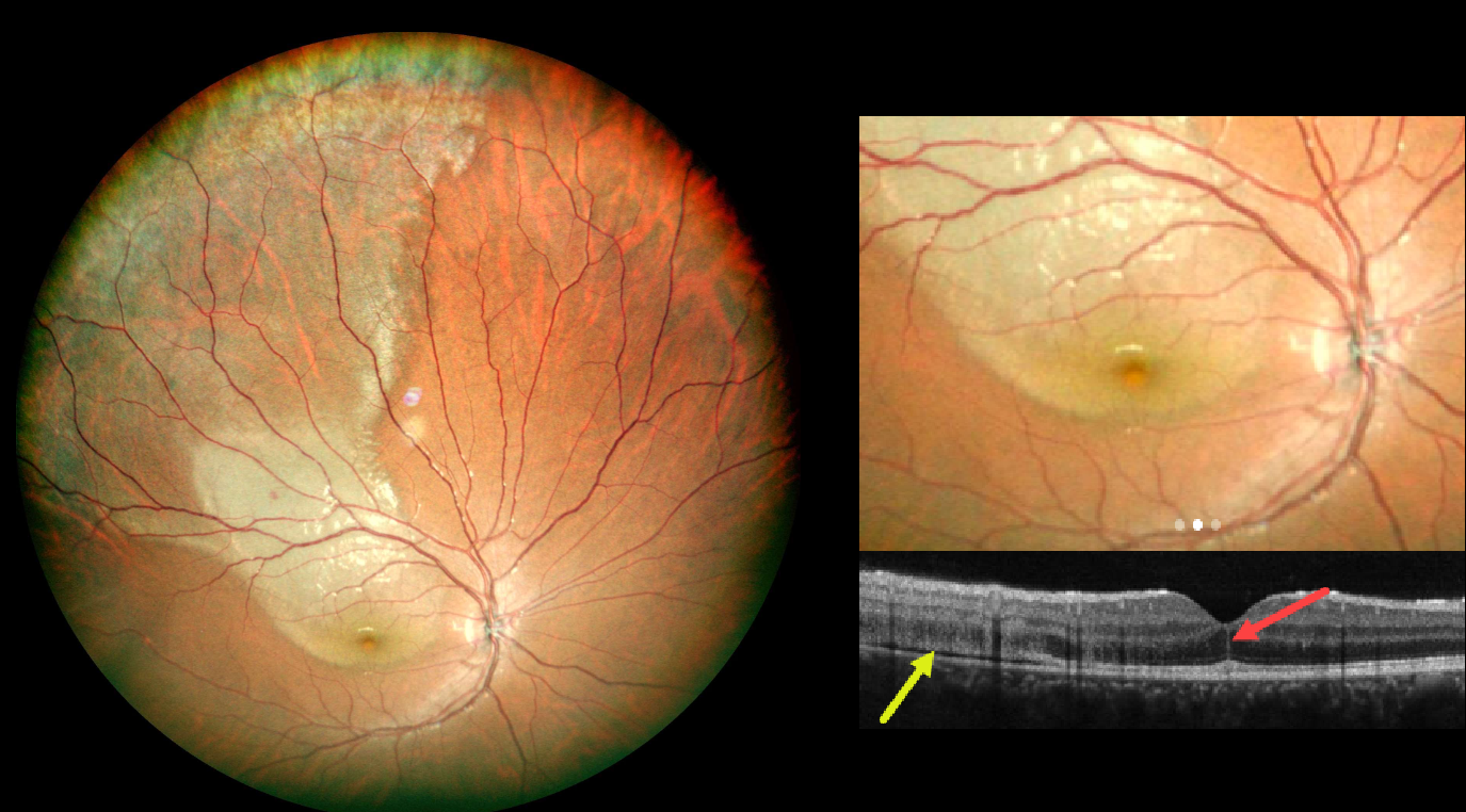
COMMOTIO RETINAE WITH FOVEAL CRACK SIGN
Andree Henaine-Berra and Gerardo Garcia-Aguirre
Originally posted on @retina.rocks July 7, 2026
This 15YO boy presented 1 day following blunt injury to his right eye from a soccer ball. Despite complaints of a central scotoma, vision was 2025.
Color photography shows a broad area of commotio retinae extending from the central macula into the superotemporal periphery. OCT scanning shows disruption of the outer retinal bands and shallow subretinal fluid temporally. A vertical hyperreflective line is present in the central outer nuclear layer.
Learning Points:
The foveal crack sign (FCS) is a central foveal hyperreflective vertical line, first described by Ishibashi et al. as an OCT biomarker for future macular hole development following vitrectomy for rhegmatogenous retinal detachment (AJO 2020;218:192-198). It has since been described in numerous other conditions, including macular pucker, diabetic macular edema, idiopathic macular telangiectasia type 2, vitreomacular traction, NAION with CRAO, AMD, non-infectious uveitis, Coats disease, and commotio retina (Kayabasi et al, Med Hypothesis Discov Innov Ophthalmol 2024;13:129-138).
Commotio retinae is caused by direct, blunt eye trauma. The outer retinal whitening is due to shearing of the photoreceptor outer segments. The term “Berlin’s edema” is a misnomer and should not be used, as the retina is not edematous. The acute white retinal changes usually resolve within several weeks. With resolution, the fundus appearance can normalize. With more severe injury, outer retinal loss can allow RPE cells to migrate into the retina, giving a pseudo-retinitis pigmentosa appearance. Patients can also develop retinal breaks and detachment.
We will follow our patient closely and hope that the FCS is not an omen for a future macular hole.
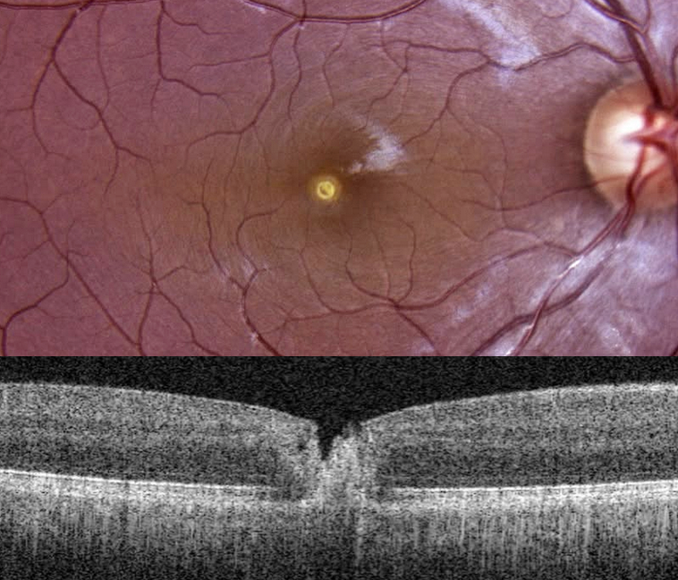
LASER MACULOPATHY
The European VitreoRetinal Society (EVRS) and Aniruddh Soni
Originally posted on @retina.rocks June 12, 2026
This 26YO male DJ noticed immediate blurred vision in his right eye after looking at a laser disco light during an event 5 days earlier. Vision was 20/120 OD.
Color photography shows a small, round, yellow lesion involving or quite close to the foveal center. OCT scanning shows full-thickness foveal disruption and hyperreflectivity.
Learning Points:
Most cases of laser maculopathy in the literature are caused by purposeful or inadvertent exposure to laser pointers (Bhavsar et al, Survey Ophthalmology 2021;66:231-260). However, more recently, there have been reports of macular injury from exposure to high-power lasers during concerts or from DJ laser machines (Perz-Montano et al, Doc Ophthalmol 2019;138:71-76). Most eyes recover fairly good vision with conservative management.
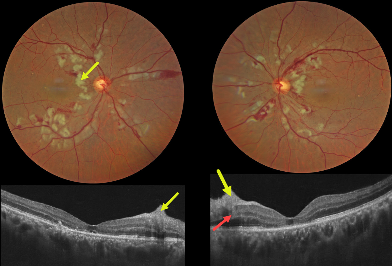
PURTSCHER RETINOPATHY
Ayushi Gupta and Vishal Agrawal
Originally posted on @retina.rocks June 1, 2026
This 55YO male presented with sudden bilateral vision loss that developed immediately following a motor vehicle accident, which included blunt thoracoabdominal trauma and transient loss of consciousness. Vision was 20/200 OU.
Color photography shows bilateral inner and mid-retinal patches of white ischemic retina. The nerve fiber layer lesions appear as inner retinal hyperreflective thickening on OCT. Mid-retinal hyperreflectivity involving the outer plexiform/inner nuclear/outer plexiform layers is noted nasally OS. The outer photoreceptor bands are variably disorganized OU, with a small amount of subfoveal subretinal fluid OD and a foveal cyst OS.
Learning Points:
Purtscher retinopathy was first described by Otmar Purtscher in 1910. Although originally described in a man who fell from a tree with cranial trauma, these white patches of retinal ischemia and hemorrhages are found in numerous other etiologies, including pancreatitis, chest trauma, and collagen vascular diseases. The findings are called Purtscher retinopathy when due to trauma, and Purtscher-like retinopathy when seen from other causes.
The underlying cause of Purtscher is unknown, but it is thought to represent leukoembolization by complement activation. The areas of ischemia include more superficial nerve fiber layer infarcts (cotton wool spots) and deeper ischemia involving the middle retinal layers. This recently described retinal ischemic phenotype, paracentral middle maculopathy (PAMM), occurs as an idiopathic entity or in other ocular disorders, including Purtscher retinopathy, diabetic retinopathy, retinal artery and retinal vein occlusions, and acute macular neuroretinopathy (see Retina 2015;35:1921-1930).
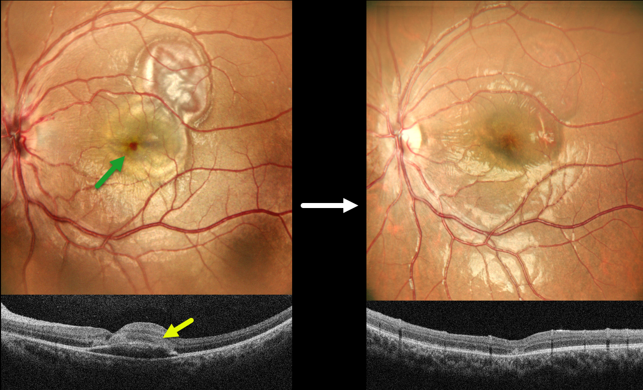
CHOROIDAL RUPTURE WITH LIKELY CONTUSIVE RPE FLUID
Akansha Sharma, Manish Nagpal and Navneet Mehrotra
Originally posted on @retina.rocks May 18, 2026
This 8YO boy presented with 2 days of vision loss in his left eye following a firecracker injury. Vision was 20/200 OS.
MultiColor SLO imaging shows a blister of foveal fluid with an overlying dot of foveal blood, and more superotemporal subretinal blood. OCT scanning shows foveal subretinal fluid with overlying hyperreflectivity from the blood. Intravenous methylprednisolone was given for 3 days, followed by a short course of oral steroids. One month later, vision improved to 20/60. The fluid and blood resolved with a choroidal rupture now visible temporally.
Learning Points:
An acute submacular hemorrhage following blunt trauma almost always indicates an underlying choroidal rupture as the source of the blood. The final vision is generally good unless the rupture extends through the macular center. These patients need long-term follow-up due to the increased risk of macular neovascularization.
Transient subretinal fluid is an uncommon finding with blunt trauma. Injury to the choroid and RPE allows fluid to accumulate in the subretinal space and outer retina (Mishra et al, Journal of Vitreoretinal Diseases 2021;5:165-169), as likely occurred in our patient.
CRAO FOLLOWING DOUBLE PERFORATION FROM PERIBULBAR INJECTION
Ayushi Gupta and Vishal Agrawal
Originally posted on @retina.rocks April 6, 2026
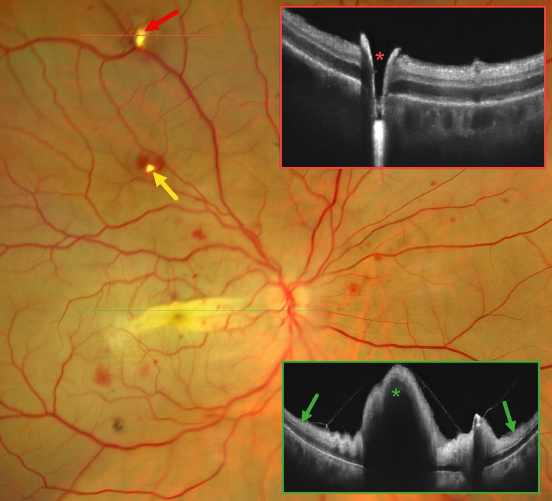
This 42YO female immediately lost vision in her right eye 4 days earlier following pterygium excision surgery performed under peribulbar anesthesia elsewhere. Vision in our office was light perception.
Fundus photography shows an opaque retina throughout the posterior pole consistent with an acute central retinal artery occlusion (CRAO). There is a horizontal retinal fold throughout the macula with scattered blot retinal hemorrhages. Entry and exit needle penetration sites are noted. OCT scan through the superior entry site confirms a full-thickness retinal break. Macular OCT shows diffuse inner retinal hyperreflectivity with elevation through the fold.
Learning Points:
Peribulbar anesthesia is considered to have a lower risk of inadvertent globe perforation, although this risk is not zero (Lim et al., J Clin Ophthalmol 2021;5:414-416). CRAO after such an injury is exceedingly rare and was reported by Lake et al. (J Cataract Refract Surg 2003;29:2234-2235), who felt the occlusion might have resulted from increased IOP due to intraocular anesthetic. A CRAO was also reported by Gyasi et al. following peribulbar anesthesia for pterygium excision, although they found no evidence of globe perforation (Ghana Medical Journal 2012;46:48-50).
INTRAOCULAR FOREIGN BODY
Kanwaljeet Harjot Madan
Originally posted on @retina.rocks April 2, 2026
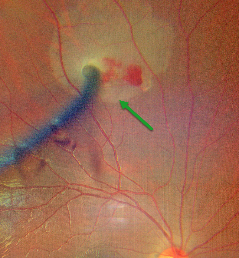
This 20YO male developed sudden, painful vision loss in his right eye while welding. Vision was counting fingers. Examination revealed a corneal laceration with a metal wire piercing the iris temporally and stuck in the superior retina with a localized retinal detachment.
Emergent surgery included corneal wound repair, lensectomy, and vitrectomy with removal of the intraocular foreign body (IOFB), fluid-air exchange, and endolaser photocoagulation. Four weeks postoperatively, vision was 20/80 with an aphakic correction, and the retina remained completely attached.
Learning Points:
Penetrating ocular injuries from IOFB are a true ocular emergency. These IOFBs are usually metallic and need to be removed due to the risk of siderosis, endophthalmitis, cataract, glaucoma, iris defects, and retinal degeneration with depressed ERG.
COMMOTIO RETINAE
The European VitreoRetinal Society (EVRS) and Gil Calvão-Santos
Originally posted on @retina.rocks March 20, 2026
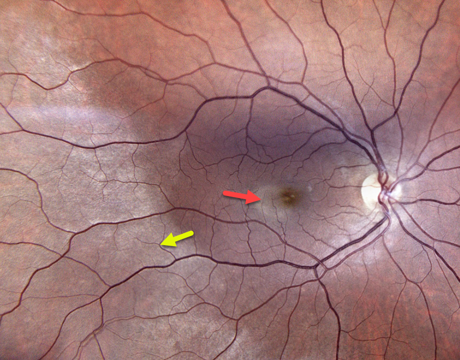
This 30YO male presented two hours following a paddleball injury to his right eye. Vision had decreased from a baseline of 20/20 to 20/80 at presentation.
Color photography shows a large area of temporal and inferotemporal outer retinal whitening, along with a smaller area of temporal macula commotio.
Learning Points:
Commotio retinae is caused by direct, blunt eye trauma. The outer retinal whitening is due to shearing of the photoreceptor outer segments. The term “Berlin’s edema” is a misnomer and should not be used, as the retina is not edematous. The acute white retinal changes usually resolve within several weeks. With resolution, the fundus appearance can normalize. With more severe injury, outer retinal loss can allow RPE cells to migrate into the retina, giving a pseudo-retinitis pigmentosa appearance. Patients can also develop retinal breaks and detachment.
LASER MACULOPATHY
Vaibhav Sethi
Originally posted on @retina.rocks January 20, 2026
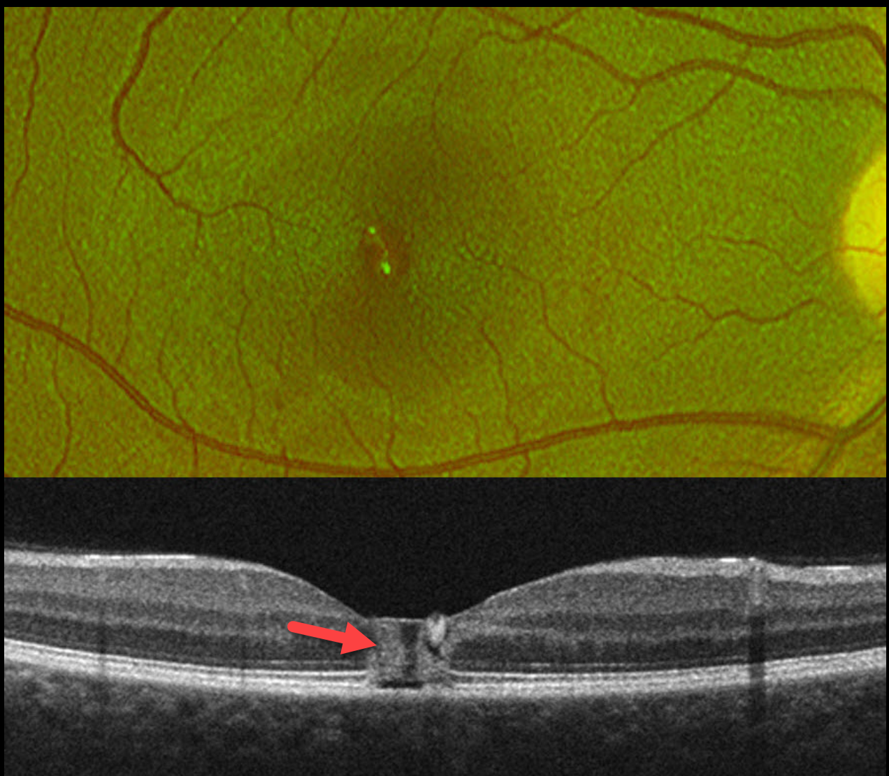
This healthy 19YO female noticed immediate blurred vision in her right eye after looking at a laser during a party. Her vision in this eye was 20/60 when she presented to us the following day.
Optos color RG imaging shows a foveal pseudohole. OCT scanning shows a central foveal column of full-thickness hyperreflectivity. There is loss of the outer segment bands overlying the RPE.
Learning Points:
Most cases of laser maculopathy reported in the literature are caused by intentional or inadvertent exposure to laser pointers (Bhavsar et al, Survey Ophthalmology 2021;66:231-260). However, more recently, there have been reports of macular injury from exposure to high-power lasers during concerts or from DJ laser machines (Perz-Montano et al, Doc Ophthalmol 2019;138:71-76). Most eyes recover fairly good vision with conservative management.
Our patient was immediately lost to follow-up.
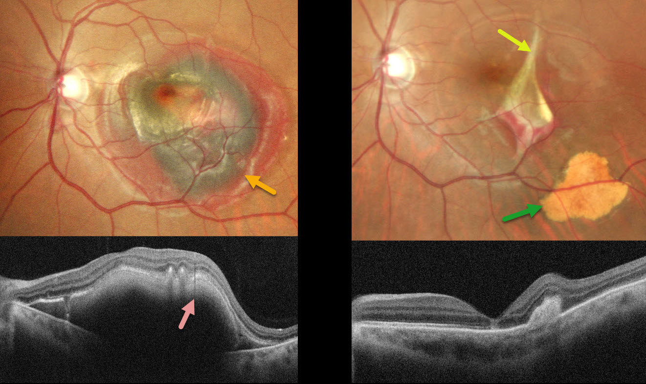
CHOROIDAL RUPTURE
Malvika Singh and Manish Nagpal
Originally posted on @retina.rocks January 14, 2026
This 38YO male was hit by a stone in his left eye two days earlier. Vision was 20/20 in his normal OD and counting fingers OS.
Pseudocolor SLO imaging shows central macular greenish-brown subretinal blood surrounded by a rim of thinner red blood. OCT shows the hyperreflective blood with posterior shadowing. An in-office intravitreal C3F8 gas injection was performed, followed by face-down positioning. Six weeks later, the blood is virtually resolved except for some residual distal inferotemporal yellow devitalized subretinal blood. The causative choroidal rupture can be seen just temporal to the foveal center.
Learning Points:
An acute submacular hemorrhage following blunt trauma almost always indicates an underlying choroidal rupture as the source of the blood. The final vision is usually good unless the rupture extends through the macular center. These patients need long-term follow-up due to the increased risk of macular neovascularization.
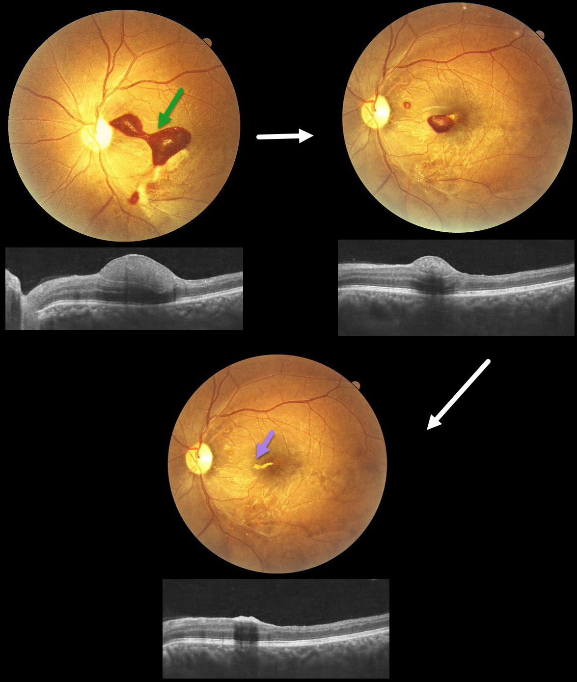
TERSON SYNDROME
Anand Temkar, Surendra Pal, Nagamani Gunasekar
Originally posted on @retina.rocks January 12, 2026
This 32YO male presented with 20 days of vision loss in his left eye. He underwent cardiovascular surgery 3 weeks earlier to remove a clot, followed 1 week later by a subarachnoid hemorrhage. Vision was 20/20 in his normal OD and 20/400 in his OS.
Fundus photography shows a subinternal limiting membrane (ILM) hemorrhage extending from the central macula, superonasally, towards the disc. This blood is hyperreflective on OCT. Observation was recommended.
One month later, vision improved to 20/70, with a marked decrease in sub-ILM blood. One month later, vision improved to 20/30, with near-complete resolution of the blood, except for a small amount of devitalized yellow blood extending nasally from the foveal center.
Learning Points:
Terson syndrome consists of intraocular hemorrhage (vitreous, subhyaloid, or retinal) associated with spontaneous or traumatic subarachnoid hemorrhage. Traditionally, the cause was attributed to elevated intracranial pressure (ICP), which caused cerebrospinal fluid effusion into the optic nerve sheath, compressing the central retinal vein and leading to subsequent rupture of retinal vessels. More recent evidence suggests glymphatic reflux, in which raised ICP causes subarachnoid blood to reflux through glymphatic channels into the globe (Kumaria et al, J Neurology 2022;269:1264-1271).
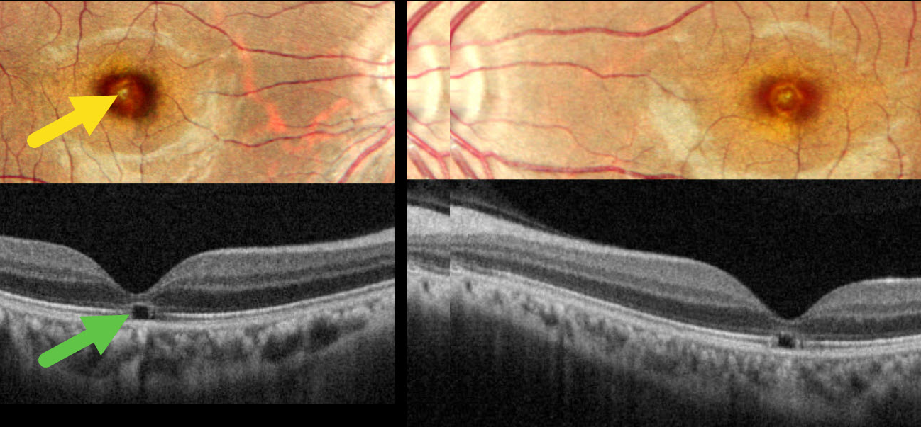
WELDING ARC MACULOPATHY
Malvika Singh and Manish Nagpa
Originally posted on @retina.rocks December 24, 2025
This 31YO male presented with 2 years of bilateral vision loss. He works as a welder and admits to rarely wearing his safety glasses. Vision was 20/30 OU.
Pseudocolor SLO imaging shows symmetrical, tiny yellow foveal scars. OCT shows outer foveal microdefects characterized by hyporeflectivity due to loss of all bands deep to the external limiting membrane.
Learning Points:
The welding process emits optical radiation across various wavelengths and intensities, including infrared, visible, and ultraviolet (UV) light. Exposure to this UV light without proper eye protection can result in keratoconjunctivitis (the most common clinical presentation) and foveal lesions similar to solar retinopathy. Findings include a small yellowish foveal spot or scar with an outer foveal microdefect on OCT (Singh et al, Surv Ophthalmology 2023;68:655-668). Vision is usually good despite the persistent clinical and OCT findings.
Our patient was lucky to have good vision despite his poor work habits. He was advised to use protective eyewear during welding.
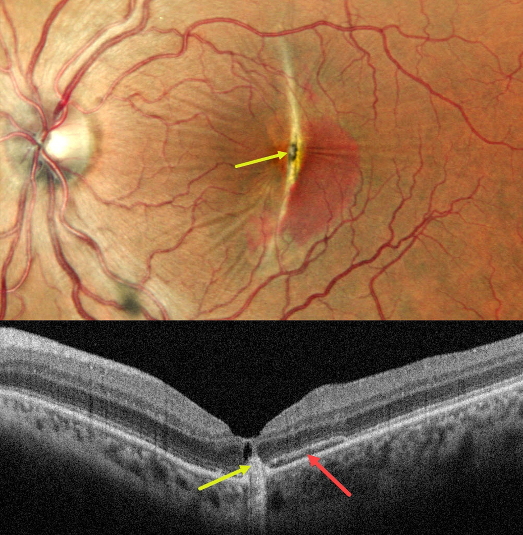
CHOROIDAL RUPTURE
Malvika Singh, Akansha Sharma and Manish Nagpal
Originally posted on @retina.rocks December 10, 2025
This 33YO female noted decreased vision in her left eye immediately following blunt trauma when a ceiling an fell on her face. Vision was 20/20 in her normal OD and 20/125 OS.
Pseudocolor SLO imaging shows a vertical choroidal rupture bisecting the central macula with surrounding subretinal blood. Radiating retinal striae extend outwards from the rupture. There is a central hyperpigmented lesion centrally, which appears hyperreflective in the outer retina on OCT. There is a hyporeflective cystic space anterior to this lesion. The subretinal blood appears as a hyper- and hyporeflective band on OCT.
This case was submitted by Malvika Singh, Akansha Sharma and Manish Nagpal.
Learning Points:
An acute submacular hemorrhage following blunt trauma almost always indicates an underlying choroidal rupture as the source for the blood. The final vision is usually good unless the rupture extends through the macular center. These patients need to be followed long-term due to the increased risk for macular neovascularization (MNV).
The central pigmented lesion with overlying fluid was suggestive for a MNV. An anti-VEGF injection was given, but she was unfortunately immediately lost to follow up.
CHOROIDAL RUPTURE WITH SECONDARY MACULAR NEOVASCULARIZATION
Nilesh Kumar
Originally posted on @retina.rocks November 24, 2025
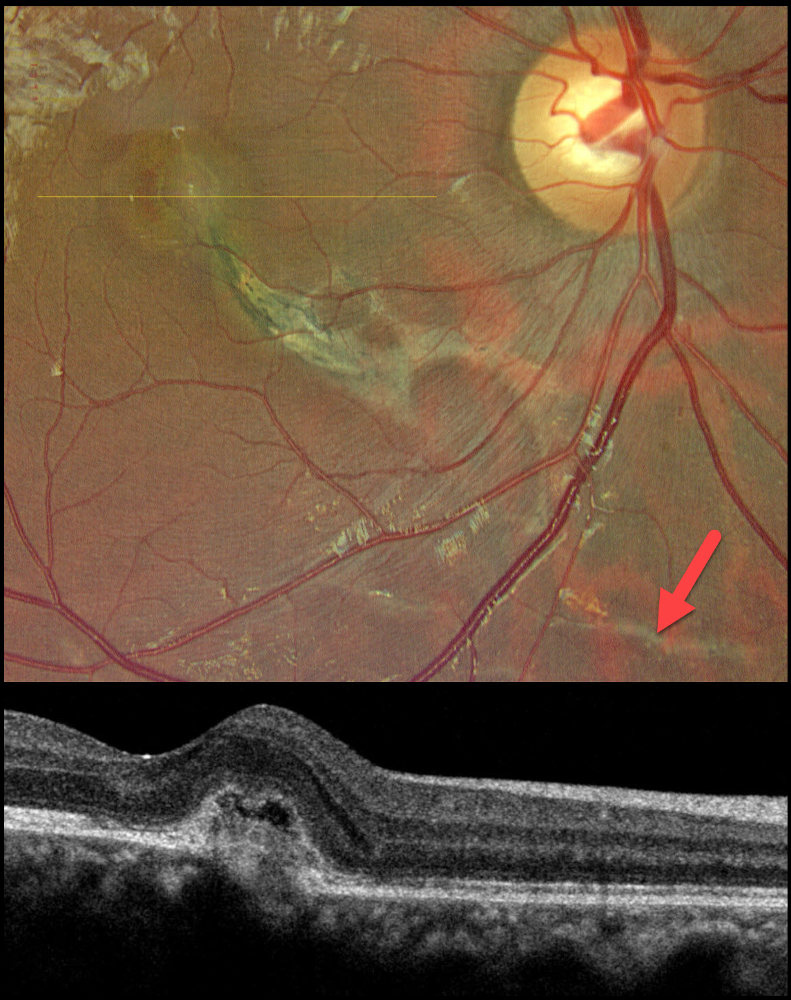
This 20YO male presented with 1 year of decreased vision in his right eye following blunt trauma from a small pebble that flew off a firecracker blast. Vision was 20/100 OD and 20/20 in his normal left eye.
Color photography shows an inferonasal macular choroidal rupture that extends through the fovea. A faint choroidal rupture is also noted inferior to the nerve. Subtle fluid and blood surround the foveal portion of the rupture. OCT scanning shows a thickened macular neovascularization (MNV) with mild overlying subretinal fluid.
Learning Points:
Choroidal ruptures are caused by blunt ocular trauma and are always circumferential with respect to the optic nerve. Subretinal blood following blunt trauma virtually always indicates the presence of an underlying choroidal rupture. The visual prognosis is generally good unless the rupture extends through the macular center. Patients need to be followed long-term due to the risk for MNV, as developed in our patient. We elected to closely follow him and will begin anti-VEGF therapy if increasing exudation develops.
LASER MACULOPATHY
The European VitreoRetina Society (EVRS) and Shraddha Chandorkar
Originally posted on @retina.rocks October 17, 2025
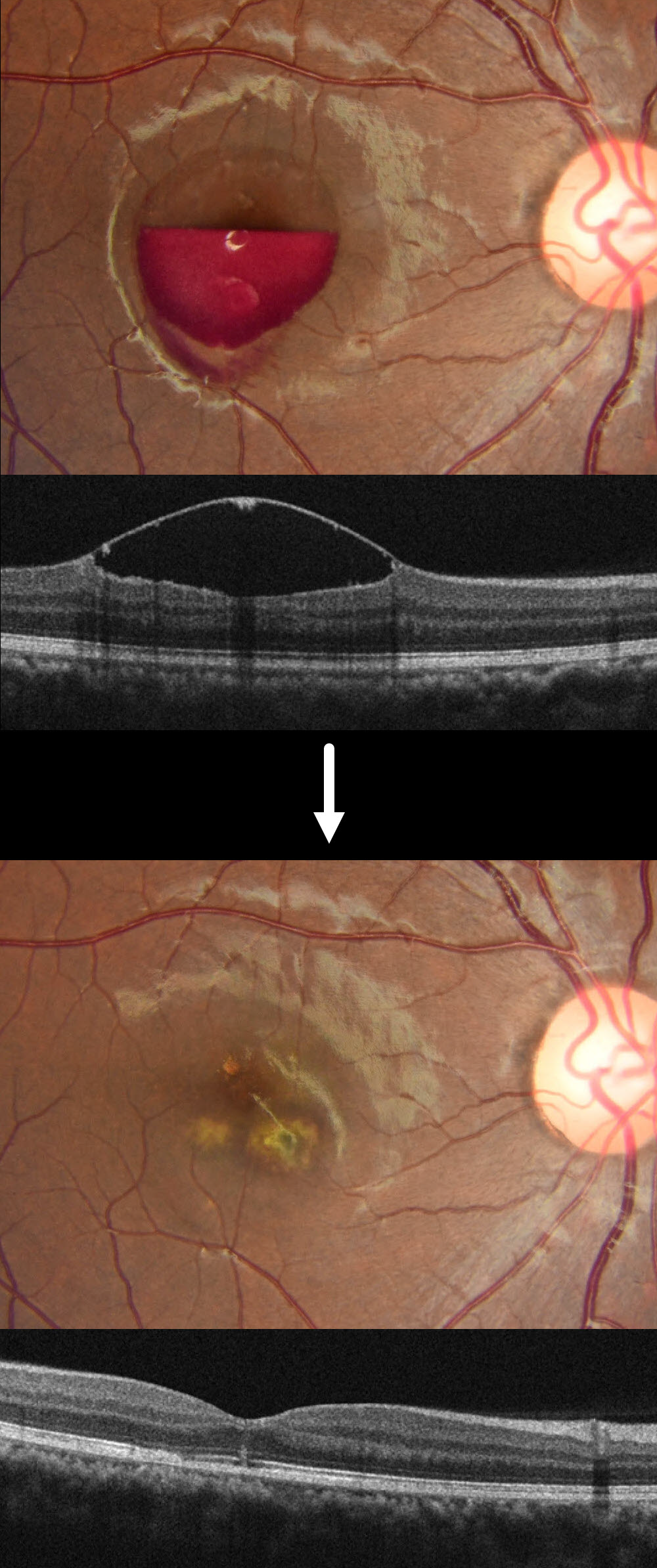
This healthy 19YO female noted sudden vision loss in her right eye immediately after looking directly at the light while attending a DJ laser party. When she presented to us 2 days later, her vision was hand motion OD and 20/20 in her normal OS.
Fundus photography and OCT show a partially layered central macular sub-internal limiting membrane (ILM) hemorrhage.
When examined 1 week later, the blood was drained via a YAG laser due to no improvement in her vision or sub-ILM blood. Two months later, vision improved to 20/20 despite multiple paracentral scars.
Learning Points:
DJ laser machines typically fall into Class 3R or Class 4, depending on their power output. These lasers emit visible light in the red (635-650 nm), green (520-532 nm), and blue (445-460 nm) wavelengths, often combined for full RGB effects. While visually stunning, high-powered lasers, especially Class 4, can pose serious risks, including retinal burns, if beams enter the audience area or are viewed directly. Blue wavelengths are particularly hazardous due to their shorter wavelength and higher energy density. A strikingly similar case to our patient was reported by Perz-Montano et al (Doc Ophthalmol 2019;138:71-76).
To protect audiences, professional setups use a combination of engineering controls and regulatory compliance. This includes fail-safe scanning systems, beam termination zones, and power-limiting protocols to remain below the Maximum Permissible Exposure. In the US, the FDA’s Center for Devices and Radiological Health oversees these devices, requiring manufacturers to submit safety documentation and operators to file a Laser Light Show Variance for public performances. Proper mounting, trained personnel, and adherence to ANSI and IEC standards are essential to ensure safe and compliant laser shows.
THORN INTRAOCULAR FOREIGN BODY
Ayushi Gupta and Vishal Agrawal
Originally posted on @retina.rocks October 13, 2025
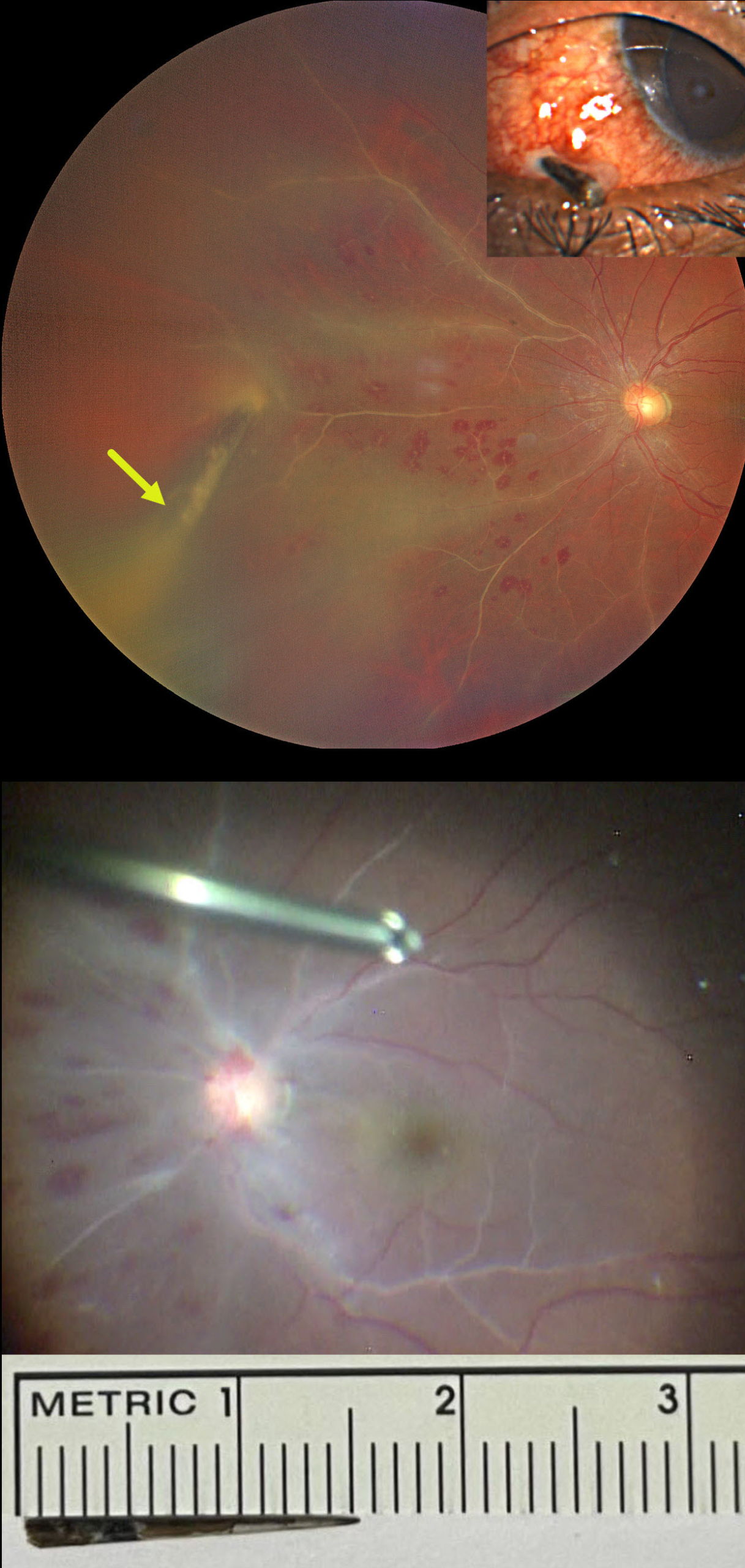
This 28YO male presented 5-6 hours following a perforating thorn injury to his left eye. He was standing in an open-moving loading vehicle when an Acacia tree branch suddenly hit his eye. Vision was 20/20 in his normal OD and 20/40 OS.
External examination showed a thorn lodged in the inferonasal sclera. There were moderate anterior chamber cells on slit lamp examination with a fibrinous pupillary membrane.
Fundus examination revealed vascular sheathing predominantly in the nasal quadrant, multiple, white-centered retinal hemorrhages, and vitreous exudates. The thorn penetrated the retina, extending into the vitreous cavity.
Despite being taken to surgery approximately two hours later, intraoperatively, there was a dramatic increase in vascular sheathing extending into the macula. Pars plana vitrectomy was performed with removal of the thorn foreign body, repair of the scleral laceration, cryotherapy, endolaser photocoagulation around the retinal break, silicone oil tamponade, and intravitreal antibiotic injection. The thorn measured approximately 15mm.
On the first postoperative day, the posterior segment appeared healthy with resolved vascular sheathing. One week postop, vision was 20/50 with a +5-diopter sphere, the retina remained completely attached, and good laser scarring was noted around the penetration site (not shown).
TRAUMATIC RETINAL HOLES
Fatma Shakarchi and Ahmed Sallam
Originally posted on @retina.rocks October 6, 2025
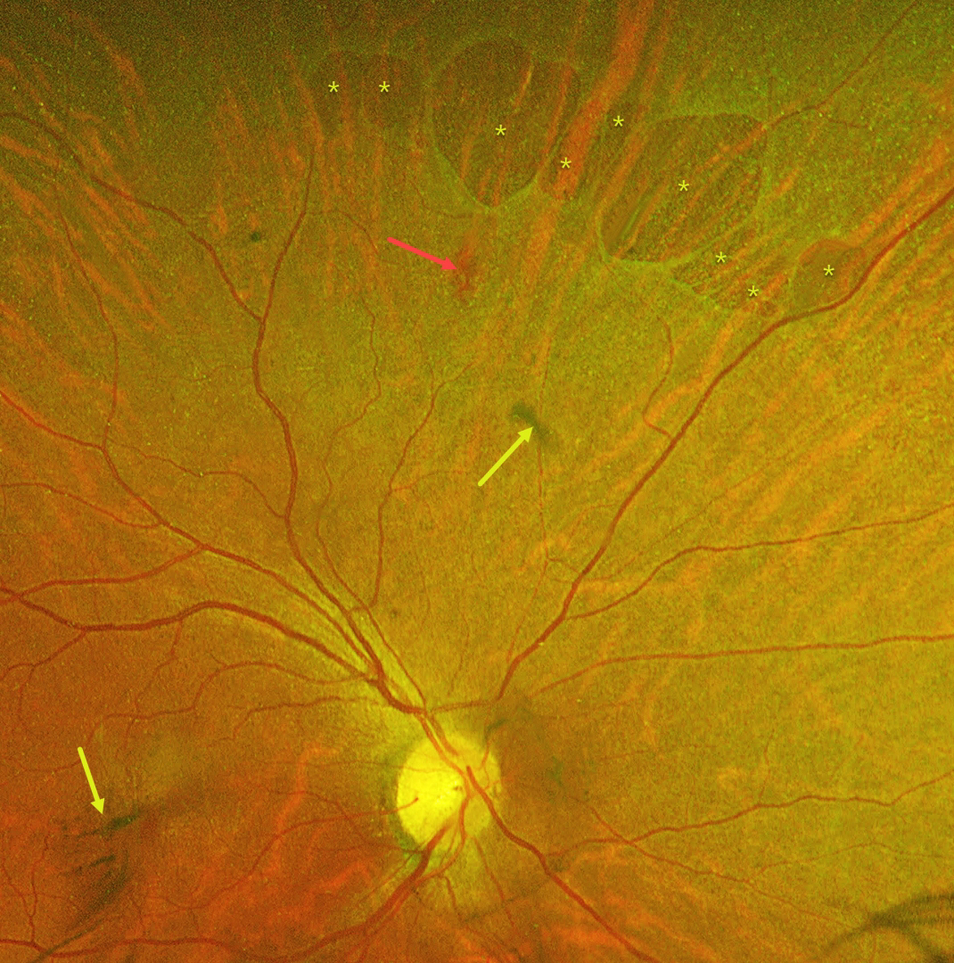
This 55YO female was referred with mildly decreased vision in her right eye two days following blunt ocular trauma. Vision was 20/40 OD and 20/20 in her normal OS.
Optos color RG imaging reveals numerous irregularly oval superonasal retinal holes, a retinal hemorrhage, and some clumps of vitreous blood. There was no vitreous detachment clinically.
Learning Points:
Retinal breaks following blunt trauma include retinal dialysis, horseshoe tears, and giant retinal tears. Breaks can also develop from concussive injury at the impact site or from a coup-contrecoup injury, so-called impact or necrotic breaks, which may be associated with varying degrees of commotio retinae acutely (Cox et al, Arch Ophthalmol 1966;76:678-685) or following its resolution (Longstaff et al, Br J Ophthalmol 1987;71:375-376).
In this case, the breaks identified 2 days after the injury were not associated with commotio. However, their appearance and the adjacent hemorrhage support an acute traumatic etiology. Therefore, prophylactic laser retinopexy was promptly applied around all breaks.
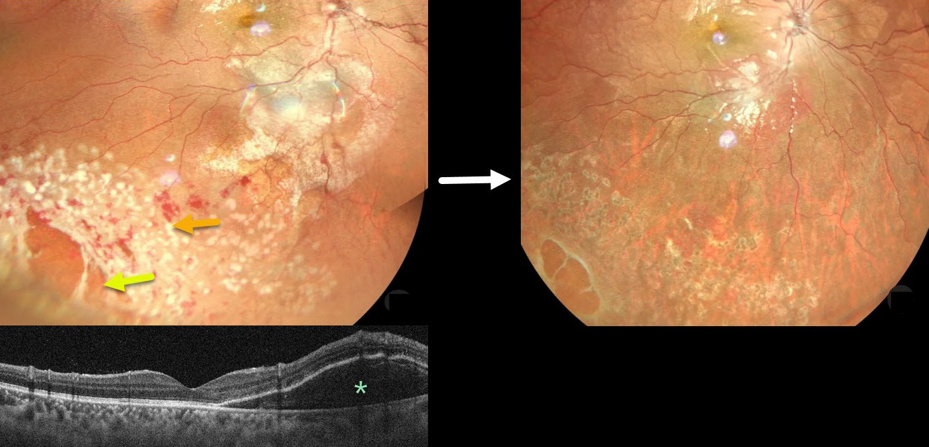
COMMOTIO RETINAE WITH RETINAL BREAKS AND CONTUSIVE SEROUS MACULAR DETACHMENT
Tejaswita Verma and Manish Nagpal
Originally posted on @retina.rocks September 24, 2025
This 13YO male was hit in his right eye with a cricket ball. When he presented later that day vision was 20/30.
Color photography shows a large area of commotio retinae with scattered retinal hemorrhages involving the inferotemporal fundus. There is a poor foveal reflex with possible nasal macular subretinal fluid, which is confirmed on OCT. Several atrophic retinal breaks are noted peripherally. Immediate prophylactic laser was applied around all breaks. Seven weeks later, the commotio and hemorrhages are resolved, and all breaks are well-surrounded with laser scarring. Vision was 20/20.
Although blunt trauma is a common cause of retinal breaks, acute or subsequent dissolution of the retina within areas of commotio with secondary holes is quite rare in our experience (Cox et al Arch Ophthalmol 1966;76:678-685 and Longstaff et al Br J Ophthalmology 1987;71:375-376). The acute white retinal changes usually resolve within several weeks. With resolution, the fundus appearance can normalize. With more severe injury, outer retinal loss can allow RPE cells to migrate into the retina, giving a pseudo-retinitis pigmentosa appearance.
Transient subretinal fluid is an uncommon finding with blunt trauma. Injury to the choroid and RPE allows fluid to accumulate in the subretinal space and outer retina (Mishra et al, Journal of Vitreoretinal Diseases 2021;5:165-169).
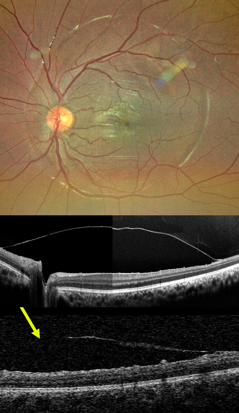
POST-TRAUMATIC ILM DETACHMENT
Nilesh Kumar
Originally posted on @retina.rocks September 2, 2025
This 12YO boy came in for a routine eye examination. Although vision was 20/20 bilaterally, the right fundus showed a striking premacular sheen.
Ultra-widefield imaging reveals a round internal limiting membrane (ILM) elevation surrounding the posterior pole. On further questioning, he gave a history of blunt trauma to the right eye 2 years earlier that was treated elsewhere with the ND:YAG laser. The opening in the ILM is noticed inferiorly on OCT.
This case demonstrates a presumed chronic and stable sub-ILM detachment likely secondary to prior sub-ILM hemorrhage. Fortunately, he remains completely asymptomatic with perfect vision and no distortion.
A valuable reminder that retinal imaging can unearth forgotten chapters of ocular history, and that fundus examination with multimodal imaging can reveal prior stories.
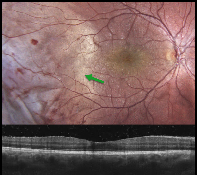
COMMOTIO RETINAE
Anastasia Petrus
Originally posted on @retina.rocks August 25, 2025
This 16YO male was chopping wood one day earlier when a branch recoiled and struck his right eye. Vision was 20/30 OD and 20/20 in his normal OS. The right anterior segment showed localized conjunctival chemosis with a subluxed crystalline lens.
Color photography shows temporal commotio retina extending into the temporal macula, along with scattered retinal hemorrhages. Although there appear to be fine inner macular striae, OCT scanning is fairly normal. The patient was referred for further management.
Learning Points:
Commotio retinae is caused by direct, blunt eye trauma. The outer retinal whitening is due to shearing of the photoreceptor outer segments. The term “Berlin’s edema” is a misnomer and should not be used, as the retina is not edematous. The acute white retinal changes usually resolve within several weeks. With resolution, the fundus appearance can normalize. With more severe injury, outer retinal loss can allow RPE cells to migrate into the retina, giving a pseudo-retinitis pigmentosa appearance. Patients can also develop retinal breaks and detachment.
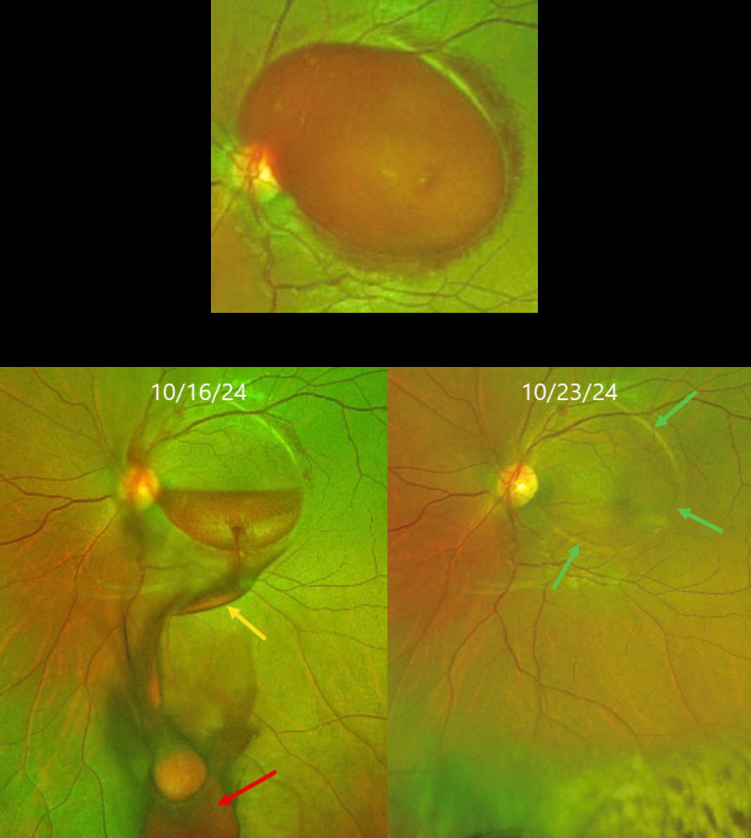
VALSALVA RETINOPATHY
The European VitreoRetina Society (EVRS) and Srishti Ramamurthy
Originally posted on @retina.rocks August 1, 2025
This 29YO pregnant female presented with sudden vision loss in her left eye immediately following a bout of severe vomiting. Vision was 20/20 in her normal OD and counting fingers OS.
Optos color RG imaging shows a large sub-internal limiting membrane (ILM) hemorrhage extending throughout the macula. Blood immediately drained into the subhyaloid and vitreous spaces following the YAG laser. One week later, the macular blood completely resolved. The margins of the prior elevated ILM are still visible, and some far inferior vitreous blood is noted. Vision was 20/20.
Learning Points:
Valsalva retinopathy is characterized by usually unilateral retinal and preretinal blood caused by rupture of the superficial retinal capillaries from suddenly raised intrathoracic or intra-abdominal pressure. Various causes include coughing, heavy lifting, and vomiting. These hemorrhages virtually always resolve without sequelae, although the preretinal blood in severe cases can be drained into the inferior vitreous by creating a small ILM opening using either a thermal or Nd:YAG laser (Durukan et al, Eye 2008;22:214-218).
The site chosen for laser delivery is ideally at the inferior margin of thick blood, well away from the fovea to avoid underlying retinal damage. For the Nd:YAG, the laser energy should begin at 4 mJ and be gradually titrated upward until an opening is created that allows the blood to drain inferiorly into the vitreous.
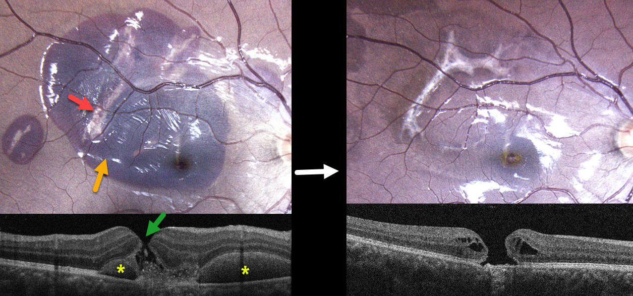
ACUTE CHOROIDAL RUPTURES WITH SUBRETINAL BLOOD + TRAUMATIC MACULAR HOLE
Originally posted on @retina.rocks July 16, 2025
This healthy 17YO male was in a motor vehicle accident 2 weeks earlier. He was not wearing a seat belt and suffered a right upper lid laceration and closed head injury. He noticed immediate vision loss in his right eye. Vision was 20/200 OD and 20/20 in his normal OS.
Color photography shows a large macular and a smaller distal temporal submacular hemorrhage. Multiple choroidal ruptures are visible beneath this blood. OCT scanning shows a central full-thickness macular hole with speckled hyperreflective subretinal material. The submacular blood is hyperreflective with posterior shadowing on either side of the macular hole.
Learning Points:
An acute submacular hemorrhage following blunt trauma always indicates an underlying choroidal rupture as its source. The final vision is generally good unless the rupture extends through the macular center. These patients need long-term follow-up due to the increased risk of macular neovascularization.
Since fresh traumatic holes can spontaneously flatten, especially in pediatric cases (Liu et al, AJO 2020;210:174-183), an initial period of observation is recommended before considering vitrectomy. When examined about 6 months following his injury, all blood had resolved, and the temporal foveal choroidal rupture appears to spare the macular center. We will likely recommend vitrectomy if the hole fails to close with several more months of observation.
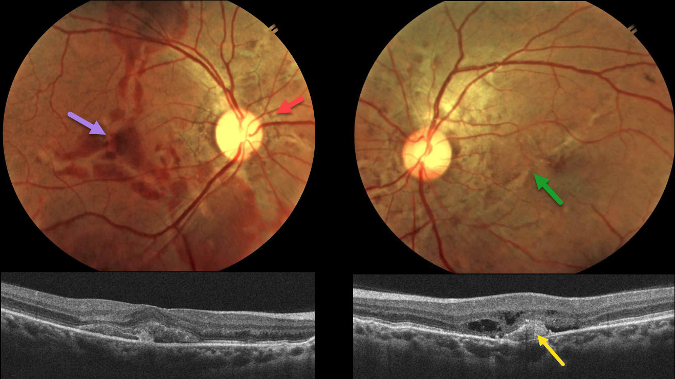
ANGOID STREAKS WITH TRAUMATIC CHOROIDAL RUPTURE AND SUBRETINAL BLOOD
Neelam Khatwani
Originally posted on @retina.rocks July 2, 2025
This 38YO male presented with 10 days of bilateral vision loss after being hit in the right eye with a fist. He denied any past medical history, and family history was negative for eye disease. Vision was 20/200 OD and 20/80 OS.
Color fundus photography shows irregular orange-gray lines radiating outwards from each nerve. Variable areas of subretinal blood are noted in the right macula, extending into the superior and inferior midperipheries. There is milder subretinal blood OS along with an irregular oblique macular angioid streak vs choroidal rupture. OCT OD shows subretinal hyperreflective material, likely blood. A possible type 1 (below the RPE) MNV with some outer retinal fluid is seen OS.
An anti-VEGF injection was given bilaterally, but unfortunately, the patient was immediately lost to follow-up.
Learning Points:
Initially described by Doyne in 1889, angioid streaks are orange-yellow, irregular, crack-like dehiscences in Bruch’s membrane associated with atrophic degeneration of the overlying RPE. The term “angioid” derives from its resemblance to blood vessels. Although they are historically mistakenly associated with numerous conditions, the only real association is with pseudoxanthoma elasticum (PXE, Nadelmann et al, Eye 2023;37:1596-1601). Patients are at significant risk for vision loss from MNV or pattern dystrophy-like changes (Murro et al, Graefe’s 2020;258:1881-1892).
Even minor trauma can cause choroidal ruptures with subretinal blood (Agrawal et al, JAMA Ophthalmology 2017;135(3):e165466). Unlike typical choroidal ruptures that are usually curvilinear with the nerve, the lesions from the brittle Bruch’s membrane in PXE appear as numerous irregular ruptures usually radiating from the nerve. Even though our patient sustained a direct injury to his right globe, the trauma was significant enough to cause choroidal ruptures in his left eye. The bilateral subretinal hemorrhages may be purely avascular, as their multifocality suggests.
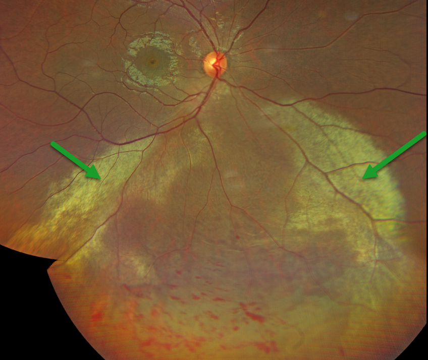
COMMOTIO RETINAE
Nilesh Kumar
Originally posted on @retina.rocks June 17, 2025
This 30YO male was hit in his right eye 2 days earlier with a cricket ball. He was visually asymptomatic, and vision was 20/20.
Color photography shows extensive commotio retinae in the inferior retina, with scattered intraretinal hemorrhages. There were no retinal breaks or detachment. Close observation was recommended.
Learning Points:
Commotio retinae is caused by direct, blunt eye trauma. The outer retinal whitening is due to shearing of the photoreceptor outer segments. The term “Berlin’s edema” is a misnomer and should not be used, as the retina is not edematous. The acute white retinal changes usually resolve within several weeks. With resolution, the fundus appearance can normalize. With more severe injury, outer retinal loss can allow RPE cells to migrate into the retina, giving a pseudo-retinitis pigmentosa appearance. Patients can also develop retinal breaks and detachment.
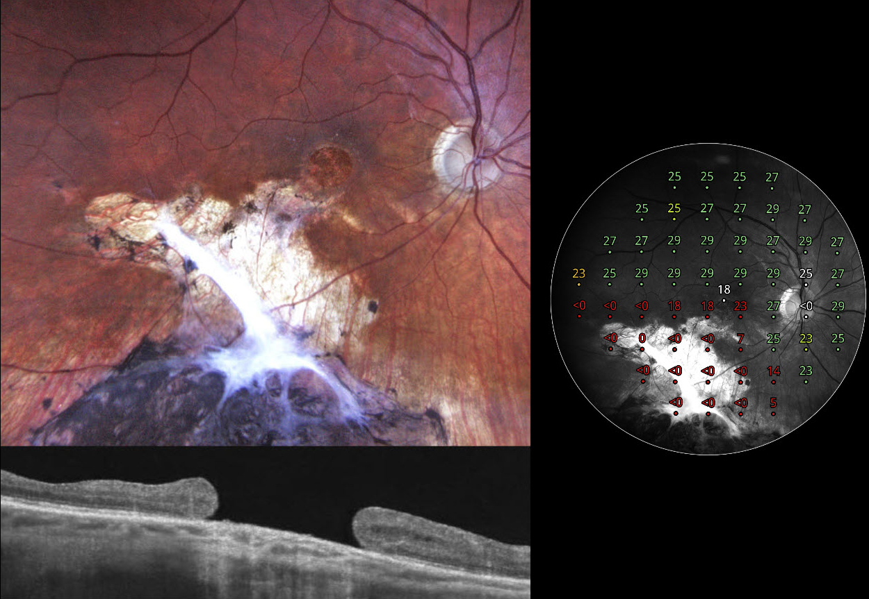
TRAUMATIC MACULAR SCARRING AND MACULAR HOLE
Liubov Sitaruk and Ophthalmocoach
Originally posted on @retina.rocks June 10, 2025
This 36YO Ukrainian male sustained a firearm-related facial injury during military combat in 2014 while in Eastern Ukraine. At the time, he was treated in a frontline hospital, where the left eye underwent primary enucleation due to a non-reconstructible globe rupture. His right eye sustained a penetrating injury with scleral rupture, which was primarily repaired. Over the following months, he underwent pars plana vitrectomy with endolaser and silicone oil due to complications, likely including retinal detachment and/or vitreous hemorrhage. Given the complexity of the trauma, the monocular status, and probable limited access to regular follow-up during wartime, silicone oil was likely retained for an extended period, potentially several years, until its removal at an undetermined later date. On our 2024 examination, he had no new visual complaints, and vision was 20/100.
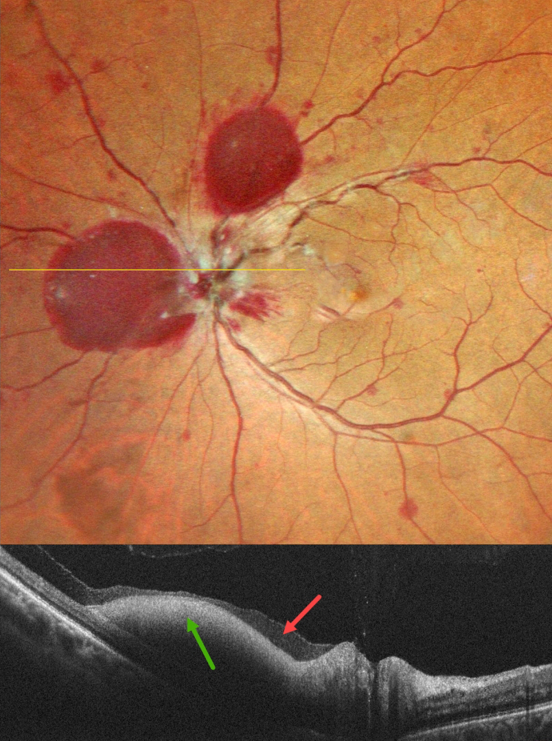
Fundus photography shows atrophic inferior macular chorioretinal scarring, which becomes hyperpigmented as it extends into the inferior midperiphery. A large macular hole is noted funduscopically and on OCT. Macular perimetry shows marked depression within the area of scarring with preservation of some central sensitivity. Observation was recommended.
VALSALVA RETINOPATHY
Malvika Singh and Manish Nagpal
Originally posted on @retina.rocks June 9, 2025
This 49YO male presented with 4 days of vision loss in his left eye. He denied any Valsalva maneuver. Vision was 20/25 in his normal OD and 20/20 OS.
Pseudocolor SLO imaging shows a large sub-internal limiting membrane (ILM) hemorrhage, nasal and superior to the nerve, along with scattered mostly deep, white-centered retinal hemorrhages. OCT scanning confirms the sub-ILM blood but also reveals shallow subhyaloid blood. Unfortunately, he was immediately lost to follow up. He then presented 1 month later with a breakthrough vitreous hemorrhage. Vitrectomy was advised due to his prior non-compliance. Vision 1 month postoperatively was 20/20, with complete resolution of all hemorrhages (not shown).
Learning Points:
Valsalva retinopathy, first described by Thomas Duane (Trans Am Ophthalmol Soc 1972; 70:298-313), is characterized by unilateral retinal and preretinal hemorrhages caused by elevated intrathoracic or intra-abdominal pressure. Various causes include coughing, heavy lifting, and vomiting. However, as in our case, patients sometimes present with these findings while denying any Valsalva maneuver. These hemorrhages virtually always resolve without sequelae, although the preretinal blood in severe cases can be drained into the inferior vitreous by creating a small ILM opening using either a thermal or Nd:YAG laser. Non -clearing break through vitreous hemorrhage is treated with vitrectomy.
WELDING ARC MACULOPATHY
Rohan Jain and Manish Nagpal
Originally posted on @retina.rocks May 1, 2025
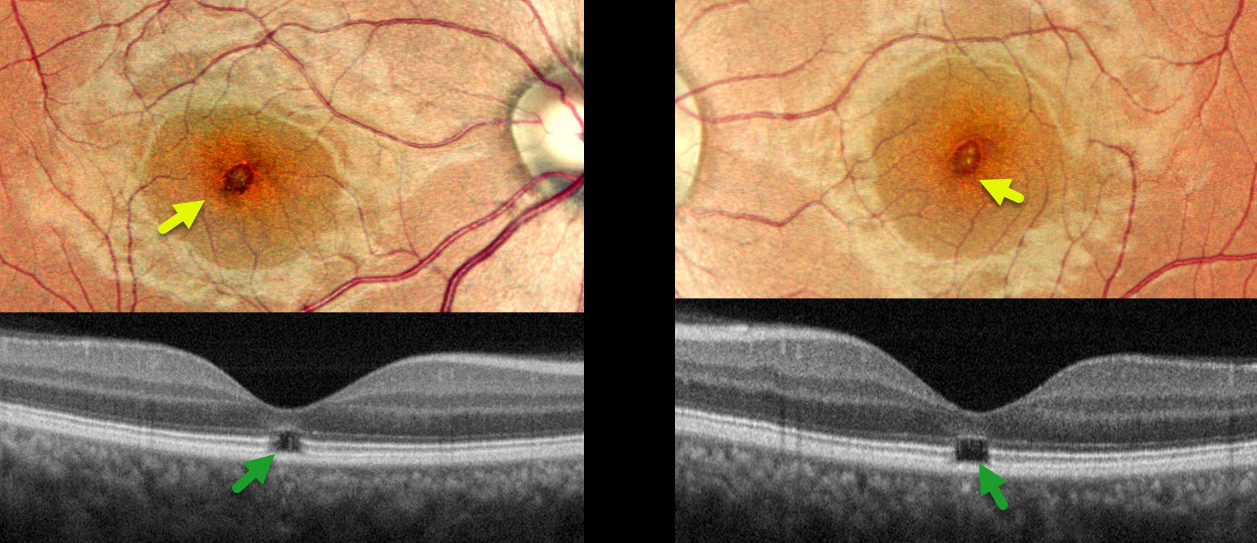
This 38YO male presented with 6-7 months of bilateral vision loss. He works as a welder and admits to rarely wearing his safety glasses. Vision was 20/25 OD and 20/20 OS.
Pseudocolor SLO imaging shows symmetrical, tiny yellow foveal scars. OCT shows outer foveal microdefects with hyporeflectivity due to loss of all bands deep to the external limiting membrane.
Learning Points:
The welding process emits optical radiation across various wavelengths and intensities, including infrared, visible, and ultraviolet (UV) light. Exposure to this UV light without proper eye protection can result in keratoconjunctivitis (the most common clinical presentation) and foveal lesions similar to those seen in solar retinopathy. Findings include a small foveal yellowish spot or scar with an outer foveal microdefect on OCT (Singh et al, Surv Ophthalmology 2023;68;655-668). Vision is usually good despite the persistent clinical and OCT findings.
Our patient was lucky to have good vision despite his poor work habits. He was advised to use protective eyewear during welding.
INTRAOCULAR FOREIGN BODY?
Originally posted on @retina.rocks April 1, 2025
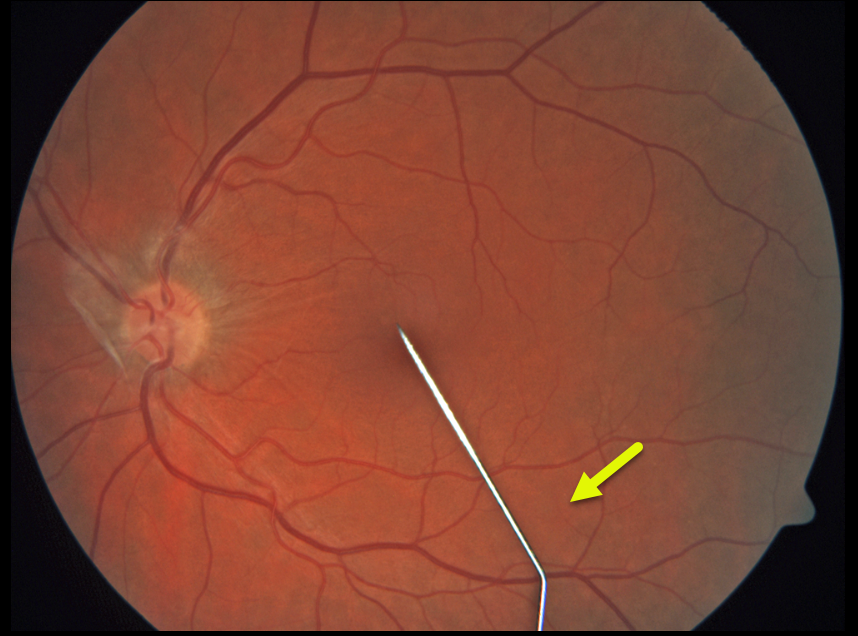
This healthy 24YO goth female presented with severe pain and a floater in her left eye immediately following a failed intraocular tattoo procedure earlier that day. Vision was 20/40.
Color photography shows a posteriorly dislocated intraocular tattoo needle lying on the inferior macular surface. Adherent vitreous is noted on the optic nerve. She was immediately taken to surgery for the removal of the metallic intraocular foreign body (IOFB).
Learning Points:
HAPPY APRIL FOOLS!!
Although this case is a fake-out from an old fundus camera utilizing a fixation stick that appears as a pseudo-IOFB, cosmetic tattooing is unfortunately a very real thing, including corneal, subconjunctival, and episcleral tattoos (https://eyewiki.org/Eye_Tattooing).
The procedure is performed by either the individual performing it on themselves or non-ophthalmic trained individuals and is obviously associated with numerous complications, including globe penetration, traumatic cataract, retinal detachment, endophthalmitis, and orbital cellulitis. Ink-related complications include dye migration into the anterior chamber, where it can coat the lens and posterior corneal surface and cause secondary
GIANT MACULAR HOLE WITH TOTAL RETINAL DETACHMENT
Anand Temkar and Manish Nagpal
Originally posted on @retina.rocks February 10, 2025
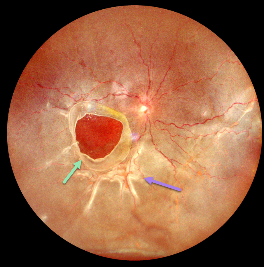
This 10YO boy presented with 1 week of vision loss in his left eye. The parents gave a vague history of trauma with a tennis ball 2 years earlier. Vision was counting fingers OD and 20/20 in his normal OS.
Ultrawidefield imaging shows a giant macular hole occupying nearly the entire macula. The hole’s edges are rolled, and proliferative vitreoretinopathy with star folds is present within the total rhegmatogenous retinal detachment (RRD).
Emergent vitrectomy surgery with silicone oil was scheduled.
Learning Points:
Blunt ocular trauma can result in numerous sequelae, including commotio retina, retinal breaks and detachment, choroidal rupture, and macular hole. A giant macular hole, also reported in Alport syndrome (Shah and Weinberg Ophthalmic Genetics 2010;31(2):93-97), is an exceedingly rare traumatic event (Hernandez-Da Mota Case Rep Ophthalmol 2011;2:283-286).
Traumatic macular holes are thought to result from a vitreous coup contrecoup injury or a jet stream of anterior chamber aqueous humor shooting through Cloquet canal into the premacular bursa.
COMMOTIO RETINAE
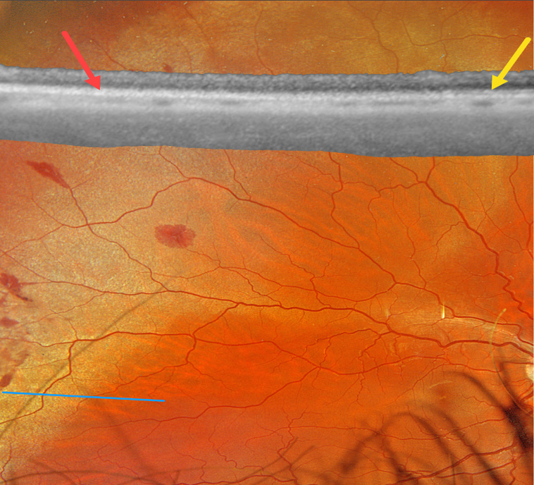
Originally posted on @retina.rocks December 24, 2024
This healthy 19YO male was hit in his right eye while playing soccer one day earlier. Vision was 20/20.
Optos color RGB imaging shows subretinal whitening with associated retinal hemorrhages in the superotemporal periphery. Optos Silverstone swept-source OCT shows at least two outer retinal hyperreflective bands in the uninvolved retina, which merge into a single hyperreflective band within the commotio. The retina was attached without breaks. The retinal appearance and OCT completely normalized 1 month later (not shown).
Learning Points:
Commotio retinae is caused by direct, blunt eye trauma. The outer retinal whitening is due to shearing of the photoreceptor outer segments. The term “Berlin’s edema” is a misnomer and should not be used, as the retina is not edematous. The acute white retinal changes usually resolve within several weeks. With resolution, the fundus appearance can normalize. With more severe injury, outer retinal loss can allow RPE cells to migrate into the retina, giving a pseudo-retinitis pigmentosa appearance. Patients can also develop retinal breaks and detachment.
TRAUMATIC MACULAR HOLE
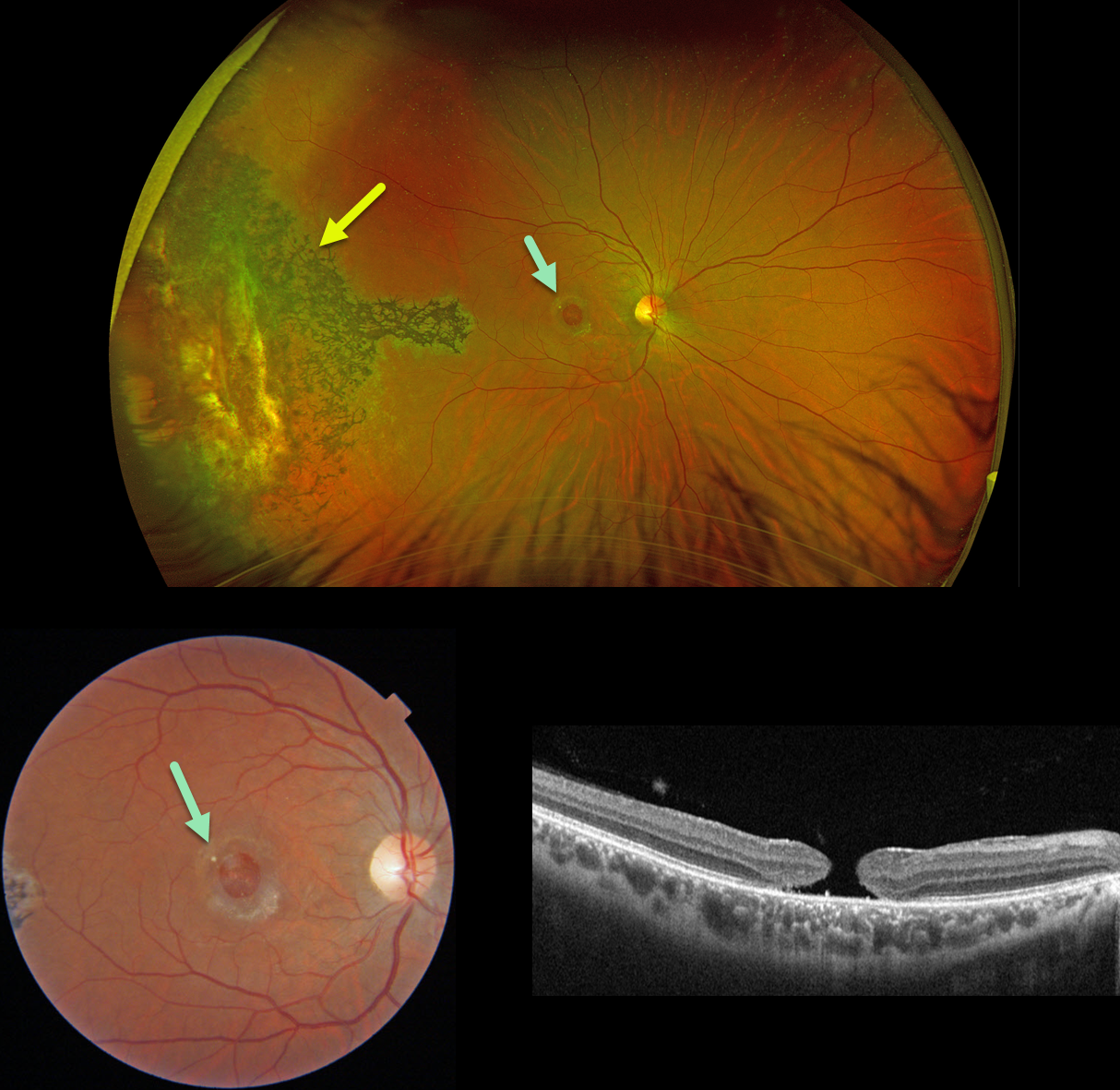
Originally posted on @retina.rocks December 18, 2024
This 31YO male suffered a paint gun injury to his right eye when he was 12YO. Vision has since been stable and poor in this eye. Vision was 20/200.
Optos color RG imaging shows temporal peripheral scarring with intraretinal pigment migration. A large macular hole with surrounding pigmentary changes is noted on Triton color imaging and swept-source OCT.
Learning Points:
Since fresh traumatic holes can spontaneously flatten, especially in pediatric cases (Liu et al, AJO 2020;210:174-183), an initial period of observation is recommended before considering vitrectomy. Given the size and chronicity of our patient’s hole, surgery was not considered.
Photoreceptor loss from numerous causes, including blunt trauma, retinal detachment, macular telangiectasia, and retinitis pigmentosa (RP), can lead to intraretinal RPE migration, a shared phenotype. Unilateral RP is exceedingly rare and requires normal electrophysiologic testing with long-term observation to ensure that delayed findings are present in the fellow eye.
LASER MACULOPATHY
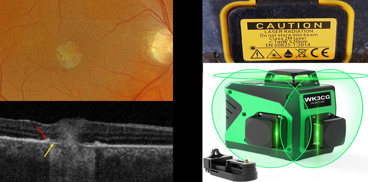
Originally posted on @retina.rocks October 3, 2024
This 46YO male presented with a several-week history of unilateral vision loss. Vision was 20/20 in his normal left eye and counting fingers in his right eye.
Optos color RGB imaging shows a mostly round foveal scar with a few temporal blood dots. Triton swept-source OCT scanning shows the normal foveal architecture replaced by full-thickness, hyperreflective, disorganized tissue. The RPE is thickened and somewhat elevated along the lesion’s edge, with bare Bruch’s membrane more centrally.
Upon further questioning, we found that the patient works as a contractor and lost vision immediately following the use of a laser leveler. He didn’t recall looking directly at the laser beam. He showed us the device, a WOKELINE WGR12L, which is used as a full-room-level laser device. A label on the back of the device defines it as a 520nm Class 2M laser with an output of less than 1 milliwatt. We found an image online of a similar device showing how this device projects a 3D green laser light in 2 vertical planes and a single horizontal plane to cover the floor, ceiling, and all walls simultaneously.
Learning Points:
A Class 2 laser device emits light in the visible spectrum, 400-700 nm. It is presumed that the natural aversion response to the very bright light will be sufficient to prevent damaging exposure, although prolonged viewing may be dangerous. Although laser pointers, Class 2 or 3A devices, are supposed to have an output of under 5 milliwatts, real-world findings reveal that the power output of many of these pointers often far exceeds what is displayed in their safety information. Laser pointer maculopathy is well reported in the literature (Bhavasar et al., Surv Ophthalmology 2021;66:231-360), although we are not aware of a prior case involving a laser-level device, as in our patient.
VALSALVA RETINOPATHY
Nilesh Kumar
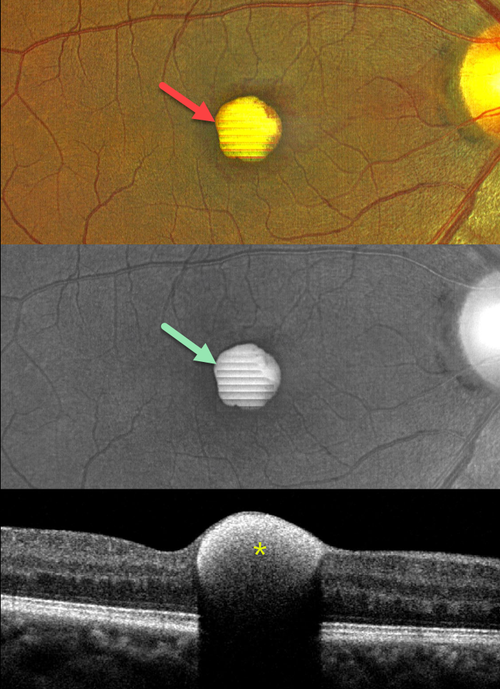
Originally posted on @retina.rocks September 26, 2024
This 43YO male presented with a 1-week history of sudden decreased vision in his right eye. He denied any Valsalva maneuver. Vision was 20/70 in his right eye and 20/30 in his left eye.
Color imaging shows a foveal yellow sub-internal limiting membrane (ILM) hemorrhage. Fundus autofluorescence (FAF) shows intense hyper-FAF. Spectral domain OCT shows a hyperreflective sub-ILM lesion that shadows posteriorly.
Our patient was treated with observation, and the blood levels were greatly decreased 3 weeks later (not shown).
Learning Points:
Valsalva retinopathy was first described by Thomas Duane (Trans Am Ophthalmol Soc 1972; 70:298-313). It is characterized by usually unilateral retinal and preretinal blood caused by raised intrathoracic or intra-abdominal pressure. Various causes include coughing, heavy lifting, and vomiting. However, as in our case, sometimes patients present with these findings, denying any Valsalva maneuver (Forshaw et al, Acta Ophthalmologica 2024;102:122-123)
These hemorrhages virtually always resolve without sequelae, although the preretinal blood in severe cases can be drained into the inferior vitreous by creating a small ILM opening using either a thermal or Nd:YAG laser (Durukan et al, Eye 2008;22:214-218).
Porphyrins are organic molecules that are part of the heme molecule in hemoglobin. When these compounds break down, the devitalized blood appears yellow and becomes intensely hyperautofluorescent (Bloom and Spaide, Retinal Cases & Brief Reports 2022;16:401-402).
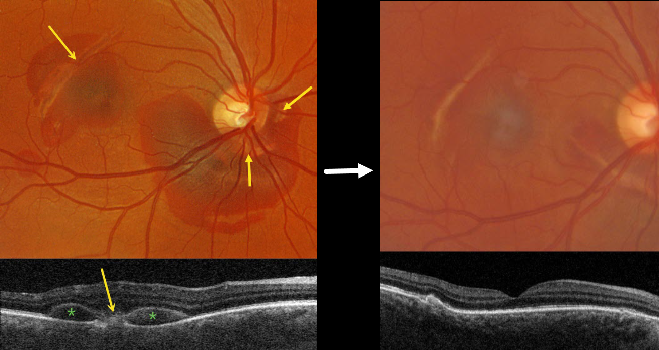
CHOROIDAL RUPTURE
Originally posted on @retina.rocks September 23, 2024
This 36YO male presented with one week of vision loss in his right eye immediately following a bungee cord injury. Vision was 20/60.
Optos color RG imaging shows areas of subretinal blood surrounding the inferior optic nerve and superotemporal macula. Multiple choroidal ruptures are evident. On Triton swept-source OCT, the superotemporal choroidal rupture appears as an outer retinal hyperreflective mound, and the blood as hyporeflective mounds between the outer retina and RPE.
Our patient was treated with observation, and one month later, all blood resolved with 20/100 vision.
Learning Points:
An acute submacular hemorrhage following blunt trauma almost always indicates an underlying choroidal rupture as the source of the blood. The final vision is usually good unless the rupture extends through the macular center. These patients need long-term follow-up due to the increased risk of macular neovascularization.
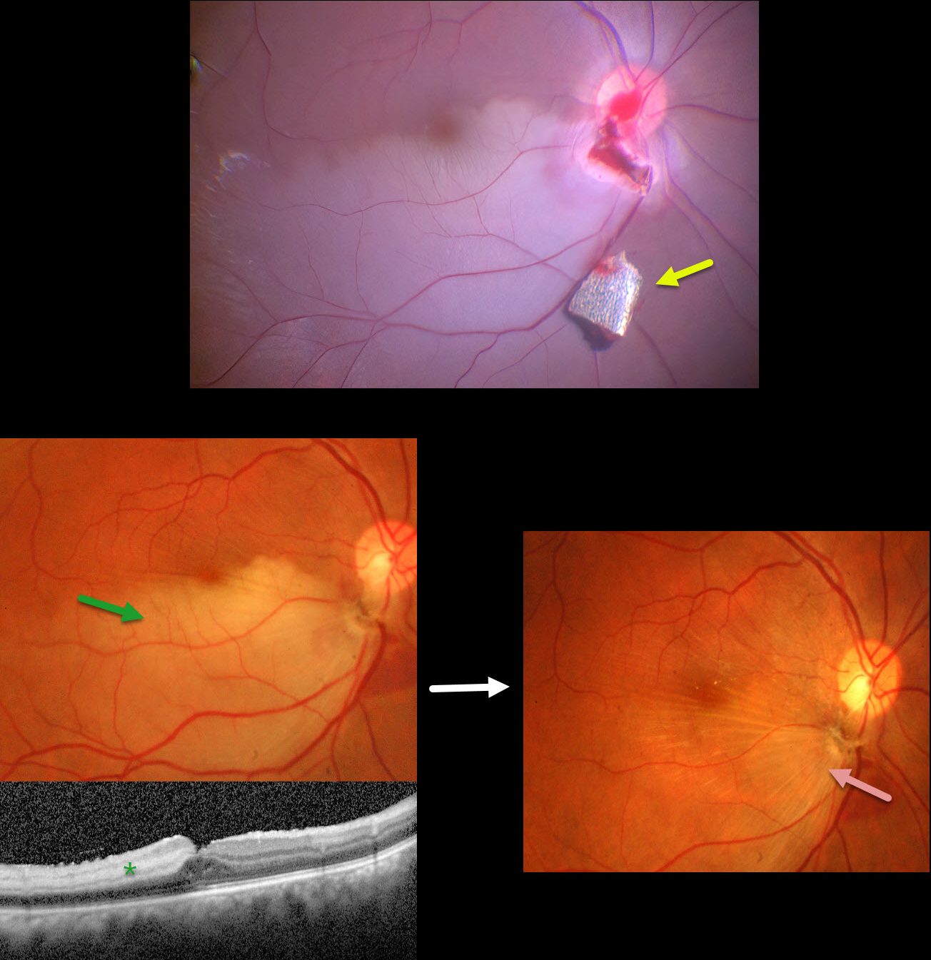
INTRAOCULAR FOREIGN BODY WITH SECONDARY BRAO
Anjana Mirajkar and Manish Nagpal
Originally posted on @retina.rocks August 5, 2024
This 34YO male presented with pain and vision loss immediately following a metal-on-metal injury at work. Vision was light perception. Slit lamp examination revealed a corneal laceration and traumatic cataract. B-scan ultrasonography and CT scanning revealed an intraocular foreign body (IOFB). The patient was immediately taken to surgery for corneal wound repair, lensectomy, and vitrectomy.
Intraoperative photography shows a metallic intraocular foreign body (IOFB) resting just inferior to the optic nerve. The impact site is just inferior to the optic nerve, with an inferotemporal branch retinal artery occlusion (BRAO) distally. The IOFB was removed with a rare-earth magnet.
Two weeks following surgery, vision was 20/120 with an aphakic correction. The inferior retina remains opaque, and the OCT shows inner retinal hyperreflectivity from the BRAO. A secondary intraocular lens implantation was performed 3 months postoperatively. Eleven months following the initial surgery, the BRAO is fully resolved, and secondary retinal striae extend outwards from the impact site. Vision remarkably improved to 20/30.
This case was published in the Indian Journal of Ophthalmology (Nagpal M et al, Ind J Ophthalmol 2018;66;146-148).
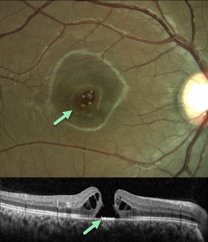
LASER-INDUCED MACULAR HOLE
Tareq Alsulami and Naser Alsaedi
Originally posted on @retina.rocks June 17, 2024
This 14YO boy presented with vision loss in his right eye following self-directed exposure to a blue wavelength laser pointer. Vision was 20/200 in his right eye and 20/20 in his normal left eye.
Color imaging and OCT scanning show an irregular full-thickness macula hole. The hole closed following vitrectomy surgery, but vision failed to improve.
Learning Points:
Laser pointers can cause significant macular injury, including solar maculopathy-like outer foveal microdefects, hemorrhage, pseudo macular dystrophy, vitelliform lesions, and macular scarring (Bhavsar et al, Surv Ophthalmology 2021;66:231-260). There are also numerous reports of a secondary full-thickness macular hole, as developed in our patient.
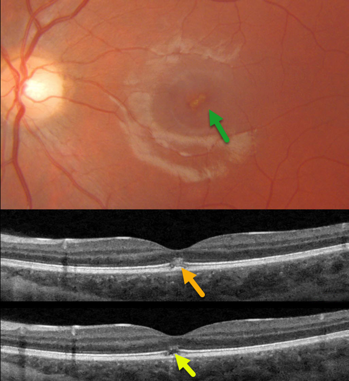
LASER MACULOPATHY
Mattie Adams
Originally posted on @retina.rocks April 22, 2024
This 13YO girl presented with immediate vision loss in her left eye after looking at a laser pointer one week earlier. Vision was 20/20 in her normal right eye and 20/70 in her left eye.
Triton color photography shows an oblique, slightly linear area of foveal pigment loss. Swept-source OCT scans through this lesion show focal hyperreflectivity with variable loss of the ellipsoid zone and outer segments.
Learning Points:
Sun gazing is thought to cause acute damage to the RPE and photoreceptors via photochemical mechanisms involving reactive oxygen species. Similar photochemical damage can follow laser pointer exposure, although the clinical findings can be different (Bloom and Singal, Retinal Cases 2022;16:
89-91).
Sun gazing acutely produces bilateral, symmetric, pinpoint subfoveal yellow-white lesions in the outer retina, followed by variable clinical depigmented foveal RPE and focal photoreceptor OCT defects extending from the external limiting membrane to the RPE.
Laser pointer maculopathy can cause a similar lesion, but presumed more prolonged viewing, saccades, and laser pointer movement can also lead to larger areas of macular damage.
Outer foveal microdefects (Cohen et al, Ophthalmology Retina 2021;5:553-561) have been described in numerous conditions, including macular telangiectasia, tamoxifen use, ABCA4 disorders (cone-rod dystrophies, Stargardt disease and fundus flavimaculatus), phototoxicity, trauma, and vitreomacular traction disorders.
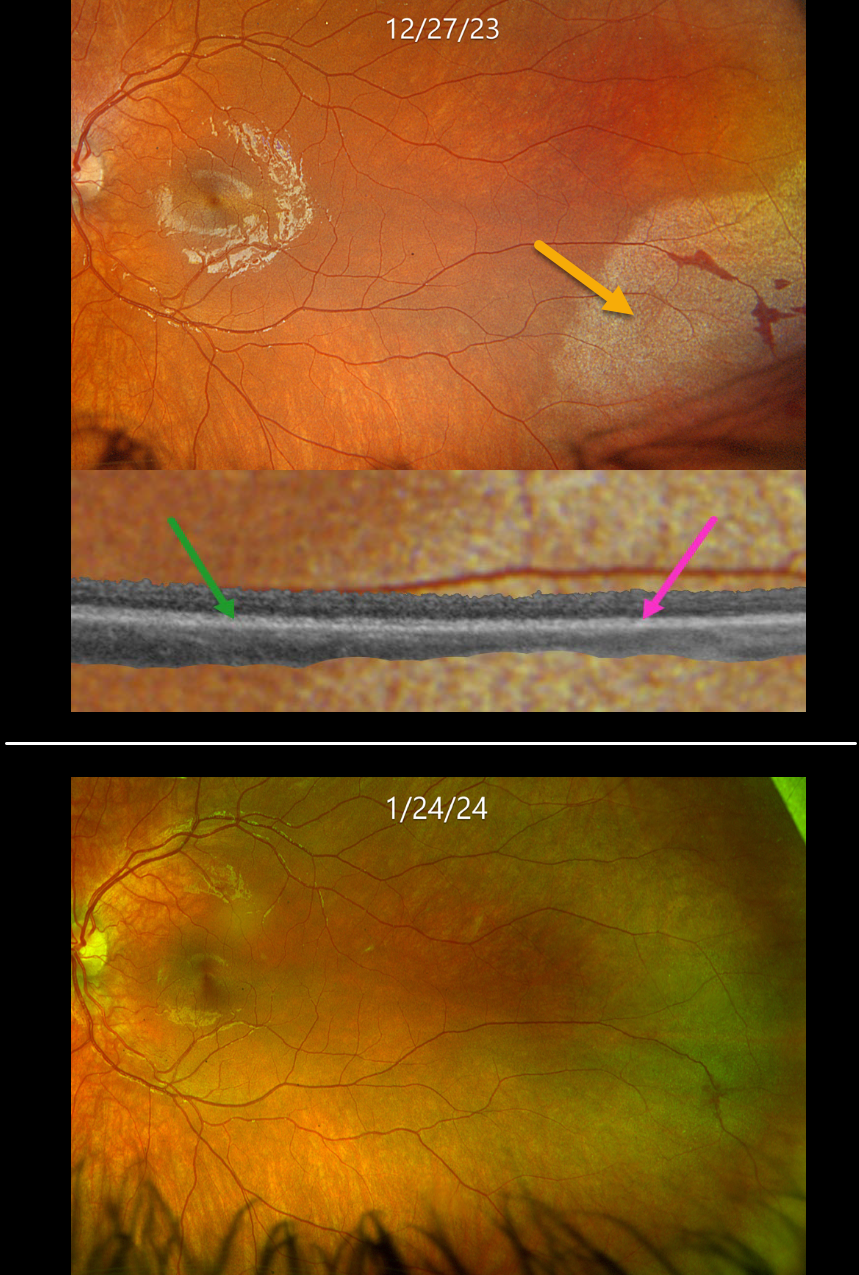
COMMOTIO RETINAE
Originally posted on @retina.rocks April 16, 2024
This healthy 10YO boy presented several hours after being hit in his left eye with a Nerf ball. Vision was 20/25.
Optos color RGB imaging shows subretinal whitening with associated retinal hemorrhages in the inferotemporal periphery. Optos Silverstone swept-source OCT shows at least two outer retinal hyperreflective bands in the uninvolved retina, which merge into a single hyperreflective band within the commotio. The retina was attached without breaks.
Observation was recommended, and the retinal findings normalized within several weeks.
Learning Points:
Commotio retinae is caused by direct, blunt eye trauma. The outer retinal whitening is due to shearing of the photoreceptor outer segments.
The term “Berlin’s edema” is a misnomer and should not be used, since the retina is not edematous. The acute white retinal changes usually resolve within several weeks. With resolution, the fundus appearance can normalize.
With more severe injury, outer retinal loss can allow RPE cells to migrate into the retina, giving a pseudo-retinitis pigmentosa appearance. Patients can also develop retinal breaks and detachment.
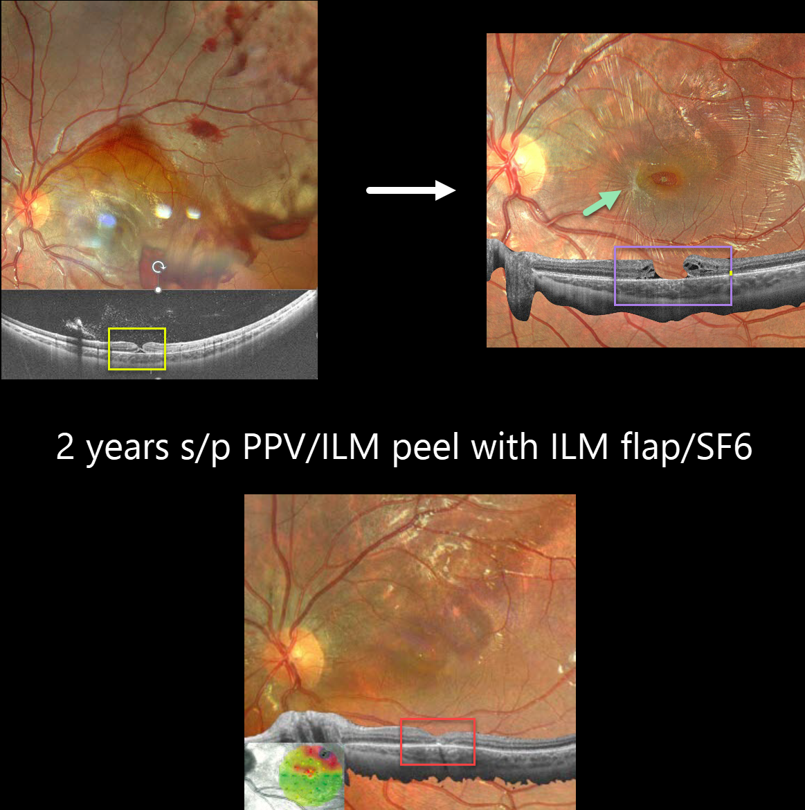
TRAUMATIC MACULAR HOLE
Barbara Parolini, Veronika Matello, Giulia Freschi, and Roberta Penzani
Originally posted on @retina.rocks April 9, 2024
This 10YO boy was referred for vision loss immediately after getting hit in his left eye by a soccer ball 7 days earlier. Vision was 20/70.
Fundus photography shows scattered intra- and preretinal hemorrhages with a large confluent area of commotio retina superiorly and temporally.
Widefield OCT shows a partial-thickness macular hole with the external retina intact but elevated. Three months later, vision was 20/100, and an irregular, full-thickness macular hole with surrounding macular pucker was seen.
25-gauge vitrectomy with ILM peeling, ILM flap, SF6 gas tamponade, and 3 days face-down positioning was performed. Two months later, vision improved to 20/40 with complete closure of the hole. Two years later, vision was 20/25.
Learning Points:
This patient illustrates several sequelae of blunt trauma, including retinal/preretinal blood, commotio retinae, and macular hole formation. Since fresh traumatic holes can spontaneously flatten, especially in pediatric cases (Liu et al, AJO 2020;210:174-183), an initial period of observation is recommended before considering vitrectomy.
In our patient’s case, the hole did not resolve, and surgery provided good anatomic and long-lasting visual outcomes.
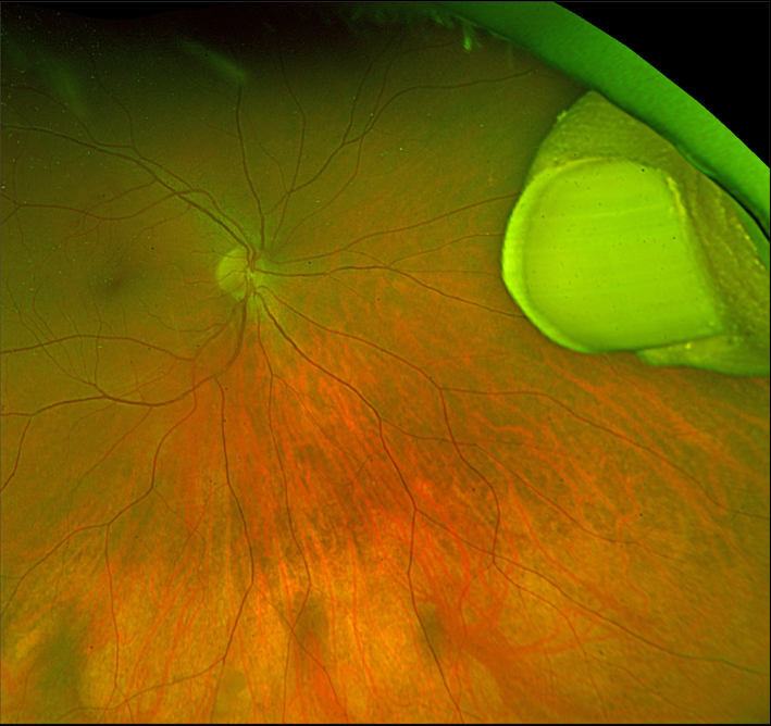
INTRAOCULAR THUMB
Originally posted on @retina.rocks April 1, 2024
This healthy 7YO boy was playing with his older brother while watching their favorite Three Stooges bit about Niagara Falls.
Horseplay literally turned into tragedy when they presented to the emergency room with the older brother’s hand impaled into his younger brother’s right eye.
Both were immediately taken to surgery, where the attacking brother’s thumb was amputated at the distal phalanx, then removed internally via an open sky pars plana vitrectomy approach. Six months later, following removal of the silicone oil, vision was 20/30.
And Happy April Fool’s Day!
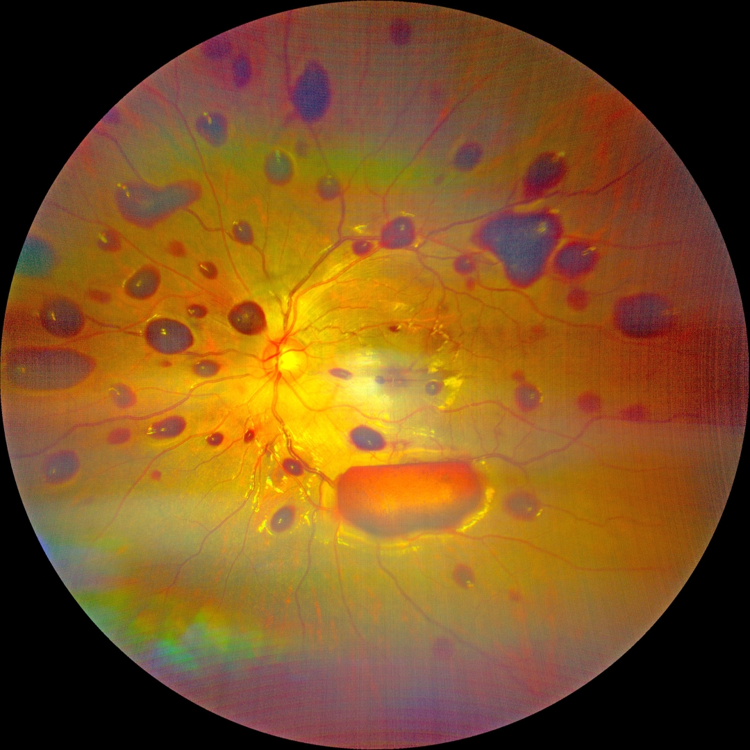
NON-ACCIDENTAL TRAUMA
Kanwaljeet Harjot Madan
Originally posted on @retina.rocks March 14, 2024
This 11YO boy presented with decreased vision in his left eye after his friend’s father thrashed him and was hit in the head 3 days earlier. There was no other sign of bodily injury. Vision was 20/20 in his normal right eye and counting fingers in his left eye.
Our patient’s findings are predominated by numerous mostly sub-ILM hemorrhages. His case is unusual given his age and unilateral findings. Fortunately, with observation, vision recovered to 20/30 three weeks later, with resolution of all findings.
Learning Points:
The shaken baby syndrome is a form of non-accidental traumatic child abuse where the young infant is vigorously shaken, causing whiplash-induced intracranial and intraocular hemorrhaging, often without obvious external trauma.
The ocular findings are usually bilateral, with the hemorrhages often having multiple layers, including subretinal, intraretinal, and preretinal.
INTRAOCULAR FOREIGN BODY
Barbara Parolini, Veronika Matello, Giulia Freschi, and Roberta Penzani
Originally posted on @retina.rocks March 12, 2024
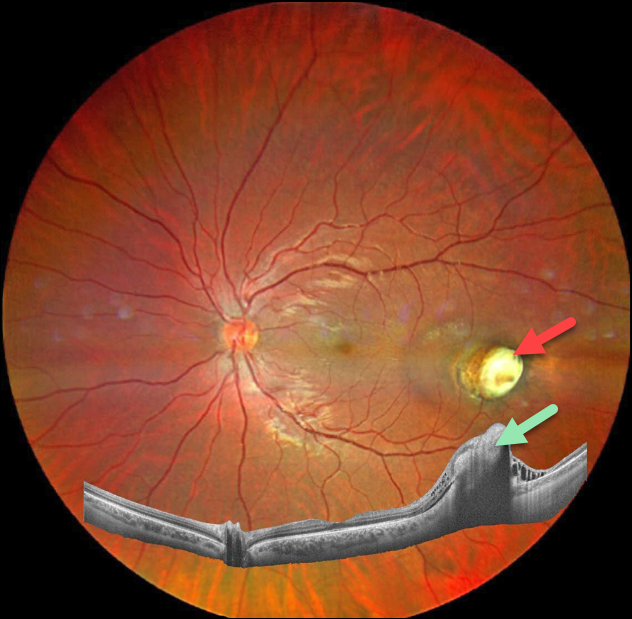
This 23YO male was referred for a second opinion regarding a traumatic intraocular foreign body that occurred about 5 months earlier.
The patient said he was pumping a large truck tire when the tube detached from the valve on the wheel. Metal powder then shot up from the ground and hit the eye. The initial surgical repair involved solely suturing the anterior wound. The foreign body was likely metallic but of an unknown type. The patient was asking why the foreign body had been left. Vision was 20/20.
Fundus photography shows a nodular fibrotic scar in the temporal midperiphery. Widefield OCT shows inner retinal hyperreflectivity with posterior shadowing from this lesion.
Learning Points:
Siderosis can develop from metallic intraocular foreign bodies (IOFBs, specifically iron) and cause cataract, glaucoma, iris defects, and retinal degeneration with depressed ERG. In this case, we felt no need to remove the IOFB given the encasement by scar tissue, lack of inflammation, and excellent vision. We plan on initially examining him monthly.
WHIPLASH MACULOPATHY
Originally posted on @retina.rocks March 5, 2024
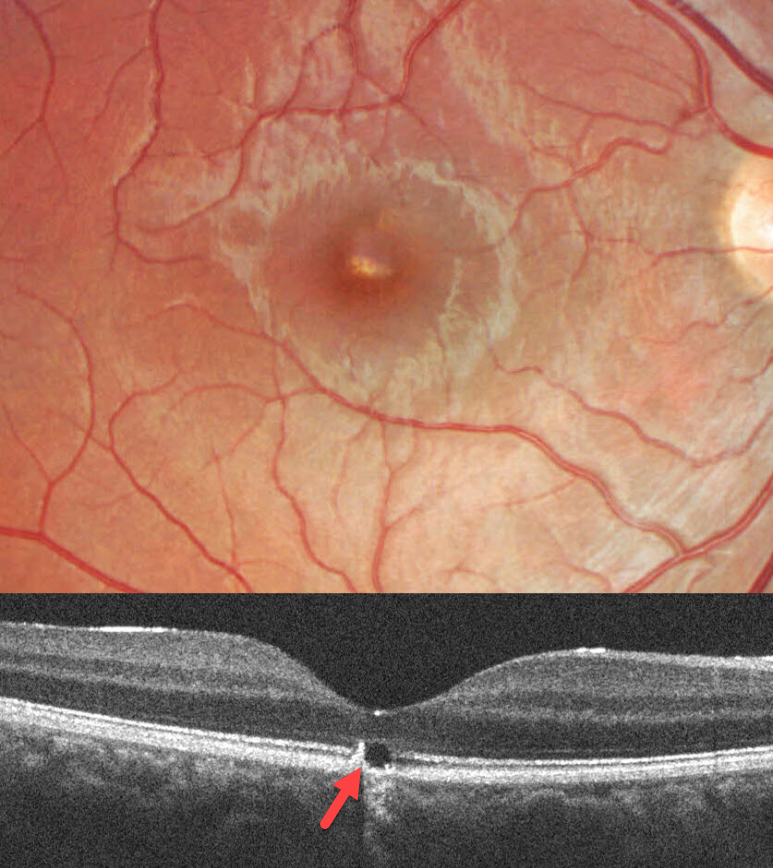
This 17YO male was in a car accident several weeks earlier with a whiplash injury. He noticed mild vision loss in his right eye since. Vision was 20/30.
Color imaging shows a somewhat linear area of foveal pigment loss. OCT scanning of this region shows an outer foveal microdefect with adjacent hyperreflectivity along its temporal edge.
Learning Points:
Initially described by Kelley et al (Arch Ophthalmol 1978;96:834-835), whiplash maculopathy is thought to occur from a vitreous coup-contrecoup injury following a motor vehicle accident. This can cause contusive RPE injury with transient macular edema (AJO 2007;143:348-350) or, more typically, a solar retinopathy-like defect in the outer retina.
Outer foveal microdefects (Cohen et al, Ophthalmology Retina 2021;5:553-561) have been described in numerous conditions, including macular telangiectasia, tamoxifen use, ABCA4 disorders (cone-rod dystrophies, Stargardt disease, and fundus flavimaculatus), phototoxicity, trauma, and vitreomacular traction disorders.
PURTSCHER-LIKE RETINOPATHY
Keissy Sousa, Gil Calvão-Santos, and Rita Gentil
Originally posted on @retina.rocks January 10, 2024
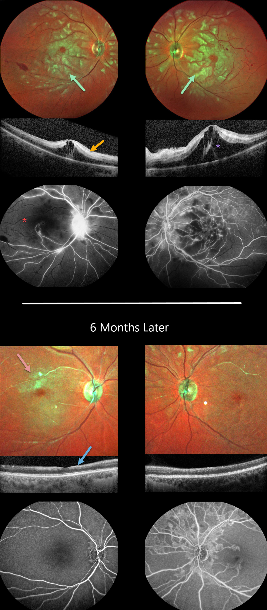
This previously healthy 29YO female presented to the emergency room with acute bilateral vision loss. Vision was counting fingers OU.
Color imaging shows bilateral white patches of inner and mid-retinal ischemia, white narrowed arterioles, and a few inner retinal hemorrhages.
OCT scanning shows variable hyperreflectivity within the inner and mid-retinal layers and cystoid edema. Fluorescein angiography confirms profound retinal ischemia, particularly in the right eye.
Extensive medical workup resulted in a diagnosis of systemic lupus erythematosus (SLE). Rheumatology started systemic rituximab. Bilateral intravitreal dexamethasone followed by intravitreal Eylea was given for the macular edema.
Six months later, although her vision remained at counting fingers OU, she was functioning better and learning to adapt to a new way of living. She was on prednisone 10mg daily and Eylea OU q12 weeks.
Although the acute patches of white retinal ischemia resolved, each fundus is clinically and angiographically markedly ischemic. OCT scanning shows no edema, although there is relative disorganization and thinning of the inner retinal layers.
Learning Points:
Purtscher retinopathy was first described by Otmar Purtscher in 1910. Although originally described in a man who fell from a tree with cranial trauma, these white patches of retinal ischemia and hemorrhages are found in numerous other etiologies, including pancreatitis, chest trauma, and collagen vascular diseases.
The findings are called Purtscher retinopathy when due to trauma, and Purtscher-like retinopathy when seen from other causes. SLE-associated retinopathy more typically appears as hypertensive retinopathy (bilateral nerve fiber layer infarcts and retinal hemorrhages), often in patients with cerebral vasculitis. However, our patient’s blood pressure was not elevated, and there were no signs of central nervous system involvement.
The underlying cause of Purtscher retinopathy is unknown, but it is thought to result from leukoembolization mediated by complement activation. The areas of ischemia include more superficial nerve fiber layer infarcts (cotton wool spots) and deeper ischemia involving the middle retinal layers.
This middle retinal ischemia, paracentral middle maculopathy (PAMM), is a recently-described phenotype found as an idiopathic entity or in other ocular disorders, including Purtscher, diabetic retinopathy, retinal artery and retinal vein occlusions, and acute macular neuroretinopathy (see Retina 2015;35:1921-1930).
VALSALVA RETINOPATHY
Asma Samsudeen and Ashish Sharma
Originally posted on @retina.rocks December 7, 2023
This 28YO male noted sudden vision loss in his left eye 1 day earlier, immediately following coughing. Vision was 20/20 in his normal right eye and counting fingers in his left eye.
Color imaging shows a large layered sub-internal limiting membrane (ILM) hemorrhage occupying the entire macula. The blood extends inferiorly in the subhyaloid space. Some peripapillary vitreous hemorrhage is also noted.
Our patient elected to be observed and, unfortunately, was subsequently lost to follow-up.
Learning Points:
Valsalva retinopathy is characterized by usually unilateral retinal and preretinal hemorrhages caused by raised intrathoracic or intra-abdominal pressure. Various causes include coughing, heavy lifting, and vomiting.
These hemorrhages virtually always resolve without sequelae, although the preretinal blood in severe cases can be drained into the inferior vitreous by creating a small ILM opening using either a thermal or Nd:YAG laser.
INTRAOCULAR FOREIGN BODY
Neelam Khatwani, Priyanka Gupta and Charu Gupta
Originally posted on @retina.rocks November 22, 2023
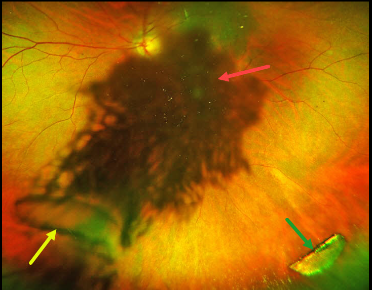
This 45YO male presented with a 4-day history of sudden vision loss in the OS immediately following penetrating trauma while working with a hammer and chisel. Vision was 20/20 in his normal OD and 20/200 OS.
Slit lamp examination OS showed a small peripheral self-sealing corneal laceration, an iris defect, and a ‘rosette cataract.’ On extreme left gaze, a semilunar defect in the posterior lens capsule and a linear defect in the anterior lens capsule were also noted.
Optos color RG imaging shows loss of central fundus detail from the cataract, the capsular defect, and a metallic intraocular foreign body (IOFB).
Fundus examination and localization of an IOFB are often difficult in the presence of secondary cataract. Ultrawide-field fundus imaging is an invaluable diagnostic tool in such cases, as it captures 200° (82% of the retina) in a single image, compared with standard fundus photography.
The rosette cataract in our patient is caused by seepage of aqueous humor through the broken lens capsule and between the lens fibers, resulting in opacification.
Urgent pars plana lensectomy, vitrectomy, removal of the IOFB, and secondary intraocular lens were performed. Vision was 20/40 six weeks following surgery.
VALSALVA RETINOPATHY
Marianna Kavalaraki
Originally posted on @retina.rocks September 20, 2023
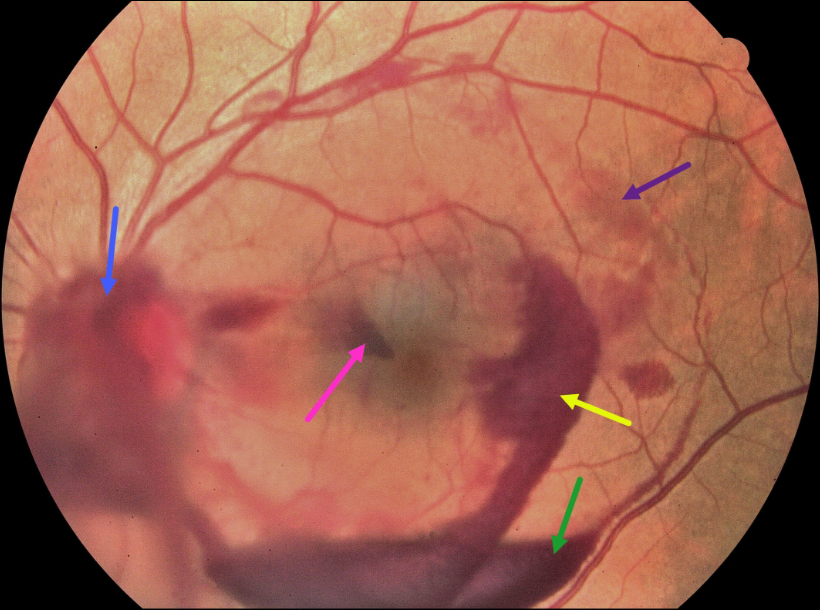
This 39YO male presented with a 3-day history of sudden floaters and central scotoma in his left eye following heavy lifting. Vision was 20/20 in his normal right eye and 20/20 OS.
Color fundus imaging shows multi-layered hemorrhages in the posterior pole, including vitreous, subhyaloid, sub-internal limiting membrane (ILM), Henle’s layer, and deep intraretinal or subretinal. The blood spontaneously resolved after 2 weeks of observation.
Learning Points:
Valsalva retinopathy is characterized by pre-retinal blood (classically sub-ILM but also subhyaloid and vitreous) following a Valsalva maneuver (heavy lifting, vomiting, coughing, sneezing, and compression injuries).
The hemorrhages virtually always resolve without sequelae, although the sub-ILM/hyaloid blood can be drained into the inferior vitreous by creating a small opening in the ILM/hyaloid using either a thermal or Nd:YAG laser.
The differential diagnosis includes other conditions causing multi-layered hemorrhages, including ruptured retinal arterial macroaneurysm, trauma, and anemic/leukemic retinopathy.
SOLAR RETINOPATHY
Originally posted on @retina.rocks August 28, 2023
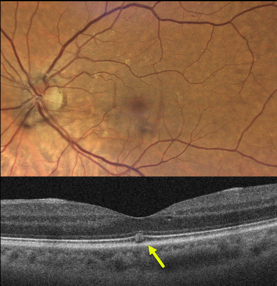
This 67YO female was referred for 3 weeks of vision loss in her left eye. Vision was 20/25 bilaterally.
The fundus photo is normal, but her OCT shows a focal area of hyperreflectivity interrupting the ellipsoid zone.
On further questioning, her symptoms developed while driving, immediately following a transformer explosion.
Learning Points:
The outer retinal damage in solar/welder’s retinopathy is thought to be caused by photochemical outer retinal and RPE damage instead of a thermal burn. Vision is usually fairly good despite the findings in the outer retina and RPE.
VALSALVA RETINOPATHY
Shivraj Tagare and Nishant Maindargi
Originally posted on @retina.rocks August 17, 2023
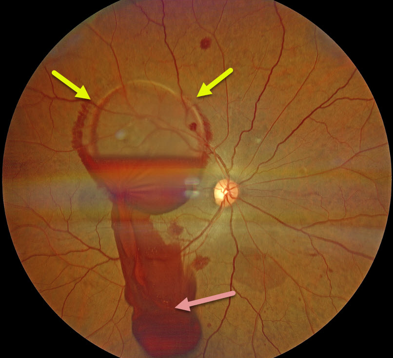
This 37YO male presented 1 day after sudden vision loss in his right eye following vigorous coughing. Vision was 20/400 in his right eye and 20/20 in his normal left eye.
Fundus examination of his right eye showed scattered posterior pole retinal hemorrhages and a large sub-internal limiting membrane (ILM) hemorrhage obscuring his right macula.
A Neodymium:YAG laser was used to create a small inferior opening in the ILM. Fundus photography shows that the blood immediately began draining into the inferior vitreous.
Learning Points:
Valsalva retinopathy is characterized by usually unilateral retinal and preretinal hemorrhages caused by raised intrathoracic or intra-abdominal pressure. Various causes include coughing, heavy lifting, and vomiting.
These hemorrhages virtually always resolve without sequelae, although the sub-ILM blood in severe cases can be drained into the inferior vitreous by creating a small ILM opening using either a thermal or Nd:YAG laser, as was done for this patient.
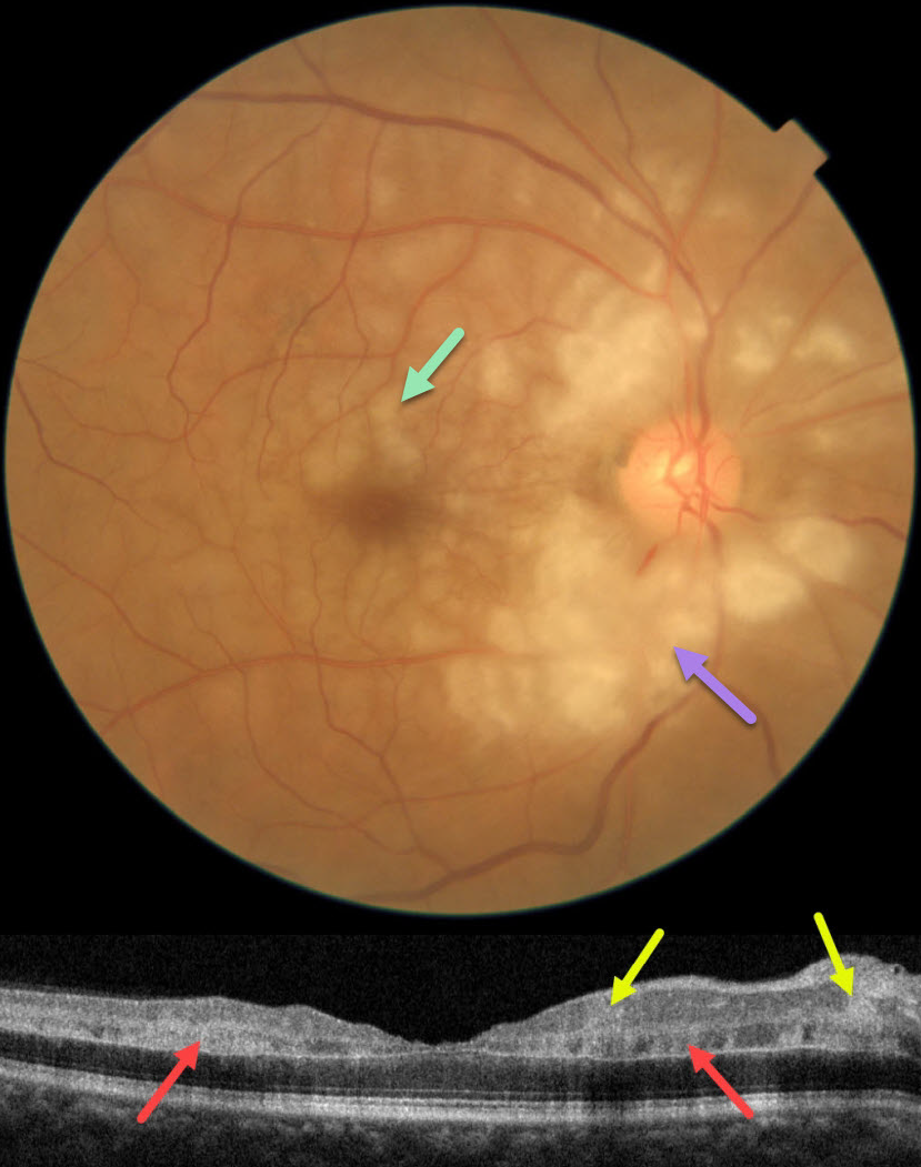
CRAO
Asma Samsudeen and Ashish Sharma
Originally posted on @retina.rocks August 10, 2023
This 60YO male presented with an abrupt, painless loss of vision in his right eye 1 week earlier. There was no prior ocular history or recent history of trauma. Vision was 20/30 in his right eye, and 20/20 in his normal left eye.
Fundus photography shows patches of mostly inner-peripapillary ischemia, with deeper ischemia throughout the macula. This is shown on OCT as patches of inner and mid-retinal hyperreflectivity. Fluorescein angiography showed a markedly delayed transit time (not shown).
We immediately referred him to his cardiologist for an emergency stroke protocol evaluation.
Learning Points:
Purtscher retinopathy was first described by Otmar Purtscher in 1910. Although originally described in a man who fell from a tree with cranial trauma, these white patches of retinal ischemia and hemorrhages are found in numerous other etiologies, including pancreatitis, chest trauma, and collagen vascular diseases.
The findings are called Purtscher retinopathy when due to trauma, and Purtscher-like retinopathy when seen from other causes.
Our patient’s fundus appearance most resembles Purtscher-like retinopathy, with patches of inner- and middle-retinal ischemia. However, given the acute symptoms and delayed angiographic filling time, we believe our patient most likely has an incomplete central retinal artery occlusion (CRAO).
See Mangla et al for the OCT findings in CRAO of varying severities (Int J Retina Vitreous 2023;9;37).
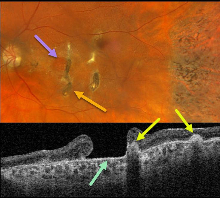
TRAUMATIC MACULAR HOLE + CHOROIDAL RUPTURE
Originally posted on @retina.rocks July 11, 2023
This 57YO female has a long history of vision loss in her left eye following blunt trauma years earlier. Vision was hand motion OS.
Optos color RGB imaging shows a large chronic macular hole with surrounding pigmentary changes. There are several hyperpigmented choroidal ruptures just temporal to the hole. Chorioretinal scarring with intraretinal pigment migration is noted in the temporal periphery.
Swept-source OCT shows a large macular hole. The choroidal ruptures appear as elevated hyperreflective subretinal mounds with posterior shadowing.
Learning Points:
This patient illustrates several sequelae of blunt trauma, including macular hole formation, choroidal rupture, and intraretinal pigment migration with a pseudo-sector retinitis pigmentosa appearance due to commotio-induced photoreceptor loss.
Since fresh traumatic holes can spontaneously flatten, especially in pediatric cases (Liu et al, AJO 2020;210:174-183), an initial period of observation is recommended before considering vitrectomy.
Given the chronicity of our patient’s hole, surgery was not considered.
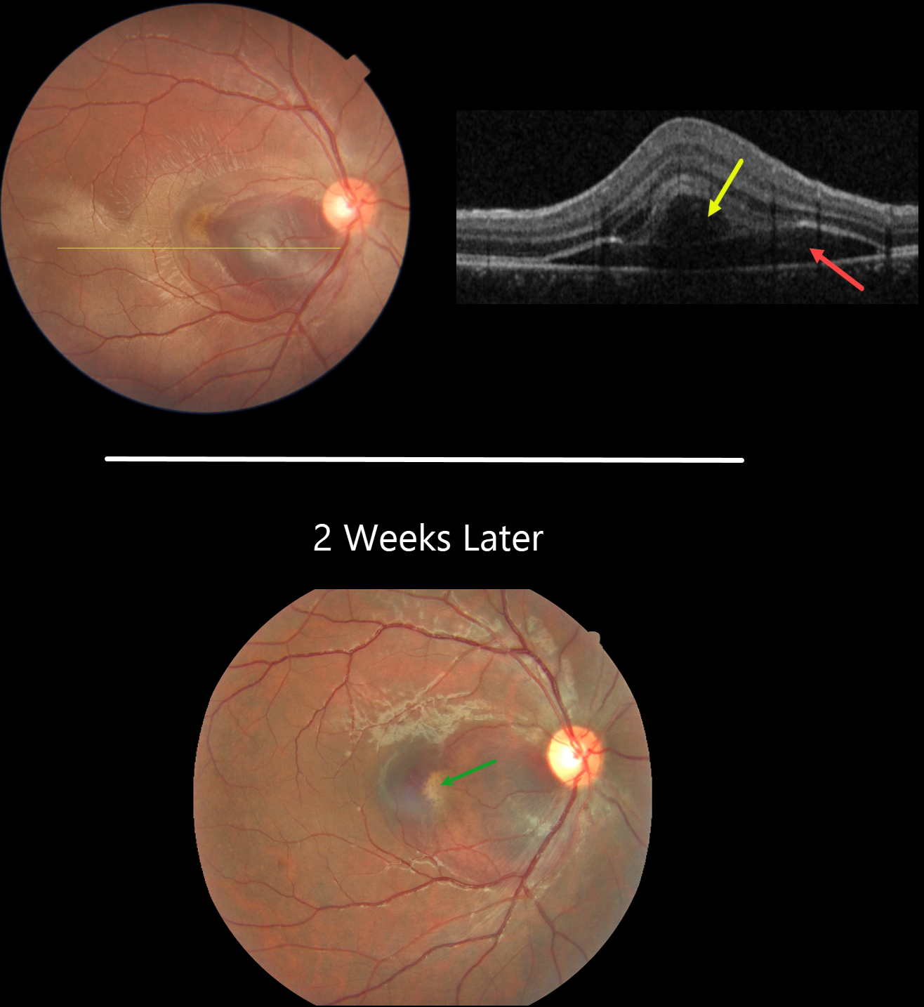
CHOROIDAL RUPTURE
Asma Samsudeen and Ashish Sharma
Originally posted on @retina.rocks July 6, 2023
This 21YO male presented with immediate vision loss after being hit with a cricket ball in his right eye one day earlier. Vision was 20/60.
Fundus photography shows a subretinal hemorrhage in the nasal macula, a small rim of surrounding subretinal fluid, and a more temporal ill-defined commotio retina.
OCT scanning shows hyporeflective retinal elevation from the subretinal blood and fluid, along with an overlying bacillary layer detachment.
Our patient shows several findings classic for acute blunt trauma, including commotio retinae and subretinal blood. This subretinal blood almost always indicates an underlying choroidal rupture, which did, in fact, reveal itself on follow-up 2 weeks later, when vision returned to 20/20. The visual prognosis for these choroidal ruptures is good as long as the rupture spares the foveal center, although patients need lifelong surveillance due to the risk of secondary macular neovascularization.
Learning Points:
A bacillary layer (cone and rod inner and outer segments) detachment (BLD) appears as a unique dome-shaped collection of intraretinal fluid from photoreceptor splitting. These detachments can be observed in an increasing number of conditions, most classically Vogt-Koyanagi-Harada disease (Cicinelli et al, Ophthalmology Retina 2020;4:454-456).
About 8% of eyes with acute non-penetrating ocular trauma have a BLD (Venkatesh et al, Can J Ophthalmol 2022;57:328-336). These resolve within 2-10 days without visual or anatomic sequelae.
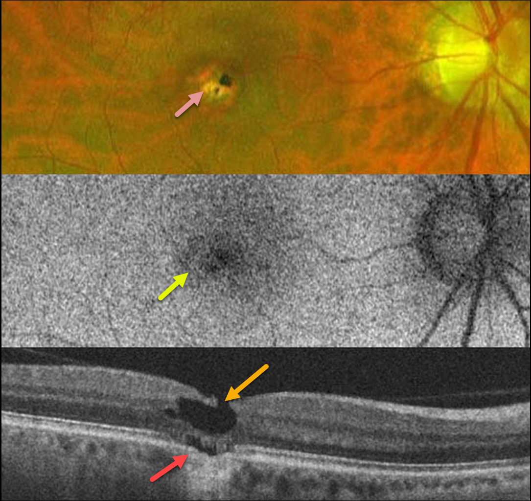
LASER MACULOPATHY
Will Gibson
Originally posted on @retina.rocks June 29, 2023
This 36YO male gave a 2-year history of immediate vision loss in his right eye following an accidental 1064nm YAG laser exposure. Vision was 20/80.
Optos color imaging shows a small, round, mostly atrophic foveal scar, which is variably hypoautofluorescent.
OCT scanning shows a degenerative lamellar macular hole (LMH) with focal loss of the underlying outer retinal bands (outer foveal microdefect), RPE loss, and a possible shallow choroidal excavation.
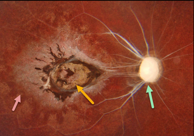
TRAUMATIC UNILATERAL WIPE-OUT
Originally posted on @retina.rocks May 4, 2023
This healthy 10YO boy presented with a 5-year history of blindness in his right eye. His mother said that he injured his eye after falling while playing at school.
Color imaging of his right eye shows a white nerve and retinal vessels, a variably pigmented macular scar, and a moth-eaten appearance to the RPE outside the macula. The left eye was normal.
Learning Points:
The differential diagnosis for unilateral optic nerve atrophy, chorioretinal scarring, and narrowing/sclerosis of the major retinal vessels is relatively small.
The term ‘unilateral wipe-out syndrome’ was coined by Dr. Gass (Ophthalmology 1978;85:521-545) in describing end-stage diffuse unilateral subacute neuroretinitis (DUSN).
Other causes include trauma, ophthalmic artery occlusion, and post-inflammatory (i.e., acute retinal necrosis syndrome, ARNS). Despite the history, it is hard to imagine how a 5-year-old child falling at a playground could cause this type of injury.
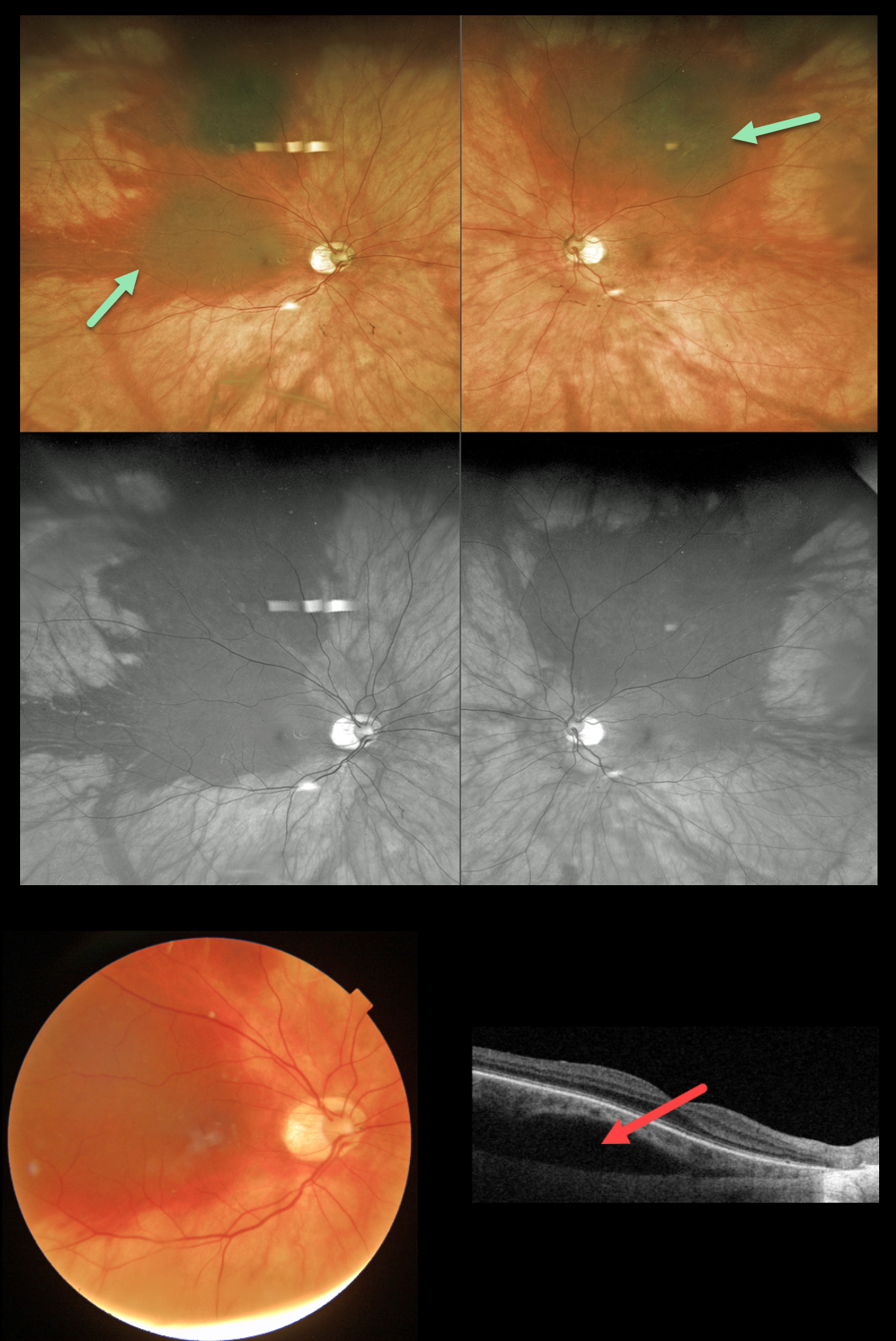
VALSALVA-INDUCED CHOROIDAL HEMORRHAGE
Originally posted on @retina.rocks May 2, 2023
This 61YO healthy female presented with sudden blurred vision immediately following forceful blowing of her nose 3 days earlier. Vision was 20/25 OU.
Optos imaging shows bilateral superior suprachoroidal blood, which extends into the right macula and temporal periphery.
Swept-source OCT through the macula hemorrhage shows hyporeflective suprachoroidal elevation. Observation was recommended, and we expect the blood to absorb over the coming months.
Learning Points:
Valsalva-induced retinal findings almost always present with sub-internal-limiting-membrane blood. Suprachoroidal hemorrhage most commonly develops intra- or postoperatively, following trauma or with thrombolytic agents (Ghu and Green, Survey Ophthalmology 1999;43:471-486).
Valsalva-induced choroidal hemorrhage is exceedingly rare, with only a handful of cases reported in the literature, including a prior Retina Rocks case (see Image Gallery, Valsalva retinopathy folder, Valsalva UIV-20220511).
As with other localized suprachoroidal hemorrhages, these lesions may simulate uveal melanoma (Marous et al, Retina 2018;12:336-341).
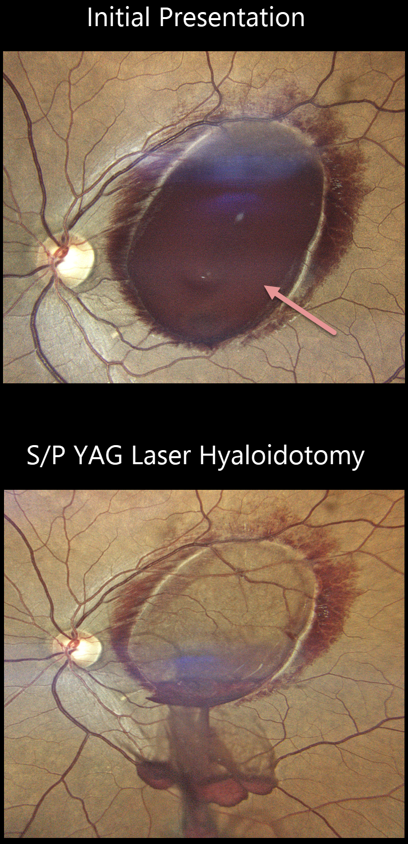
VALSALVA RETINOPATHY
Akansha Sharma, Urmil Shah and Denish Patel
Originally posted on @retina.rocks May 1, 2023
This 40YO male developed sudden loss of vision in his left eye after sneezing 4 days earlier. Vision was 20/1200 OS.
Fundus imaging shows a large sub-internal limiting membrane (ILM) hemorrhage covering the left macula. There is a rim of surrounding nerve fiber layer vs Henle’s blood.
He underwent a YAG laser hyaloidotomy on the same day with immediate improvement to 20/60. Final follow-up vision was 20/30 at 8 weeks.
Learning Points:
Valsalva hemorrhages are usually in the sub-ILM space. These hemorrhages virtually always resolve without sequelae, although the sub-ILM blood in severe cases can be drained into the inferior vitreous by creating a small ILM opening using either a thermal or Nd:YAG laser.
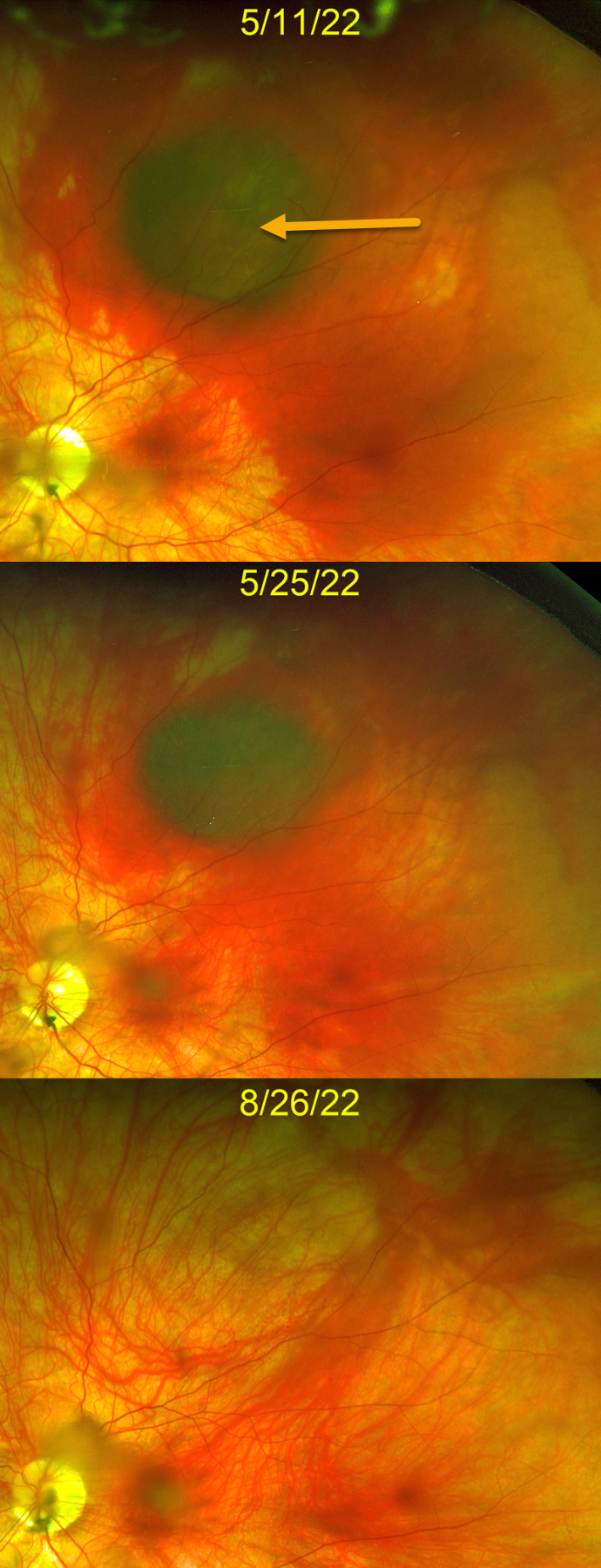
VALSALVA RETINOPATHY
Michael Wong and Jonathan Tsui
Originally posted on @retina.rocks March 9, 2023
This 38YO healthy female presented with one day of peripheral vision loss in her left eye after an episode of forceful vomiting from a migraine headache. Vision was 20/30 OS.
Optos color imaging shows a dark, round, localized suprachoroidal hemorrhage in the superior midperiphery surrounded by subretinal blood. The right fundus was normal (not shown).
The blood gradually improved with observation, completely resolving over the next 3-4 months with return of 20/20 vision.
Learning Points:
Valsalva-induced retinal findings almost always present with sub-internal-limiting-membrane blood. Suprachoroidal hemorrhage most commonly develops intra- or postoperatively, following trauma or with thrombolytic agents (Ghu and Green, Survey Ophthalmology 1999;43:471-486).
Valsalva-induced choroidal hemorrhage is exceedingly rare, with only a handful of cases reported in the literature. As with other localized suprachoroidal hemorrhages, these lesions may simulate uveal melanoma (Marous et al, Retina 2018;12:336-341).
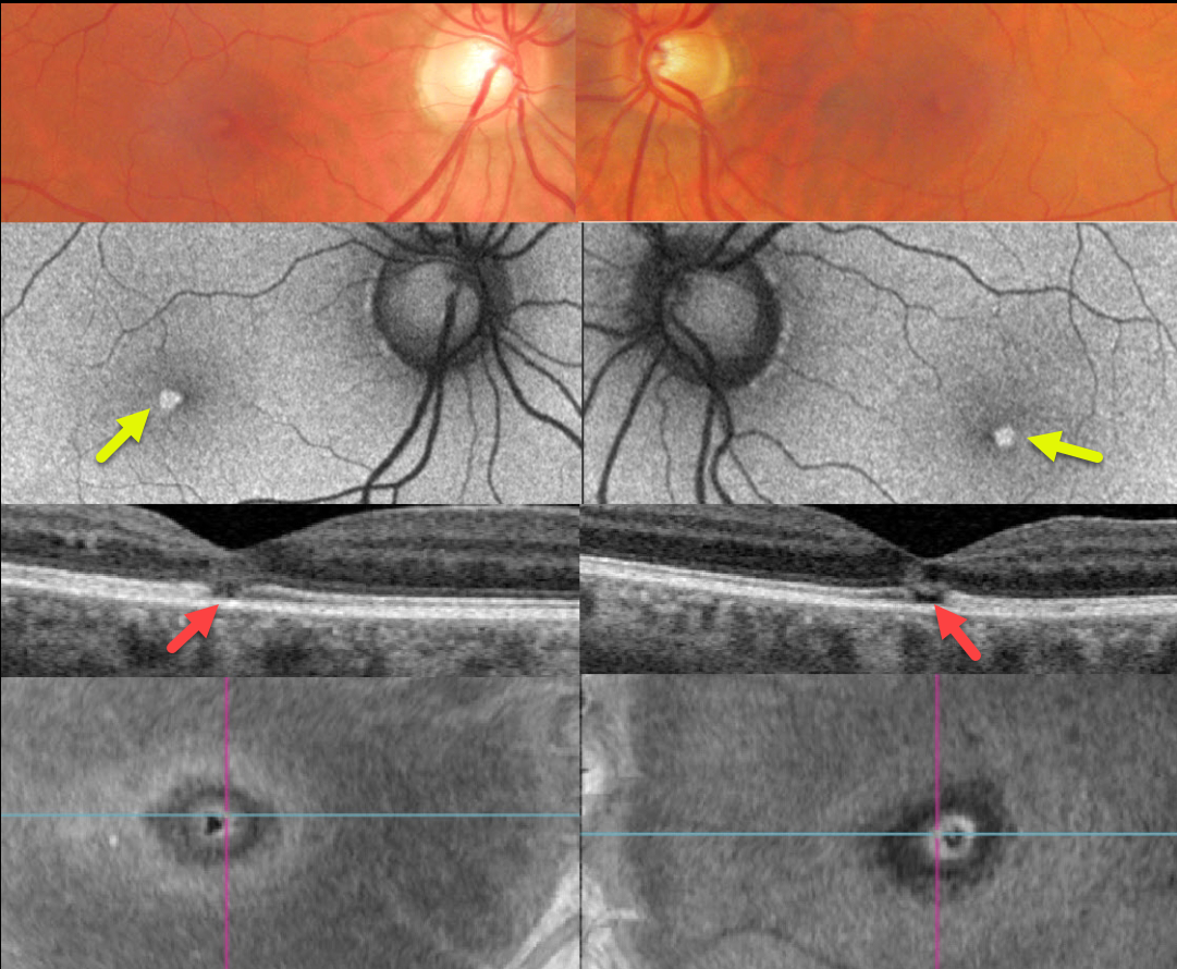
SOLAR RETINOPATHY
Originally posted on @retina.rocks February 27, 2023
This healthy 46YO male complained of mild bilateral central vision loss for about 5 years. Vision was 20/25 OD and 20/30 OS.
Optos color imaging shows subtle central foveal pigment loss. Fundus autofluorescence (FAF) shows these lesions to be hyper-FAF.
Swept-source OCT shows small central foveal outer retinal defects, and en face OCT of the outer retina shows a unique perspective of these lesions.
On further questioning, he noted that his visual symptoms immediately followed his viewing the Great Eclipse of 2017 without proper eye protection.
Learning Points:
The outer retinal damage in solar retinopathy is thought to be caused by photochemical outer retinal and RPE damage instead of a thermal burn. Vision is usually fairly good despite the outer retinal and RPE findings.
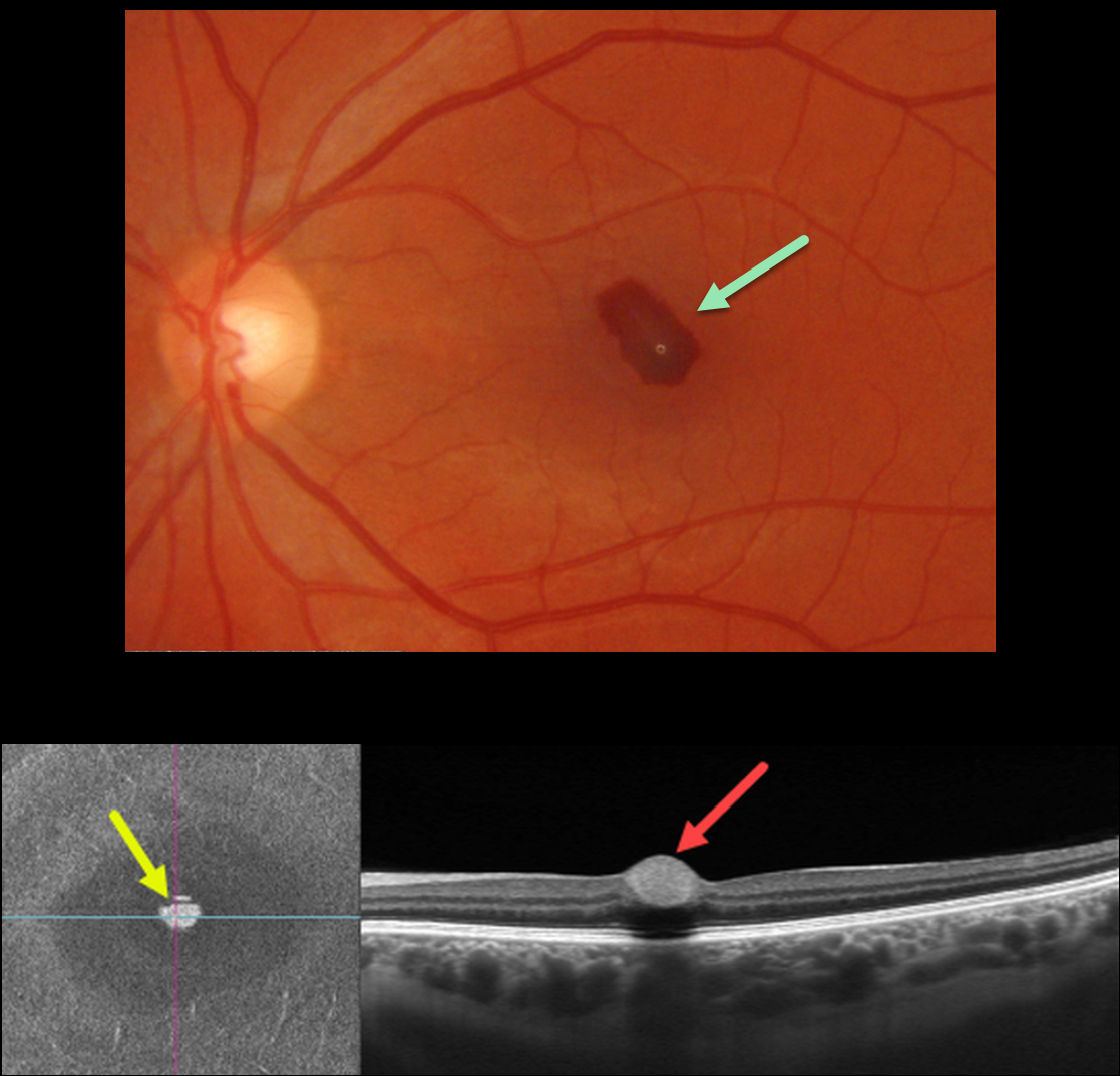
VALSALVA RETINOPATHY
Originally posted on @retina.rocks January 17, 2023
This 33YO female had a severe upper respiratory illness with intense coughing and sneezing. She woke one day earlier with a central scotoma in her left eye.
Optos color imaging shows a teardrop-shaped sub-internal limiting membrane (ILM) hemorrhage. Swept-source OCT B-scan shows a hyperreflective sub-ILM lesion with underlying shadowing. En face scanning of the inner retina provides a unique perspective on the blood.
Learning Points:
Valsalva hemorrhages are usually in the sub-ILM space. These hemorrhages virtually always resolve without sequelae, although the sub-ILM blood in severe cases can be drained into the inferior vitreous by creating a small ILM opening using either a thermal or Nd:YAG laser.
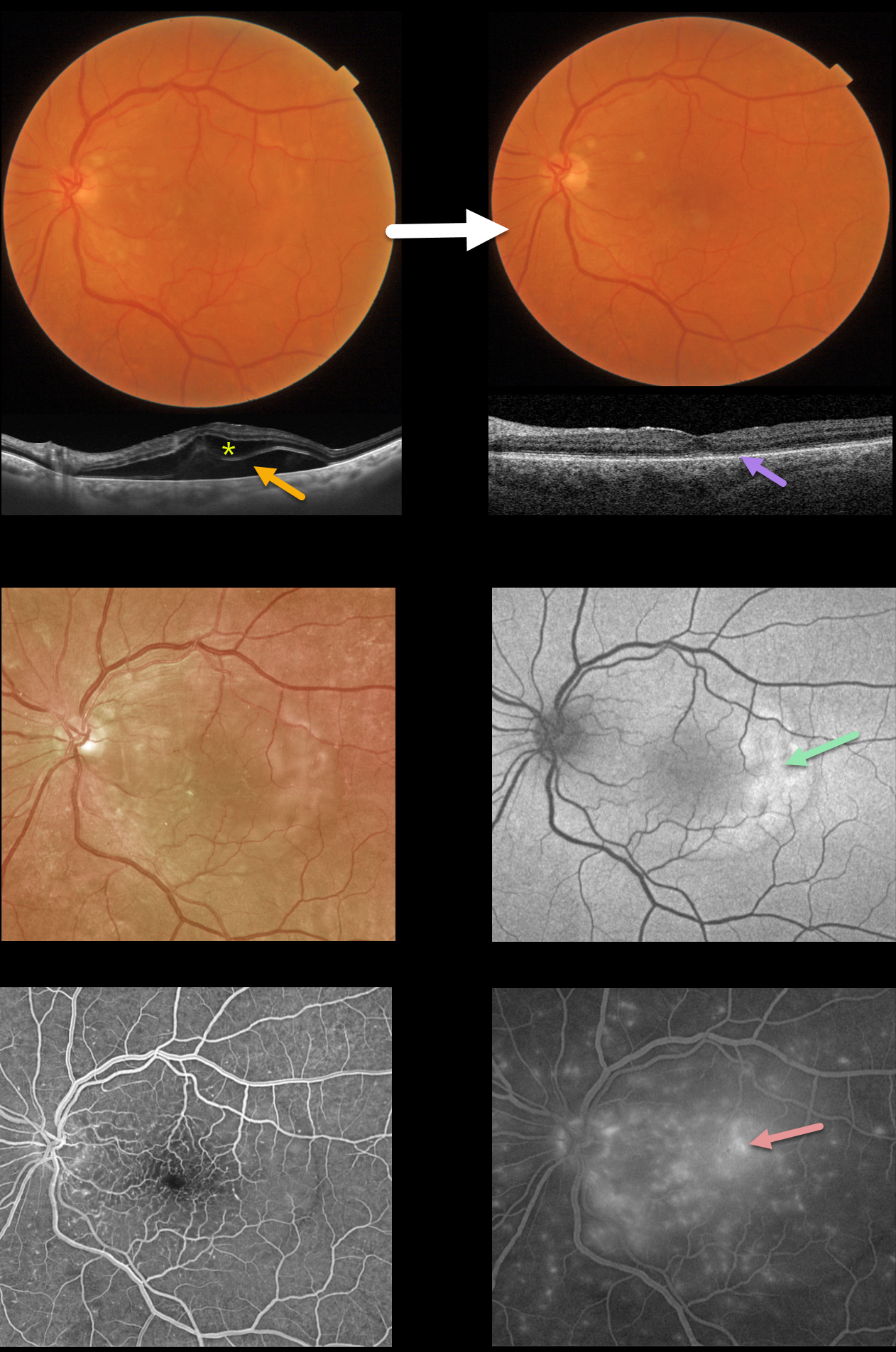
CONTUSIVE TRAUMA WITH SEROUS MACULAR DETACHMENT
Originally posted on @retina.rocks January 16, 2023
This 71YO male presented with decreased vision in his left eye one day following a motor vehicle accident with airbag deployment. Vision was 20/100.
Color photography shows variable faint patches of outer retinal whitening. Swept-source OCT shows outer retinal and subretinal fluid.
Optos fundus autofluorescence (FAF) shows temporal macular hyper-FAF. Fluorescein angiography shows early dots of subretinal hyperfluorescence with late deep leakage extending beyond the arcades.
Two weeks later, he was subjectively much improved, although he was bothered by metamorphopsia and some paracentral scotomas. Vision was 20/60. The macular pigmentary changes improved funduscopically. All macular fluid resolved, but the outer photoreceptor bands were disorganized and thinned.
Learning Points:
Posterior segment findings from blunt ocular trauma most commonly include commotio retinae or choroidal rupture.
Our patient’s unusual findings were from a contusive injury to the choroid and RPE. RPE dysfunction led to fluid accumulation in the subretinal space and the outer retina.
A strikingly similar case was reported by Mishra et al (Journal of Vitreoretinal Diseases 2021;5:165-169).
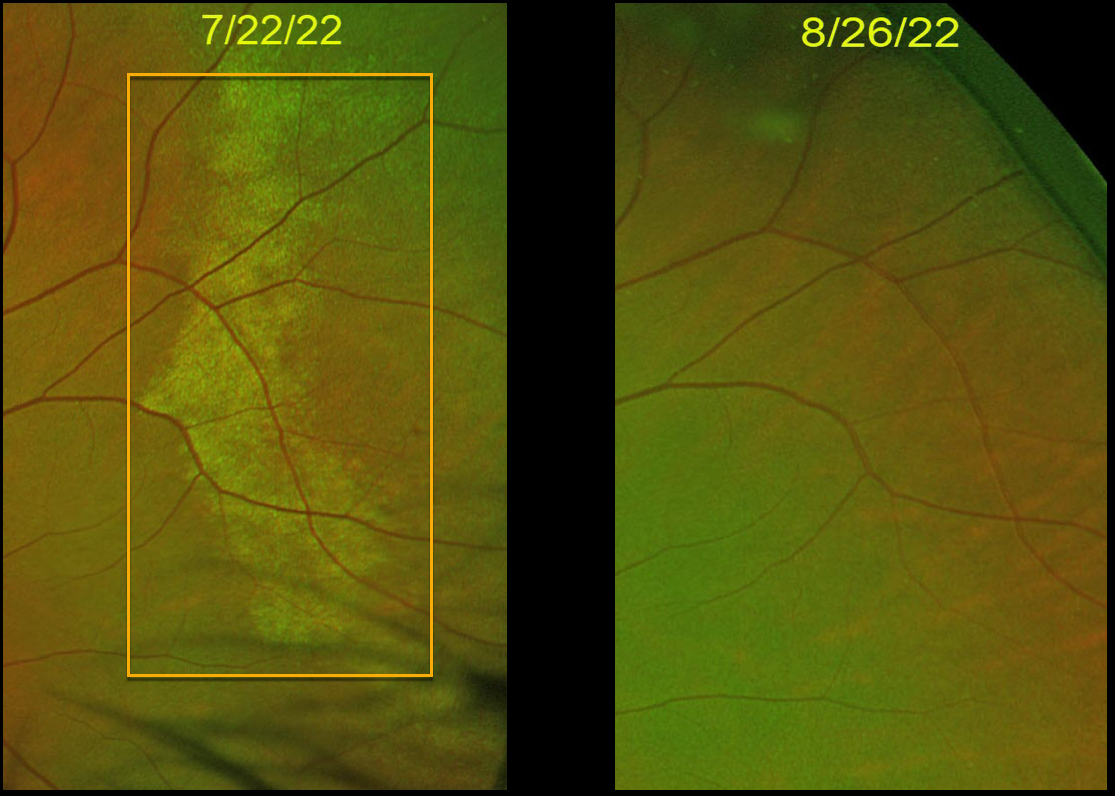
COMMOTIO RETINAE
Originally posted on @retina.rocks September 27, 2022
This 10YO boy was hit in the right eye with a Nerf ball and developed commotio retinae. One month later, the retinal appearance normalized.
Vision was 20/20 at both visits.
Learning Points:
Commotio retinae is caused by direct, blunt eye trauma. The outer retinal whitening is due to shearing of the photoreceptor outer segments.
The term “Berlin’s edema” is a misnomer and should not be used, as the retina is not edematous. The acute white retinal changes usually resolve within several weeks. With resolution, the fundus appearance can normalize, as in this patient.
With more severe injury, outer retinal loss can allow RPE cells to migrate into the retina, giving a pseudo-retinitis pigmentosa appearance. Patients can also develop retinal breaks and detachment.
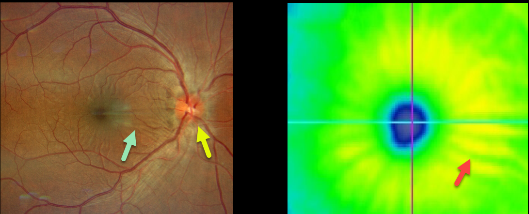
AIRBAG TRAUMA
Will Gibson
Originally posted on @retina.rocks August 25, 2022
This 35YO male presented with a unilateral swollen nerve and retinal striae following blunt trauma from airbag deployment Vision was 20/25. The intraocular pressure was 15, and there were no signs of open-globe injury.
Optos imaging shows retinal folds radiating from the macular center. These striae are also evident on the OCT thickness map.
He was started on oral prednisone, and 10 days later, the ocular findings had improved (images not available).
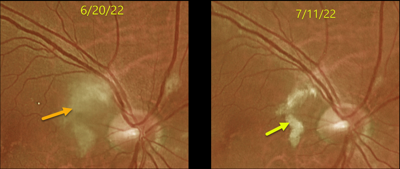
PURTSCHER-LIKE RETINOPATHY
Originally posted on @retina.rocks July 28, 2022
This 65YO male developed inferior field loss in his right eye immediately following cardiac valve replacement on 6/10/22. Vision was 20/30 when he presented to our office 10 days later.
Optos imaging shows patches of inner retinal opacification in the nasal macula and around the nerve, consistent with Purtscher ’s-like retinopathy. Several weeks later, the opacification was resolving with improving symptoms.
Learning Points:
Purtscher’s retinopathy was first described by Otmar Purtscher in 1910. Although originally described in a man who fell from a tree with cranial trauma, these white patches of retinal ischemia and hemorrhages are found in numerous other etiologies, including pancreatitis, chest trauma, and collagen vascular diseases.
The findings are called Purtscher’s retinopathy when due to trauma, and Purtscher-like retinopathy when seen from other causes. The underlying cause of Purtscher’s is unknown, but it is thought to represent leukoembolization by complement activation. The areas of ischemia include more superficial nerve fiber layer infarcts (cotton wool spots) and deeper ischemia involving the middle retinal layers.
This middle retinal ischemia, paracentral middle maculopathy (PAMM), is a recently-described phenotype found as an idiopathic entity or in other ocular disorders, including Purtscher’s, diabetic retinopathy, retinal artery and retinal vein occlusions, and acute macular neuroretinopathy (see Retina 2015;35:1921-1930).
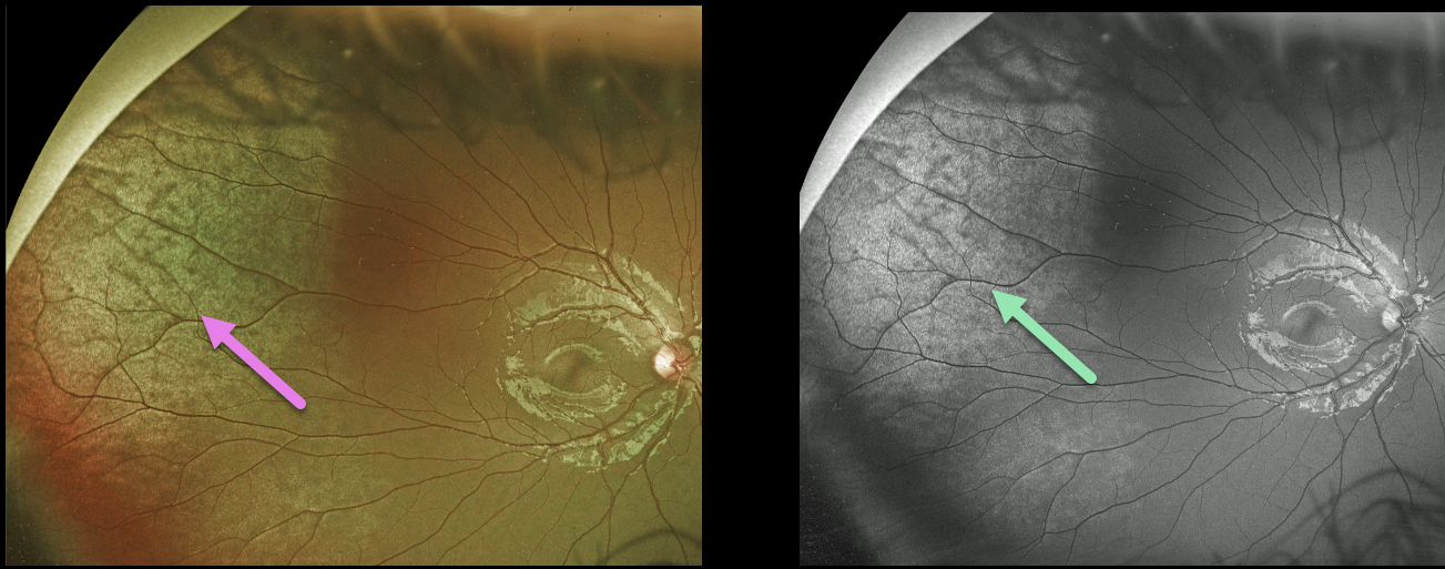
COMMOTIO RETINAE
Originally posted on @retina.rocks July 12, 2022
This 11YO male presented a few hours after being hit in his right eye with a toy gun. Optos color and green channel imaging show a broad area of commotio retina in the superotemporal periphery. Vision was 20/20.
Learning Points:
Commotio retinae is caused by direct, blunt eye trauma. The outer retinal whitening is due to shearing of the photoreceptor outer segments. The term “Berlin’s edema” is a misnomer and should not be used, since the retina is not edematous. The acute white retinal changes usually resolve within several weeks. With resolution, the fundus appearance can normalize.
With more severe injury, outer retinal loss can allow RPE cells to migrate into the retina, giving a pseudo-retinitis pigmentosa appearance. Patients can also develop retinal breaks and detachment.
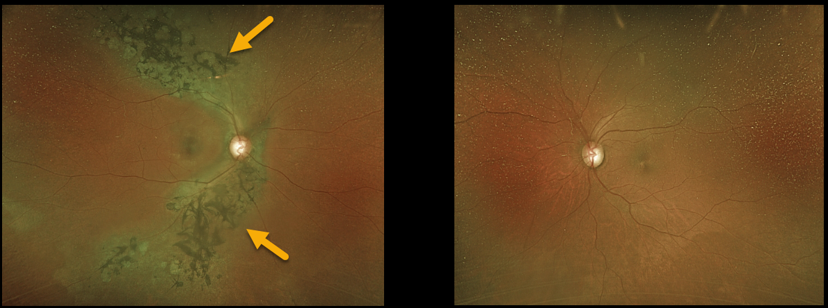
TRAUMATIC PSEUDO-RP
Originally posted on @retina.rocks May 31, 2022
A 67YO female was referred for asymptomatic retinal findings in her right eye. She gave a history of unspecified head trauma as a child.
Optos imaging shows chorioretinal scarring with intraretinal pigment migration, extending superotemporally and inferotemporally from the optic nerve. The left fundus was completely normal.
Learning Points:
Photoreceptor loss from numerous causes, including blunt trauma, retinal detachment, macular telangiectasia, and retinitis pigmentosa (RP), can allow for intraretinal RPE migration with this shared phenotype.
Unilateral RP is exceedingly rare and requires normal electrophysiologic findings and long-term observation to ensure delayed findings in the fellow eye.
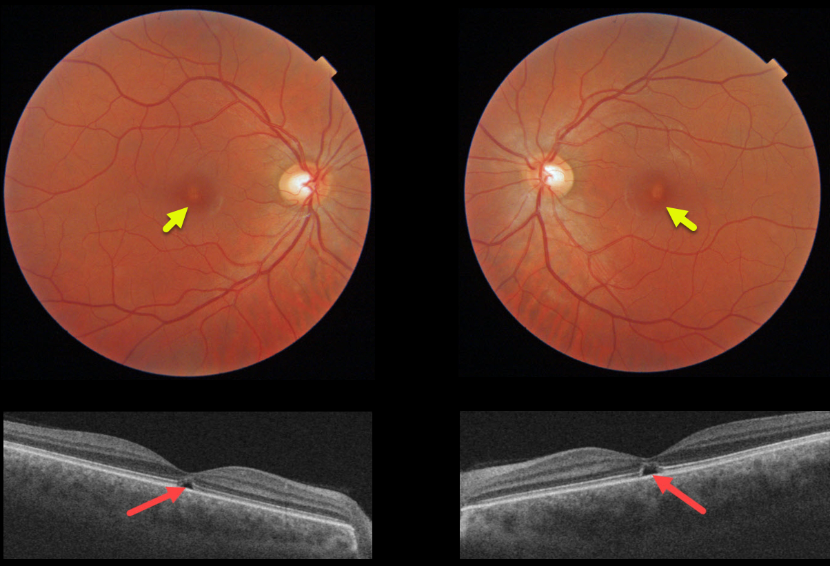
SOLAR RETINOPATHY
Originally posted on @retina.rocks April 27, 2022
This 31YO male was referred for relatively asymptomatic central macular pigmentary changes. He gave a long history of twice-daily sun gazing, which always caused a very pleasurable sneezing episode. Vision was 20/30 OD and 20/40 OS.
Triton color imaging shows tiny foveal pigment loss. Swept-source OCT shows bilateral ellipsoid zone and outer segment defects.
We diagnosed him with the ACHOO reflex (Autosomal Dominant Compelling Helioophthalmic Outburst). And no, this isn’t an April Fools post, and we’re not talented enough to even make this stuff up if we wanted to! Patients with the ACHOO reflex sneeze when exposed to bright lights, typically sunlight.
We advised him to stop his sun gazing ritual, although we’re not sure this will be possible for him.
Learning Points:
The outer retinal damage in solar retinopathy is thought to be caused by photochemical outer retinal and RPE damage instead of a thermal burn. Vision is usually fairly good despite the outer retinal and RPE findings.
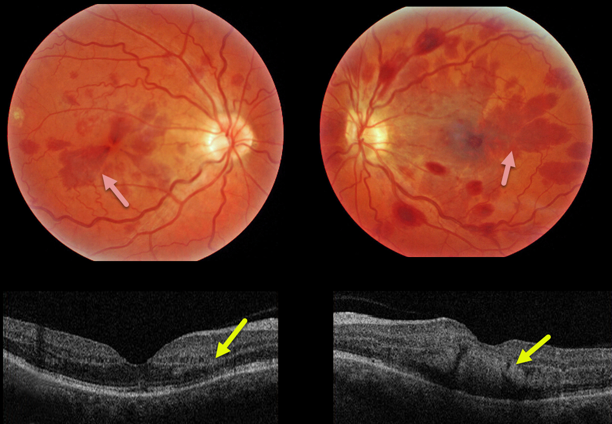
HENLE LAYER HEMORRHAGE
Originally posted on @retina.rocks April 18, 2022
This 47YO female presented with bilateral vision loss. She had a seizure 10 days prior, losing consciousness and hitting her head. Upon awakening, she had reduced vision in her right eye and complete central loss in her left. Vision was 20/60 OD and counting fingers at 3 feet OS. Head CT scanning was normal.
Triton color imaging shows deep radiating wedge-shaped retinal hemorrhages centered in each macula. Swept-source OCT shows that these hemorrhages are located primarily in the outer plexiform layer (OPL).
Learning Points:
Radiating deep macular hemorrhages are seen in multiple conditions, including Valsalva (likely this case) and trauma. Their unique radiating appearance is caused by blood accumulating in Henle’s layer.
Kon Graversen et al described hemorrhagic unilateral retinopathy, an idiopathic disorder with a similar appearance (Retina 2014;34:483-489).
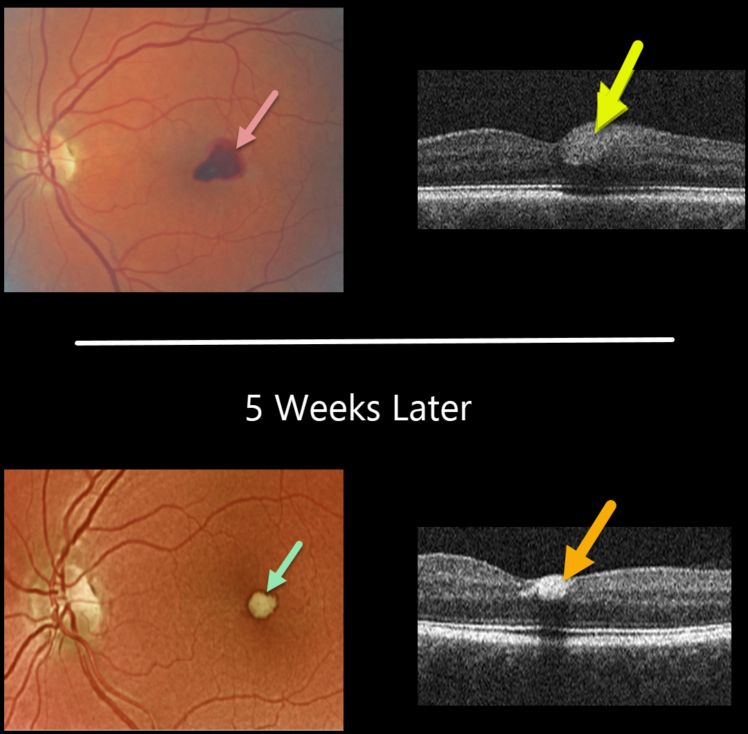
VALSALVA RETINOPATHY
Originally posted on @retina.rocks March 24, 2022
This 56YO female presented with acute vision loss in her left eye from Valsalva retinopathy. Vision was 20/100. A temporal foveal retinal hemorrhage is seen, located within the inner retina on OCT B-scan.
Five weeks later, the devitalized blood is contracting and turning yellow. The blood also became more hyperreflective on the OCT B-scan. All blood subsequently resolved with the return of normal vision.
Learning Points:
Valsalva hemorrhages are usually in the sub-internal limiting membrane (ILM) space, although this case was in the inner retina. These hemorrhages virtually always resolve without sequelae, although the sub-ILM blood can be drained into the inferior vitreous by creating a small opening in the ILM using either a thermal or Nd:YAG laser.
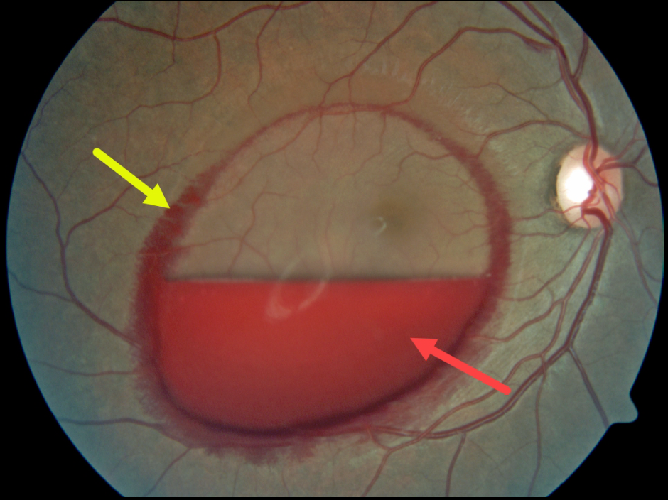
VALSALVA RETINOPATHY
Originally posted on @retina.rocks March 3, 2022
This patient presented with an acute layered sub-internal limiting membrane (ILM) hemorrhage due to a Valsalva maneuver. There is also a rim of blood along the circumference of the ILM detachment, which is most likely located in Henle’s layer, given its radiating deep retinal pattern.
Learning Points:
Sub-ILM hemorrhages most commonly follow a Valsalva maneuver or trauma, but can also happen spontaneously, as with anemia.
These hemorrhages virtually always resolve without sequelae, although the blood can be drained into the inferior vitreous by creating a small opening in the ILM using either a thermal or Nd:YAG laser.
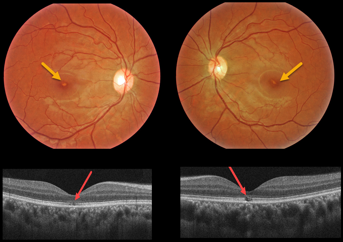
SOLAR RETINOPATHY
Originally posted on @retina.rocks February 24, 2022
This 32YO female with schizophrenia reported regular sun gazing every morning for many years. Vision was 20/40 OU.
Triton color imaging shows tiny foveal pigment loss. Swept-source OCT shows bilateral ellipsoid zone and outer segment defects.
Learning Points:
The outer retinal damage in solar retinopathy is thought to be caused by photochemical outer retinal and RPE damage instead of a thermal burn.
Vision is usually fairly good despite the outer retinal and RPE findings.
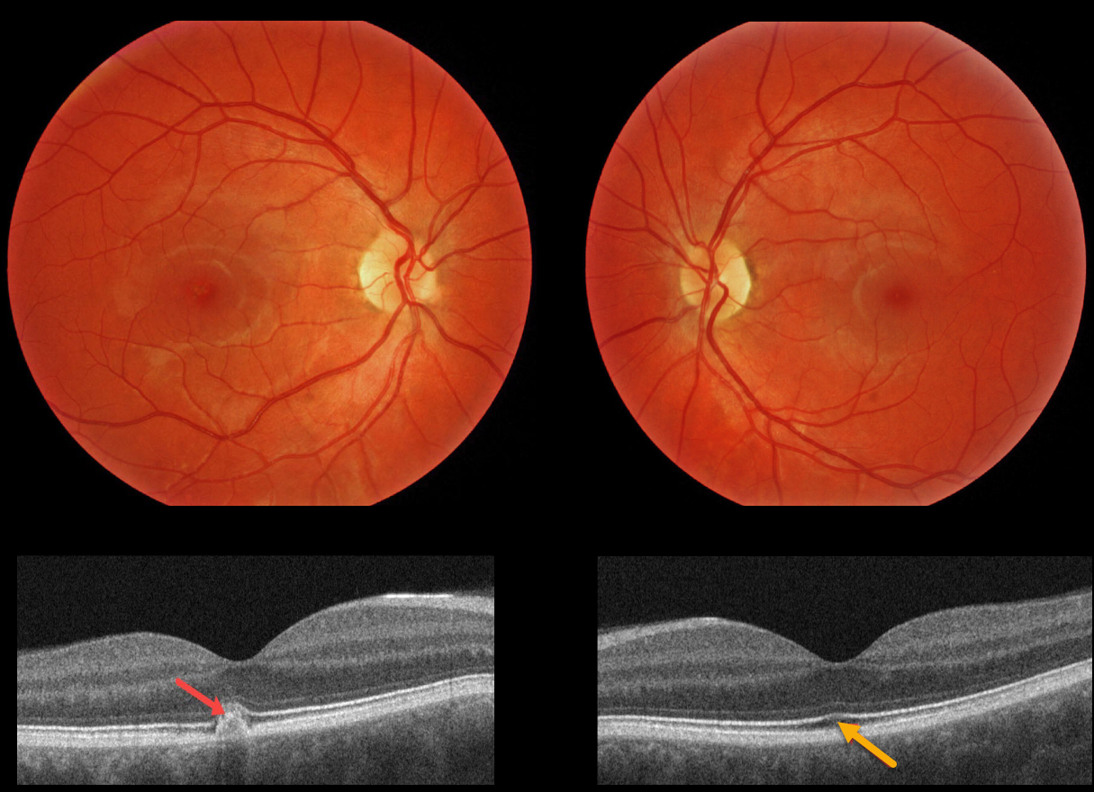
WELDER’S FLASH RETINOPATHY
Originally posted on @retina.rocks December 16, 2021
This 29YO male subjectively noted a persistent central visual disturbance in his right eye following a welder’s flash. Vision was 20/20 OU.
Triton color imaging shows subtle, tiny foveal pigmentary changes. Swept-source OCT shows an RPE and outer retinal hyperreflective mound OD, and very subtle ellipsoid zone and outer segment changes OS.
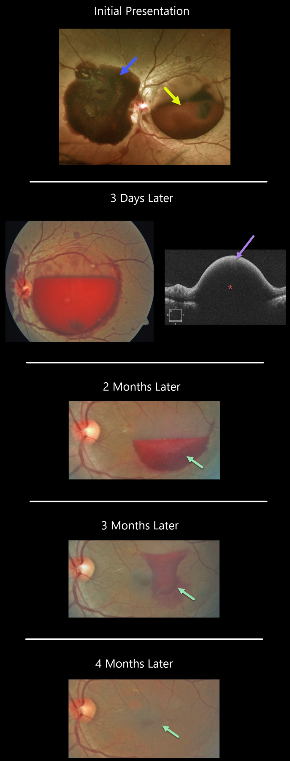
VALSALVA RETINOPATHY
Originally posted on @retina.rocks November 16, 2021
This patient developed sudden vision loss in their left eye while on the toilet.
A large sub-internal limiting membrane (ILM) hemorrhage occupies the macula, while a large subhyaloid hemorrhage extends from the nerve nasally. Scattered retinal hemorrhages are also seen.
Blood pressure was 191/141, and they were sent to the emergency room for hypertensive control.
When examined 3 days later, the macular blood had already begun to decrease. OCT scanning shows marked hyperreflectivity from the blood with posterior shadowing.
The blood gradually resolved over the next four months, and vision improved to 20/30.
Learning Points:
Acute sub-internal limiting membrane (ILM) hemorrhage most commonly is a result of a Valsalva maneuver or trauma, but can also happen spontaneously, as with anemia. The presence of subhyaloid hemorrhage, as well as the association with severe hypertension as occurred in our patient, is quite unusual.
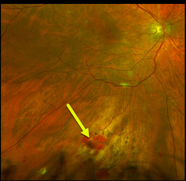
OCULAR PERFORATION FROM SUBTENON INJECTION
Originally posted on @retina.rocks September 28, 2021
This 73YOF was referred for persistent bilateral symptomatic pseudophakic cystoid macular edema (CME) despite a course of topical therapy. Vision was 20/70 OU.
During the triamcinolone subtenon injection, given through the inferior conjunctival cul-de-sac, the patient complained of severe pain, and the red reflex turned white. We immediately removed the needle. Triamcinolone was dispersed throughout the vitreous cavity; no retinal detachment or break could be identified, and the intraocular pressure was remarkably normal.
When examined two days later, vision was hand motion due to residual triamcinolone, and the retina remained attached. One week later, the examination was unchanged, and she requested that the material be removed. At vitrectomy, the penetration was noted in the inferior midperiphery without open breaks or detachment. Three weeks following vitrectomy, vision was 20/40, with faint residual blood and chorioretinal scarring.
Not knowing how to proceed immediately after the initial injection, the patient allowed us to perform a subtenon injection in the left eye, which was thankfully completely uneventful. Vision returned to 20/40 within a few weeks with resolved CME.
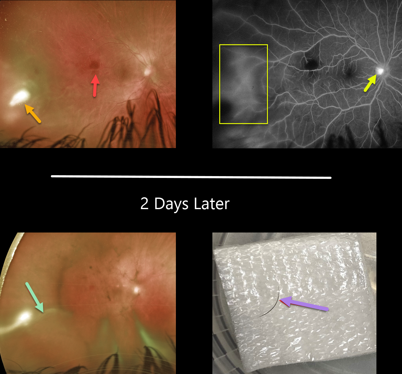
INTRAOCULAR FOREIGN BODY
Originally posted on @retina.rocks September 21, 2021
This 18YO female presented with a 10-day history of pain, redness, and blurred vision in her right eye. Her referring doctor put her on topical prednisolone acetate 1% and ciprofloxacin several days earlier.
Vision was 20/60 OD and 20/20 in her normal left eye. There was significant temporal conjunctival injection overlying a deeper scleral nodule with moderate chemosis. There were fine KP on the corneal endothelium, severe cell and moderate flare, and a 0.8mm hypopyon with scattered fibrin clumps (not shown).
Fundus examination shows an outer retinal hemorrhage in the distal macula, along with a patch of white retinal/vitreous inflammation in the temporal periphery. Fluorescein angiography shows diffuse leakage temporally, along with some optic nerve leakage.
She was placed empirically on oral fluconazole. Laboratory workup for various infectious (toxoplasmosis, bartonella, syphilis, tuberculosis, toxoplasma) and inflammatory (Behcet’s, HLA-B27, ANA, ESR) causes was ordered.
One week later, vision was stable, and the scleral and anterior chamber inflammation were only minimally better. Given the lack of significant improvement, oral Bactrim was started.
She returned 2 days later with increasing vitreous inflammation and elongation of the temporal white vitreous opacity. Laboratory results were completely negative. She was taken to surgery the following day to both clear the visual axis and to obtain vitreous for cultures and sensitivities.
During core vitrectomy, the vitreous was so thick that the cutter had to be pulsed on and off due to the tip being occluded. As the elongated vitreous opacity was excised, an embedded underlying eyelash cilia was discovered. The lash was ‘plucked’ with intraocular forceps and removed from the eye, and the site was prophylactically surrounded with laser.
One week later, vision improved to 20/30. The external and intraocular inflammation were dramatically improved. However, a new superotemporal dialysis with peripheral retinal detachment was found, which was repaired with vitrectomy.
Learning Points:
This is an unbelievable case, and it is difficult to fathom how the eyelash entered the vitreous cavity. There was no history of trauma or ocular surgery.
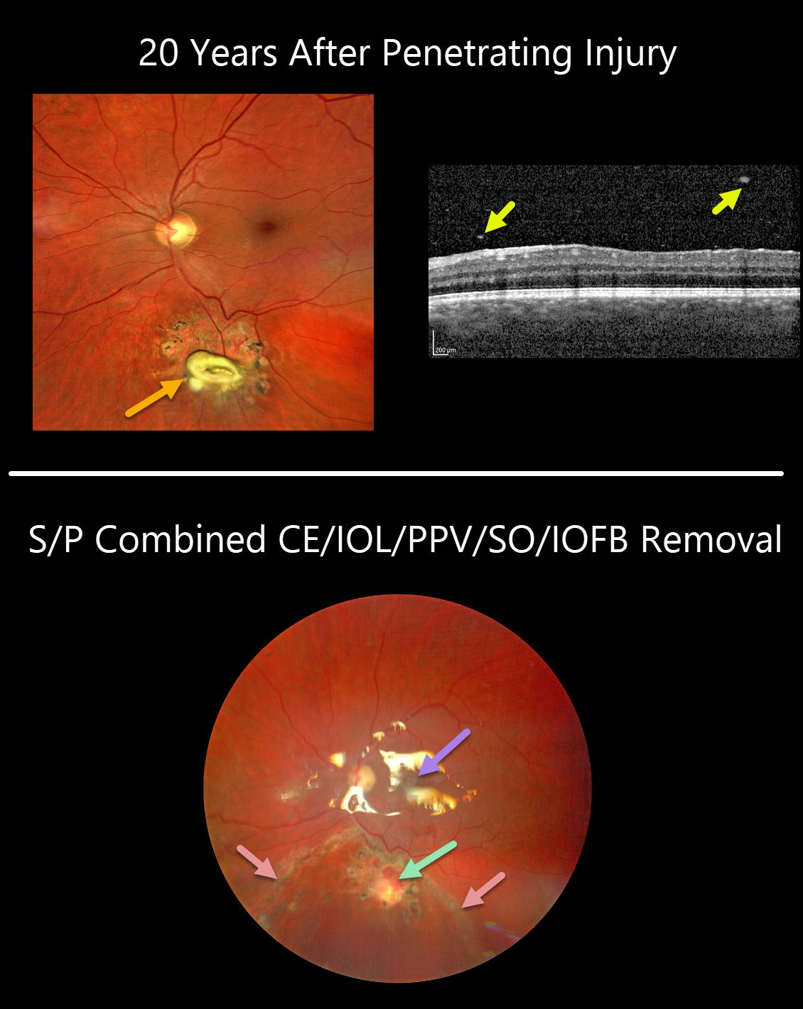
INTRAOCULAR FOREIGN BODY
Originally posted on @retina.rocks September 9, 2021
This 36YO male presented with recent blurred vision of 20/32 in his left eye.
Clarus imaging shows a whitish elevated lesion with surrounding chorioretinal scarring just below the inferotemporal arcade. OCT scanning shows overlying vitreous cells.
He gave a history of a retained retinal plastic foreign body 20 years earlier, following a penetrating injury from an exploding detonator. He had no prior visual complaints, and given the presumed inert nature of the intraocular foreign body (IOFB), surgery was not recommended at that time.
Combined phacoemulsification and vitrectomy with foreign body removal was performed due to increasingly symptomatic vitritis. The retina around the IOFB was necrotic and excised, and the area was surrounded with laser retinopexy. The retina was completely attached under silicone oil one week later.
After removal, the central portion of the IOFB was found to be metallic but coated in plastic. Presumably, as the plastic degraded over time, the metal’s toxicity caused secondary inflammation and visual symptoms.
Learning Point:
In retrospect, a CT scan should have been performed following the initial injury to ensure that the IOFB was fully non-metallic.
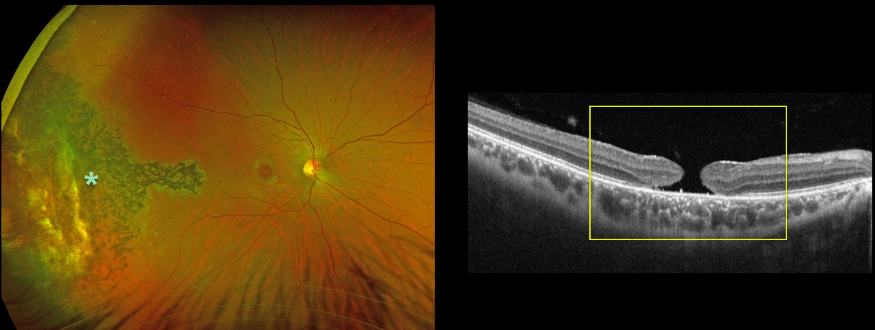
MACULAR HOLE
Originally posted on @retina.rocks August 24, 2021
This 31YO male was hit in his right eye by a paintball when he was 12 years old. His vision was 20/200 due to a large, chronic 1100-micron full-thickness macular hole.
Optos imaging shows chorioretinal scarring with intraretinal pigment migration extending from the distal macula into the temporal periphery, underlying the likely impact site.
Learning Points:
Traumatic macular holes are more common in younger patients, often from recreational or sports injury, and tend to be more eccentric in shape.
The mechanism could be sudden vitreous separation or stretching of the posterior pole resulting from anteroposterior ocular compression. The holes can spontaneously close.
Unfortunately for our patient, the hole remained open, and at this point, we saw little possible surgical benefit given its chronicity, size, and secondary pigmentary changes.
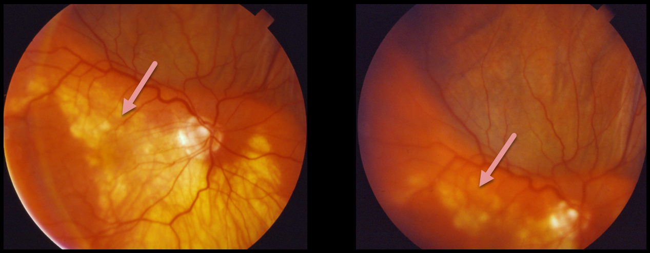
SUPRACHOROIDAL HEMORRHAGE
Originally posted on @retina.rocks August 20, 2021
This patient presented with a pigmented, well-defined, and irregularly lumpy superior subretinal mass due to a suprachoroidal hemorrhage. This simulated a uveal melanoma.
Unfortunately, we don’t have any further information or images for this patient. However, we suspect that the blood spontaneously resolved with normalization of the fundus appearance.
Learning Points:
Patients presenting with suprachoroidal hemorrhage will usually have a history of recent intraocular surgery, trauma, or Valsalva. A clinical clue to this being a hemorrhage and not a melanoma includes the often somewhat lumpy and corrugated appearance, as in this patient.
See Chu and Green for a good review of suprachoroidal hemorrhage (Surv Ophthalmol 1999;43:471-486).
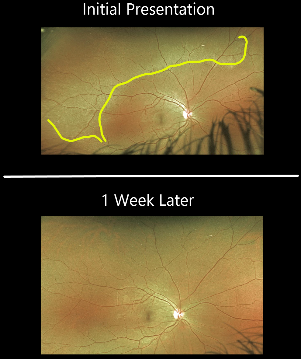
COMMOTIO RETINAE
Originally posted on @retina.rocks August 9, 2021
This 27YO female presented with eye pain, 3 days following falling down the stairs and hitting her right eye. Prior CT imaging revealed an associated right nasal fracture.
Optos imaging shows a broad area of commotio retinae superiorly. The commotio completely resolved 1 week later.
Learning Points:
Commotio retinae is caused by direct, blunt eye trauma. The outer retinal whitening is due to shearing of the photoreceptor outer segments.
The term “Berlin’s edema” is a misnomer and should not be used, since the retina is not edematous.
The acute white retinal changes usually resolve within several weeks. With resolution, the fundus appearance can normalize.
With more severe injury, outer retinal loss can allow RPE cells to migrate into the retina, giving a pseudo-retinitis pigmentosa appearance. Patients can also develop retinal breaks and detachment.
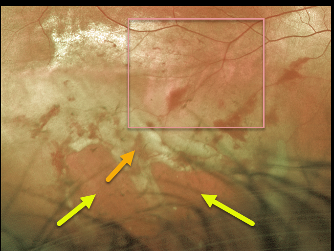
COMMOTIO RETINAE
Originally posted on @retina.rocks July 6, 2021
This 28YO male presented with 20/60 vision after being hit the night before in the right eye with a Nerf arrow.
Optos imaging shows outer retinal whitening and hemorrhages extending from the inferior macula into the inferotemporal periphery. Multiple ragged retinal breaks with a shallow retinal detachment are noted more peripherally.
The patient was taken to the operating room later that day for vitrectomy surgery.
Learning Points:
Commotio retinae follows direct, blunt eye trauma. The outer retinal whitening is due to shearing of the photoreceptor outer segments.
The term “Berlin’s edema” is a misnomer and should not be used, since the retina is not edematous.
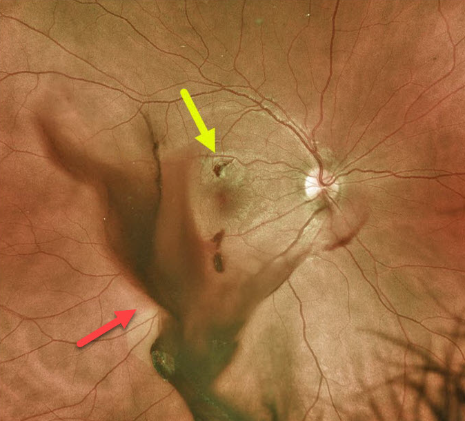
INTRAOCULAR FOREIGN BODY
Originally posted on @retina.rocks March 25, 2021
This 25YO male was utilizing a pneumatic hammer without appropriate eye protection when a metal projectile entered his eye and lodged in the superior macula with associated vitreous hemorrhage.
He underwent immediate vitrectomy with removal of the foreign body.
Learning Points:
Siderosis can develop from metallic intraocular foreign bodies (specifically iron) and lead to cataract, glaucoma, iris defects, and retinal degeneration with depressed ERG potentials.
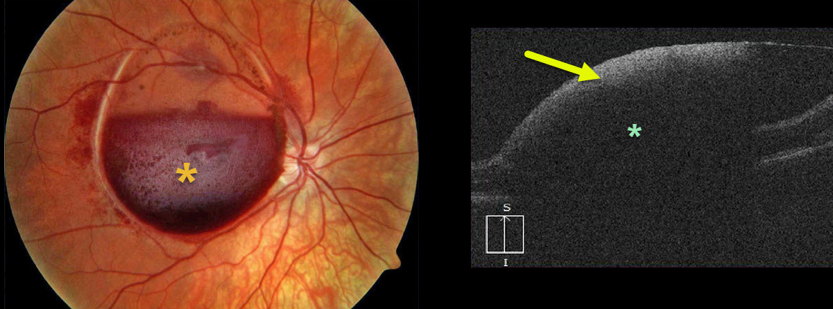
VALSALVA RETINOPATHY
Originally posted on @retina.rocks March 15, 2021
This is a beautiful photograph of an acute sub-internal limiting membrane (ILM) hemorrhage, most commonly found following a Valsalva maneuver or trauma, but can also happen spontaneously, as with anemia.
OCT scanning shows marked hyperreflectivity from the sub-ILM blood with posterior shadowing.
Learning Points:
Sub-ILM hemorrhages virtually always resolve without sequelae, although the blood can be drained into the inferior vitreous by creating a small opening in the ILM using either a thermal or Nd:YAG laser.
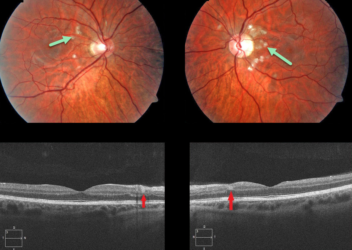
PURTSCHER-LIKE RETINOPATHY
Originally posted on @retina.rocks December 23, 2020
This 24YO male had a history of acute alcohol-related pancreatitis one month earlier and presented with bilateral multifocal areas of superficial peripapillary retinal whitening.
OCT scanning showed small hyperreflective areas spanning the inner plexiform, inner nuclear, and outer plexiform layers.
Learning Points:
Purtscher’s retinopathy was first described by Otmar Purtscher in 1910. Although originally described in a man who fell from a tree with cranial trauma, these white patches of retinal ischemia and hemorrhages are found in numerous other etiologies, including pancreatitis, chest trauma, and collagen vascular diseases.
The findings are called Purtscher’s retinopathy when due to trauma, and Purtscher ‘s-like retinopathy when seen from other causes, as in this case.
The underlying cause of Purtscher’s is unknown, but it is thought to involve leukoembolization due to complement activation. The areas of ischemia include more superficial nerve fiber layer infarcts (cotton wool spots) and deeper ischemia involving the middle retinal layers (paracentral middle maculopathy).
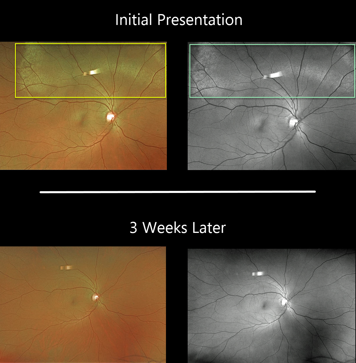
COMMOTIO RETINAE
Originally posted on @retina.rocks November 27, 2020
This patient presented with superior commotio retinae after being hit in the eye with a bungee cord. The outer retinal opacification was best visualized on the Optos green channel. Vision was 20/30.
Fortunately, the retina findings completely resolved 3 weeks later.
Learning Points:
Commotio retinae follows direct blunt eye trauma. Large areas of peripheral commotio may be associated with an increased risk for retinal detachment.
In some cases, the photoreceptors are permanently damaged, allowing RPE cells to migrate into the retina along capillaries, giving a pseudo-retinitis pigmentosa appearance.
The term “Berlin’s edema” is a misnomer and should not be used, since the retina is not edematous.
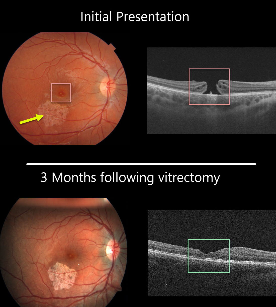
MACULAR HOLE
Originally posted on @retina.rocks November 6, 2020
This 13YO girl was hit in her right eye while playing volleyball. She presented to us several months later with 20/200 vision, an inferotemporal macular scar from resolved commotio retina, and a full-thickness macular hole.
The macular hole was closed following successful vitrectomy, and she was 20/100 one month postoperatively. Vision improved to 20/40 three months following surgery. Since the scotoma from the macular scar was superior, she is now virtually completely visually asymptomatic and extremely lucky!
Learning Points:
Traumatic macular holes are more common in younger patients, often from recreational or sports injury, and tend to be more eccentric in shape.
The mechanism could be due to sudden vitreous separation or to stretching of the posterior pole resulting from anteroposterior ocular compression. The holes can spontaneously close and have a high surgical success rate, as in our patient (see Liu et al AJO 2020;210:174-183).
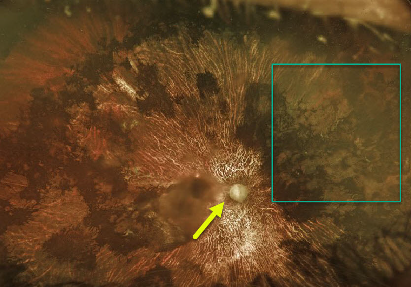
TRAUMA
Originally posted on @retina.rocks October 5, 2020
There is extensive chorioretinal scarring with optic atrophy in this patient who suffered severe blunt trauma 10 years earlier following a heroin overdose.
Vision is now counting fingers (CF), but thankfully, the other eye is normal with 20/20 acuity.
Note that the areas of intraretinal pigment migration mimic the bone spicules found with RP.
Learning Points:
Near total unilateral chorioretinal scarring has a relatively small differential diagnosis, including severe trauma, diffuse unilateral subacute neuroretinitis (DUSN), acute retinal necrosis syndrome (ARNS), and Saturday night retinopathy.
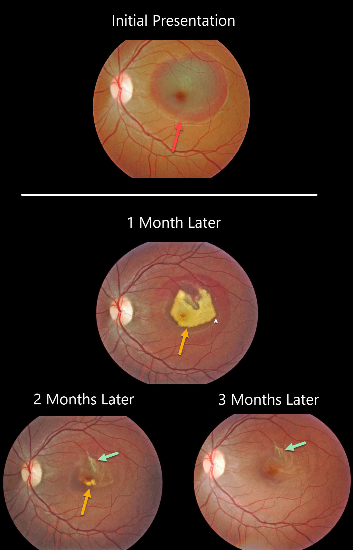
CHOROIDAL RUPTURE
Originally posted on @retina.rocks October 1, 2020
This patient presented with an acute submacular hemorrhage following a finger-to-eye injury.
The subretinal blood slowly resolved over the following three months. An underlying choroidal rupture is almost always the source of this traumatic blood.
Final vision was 20/400, either due to contusive foveal damage or possible toxicity from the subretinal blood.
Learning Point:
Choroidal ruptures are caused by blunt ocular trauma and are always circumferential with respect to the optic nerve.
These patients need to be followed since macular neovascularization can develop later in life.
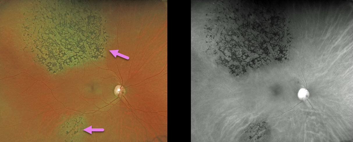
TRAUMA
Originally posted on @retina.rocks September 29, 2020
A 49YO female was involved in a motor vehicle accident 30 years ago.
Her retina showed signs of pigmentary retinopathy from the ocular injury, which could be mistaken for retinitis pigmentosa (RP). Unlike typical RP, our patient had multifocal, sectoral, unilateral findings.
Learning Point:
Intact photoreceptors inhibit RPE migration. Photoreceptor loss from numerous causes, including blunt trauma, retinal detachment, macular telangiectasia, and retinitis pigmentosa, can allow intraretinal RPE migration with this shared phenotype.
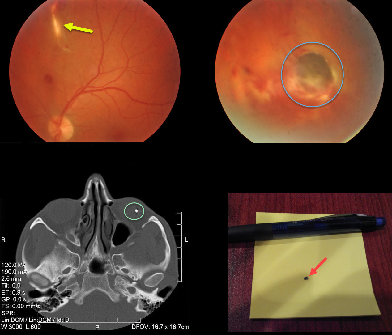
INTRAOCULAR FOREIGN BODY
Originally posted on @retina.rocks September 3, 2020
This patient was hammering so hard that a piece of metal was sent flying through his upper lid, into his orbit, bounced off the orbital roof, penetrated his globe from behind in the superior midperiphery, and lodged in the inferior retina.
The eye looked completely normal externally since the penetrating injury occurred from behind the eye. The entrance wound through the upper lid was also self-sealing, and the lid looked fairly normal when he presented a few days after the injury.
Take a look at the metal object on the CT scan and the removed foreign body.
Learning Points:
Siderosis can develop from metallic intraocular foreign bodies (specifically iron) and lead to cataract, glaucoma, iris defects, and retinal degeneration with depressed ERG potentials.
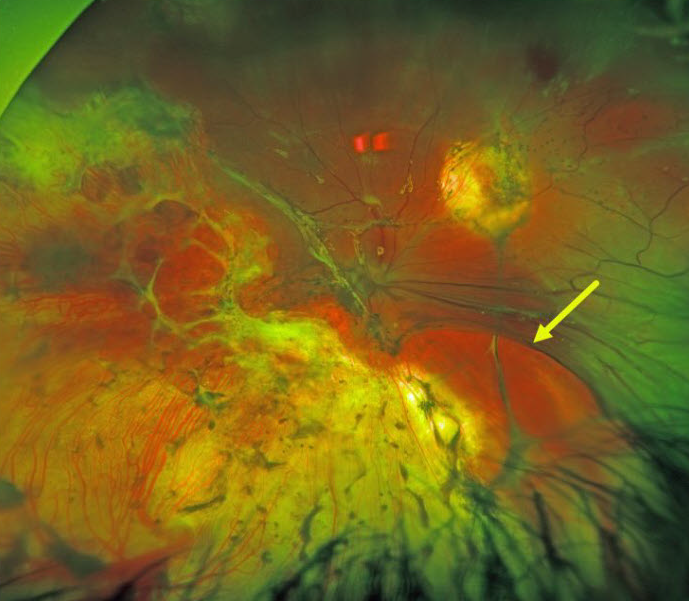
TRAUMA
Originally posted on @retina.rocks August 7, 2020
This patient was shot several times in the head and remarkably survived.
He underwent numerous retinal procedures at an outside hospital for a retinal detachment, and currently has light perception (LP) vision.
Examination revealed extensive chorioretinal scarring with a mostly detached retina under silicone oil.
A large retinal break, possibly from an intraoperative retinectomy, was seen nasally.
Learning Points:
Traumatic retinal detachments can occur from blunt trauma or from perforating injury to the eye.
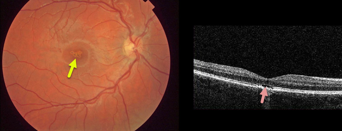
SOLAR RETINOPATHY
Originally posted on @retina.rocks July 31, 2020
This asymptomatic 10YO boy presented with 20/40 vision OD and 20/20 vision OS.
There were multifocal small subretinal pigment clumps with surrounding pigment loss in the right central macula.
OCT shows foveal outer segment loss with ragged RPE loss. The left macula was normal.
He denied looking at a laser pointer but did admit to a history of sun-gazing. He likely closed his left eye each time he looked at the sun, which explains the unusual finding of unilateral solar retinopathy.
Learning Points:
The outer retinal damage in solar retinopathy is thought to be caused by photochemical outer retinal and RPE damage instead of a thermal burn. Vision is usually fairly good.
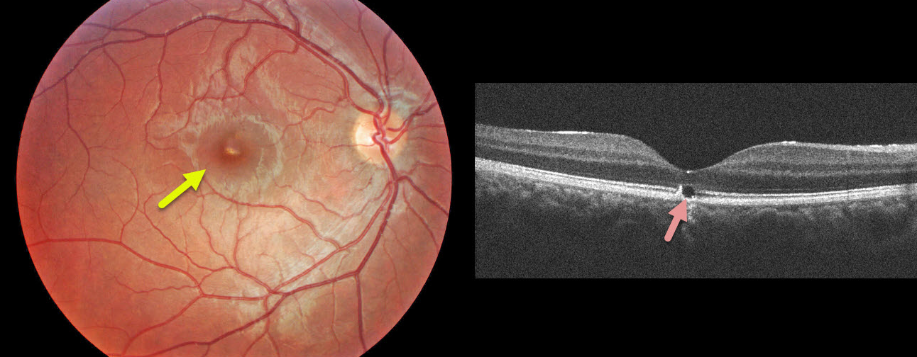
WHIPLASH MACULOPATHY
Originally posted on @retina.rocks July 1, 2020
This 17YO male presented with sudden unilateral vision loss immediately following a whiplash from a motor vehicle accident.
Clinically, he has a yellowish linear subretinal foveal discoloration. OCT shows a focal loss of the outer subfoveal retina.
Learning Points:
These findings are classic for whiplash maculopathy, in which sudden acceleration/deceleration causes the vitreous to tug on or impact the retina, resulting in outer retinal and RPE damage.
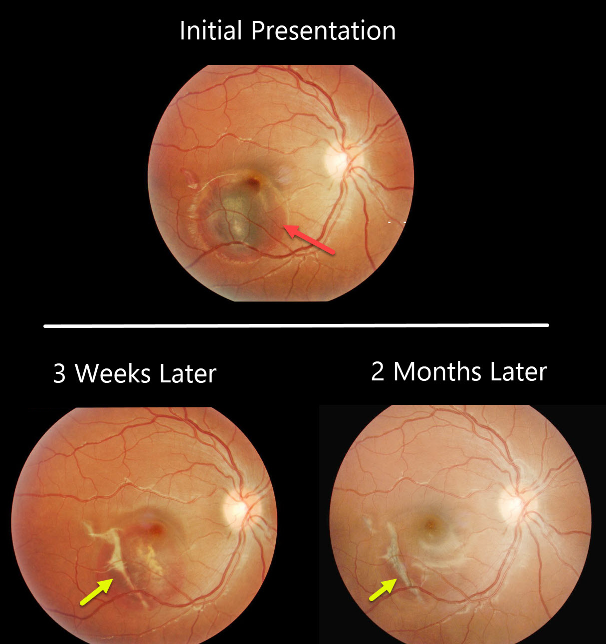
CHOROIDAL RUPTURE
Originally posted on @retina.rocks June 4, 2020
This patient presented with vision loss from an acute submacular hemorrhage following blunt trauma.
Subsequent images, 3 weeks later and 2 months later, show gradual resolution of the blood with the appearance of an underlying choroidal rupture.
Learning Point:
Choroidal ruptures are caused by blunt ocular trauma and are always circumferential with respect to the optic nerve. Subretinal blood following blunt trauma virtually always indicates the presence of an underlying choroidal rupture.
The initial visual prognosis is generally good if the rupture does not extend through the macular center, as was fortunately the case in this patient.
These patients need to be followed since macular neovascularization can develop later in life.
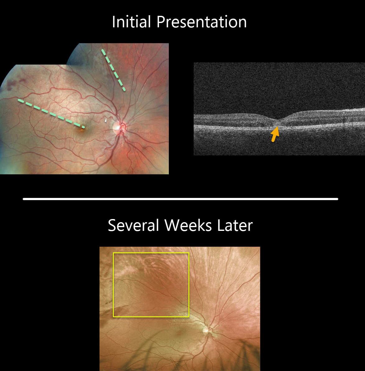
COMMOTIO RETINAE
Originally posted on @retina.rocks April 8, 2020
This patient was hit in the eye with a soccer ball and developed outer retinal whitening due to shearing of the outer segments. OCT reveals subfoveal ellipsoid zone (EZ) disruption and hyperreflectivity.
The retinal whitening usually disappears within a few weeks due to regeneration of the photoreceptors
Learning Points:
Commotio retinae follows direct blunt eye trauma. Large areas of peripheral commotio may be associated with an increased risk for retinal detachment.
In some cases, the photoreceptors are permanently damaged, allowing RPE cells to migrate into the retina along capillaries, giving a pseudo-retinitis pigmentosa appearance.
The term, Berlin’s edema, is a misnomer and should not be used, since the retina is not edematous.
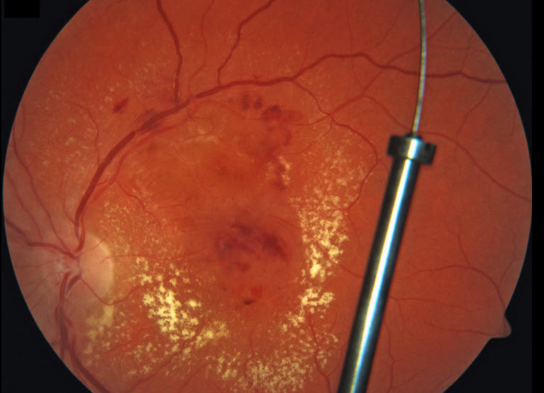
INTRAOCULAR FOREIGN BODY
Originally posted on @retina.rocks April 1, 2020
An automobile manufacturing worker from our local Ford plant presented with an acute injury to his left eye while installing a catalytic converter. A large metallic intraocular foreign body (IOFB) shot through the pars plana in a self-sealing wound and lodged in the posterior vitreous without any direct retinal injury. Since the material was made primarily of inert platinum, the IOFB was left in place and the patient has remained remarkably stable for 3 years.
We hypothesize that the platinum exhibited powerful anti-fungal and anti-bacterial effects helping to prevent infection. The diffuse diabetic macular edema with surrounding lipid also spontaneously resolved, sparking interest in intravitreal platinum as a more durable treatment for macular leakage. The results of the NIH-sponsored Phase 3 MORON Study, Might IntraOcular Platinum ResOlve Edema Better ThaN Really Expensive Anti-VEGF Agents, are eagerly anticipated in late 2020..
Yes, of course it’s APRIL FOOLS!
This patient has diabetic retinopathy with macular edema. The “IOFB” is really a broken fundus camera fixation stick projected onto the posterior pole image.
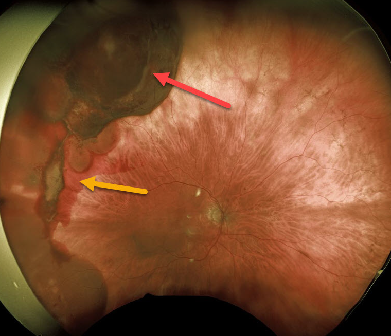
TRAUMA
Originally posted on @retina.rocks March 26, 2020
This patient presented with unilateral subretinal and suprachoroidal hemorrhages from a fall 1 month earlier causing blunt facial trauma.
Vision was normal and she was asymptomatic. She is being followed without treatment.
Learning Points:
There will likely be at least one choroidal rupture visible as the overlying blood clears over time. Subretinal blood following blunt trauma virtually always indicates the presence of an underlying choroidal rupture.
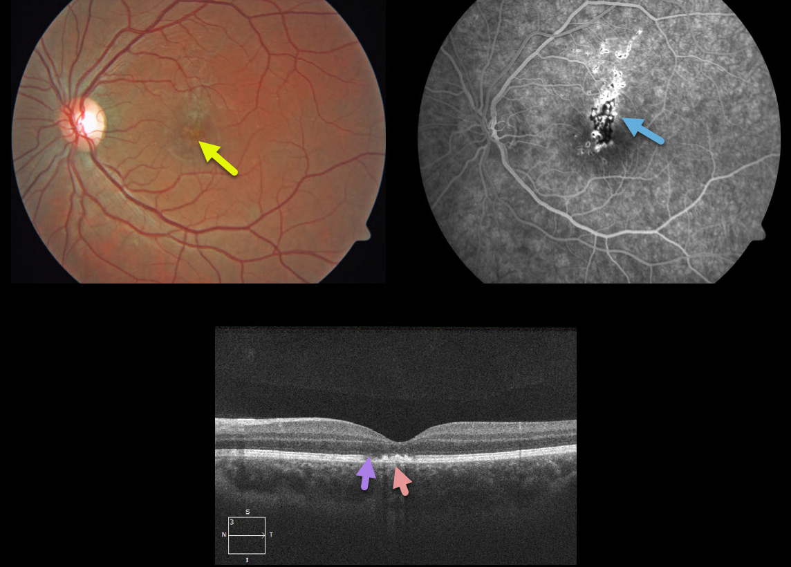
LASER MACULOPATHY
Originally posted on @retina.rocks February 21, 2020
This 18YO patient presented with sudden painless 20/400 vision loss in his left eye. He admitted to staring 3 days earlier directly into his recently purchased green laser pointer.
There are multifocal small subretinal pigment clumps in the left central macula and fluorescein angiogram reveals variable areas of hypo and hyperfluorescence. OCT shows EZ attenuation with hyperreflectivity above the RPE.
Learning Points:
Ocular damage from these lasers depends highly on the laser wavelength, power and duration. The US regulates hand-held laser pointers as a category 3R, which cannot exceed 5 mW. However, a 2013 study looking at 122 laser pointers labeled as 3R found that 90% of green and 44% of red laser pointers actually exceeded the 5mW limit.
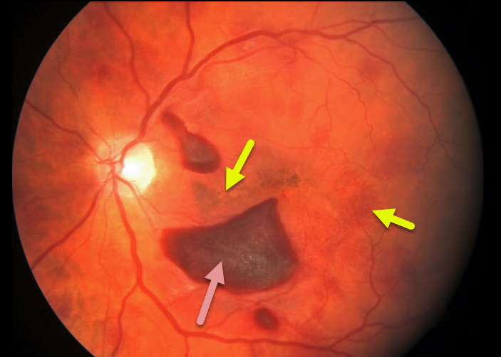
TERSON SYNDROME
Originally posted on @retina.rocks January 17, 2020
This patient presented with these retinal findings following a ruptured brain aneurysm.
The fundus photo of the left eye shows subhayloid and deep, radiating retinal hemorrhages in the outer plexiform layer.
Learning Points:
Terson’s syndrome is a condition in which retinal and/or vitreous hemorrhages are associated with spontaneous or traumatic intracranial hemorrhage. These deeper retinal hemorrhages are somewhat similar to those seen in hemorrhagic unilateral retinopathy.
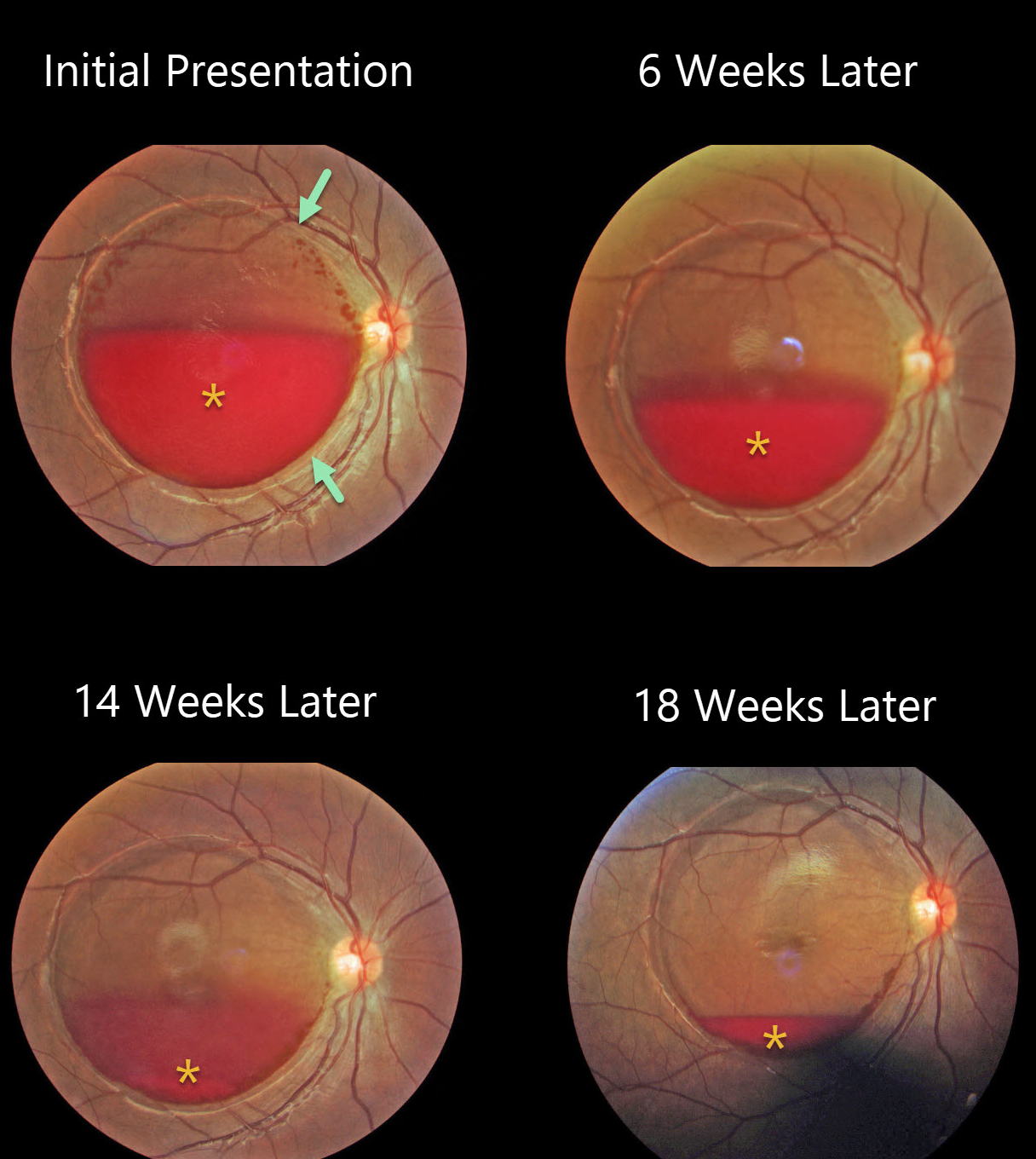
VALSALVA RETINOPATHY
Originally posted on @retina.rocks January 9, 2020
This pregnant female developed sudden vision loss in her right eye following delivery of her baby.
There is a large, round detachment of the internal limiting membrane (ILM) throughout the entire macula with layered blood initially blocking the central fovea.
The blood gradually resolved 6 weeks, 14 weeks, and 18 weeks thereafter.
Learning Points:
Valsava retinopathy is characterized by preretinal hemorrhages, which virtually always resolve without sequelae over time. The blood can also be drained into the inferior vitreous by creating a small opening in the ILM using either a thermal or Nd:YAG laser.


