

08 Toxic Disorders
<< Back to Cases
HEMORRHAGIC OCCLUSIVE RETINAL VASCULITIS (HORV)
Ayushi Gupta and Vishal Agrawal
Originally posted on @retina.rocks May 20, 2026
This 35YO male underwent uneventful cataract surgery in his left eye at an outside facility about two weeks earlier. On postoperative day 4-5, he developed mild blurring and floaters, raising suspicion for early postoperative endophthalmitis. He underwent vitreous tap with intravitreal vancomycin and ceftazidime. Although he noticed initial mild symptomatic improvement, over the next 5-7 days, he experienced progressive and profound vision loss. Vision in our office was light perception. The anterior segment was quiet.
Fundus photography shows a circular band of midperipheral fibrosed retinal neovascularization encircling the posterior pole. The pericentral macula shows inner retinal white ischemia, and there are scattered retinal hemorrhages. Fluorescein angiography shows dramatic capillary loss encircling the midperiphery and temporal macula. The foveal avascular zone is irregularly enlarged. Panretinal photocoagulation (PRP) was advised, but she was lost to follow-up immediately.
Learning Points:
Hemorrhagic occlusive retinal vasculitis (HORV) is a rare but devastating complication strongly associated with intraocular vancomycin exposure, typically presenting 1-21 days after cataract surgery or intravitreal injection with delayed-onset painless vision loss (Witkin et al, Ophthalmology 2017;14:583-595). It is characterized by sectoral ischemia with retinal hemorrhages, mild anterior chamber and vitreous inflammation, often with a deceptively unremarkable postoperative day 1 examination. The findings are likely due to a delayed hypersensitivity reaction rather than direct toxicity (Todorich et al, AJO 2018;188:131-140).
The visual prognosis is poor, with approximately 61% of eyes achieving <20/200 vision and 22% progressing to no light perception. Neovascular glaucoma develops in approximately 56% of cases due to extensive retinal ischemia. Management includes early systemic and/or intravitreal corticosteroids, and subsequent PRP and anti-VEGF therapy.
PENTOSAN POLYSUFATE SODIUM TOXICITY
Originally posted on @retina.rocks May 12, 2026
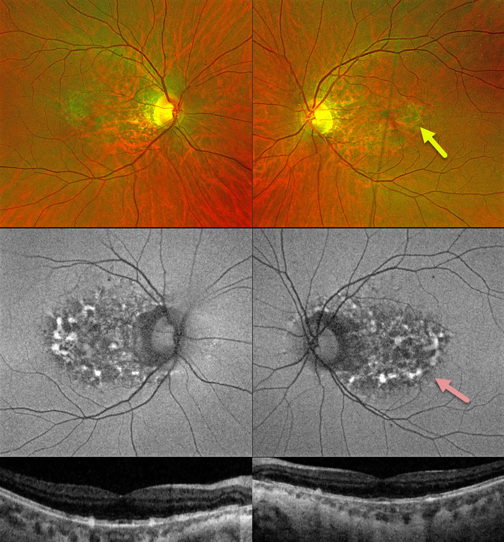
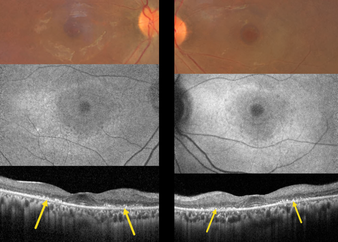
This 48YO was on pentosan polysulfate sodium (PPS, Elmiron) for interstitial cystitis for 2-3 years, but discontinued this drug 2 years earlier. Vision was 20/30 bilaterally.
Fundus color imaging shows bilateral ill-defined macular pigmentary changes. Swept-source OCT shows variable loss of the ellipsoid zone, outer segment bands, and RPE with clumps of hyperreflective material involving or located above the RPE. Optos fundus autofluorescence (FAF) shows patchy hyper- and hypo- FAF extending from the central macula to the nerve.
Learning Points:
Elmiron was approved by the FDA in 1996 for the treatment of interstitial cystitis. A unique PPS retinopathy was recently described, and our patient shows classic findings. Toxicity seems to develop over many years and can mimic more common disorders, including age-related macular degeneration and macular dystrophies.
The risk of toxicity is dose-dependent (Tao et al, Ophthalmology Retina 2025;9:870-882), although toxicity can rarely occur with relatively minimal exposure, as occurred in our patient (Yousif and Johnson, JAMA Ophthalmology 2025;143:362-363). The drug should be discontinued once retinal findings are discovered, although progressive functional and structural changes can develop even after drug cessation (Jung et al, JAMA Ophthalmol 2023;141:260-266).
A recent report found severe asymptomatic adenomatous polyposis in 75% of their PPS maculopathy patients (Feo et al, AJO 2026;287:1-13). Most of their patients required partial or total colectomy or endoscopic resection. They therefore recommended colonoscopy screening in all exposed patients.
ETHAMBUTOL TOXICITY WITH CONCOMITANT DIABETIC RETINOPATHY
Ayushi Gupta and Vishal Agrawal
Originally posted on @retina.rocks May 4, 2026
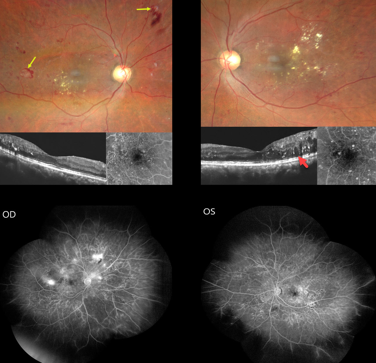
This 54YO male presented for a second opinion with several weeks of progressive bilateral painless vision loss. He gave a history of active pulmonary tuberculosis, diagnosed about 6 months earlier, which was being treated with antitubercular therapy (ATT) including ethambutol. There was also a 5-year history of well-controlled type 2 diabetes. Vision was 20/200 OU.
Fundus photography shows bilateral non-central diabetic macular edema (DME) with mostly temporal lipid. The central macula looks fairly normal bilaterally on OCT, and fundus fluorescein angiography (FFA) shows relatively preserved foveal avascular zones. Retinal neovascularization (NV) with some associated preretinal blood is noted OD. Widefield FFA confirms scattered retinal NV OD with moderate bilateral capillary nonperfusion. Disc OCT showed a normal retinal nerve fiber layer, and 30-2 visual fields showed severe generalized depression (not shown).
Learning Points:
Our patient was previously seen by other specialists who felt that diabetic retinopathy was the sole reason for his complaints and findings, despite the lack of central foveal fluid or ischemia. Our patient reminds us of 2 dictums in medicine: 1) we try to explain all findings by a single disease, and 2) that a patient can have as many diseases as they so desire. In our patient’s case, we felt that ethambutol better explained his clinical picture.
Ethambutol is one of the agents used as part of ATT, a multi-drug regimen for treating active tuberculosis. About 1-3% of patients can develop drug-induced optic neuropathy, most commonly following 2 months of therapy but rarely developing within days of starting treatment. Symptoms include decreased visual acuity, scotoma, color blindness, and visual defects in one or both eyes.
Although the FDA recommends baseline visual acuity and color vision testing, followed by monthly color discrimination testing during therapy, there are no studies demonstrating any benefit of such screening. Ethambutol should be discontinued if toxicity is suspected. Although vision loss is often thought to be permanent, a recent systematic review showed significant improvement in vision, color vision, and visual field loss with discontinuation of the drug (Sabhapandit et al., Indian J Ophthalmol 2023;71:729-735).
We contacted our patient’s pulmonologist, who stoppe therapy immediately. Anti-VEGF therapy followed by panretinal photocoagulation was recommended.
HYDROXYCHLOROQUINE TOXICITY
Originally posted on @retina.rocks April 14, 2026
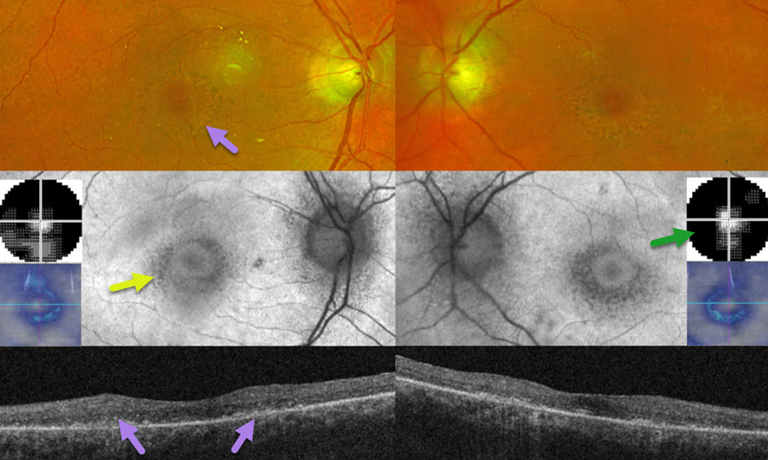
This 78YO female was on Plaquenil (hydroxychloroquine) for 7 years for her “arthritis.” After noticing progressive reading difficulties, she decided to stop this medicine about 3 months earlier. She was screened annually by her local eye doctor, who referred her for possible toxicity. Vision was 20/40 OD and 20/30 OS.
Optos color imaging and OCT show classic findings for severe hydroxychloroquine toxicity, including an oval area of foveal pigment loss/mottling and outer retinal atrophy. These areas of pigment loss hypo-autofluoresce. 10-2 visual fields reveal small islands of preserved central vision.
Learning Points:
Current screening guidelines should make Plaquenil toxicity a thing of the past. The American Academy of Ophthalmology updated its screening guidelines in 2025 (Marmor et al, Ophthalmology 2026;133:439-450).
A baseline screening examination with fundus photography, spectral-domain or swept-source OCT, and fundus autofluorescence (FAF) should be performed as soon as treatment is started to note preexisting conditions and for future comparison. Annual screening is recommended but may be deferred during the first 5 years if there are no significant risk factors, ie, high daily dosage, renal disease, concomitant drugs such as tamoxifen, macular disease, and older age.
The recommended daily dosage remains unchanged at <=5.0 mg/kg/day real weight, with the dosage under 400 mg/day for severely obese individuals. The primary screening tools are OCT with widefield FAF. Secondary confirmatory tests include 24-2C visual field testing (which screens the parafoveal and pericentral regions simultaneously) and multifocal ERG. The pericentral field needs to be evaluated, as Asians typically have more eccentric macular and midperipheral involvement.
BRAF AND MEK INHIBITOR-ASSOCIATED RETINOPATHY
The European VitreoRetina Society (EVRS) and Gerardo Garcia
Originally posted on @retina.rocks December 12, 2025
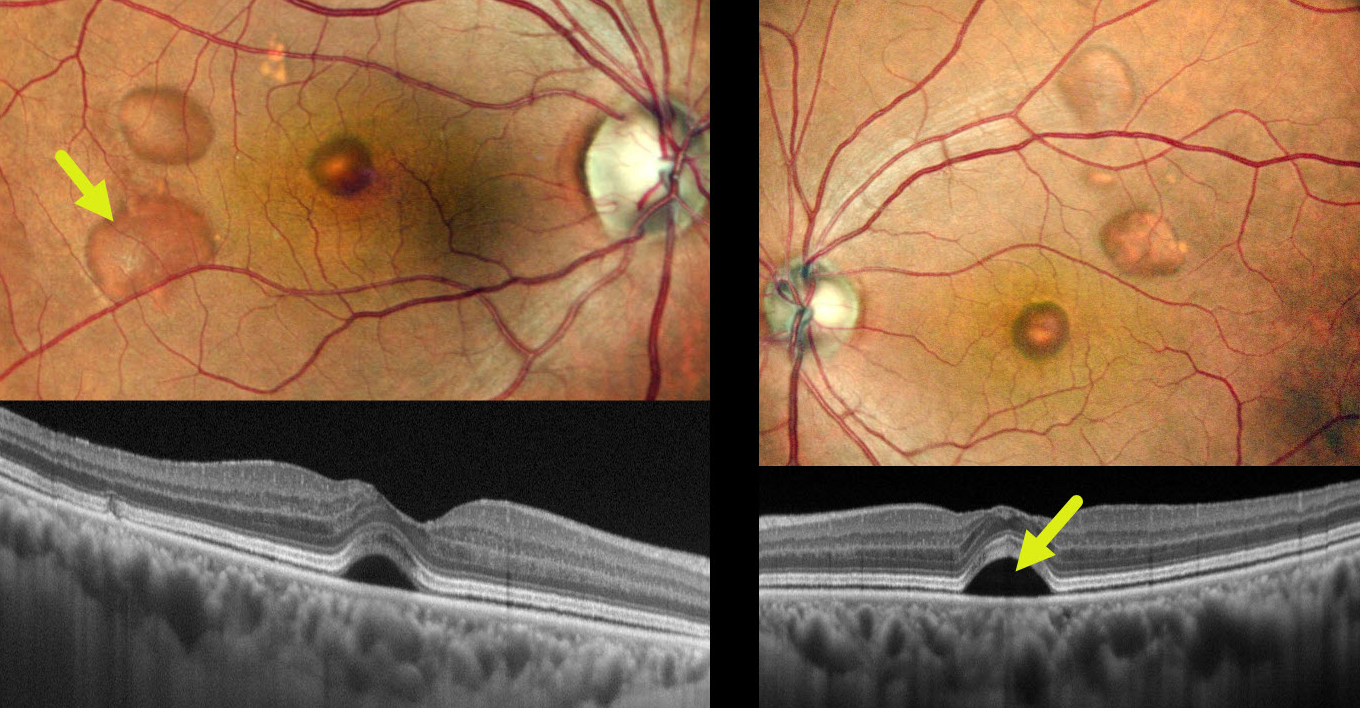
This 67YO male presented with 3 weeks of mild bilateral blurred vision after starting dabrafenib (BRAF inhibitor) and trametinib (MEK inhibitor) for metastatic melanoma. Vision was 20/25 OU.
Pseudocolor SLO imaging shows multifocal bilateral macular blisters with serous subretinal fluid, confirmed on OCT.
Learning Points:
Recent drugs targeting the BRAF and MAPK pathways have emerged as therapy for metastatic melanoma. These agents, which include BRAF and MEK inhibitors, are characterized by transient, bilateral, multifocal serous retinal detachments and subretinal fluid often mimicking central serous chorioretinopathy on OCT but without angiographic leakage (Bravetti et al, Acta Ophthalmologica 2024;102:e651-e656). The serous detachments usually develop within days to weeks of initiating therapy, are dose- and time-dependent, and are usually reversible upon drug discontinuation or even with continued treatment. The pathogenesis is thought to involve disruption of the retinal pigment epithelium’s pump function due to inhibition of the MAPK pathway.
HYDROXYCHLOROQUINE TOXICITY
The European VitreoRetina Society (EVRS) and Debolina Deb
Originally posted on @retina.rocks October 31, 2025
This 28YO Indian female presented with several months of seeing occasional black spots. She has systemic lupus erythematosus, which has been treated with hydroxychloroquine (HCQ) for the past 5 years. Her calculated cumulative dose of HCQ was 365 grams. She denied having prior baseline or screening examinations for HCQ toxicity. Vision was 20/30 OU.
Color photography shows bilateral perifoveal pigmentary changes. Fundus autofluorescence (FAF) shows a rim of parafoveal hyper-FAF. OCT scanning shows paracentral outer retinal thinning, with disorganized clumps of hyperreflective material overlying thinned RPE.
Learning Points:
Current screening guidelines should make hydroxychloroquine toxicity, as found in our patient, a thing of the past. Last revised in 2016, the American Academy of Ophthalmology (AAO) recommends that a baseline exam be performed before starting the medication, with annual screenings beginning at least 5 years after initiating treatment, unless major risk factors are present. However, in practice, patients are usually screened yearly once they are placed on this medication.
OCT is performed annually to assess outer retinal findings, including ellipsoid zone loss. Humphrey 10-2 visual field testing (24-2 for Asians, since their macular involvement is usually more peripheral) is also needed annually, as about 10% of patients will have field loss despite normal examinations and OCT testing (Ahn et al., AJO 2017;184:11-18).
Plaquenil dosing should also be based on real, not ideal, weight to better predict the optimal dose. See Marmor et al Ophthalmology 2016;123:1386-1394 for the full screening guidelines. Despite the most recent AAO guidelines being almost 10 years old, a recent publication found that over one-third of patients are still overdosed (Moussa et al, Ophthalmology Retina 2025;9:814-817).
Toxicity still developed in our patient despite not falling into any of the high-risk categories (cumulative HCQ dose < 1000 grams, young age, not obese). We have written a letter to her rheumatologist to immediately stop HCQ treatment.
HYPERTENSIVE RETINOPATHY FROM METHAMPHETAMINE
Originally posted on @retina.rocks May 22, 2025
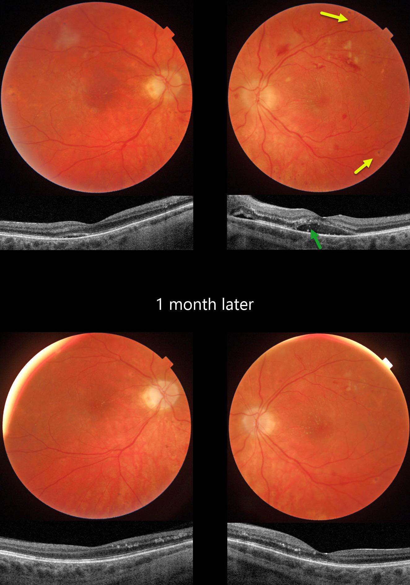
This 21YO female presented with bilateral vision loss. She was admitted to the hospital 2 weeks earlier for malignant hypertension (HTN), likely from methamphetamine abuse. Blood pressure at that time was 292/208. Vision was hand motion OD and 20/80 OS.
Triton color photography of her left eye shows a somewhat pale and swollen nerve, mostly inner retinal hemorrhages, and resolving nerve fiber layer infarcts (cotton-wool spots, CWS). Faint lipid exudates radiate nasally and superiorly from the macular center. Multifocal small hyperpigmented choroidal lesions are noted outside the arcades. Swept-source OCT shows subretinal fluid with hyperreflective lipid in the subretinal space and outer nuclear layer. Similar but much milder findings are noted in her right eye.
When examined 1 month later, vision remained at hand motion OD and improved to 20/40 OS with improved funduscopic and OCT findings.
Learning Points:
Malignant hypertension, defined as blood pressure above 180 systolic and/or 120 diastolic, is a life-threatening medical emergency. Eye doctors are in a unique position to often diagnose this condition. Patients will often present with bilateral optic nerve swelling, flame-shaped retinal hemorrhages, CWS, and, with more chronic disease, lipid precipitates in the nasal macular outer plexiform layer.
Acute hypertensive choroidopathy outside the setting of pre-eclampsia, in our experience, is quite rare. Independent of the disc and neurosensory retinal findings, patients present with localized multifocal serous retinal detachments (de Venecia and Jampol, Arch Ophthalmol 1984;102:68-73). With blood pressure control, these resolve often with minimal funduscopic changes. Focal (Elschnig spots) or linear (Siegrist streaks) choroidal pigmentary changes may result from more severe disease.
TAMOXIFEN RETINOPATHY
Mauli Shah and Alay Banker
Originally posted on @retina.rocks March 4, 2025
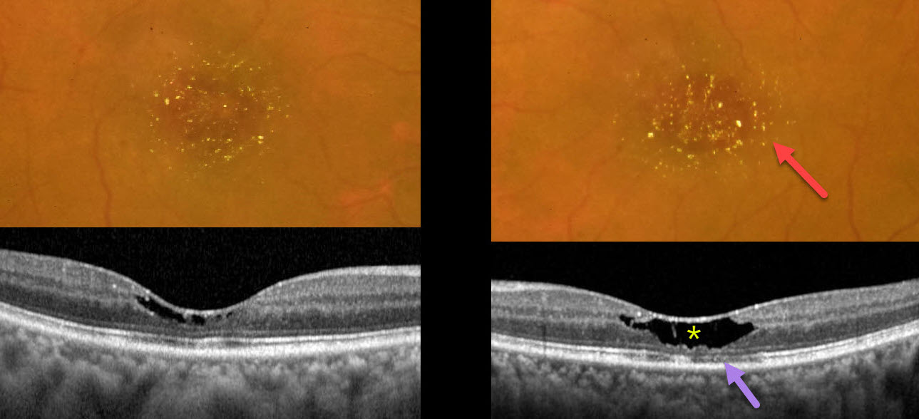
This 51YO female presented with 3 months of bilateral vision loss. She was diagnosed 5 years earlier with breast cancer, followed by a radical mastectomy. Since then, she has been treated with tamoxifen 20mg PO daily. Vision was 20/60 OD and 20/40 OS.
Color photography shows bilateral, symmetric, central macular yellowish crystalline deposits, corresponding to tiny hyperreflective inner retinal lesions on OCT. Central inner retinal cavitations, outer nuclear layer loss, and disruption of the ellipsoid zone and outer segment bands are noted, particularly in the left eye.
Learning Points:
The prevalence of tamoxifen retinopathy for patients taking tamoxifen 20mg daily may be as high as 12% (Kim et al, Ophthalmology 2020;127:555-557). Although most oncologists do not require this, it is not unreasonable to perform ophthalmic screening examinations with OCT following 2 years of therapy (Tenney et al, Surv Ophthalmology 2023;69:42-50).
Tamoxifen inhibits the glutamate-aspartate transporter, leading to excessive intracellular accumulation of glutamate in Müller cells, which are vital in maintaining retinal cell integrity and homeostasis. Tamoxifen retinopathy shows findings very similar to macular telangiectasia type 2 (MacTel 2; Lee et al., Ophthalmology Retina 2020;3:681-689), including retinal cavitations, right-angle venules, and inner retinal crystals. Both disorders likely share Muller cell injury as a common etiology. The changes in MacTel2 are usually in the temporal fovea, whereas they seem to be more diffusely distributed throughout the central macula with tamoxifen (Hess et al, Retina 2023;7:101-110).
Ocular toxicity is more common with a cumulative dose over 100 grams, although our patient still developed classic toxicity despite a cumulative dose of only 35.2 grams. The tamoxifen was discontinued after discussion with the treating oncologist. Three months later, the vision and retinal findings were unchanged (not shown).
MACULAR HOLE WITH PSEUDO BULLS-EYE MACULOPATHY
Originally posted on @retina.rocks June 24, 2024
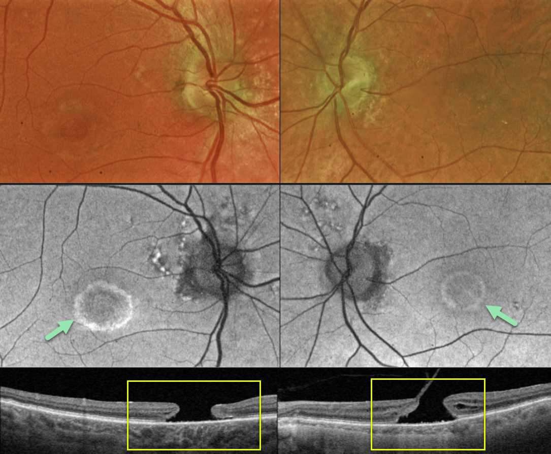
This 83YO male presented with a history of chronic bilateral vision loss. Vision was 20/150 OD and 20/200 OS.
Optos color RG imaging shows a bull’s eye pattern of bilateral pigmentary changes along with a large macular hole OU. Fundus autofluorescence (FAF) shows the bull’s-eye lesions as hyper-FAF. Triton swept-source OCT confirms bilateral macular holes.
Our patient has bilateral chronic macular holes with secondary RPE depigmentation, resulting in a bull’s-eye appearance. Due to the chronicity of the holes, observation was recommended.
Learning Points:
Bull’s eye maculopathy is characterized by a rim of subretinal pigment loss and outer retinal atrophy, most commonly found in inherited retinal diseases (usually ABCA4 disorders) and hydroxychloroquine/chloroquine toxicity.
HYDROXYCHLOROQUINE TOXICITY + PACHYCHOROID
Originally posted on @retina.rocks March 11, 2024
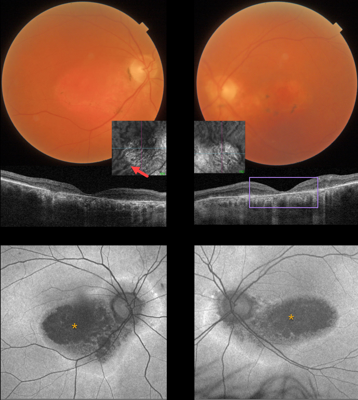
This 67YO female has a known history of Plaquenil toxicity. Her bilateral 20/200 vision and macular findings have been stable since discontinuing this medication 30 years ago.
Triton color imaging and swept-source OCT show classic findings for severe hydroxychloroquine toxicity, including an oval area of foveal pigment loss and outer retinal atrophy.
Most patients will have preserved central foveal pigment giving a bulls-eye appearance, although our patient has loss of outer retina and RPE throughout. Interestingly, choroidal en face imaging shows bilateral dilated choroidal pachyvessels.
On fundus autofluorescence (FAF), the central maculas are hypo-FAF, although an atypical hyper-FAF ring extends around each optic nerve.
Learning Points:
Current screening guidelines should make Plaquenil toxicity, as found in our patient, a thing of the past. Last revised in 2016, the American Academy of Ophthalmology (AAO) recommends that a baseline exam be performed before starting the medication, with annual screenings beginning at least 5 years after initiating treatment, unless major risk factors are present. However, in practice, patients are usually screened yearly once they are placed on this medication.
Optical coherence tomography (OCT) is performed annually to assess outer retinal findings, including ellipsoid zone loss. Humphrey 10-2 visual field testing (24-2 for Asians, since their macular involvement is usually more peripheral) is also needed annually, as about 10% of patients will have field loss despite normal examinations and OCT testing.
Plaquenil dosing should also be based on real, not ideal, weight to better predict the optimal dose. See Marmor et al Ophthalmology 2016;123:1386-1394 for the full screening guidelines.
PENTOSAN POLYSULFATE SODIUM TOXICITY VS AMD MACULAR ATROPHY
Will Gibson
Originally posted on @retina.rocks September 4, 2023
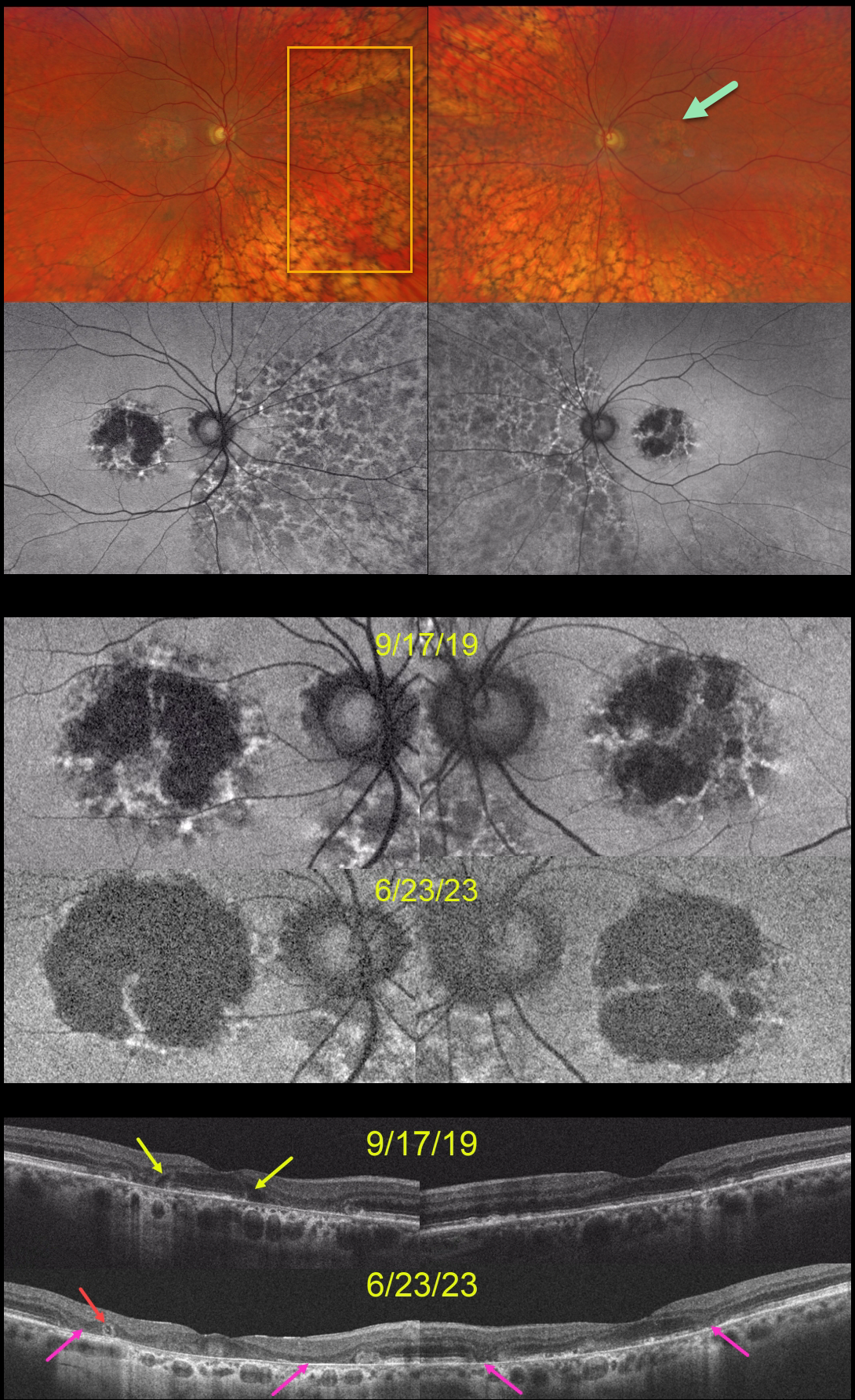
This 59YO was examined on 9/17/19. She was taking Elmiron (pentosan polysulfate sodium, PPS) for 17 years for interstitial cystitis. Vision was 20/40 OD and 20/30 OS.
Optos color RG imaging shows bilateral central-sparing macular atrophy (MA) and extensive, posterior peripheral reticular degeneration of the RPE (PRDRPE).
Fundus autofluorescence shows hypo-FAF from the MA with surrounding punctate and linear areas of hyper-FAF. Based on these findings, she decided to stop taking Elmiron.
When last examined on 6/23/23, vision dropped to 20/800 OD and was relatively stable at 20/40 OS. The MA progressed bilaterally clinically and on FAF and OCT. Hyporeflective OPL degeneration (wedge defects) is noted on the initial 9/17/19 OCT. On the 6/23/23 OCTs, an area of outer retinal tubulation (ORT) is seen temporally OD, and progressive outer retinal and RPE atrophy are noted bilaterally.
Learning Points:
Elmiron was approved by the FDA in 1996 for the treatment of interstitial cystitis. Recently, a unique PPS retinopathy has been described. Toxicity seems to develop over many years and can mimic more common disorders, including age-related macular degeneration and macular dystrophies.
Peripapillary hypoautofluorescence, more densely packed macular autofluorescent changes, and earlier central macular involvement suggest PPS toxicity over other causes (see Barnes et al Ophthalmology Retina 2020;4:1196-1201), including maternally inherited diabetes and deafness (MIDD).
ORT is often noted overlying inactive macular neovascularization with ongoing anti-VEGF therapy and should not be confused with exudative fluid or cysts, which lack a hyperreflective border. The outer hyperreflective band likely represents inner segment mitochondria undergoing fission and translocation towards the nucleus (Litts et al, Retina 2018;38:445-461).
ORT, initially described by Zweifel et al (Arch Ophthalmol 2009;127:1596-1602), is a neurodegenerative condition of the photoreceptors and Muller cells associated with outer retinal and retinal pigment epithelium atrophy, including advanced AMD and inherited retinal diseases.
The hyper-FAF surrounding the initial MA is more typical for PPS toxicity, and progressive atrophy is not uncommon even with stopping therapy (Jung et al, JAMA Ophthalmol 2023;141:260-266). However, in our experience, PPS usually produces more widespread macular findings. Given the PRDRPE, the MA could also be age-related. Or maybe our patient had baseline age-related MA superimposed on Elmiron toxicity?
TAMOXIFEN RETINOPATHY
Aaron McNulty
Originally posted on @retina.rocks June 19, 2023
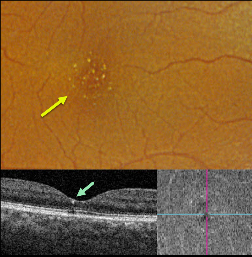
This 56YO female was referred for asymptomatic macular findings. The referring doctor mentioned that she may have retinal changes from the Tamoxifen that she was taking for over 3 years for breast cancer. Vision was 20/50 OD and 20/40 OS.
Optos color RGB imaging shows numerous tiny inner retinal crystalline deposits scattered throughout each fovea. These deposits appeared as tiny hyperreflective dots beneath the ILM on OCT B-scan and en face.
Our patient had already spoken with her oncologist before seeing us and had already stopped the Tamoxifen. The crystalline changes will likely remain and, hopefully, not progress going forward.
Learning Points:
Tamoxifen retinopathy shares phenotypes very similar to those of macular telangiectasia type 2 (MacTel 2, Lee et al., Ophthalmology Retina 2020;3:681-689), especially in the early stages, including retinal cavitations, right-angle venules, and inner retinal crystals.
The changes in Tamoxifen are confined to the central macula, whereas in MacTel2 they are present in a slightly larger area with an epicenter temporal to the foveal center (Hess et al, Ophthalmology Retina 2023;7:101-110). The retinal changes for both disorders likely share Muller cell dysfunction as the common cause.
PENTOSAN POLYSULFATE SODIUM TOXICITY
Originally posted on @retina.rocks March 28, 2023
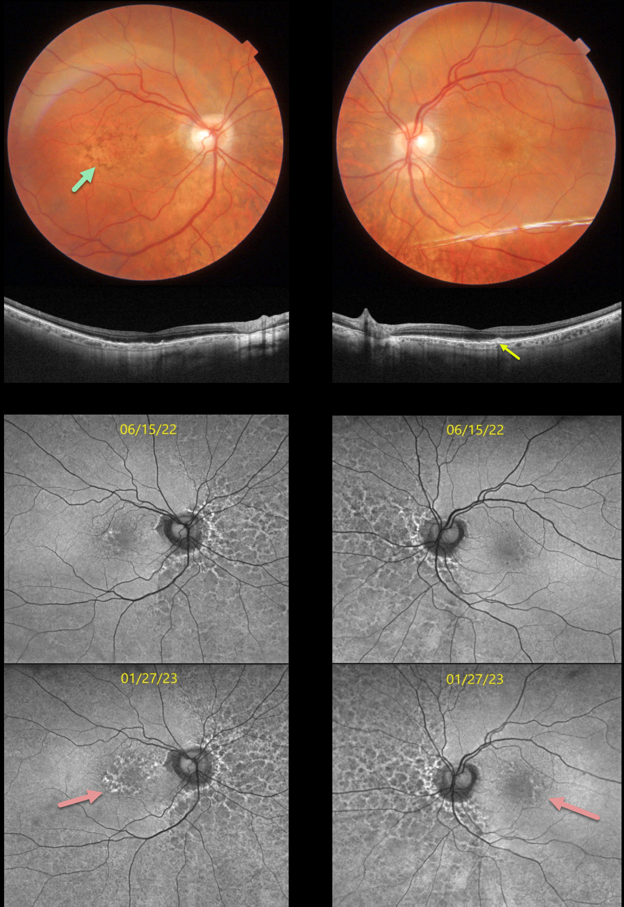
This 74YO female has been followed by us for a few years with known pentosan polysulfate (PPS, Elmiron) retinal toxicity. She has been taking Elmiron for 25 years.
When we initially diagnosed her with these retinal findings, she was hesitant to stop treatment since her interstitial cystitis symptoms were intolerable and dramatically improved with this medicine. She returned for her 6-month examination on 1/27/23 with increased central visual and contrast complaints.
Optos color imaging looked fairly stable. However, fundus autofluorescence (FAF) shows definite progressive macular changes. Macular color photography with swept-source OCT shows variable loss of the outer retinal layers with some focal outer retinal hyperreflective deposits.
Learning Points:
Elmiron was approved by the FDA in 1996 for treating interstitial cystitis. A unique PPS retinopathy was described in 2018, and our patient shows classic findings. Toxicity seems to develop over many years and can mimic more common disorders, including age-related macular degeneration and macular dystrophies.
The drug should be discontinued once retinal findings are discovered, although progressive functional and structural changes can develop even after drug cessation (Jung et al, JAMA Ophthalmol 2023;141:260-266).
We strongly recommend that she discontinue her Elmiron therapy. However, she felt that she would rather completely lose her central vision than face life with the pain from untreated interstitial cystitis and will remain on treatment.
She will discuss other potential therapeutic options with her nephrologist in the meantime.
HYDROXYCHLOROQUINE TOXICITY
Originally posted on @retina.rocks February 9, 2023
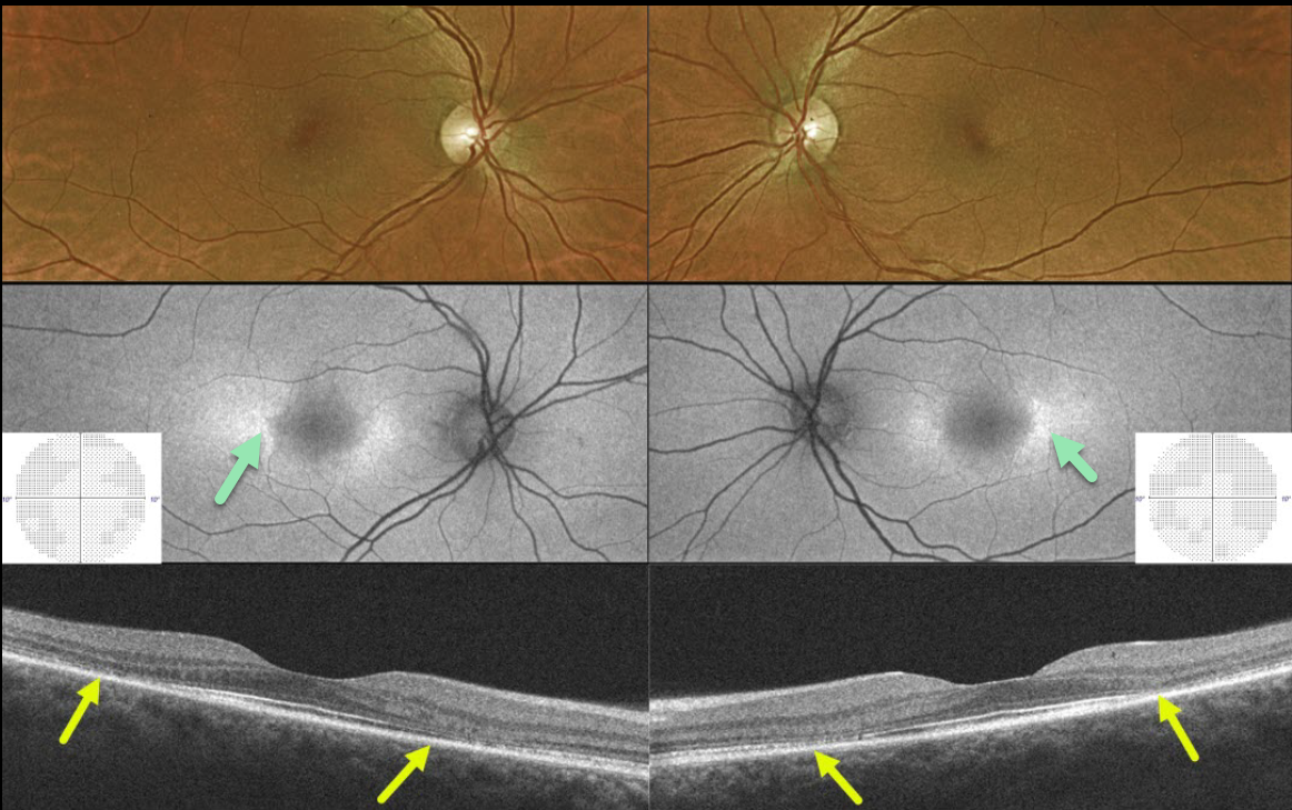
This 43YO female presented without visual complaint and 20/25 vision bilaterally. She gave a 27-year history of Plaquenil use for lupus.
Optos color imaging shows subtle pigment loss surrounding each fovea. Fundus autofluorescence (FAF) shows more obvious changes with perifoveal hyper-FAF nasally and temporally. 10-2 visual fields were normal.
Swept-source OCT B-scan imaging shows classic pericentral outer retinal atrophy.
Learning Points:
Current screening guidelines should make Plaquenil toxicity a thing of the past. Last revised in 2016, the American Academy of Ophthalmology (AAO) recommends that a baseline exam be performed before starting the medication, with annual screenings beginning at least 5 years after initiating treatment, unless major risk factors are present.
However, in practice, patients are usually screened yearly once they are placed on this medication.
Optical coherence tomography (OCT) is performed annually to assess outer retinal findings, including ellipsoid zone loss.
Our case emphasizes the importance of using multiple testing modalities to detect toxicity at the earliest possible stage.
HYDROXYCHLOROQUINE TOXICITY
Will Gibson
Originally posted on @retina.rocks November 30, 2022
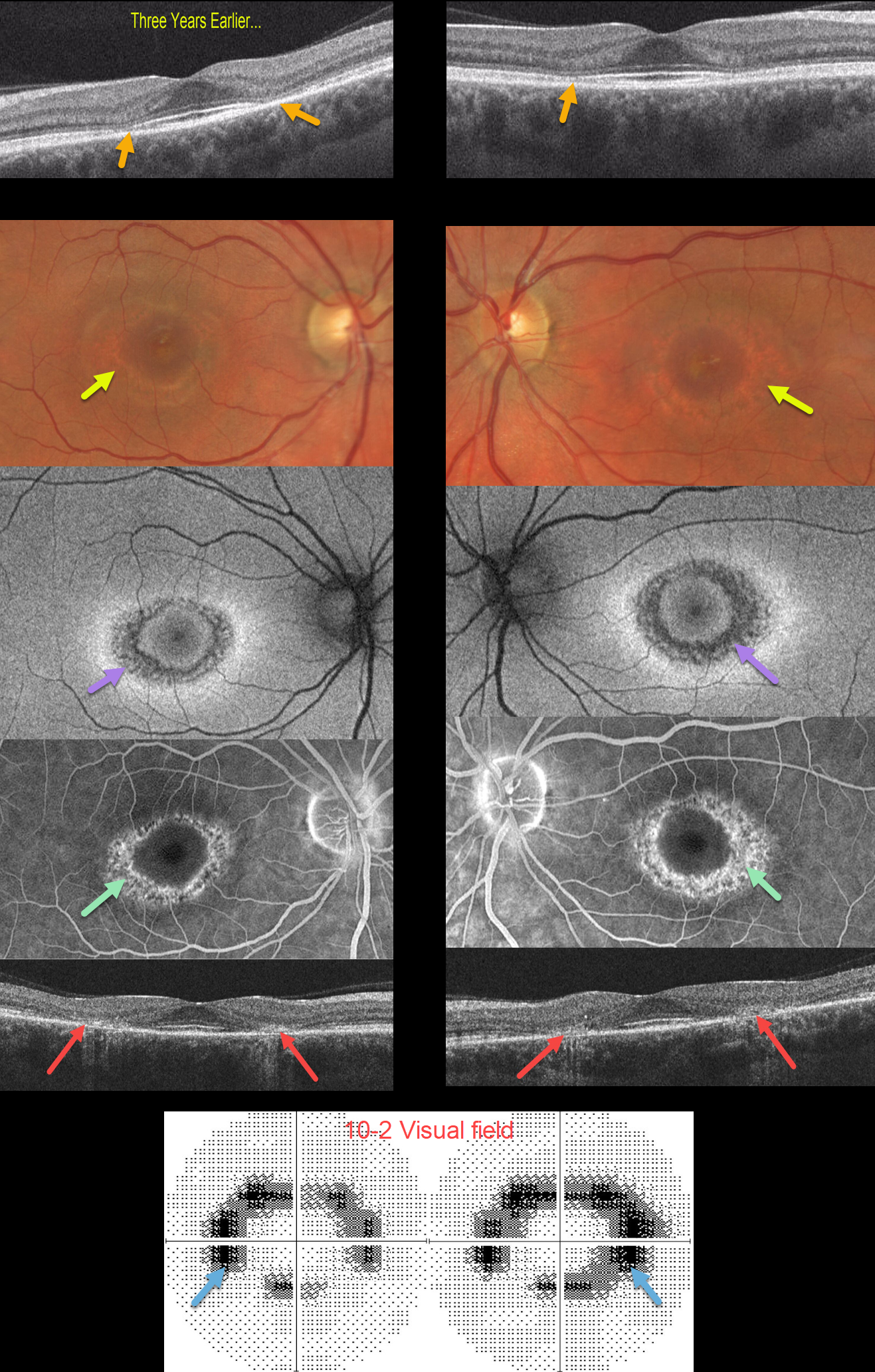
This 43YO female was seen in 2019 by a general ophthalmologist at an outside practice with somewhat subtle paracentral outer retinal atrophy. Vision was 20/20 without symptoms. She was taking Plaquenil for 8 years for rheumatoid arthritis. Unfortunately, these OCT findings for early Plaquenil toxicity were missed.
She was subsequently lost to followup until she presented 3 years later. Although vision remained at 20/20 OU, she now complained of donut-shaped scotomas bilaterally. There were now dramatic and classic findings for Plaquenil toxicity captured on multimodal imaging.
Optos color imaging shows a bullseye pattern of pigmentary loss encircling each fovea. Fundus autofluorescence shows hypo-FAF and fluorescein angiography shows window defects corresponding to these bullseye changes.
OCT scanning shows symmetrical paracentral outer retinal and RPE atrophy. Finally, 10-2 visual fields show a ring of bilateral paracentral visual field loss.
Learning Points:
Current screening guidelines should make Plaquenil toxicity a thing of the past. Last revised in 2016, the American Academy of Ophthalmology (AAO) recommends that a baseline exam be performed before starting the medication with annual screenings beginning at least 5 years after initiating treatment, unless major risk factors are present. However, in practicality, patients are usually screened yearly once they are placed on this medication.
Optical coherence tomography (OCT) is performed annually, looking for outer retinal findings, including ellipsoid zone loss. Humphrey 10-2 visual field testing (24-2 for Asians since their macular involvement is usually more peripheral) is needed annually as well, since about 10% of patients will have field loss despite normal examinations and OCT testing.
Plaquenil dosing should also be based on real, not ideal, weight to better predict the optimal dose. See Marmor et al Ophthalmology 2016;123:1386-1394 for the full screening guidelines.
CARBOPLATIN TOXICITY
Originally posted on @retina.rocks August 24, 2022
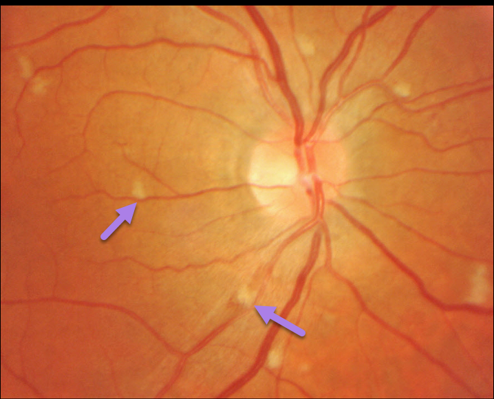
This patient, undergoing chemotherapy with carboplatin, developed these scattered nerve fiber layer infarcts (cotton wool spots).
Learning Points:
Carboplatin is an alkylating agent used to treat various cancers, including ovarian, lung, head and neck, brain, and neuroblastoma.
Retinal ischemia and optic neuropathies can rarely be found in patients taking both high-dose and cumulative-dose intravenous therapy.
The retinal ischemia can resolve with discontinuation of therapy, but those who experience optic neuropathies suffer irreversible loss.
HYDROXYCHLOROQUINE TOXICITY
Originally posted on @retina.rocks August 19, 2022
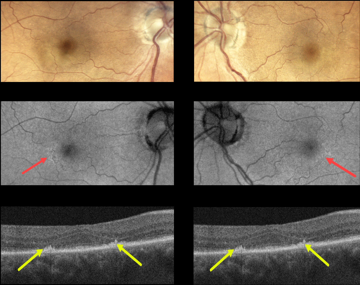
This 73YO female presented with bilateral hydroxychloroquine (Plaquenil) macular toxicity despite being treated for only 5 years. Vision was 20/40 OD and 20/30 OS.
Color photography shows very subtle pigmentary changes encircling each fovea. Fundus autofluorescence (FAF) shows subtle changes as well. The findings are most evident with OCT, which shows symmetrical paracentral outer retinal atrophy.
Learning Points:
Current screening guidelines should make Plaquenil toxicity a thing of the past. Last revised in 2016, the American Academy of Ophthalmology (AAO) recommends that a baseline exam be performed before starting the medication with annual screenings beginning at least 5 years after initiating treatment, unless major risk factors are present.
However, in practicality, patients are usually screened yearly once they are placed on this medication. Optical coherence tomography (OCT) is performed annually, looking for outer retinal findings, including ellipsoid zone loss.
Humphrey 10-2 visual field testing (24-2 for Asians since their macular involvement is usually more peripheral) is needed annually as well, since about 10% of patients will have field loss despite normal examinations and OCT testing.
Plaquenil dosing should also be based on real, not ideal, weight to better predict the optimal dose. See Marmor et al Ophthalmology 2016;123;1386-1394 for the full screening guidelines.
Our patient’s case is somewhat unique in that macular toxicity developed after only 4 years of therapy. This reinforces that screening and treatment guidelines must always be individualized.
PENTOSAN POLYSULFATE SODIUM MACULOPATHY
Originally posted on @retina.rocks August 18, 2022
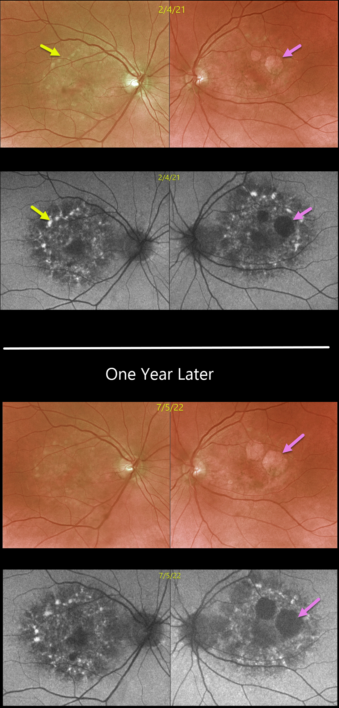
This 64YOF presented in February 2020 with findings of Pentosan polysulfate sodium (PPS, Elmiron) retinal toxicity. She used PPS for 4 years, but discontinued the drug 9 years earlier. We have since followed her yearly.
Optos color imaging of the prior (17 months earlier) and current examinations shows variable yellow-orange atrophic macular pigmentary changes. These findings are best visualized with fundus autofluorescence.
Close inspection of these images shows definite enlargement of the RPE atrophy, especially in her left eye. She remains completely free of visual symptoms with vision of 20/30 OD and 20/25 OS.
Learning Points:
Elmiron was approved by the FDA in 1996 for treating interstitial cystitis. A unique PPS retinopathy was recently described, and our patient shows classic findings.
Toxicity seems to develop over many years and can mimic more common disorders, including age-related macular degeneration and macular dystrophies.
Progressive changes, as in our patient, usually continue despite discontinuation of the causative drug (Shah et al., JAMA Ophthalmology 2020;138:894-900).
PENTOSAN POLYSULFATE SODIUM MACULOPATHY + OUTER RETINAL TUBULATION
Originally posted on @retina.rocks March 1, 2022
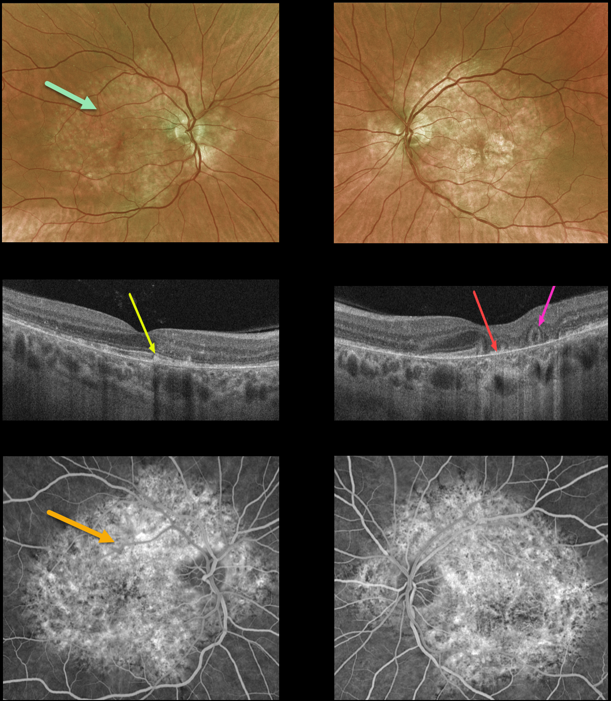
This 54YO female presented with a 9-month history of blurred vision in her left eye. She was on Elmiron (pentosan polysulfate sodium, PPS) for about 20 years due to interstitial cystitis, but stopped the medication before seeing us due to the widespread publicity about its potential retinal toxicity.
Optos color imaging shows variable yellow-orange atrophic pigmentary changes throughout the macular and peripapillary posterior poles.
OCT of the right macula shows scattered hyperreflective lesions mostly within the outer segment layers. The left macular OCT shows variable outer retinal and RPE loss, especially centrally, where bare Bruch’s membrane is visible. There is also an area of outer retinal tubulation (ORT) more temporally.
Fluorescein angiography shows window defects within these regions.
Learning Points:
Elmiron was approved by the FDA in 1996 for treating interstitial cystitis. Recently, a unique PPS retinopathy has been described, and our patient shows classic findings.
Toxicity seems to develop over many years and can mimic more common disorders, including age-related macular degeneration and macular dystrophies.
Peripapillary hypoautofluorescence, more densely packed macular autofluorescent changes, and earlier central macular involvement suggest PPS toxicity over other causes (see Barnes et al Ophthalmology Retina 2020;4:1196-1201), including maternally inherited diabetes and deafness (MIDD).
ORT is often noted overlying inactive macular neovascularization with ongoing anti-VEGF therapy and should not be confused with exudative fluid or cysts, which lack a hyperreflective border. The outer hyperreflective band likely represents inner segment mitochondria undergoing fission and translocation towards the nucleus (Litts et al, Retina 2018;38:445-461).
ORT, initially described by Zweifel et al (Arch Ophthalmol 2009;127:1596-1602), is a neurodegenerative condition of the photoreceptors and Muller cells associated with atrophy affecting the outer retina and retinal pigment epithelium, including advanced AMD and inherited retinal diseases.
HYDROXYCHLOROQUINE TOXICITY
Originally posted on @retina.rocks January 17, 2022
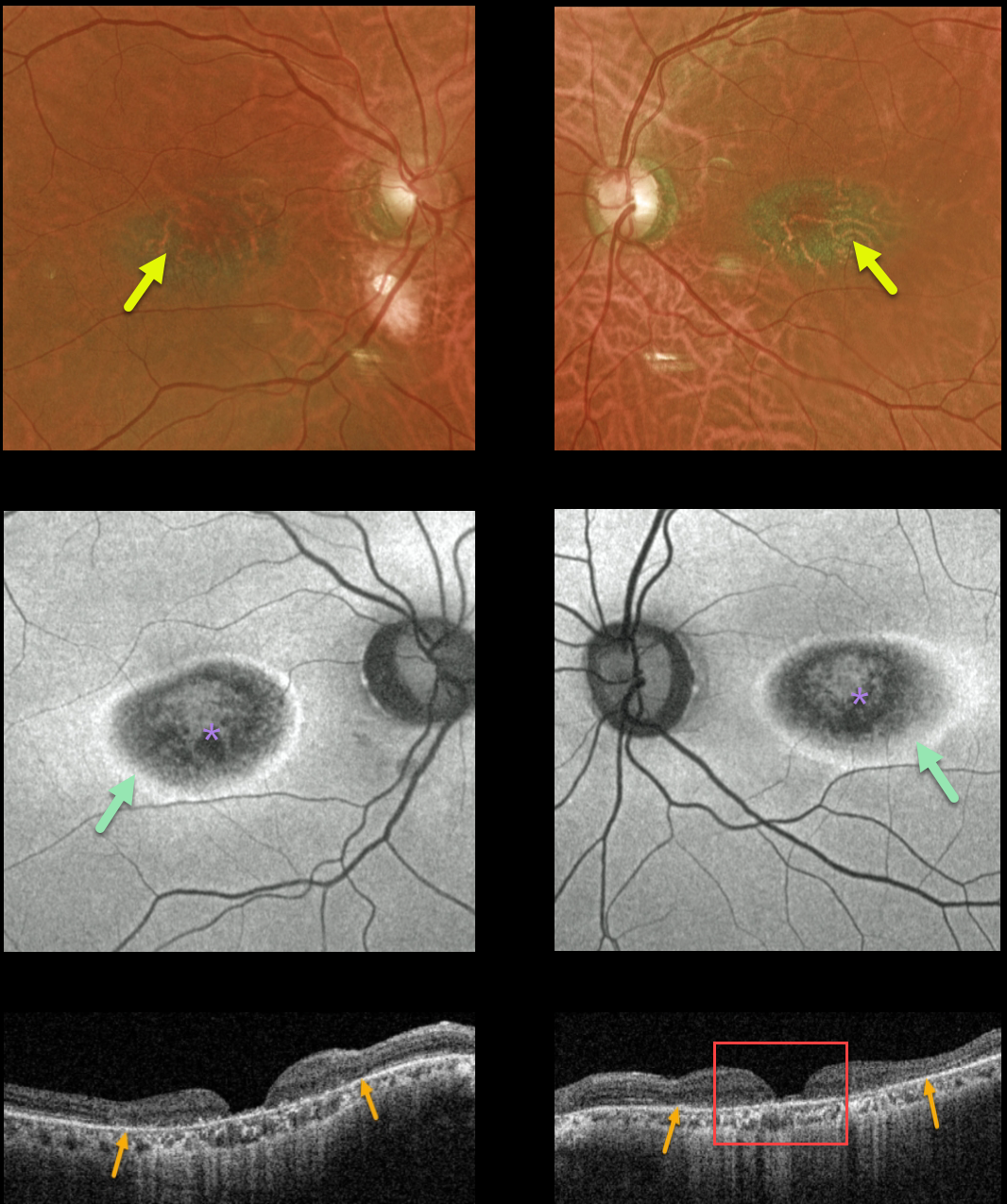
This 77YO female had a long prior history of Plaquenil (hydroxychloroquine) use for lupus. Although it was discontinued 10 years ago, vision was 20/200 OU with classic findings for Plaquenil toxicity.
Optos color imaging shows a hyperpigmented bull’s-eye pattern of pigmentary changes surrounding each macular center. Fundus autofluorescence (FAF) shows pericentral hypo-FAF from RPE damage, with a surrounding narrow ring of hyper-FAF.
Triton swept-source OCT shows outer retinal thinning with more severe near-total central neurosensory retinal thinning with pseudo-macular holes. To further confuse the OCT findings, she also gave a history of macular hole surgery in one of her eyes!
Learning Points:
Current screening guidelines should make Plaquenil toxicity a thing of the past. Last revised in 2016, the American Academy of Ophthalmology (AAO) recommends that a baseline exam be performed before starting the medication, with annual screenings beginning at least 5 years after initiating treatment, unless major risk factors are present.
However, in practice, patients are usually screened yearly once they are placed on this medication. Optical coherence tomography (OCT) is performed annually to assess outer retinal findings, including ellipsoid zone loss.
Humphrey 10-2 visual field testing (24-2 for Asians, since their macular involvement is usually more peripheral) is also needed annually, as about 10% of patients will have field loss despite normal examinations and OCT testing.
Plaquenil dosing should also be based on real, not ideal, weight to better predict the optimal dose. See Marmor et al Ophthalmology 2016;123;1386-1394 for the full screening guidelines.
PENTOSAN POLYSULFATE SODIUM MACULOPATHY
Originally posted on @retina.rocks October 21, 2021
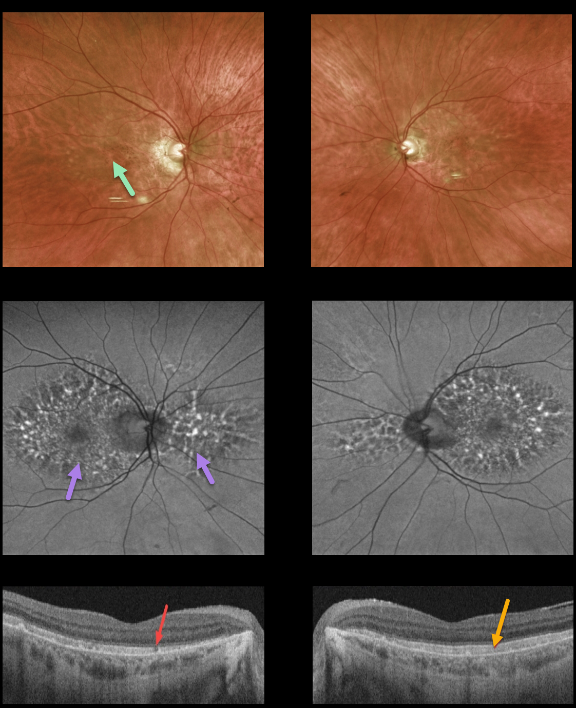
This 74YO female had a long history of Elmiron (pentosan polysulfate, PPS) use. Vision was 20/30 OU.
Variable yellow-orange atrophic pigmentary changes were noted throughout each macula, with more dramatic patches of macular and peripapillary hyper- and hypoautofluorescence.
Triton swept-source OCT showed some patchy ellipsoid loss OD and diffuse hyperreflectivity of the outer segments.
Learning Points:
Elmiron was approved by the FDA in 1996 for treating interstitial cystitis. Recently, a unique PPS retinopathy has been described, and our patient shows classic findings. Toxicity seems to develop over many years and can mimic more common disorders, including age-related macular degeneration and macular dystrophies.
Peripapillary hypoautofluorescence, more densely packed macular autofluorescent changes, and earlier central macular involvement suggest PPS toxicity over other causes (see Barnes et al, Ophthalmology Retina 2020;4:1196-1201), including MIDD. Mitochondrial disease, pattern macular dystrophies, and PPS maculopathy have similar yet distinct findings. We hope the last 3 days of posts will help to clarify these clinically overlapping phenotypes.
PACLITAXEL MACULOPATHY
Originally posted on @retina.rocks September 2, 2021
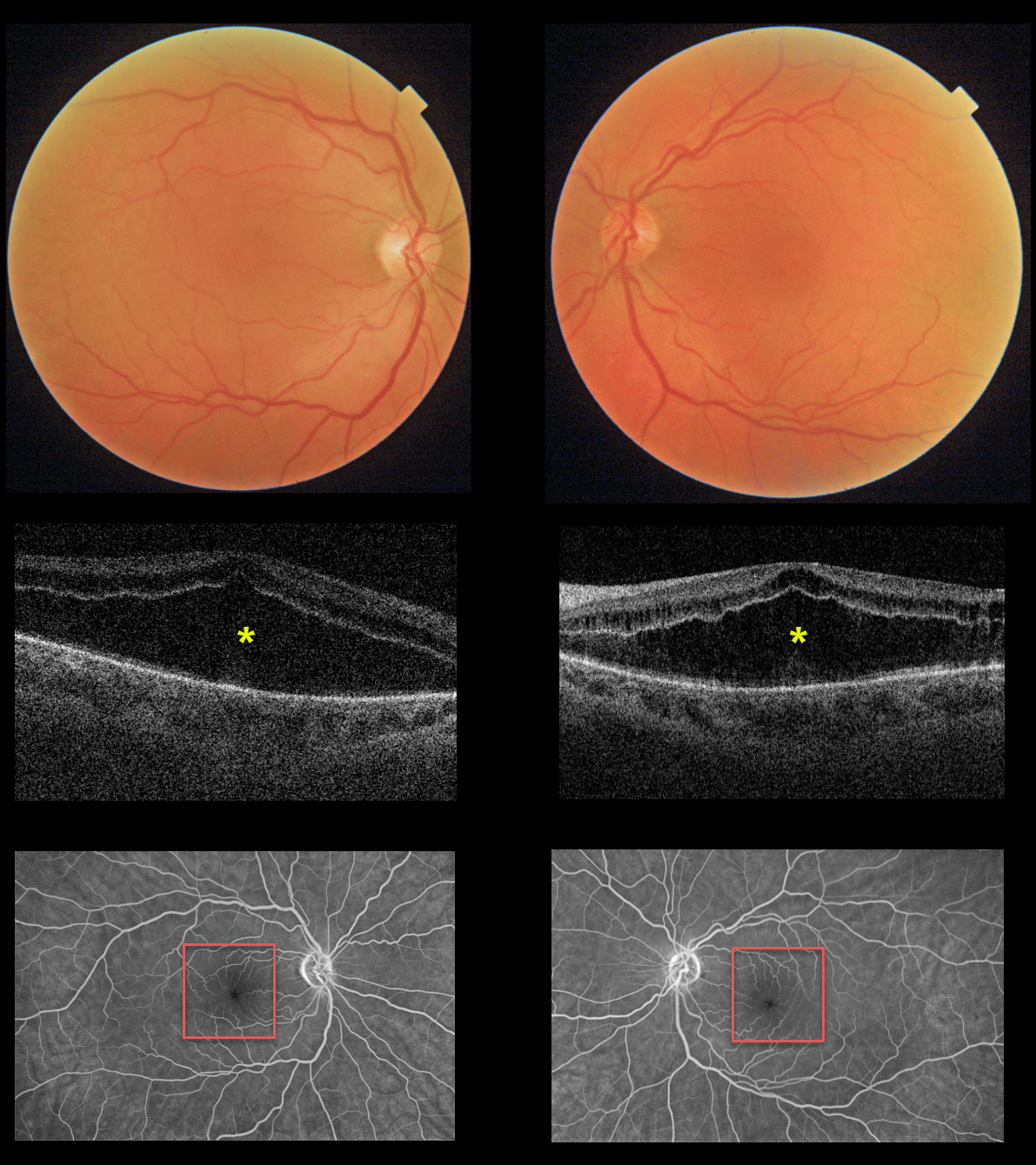
This 55YO female presented with an 8-month history of bilateral vision loss. She had been treated for metastatic breast cancer with Taxol (paclitaxel) for 1 year prior to this exam. Vision was 20/200 OU.
Triton imaging and swept-source OCT show severe symmetrical bilateral cystoid changes in each macula. The cystoid changes are most pronounced in the outer nuclear layer and less so in the inner nuclear layer. Fluorescein angiography shows no leakage.
We have consulted with her oncologist, and expect him to discontinue the paclitaxel. The macular changes and vision should improve within several months.
Learning Points:
Paclitaxel is a member of the taxane family of microtubule-stabilizing agents that is used to treat various solid malignant tumors. Their major adverse effects are bone marrow toxicity and peripheral neuropathy. Ophthalmic complications are rare, including taxane-related cystoid macular edema (CME).
Unlike more common exudative causes for CME, there is no angiographic leakage, and OCT shows intact and continuous inner and outer plexiform layers (see Perez et al, Graefe’s 2020;258:1607-1615).
The cause for CME is uncertain but may be due to Müller cell dysfunction (see Nakao et al, Ophthalmic Surg Lasers Retina 2016;47:81-84). There is no treatment for the CME other than stopping the causative drug.
HYDROXYCHLOROQUINE TOXICITY
Originally posted on @retina.rocks May 25, 2021
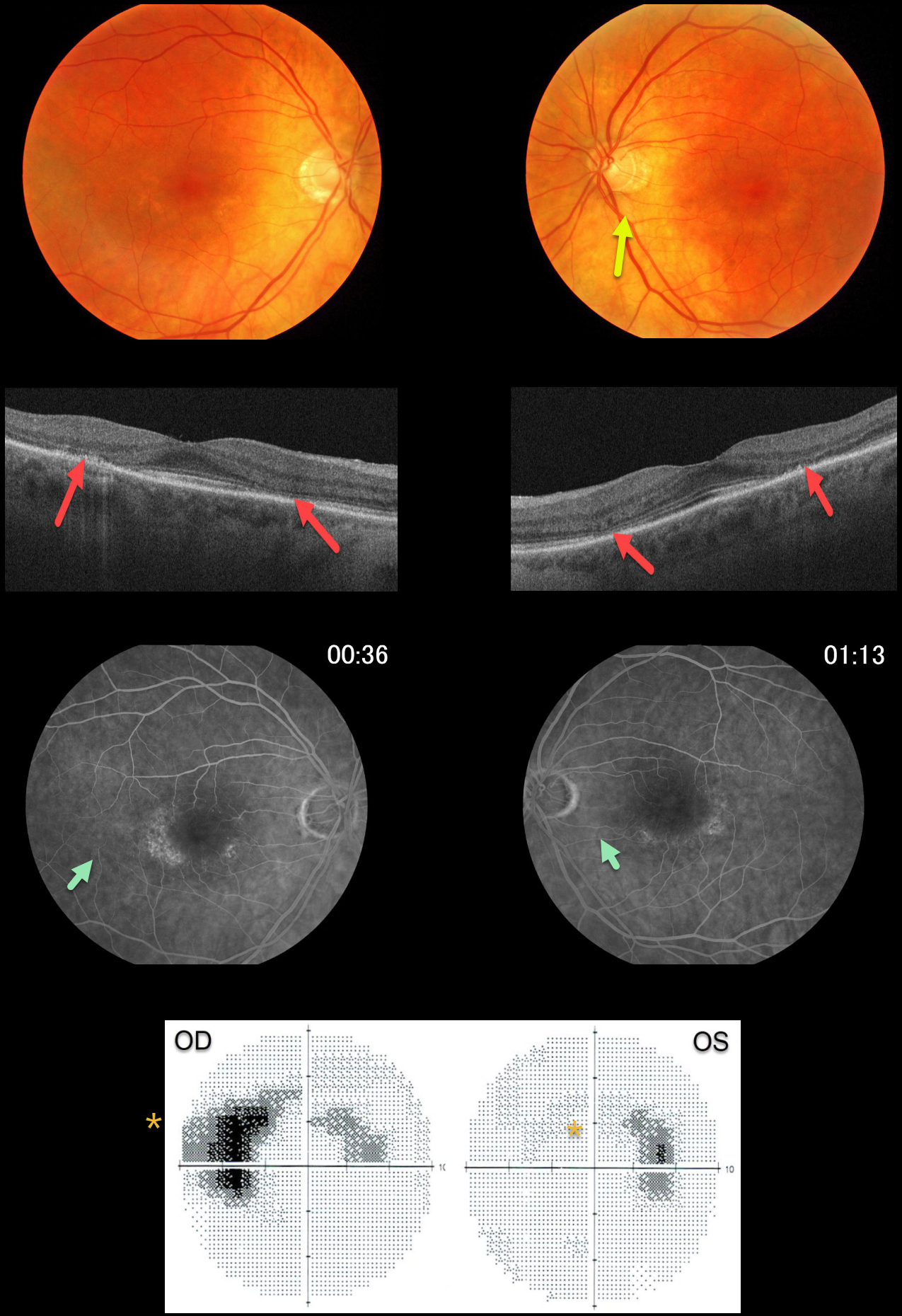
This 72YO female gave a 30-year history of Plaquenil (hydroxychloroquine) use for lupus. Vision was 20/50 OD and 20/20 OS.
There are subtle yet classic findings for Plaquenil toxicity, including a bull’s-eye pattern of pericentral outer retinal atrophy and pigment loss. This is seen clinically as loss of the pericentral orange RPE pigment, on swept-source OCT as thinning of the pericentral photoreceptor elements, and on fluorescein angiography as hyperfluorescent window defects. 10-2 visual fields show bilateral paracentral scotomas.
In the pre-OCT era, patients were followed until clinical or angiographic pigmentary changes developed, at which point the medication was discontinued. Unfortunately, this patient was not properly screened and developed irreversible changes.
Learning Points:
Current screening guidelines should make Plaquenil toxicity a disease of the past
Optical coherence tomography (OCT) is performed annually to assess outer retinal findings, including ellipsoid zone loss.
Humphrey 10-2 visual field testing (24-2 for Asians, since their macular involvement is usually more peripheral) is also needed annually, as about 10% of patients will have field loss despite normal examinations and OCT testing. Plaquenil dosing should also be based on real, not ideal, weight to better predict the optimal dose.
See Marmor et al, Ophthalmology 2016;123:1386-1394 for the full screening guidelines.
PENTOSAN POLYSULFATE SODIUM MACULOPATHY
Originally posted on @retina.rocks March 24, 2021
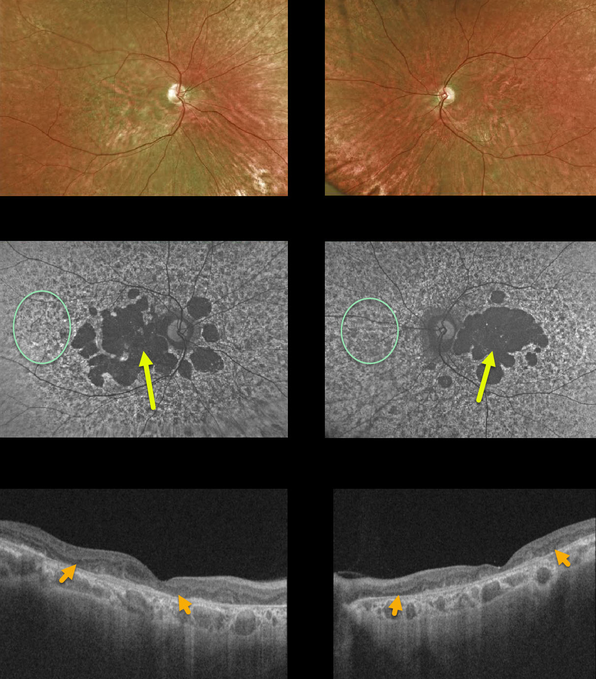
This 65YO female was taking Elmiron (pentosan polysulphate, PPS) for many years beginning in the mid-1990s. Vision was 20/400 OD and counting fingers OS.
Variable yellow-orange atrophic pigmentary changes were noted funduscopically throughout each macula, with more dramatic patches of macular hypoautofluorescence with a peripheral granular hyper- and hypoautofluorescent appearance.
OCT scanning showed variable outer retinal loss.
Learning Points:
Elmiron was approved by the FDA in 1996 for treating interstitial cystitis. Recently, a unique PPS retinopathy has been described, and our patient shows classic findings. Toxicity seems to develop over many years and can mimic more common disorders, including age-related macular degeneration and macular dystrophies.
Peripapillary hypoautofluorescence, more densely packed macular autofluorescent changes, and earlier central macular involvement suggest PPS toxicity over other causes (see Barnes et al, Ophthalmology Retina 2020;4:1196-1201).
TAMOXIFEN RETINOPATHY
Originally posted on @retina.rocks September 9, 2020
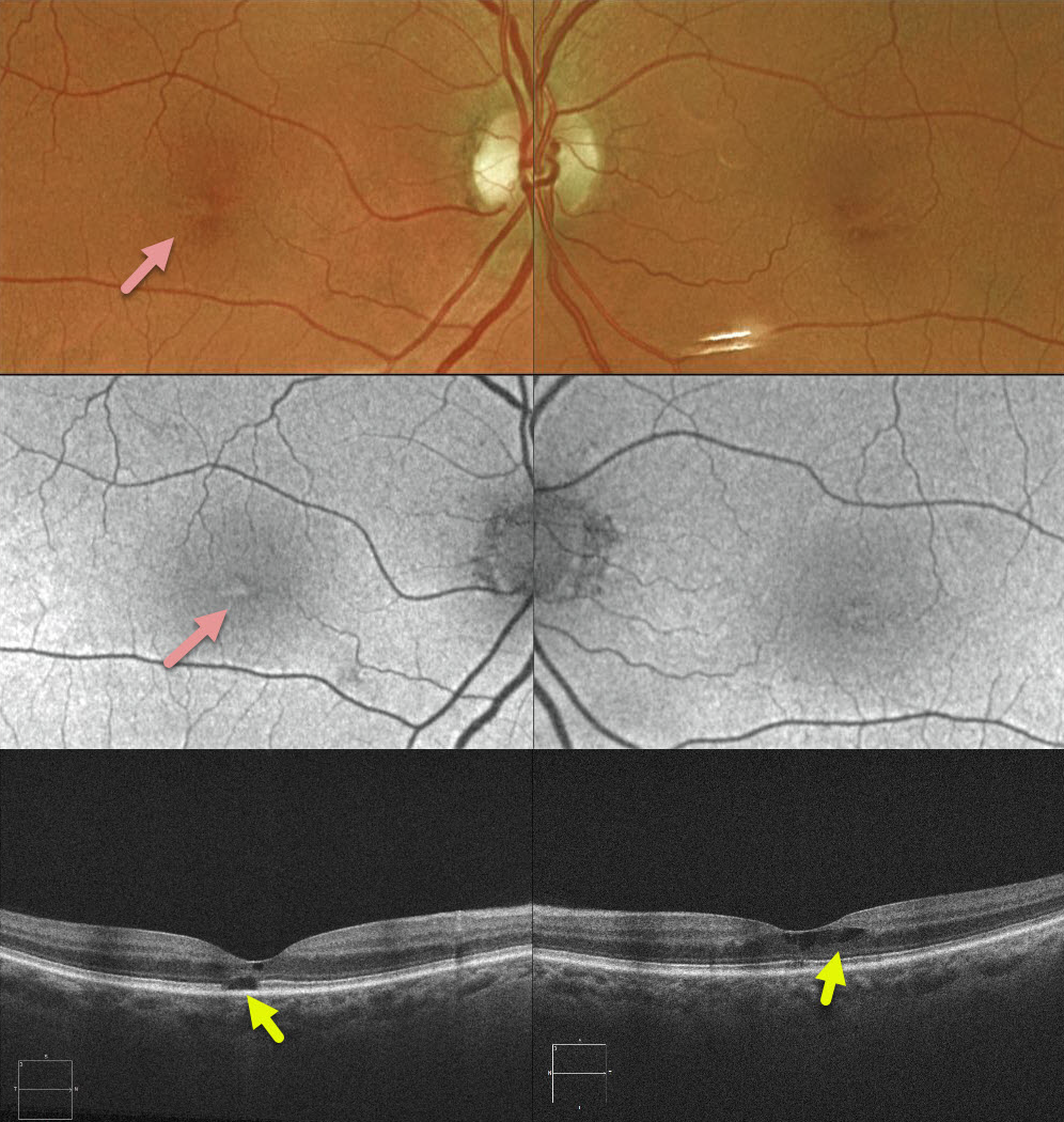
This 72YO female recently completed a 5-year course of Tamoxifen as adjunctive treatment for breast cancer. She had no visual symptoms with 20/30 vision bilaterally.
Clinically, there were subtle bilateral foveal pigmentary changes. B-scan and en-face OCT showed bilateral cavitary changes.
Learning Points:
Tamoxifen retinopathy shares findings very similar to those of macular telangiectasia type 2 (MacTel2), including retinal cavitations, right-angle venules, and inner retinal crystals. The changes in MacTel2 are usually confined to the temporal fovea, whereas with Tamoxifen, they seem more diffusely distributed throughout the entire central macula. The retinal changes for both disorders likely share Muller cell injury as the underlying cause.
The prevalence of Tamoxifen retinopathy may be as high as 12%. Although most oncologists do not require this, periodic ophthalmic screening, OCT, and examinations may be indicated since patients can lose central vision if toxicity develops.
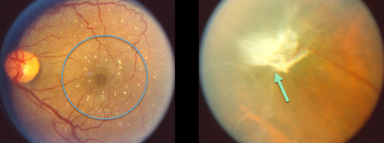
TALC RETINOPATHY
Originally posted on @retina.rocks June 18, 2020
Glistening crystals in the posterior pole are prevalent in this intravenous drug user with talc retinopathy.
There is also a large fibrosed area of neovascularization along the border between the perfused retina to the right and the ischemic retina to the left, a neovascularization pattern somewhat similar to that seen in Eale’s disease.
Learning Points:
Intravenous drug users may prepare a suspension of crushed tablets that include inadequately filtered insoluble fillers, including talc and cornstarch.
Talc emboli lodge in the pulmonary circulation and eventually enter the arterial circulation through pulmonary collaterals. These talc emboli can eventually enter the retinal arterial circulation, leading to retinal ischemia and neovascularization.
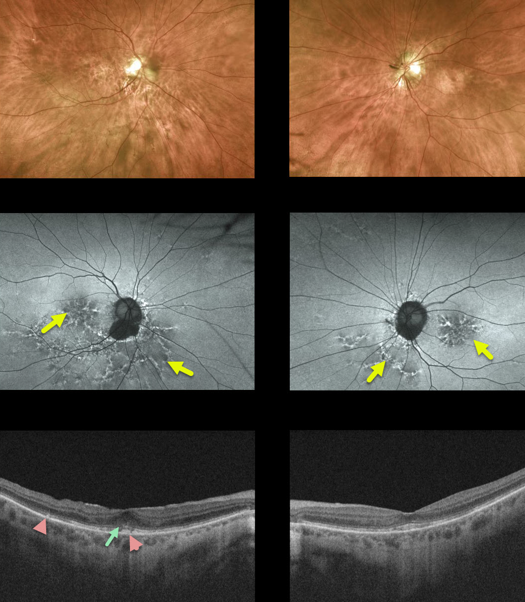
PENTOSAN POLYSULFATE SODIUM MACULOPATHY
Originally posted on @retina.rocks June 2, 2020
This 72yo female was on pentosan polysulfate sodium (PPS, Elmiron) for 5-10 years in the 1990s and was previously misdiagnosed with dry age-related macular degeneration (AMD).
While her fundus exam may resemble pigmentary changes associated with dry AMD, there are no drusen.
Fundus autofluorescence (FAF) shows a mixture of hyper- and hypo-autofluorescent spots in the macula extending outwards into the mid-peripheries. Drusen and pigmentary changes in AMD typically exhibit variable hypoautofluorescence.
OCT shows some variable attenuation of the EZ band along with hyperreflective deposits above the RPE.
Learning Points:
PPS, sold under the brand name Elmiron, is used to treat interstitial cystitis. PPS maculopathy seems to develop over many years and can mimic more common disorders, including AMD and macular dystrophies. As with hydroxychloroquine, macular changes can progress despite discontinuation of the drug. In our experience, wide-field FAF is critical in establishing the diagnosis.
Although there is no treatment for the underlying maculopathy, patients need to be followed and treated if macular neovascularization develops.
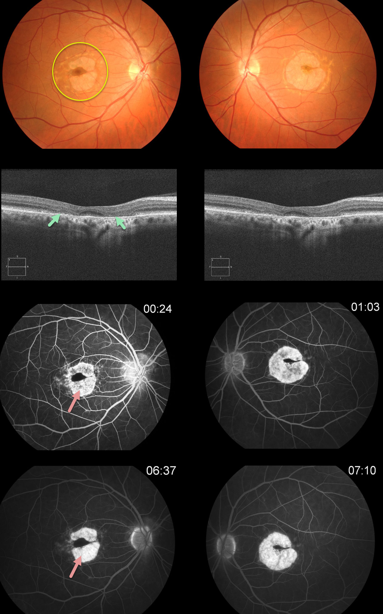
HYDROXYCHLOROQUINE TOXICITY
Originally posted on @retina.rocks May 20, 2020
This patient has classic findings for Plaquenil (hydroxychloroquine) toxicity, including a bull’s-eye pattern of pericentral RPE and outer retinal atrophy. This is seen clinically as loss of the orange RPE pigment, on OCT as loss of the photoreceptor elements and RPE, and on fluorescein angiography as hyperfluorescent window defects.
In the pre-OCT era, patients were followed until clinical or angiographic pigmentary changes developed, at which point the medication was discontinued. Unfortunately, this patient was not properly screened and developed irreversible changes.
Learning Points:
Current screening guidelines should make Plaquenil toxicity a disease of the past
Optical coherence tomography (OCT) is performed annually to assess outer retinal findings, including ellipsoid zone loss.
Humphrey 10-2 visual field testing (24-2 for Asians, since their macular involvement is usually more peripheral) is also needed annually, as about 10% of patients will have field loss despite normal examinations and OCT testing. Plaquenil dosing should also be based on real, not ideal, weight to better predict the optimal dose.
See Marmor et al, Ophthalmology 2016;123:1386-1394 for the full screening guidelines.
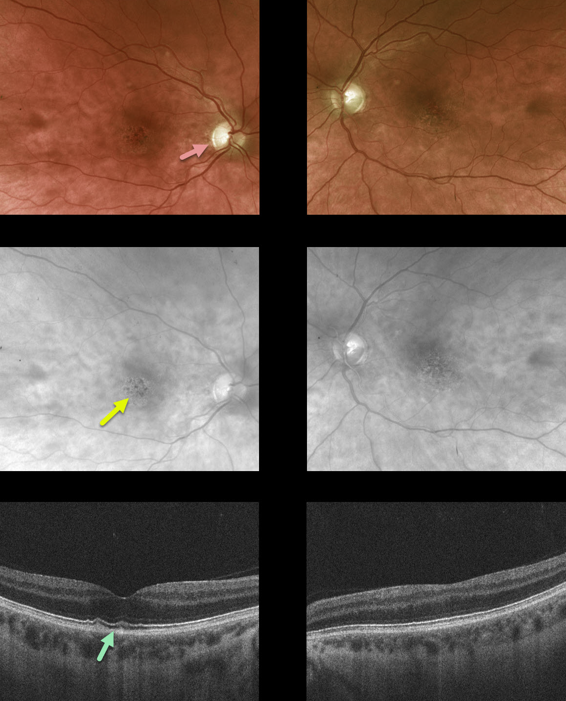
METHAMPHETAMINE TOXICITY
Originally posted on @retina.rocks January 28, 2020
This former methamphetamine and heroin addict presented with 20/50 vision OU, symmetric foveal RPE mottling, mild optic atrophy and some thickening and hyper-reflectivity of the foveal outer segments on OCT.
Learning Points:
Retinal changes that occur due to methamphetamine use include hemorrhages, cotton wool spots and vasculitis. We presume the findings in this patient were a sequela of prior toxicity.
Similar findings can be found in long-term dextroamphetamine/amphetamine (Adderall) users.


