

09 Infectious Disorders
<< Back to Cases
DENGUE RETINITIS
Rohan Jain and Manish Nagpal
Originally posted on @retina.rocks September 9, 2026
This 63YO male presented with 1 month of bilateral decreased vision following dengue fever, with positive Dengue NS1 antigen during the acute illness. Vision was 20/200 OD and counting fingers OS.
Pseudocolor SLO imaging shows extensive peripapillary cotton wool spots, retinal whitening, and significant macular edema. The fundus detail is partially obscured by overlying vitreous debris from vitritis. OCT scanning neurosensory retinal detachment with subretinal hyperreflective material, intraretinal cystoid edema, and subretinal fluid involving the macula in both eyes, consistent with post-dengue inflammatory retinitis. A possible bacillary layer detachment is also noted in the left eye.
Topical steroids and a 9-week course of tapering oral steroids, as well as a 3-week course of oral doxycycline, were started. Four months later, vision improved to 20/20 OD and 20/30 OS. The patches of white inner retinal ischemia are fading, and some lipid flecks have precipitated out in the mid-retina.
Learning Points:
Epidemic (post-fever) retinitis is an infectious or para-infectious entity that develops in immunocompetent individuals 2-6 weeks following a bacterial, viral, or protozoal infection. Rickettsiosis is the most identified cause, though a large proportion of cases remain idiopathic despite serologic testing; dengue, chikungunya, West Nile, Zika, leptospirosis, typhoid, and Ebola have also been implicated. Patients present with sudden painless vision loss. Fundus findings include unilateral or bilateral unifocal or multifocal patches of retinitis with possible nerve involvement, neuroretinitis with macular star, serous macular detachment, cystoid macular edema, and vasculitis or frosted branch angiitis.
Management is stratified by etiology and lacks controlled data: bacterial causes are treated with systemic antibiotics with or without corticosteroids, viral causes with corticosteroids alone, and many authors advocate observation, as the disease often resolves on its own. Corticosteroids should not be used as sole therapy while infectious causes (including TB, syphilis, and toxoplasmosis) remain in the differential. See Mahendradas et al (Indian J Ophthalmol 2020;68:1775-1786) for an excellent post-fever retinitis review.
POST-FEVER RETINITIS
Vedant Gambhir
Originally posted on @retina.rocks August 3, 2026
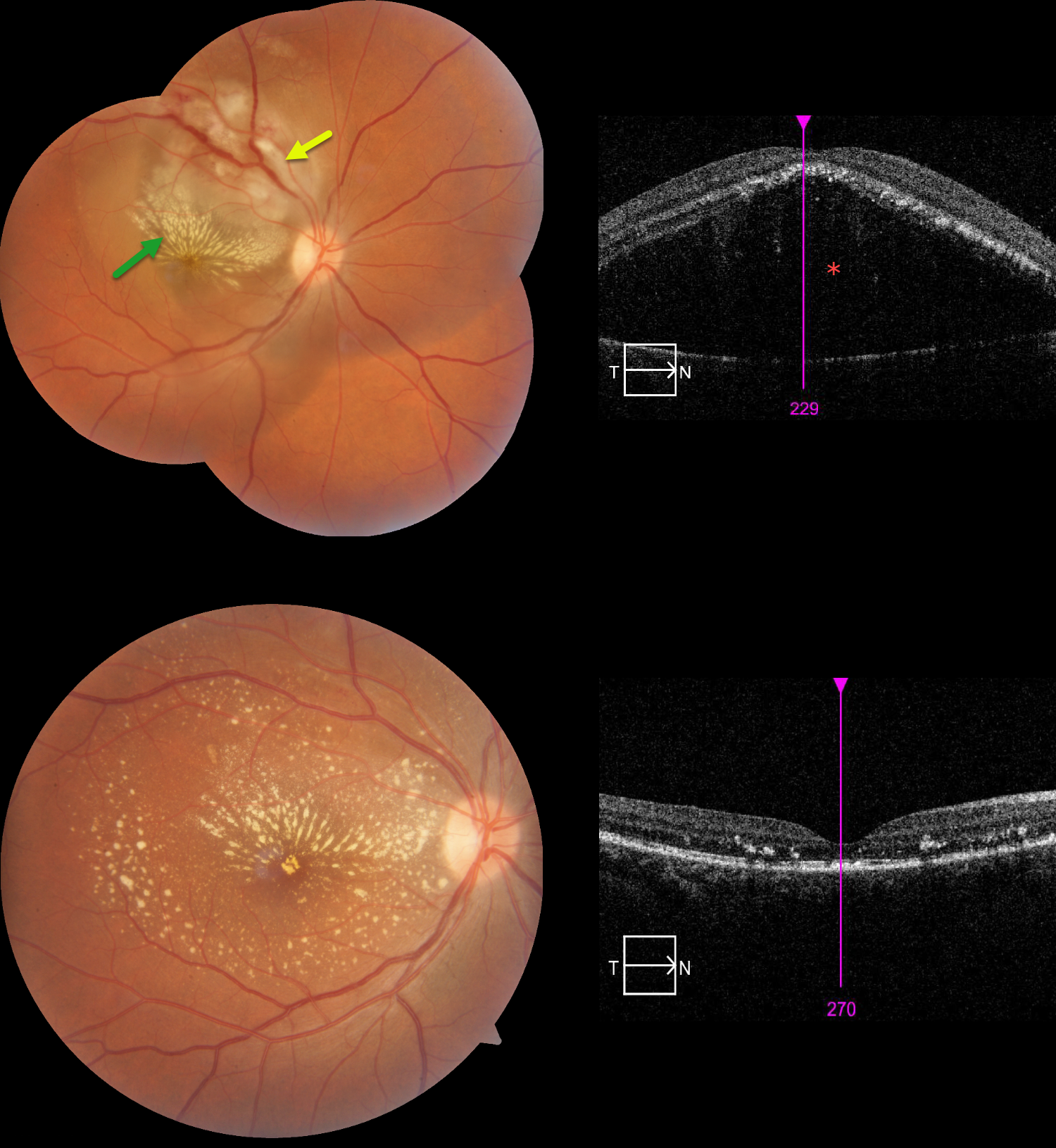
This 31YO female presented with 1 week of decreased vision in her right eye following a febrile illness. Vision was 20/200 OD and 20/20 in her normal OS.
Color photography shows opaque, white inner and deep retinal whitening centered in the superonasal macula, with surrounding subretinal fluid extending into the temporal and inferior macula. A lipid star radiates superiorly and nasally from the foveal center. OCT scanning confirms a highly elevated serous detachment.
An extensive infectious workup was negative for common bacterial, viral, and parasitic etiologies. Given the history of antecedent fever and characteristic retinal findings, a diagnosis of post-fever (epidemic) retinitis was favored. She was treated with oral corticosteroids and doxycycline. At follow-up, visual acuity improved dramatically from 20/200 to 20/30.
Learning Points:
Epidemic (post-fever) retinitis is an infectious or para-infectious entity that develops in immunocompetent individuals several weeks following a bacterial, viral, or protozoal infection. Causes include Dengue, Rickettsiosis, West Nile, Leptospirosis, Chikungunya, Typhoid, and Ebola. Serologic workup is negative in about 60% of cases (Kawali et al, Indian J Ophthalmol 2020;68:1916-1919), due to patients often presenting when acute-phase serologies are difficult to interpret, and viremia has resolved; limitations of available serologic testing; heterogeneous and overlapping etiologies; and a potential immune-mediated response.
Patients present with sudden, painless vision loss. Fundus findings include unilateral or bilateral unifocal or multifocal patches of retinitis with possible nerve involvement, serous macular detachment, and vasculitis or frosted branch angiitis. A macular lipid star in post-fever retinitis is uncommon, found in only 3.3% of affected eyes in rickettsial retinitis (the best-studied cause).
The exact pathogenesis remains uncertain; however, a post-infectious immune-mediated mechanism has been proposed. There is usually a favorable response to steroids and treating the underlying infectious cause with appropriate systemic therapy. See Mahendradas et al (Indian J Ophthalmol 2020;68:1775-1786) for an excellent post-fever retinitis review.
TUBERCULOUS POSTERIOR SCLERITIS
Aniruddh Soni
Originally posted on @retina.rocks July 31, 2026
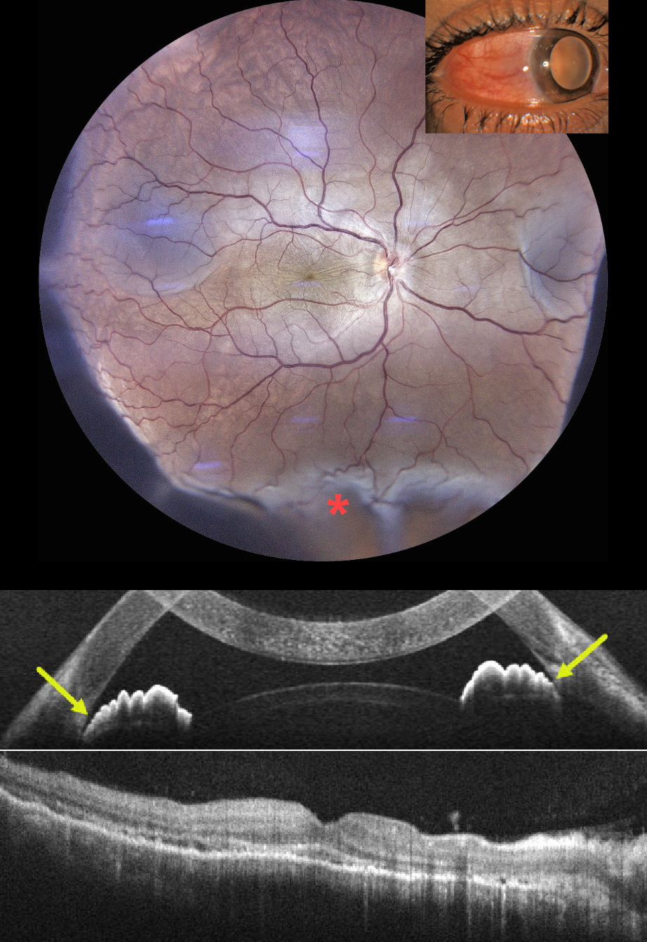
This previously healthy 35YO male presented with 1 week of pain, redness, and decreased vision in his right eye. He had been treated by an outside doctor for conjunctivitis. Vision 20/200 OD and 20/20 in his normal OS. Slit lamp examination showed temporal scleral injection. Intraocular pressure was 23 mm Hg.
Color photography shows radiating chorioretinal folds throughout the macula with an inferior exudative retinal detachment. OCT scanning of the macula shows chorioretinal folds, with anterior chamber OCT showing a narrow-to-closed angle. B-scan ultrasonography showed subtenon’s fluid (not shown).
Following a 3-week course of tapering oral steroids and glaucoma drops, vision improved to 20/40 with reduced chorioretinal folds and complete resolution of the exudative detachment. Rheumatologic workup was negative, but Mantoux skin testing and QuantiFERON-TB GOLD were positive, with a subsequent diagnosis of latent tuberculosis (TB). Antituberculous therapy was recommended, but the patient refused treatment.
Learning Points:
Tuberculous posterior scleritis is a rare manifestation of ocular TB (Agarwal and Majumder, Indian J Ophthalmol 2019;67:1362-1365), which itself occurs in approximately 1.5% of patients with confirmed systemic TB. Ocular findings include optic disc edema, choroiditis, and sclerochoroidal thickening with widening of the sub-Tenon space. It may occur via direct hematogenous dissemination of Mycobacterium tuberculosis or through a hypersensitivity-mediated immune response to the organism and can develop even in the absence of clinically active pulmonary disease.
Diagnosis, as in our patient, is largely presumptive, requiring a high index of suspicion supported by bloodwork, chest imaging to assess pulmonary involvement, and exclusion of mimics such as sarcoidosis, syphilis, and metastatic disease.
TUBERCULAR SERPIGINOUS-LIKE CHOROIDITIS
Aniruddh Soni
Originally posted on @retina.rocks July 6, 2026
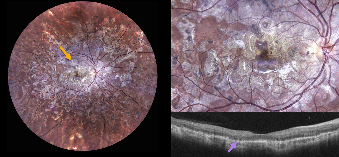
A 30YO female presented with a 1.5-year history of bilateral floaters and blurred vision. She was diagnosed elsewhere with ‘choroiditis’ and treated with oral steroids and azathioprine, with 2 recurrences. When seen in our office, vision was 20/30 OU.
Color photography OD shows extensive, variably confluent, and meandering chorioretinal scarring extending from the disc and macula to the retinal periphery. The foveal center is just spared. OCT scanning shows variable outer retinal and RPE atrophy. Identical findings were noted in her left eye (not shown).
Tuberculin skin testing and QuantiFERON-TB Gold were positive. She was referred to pulmonary and infectious disease specialists, was diagnosed with latent TB, and started on anti-tuberculosis treatment (ATT) with tapering oral steroids.
Learning Points:
Although TB most commonly presents with pulmonary involvement, extrapulmonary sites can involve the gastrointestinal, skin, cardiovascular, genitourinary, and central nervous systems, as well as the eyes. Ocular involvement, like syphilis, can mimic virtually any type of uveitis, including anterior, intermediate, posterior, and panuveitis, retinitis, and retinal vasculitis, neuroretinitis, optic neuropathy, choroidal granuloma, choroiditis, and scleritis.
Tubercular serpiginous-like choroiditis mimics serpiginous but affects younger patients with more multifocal and peripheral recurrences, foveal sparing, vitritis, and progression if untreated (Agarwal et al, AJO 2020;220:160-169). Although the exact mechanism is not yet clear, a direct or indirect infectious trigger by the Mycobacterium tuberculosis is thought to cause choroiditis. Its origin is probably immunogenic, as it appears to respond to corticosteroids and other immunosuppressants. Our patient was a bit unusual in that he required systemic steroids, ATT, and adalimumab to finally control his disease.
SCRUB TYPHUS POST-FEVER RETINITIS
Anand Temkar, Surendra Pal, Vasumathi Vijay, and Manorama Baheti
Originally posted on @retina.rocks June 3, 2026
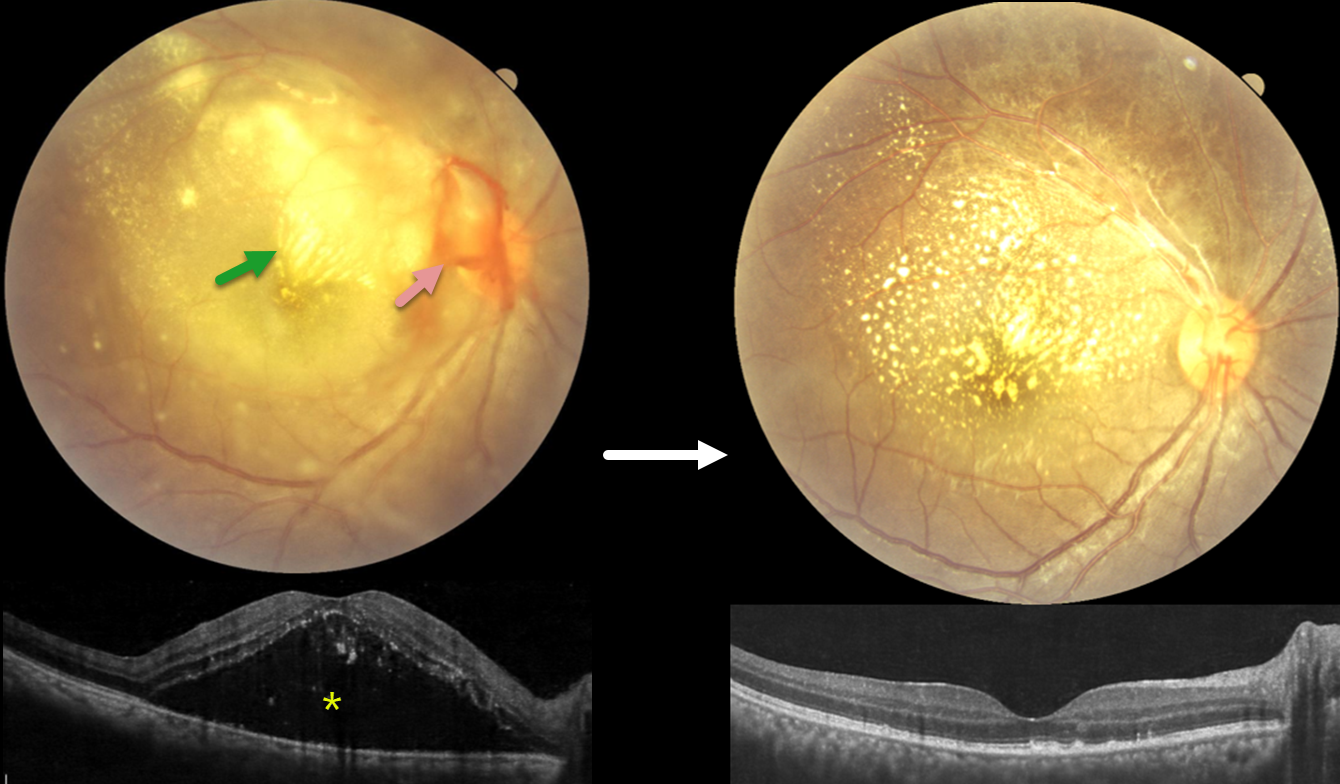
This 28YO female presented with 4 days of blurred vision in her right eye. For the past 2 weeks, she has complained of generalized weakness, malaise, and body pain. Vision was counting fingers OD and 20/20 in her normal OS.
Fundus photography shows variable, dense subretinal lipid, particularly confluent in the superonasal macula. There is some preretinal blood around the nerve, and the retina is somewhat yellow and opaque temporally and inferiorly. Patches of inner retinal opacification are noted outside the macula superiorly. OCT shows hyperreflective dots from lipid exudate in the outer retina, with a large serous detachment. Laboratory testing, including CBC, ESR, syphilis, TB, HIV, TORCH profile, and chest X-ray, was negative. We started a 1-week tapering course of oral prednisone 40mg and doxycycline 100mg BID. Three days after our initial examination, she mentioned that she had 2 weeks of mild intermittent fever. Weil-Felix testing was positive, and she was subsequently diagnosed with scrub typhus and asked to continue the doxycycline for a full 2-week course.
The clinical picture gradually improved, and about 6 weeks following presentation, vision improved to 20/40 despite significant residual lipid. OCT scanning through the macular center was mostly normalized. We continue to follow her closely.
Learning Points:
Rickettsial infections cause posterior segment involvement in a substantial proportion of patients, often asymptomatic despite significant fundoscopic findings. The most common rickettsial organisms causing retinal disease include Rickettsia rickettsii (Rocky Mountain spotted fever), Rickettsia conorii (Mediterranean spotted fever), and Rickettsia typhi (murine typhus).
Scrub typhus, caused by Orientia tsutsugamushi and transmitted by a larval mite (chigger) bite, has ocular involvement in about 20% of patients. Retinal findings include retinal hemorrhages, branch retinal vein occlusion, retinal vasculitis/retinitis, and papillitis (Ganekal et al, Ind J Ophthalmol 2021;69:1167-1171).
The ocular and systemic findings of vasculitis and perivasculitis result from both direct endothelial cell destruction by the organism and exaggerated immune responses. Doxycycline is the drug of choice; treatment should last at least 7 days and continue for at least 3 days after fever has subsided and clinical improvement is evident. Visual prognosis is generally favorable with appropriate treatment.
TUBERCULAR RETINITIS AND VASCULITIS
The European VitreoRetinal Society (EVRS), Saarang Hansraj and Sanjiv Hansraj
Originally posted on @retina.rocks May 8, 2026
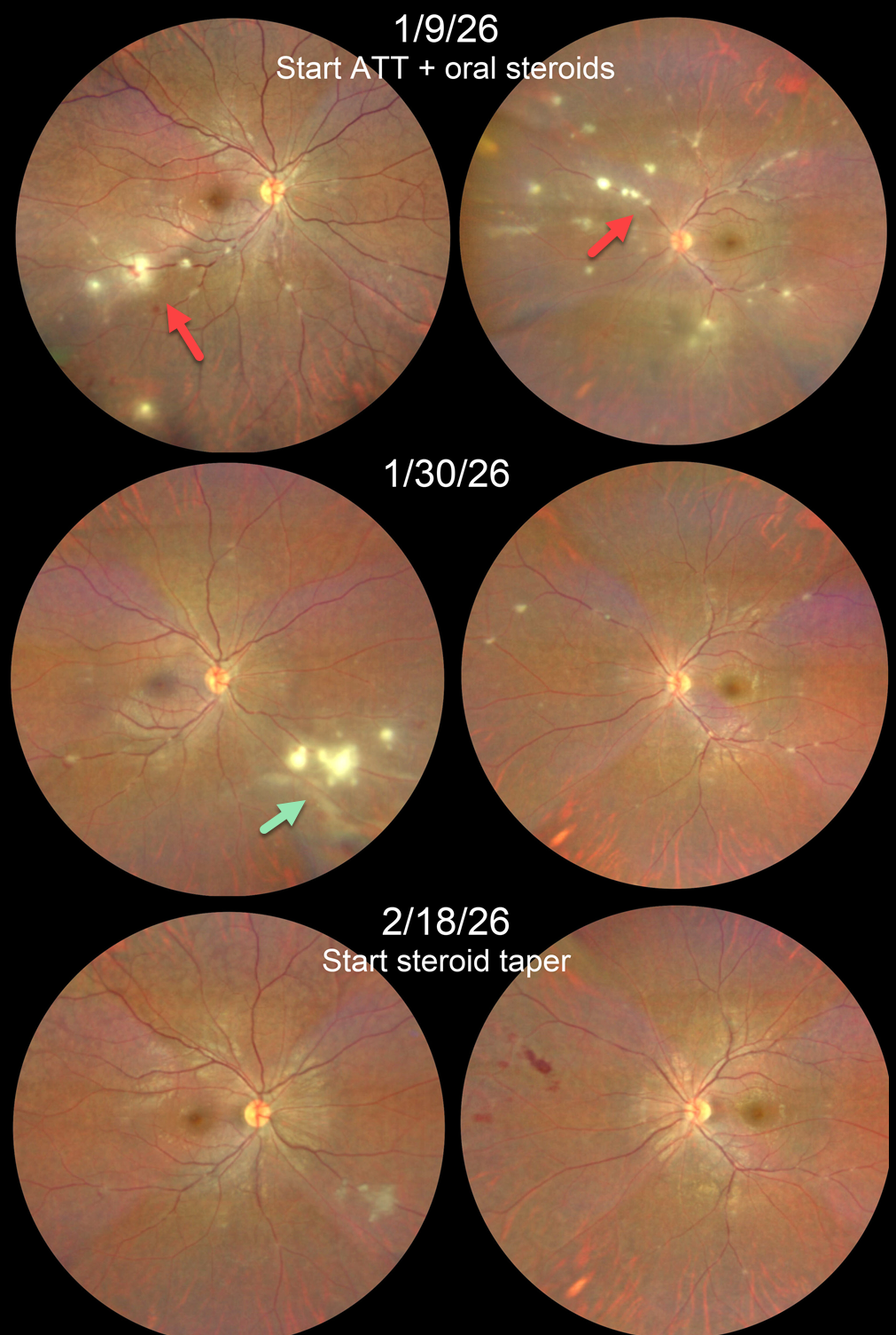
This 22YO male presented with a 1-week history of blurred vision in his left eye. His sister had been undergoing treatment for pulmonary tuberculosis 1 year ago. She also had a history of Takayasu arteritis requiring renal artery embolization and was on multiple immunosuppressants. Vision was 20/20 OD and 20/25 OS.
Color photography shows multifocal areas of retinitis/phlebitis bilaterally. Chest CT revealed paratracheal and subcarinal lymph node calcification, and Mantoux skin testing was strongly positive. Anti-tubercular therapy (ATT) and oral steroids were begun.
He returned 3 weeks later with 20/20 OU. Both eyes were markedly improved except for a new area of active inflammation inferonasally OD. Two weeks later, the retinitis/phlebitis was inactive bilaterally, although some new retinal hemorrhages were noted in the left nasal midperiphery. He was then asked to taper his steroids.
Learning Points:
Although TB most commonly presents with pulmonary involvement, extrapulmonary sites can include the gastrointestinal, skin, cardiovascular, genitourinary, and central nervous systems, as well as the eyes. Ocular involvement, like syphilis, can mimic virtually any type of uveitis, including anterior, intermediate, posterior, and panuveitis, retinitis and retinal vasculitis, neuroretinitis, optic neuropathy, choroidal granuloma, choroiditis, and scleritis.
Tubercular retinitis and vasculitis are prevalent in endemic countries such as India. Laboratory investigations should validate/rule out, not determine, the diagnosis. It is important to closely follow these patients after starting treatment. Despite initial improvement in our patient’s retinitis and vasculitis, a new area developed that needed an additional 2 weeks of therapy before we could start steroid tapering.
TOXOPLASMOSIS
Shraddha Raj Shrivastava, Akansha Sharma and Manish Nagpal
Originally posted on @retina.rocks March 31, 2026
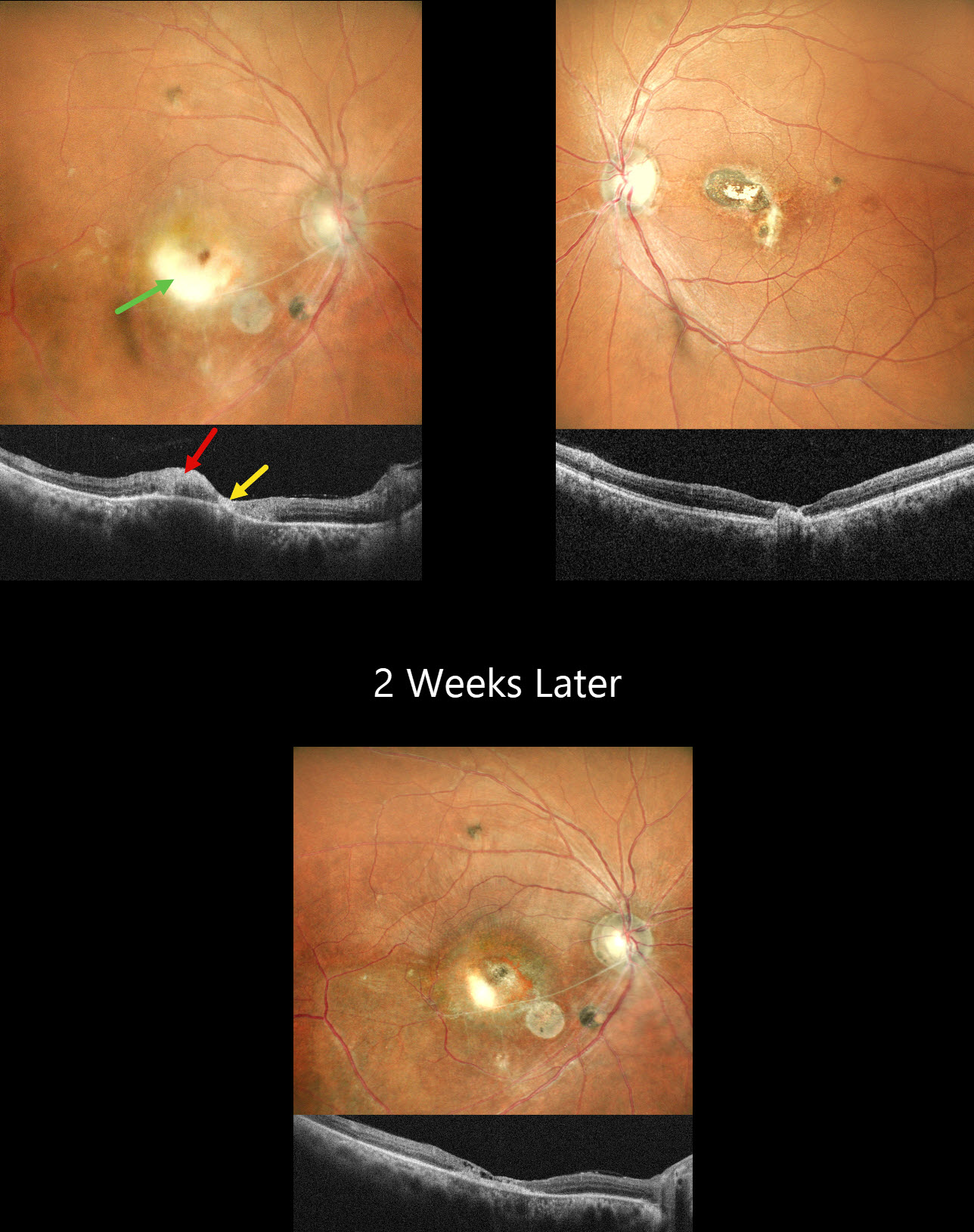
This healthy 45YO male presented with a 3-week history of decreased vision in his right eye. He reported a similar episode in the left eye 10 years earlier, with poor vision since. Vision was 20/120 OD and 20/200 OS.
Pseudocolor SLO imaging OD shows active white retinitis adjacent to inactive nasal scarring. The vessels appear sclerosed in the inferonasal macula. OCT shows hyperreflectivity within the area of active retinitis with atrophy within the nasal scarring. The left eye has inactive nasal macular scarring. He was diagnosed with recurrent toxoplasmosis and started on a 6-week course of oral Bactrim DS. Oral steroids were also started 3 days later. When he returned 2 weeks later, he was unchanged at 20/120 with resolution of the active retinitis.
Learning Points:
Toxoplasmosis, the most common cause of infectious chorioretinitis in humans, is caused by the protozoan parasite, Toxoplasma gondii. It is usually transmitted through infected felines or by eating raw meat. Most infections are acquired, although it can also be transmitted congenitally. The body’s immune system is unable to completely kill the organism, which often lies dormant within a chorioretinal scar for years. When the immune balance favors the encysted organism, active chorioretinitis develops. Focal retinitis usually develops adjacent to a pigmented retinochoroidal scar. Overlying retinal arterial vasculitis is not uncommon.
The inflammation usually resolves spontaneously within 6 weeks. If the optic nerve or macula are threatened, as in our patient, a six-week course of Bactrim DS is recommended (Soheilian et al, Ophthalmology 2004;112:1876-1882). Oral prednisone can be added as well to help with severe inflammation.
TUBERCULAR SERPIGINOUS-LIKE CHOROIDITIS
Akansha Sharma
Originally posted on @retina.rocks March 9, 2026
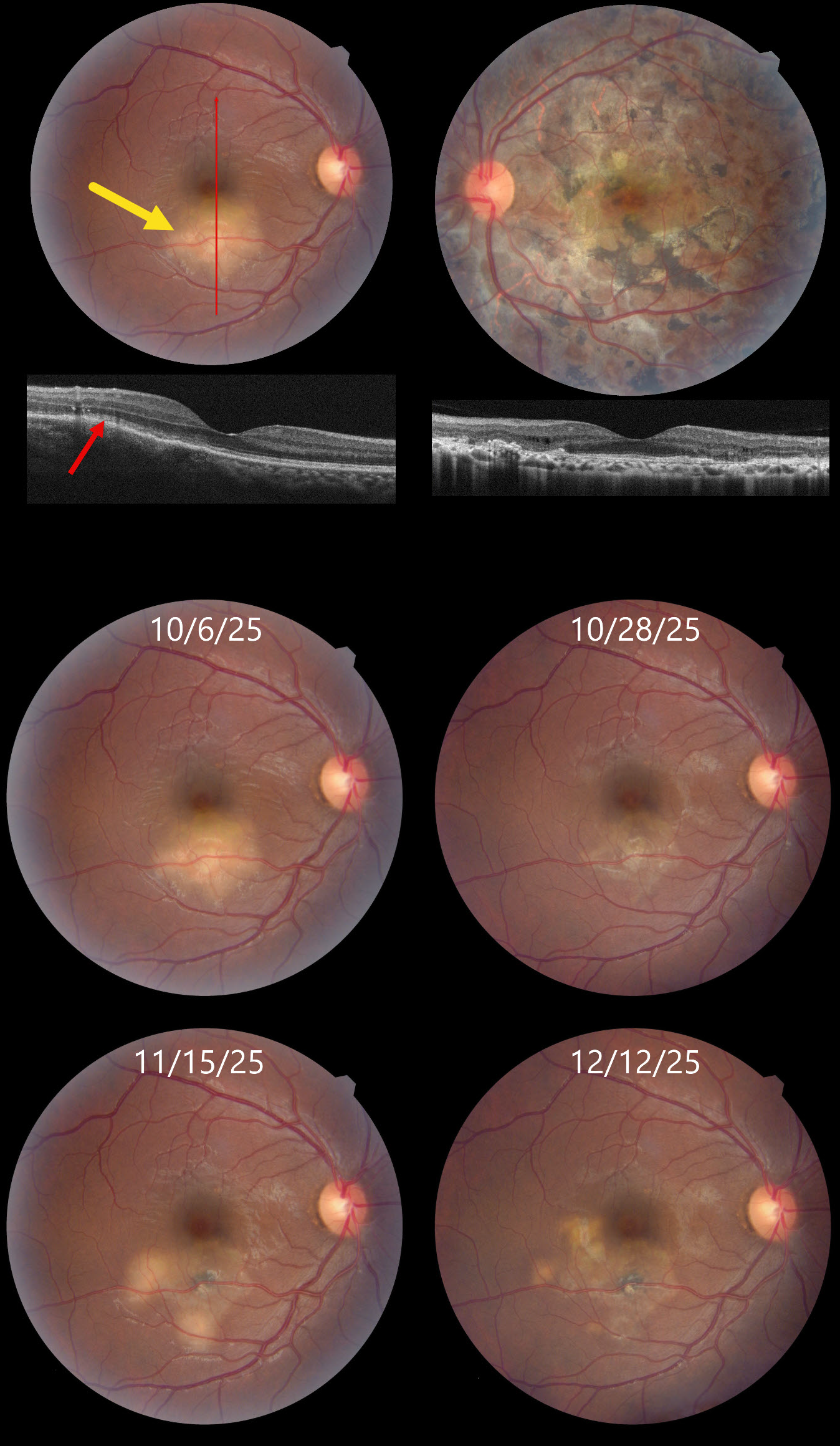
This 32YO male presented on 10/6/25 with a 6-day history of decreased vision in his right eye. About 2 years earlier, he was diagnosed elsewhere with tubercular serpiginous-like choroiditis in his left eye. Extensive workup at the time, including a chest X-ray, was negative except for a positive QuantiFERON TB Gold result. He was lost to follow-up without treatment for his latent tuberculosis (TB). Vision in our office was 20/30 OU.
Color photography shows a deep, creamy-white retinal inflammatory lesion in the inferior macula. OCT shows outer retinal hyperreflectivity with loss of the outer retinal band details. Extensive scarring is noted throughout the left posterior pole, with a small island of preserved foveal tissue.
Tapering systemic corticosteroid therapy was begun, followed by anti-tuberculosis treatment. Over the ensuing 2 months, he experienced waxing and waning inflammation necessitating intravitreal Ozurdex, oral steroids, and adalimumab while vision remained stable at 20/30. We continue to follow him closely.
Learning Points:
Although TB most commonly presents with pulmonary involvement, extrapulmonary sites can include the gastrointestinal, skin, cardiovascular, genitourinary, and central nervous systems, as well as the eyes. Ocular involvement, like syphilis, can mimic virtually any type of uveitis, including anterior, intermediate, posterior, and panuveitis, retinitis, and retinal vasculitis, neuroretinitis, optic neuropathy, choroidal granuloma, choroiditis, and scleritis.
Tubercular serpiginous-like choroiditis mimics serpiginous but affects younger patients with more multifocal and peripheral recurrences and progression if untreated (Agarwal et al, AJO 2020;220:160-169). Although the exact mechanism is not yet clear, a direct or indirect infectious trigger by the Mycobacterium tuberculosis is thought to cause choroiditis. Its origin is probably immunogenic since it seems to respond to corticosteroids and other immunosuppressants. Our patient was a bit unusual in that he required systemic steroids, ATT, and adalimumab to finally control his disease.
NEURORETINITIS
Anand Temkar, Surendra Pal and Prasanna Suresh
Originally posted on @retina.rocks February 25, 2026
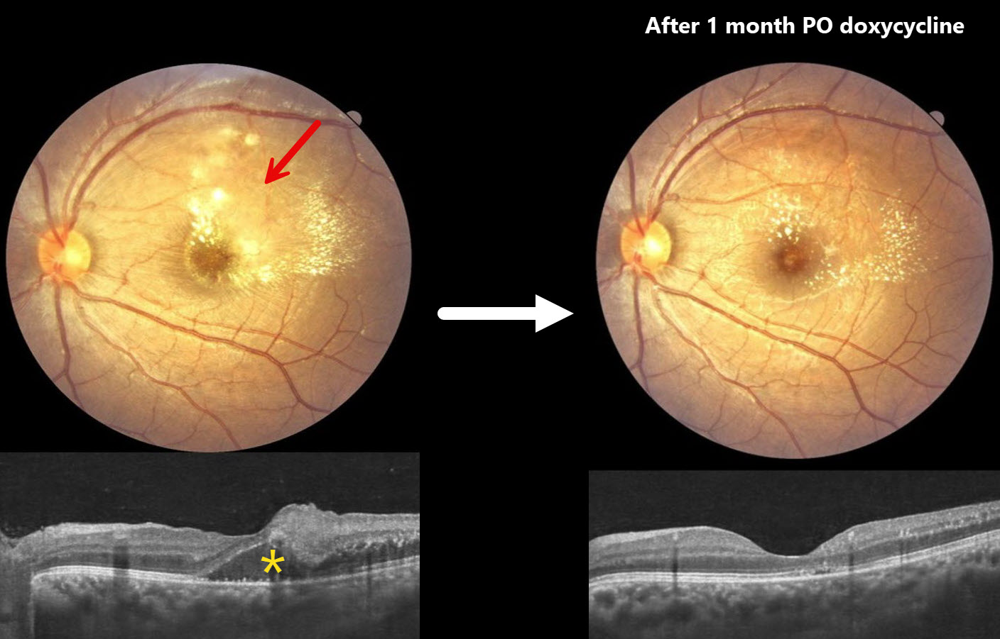
This 21YO male presented with one month of intermittent headaches and vision loss in his left eye. Vision was 20/20 in his normal OD and 20/60 OS.
Color photography of the macula shows patches of yellow-white inner retinal opacification with superior and temporal lipid. OCT shows outer nuclear layer fluid with hyperreflective dots of lipid in the outer plexiform layer. Mild vitreous cells are present.
A presumed clinical diagnosis of Bartonella neuroretinitis was made, although serology could not be performed due to cost concerns. He was started on oral doxycycline 100mg PO BID and oral corticosteroids. Two weeks later, vision was 20/40 with resolving retinal opacification and fluid (not shown). Following a full month of oral therapy, vision improved to 20/20 with decreasing lipid and no fluid.
Learning Points:
The diagnosis for a unilateral swollen nerve is extensive. However, it becomes much smaller when associated with acute inner retinal ischemia and later lipid exudation within Henle’s layer (neuroretinitis), including cat scratch (Bartonella) and syphilis. When no underlying cause is found, the entity is called Leber’s idiopathic stellate neuroretinitis.
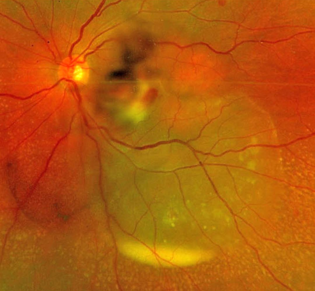
SEPTIC RETINAL EMBOLUS WITH SUBRETINAL HYPOPYON
The European VitreoRetinal Society (EVRS) and Mohit Dogra
Originally posted on @retina.rocks February 6, 2026
This previously healthy 37YO female presented to us for a second opinion regarding 4 days of decreased vision in her left eye. There was a recent history of a urinary tract infection secondary to E. coli. Vision was 20/20 in her normal OD and 20/400 OS. There was a moderate non-granulomatous uveitis with vitreous cells.
Optos color RG imaging shows opaque white retina in the inferonasal macula with overlying retinal and vitreous blood. An exudative retinal detachment extends inferotemporally with an inferior subretinal hypopyon.
She was referred back to the initially treating ophthalmologist and was subsequently lost to follow-up.
Learning Points:
Posterior segment chorioretinitis from septic embolization is a rare event, most commonly found in patients with bacterial endocarditis. Our patient presented with a constellation of findings, including retinitis, retinal and vitreous hemorrhage, and an exudative retinal detachment with an inferior subretinal hypopyon.
Subretinal hypopyon is characterized by yellowish inflammatory material settling inferiorly in the subretinal space. It has been documented in infectious (bacterial and fungal endophthalmitis, acute retinal necrosis, tuberculosis, syphilis), inflammatory (sympathetic ophthalmia), and neoplastic (leukemia, lymphoma) disorders.
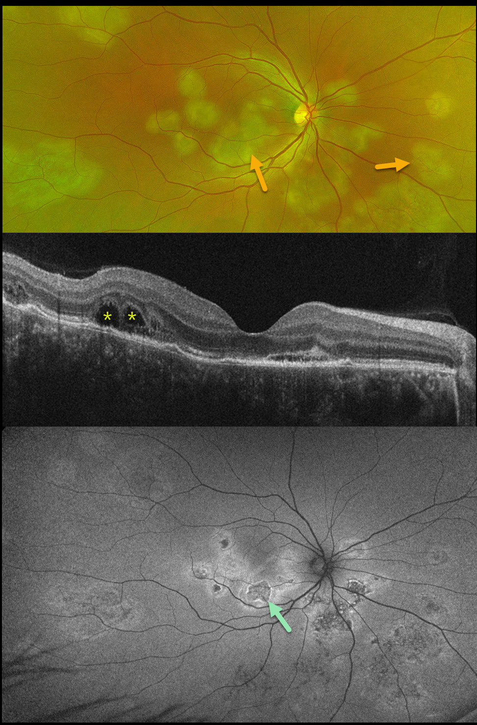
TUBERCULAR SERPIGINOUS-LIKE CHOROIDITIS
The European VitreoRetinal Society (EVRS) and Vaibhav Sethi
Originally posted on @retina.rocks January 2, 2026
This previously healthy 41YO male presented with 4 days of blurred vision in his right eye. Vision was 20/80 OD and 20/20 in his normal OS.
Optos color RG imaging shows multifocal subretinal creamy white placoid lesions in the macula, peripapillary retina, and midperiphery. OCT scanning shows variable disruption of the outer retinal bands, as well as bacillary layer detachments (BALAD) temporally. The placoid lesions on fundus autofluorescence (FAF) show variable central hypo-FAF with more hyper-FAF borders.
Mantoux skin testing was positive at 24mm, and chest CT revealed left lower lobe ground glass opacities with hilar adenopathy. He was started on anti-tubercular therapy and oral prednisone.
Although tuberculosis most commonly presents with pulmonary involvement, extrapulmonary sites can include the gastrointestinal, skin, cardiovascular, genitourinary, and central nervous systems, as well as the eyes. Ocular involvement, like syphilis, can mimic virtually any type of uveitis, including anterior, intermediate, posterior, and panuveitis, retinitis, and retinal vasculitis, neuroretinitis, optic neuropathy, choroidal granuloma, choroiditis, and scleritis.
Learning Points:
Tubercular serpiginous-like choroiditis mimics serpiginous but affects younger patients with more multifocal and peripheral recurrences and progression if untreated (Agarwal et al, AJO 2020;220:160-169). Although the exact mechanism is not yet clear, a direct or indirect infectious trigger by the Mycobacterium tuberculosis is thought to cause choroiditis. Its origin is probably immunogenic since it seems to respond to corticosteroids and other immunosuppressants.
BALAD appears as a unique dome-shaped collection of intraretinal fluid from photoreceptor splitting. These detachments can be observed in an increasing number of conditions, most classically Vogt-Koyanagi-Harada disease (Cicinelli et al, Ophthalmology Retina 2020;4:454-456).
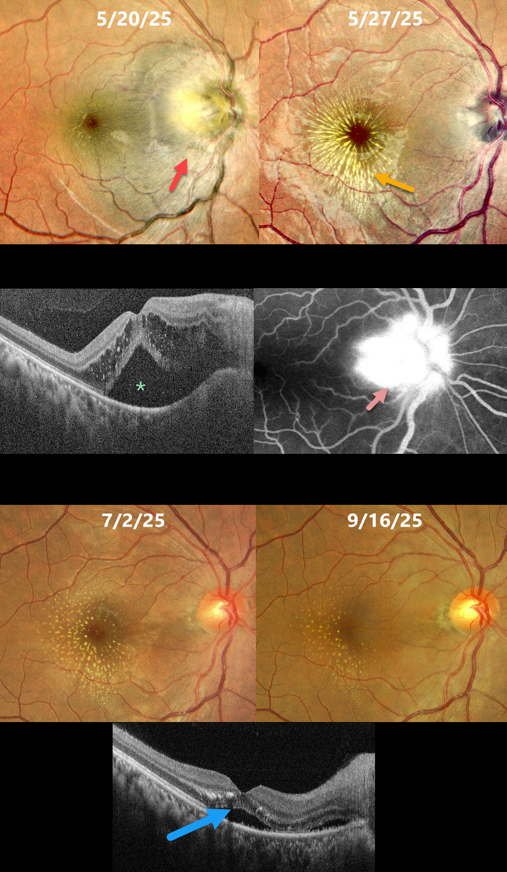
LEBER’S IDIOPATHIC STELLATE NEURORETINITIS
Rohan Jain and Manish Nagpal
Originally posted on @retina.rocks December 29, 2025
This healthy 32YO female presented with a 10-day history of headaches, followed by vomiting and blurred vision in her right eye. She also reported a rash on her hands and legs that appeared before her visual symptoms. Vision was 20/300 OD and 20/20 in her normal OS.
Pseudocolor SLO imaging shows a swollen optic nerve, a serous detachment extending through the fovea, and some mostly temporal radiating foveal lipid. OCT scanning shows outer nuclear layer fluid with hyperreflective lipid deposits and subretinal fluid. Fundus fluorescein angiography shows marked disc leakage. Infectious disease consultation revealed a negative infectious workup, and she was started on intravenous followed by a tapering dose of oral steroids.
One week later, vision was 20/300 with a striking new lipid star from lipid in the outer plexiform layer. There is a marked reduction in the intra- and subretinal fluid. Her findings continued to improve, and 4 months following presentation, vision was 20/20 with faint residual lipid.
Learning Points:
The diagnosis for a unilateral swollen nerve is extensive. However, it becomes much smaller when associated with acute inner retinal ischemia (not seen in our case) and later lipid exudation within Henle’s layer (neuroretinitis), including cat scratch (Bartonella) and syphilis. When no underlying cause is found, as in our patient, the entity is called Leber’s idiopathic stellate neuroretinitis.
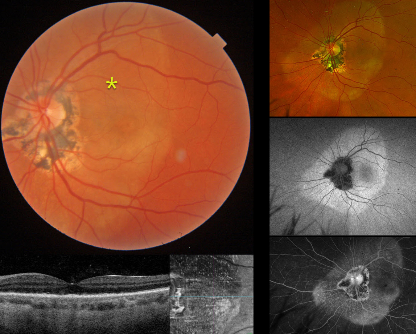
SYPHILIS
Originally posted on @retina.rocks December 9, 2025
This 61YO female presented with 1 week of vision loss in her left eye. Vision was 20/25 in her normal OD and counting fingers OS.
Triton color imaging shows a subretinal yellow-white placoid lesion extending beyond the macula, along with ocular histoplasmosis-related peripapillary scarring. Swept-source OCT shows replacement of the outer retinal bands with hyperreflective material, which is seen as innumerable hyperreflective dots on en face imaging. Optos color RGB imaging shows the full extent of the placoid lesion. Fundus autofluorescence (FAF) of this lesion shows homogenous hyper-FAF. It diffusely stains on fluorescein angiography. Laboratory testing was positive for syphilis and negative for HIV. She was referred to an infectious disease specialist and placed on oral doxycycline 100mg BID due to a penicillin allergy. When last seen two and a half months after her initial presentation, and after completing a full 1-month course of antibiotics, vision improved to 20/40 with near-complete resolution of the retinal findings (not shown).
Learning Points:
Ocular manifestations of syphilis vary widely and include virtually all variations of anterior and posterior segment inflammation. Originally described by Gass (Ophthalmology 1990;97:1288-1297), placoid chorioretinitis is one of the more common presentations. Inflammation is localized to the outer retina and RPE and appears on OCT as disruption of the outer retinal bands and hyperreflective pyramidal lesions as seen in our patient (Hu et al, Ophthalmology Retina 2022;6:172-178). The outer retinal damage unmasks the underlying RPE, accounting for the hyperfluorescent placoid lesions.
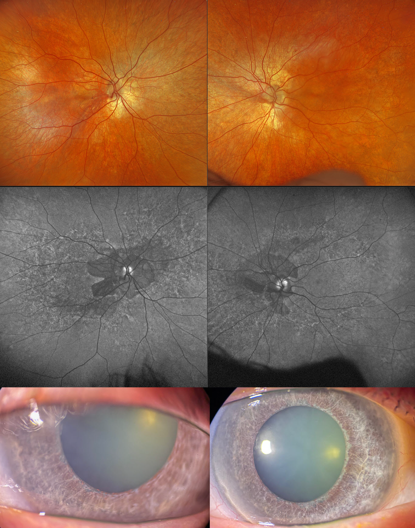
CONGENITAL RUBELLA
Fraser McKay
Originally posted on @retina.rocks December 3, 2025
This 59YO male with type 2 diabetes was examined for his yearly screening. He has a known history of congenital rubella with deafness. Vision was 20/30 OD and 20/25 OS.
Optos color RGB imaging shows a diffuse salt-and-pepper appearance in each fundus, with peripapillary atrophy. On fundus autofluorescence (FAF), the peripapillary atrophy is hypo-FAF with a speckled hyper- and hypo-FAF appearance to each posterior pole. Slit lamp photographs show diffuse iris atrophy with a relative lack of pigmentation. Triton swept-source OCT shows variable outer retinal thinning with ellipsoid zone disruption.
Learning Points:
The classic ocular findings in congenital rubella include cataracts, pigmentary retinopathy (“salt-and-pepper” fundus), congenital glaucoma, and microphthalmia. Nuclear cataracts are the most common ocular abnormality, although our patient had just trace nuclear sclerosis. Pigmentary retinopathy is characterized by diffuse mottling of the retinal pigment epithelium, which is best visualized with FAF. Systemic associations include sensorineural hearing loss and congenital heart defects.
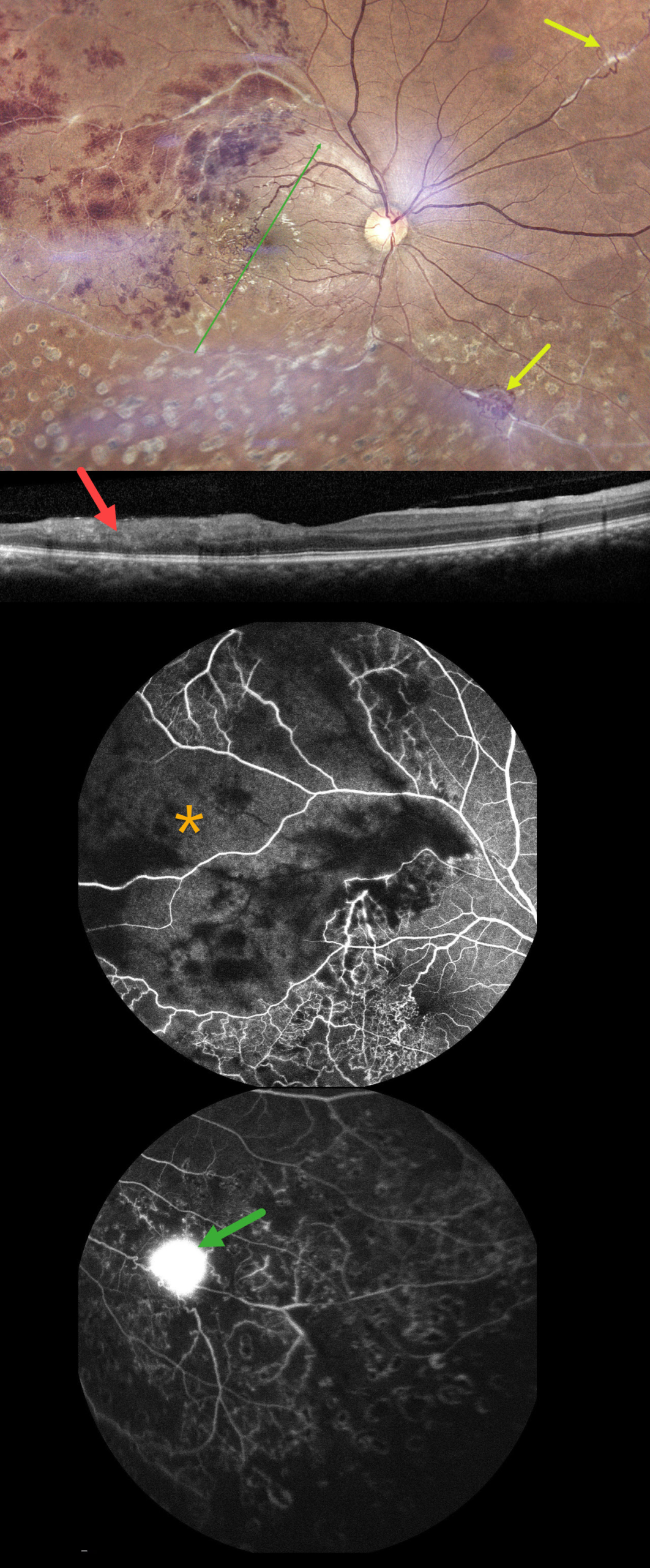
TUBERCULAR ISCHEMIC VASCULITIS
The European VitreoRetina Society (EVRS) and Abhishek Upadhyaya
Originally posted on @retina.rocks November 28, 2025
This 40YO male presented with 10 days of vision loss in his right eye. There was a history of prior laser treatment to this eye elsewhere. Vision was 20/40 OD and 20/20 in his normal OS.
Fundus photography shows superotemporal retinal hemorrhages consistent with a branch retinal vein occlusion (BRVO), with a possible additional BRVO inferotemporally. Collateral vessels are noted in the temporal fovea with scatter laser scarring in the inferior hemiretina. Skip vasculitis can be seen in the superotemporal quadrant along with venous sheathing in other areas as well. There are a few areas of fibrosed peripheral neovascularization (NV). OCT scanning shows variable temporal macular thinning with disorganization of retinal inner layers (DRIL). Fluorescein angiography shows profound ischemia within the superotemporal BRVO, along with vascular leakage and peripheral ischemia. Leakage from the Inferonasal NVE can be seen in the late stages.
Systemic workup was consistent with Ocular Tuberculosis (TB), including a positive Mantoux skin test (12mm), positive Quantiferon TB testing, and calcified lung nodules on contrast-enhanced computed tomography. He was started on oral steroids and antitubercular therapy, and scatter laser was planned for the areas of angiographic ischemia.
Learning Points:
The differential for occlusive peripheral retinal vasculitis (Huvard et al, Ophthalmology Retina 2022;6:43-48) includes TB, rheumatologic disorders/systemic vasculitides (granulomatosis with polyangiitis, Bechet’s disease, systemic lupus, etc.), idiopathic retinal vasculitis and neuroretinitis, and sarcoidosis. Historically, Eales disease has been used to describe a variety of heterogeneous disorders, including tuberculous vasculitis, and, due to its ambiguity, we prefer not to use this term.
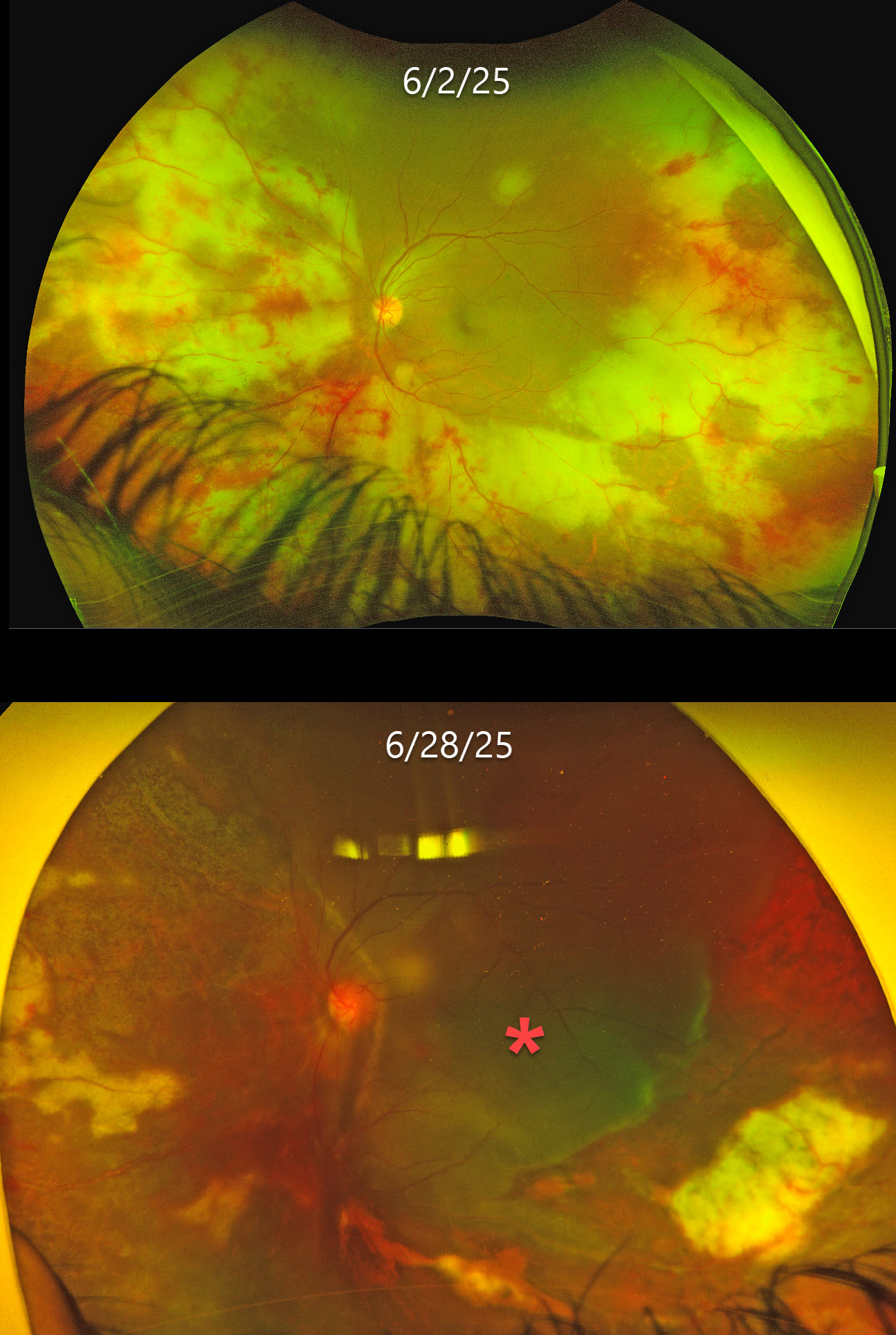
ACUTE RETINAL NECROSIS SYNDROME
César Adrián Gómez Valdivia
Originally posted on @retina.rocks November 26, 2025
This previously healthy 35YO male presented with 1 week of floaters in his left eye. Vision was 20/20 in his normal OD and 20/20 OS. Anterior segments were normal.
Optos color RG imaging shows variably confluent outer retinal opacification with retinal hemorrhages extending from the periphery into the posterior pole. The retinal vessels are variably narrowed. He was initially diagnosed with possible cytomegalovirus (CMV) retinitis, started on valganciclovir, and referred to infectious diseases (ID).
He returned 3 weeks later with counting fingers vision despite improved retinitis and stopping systemic treatment after 10 days due to financial limitations. He returned 5 days later with sudden loss of vision. Vision was light perception with improved retinitis but with a new macula-off retinal detachment, which was repaired with lensectomy, vitrectomy, scleral buckle, retinectomy, and silicone oil. Two months later, vision was 20/150, and the retina remained completely attached.
Learning Points:
Acute retinal necrosis syndrome (ARNS) is usually caused by the herpes zoster virus. It generally occurs in immunocompetent individuals and presents as rapidly progressive panuveitis with hemorrhagic and ischemic unilateral (ARNS) or bilateral (BARNS) retinal necrosis, typically beginning in the retinal periphery.
Our patient was eventually evaluated by ID, who diagnosed him with HIV and a CD4 count of 45 with initiation of HARRT therapy. It is unclear if our patient’s underlying previously undiagnosed HIV infection was contributory to his retinitis. The lack of the usual anterior chamber and vitreous inflammation is likely due to his immunosuppression.
Both CMV retinitis and ARNS cause retinal necrosis and hemorrhages. However, CMV usually starts in the posterior pole, often beginning in a paravascular location, which then spreads outwards. The retinitis involves the full-thickness retina, so the hemorrhages and opaque retina also involve the inner retina and obscure the underlying larger retinal vessels. Our patient’s findings are more pathognomonic for ARNS, given the outer retinal findings that likely began in the retinal periphery and extended posteriorly, with no paravascular location.
ARNS is often a devastating and blinding infection due to the high risk for retinal detachment as well as macular and optic nerve involvement. The latest meta-analysis argues for systemic antiviral therapy and prophylactic vitrectomy, with uncertain benefits for prophylactic laser photocoagulation or adjunctive intravitreal antivirals (Zhao et al, Retina 2021;41:965-978).
Treatment should be initiated as soon as the diagnosis is suspected, without waiting for laboratory results. Oral valacyclovir is clinically equivalent to intravenous acyclovir (Baltinas et al, AJO 2018;188:173-180), and aspirin is often recommended to minimize vascular thrombosis. Although valganciclovir is not the treatment of choice, our patient’s retinitis still rapidly improved despite only 10 days of therapy. Treatment is continued until all retinitis becomes inactive, and usually for at least 3-6 months thereafter to reduce the risk of fellow-eye involvement.
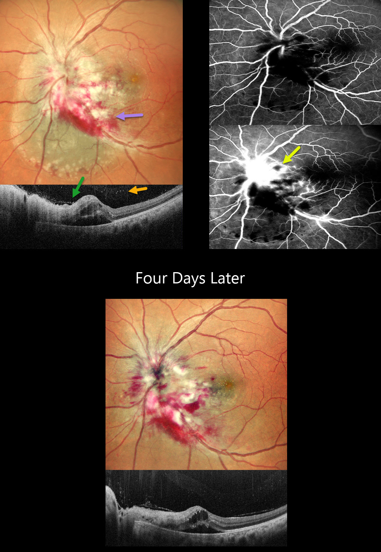
CMV PAPILLITIS AND RETINITIS
Akansha Sharma and Manish Nagpal
Originally posted on @retina.rocks September 18, 2025
This 42YO female with a known history of HIV infection presented with 1 month of vision loss in her left eye. Her last CD4 count was 38 despite being compliant with HAART therapy. Her husband was HIV positive. Vision was 20/20 in her normal OD and 20/400 in OS.
Multicolor SLO imaging shows active cytomegalovirus (CMV) retinitis involving the nerve, which extends nasally and inferotemporally. Subretinal fluid is noted clinically and on OCT. OCT also shows full-thickness disorganization and hyperreflectivity, along with overlying vitreous cells. Fluorescein angiography shows early blockage with late disc and retinal leakage.
Oral valganciclovir and biweekly intravitreal injections were started. Four days later, vision improved to 20/90 with improvement in the papilledema and retinitis.
Learning Points:
CMV retinitis develops as a reactivation of latent CMV in immunosuppressed individuals. Before effective antiretroviral treatment emerged in the mid to late 1990’s, CMV retinitis developed in up to 40% of HIV/AIDS patients, often within the last 6 months of life. Treatment includes a combination of systemic and intravitreal medications, including ganciclovir, foscarnet, and cidofovir. Rhegmatogenous retinal detachment is a common late sequela and requires vitrectomy with silicone oil.
POST-FEVER RETINITIS
Tejaswita Verma,Manish Nagpal and Anjana Mirajkar
Originally posted on @retina.rocks September 9, 2025
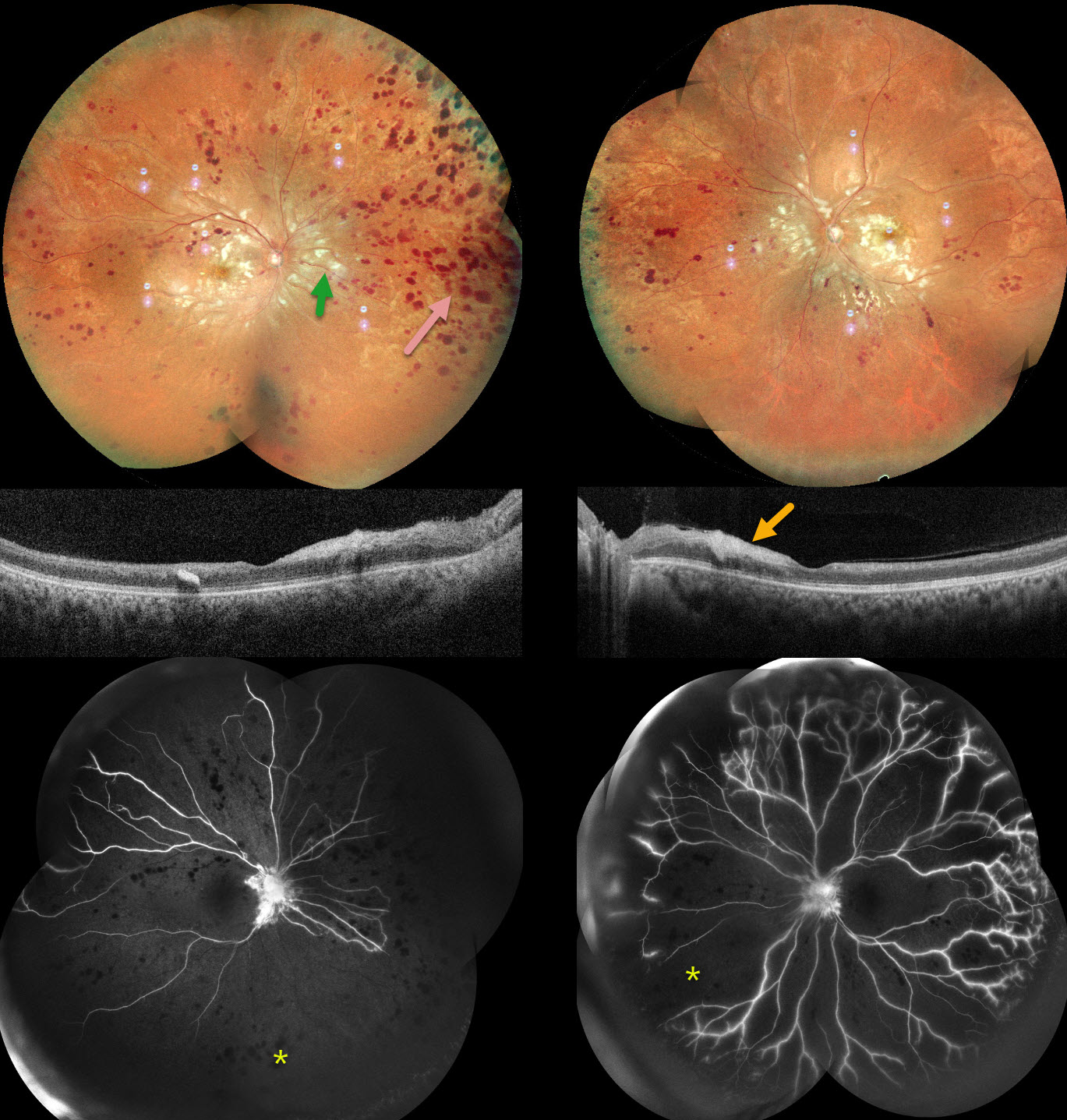
This previously healthy 32YO male presented with a 3-month history of bilateral vision loss following a viral illness. He was treated elsewhere for post-fever retinitis with intravitreal triamcinolone and oral steroids with antivirals. He also had a right lobe pneumonia. Testing was positive for CMV IgG and IgM and negative for TB.
When examined in our office, vision was 20/200 OU. Anterior segments were normal, with mild bilateral vitreous cells.
Pseudocolor SLO imaging shows extensive cotton-wool spots throughout each posterior pole, with more distal retinal hemorrhages that extend into the peripheries. OCT scanning shows possible vitreous cells with variable inner retinal thickened/thinned hyperreflectivity. Fluorescein angiography shows profound bilateral ischemia and disc leakage. We advised bilateral panretinal photocoagulation, but he was unfortunately immediately lost to follow-up.
Learning Points:
Epidemic (post-fever) retinitis is an infectious or para-infectious entity that develops in immunocompetent individuals several weeks following a bacterial, viral, or protozoal infection. Causes include Dengue, Rickettsiosis, West Nile, Leptospirosis, Chikungunya, Typhoid, and Ebola (none of which were tested in our patient).
Patients present with sudden, painless vision loss. Fundus findings include unilateral or bilateral unifocal or multifocal patches of retinitis with possible nerve involvement, serous macular detachment, and vasculitis or frosted branch angiitis. There is usually a favorable response to steroids and treating the underlying infectious cause with appropriate systemic therapy. See Mahendradas et al (Indian J Ophthalmol 2020;68:1775-1786) for an excellent post-fever retinitis review.
MULTIFOCAL TUBERCULOSIS CHOROIDAL GRANULOMAS
Will Gibson
Originally posted on @retina.rocks July 3, 2025
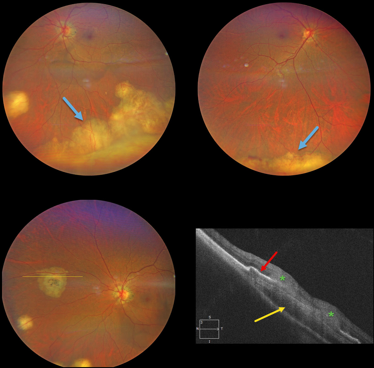
This previously healthy 49YO Indian female was visiting the United States and presented with new floaters in her left eye. Vision was 20/25 OD and 20/40 OS. There was no anterior uveitis, and the vitreous was clear bilaterally.
Color photography shows multifocal bilateral amelanotic choroidal lesions, some isolated and others confluent in the inferior periphery. OCT scanning through a nasal lesion OS shows a relatively hyporeflective choroidal lesion, irregular overlying RPE elevation, and probable infiltration of the overlying retina.
QuantiFERON-TB testing was positive with a negative chest X-ray. The patient was referred to the infectious disease department and started on anti-tuberculosis treatment (ATT).
Learning Points:
Although TB most commonly presents with pulmonary involvement, extrapulmonary sites can include the gastrointestinal, skin, cardiovascular, genitourinary, and central nervous systems, as well as the eyes. Ocular involvement, like syphilis, can mimic virtually any type of uveitis, including anterior, intermediate, posterior, and panuveitis, retinitis and retinal vasculitis, neuroretinitis, optic neuropathy, choroidal granuloma (our patient), choroiditis, and scleritis. For an excellent review of intraocular tuberculosis, see Bupta et al, Survey Ophthalmology 2007;52:561-587.
Tubercular granulomas can be distinguished from sarcoid lesions by being solitary, yellow, larger, and vascularized (Agarwal et al, AJO 2021;226:42-55). Our patient is therefore somewhat unusual with bilateral multifocal lesions. Sarcoid workup was negative.
ACUTE RETINAL NECROSIS SYNDROME
Ayushi Gupta and Vishal Agrawal
Originally posted on @retina.rocks June 26, 2025
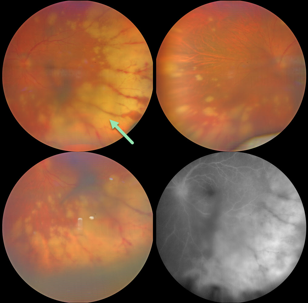
This 56YO female presented with 2 weeks of decreased vision in her left eye. She was treated for herpes zoster one month earlier, and there were a few crusted lesions on the right side of the forehead. Vision was 20/20 in her normal OD and 20/200 OS. There were moderate anterior chamber and vitreous cells.
Color photography shows scattered, midperipheral, variably confluent patches of creamy white deep retinitis. Fluorescein angiography shows optic nerve and deep retinal leakage.
An anterior chamber tap was performed, and PCR analysis confirmed varicella zoster. Intravenous acyclovir was given for 1 week, followed by oral valacyclovir 1 gram daily. When examined 3 weeks later, vision improved to 20/40 with resolving retinitis (not shown).
Learning Points:
The acute retinal necrosis syndrome is usually caused by the herpes zoster virus. It generally occurs in immunocompetent individuals and presents as a rapidly progressive panuveitis with hemorrhagic and ischemic unilateral (ARNS) or bilateral (BARNS) retinal necrosis, typically beginning in the retinal periphery.
It is often a devastating and blinding infection due to the high risk for retinal detachment as well as macular and optic nerve involvement. The latest meta-analysis argues for systemic antiviral therapy and prophylactic vitrectomy, with uncertain benefits for prophylactic laser photocoagulation or adjunctive intravitreal antivirals (Zhao et al, Retina 2021;41:965-978).
Treatment should be initiated as soon as the diagnosis is suspected, without waiting for laboratory results. Oral valacyclovir is clinically equivalent to intravenous acyclovir (Baltinas et al, AJO 2018;188:173-180), and aspirin is often recommended to minimize vascular thrombosis. Treatment is continued until all retinitis becomes inactive, and usually for at least 3-6 months thereafter to reduce the risk of fellow-eye involvement.
SYPHILIS
César Adrián Gómez Valdivia
Originally posted on @retina.rocks June 5, 2025
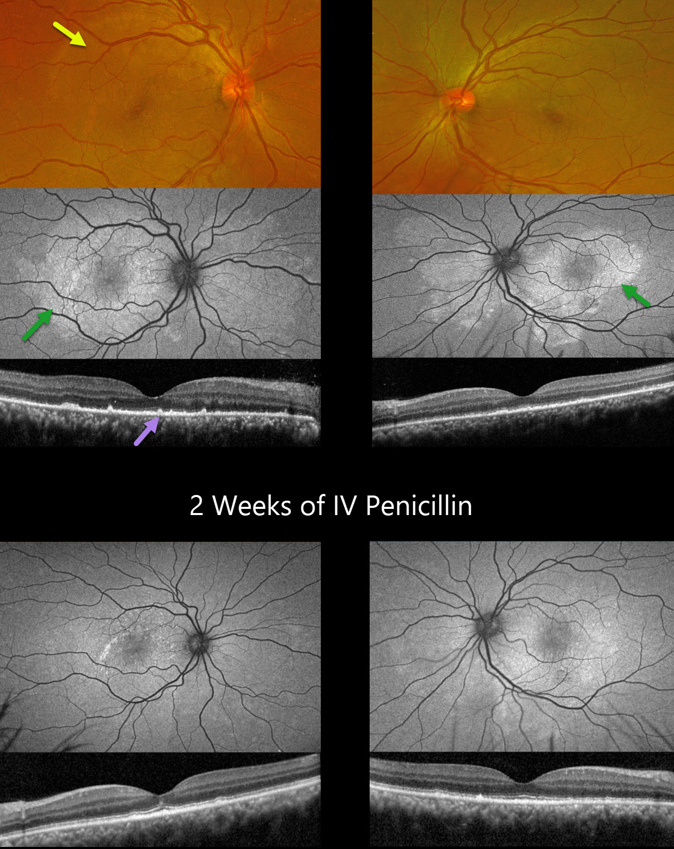
This 39YO HIV positive male presented with 5 days of bilateral decreased vision. Vision was 20/100 OD and 20/200 OS.
Optos color RG imaging shows bilateral whitish macular and peripapillary placoid lesions extending nasal to the nerves. Fundus autofluorescence (FAF) more dramatically shows these hyper-FAF placoid lesions, which resemble a butterfly in his left eye. OCT scanning shows variable loss of the outer retinal bands associated with hyperreflective deposits above the RPE.
VDRL was positive, and he was referred to his social security unit for systemic treatment. Seven weeks later, following two weeks of parenteral penicillin, vision improved to 20/20 OD and 20/20-2 OS, with improved funduscopic and OCT findings.
Learning Points:
Ocular manifestations of syphilis vary widely and include virtually all variations of anterior and posterior segment inflammation. Originally described by Gass (Ophthalmology 1990;97:1288-1297), placoid chorioretinitis is one of the more common presentations.
Inflammation is localized to the outer retina and RPE and appears on OCT as disruption of the outer retinal bands and hyperreflective pyramidal lesions as seen in our patient (Hu et al, Ophthalmology Retina 2022;6:172-178). The outer retinal damage unmasks the underlying RPE, accounting for the hyperfluorescent placoid lesions.
POSSIBLE EPIDEMIC RETINITIS
Sharat Hegde
Originally posted on @retina.rocks June 2, 2025
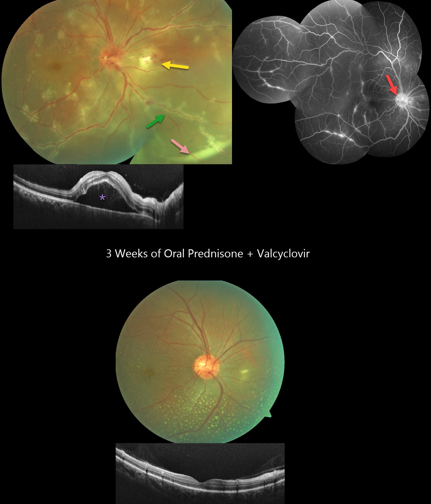
This healthy 36YO male presented with 3 days of floaters and decreased vision in his right eye. Vision was 20/60 OD and 20/20 in his healthy OS. The right eye contained mild anterior chamber cells and moderate vitreous cells.
Fundus photography shows a swollen nerve, a frosted branch pattern of vasculitis involving the retinal veins, an area of retinitis just nasal to the nerve, and a subretinal hypopyon in the inferonasal midperiphery. Some multifocal areas of outer retinal inflammation are noted, along with some retinal hemorrhages. OCT scanning shows variable hyperreflective foveal subretinal fluid. Fluorescein angiography shows variable venous staining and leakage, including from the disc and in nasal retinitis.
The workup included elevated ESR and CRP levels, along with an increased IgG titer for Varicella zoster virus. Mantoux testing, syphilis, Bartonella, typhus, HIV, and chest X-ray were all negative. Although very atypical for viral uveitis, we started oral prednisone and valaciclovir. After one week, the hypopyon resolved, with decreased vasculitis and new scattered flecks of subretinal lipid throughout the posterior pole. By three weeks, vision improved to 20/20 with continued funduscopic improvement.
Learning Points:
Epidemic (post-fever) retinitis is an infectious or para-infectious entity that develops in immunocompetent individuals several weeks following a bacterial, viral, or protozoal infection. Causes include Dengue, Rickettsiosis, West Nile, Leptospirosis, Chikungunya, Typhoid, and Ebola. Patients present with sudden, painless vision loss.
Fundus findings include unilateral or bilateral unifocal or multifocal patches of retinitis with possible nerve involvement, serous macular detachment, and vasculitis or frosted branch angiitis. There is usually a favorable response to steroids and treating the underlying infectious cause with appropriate systemic therapy. See Mahendradas et al (Indian J Ophthalmol 2020;68:1775-1786) for an excellent post-fever retinitis review.
Frosted branch angiitis is a retinal vasculitis that gets its name from the involved vessels resembling branches of a tree that are “frosted” with snow (Kleiner et al, AJO 1988;106:27-34). Although these findings may be isolated, they are often considered a phenotype found in many inflammatory conditions, most commonly CMV retinitis. The inflammation is usually highly responsive to steroids, and the visual prognosis is generally good, although some may develop secondary neovascularization in the anterior and posterior segments due to widespread ischemia.
Subretinal hypopyon is characterized by yellowish inflammatory material settling inferiorly in the subretinal space. It has been documented in infectious (bacterial and fungal endophthalmitis, acute retinal necrosis, tuberculosis, syphilis), inflammatory (sympathetic ophthalmia), and
LEBER’S IDIOPATHIC STELLATE NEURORETINITIS
Originally posted on @retina.rocks April 23, 2025
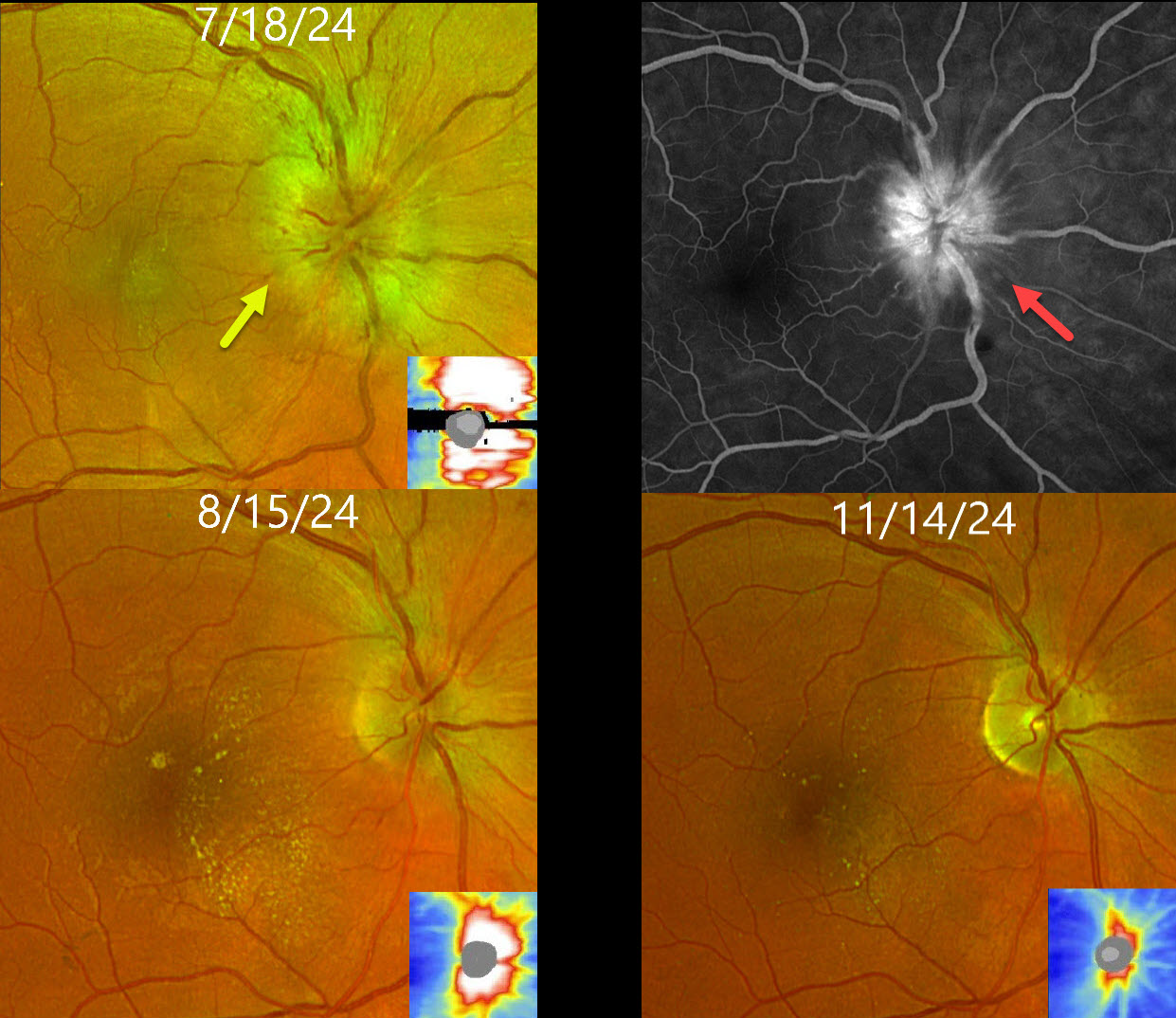
This healthy 35YO male presented with recent severe vision loss in his right eye. Vision was counting fingers OD and 20/20 in his normal OS.
Optos color RG imaging shows moderate optic nerve swelling, which is confirmed by OCT. There is moderate nerve leakage on fluorescein angiography. Bloodwork, including syphilis and Bartonella, was negative, so he was observed without treatment.
One month later, vision improved to 20/200. The disc edema was markedly improved, and new lipid radiated nasally from the macular center. Three months later, the disc edema was fully resolved with secondary atrophy, and the lipid was decreasing. Vision was 20/25.
Learning Points:
The diagnosis for a unilateral swollen nerve is extensive. However, it becomes much smaller when associated with acute inner retinal ischemia (not seen in our case) and, later, with lipid exudation within Henle’s layer (neuroretinitis), including cat-scratch (Bartonella) and syphilis. When no underlying cause is found, as in our patient, the entity is called Leber’s idiopathic stellate neuroretinitis.
SYPHILIS
Fraser McKay
Originally posted on @retina.rocks April 21, 2025
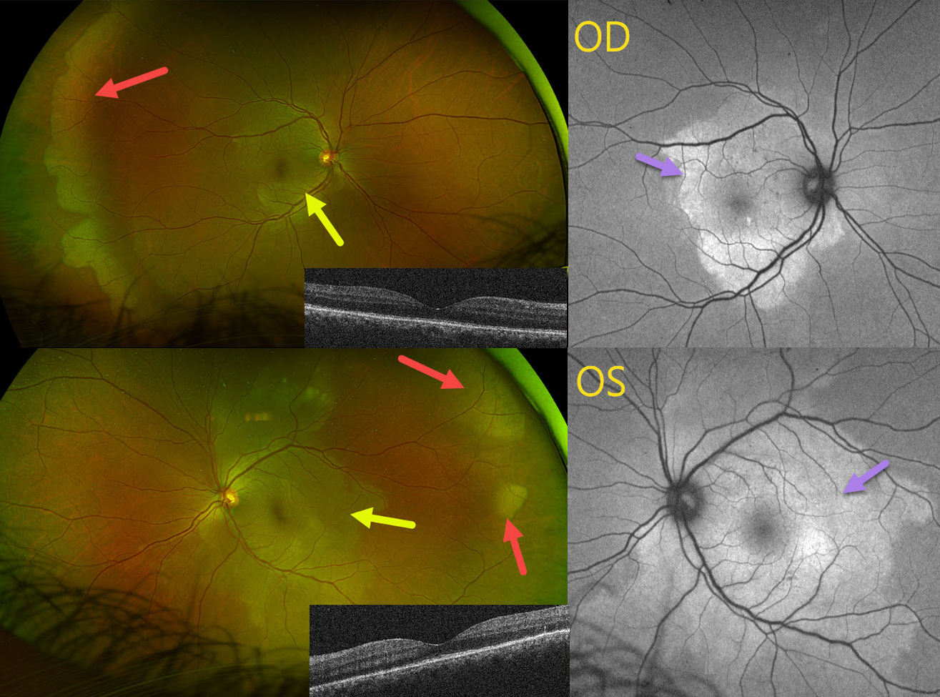
This 42YO female presented with 1 month of bilateral blurred vision. Vision was 20/30 OU.
Optos color RG imaging shows bilateral peripheral and posterior white areas without pressure. Correct??
WRONG! These posterior lesions are hyperautofluorescent. Macular OCT scanning shows variable loss of the outer retinal bands. Syphilis testing was positive, and she was immediately referred to an infectious disease specialist for treatment. When she returned 4 months later following a full course of parenteral penicillin, vision was 20/40 OD, 20/20 OS, with normalization of all fundus findings (not shown).
Learning Points:
Ocular manifestations of syphilis vary widely and include virtually all variations of anterior and posterior segment inflammation. Originally described by Gass (Ophthalmology 1990;97:1288-1297), placoid chorioretinitis is one of the more common presentations. Inflammation is localized to the outer retina and RPE and appears on OCT as disruption of the outer retinal bands and hyperreflective pyramidal lesions as seen in our patient (Hu et al, Ophthalmology Retina 2022;6:172-178). The outer retinal damage unmasks the underlying RPE, accounting for the hyperfluorescent placoid lesions.
TOXOPLASMOSIS
Tejaswita Verma and Manish Nagpal
Originally posted on @retina.rocks March 19, 2025
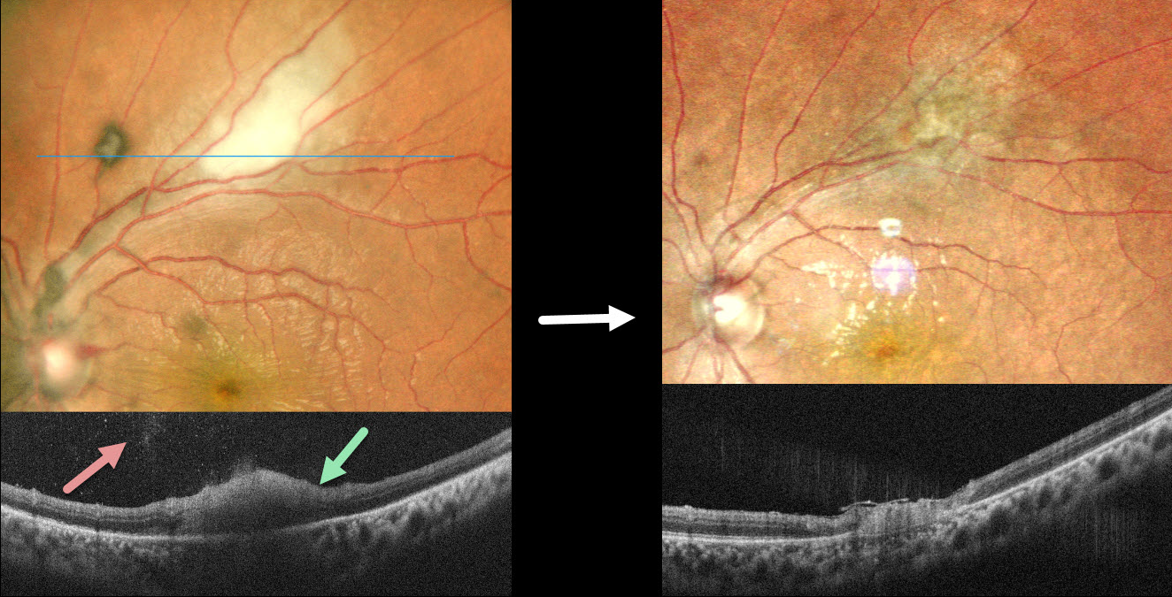
This 26YO male presented with 6 days of blurred vision in his left eye. Vision was 20/20 in his normal OD and 20/40 OS. The left cornea was edematous with a moderate nongranulomatous anterior uveitis and moderate vitreous cells. The intraocular pressure (IOP) was 47 mm HG.
Pseudocolor SLO imaging shows active white retinitis just superior to the macula. OCT scanning shows full-thickness hyperreflective retinal thickening with moderate overlying vitreous cells. He was diagnosed with toxoplasmosis panuveitis and started on a 6-week course of oral Bactrim DS. Oral steroids were also started 3 days later. The elevated IOP was aggressively treated with topical drops and oral acetazolamide. Six weeks later, vision improved to 20/30, and the IOP was 12. The uveitis and retinitis resolved, leaving behind inactive chorioretinal scarring.
Learning Points:
Toxoplasmosis, the most common cause of infectious chorioretinitis in humans, is caused by the protozoan parasite, Toxoplasma gondii. It is usually transmitted through infected felines or by eating raw meat. Most infections are acquired, although they can also be transmitted congenitally. The body’s immune system is unable to completely kill the organism, which often lies dormant within a chorioretinal scar for years. When the immune balance favors the encysted organism, active chorioretinitis develops. Focal retinitis usually develops adjacent to a pigmented retinochoroidal scar. Overlying retinal arterial vasculitis is not uncommon.
The inflammation usually resolves spontaneously within 6 weeks. If the optic nerve or macula is threatened, as in our patient, a six-week course of Bactrim DS is recommended (Soheilian et al, Ophthalmology 2004;112:1876-1882). Oral prednisone can be added as well to help with severe inflammation.
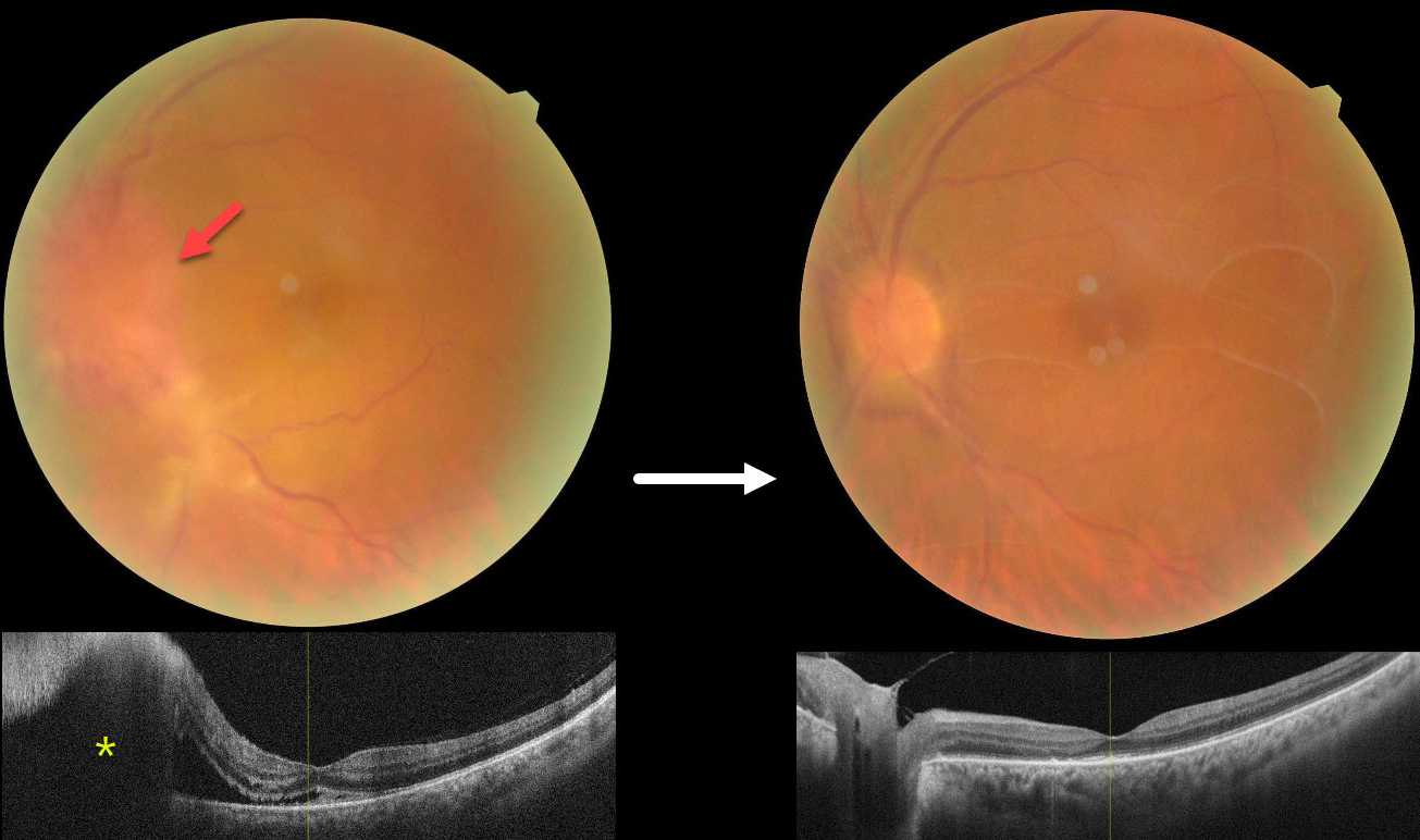
SYPHILITIC PAPILLITIS
Navneet Mehrotra and Tamanna Patel
Originally posted on @retina.rocks March 12, 2025
This 52YO male presented with 3 weeks of vision loss in his left eye. He has a history of diabetes, hypertension, and HIV. He was diagnosed elsewhere with optic neuritis and started on intravenous methylprednisolone with no improvement. Vision was 20/30 in his normal OD and counting fingers OS. There was a mild nongranulomatous anterior uveitis OS on slit lamp examination.
Color photography shows a markedly swollen nerve with possible retinitis extending inferotemporally. OCT scanning confirms the disc elevation with subretinal fluid extending beneath the nasal macula.
On further questioning, there was a recent history of a genital chancre, and the treponema pallidum hemagglutination assay was positive for syphilis. Following a 2-week course of parenteral penicillin, vision improved to 20/120 with marked improvement in the posterior segment findings. Unusual premacular vitreous membranes are noted.
Learning Points:
Ocular manifestations of syphilis vary widely and include virtually all variations of anterior and posterior segment inflammation. In our experience, placoid syphilis is the most common posterior segment presentation, although a recent systematic review and meta-analysis found papillitis to be the most reported finding in syphilitic uveitis (Zhang et al, J of Ophthalmology 2017;1; 6594849). Papillitis can be isolated or associated with other findings.
Any patient who tests positive for syphilis should always be checked for HIV since coinfection, as in our patient, is common.
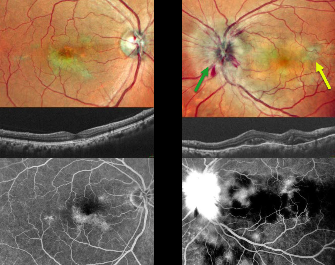
TUBERCULAR SERPIGINOUS-LIKE CHOROIDITIS
Originally posted on @retina.rocks March 11, 2025
This 54YO male presented with 2 weeks of vision loss in his left eye. He was diagnosed elsewhere with papilledema, and brain MRI scanning was normal. Vision was 20/20 in his asymptomatic OD and counting fingers OS. Pseudocolor SLO imaging of the OS shows marked disc swelling with radiating hemorrhages in the nerve fiber layer. Some deep, hypopigmented retinal lesions are also seen.
OCT scanning shows irregular choroidal thickening vs choroidal folds with overlying pockets of variably reflective subretinal fluid. The right eye shows multifocal serpiginous-like areas of chorioretinal scarring with variable outer retinal thinning on OCT.
On fluorescein angiography, the macular lesions stain OD. The left nerve shows profound leakage, and the choroidal lesions show variable blockage and staining.
We suspected tubercular serpiginous-like choroiditis OD with acute tubercular papillitis and choroiditis OS. Chest X-ray showed a mass pressing on the trachea. Chest CT confirmed the mass, and subsequent biopsy revealed granulomatous thyroiditis. His Mantoux skin test was positive. We advised emergent Infectious disease consultation regarding starting anti-tubercular therapy. Unfortunately, he was immediately lost to follow-up.
Learning Points:
Although tuberculosis most commonly presents with pulmonary involvement, extrapulmonary sites can include the gastrointestinal, skin, cardiovascular, genitourinary, and central nervous systems, as well as the eyes. Ocular involvement, like syphilis, can mimic virtually any type of uveitis, including anterior, intermediate, posterior, and panuveitis, retinitis, and retinal vasculitis, neuroretinitis, optic neuropathy, choroidal granuloma, choroiditis, and scleritis.
Tubercular serpiginous-like choroiditis mimics serpiginous but affects younger patients with more multifocal and peripheral recurrences and progression if untreated (Agarwal et al, AJO 2020;220:160-169). Although the exact mechanism is not yet clear, a direct or indirect infectious trigger by the Mycobacterium tuberculosis is thought to cause the choroiditis. Its origin is probably immunogenic since it seems to respond to corticosteroids and other immunosuppressants.
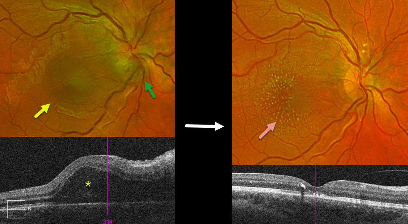
BARTONELLA
Originally posted on @retina.rocks February 20, 2025
This 18YO female presented with a several-day history of vision loss in her right eye. Vision was 20/70 OD and 20/40 in her normal OS.
Optos color RG imaging shows inferior disc edema with a serous macular detachment. Triton swept-source OCT confirms the serous detachment extending towards the nerve.
Bartonella serology was positive, and she was placed on a 6-week course of oral antibiotics. Two months following presentation, vision improved to 20/40. The disc edema and serous detachment resolved, with the development of a lipid star.
Learning Points:
The diagnosis for a unilateral swollen nerve is extensive. However, it becomes much smaller when associated with acute inner retinal ischemia (not seen in our case) and, later, with lipid exudation within Henle’s layer (neuroretinitis), including cat-scratch (Bartonella) and syphilis. When no underlying cause is found, the entity is called Leber’s idiopathic stellate neuroretinitis
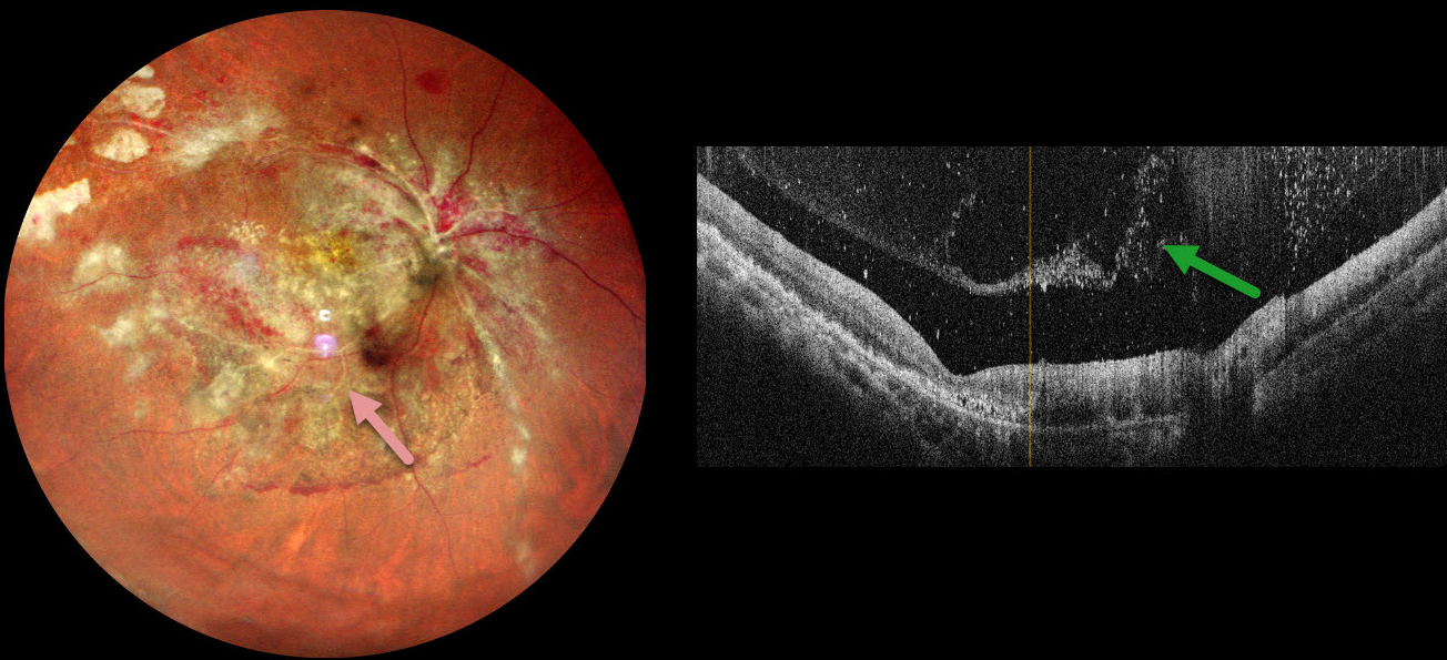
CMV RETINITIS
Rohan Jain and Manish Nagpal
Originally posted on @retina.rocks February 6, 2025
This 34YO male with a known history of HIV infection presented with 1 month of vision loss in his right eye. He was treated with a six-month course of systemic anti-tuberculous therapy for pulmonary tuberculosis two years earlier. One month earlier, he was diagnosed with HIV AIDS with a CD4 count of 43 and was started on systemic anti-retroviral therapy. Vision was counting fingers OD and 20/20 in his normal OS.
Multicolor imaging shows variably active cytomegalovirus (CMV) retinitis throughout the right posterior pole, including patches of necrotic white retina and retinal hemorrhages. The fundus is ischemic with sheathed arterioles. OCT scanning shows a vitreous separation with extensive vitreous cells.
Learning Points:
CMV retinitis develops as a reactivation of latent CMV in immunosuppressed individuals. Before effective antiretroviral treatment emerged in the mid to late 1990’s, CMV retinitis developed in up to 40% of HIV/AIDS patients, often within the last 6 months of life. Treatment includes a combination of intravenous and intravitreal medications, including ganciclovir, foscarnet, and cidofovir. Rhegmatogenous retinal detachment is a common late sequela and requires vitrectomy with silicone oil.
Our patient’s funduscopic findings are classic for CMV retinitis. However, patients with this degree of retinitis will usually have bilateral findings, and our patient’s left fundus was normal. These immunosuppressed patients also cannot mount an adequate immune response and therefore usually have a quiet vitreous, unlike our patient. It is therefore possible that there may also be an underlying tuberculous component. Unfortunately, our patient was immediately lost to follow-up before we could start intravitreal and systemic CMV therapy.
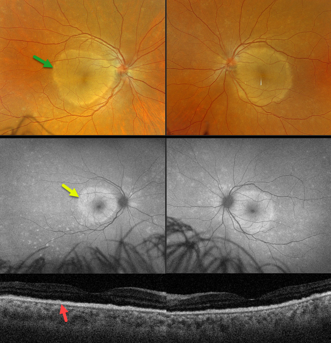
SYPHILIS
Originally posted on @retina.rocks December 2, 2024
This 45YO female presented with one week of bilateral vision loss. Vision was counting fingers in each eye.
Optos color RGB imaging shows symmetrical, discrete subretinal yellow-white placoid lesions occupying each macula. Fundus autofluorescence (FAF), however, shows multifocal hyper-FAF spots extending peripherally to the hyper-FAF macular lesions. Swept-source OCT shows replacement of the outer retinal bands with hyperreflective material.
Laboratory testing was positive for syphilis and negative for HIV. She was referred to an infectious disease specialist for systemic penicillin treatment. Unfortunately, she was immediately lost to follow-up.
Learning Points:
Ocular manifestations of syphilis vary widely and include virtually all variations of anterior and posterior segment inflammation. Originally described by Gass (Ophthalmology 1990;97:1288-1297), placoid chorioretinitis is one of the more common presentations. The discrete placoid lesions were quite dramatic on our patient’s multimodal imaging.
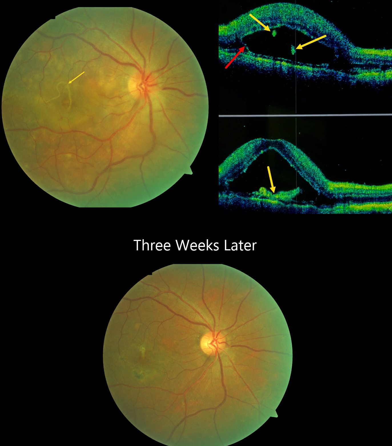
DIFFUSE UNILATERAL SUBACUTE NEURORETINITIS (DUSN)
Sharat Hegde
Originally posted on @retina.rocks November 27, 2024
This 41YO male presented with 4 days of severe vision loss in his right eye. Vision was hand motion OD and 20/20 in his normal left eye.
Color photography shows multiple focal yellow-white deep retinal lesions with a submacular worm and overlying neurosensory detachment. OCT scanning shows a bacillary layer detachment with various cuts through the hyperreflective worm.
Direct photocoagulation of the worm was impossible due to its submacular location and continued movement. The worm subsequently died and was absorbed following a 2-week course of oral albendazole 400mg BID and steroids. Three weeks later, vision improved to 20/30. Chorioretinal scarring is noted in the region of the prior worm, and the inflammatory lesions are mostly resolved.
Learning Points:
Originally described by Gass (Ophthalmology 1978;85:521-545), diffuse unilateral subacute neuroretinitis (DUSN) is a syndrome in healthy young adults characterized by unilateral decreased vision, vitritis, and papillitis, with recurrent crops of transient gray-white outer retinal/RPE lesions. Optic atrophy, diffuse RPE changes, decreased visual field, and electroretinogram are long-term sequela in untreated cases.
DUSN is caused by at least 2 different-sized subretinal nematodes that can remain viable in the eye for up to 3 years. If identified, the extramacular worm can be treated with thermal photocoagulation, surgically removed, or treated with systemic anthelmintic therapy.
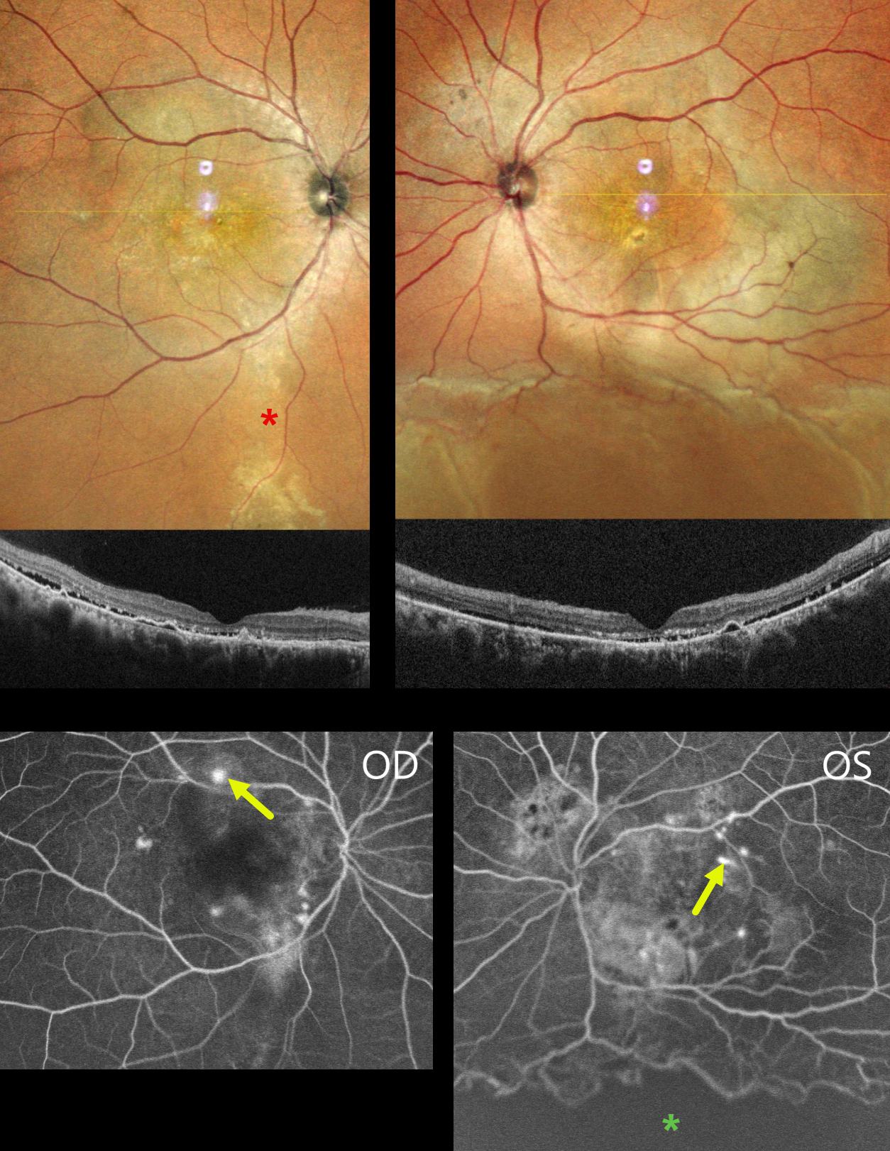
CENTRAL SEROUS CHORIORETINOPATHY WITH EXUDATIVE RD AND COMCOMITANT TB
Anand Temkar, Navneet Mehrotra, and Manish Nagpal
Originally posted on @retina.rocks November 7, 2024
This 39YO male presented with 4 months of bilateral vision loss. He was diagnosed elsewhere with chronic idiopathic central serous chorioretinopathy (ICSC) and was placed on azathioprine and spironolactone 3 months earlier. There was no history of steroid use or injections. Vision was 20/120 OD and 20/200 OS.
MultiColor imaging shows macular pigmentary changes that extend in a gutter inferiorly OD. Diffuse macular pigmentary changes are noted in his left eye that communicate with an inferior exudative retinal detachment. OCT scanning shows shallow subretinal fluid with hyperreflective shed outer segments, outer retinal hyperreflective foci, and RPE detachments. Fluorescein angiography shows bilateral multifocal subretinal blot leakage. A broad area of ischemia is present overlying the inferior detachment of the OS.
Mantoux skin testing (15x18mm) and QuantiFERON-TB Gold testing were positive. Chest CT showed fibrotic strands in the right lung. He was referred for systemic antituberculosis treatment. A week later, his local ophthalmologist reported that his vision and retinal findings improved, but he was then lost to follow-up.
Learning Points:
Our patient presented with severe bilateral idiopathic central serous chorioretinopathy (ISCS) with multifocal leaks in each eye and a secondary exudative detachment in his left eye. There are rare case reports of TB occurring in patients with ICSC, and these are likely coincidental findings (Khan et al, BMJ Case Rep 2017:bcr-2016-216471).
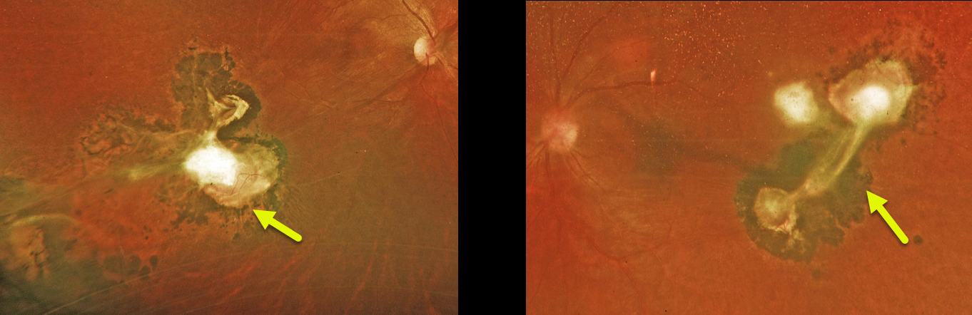
TOXOCARA
Matt Meredith
Originally posted on @retina.rocks October 23, 2024
This healthy 29YO female was referred for asymptomatic bilateral retinal findings. Vision was 20/40 OU.
Optos color RG imaging shows a large area of variably atrophic and fibrotic subretinal scarring inferotemporally OD, with a smaller area of scarring temporally OS. The scarring in her left eye has a more triangular appearance. Toxocara canis ELISA bloodwork was positive.
Learning Points:
Toxocariasis is a rare infection caused by the toxocara canis roundworm, which matures in the dog intestine. The eggs are passed in the stool, which can then be ingested when young children come into contact with infected sand or dirt and then touch their mouth. The ingested egg matures into a larva, which enters the systemic circulation from the intestine and can migrate into the eye. The death of the organism can cause variable findings, including endophthalmitis (typically a completely white, quiet eye), posterior granuloma, or peripheral granuloma.
Most commonly in our experience, they present as a triangular-shaped area of vitreous base organization/scarring, with a fold of retina extending more posteriorly. Since there is usually a single larva, the eye findings are almost always unilateral. Our case is therefore quite unusual in its bilaterality.
SYPHILIS
Mattie Adams
Originally posted on @retina.rocks October 22, 2024
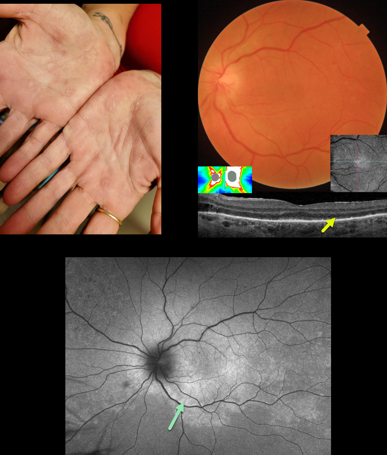
This 51YO female presented with one month of flashes and vision loss in her left eye. Vision was 20/20 in her normal OD and counting fingers OS.
When asked about any rashes, she showed us her palms. What’s your diagnosis??
Triton color imaging of the left posterior pole shows mild optic nerve swelling, with some subtle subretinal whitish pigmentary changes. The nerve swelling was confirmed on OCT. Swept-source OCT shows variable loss of the outer retinal bands and ellipsoid zone with hyperreflective material above the RPE. En face imaging of the outer retina shows this hyperreflective material as numerous bright dots.
Fundus autofluorescence (FAF) shows confluent macular and peripapillary hyper-FAF with more patchy areas of hyper-FAF extending peripherally. Laboratory testing was positive for syphilis and negative for HIV. She was referred to an infectious disease specialist for systemic penicillin treatment and was subsequently lost to follow-up.
This case was submitted by Mattie Adams.
Learning Points:
Ocular manifestations of syphilis vary widely and include virtually all variations of anterior and posterior segment inflammation, including a recently described presentation as the multiple evanescent white dot syndrome phenotype (Russel et al Int Ophthalmol 2020;40:627-738). Hence, the reason it’s been dubbed the great imitator or masquerader. One of the more common presentations is placoid chorioretinitis (Eandi et al, Retina 2012;32:1915-1941), which was evident in our patient’s multimodal imaging.
The rates of syphilis in the US have steadily climbed since 2000, and according to the CDC, its incidence rose nearly 80% between 2018 and 2022 (https://www.cdc.gov/std/statistics/2022/default.htm). Syphilis is prevalent in men having sex with men, among heterosexuals, and in both sexes. These numbers are reflected in how often we are now finding and diagnosing syphilis in our practice. We must always remember that as eye doctors, we are uniquely privileged to not only treat eye problems, but also diagnose systemic diseases. In our case, the painless palmar rash and macular placoid findings were both pathognomonic and led to a rapid diagnosis with referral for treatment.
TOXOPLASMOSIS WITH RETINAL CHOROIDAL ANASTOMOSIS
Originally posted on @retina.rocks October 21, 2024
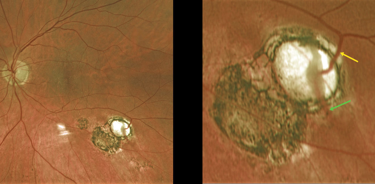
This 60YO female was referred for asymptomatic scarring in her left eye. Vision was 20/50 OD and 20/40 OS due to early bilateral cataracts. Small macular drusen were noted bilaterally.
Optos color RG imaging of her left shows multifocal variably pigmented scars below the inferotemporal arcade. A magnified inferotemporal view shows a retinal vein diving into an atrophic scar and directly connecting to a choroidal vein.
Learning Points:
Under normal conditions the retinal and choroidal circulations remain separate and distinct. Rarely, the two circulations directly connect with each other through a chorioretinal anastomosis. These can occur in chorioretinal scars (typically from inactive toxoplasmosis lesions as in this patient), in macular telangiectasia, and in disciform scars. We felt our patient’s findings most consistent with inactive toxoplasmosis scarring.
TUBERCULOSIS VASCULITIS
Tejaswita Verma, Navneet Mehrotra and Manish Nagpal
Originally posted on @retina.rocks September 18, 2024
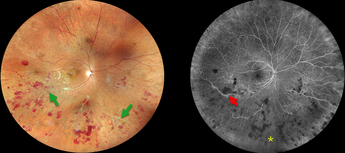
This 27YO male presented with 10 days of floaters and photophobia in his right eye. He was tentatively diagnosed with tuberculosis (TB) several days earlier and sought a second opinion with us. Recent Mantoux skin testing was markedly positive (20 mm induration), and QuantiFERON-TB Gold was positive as well. Vision was 20/20 bilaterally.
Fundus photography shows inferior and nasal vascular sheathing, inner and deep retinal hemorrhages in the inferior hemisphere, and focal areas of whitish inner retinal ischemia in the inferior macula. Fluorescein angiography shows variable midperipheral ischemia with focal vascular staining. The left eye was normal.
Chest CT was supportive of active pulmonary TB, and the patient was started on antitubercular treatment (ATT) by the pulmonary team on the day of our exam.
When he returned four days later, his vision was 20/40 OD. We recommended an intravitreal anti-VEGF injection followed by scatter laser, but unfortunately, he was then lost to follow-up.
Learning Points:
The differential for occlusive peripheral retinal vasculitis (Huvard et al, Ophthalmology Retina 2022;6:43-48) includes TB, rheumatologic disorders / systemic vasculitides (granulomatosis with polyangiitis, Bechet’s disease, systemic lupus, etc), idiopathic retinal vasculitis and neuroretinitis, and sarcoidosis. Historically, Eales’ disease has been used for a variety of heterogeneous disorders, including tuberculous vasculitis, and due to its ambiguity, we prefer not to use this term.
The diagnosis of tubercular vasculitis is often presumptive and difficult to diagnose without isolation of TB from ocular tissue or fluids. However, as with our patient, it is a reasonable diagnosis in the proper clinical setting, including in those from endemic areas and with a systemic diagnosis of TB (Agrawal et al, 2017 37:112-117). Systemic ATT, along with local ocular treatment for neovascular complications (anti-VEGF injections, scatter laser photocoagulation), is recommended.
POST-FEVER RETINITIS
Tejaswita Verma, Navneet Mehrotra and Manish Nagpal
Originally posted on @retina.rocks September 12, 2024
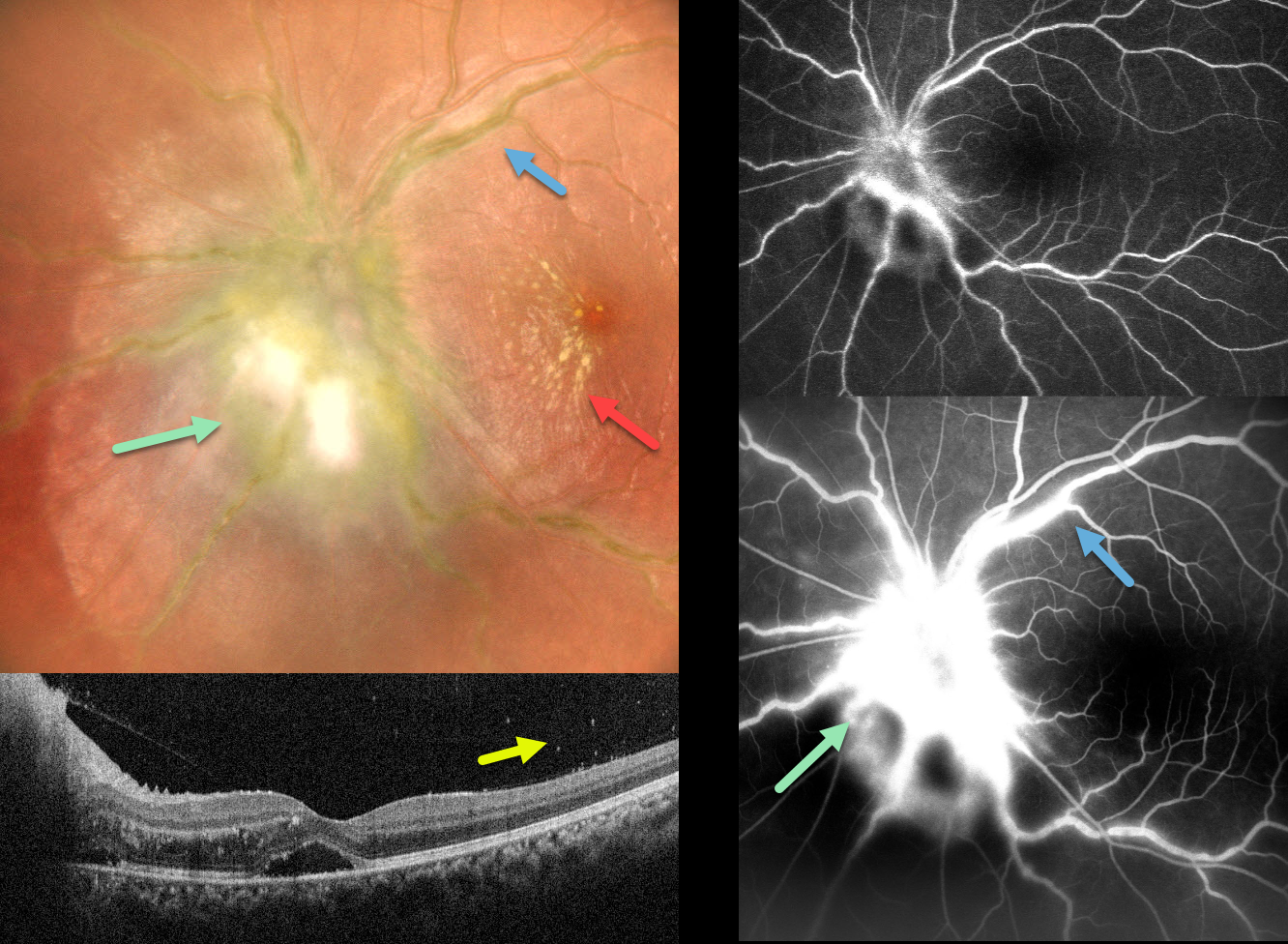
This 20YO female presented with sudden vision loss in her left eye that followed a 1-week febrile illness. Vision was 20/20 in her normal right eye and 20/30 in her left eye.
Multicolor imaging shows marked disc edema with surrounding retinitis, frosted branch vasculitis, and a nasal macular star. OCT shows marked nasal macular retinal thickening and foveal subretinal fluid. Vitritis was noted clinically, and vitreous cells were also noted on the OCT. Fluorescein angiography shows marked optic nerve leakage and vascular staining.
Oral steroids and doxycycline were recommended, along with Weill-Felix testing to rule out typhoid fever, but unfortunately, she was immediately lost to follow-up.
Learning Points:
Epidemic (post-fever) retinitis is an infectious or para-infectious entity that develops in immunocompetent individuals several weeks following a bacterial, viral, or protozoal infection. Causes include Dengue, Rickettsiosis, West Nile, Leptospirosis, Chikungunya, Typhoid, and Ebola. Patients present with sudden, painless vision loss.
Fundus findings include unilateral or bilateral unifocal or multifocal patches of retinitis with possible nerve involvement, serous macular detachment, and vasculitis or frosted branch angiitis. There is usually a favorable response to steroids and treating the underlying infectious cause with appropriate systemic therapy.
See Mahendradas et al (Indian J Ophthalmol 2020;68:1775-1786) for an excellent post-fever retinitis review.
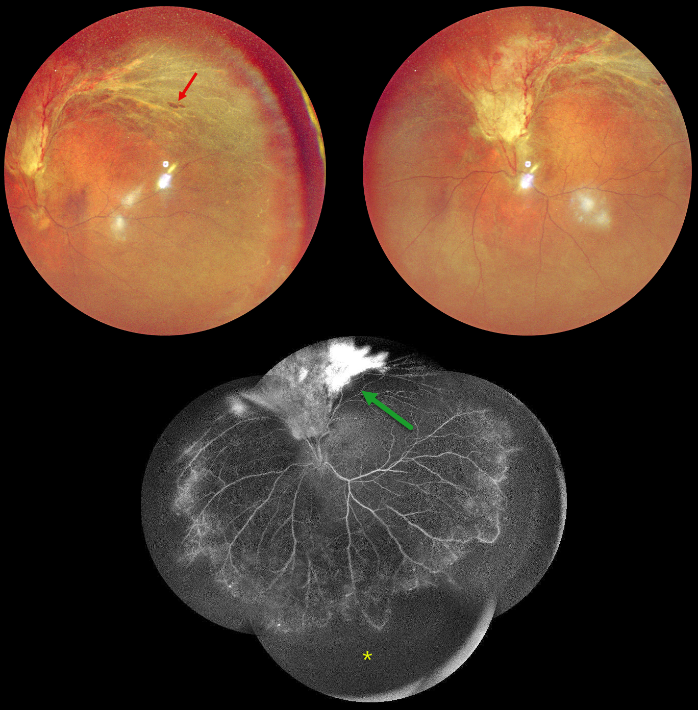
CMV RETINITIS WITH RETINAL DETACHMENT
Originally posted on @retina.rocks September 2, 2024
This 48YO HIV positive male on highly active antiretroviral therapy (HAART) presented with 2 months of vision loss in his left eye. His last CD4 count was 81. Vision was 20/20 OD and counting fingers OS.
Multicolor imaging shows active cytomegalovirus (CMV) retinitis with retinal detachment extending superiorly from the optic nerve into the superotemporal midperiphery. A few atrophic retinal breaks are noted. Fluorescein angiography of his left eye shows variable leakage within the area of retinitis superiorly and severe encircling peripheral ischemia. Vitrectomy with silicone oil was scheduled to repair the detachment in his left eye.
Learning Points:
CMV retinitis develops as a reactivation of latent CMV in immunosuppressed individuals. Before effective antiretroviral treatment emerged in the mid to late 1990’s, CMV retinitis developed in up to 40% of HIV/AIDS patients, often within the last 6 months of life. Treatment includes a combination of intravenous and intravitreal medications, including ganciclovir, foscarnet and cidofovir. Rhegmatogenous retinal detachment is a common late sequela and requires vitrectomy with silicone oil.
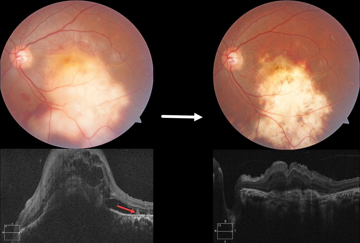
TUBERCULOSIS CHOROIDAL GRANULOMA
Divya Nair
Originally posted on @retina.rocks July 22, 2024
This 36YO male presented with 10 days of vision loss in his left eye. One month earlier, he was diagnosed with disseminated tuberculosis (TB), peritonitis, and pleural effusion, and was placed on systemic antitubercular therapy (ATT). Vision was 20/20 in his normal right eye and counting fingers in his left eye.
Fundus photography shows a large elevated yellowish submacular lesion that extends into the inferior midperiphery. Subretinal blood is noted along its superior and inferior margins. Faint lipid is present superotemporally, and some retinal striae are seen temporally. Spectral domain OCT shows a large dome-shaped hyporeflective choroidal lesion with overlying cystic retinal thickening, subretinal fluid, and subretinal hyperreflective material. A few hyperreflective pyramidal lesions are noted above the RPE temporally.
We started him on oral corticosteroids. In view of the vascularized lesion with subretinal blood, an intravitreal injection of Avastin was also given. Six weeks later, there is a relatively flat scar with near complete resolution of the blood. The choroidal lesion has resolved with an overlying hyperreflective subretinal scar and decreased retinal thickening. Unfortunately, vision remained at counting fingers.
Learning Points:
Although TB most commonly presents with pulmonary involvement, extrapulmonary sites can include the gastrointestinal, skin, cardiovascular, genitourinary, and central nervous systems, including the eyes. Ocular involvement, like syphilis, can mimic virtually any type of uveitis, including anterior, intermediate, posterior, and panuveitis, retinitis and retinal vasculitis, neuroretinitis, optic neuropathy, choroidal granuloma, choroiditis, and scleritis.
For an excellent review of intraocular tuberculosis, see Bupta et al, Survey Ophthalmology 2007;52:561-587.
Tubercular granulomas can be distinguished from sarcoid lesions by their solitary nature, yellow color, larger size, and vascularization (Agarwal et al., AJO 2021;226:42-55). All these features were found in our patient. Vascularization is thought to arise from localized hypoxia, leading to increased levels of vascular endothelial growth factor. Anti-VEGF injections and systemic corticosteroids, in addition to ATT, are helpful in hastening granuloma resolution and reducing secondary exudation (Lee et al, Graefe’s 2022;260:1641-1650).
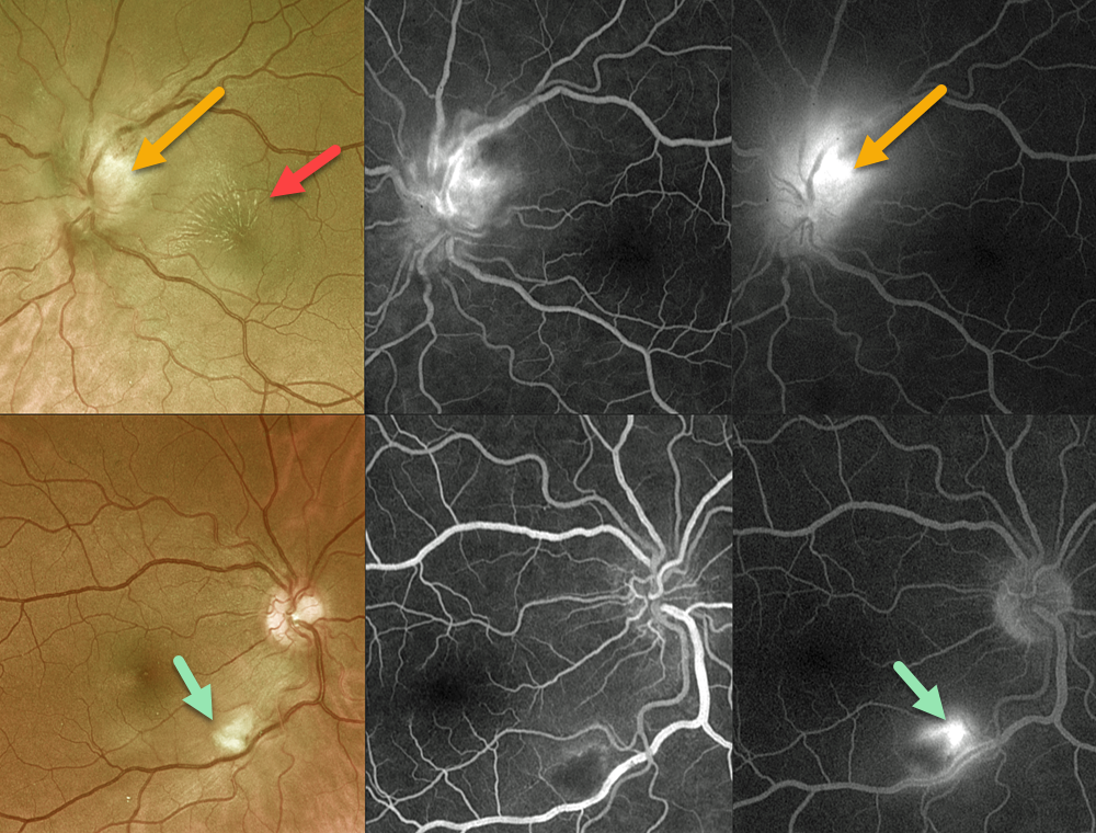
BARTONELLA
Originally posted on @retina.rocks July 3, 2024
This 52YO female presented with one week of bilateral vision loss. She has multiple cats at home. Vision was 20/80 OD and counting fingers OS.
Optos color RG imaging shows an area of inferonasal macular inner retinal opacification OD, and a superiorly swollen left optic nerve with a superonasal macular lipid star.
Fluorescein angiography shows early blockage with late staining of the retinal lesion OD with an adjacent area of vasculitis. Marked optic nerve leakage is seen OS.
Bartonella henselae antibody testing was positive for both IgG >1:1024 and IgM 1:126. She was subsequently lost to follow-up.
Learning Points:
Bartonella henselae is the bacterium associated with cat scratch disease. It is transmitted from cat fleas carrying the bacterium or from flea feces that can be present on the cat’s claws/teeth, and transferred to humans from a cat scratch, lick, or bite. Common systemic findings include fever, malaise, and progressive lymphadenopathy. These findings usually occur within 1-2 weeks of a cat scratch/bite. Ocular effects primarily include neuroretinitis with rare cases of Parinaud’s ocular glandular syndrome, or, as in our patient’s case, chorioretinitis with vasculitis.
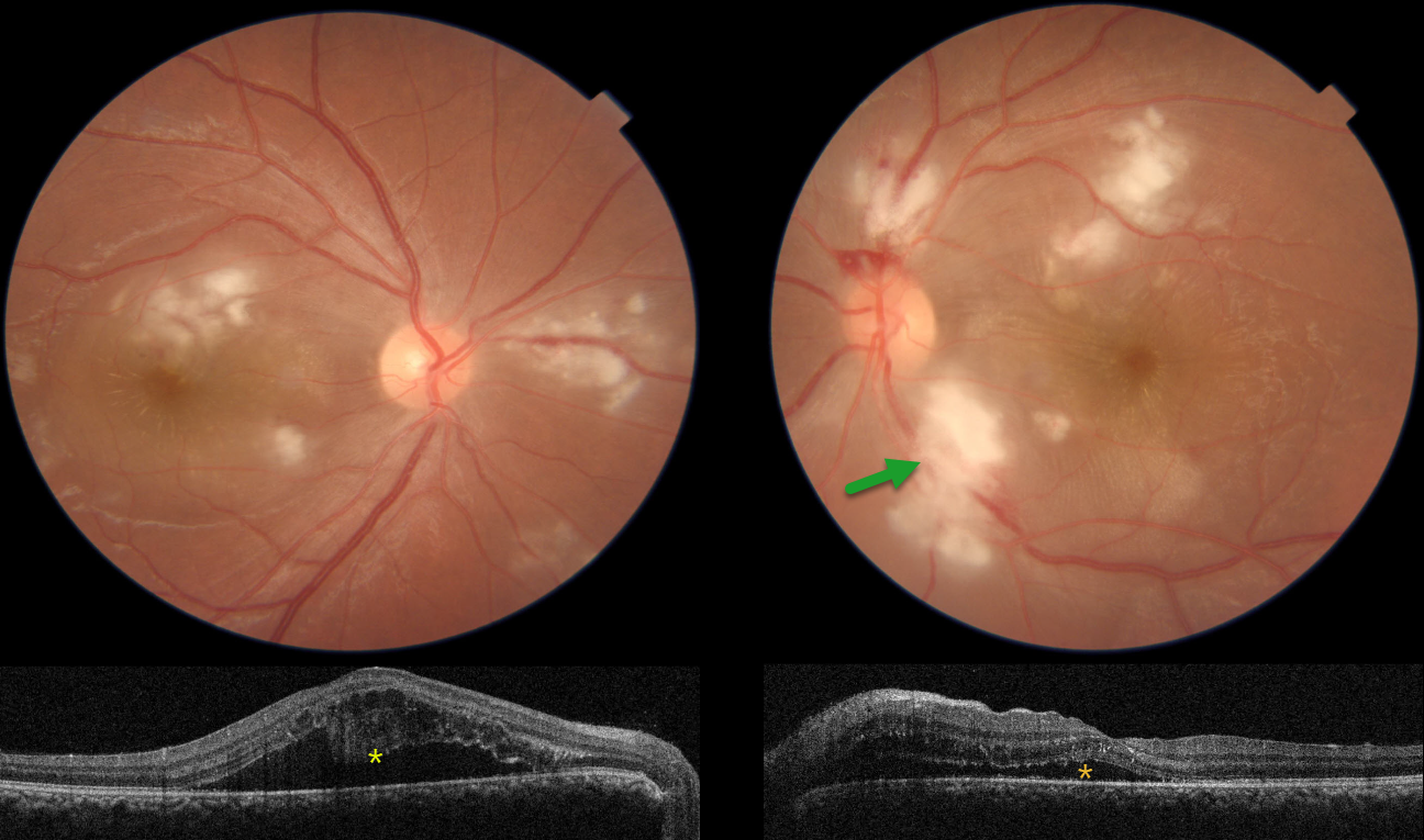
POST-FEVER RETINITIS
Asma Samsudeen and Ashish Sharma
Originally posted on @retina.rocks June 19, 2024
This 33YO male started to complain of blurred vision 10 days earlier. He was hospitalized two months prior with severe headaches, fever, and chills. Vision was 20/30 OU.
Color imaging shows bilateral Purtscher ‘s-like acute inner macular ischemic whitening with a few retinal hemorrhages. Faint radiating foveal lipid is also present.
OCT scanning shows outer retinal cystic edema and subretinal fluid. Hyperreflective material is adherent to the overlying detached right macula. He was referred for viral bloodwork, but unfortunately was subsequently lost to follow-up.
Learning Points:
Epidemic (post-fever) retinitis was first described by Kawali et al (Ocular Immunology & Inflammation 2019;27:571-577) as an infectious or para-infectious uveitis days to weeks following a febrile illness with Dengue, Rickettsiosis, West Nile, Leptospirosis, Chikungunya, Typhoid, or Ebola. It appears as a usually bilateral Purtscher ’s-like retinitis involving the posterior pole with a favorable response to steroids. It is most common in tropical countries like India.
See Mahendradas et al (Indian J Ophthalmol 2020;68:1775-1786) for an excellent post-fever retinitis review.
SYPHILIS
Anjana Mirajkar, Navneet Mehrotra and Manish Nagpal
Originally posted on @retina.rocks May 15, 2024
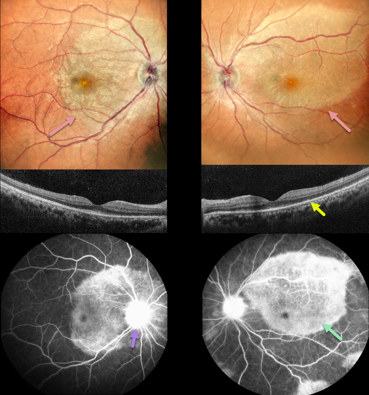
This 36YO female presented with a month of bilateral vision loss, which got worse over the prior 2 days. She also complained of 3 months of headaches and painful urination. Vision was 20/30 OD and 20/120 OS.
Multicolor imaging shows bilateral creamy placoid lesions. OCT scanning shows variable ellipsoid zone loss with small hyperreflective material above. Some vitreous cells are also noted. Fluorescein angiography shows late staining of these placoid lesions with optic nerve leakage.
Bloodwork was positive for syphilis, and she was referred to an infectious disease specialist for further management.
Learning Points:
Ocular manifestations of syphilis vary widely and include virtually all variations of anterior and posterior segment inflammation, including a recently described presentation as the multiple evanescent white dot syndrome phenotype (Russel et al Int Ophthalmol 2020;40:627-738). Hence, the reason it’s been dubbed the great imitator or masquerader.
One of the more common presentations is placoid chorioretinitis (Eandi et al, Retina 2012;32:1915-1941), which was evident in our patient’s multimodal imaging.
TUBERCULAR SERPIGINOUS-LIKE CHORIORETINITIS
Ogugua Okonkwo, Adekunle Olubola Hassan, Ayodele Harriman, Ogochukwu Sibeudu, and Idris Akintayo Oyekunle
Originally posted on @retina.rocks May 1, 2024
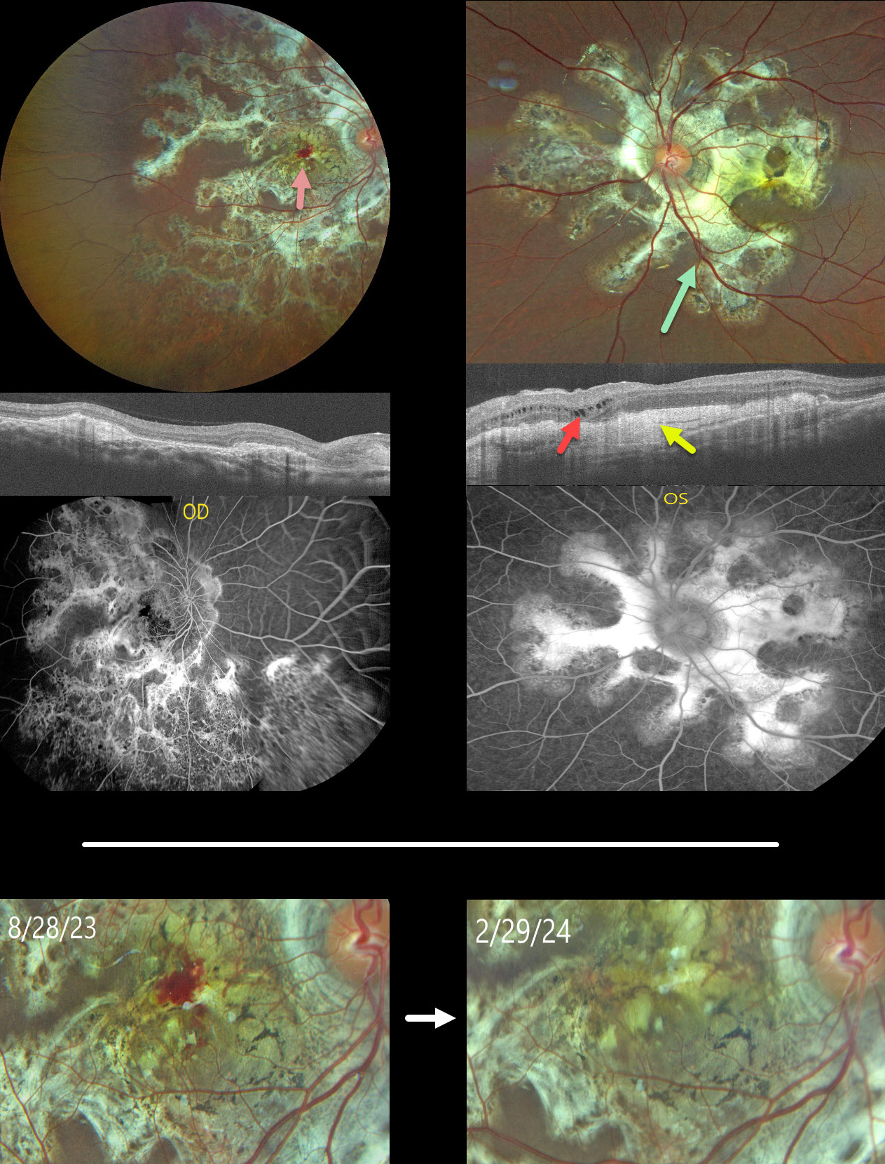
This healthy 13YO male presented with a 5-month history of bilateral vision loss. Vision was 20/200 OU.
Fundus photography shows bilateral subretinal, variably fibrotic peripapillary scarring that extends outwards with finger-like projections. A foveal hemorrhage is noted in the right macula. More mottled pigmented scarring extends into the right inferior periphery.
OCT scanning shows variable bilateral outer retinal atrophy and hyperreflective subretinal fibrosis with mild outer nuclear cysts in the nasal left macula. This scarring stains angiographically.
We were hesitant to start anti-VEGF therapy in a young boy and felt that the macular neovascularization causing the right macular blood might be inflammatory. He was therefore placed on a rapidly tapering course of oral prednisone.
At the 6-month follow-up, vision remained stable at 20/200 in this eye, with resolution of the blood.
Mantoux skin testing was negative. We have made multiple attempts to have the patient get QuantiFERON-TB Gold testing, but to date have been unsuccessful.
Learning Points:
Serpiginous choroiditis is an idiopathic, usually bilateral chorioretinal inflammatory condition, most commonly found in middle-aged men (Khanamiri and Rao, Surv Ophthalmology 2013;58:203-232). Recurrent areas of inflammation develop along the edge of a previous scar, producing scarring that meanders from the optic nerve outwards. Vision is good unless scarring or macular neovascularization extends through the macular center.
Tubercular serpiginous-like choroiditis mimics serpiginous but affects younger patients with more multifocal and peripheral recurrences and progression if untreated (Agarwal et al, AJO 2020;220:160-169). Although the exact mechanism is not yet clear, a direct or indirect infectious trigger by the Mycobacterium tuberculosis is thought to cause the choroiditis. We believe our patient has this diagnosis and will continue to confirm it with further testing.
TUBERCULOUS NEURORETINITIS
Keith Slayden
Originally posted on @retina.rocks April 2, 2024
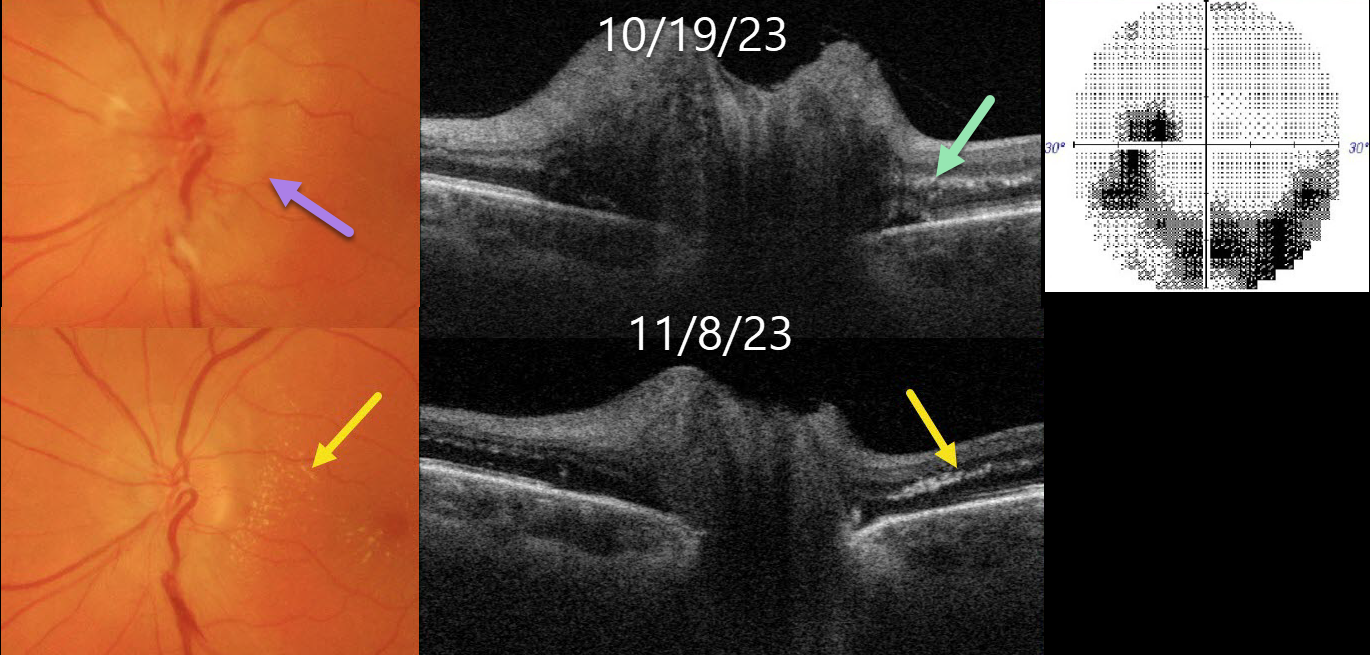
This healthy 56YO female presented with 1-2 weeks of blurred vision in her left eye. Vision was 20/25 in her normal OD and 20/100 OS.
Color imaging shows a diffusely swollen left nerve. Optic nerve Triton swept-source OCT B-scan confirms the diffuse retinal nerve fiber layer edema. A few dots of subclinical hyperreflective lipid are noted in the outer plexiform layer. 30-2 visual field testing shows an inferior unilateral arcuate scotoma extending from the blind spot.
Extensive blood work and chest X-ray were negative except for a positive QuantiFERON-TB Gold. Infectious disease consultation led to the diagnosis of latent tuberculosis, and she was started on systemic anti-tuberculous therapy.
Over the following several weeks, as the optic nerve edema resolved, the papillomacular outer plexiform layer lipid became more prominent.
Learning Points:
The diagnosis for a unilateral swollen nerve is extensive. However, it becomes much smaller when associated with acute inner retinal ischemia (not seen in our case) and later lipid exudation within Henle’s layer (neuroretinitis), including cat scratch (Bartonella), Lyme disease, and syphilis. When no underlying cause is found, the entity is called Leber’s idiopathic stellate neuroretinitis.
Tuberculosis is a rare cause of neuroretinitis (Stechschulte et al, J Neuroophthalmology 1999;19:201-204). It is unclear whether our patient’s findings are idiopathic or related to her latent tuberculosis.
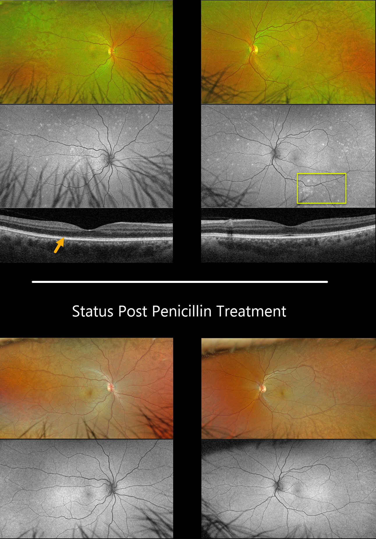
SYPHILIS
Originally posted on @retina.rocks March 7, 2024
This 57YO male presented with 3 weeks of blurred vision in his right eye. Vision was 20/40 OD and 20/25 OS.
Optos color RG imaging shows bilateral plaques of whitish deep retinal discoloration. Fundus autofluorescence (FAF) shows multifocal MEWDS-like hyper-FAF spots scattered throughout each fundus. Triton swept-source OCT shows an indistinct temporal foveal ellipsoid zone OD and a fairly normal B-scan OS.
RPR testing was positive, and the patient was started on parenteral penicillin treatment. The autofluorescent findings dramatically resolved seven weeks later.
Learning Points:
Ocular manifestations of syphilis vary widely and include virtually all variations of anterior and posterior segment inflammation, including a recently described presentation as the multiple evanescent white dot syndrome phenotype (Russel et al Int Ophthalmol 2020;40:627-738). Hence, the reason it’s been dubbed the great imitator or masquerader.
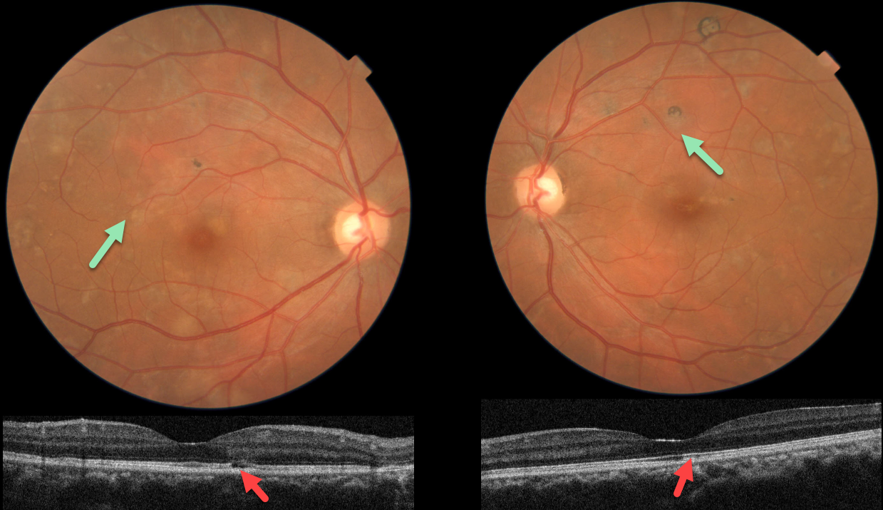
TUBERCULAR SERPIGINOUS-LIKE CHORIORETINITIS
Asma Samsudeen and Ashish Sharma
Originally posted on @retina.rocks January 18, 2024
This 38YO male presented with floaters in each eye. He denied any past medical history. Vision was 20/20 OU.
Fundus photos show multifocal patches of variably pigmented chorioretinal scarring throughout each posterior pole.
OCT scanning in each eye shows variable outer retinal atrophy and ellipsoid disruption. Unfortunately, fundus autofluorescence and fluorescein angiography are not available.
Subsequent workup was consistent for active tuberculosis, including a positive QuantiFERON-TB Gold and chest x-ray findings. Systemic anti-tuberculous therapy was begun.
Learning Points:
Serpiginous choroiditis is an idiopathic, usually bilateral chorioretinal inflammatory condition most commonly found in middle-aged men (Khanamiri and Rao, Surv Ophthalmology 2013;58:203-232).
Recurrent areas of inflammation develop along the edge of a previous scar, producing scarring that meanders from the optic nerve outwards. Vision is good unless scarring or macular neovascularization extends through the macular center.
Tubercular serpiginous-like choroiditis mimics serpiginous, but affects younger patients with more multifocal and peripheral recurrences and progression if untreated (Agarwal et al, AJO 2020;220:160-169).
Although the exact mechanism remains unclear, a direct or indirect infectious trigger by Mycobacterium tuberculosis is believed to cause choroiditis.
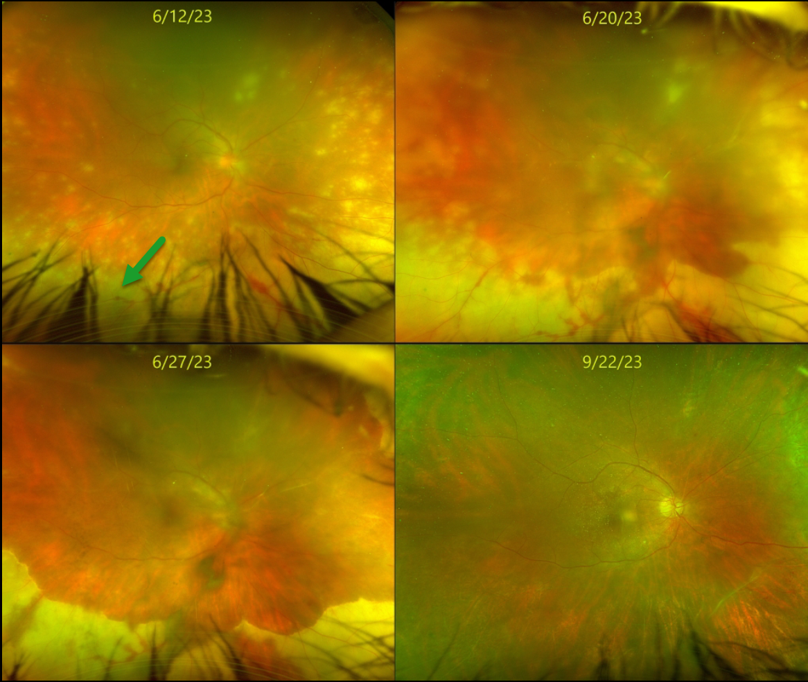
ACUTE RETINAL NECROSIS SYNDROME
Originally posted on @retina.rocks November 29, 2023
This healthy 55YO female presented with about 2 weeks of vision loss in her right eye. Vision was counting fingers OD and 20/30 in her normal left eye.
Slit lamp showed moderate anterior granulomatous inflammation. Moderate vitreous debris was noted. Optos color RG imaging shows multifocal white patches of variably confluent outer retinal inflammation extending anteriorly from the midperiphery. She was started on topical steroids and atropine, valacyclovir 1 gram PO BID, and a baby aspirin daily for acute retinal necrosis syndrome (ARNS).
After 1 week, vision remained at counting fingers but with subjective improvement. The retinitis was consolidating. The retinitis gradually improved over the following 3 months. When last examined on 9/22/23, vision was 20/70, with complete resolution of disease on valacyclovir 1 gram PO daily. We continue to follow her closely.
Learning Points:
ARNS is most commonly caused by the Herpes zoster virus. It usually occurs in immunocompetent individuals, and is a rapidly progressive panuveitis with hemorrhagic and ischemic unilateral (ARNS) or bilateral (BARNS) retinal necrosis that typically begins in the retinal periphery.
ARNS is often a devastating and blinding infection due to the high risk for retinal detachment as well as macular and optic nerve involvement. The latest meta-analysis argues for systemic antiviral therapy and prophylactic vitrectomy, with uncertain benefits for prophylactic laser photocoagulation or adjunctive intravitreal antivirals (see Zhao et al, Retina 2021;41:965-978).
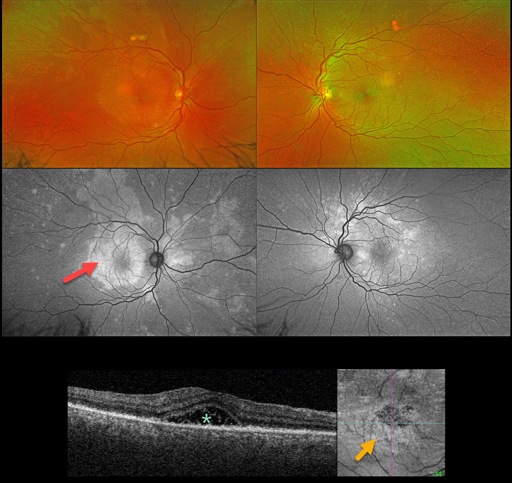
SYPHILIS
Originally posted on @retina.rocks November 16, 2023
This 52YO male presented with a one-week history of blurred central vision in his right eye. There was no past medical history. Vision was 20/200 OD and 20/25 OS. Anterior segments were normal.
An oval, whitish submacular lesion is noted OD. Fundus autofluorescence (FAF) reveals much more extensive findings than those noted funduscopically. In particular, the placoid lesion seen in the right macula is markedly hyper-FAF and extends into the right superior midperiphery.
Triton swept-source OCT of the right eye shows variable hyperreflective subfoveal fluid, and en face OCT shows fine scattered hyperreflective dots in the outer retina.
Treponema pallidum antibodies were reactive with a high quantitative RPR (1:64). He was started on parenteral penicillin.
Learning Points:
Ocular manifestations of syphilis vary widely and include virtually all variations of anterior and posterior segment inflammation, including a recently described presentation as the multiple evanescent white dot syndrome phenotype (Russel et al Int Ophthalmol 2020;40:627-738). Hence, the reason it’s been dubbed the great imitator or masquerader.
One of the more common presentations is placoid chorioretinitis (Eandi et al, Retina 2012;32:1915-1941), which was evident in our patient’s multimodal imaging.
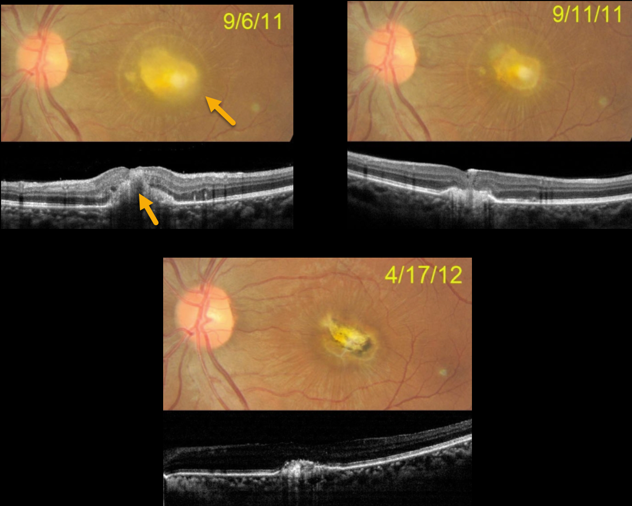
CANDIDA ENDOPHTHALMITIS
Santosh Bhide
Originally posted on @retina.rocks October 11, 2023
In 2011, this 27YO female was recently hospitalized and treated with intravenous amphotericin. Upon discharge, she was examined for vision loss in her left eye. Vision was hand motion.
Color photography shows a thickened yellow subfoveal lesion that extends through the full-thickness retina on OCT. One week later, vision improved to counting fingers, and the lesion is flattening and contracting. Seven months later, a small contracted foveal scar remains, with 4/60 vision. She was subsequently lost to follow-up.
Learning Points:
Ocular candidiasis is characterized by single or multiple focal yellow-white chorioretinal lesions with overlying vitritis, vitreous haze, vitreous abscess (fluff balls or string of pearls), and other more generalized presentations of infection and inflammation, such as endophthalmitis, hypopyon, scleritis, exudates, and anterior chamber cells. Ocular candidiasis can be caused by exogenous or endogenous sources. Exogenous routes include trauma and inoculation from surgical procedures. Endogenous ocular candidiasis arises from hematogenous seeding of chorioretinal blood vessels via fungemia, indwelling catheters, a severely compromised immune system, or broad-spectrum antibiotic abuse, etc.
Treatment includes removal of the inciting source of candidiasis (i.e., indwelling catheter) and systemic therapy with possible intravitreal antifungal therapy.
Routine ophthalmic screening for known systemic candida sepsis is not recommended unless there are suggestive signs or symptoms for ocular involvement (see Breazzano et al, Ophthalmology 2011;129:73-76).
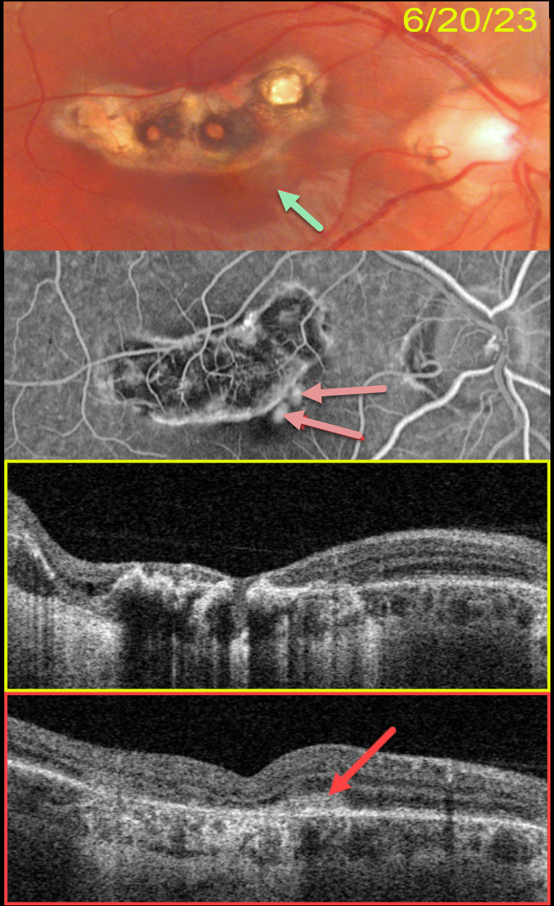
TOXOPLASMOSIS
Originally posted on @retina.rocks August 24, 2023
This 22YO male presented with several days of central blurred vision in his left eye. There was no prior ocular or medical history. Vision was 20/25 in his normal right eye and 20/25 in his left eye.
Color photography shows a variably pigmented comma-shaped colobomatous scar in the superior macula. A tiny depigmented scar is noted along its inferonasal edge. OCT scanning through this lesion showed no fluid (not shown).
He returned about 2 months later, complaining of increasing vision loss in his eye. Vision had decreased to 20/400. A faint area of subretinal pigment is now noted.
Optos fluorescein angiography shows leaking macular neovascularization (MNV) within this region. Swept-source OCT confirms a type 2 MNV (located above the RPE).
Intravitreal Avastin was injected.
Learning Points:
Virtually any disorder that affects the RPE-Bruch membrane layer can cause MNV, the most common being age-related macular degeneration, ocular histoplasmosis, idiopathic high myopia (lacquer cracks), trauma (choroidal rupture), and angioid streaks.
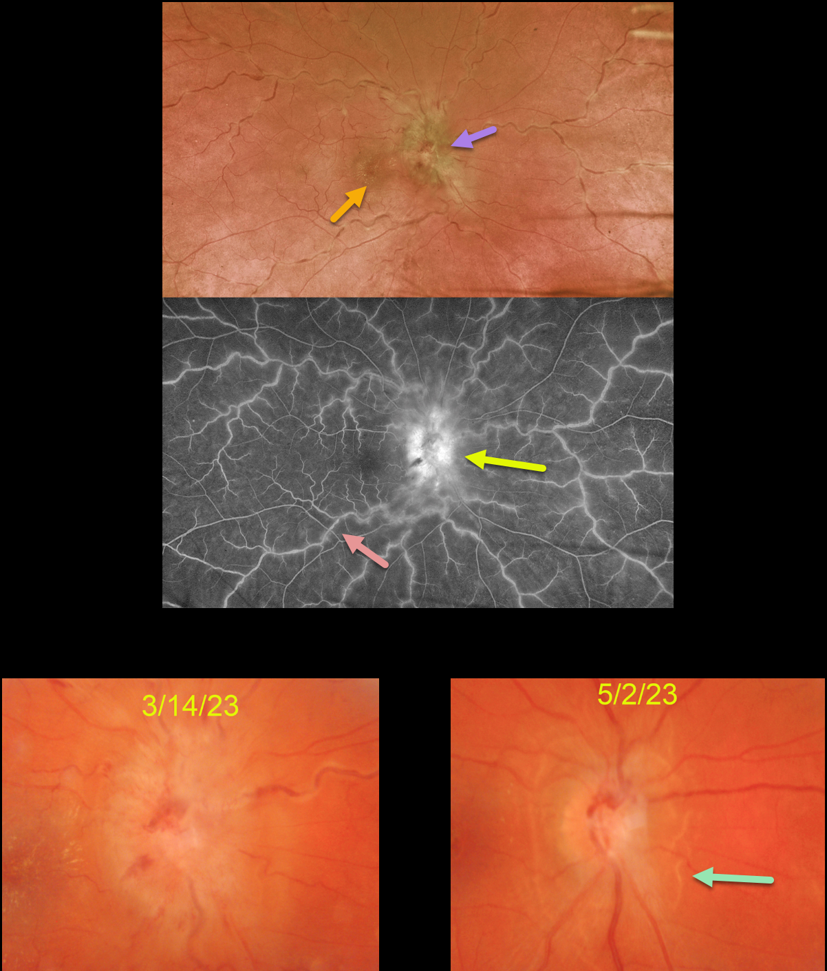
BARTONELLA
Originally posted on @retina.rocks July 13, 2023
This 36YO female presented with unilateral vision loss in her left eye. She denied any systemic symptoms except for headaches for 3 months. Vision was 20/70 in her right eye and 20/25 in her left eye.
Optos color imaging shows severe optic nerve head edema, a foveal lipid star, dilated and tortuous vessels, and a variable frosted branch appearance to the retinal veins. Fluorescein angiography shows diffuse venous staining with optic nerve leakage.
Extensive blood work was positive for both Bartonella henselae (IgG 1:64) and Bartonella quintana (IgG 1:128), and negative for Bartonella IgM antibodies. Although she has cats at home, she denied cat scratches or exposure to cat fleas. She was referred to an infectious disease specialist who started her on doxycycline 100mg PO BID.
When she returned 6 weeks later, her headaches had resolved, and her vision was 20/80. The optic nerve swelling was dramatically improved with secondary peripapillary pigmentary changes.
Learning Points:
Bartonella henselae is the bacterium associated with cat scratch disease. It is transmitted from cat fleas carrying the bacterium or from flea feces that can be present on the cat’s claws/teeth, and transferred to humans from a cat scratch, lick, or bite.
Common systemic findings include fever, malaise, and progressive lymphadenopathy. These findings usually occur within 1-2 weeks of a cat scratch/bite.
Ocular effects primarily include neuroretinitis, with rare cases of Parinaud’s ocular glandular syndrome and, as in our patient’s case, chorioretinitis with vasculitis.
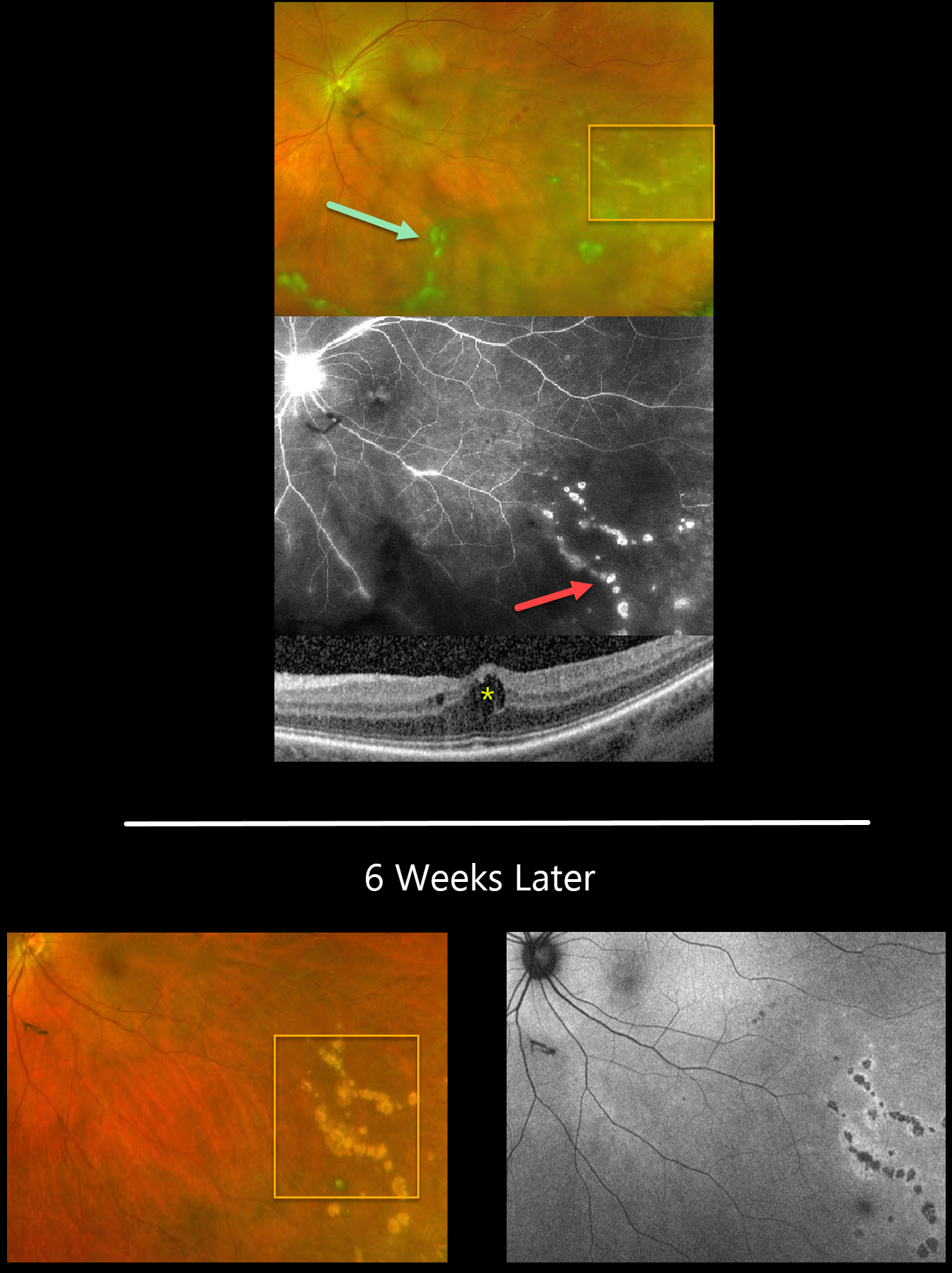
WEST NILE VIRUS
Lisa Faia
Originally posted on @retina.rocks June 28, 2023
This 51YO female presented with a recent history of floaters in her left eye. There was no significant past ocular or medical history. Vision was 20/30 OS, and the right eye was normal.
Optos RG imaging shows clumps of vitreous floaters over the posterior pole, extending into the inferior periphery. A few retinal hemorrhages are noted in the temporal midperiphery, and faint, small, multifocal atrophic scars are seen inferotemporally.
Fluorescein angiography better shows these multifocal scars with late staining. There is late optic nerve leakage and variable focal staining of the retinal vessels. OCT scanning shows mild cystoid edema.
We were concerned for possible herpes viral retinitis, and she was started on valtrex, had an anterior chamber tap, and a uveitis workup. The tap came back negative for herpes viruses. West Nile titers were ordered, and these came back positive for IgG (2.48) and low but detectable for IgM (0.10). Valtrex was stopped, and Durezol QID OS was started with a slow taper.
When she returned 6 weeks later, the vitreous was clear, and linear clumps of atrophic scars followed the retinal vessels. The scars were hyper-autofluorescent with small rims of increased autofluorescence.
Learning Points:
West Nile virus is an RNA virus transmitted by the bite of a mosquito that has fed on an infected bird. Although most infections are asymptomatic, about 20% will develop fever, weakness, headache, myalgia, nausea, and skin rash.
Multifocal chorioretinitis with or without vitreous inflammation is the most common ocular finding, occurring in about 80% of cases. Acutely, there are usually about 10-50 deep, 200-1000-micron yellow-white, scattered or radiating, curvilinear, clustered lesions that are relatively depigmented upon resolution.
Treatment of the primary viral infection is supportive. See a great review on this topic by Garg and Jampol (Surv Ophthalmology 2005;50:3-13).
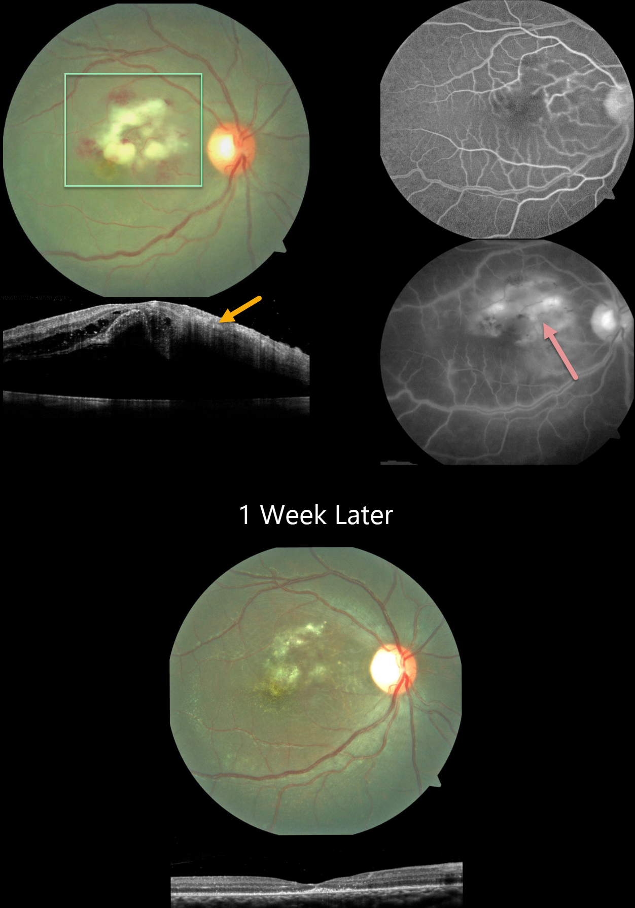
POST-FEVER DENGUE RETINITIS
Vidhya Nadahalli, Asma Samsudeen and Ashish Sharma
Originally posted on @retina.rocks June 8, 2023
This 15YO boy had a 3-week history of Dengue fever followed by severe vision loss in his right eye. Vision was counting fingers OD and 20/20 in his normal left eye.
Color imaging shows Purtscher-like central macular findings of acute inner macular ischemic whitening with retinal hemorrhages. OCT scanning shows retinal thickening with increased inner retinal hyperreflectivity. Fluorescein angiography shows leakage and staining from the ischemic tissue.
Dengue testing was positive for IgG and negative for IgM antibodies. Weil Felix testing was positive for OX2 and OX19, indicating a likely rickettsial coinfection. Oral antibiotics and steroids were started.
One week later, vision improved to 20/120. The macular findings were resolving. OCT scanning shows complete resolution of the prior edema with secondary thinning and disorganization of the outer foveal layers.
Learning Points:
Epidemic (post-fever) retinitis was described by Kawali et al (Ocular Immunology & Inflammation 2019;27:571-577) as an infectious or para-infectious uveitis occurring days to weeks following a febrile illness due to Dengue, Rickettsiosis, West Nile, Leptospirosis, Chikungunya, Typhoid, and Ebola. It appears as a usually bilateral Purtscher ’s-like retinitis involving the posterior pole with a favorable response to steroids. It is most common in tropical countries like India. See Mahendradas et al (Indian J Ophthalmol 2020;68:1775-1786) for an excellent post-fever retinitis review.
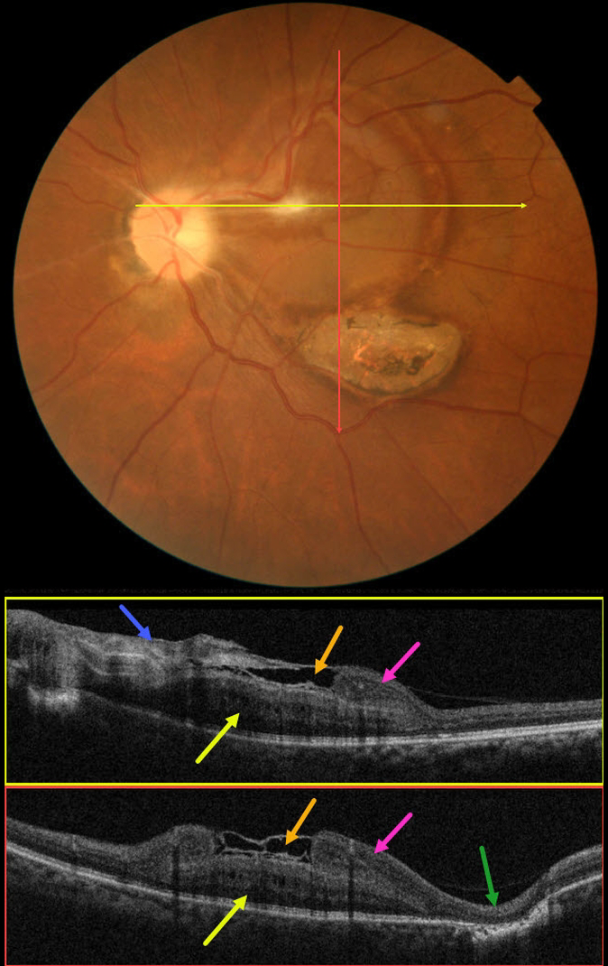
MYSTERY CASE
Asma Samsudeen and Ashish Sharma
Originally posted on @retina.rocks April 7, 2023
This 61YO male presented with 16 days of blurred vision and floaters in his left eye. There was no prior ocular history, and the past medical history was normal. Vision was 20/30 in his normal right eye and 20/200 in his left eye.
Color imaging shows an unusual circular area of subretinal yellow-orange pigmentary changes extending temporally from the optic nerve throughout the central and superior macula. A contracted localized area of premacular fibrosis is noted in the superonasal fovea. A flat area of variably pigmented chorioretinal scarring is found inferotemporally.
OCT B-scans show a constellation of interesting findings. The nasal epimacular fibrosis extends temporally, giving the appearance of either an ILM drape or local separation over the macular center. More temporally disorganized hyperreflective tissue descends and seems to merge into definite retinal inner tissue. There is complete outer retinal loss overlying the inferotemporal area of scarring.
The deeper retina is occupied by a mound-like area of alternating hyper- and hyporeflective bands. It is unclear whether this represents thickened outer retina or an unusual scar, as it appears distinct and anterior to the well-visualized ellipsoid zone below.
We believe this constellation of findings most likely represents changes following toxoplasmosis chorioretinitis, but we also can’t rule out an independent combined hamartoma. Or maybe we’re dealing with another diagnosis. What do you think?
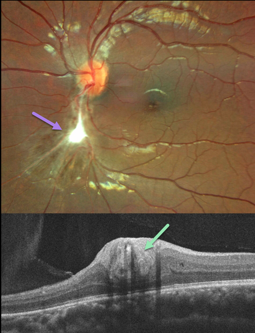
TOXOCARA
Will Gibson
Originally posted on @retina.rocks March 23, 2023
This healthy 13YO boy presented with a history of reduced vision in the left eye for 3 months. At the time of onset, he also reported having a red eye and headache.
Vision was 20/20 in his normal right eye and 20/70 in his left eye. Optos color imaging shows a triangular area of chorioretinal scarring extending inferiorly from the optic nerve. White fibrotic inner retinal scarring is noted more centrally.
OCT scanning through this region shows a thickened, featureless, hyperreflective inner retina.
After examination, further questioning revealed he was around multiple dogs at home, one of which had recalcitrant worms. Toxocara IgG antibody testing was surprisingly negative.
Learning Points:
Toxocariasis is a rare infection caused by the Toxocara canis roundworm, which matures in the dog intestine. The eggs are passed in stool, which can be ingested when young children come into contact with infected sand or dirt and then touch their mouths.
The ingested egg matures into a larva, which enters the systemic circulation from the intestine and can then migrate into the eye.
Death of the organism can cause variable findings, including endophthalmitis (typically a completely white and quiet eye), posterior granuloma, or peripheral granuloma. Most commonly in our experience, they present as a triangular-shaped area of vitreous base organization/scarring, with a fold of retina extending more posteriorly.
A positive test can help establish the diagnosis, although a false-negative result may occur, as sensitivity depends on the site of infection, the level of parasitic burden, and the timing of sample collection relative to exposure.
In our patient, despite the negative antibody testing, we still feel the history and clinical findings are most consistent with Toxocara. Other possible diagnoses include a combined hamartoma and posterior persistent fetal vasculature.
Regardless of the diagnosis, we expect the macular appearance to remain stable going forward and not require any treatment.
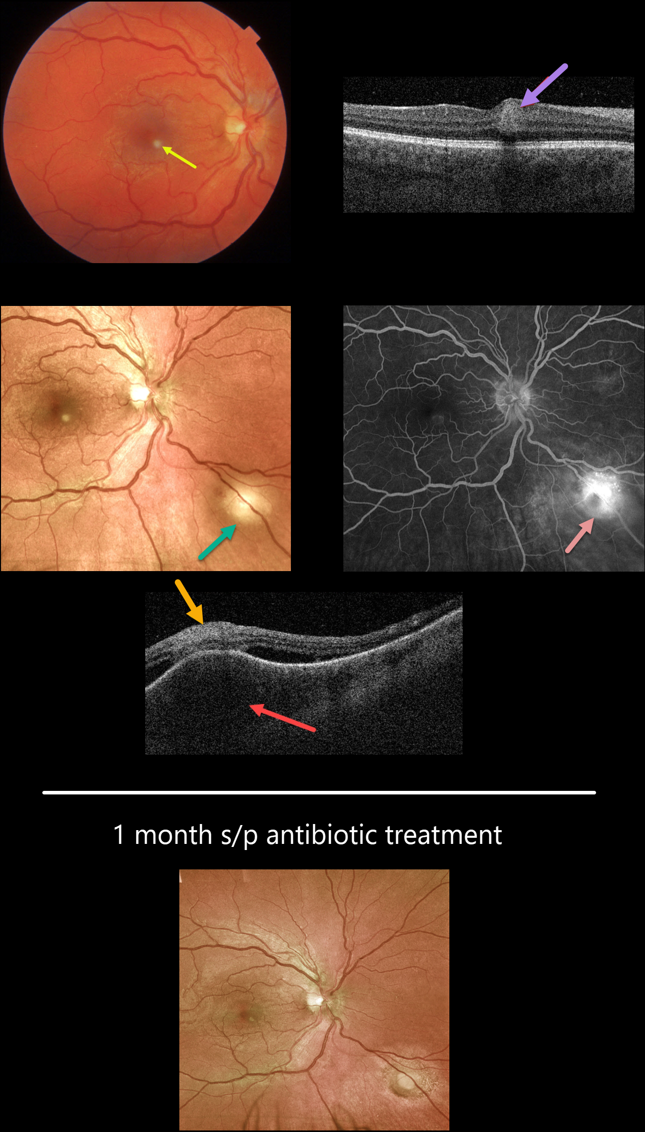
BARTONELLA
Originally posted on @retina.rocks January 24, 2023
This healthy 27YO male presented with a few days of a paracentral scotoma in his right eye. Vision was 20/25 OD and 20/20 OS.
Color imaging shows a small inferonasal foveal area of inner retinal opacification. Swept-source OCT through this region shows inner retinal hyperreflectivity. An additional, larger subretinal lesion is noted in the inferonasal midperiphery. OCT scanning through this lesion shows marked choroidal thickening, overlying retinal disorganization and hyperreflectivity, and some surrounding subretinal fluid. Fluorescein angiography shows moderate leakage from this choroidal lesion.
On further questioning, the patient told us that he recently adopted 2 stray cats. He didn’t recall any cat scratches, rashes, or adenopathy, but Bartonella titers were markedly elevated (IgG 1:1280, IgM 1:400).
Following infectious disease consultation, he started a 6-week course of oral doxycycline and rifampin. Four weeks later, vision was 20/20, and both lesions had significantly improved.
Learning Points:
Bartonella Henselae is the bacterium associated with cat scratch disease. It is transmitted from cat fleas (carrying the bacterium) or from flea feces that can be present on the cat’s claws/teeth, and transferred to humans from a cat scratch or bite.
Common systemic findings include fever, malaise, and progressive lymphadenopathy. These findings usually occur within 1-2 weeks of a cat scratch/bite.
Ocular effects primarily include neuroretinitis with rare cases of Parinaud’s ocular glandular syndrome, or, as in our patient’s case, chorioretinitis.
In our practice, posterior segment Bartonella involvement most commonly presents with neuroretinitis. Our patient’s inflammation spared the nerve, with multifocal involvement including the inner retina and choroid.
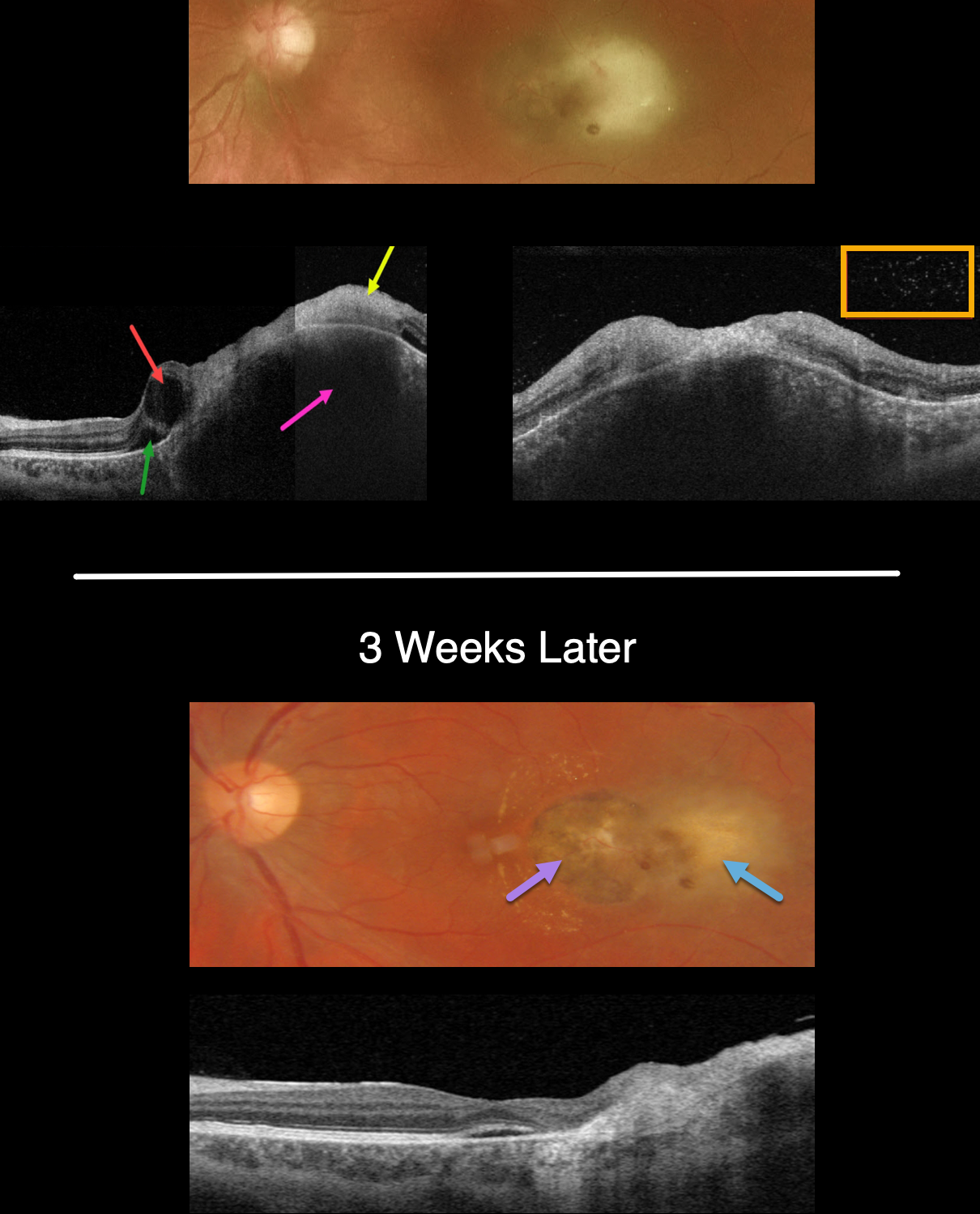
TOXOPLASMOSIS
Originally posted on @retina.rocks December 19, 2022
This 34YO male presented with recent vision loss of counting fingers vision in his left eye. He gave a history of prior ocular toxoplasmosis.
Optos color imaging shows opaque and white retina temporal to the fovea. Swept-source OCT shows the clinical retinitis involving full-thickness retina and is markedly hyperreflective. The underlying choroid is markedly thickened. The temporal fovea has a large cyst with a small amount of subretinal fluid. Overlying vitreous cells are noted.
The patient was started on a six-week course of oral Bactrim-DS BID. Laboratory testing for toxoplasmosis showed high IgG and negative IgM titers. ACE, lysozyme and QuantiFERON TB were negative.
The chorioretinal inflammation improved at each visit. When last seen 3 weeks into therapy, the likely pre-existing temporal pigmented toxoplasmosis scar is seen with resolving temporal retinitis. A few dots of retinal blood are seen, and faint lipid is noted nasally.
OCT scanning shows resolved vitreous cells and markedly decreased choroidal thickening.
Learning Points:
Toxoplasmosis, the most common cause of infectious chorioretinitis in humans, is caused by the protozoan parasite, Toxoplasma gondii.
It is most commonly transmitted through infected felines or by eating raw meat. The majority of infections are acquired, although it can also be transmitted congenitally.
The body’s immune system is unable to completely kill the organism, which often lies dormant within a chorioretinal scar for years. When the immune balance favors the encysted organism, active chorioretinitis develops. This usually resolves spontaneously within 6 weeks.
If the optic nerve or macular are threatened, a six-week course of Bactrim DS is recommended (see Ophthalmology 2004;112:1876-1882). Oral prednisone can be added as well to help with severe inflammation.
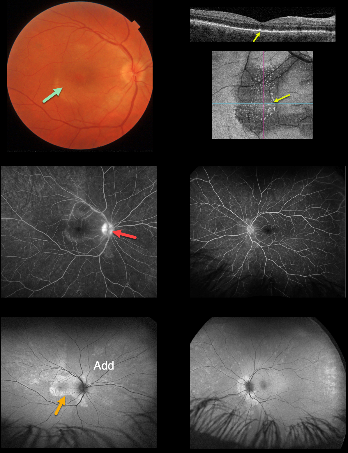
SYPHILIS
Originally posted on @retina.rocks October 14, 2022
This 49YOM presented with a one-week history of blurred central vision in his right eye. There was no past medical history.
Vision was 20/400 OD and 20/25 OS. Anterior segments were normal. An oval, whitish submacular lesion was noted, and the optic nerve also appeared mildly swollen in the right eye.
OCT scanning of the right macula shows numerous, needle-like projections extending from the RPE into the outer retina. En face imaging best shows the hyperreflective outer retinal opacities.
Fluorescein angiography shows staining of the right nerve, mild peripapillary subretinal staining OU, and macular staining OD.
Fundus autofluorescence (FAF) shows much more extensive findings than those noted clinically, with peripapillary hyper-FAF OU, macular hyper-FAF OD corresponding to the placoid lesion, and peripheral hyperreflective dots bilaterally.
Treponema pallidum antibodies were reactive, with a high quantitative RPR (1:32). He was started on parenteral penicillin.
Learning Points:
Ocular manifestations of syphilis vary widely and include virtually all variations of anterior and posterior segment inflammation, including a recently described presentation as the multiple evanescent white dot syndrome phenotype (Russel et al Int Ophthalmol 2020;40:627-738). Hence, the reason it’s been dubbed the great imitator or masquerader.
One of the more common presentations is placoid chorioretinitis (Eandi et al., Retina 2012;32:1915-1941), which was evident on FAF imaging in our patient.
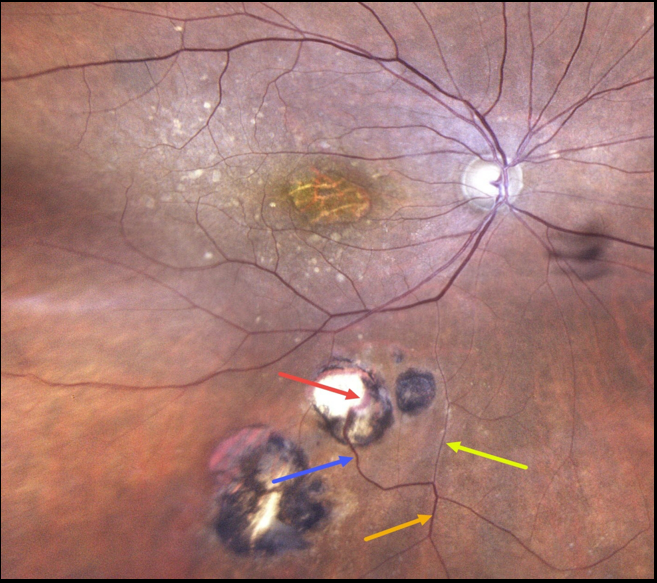
TOXOPLASMOSIS WITH RETINAL CHOROIDAL ANASTOMOSIS
Originally posted on @retina.rocks September 20, 2022
This 86YO female has a history of atrophic age-related macular degeneration with vision of 20/400 OD and 20/60 OS. Optos imaging of her right eye shows foveal atrophy with surrounding mixed drusen.
Multifocal inactive toxoplasmosis scars are noted inferiorly. An inferior retinal vein bifurcates, with a markedly attenuated branch continuing on its path towards the optic nerve. The other branch has a more normal caliber, dives into one of the toxoplasmosis scars, and drains into the choroid.
Learning Points:
Under normal conditions, the retinal and choroidal circulations remain separate and distinct. Rarely, the two circulations connect directly through a chorioretinal anastomosis.
These can occur in chorioretinal scars (typically from inactive toxoplasmosis lesions, as in this patient), in macular telangiectasia, and in disciform scars.
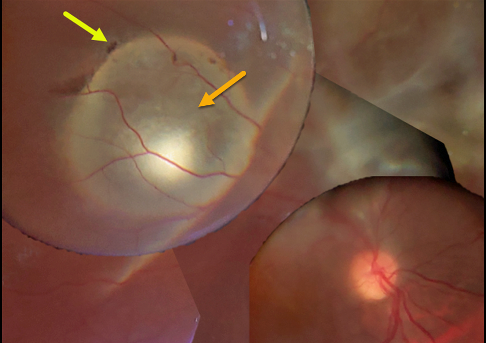
CYSTICERCOSIS WITH RHEGMATOGENOUS RETINAL DETACHMENT
Chhaya Bharti
Originally posted on @retina.rocks September 14, 2022
This 15YO boy presented with 20/200 vision. A large subretinal cysticercosis cyst is noted in the right distal superotemporal macula.
There is an overlying rhegmatogenous retinal detachment caused by a small retinal break. Unfortunately, the patient was subsequently lost to follow-up.
Learning Points:
Ocular cysticercosis is a rare parasitic eye infection caused by a pork tapeworm (larval form of the cestode, Taenia solium).
The ocular adnexa are most commonly affected, but the encysted organism can appear within the eye, including the anterior segment, vitreous cavity, and the subretinal space, as in this patient.
Vitrectomy with complete removal of the subretinal cyst is required since the dead organism can cause a severe inflammatory response.
For a great recent cysticercosis in ophthalmology review article, see Pujari et al, Surv Ophthalmology 2022;67:544-569.
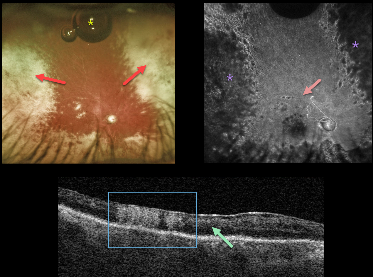
ACUTE RETINAL NECROSIS SYNDROME
Originally posted on @retina.rocks July 15, 2022
This 58YO male has an extremely complicated past ocular history beginning with pseudoexfoliative glaucoma with a steroid response. Following cataract surgery in 2012, his intraocular lens dislocated in February 2016, requiring vitrectomy with a secondary anterior chamber implant. Pseudophakic bullous keratopathy developed, requiring DMEK surgery in July 2018.
His intraocular pressure subsequently increased, eventually requiring multiple glaucoma procedures, including Baerveldt implantation. Additional DMEK procedures were performed in September 2020 and December 2021. A minor additional corneal procedure with injection of gas into the anterior chamber was performed on 5/5/22 for some host Descemet’s that extended under the donor graft, causing recurrent corneal edema.
When examined on 6/24/22, the cornea was finally clear enough to allow for visualization of the posterior segment, which hadn’t been possible during his multiple anterior segment procedures. Vision was light perception.
Optos imaging shows white chorioretinal scarring in the nasal and temporal peripheries with a small amount of residual intraocular gas that had migrated into the vitreous cavity. Triton swept source OCT shows diffuse disorganization of all retinal layers, with some areas of full-thickness hyperreflectivity. Fluorescein angiography shows near total loss of all retinal perfusion, with peripheral hypofluorescence from the white peripheral chorioretinal scarring.
We felt his findings were most consistent with acute retinal necrosis, and oral valaciclovir was started.
Learning Points:
ARNS is most commonly caused by the Herpes zoster virus. It usually occurs in immunocompetent individuals, and is a rapidly progressive panuveitis with hemorrhagic and ischemic unilateral (ARNS) or bilateral (BARNS) retinal necrosis that typically begins in the retinal periphery.
ARNS is often a devastating and blinding infection due to the high risk for retinal detachment as well as macular and optic nerve involvement. The latest meta-analysis argues for systemic antiviral therapy and prophylactic vitrectomy, with uncertain benefits for prophylactic laser photocoagulation or adjunctive intravitreal antivirals (see Zhao et al, Retina 2021;41:965-978).
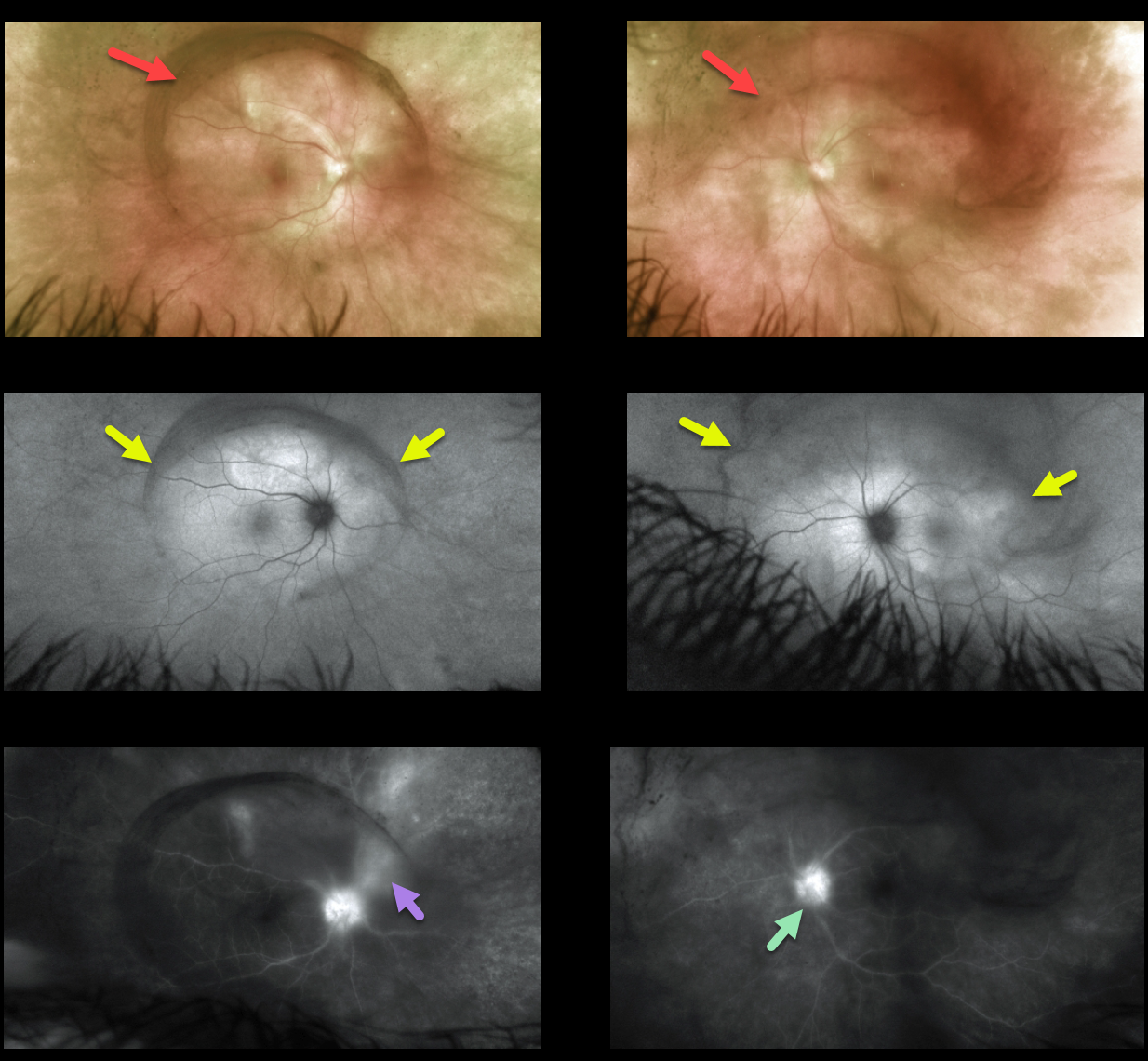
SYPHILIS
Originally posted on @retina.rocks June 2, 2022
This 49YO female presented with a 1-month history of bilateral vision loss and eye pain. Vision was 20/200 OU. There was a mild-moderate, bilateral nongranulomatous anterior uveitis.
Vitreous debris was centered over each posterior pole, which was grossly normal.
Fundus autofluorescence (FAF) shows an oval region of placoid hyper-FAF in each posterior pole. Fluorescein angiography shows bilateral nerve leakage and vasculitis.
RPR and treponema pallidum antibody testing were positive, and she was immediately started on intravenous penicillin.
Learning Points:
Ocular manifestations of syphilis vary widely and include virtually all variations of anterior and posterior segment inflammation, including a recently described presentation as the multiple evanescent white dot syndrome phenotype (see Russel et al Int Ophthalmol 2020;40:627-738). Hence, the reason it’s been dubbed the great imitator or masquerader.
One of the more common presentations is placoid chorioretinitis, which was evident on FAF imaging in our patient.
Patients also need to be evaluated for HIV since syphilis and HIV infections often coexist. Fortunately, our patient was HIV negative.
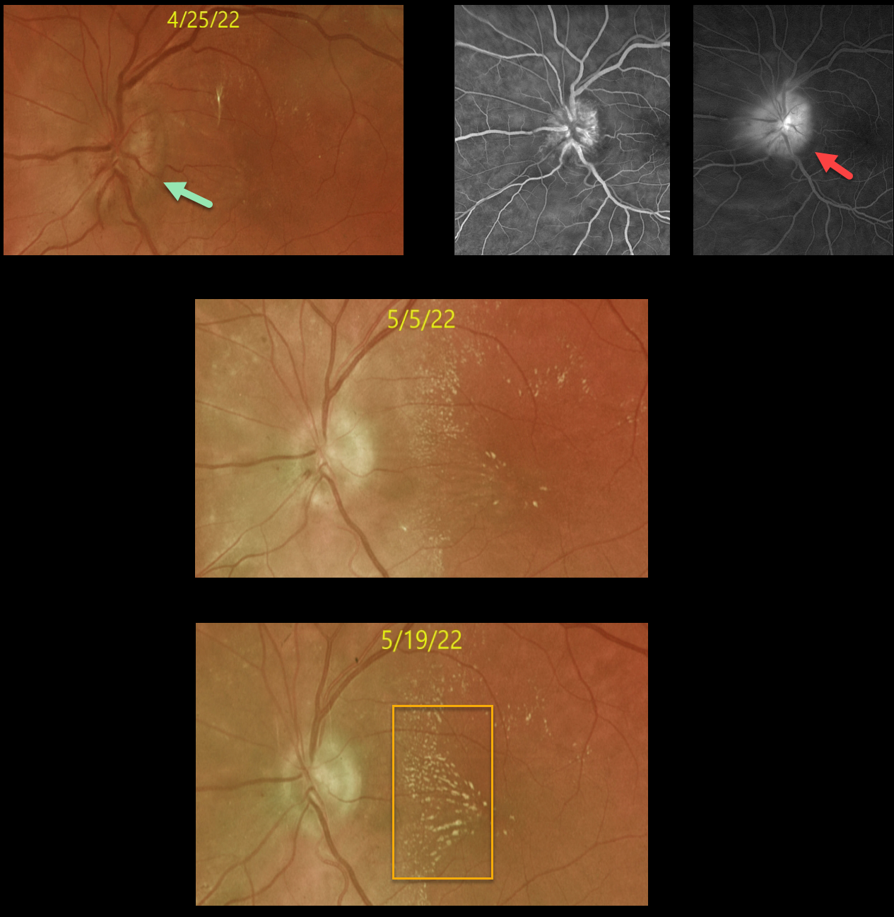
LEBERS IDIOPATHIC STELLATE NEURORETINITIS
Originally posted on @retina.rocks June 1, 2022
A 44YO female with type 2 diabetes presented on 4/25/22 with a 2-week history of severe vision loss in her left eye. Vision was counting fingers OS.
Ocular examinations were normal except for a markedly swollen left optic nerve. There were very faint, scattered, hard exudates.
OCT showed an average nerve fiber layer thickness of 370 microns (not shown). Fluorescein angiography shows diffuse optic nerve leakage.
Extensive bloodwork was negative, including Bartonella, syphilis, Lyme, Rocky Mountain spotted fever, CRP, ACE, and serum lysozyme. MRI scanning was also normal.
Over the next 3-4 weeks, the optic nerve edema resolved with the development of a nasal lipid star. Unfortunately, vision remained at counting fingers.
Learning Points:
The diagnosis for a unilateral swollen nerve is extensive. However, it becomes much smaller when associated with acute inner retinal ischemia (not seen in our case) and later lipid exudation within Henle’s layer (neuroretinitis), including cat scratch (Bartonella), Lyme disease, and syphilis.
When no underlying cause is found, the entity is called Leber’s idiopathic stellate neuroretinitis.
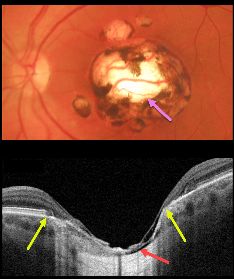
TOXOPLASMOSIS
Originally posted on @retina.rocks May 16, 2022
This 48YO male gave a history of lifelong poor vision in his left eye. Vision was 20/40 OD and 20/200 OS.
Triton color imaging shows a large, round, variably pigmented, excavated macular scar with a few smaller surrounding scars.
Swept-source OCT reveals a thinned atrophic retina and RPE overlying a shallow coloboma. Within the coloboma, the choroid is virtually absent, and the sclera is excavated. The yellow arrows mark the hyper-reflective plane of the surrounding intact retina and RPE. The right fundus was normal.
Learning Points:
For a great discussion on how to differentiate these infectious macular colobomatous lesions (congenital toxoplasmosis, Zika, and cytomegalovirus) from inherited dystrophies like North Carolina Macular Dystrophy (NCMD), see Kumar and Mahalingam AJO 2019;200:47-56.
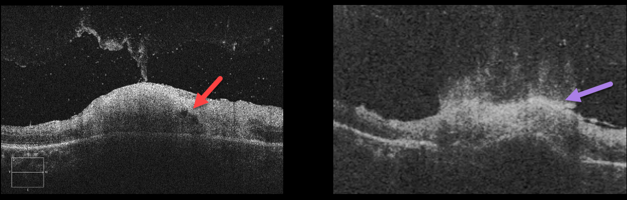
TOXOPLASMOSIS
Originally posted on @retina.rocks May 4, 2022
This is a great case of active toxoplasmosis.
The OCT B-scans show full-thickness retinitis with a volcanic-like eruption of the inflammatory process extending into the attached vitreous.
Learning Points:
Toxoplasmosis, the most common cause of infectious chorioretinitis in humans, is caused by the protozoan parasite, Toxoplasma gondii.
It is most commonly transmitted through infected felines or by eating raw meat. The majority of infections are acquired, although they can also be transmitted congenitally.
The body’s immune system is unable to completely kill the organism, which often lies dormant within a chorioretinal scar for years. When the immune balance favors the encysted organism, active chorioretinitis develops. This usually resolves spontaneously within 6 weeks.
If the optic nerve or macula is threatened, a six-week course of Bactrim DS is recommended (see Ophthalmology 2004;112:1876-1882).
Oral prednisone can be added as well to help with severe inflammation.
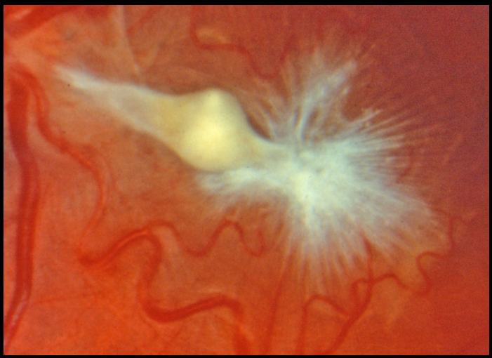
MACULAR PUCKER FOLLOWING ENDOPHTHALMITIS
Originally posted on @retina.rocks March 31, 2022
This eye shows a severely contracted and complex epimacular membrane in a patient who recovered from prior endophthalmitis. Unfortunately, we don’t have any further clinical information.
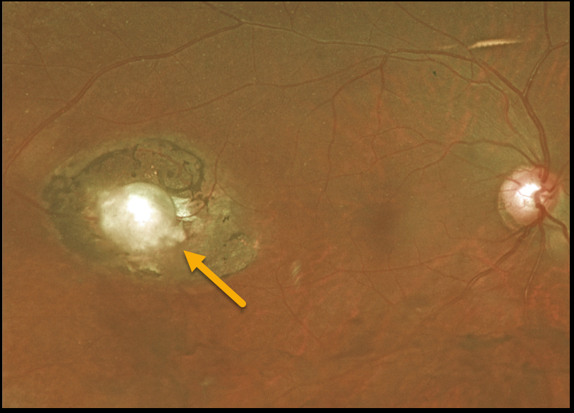
TOXOPLASMOSIS
Originally posted on @retina.rocks March 11, 2022
This 55YO female presented with 20/30 vision in her right eye and an asymptomatic colobomatous scar in her distal temporal macula. The left eye was normal.
We felt this lesion most likely represented an inactive toxoplasmosis scar.
Learning Points:
For a great discussion on how to differentiate these infectious macular colobomatous lesions (congenital toxoplasmosis, Zika, and cytomegalovirus) from inherited dystrophies, see Kumar and Mahalingam, AJO 2019;200;47-56.
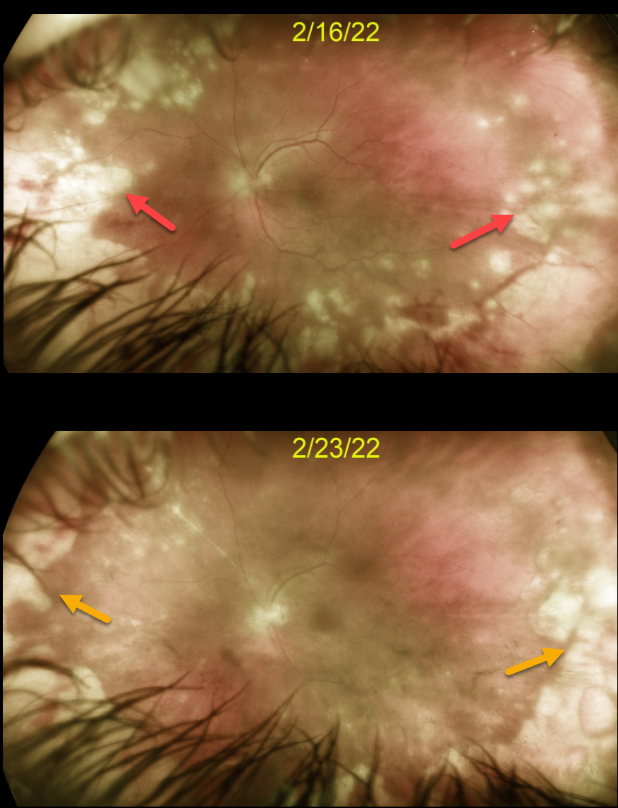
ACUTE RETINAL NECROSIS SYNDROME
Originally posted on @retina.rocks March 10, 2022
This 44YO otherwise healthy male presented with 1 week of floaters, blurred vision and pain in his left eye. Vision was 20/20 in his normal right eye and 20/100 in his left eye. There was panuveitis in his left eye, including a moderate granulomatous anterior uveitis, vitritis, and peripheral multifocal mostly outer white retinitis.
We diagnosed him with acute retinal necrosis syndrome (ARNS), and started him on oral valganciclovir 900 mg BID and a baby aspirin daily. Several days later, oral prednisone 60mg/day was begun.
After 1 week, vision remained at 20/100 but with subjective improvement. The retinitis was significantly improving. We continue to follow him very closely.
Learning Points:
The Herpes zoster virus most commonly causes ARNS. It usually occurs in immunocompetent individuals and is a rapidly progressive panuveitis with hemorrhagic and ischemic unilateral (ARNS) or bilateral (BARNS) retinal necrosis, typically beginning in the retinal periphery.
ARNS is often a devastating and blinding infection due to the high risk for retinal detachment as well as macular and optic nerve involvement.
The latest meta-analysis argues for systemic antiviral therapy and prophylactic vitrectomy, with uncertain benefits for prophylactic laser photocoagulation or adjunctive intravitreal antivirals (see Zhao et al, Retina 2021;41:965-978).
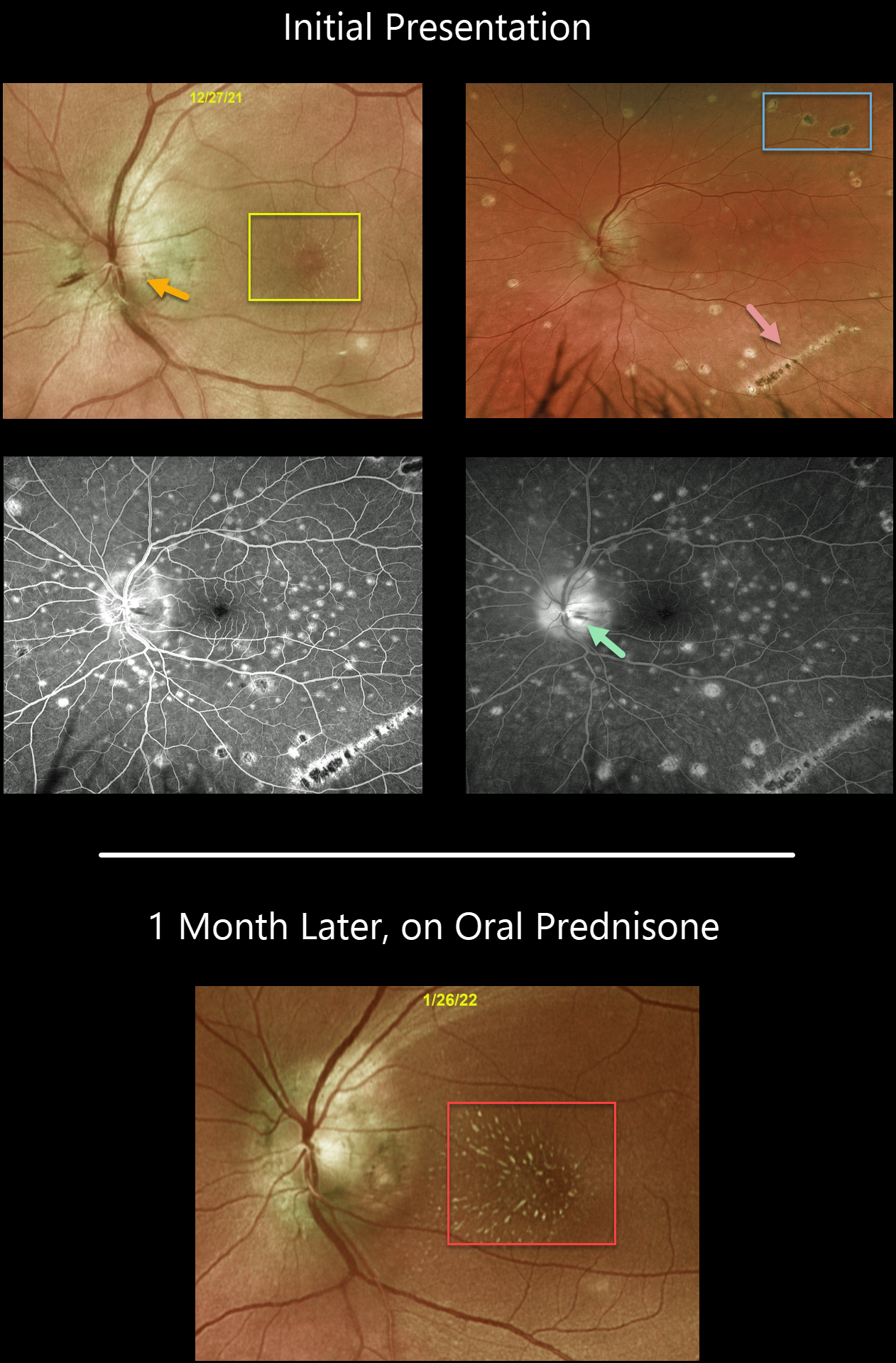
LEBER’S IDIOPATHIC STELLATE NEURORETINITIS
Originally posted on @retina.rocks February 17, 2022
This healthy 46YO male presented on 12/27/21 with 1 day of decreasing vision in his left eye. Vision was 20/25 in his normal right eye and 20/60 in his left eye.
The left nerve was swollen with a faint macular lipid star. There were bilateral multifocal scars and an inferotemporal streak lesion in his left eye. Fluorescein angiography shows minimal left optic nerve staining.
An extensive workup, including labs for Bartonella, Lyme, tuberculosis, sarcoidosis, toxoplasmosis, syphilis, and a chest x-ray, was all negative. He denied any systemic symptoms, although he recently had a COVID-19 vaccine. Serology showed positive IgG and negative IgM antibodies for COVID-19.
He was treated with a tapering 1-month course of oral prednisone, and at his 1/26/22 visit, the nerve edema had resolved, with a more prominent macular lipid star.
Learning Points:
The diagnosis for a unilateral swollen nerve is extensive. However, it becomes much smaller when associated with inner retinal ischemia and lipid exudation (neuroretinitis), including cat scratch (Bartonella), Lyme disease, and syphilis. When no underlying cause is found, the entity is called Leber’s idiopathic stellate neuroretinitis. The condition is self-limited, so treatment is not usually recommended.
We gave our patient the presumed diagnosis of Leber’s idiopathic stellate neuroretinitis due to the unilateral swollen nerve followed by a lipid star, in the absence of a positive infectious workup. The bilateral multifocal scars appeared inactive and were likely present long before he presented with a unilateral swollen nerve.
Our best guess is that the scarring represents ocular histoplasmosis or multifocal choroiditis unrelated to the swollen nerve, although we can’t rule out that the two might be related.
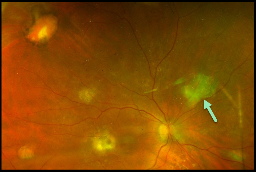
CANDIDA ENDOPHTHALMITIS
Giuseppina Monteleone
Originally posted on @retina.rocks January 25, 2022
In October 2021, this 73YO emaciated woman presented for a routine cataract evaluation. Vision was 20/600 with moderate cell and flare bilaterally. Posterior segments were grossly normal, but the views were limited due to vitreous opacification.
There was a history of colon cancer treated with chemotherapy in 2018. In 2019, she suffered a cerebral hemorrhage and was later evacuated via craniotomy. In 2020, she developed a bowel obstruction during chemotherapy treatment, and total parenteral nutrition was initiated. In August 2021, a central line infection caused Candida albicans sepsis. Voriconazole was administered until blood cultures came back negative, and she became afebrile.
Based on the clinical history, she was felt to have bilateral fungal endophthalmitis despite the negative blood cultures and negative in-office vitreous and aqueous stains and cultures.
Fluconazole 400 mg/day and intravitreal Amphotericin B (0.01 mg/0.1 ml) were administered, with improvement in anterior segment inflammation but no change in the vitritis.
Eventually, a diagnostic vitrectomy was performed in the right eye, revealing yellowish retinal foci, typical for endogenous Candida albicans endophthalmitis, although Gram stain and cultures were negative.
Vision improved to 20/100 in the right eye. We plan on performing vitrectomy in her left eye when her clinical condition allows.
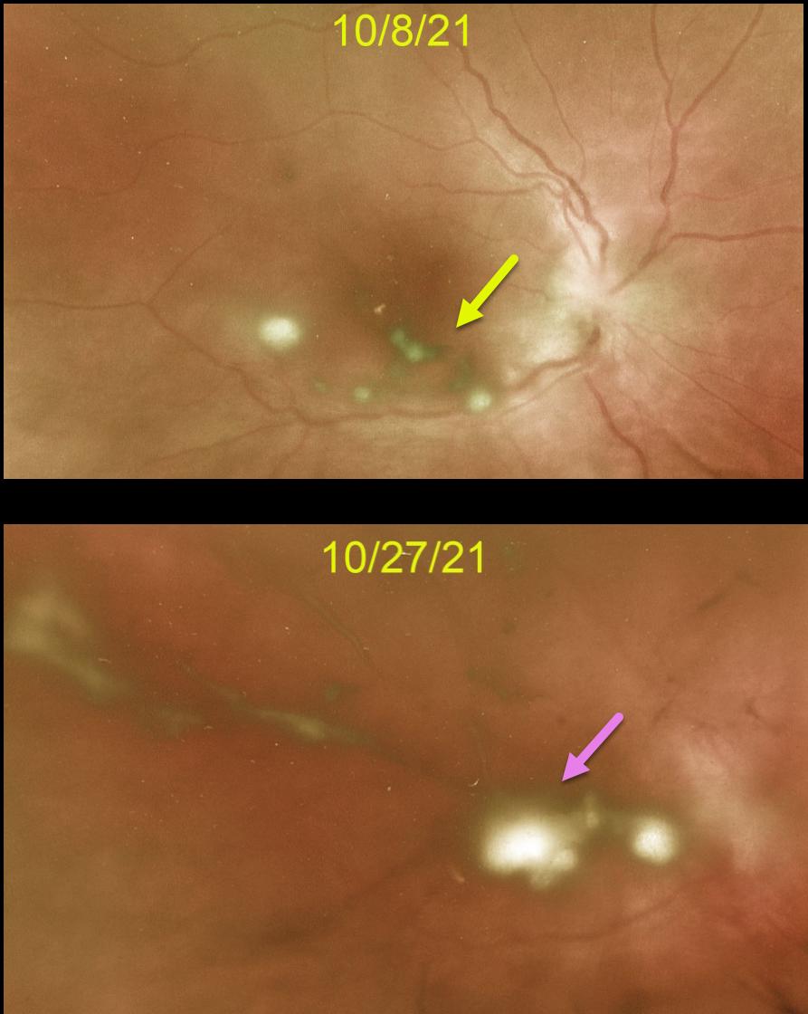
CANDIDA ENDOPHTHALMITIS
Originally posted on @retina.rocks December 31, 2021
This 37YO male presented with a 4-day history of right eye pain and blurred vision. There was no prior ocular history, surgery, recent trauma, or intravenous drug use.
Vision was counting fingers OD and 20/25 in his normal OS. Moderate cell and flare were noted OD, along with a tiny hypopyon. Optos imaging shows multiple posterior inflammatory puff balls suggestive of endogenous fungal endophthalmitis.
We ordered emergent blood work to rule out other infectious causes before initiating intravitreal or systemic antifungal therapy. Unfortunately, he was completely noncompliant about going for testing despite daily calls.
He subsequently returned to the office 2 weeks later with increased vitreous opacities. On further questioning, he gave a history of recent low back pain and inflammation, where 6 weeks earlier he underwent surgery with metal plates. We told him to go to the ER for candida sepsis workup immediately.
However, he went home and continued his noncompliance, which finally led him to our local teaching hospital 1 week later. We assume the infectious source was the hardware implanted in his back.
Learning Points:
Ocular candidiasis is characterized by single or multiple focal yellow-white chorioretinal lesions with overlying vitritis, vitreous haze, vitreous abscess (fluff balls or string of pearls), and other more generalized presentations of infection and inflammation, such as endophthalmitis, hypopyon, scleritis, and anterior chamber cells.
Exogenous or endogenous sources can cause ocular candidiasis. Exogenous routes include trauma and inoculation from surgical procedures.
Endogenous ocular candidiasis arises from hematogenous seeding of the chorioretinal blood vessels caused by fungemia, indwelling catheters, severely compromised immune systems, and abuse of broad-spectrum antibiotics.
Treatment includes removal of the inciting source of candidiasis (e.g., an indwelling catheter) and systemic therapy, with possible intravitreal antifungal therapy.
Routine ophthalmic screening for known systemic candida sepsis is not recommended unless there are suggestive signs or symptoms for ocular involvement (see Breazzano et al, Ophthalmology 2011;129:73-76).
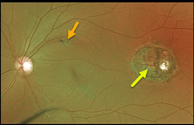
TOXOPLASMOSIS
Originally posted on @retina.rocks August 12, 2021
This 44YO male presented with 20/25 vision and a presumed inactive toxoplasmosis scar in the distal left macula. Although we do not have an OCT through this scar, it likely represents a colobomatous lesion.
This lesion somewhat resembles torpedo maculopathy, but the additional scar in the superior macula more suggests a prior inflammatory event.
Learning Points:
For a great discussion on how to differentiate these infectious macular colobomatous lesions (congenital toxoplasmosis, Zika, and cytomegalovirus) from inherited dystrophies like North Carolina Macular Dystrophy (NCMD), see Kumar and Mahalingam AJO 2019;200;47-56.
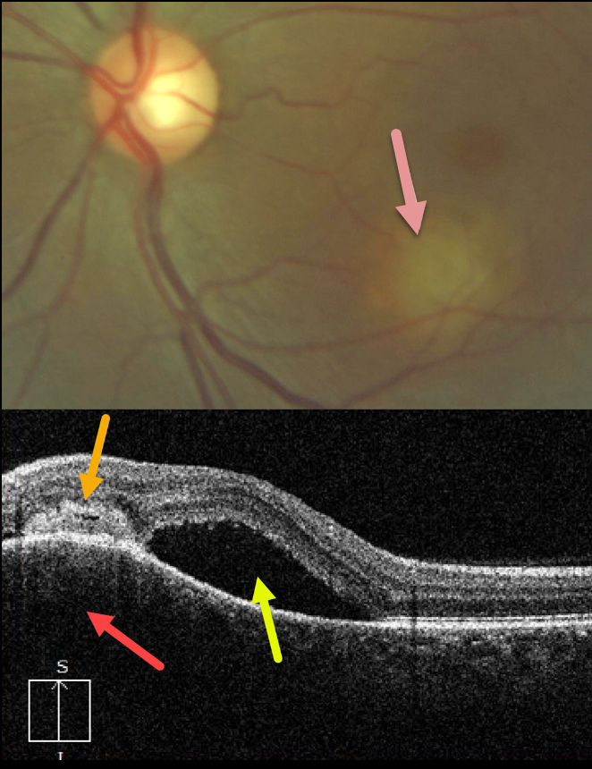
TUBERCULOSIS CHOROIDAL GRANULOMA
Originally posted on @retina.rocks July 12, 2021
This 33YO female presented with this symptomatic lesion in her left macula. Vision was 20/25 in her normal right eye and 20/300 in her left eye.
A granulomatous lesion is noted in the inferior left macula. A vertical OCT B-scan through this lesion shows choroidal hyporeflective thickening, an overlying subretinal/intraretinal hyperreflective lesion, and superior subretinal fluid.
Laboratory testing was positive for QuantiFERON-TB Gold, and negative for FTA, CBC, and ACE. Unfortunately, despite numerous attempts to reach this patient, she was lost to follow-up.
Learning Points:
Tuberculosis (TB) is a systemic disease caused by Mycobacterium tuberculosis, characterized by the formation of caseating granulomas throughout the body. Although pulmonary involvement is the most common, extrapulmonary sites can involve the gastrointestinal, skin, cardiovascular, genitourinary, and central nervous systems, as well as the eyes.
Ocular involvement, similar to syphilis, can mimic virtually any type of uveitis, including anterior, intermediate, posterior, and panuveitis, retinitis and retinal vasculitis, neuroretinitis, optic neuropathy, and choroidal granuloma, choroiditis, and scleritis.
For an excellent review of intraocular tuberculosis, see Bupta et al, Survey Ophthalmology 2007;52:561-587.
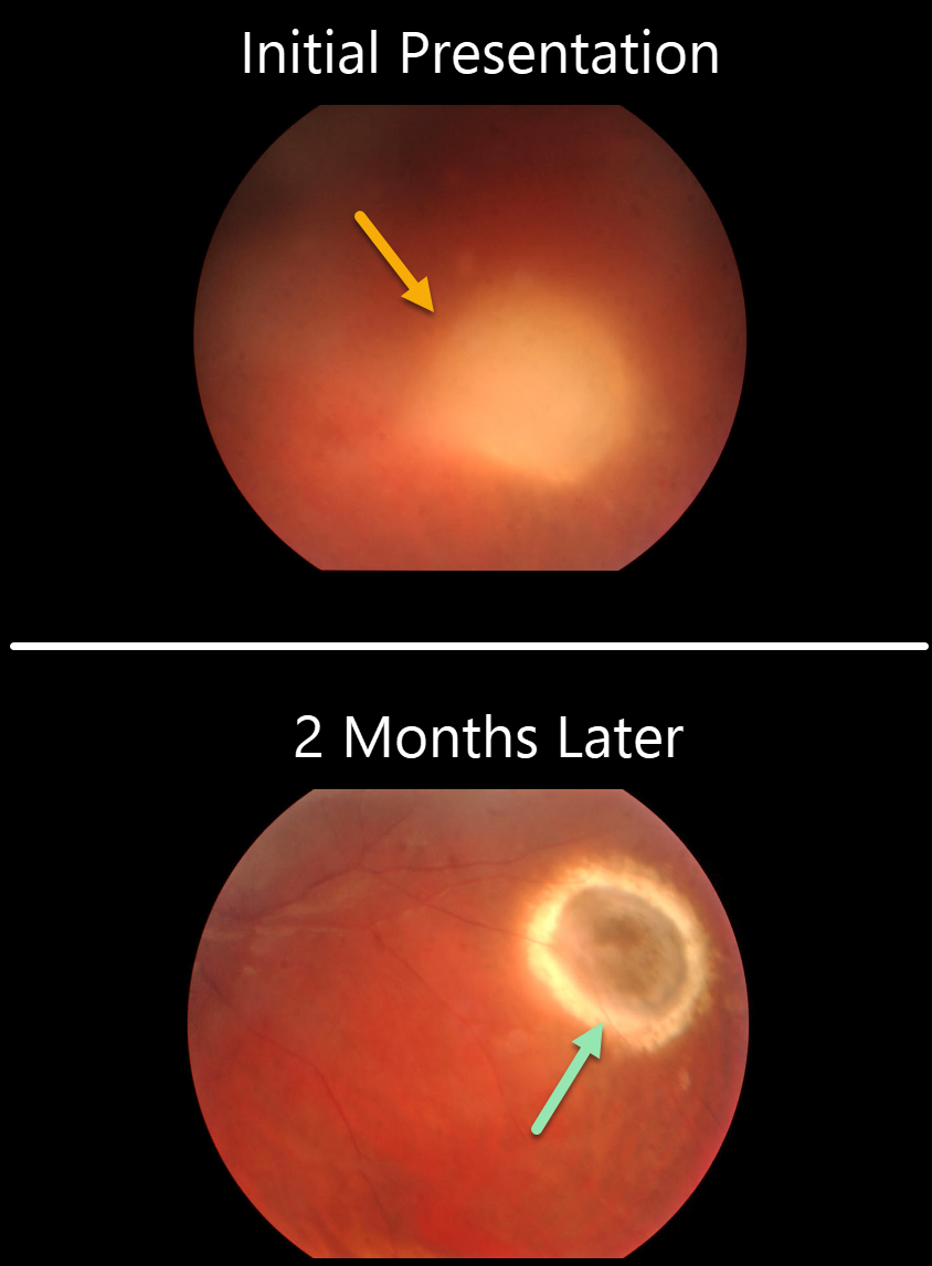
HERPES ZOSTER CHOROIDITIS
Originally posted on @retina.rocks June 24, 2021
Our patient presented with a focal, creamy-white inflammatory lesion obscured by dense vitritis. Vision was 20/80.
Interestingly, he gave a history of chickenpox five months earlier, which coincided with his visual symptoms.
Observation was recommended since he felt that his vision was improving. Two months later, the vitritis had improved, and the inflammatory lesion had resolved into a focal chorioretinal scar.
Learning Points:
We felt that our patient developed herpes zoster chorioretinitis secondary to an acute systemic varicella zoster infection.
Choroiditis is a somewhat rare finding with herpes zoster ophthalmicus (see Bloom and Snady-McCoy, AJO 1989;108;733-735). Our case beautifully shows both the acute and chronic sequelae of such an infection.
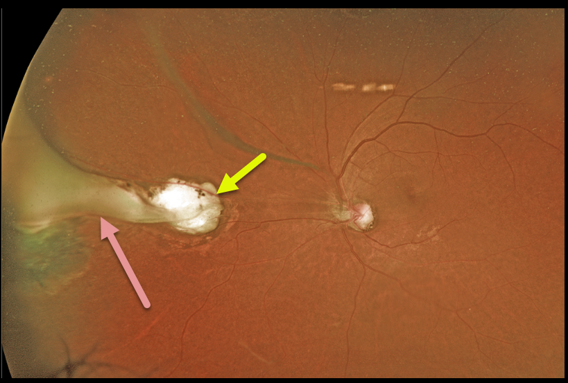
TOXOCARA
Originally posted on @retina.rocks June 8, 2021
This 63YO female was referred for these asymptomatic unilateral fundoscopic changes. Optos imaging shows a midperipheral atrophic chorioretinal scar. Associated with this scar is a whitish fold of retina that extends anteriorly as a broader area of traction and scarring in the vitreous base.
A positive ELISA test can help to establish the diagnosis. Since our patient was asymptomatic, we did not pursue any further workup and recommended observation.
Learning Points:
Toxocariasis is a rare infection caused by the Toxocara canis roundworm, which matures in the dog intestine. The eggs are passed in stool, which can be ingested when young children come into contact with infected sand or dirt and then touch their mouths.
The ingested egg matures into a larva, which enters the systemic circulation from the intestine and can then migrate into the eye.
Death of the organism can cause variable findings, including endophthalmitis (typically a completely white and quiet eye), posterior granuloma, or peripheral granuloma.
Most commonly, in our experience, they present later in life as a triangular-shaped area of vitreous base organization/scarring, with a fold of retina extending more posteriorly.
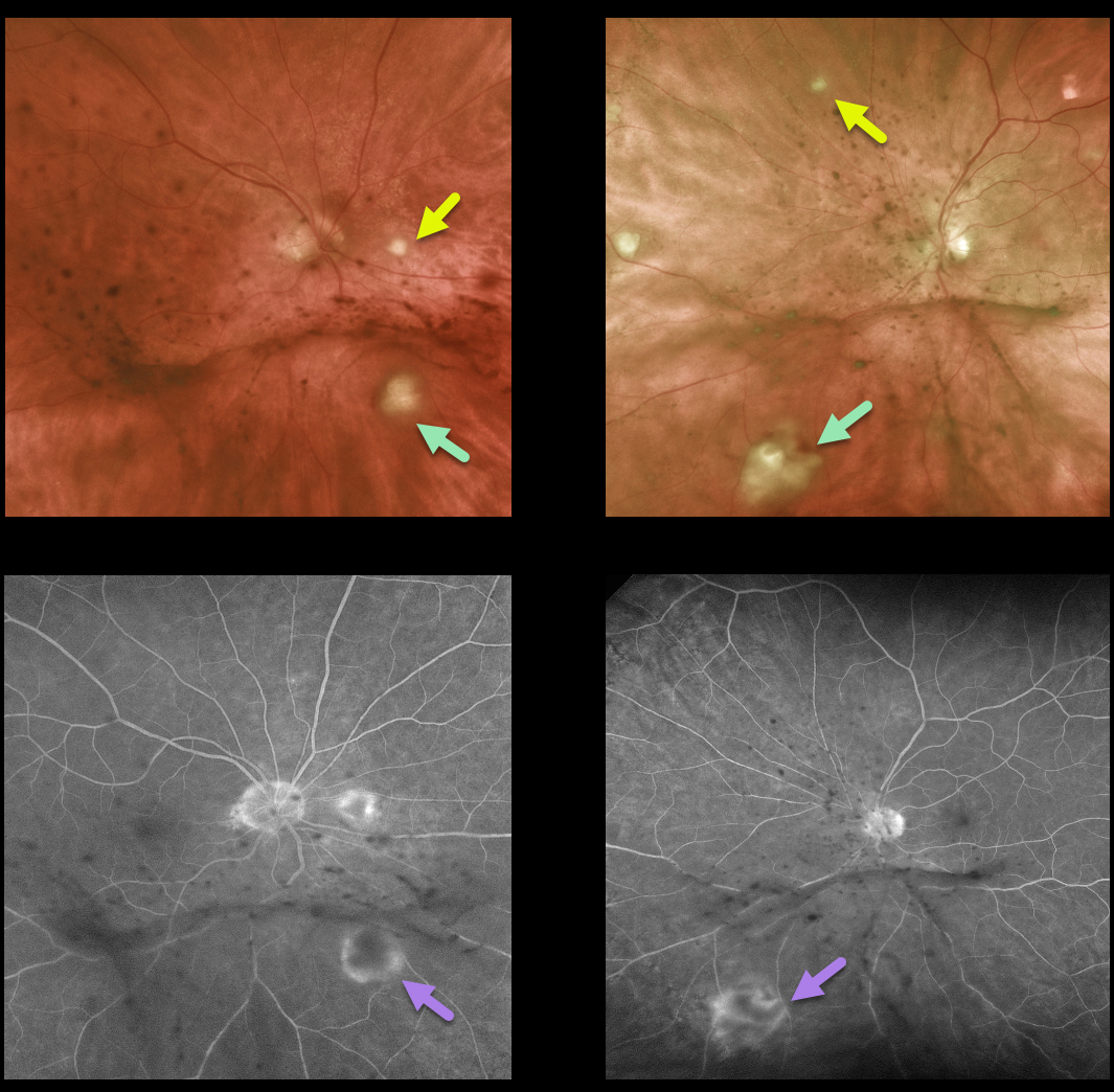
CANDIDA ENDOPHTHALMITIS
Originally posted on @retina.rocks May 20, 2021
This 75 yo retired physician presented with decreased vision and floaters for about 8 weeks. Vision was 20/25 OD and 20/70 OS. Anterior segments were normal except for mild nuclear sclerosis.
There are multifocal areas of yellow-white chorioretinal inflammation spreading into the overlying vitreous, along with clumps of inflammatory deposits on the back of each hyaloid. Fluorescein angiography reveals early blockage with late surrounding staining of these inflammatory lesions.
He gave a history of non-Hodgkin’s lymphoma, which was in remission. He still had a port in place despite having completed chemotherapy treatments. He was also being treated with oral prednisone and azithromycin for organizing pneumonia and had active oral candidiasis.
Based on his clinical findings, immunosuppression, chemotherapy port, and oral thrush, we suspected bilateral candidal endophthalmitis, although we were also concerned about an atypical presentation for intraocular lymphoma.
Diagnostic vitrectomy was performed. Cytology was negative for lymphoma, and Gram stain and fungal cultures were also surprisingly negative. Blood cultures were also negative.
Despite the negative workup, we still felt that fungal endophthalmitis was the most likely diagnosis and started oral fluconazole. Since the port was no longer in use, this was removed and did, in fact, grow out Candida. The intraocular inflammation subsequently improved.
Learning Points:
In many cases of endophthalmitis, no infectious agent is identified, and often the diagnosis is made clinically. Empiric treatment often includes intravitreal antibiotics. In this case, despite negative blood cultures and vitreous biopsy, our clinical suspicion ultimately led to the correct diagnosis and treatment.
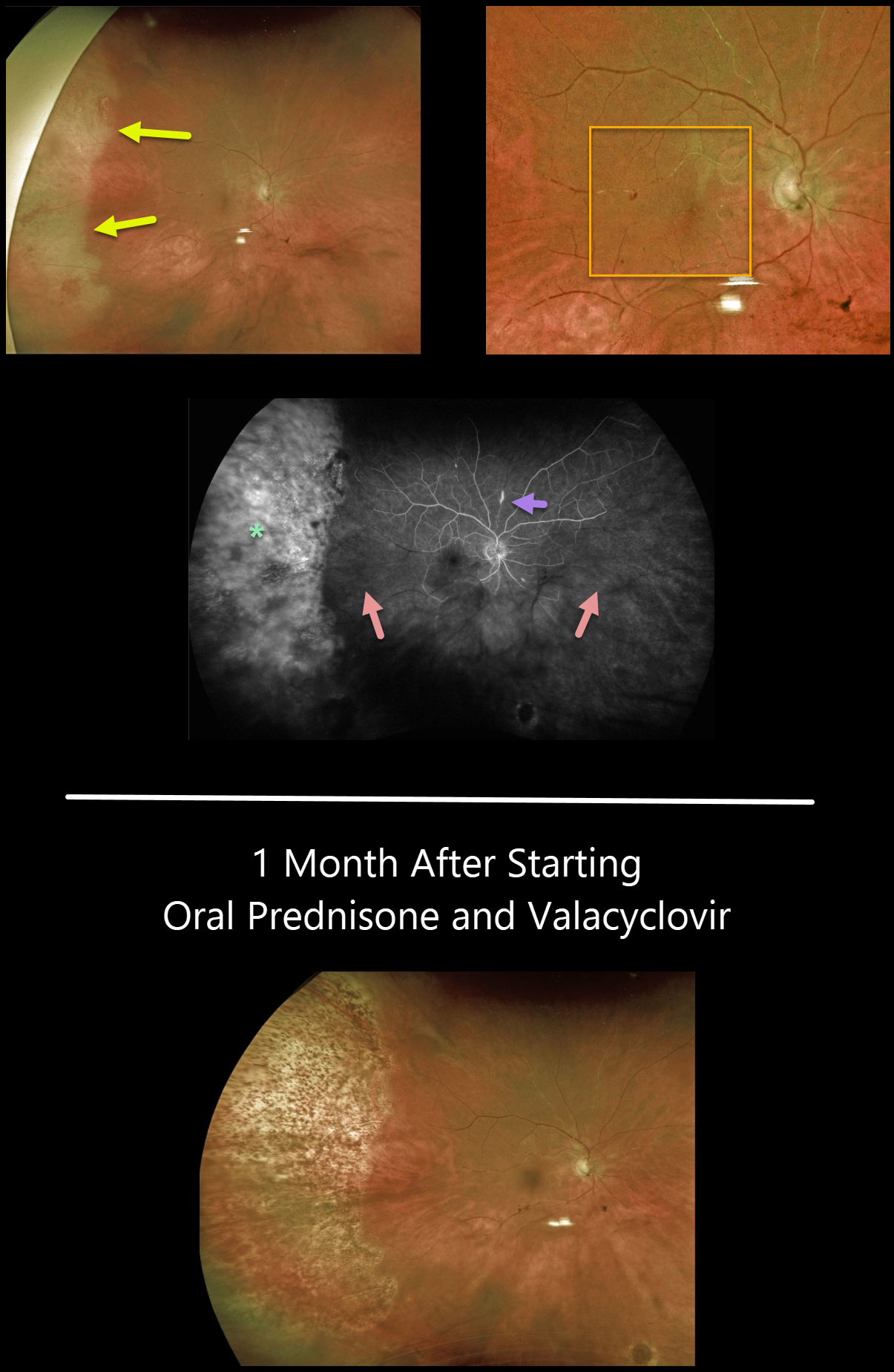
ACUTE RETINAL NECROSIS SYNDROME
Originally posted on @retina.rocks May 19, 2021
This 80YO female presented with a sudden onset of floaters while battling 2 months of persistent iridocyclitis from Herpes zoster ophthalmicus.
There was increased anterior uveitis, along with new peripheral temporal retinitis. Despite significant macular ischemia on clinical examination, vision was 20/60. Our patient’s posterior involvement was likely seeded from her anterior segment.
Optos fluorescein angiography shows diffuse staining of the peripheral retina, extensive nonperfusion throughout the posterior pole, and a few scattered areas of focal staining vasculitis.
She was started on high-dose oral prednisone and valacyclovir. One month later, vision improved to 20/40, and the retinitis resolved with secondary chorioretinal scarring.
We are slowly tapering her steroids and plan to leave her on low-dose valacyclovir as prophylaxis against recurrent infection in her right eye, as well as infection in her normal left eye.
Learning Points:
Acute retinal necrosis syndrome (ARNS) is most commonly caused by the Herpes zoster virus. ARNS, which usually occurs in immunocompetent individuals, is a rapidly progressive panuveitis with hemorrhagic and ischemic unilateral (ARNS) or bilateral (BARNS) retinal necrosis that typically begins in the retinal periphery.
ARNS is often a devastating and blinding infection due to the high risk for retinal detachment as well as macular and optic nerve involvement.
The latest meta-analysis argues for systemic antiviral therapy and prophylactic vitrectomy, with uncertain benefits for prophylactic laser photocoagulation or adjunctive intravitreal antivirals (see Zhao et al, Retina 2021;41:965-978).
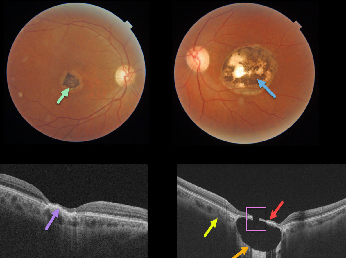
TOXOPLASMOSIS
Originally posted on @retina.rocks March 26, 2021
This 57YO male gave a history of lifelong poor vision in his left eye. Vision was 20/40 OD and 20/200 OS.
A small hyperpigmented foveal scar is noted in his right eye with neurosensory retinal atrophy and irregular elevation of the RPE.
A larger, round, variably pigmented, excavated macular scar is noted in his left eye. Triton swept-source OCT reveals a thinned atrophic retina and intact RPE suspended over an optically empty hyporeflective excavated colobomatous type lesion. The plane of the intact surrounding sclera is indicated by the yellow arrow. A small full-thickness retinal and RPE defect is noted centrally.
Learning Points:
For a great discussion on how to differentiate these infectious macular colobomatous lesions (congenital toxoplasmosis, Zika, and cytomegalovirus) from inherited dystrophies like North Carolina Macular Dystrophy (NCMD), see Kumar and Mahalingam AJO 2019;200;47-56.
In case you’re thinking our patient has NCMD, also see the first OCT description of congenital toxoplasmosis macular lesions (Garg et al, Retina 2009;29;631-637). Their Figures 4 and 6 look eerily similar to our patient’s right and left eyes!
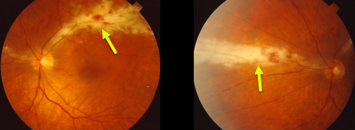
CMV RETINITIS
Originally posted on @retina.rocks February 10, 2021
This patient presented in 1994 with classic active cytomegalovirus (CMV) retinitis, which appeared as a “cottage cheese and ketchup” hemorrhagic retinitis following a vascular distribution. The retinitis spreads outwards, leaving behind thinned, necrotic retina and retinal pigment epithelium.
Learning Points:
CMV retinitis develops as a reactivation of latent CMV in immunosuppressed individuals.
Before effective antiretroviral treatment emerged in the mid to late 1990’s (highly active antiretroviral therapy, HAART), CMV retinitis developed in up to 40% of HIV/AIDS patients, often within the last 6 months of life.
Treatment includes a combination of intravenous and intravitreal anti-viral medications, including ganciclovir, foscarnet, and cidofovir. There is a high incidence of retinal detachment, especially with larger areas of involved retina.
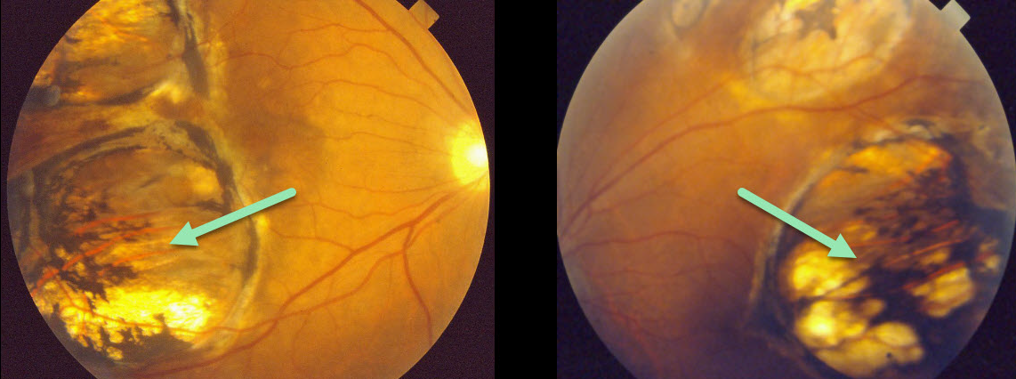
TOXOPLASMOSIS
Originally posted on @retina.rocks January 12, 2021
This patient presented in 1990 with what we assumed was bilateral inactive colobomatous congenital toxoplasmosis scarring.
Learning Points:
Choroidal colobomas are most commonly due to failure of the optic vesicle and choroidal fissure to close during fetal development. Multifocal lesions, as seen in our patient, can be caused by congenital toxoplasmosis and Zika.
For a comprehensive review of ocular coloboma, see Onwochei et al Surv Ophthalmol 2000;45:175-194.
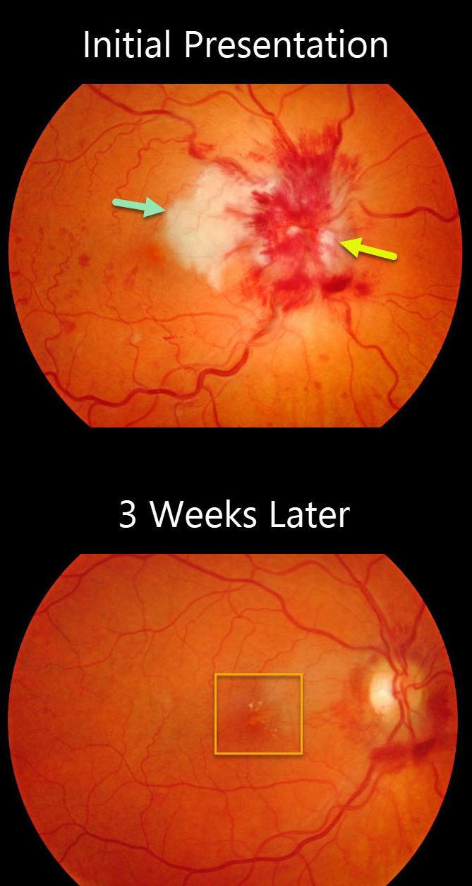
LEBER’S IDIOPATHIC STELLATE NEURORETINITIS
Originally posted on @retina.rocks December 31, 2020
This patient presented with 20/200 vision OD with a severely swollen nerve, a secondary central retinal vein occlusion (CRVO), and a large patch of nasal macular neuroretinitis. The left eye was normal.
Three weeks later, the findings had improved spontaneously and dramatically, and vision was 20/50. Some faint superior and nasal lipid was noted.
Bloodwork for syphilis, Bartonella (cat scratch), and Lyme disease was negative.
Learning Points:
The diagnosis for a unilateral swollen nerve is extensive. However, it becomes much smaller when associated with inner retinal ischemia and lipid exudation (neuroretinitis), including cat scratch (Bartonella), Lyme disease, and syphilis.
When no underlying cause is found, the entity is called Leber’s idiopathic stellate neuroretinitis. The condition is self-limited, so treatment is not usually recommended.
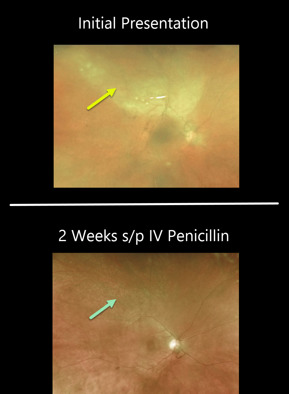
SYPHILIS
Originally posted on @retina.rocks December 2, 2020
This 46YO female presented with worsening pain and vision loss in her left eye despite being treated two days earlier by the emergency room for conjunctivitis. In our office, vision was counting fingers OS with a granulomatous panuveitis.
There is a creamy-white area of confluent retinitis superiorly. The image is hazy due to vitritis.
Stat bloodwork revealed a positive RPR and FTA-ABS. She denied any other symptoms or known history of syphilis.
Patients also need to be evaluated for HIV since syphilis and HIV infections often coexist. Fortunately, our patient was HIV negative.
After 2 weeks of parenteral penicillin, the panuveitis had improved markedly, and the retinitis had completely resolved.
Learning Points:
Ocular manifestations of syphilis vary widely and include virtually all variations of anterior and posterior segment inflammation, including a recently described presentation as the multiple evanescent white dot syndrome phenotype (see Russel et al Int Ophthalmol 2020;40:627-738).
Hence, the reason it’s been dubbed the great imitator or masquerader. One of the more common presentations is placoid chorioretinitis.
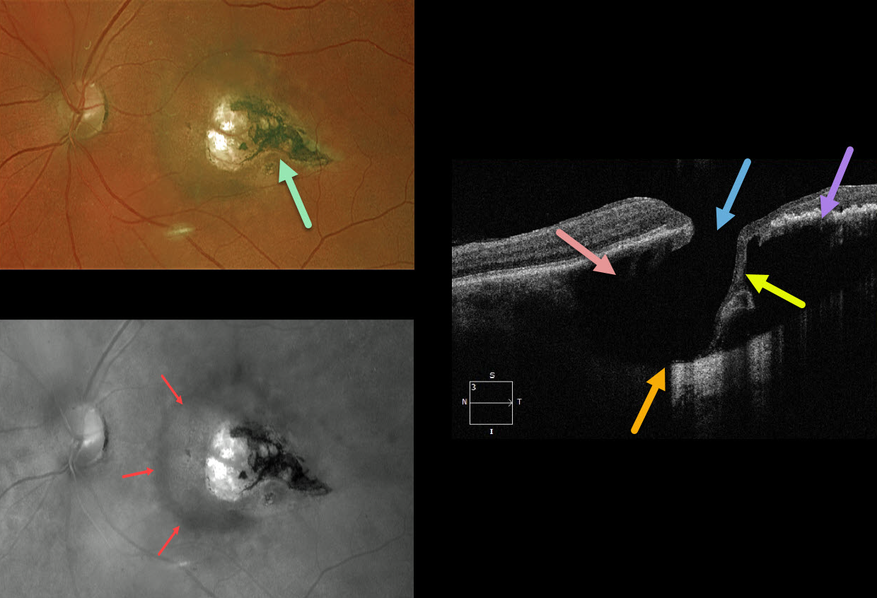
TOXOPLASMOSIS
Originally posted on @retina.rocks November 16, 2020
This 67YO female was referred with 20/30 vision and asymptomatic retinal findings. There was a tear-drop-shaped, variably pigmented chorioretinal scar in the temporal macula. The associated increased choroidal pigmentation is best seen in the red-channel image.
The OCT shows some unexpected findings. There is either a coloboma or posterior staphyloma causing marked posterior displacement of the sclero-choroidal junction. Temporally thinned retina dives posteriorly with an adjacent full-thickness retinal defect.
Temporally, the increased pigmentation of the scarring likely makes it difficult to see more posterior choroidal detail due to shadowing. Nasally, the faint, increased choroidal pigmentation also causes shadowing.
What do you think this is? Our best guess is a choroidal cavitation or an atypical colobomatous toxoplasmosis scar.
Learning Points:
For a great discussion on how to differentiate these infectious macular colobomatous lesions (congenital toxoplasmosis, Zika, and cytomegalovirus) from inherited dystrophies like North Carolina Macular Dystrophy (NCMD), see Kumar and Mahalingam AJO 2019;200:47-56. Also see the first OCT description of congenital toxoplasmosis macular lesions (Garg et al, Retina 2009;29;631-637).
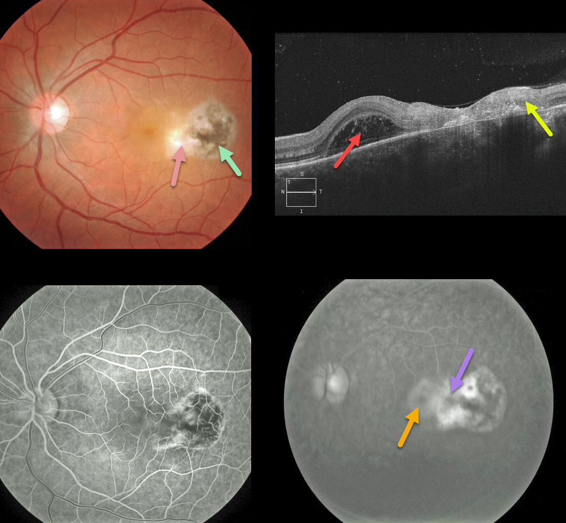
TOXOPLASMOSIS
Originally posted on @retina.rocks November 13, 2020
This patient presented with blurred vision from an area of active retinitis nasal to a prior toxoplasmosis scar.
An OCT B-scan shows full-thickness retinal necrosis and scarring temporally, with a foveal bacillary detachment. Tiny vitreous cells are scattered above the retina.
Fluorescein angiography shows staining of the inflamed retina and pooling within the bacillary detachment.
This patient was successfully treated with a 6-week course of oral Bactrim DS.
Learning Points:
Toxoplasmosis, the most common cause of infectious chorioretinitis in humans, is caused by the protozoan parasite, Toxoplasma gondii. It is most commonly transmitted through infected felines or by eating raw meat.
The majority of infections are acquired, although they can also be transmitted congenitally. The body’s immune system is unable to completely kill the organism, which often lies dormant within a chorioretinal scar for years.
When the immune balance favors the encysted organism, active chorioretinitis develops. This usually resolves spontaneously within 6 weeks.
If the optic nerve or macula is threatened, a six-week course of Bactrim DS is recommended (see Ophthalmology 2004;112;1876-1882). Oral prednisone can be added as well to help with severe inflammation.
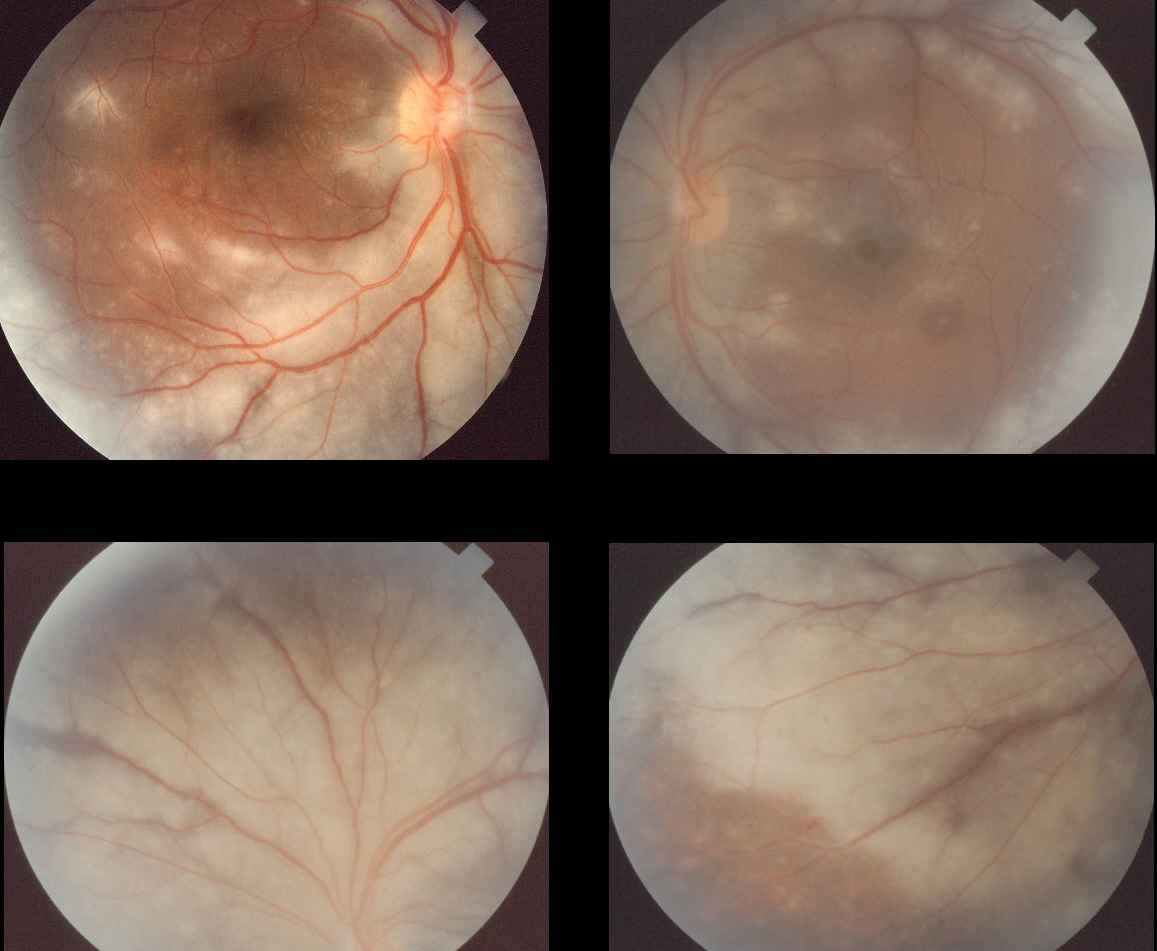
PROGRESSIVE OUTER RETINAL NECROSIS
Originally posted on @retina.rocks November 4, 2020
Progressive outer retinal necrosis (PORN) is a rare form of acute retinal necrosis (ARN) found in patients with severe immunosuppression, such as from HIV/AIDS.
This patient was lost to follow-up after the visit at which these images were taken. We are sure he went on to develop bilateral blindness.
Learning Points:
Both ARN and PORN are most commonly caused by the Herpes zoster virus. ARN, which occurs in immunocompetent individuals, is a rapidly progressive panuveitis with hemorrhagic and ischemic retinal necrosis that typically begins in the retinal periphery.
In contrast, PORN presents as a patchy, more posterior, and apparent outer non-hemorrhagic retinal necrosis in an otherwise relatively quiet eye. The white patches rapidly progress to more confluent areas of full-thickness necrosis.
Intravitreal ganciclovir and foscarnet can sometimes preserve vision, although the visual outcomes are usually extremely poor. Patients are also at risk for subsequent retinal detachment.
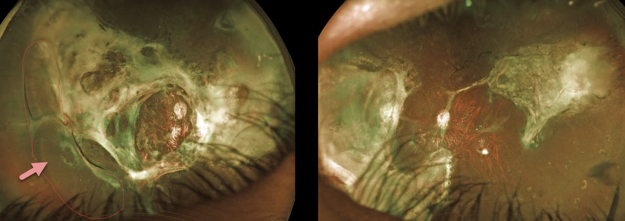
CMV RETINITIS
Originally posted on @retina.rocks October 23, 2020
This 29YO male presented to us 6 years earlier with bilateral cytomegalovirus (CMV) retinal detachments. At that time, he was severely ill from untreated HIV/AIDS.
He underwent bilateral vitrectomy surgery with silicone oil. Since then, he has done extremely well systemically, with normal CD4 counts and undetectable viral loads.
Despite severe bilateral chorioretinal scarring and a partial peripheral detachment under oil OD, vision is 20/400 bilaterally, and he is functioning well.
We have no plans to remove the oil, as his retinal appearances have remained stable since surgery.
Learning Points:
Cytomegalovirus (CMV) retinitis develops as a reactivation of latent CMV in immunosuppressed individuals. Before effective antiretroviral treatment emerged in the mid to late 1990’s (highly active antiretroviral therapy, HAART), CMV retinitis developed in up to 40% of HIV/AIDS patients, often within the last 6 months of life.
Treatment includes a combination of intravenous and intravitreal medications, including ganciclovir, foscarnet, and cidofovir.
Rhegmatogenous retinal detachment is a common late sequela of CMV infection and may require vitrectomy for repair.
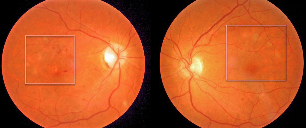
WEST NILE VIRUS
Originally posted on @retina.rocks October 8, 2020
Our patient presented with macular pigmentary changes, which were most pronounced in the right eye. Vision was counting fingers OD and normal OS.
On further questioning, they gave a history of vision loss following West Nile encephalitis 6 months earlier. Although we can’t say for sure, these findings are most likely the sequelae of resolved West Nile chorioretinitis.
Learning Points:
West Nile virus is an RNA virus transmitted by the bite of a mosquito that has fed on an infected bird. Only 1 in 150 of infected persons experience encephalitis, and far fewer will have ocular effects.
Chorioretinitis in a linear pattern is the most common ocular finding, although retinal vasculitis and optic nerve swelling can also occur. There is no proven treatment for West Nile virus infection.
See a great review on this topic by Garg and Jampol (Surv Ophthalmology 2005;50:3-13).
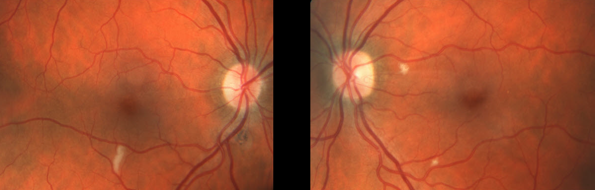
HIV RETINOPATHY
Originally posted on @retina.rocks September 24, 2020
Our patient was HIV positive with a CD4 count of 9.
There is an isolated nerve fiber layer infarct (cotton wool spot, CWS) in the right eye and two CWS in the left eye.
Learning Points:
Cotton-wool spots are nonspecific and are found in many disorders, including hypertensive and diabetic retinopathy. They can also be seen with HIV/AIDS when the CD4 count is under 200.
The etiology of CWS in HIV/AIDS is not known, although they do not contain virus particles.
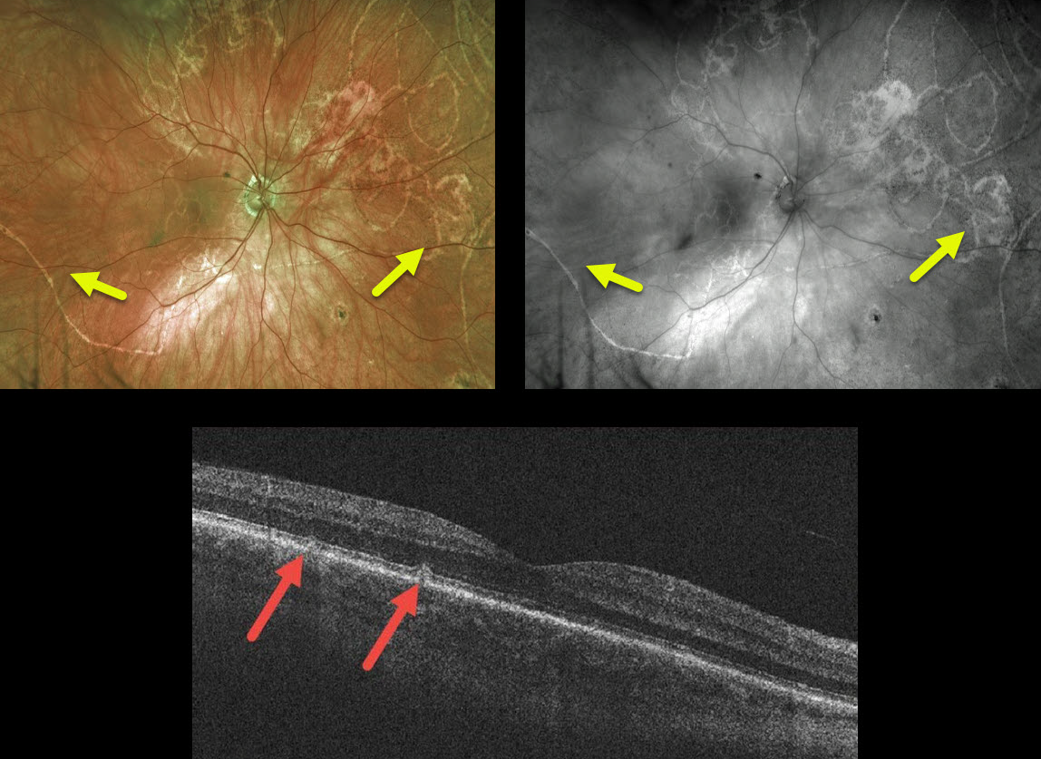
OPHTHALMOMYIASIS
Originally posted on @retina.rocks September 21, 2020
This 68YO presented with a known history of having a “questionable parasite” in his right eye. He was treated with oral and IV antibiotics at another facility many years ago.
Despite the extensive subretinal fly larva tracks, OCT shows surprisingly subtle subretinal changes. Vision is 20/30.
Learning Points:
Larvae can be found in different parts of the eye, including the conjunctiva, cornea, anterior chamber, vitreous, and subretinal space.
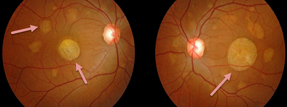
PNEUMOCYSTIS CHOROIDITIS
Originally posted on @retina.rocks September 11, 2020
This patient had a known history of treated HIV infection and presented with asymptomatic bilateral irregularly round patches of choroidal and RPE pigmentary loss.
Our patient’s findings show what the lesions of pneumocystis choroiditis look like upon resolution.
Learning Points:
In the early days of the HIV/AIDS epidemic, pneumocystis pneumonia was one of the most common opportunistic infections. Aerosolized pentamidine was therefore given as prophylaxis. The medication, however, did not reach the entire lungs, and patients often had septic pneumocystis emboli from an incompletely treated pneumonia.
Pneumocystis choroiditis was a rare manifestation of this systemic infection, where patients developed multifocal, flat, bilateral choroidal lesions that were usually asymptomatic and had a striking absence of inflammation.
Systemic prophylaxis was subsequently recommended, which better treated the underlying pneumonia and prevented systemic infection.
Highly active antiretroviral therapy (HAART), introduced in the late 1990’s, restores the health of the underlying immune system and has turned this type of finding into something for the history books.
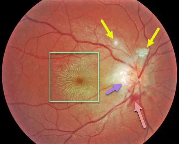
NEURORETINITIS
Originally posted on @retina.rocks August 24, 2020
This 17YO presented with 20/200 vision OD and diffuse optic disc edema with temporal peripapillary retinitis, peripapillary inner retinal blood, a few areas of focal inner retinitis, and a prominent macular lipid star. The left eye was normal.
Our patient tested positive for Bartonella and had a cat (should’ve gotten a dog and opted for a Toxocara worm instead…).
Patients will usually improve without treatment, although antibiotic therapy may sometimes be recommended.
Learning Points:
Optic nerve inflammation, often from malignant hypertension or neuroretinitis, can cause fluid and blood to leak into the optic nerve and peripapillary tissues. When the fluid absorbs, the lipid can precipitate in Henle’s layer, creating the lipid star.
There are numerous causes for neuroretinitis, including cat scratch (Bartonella), Lyme disease, syphilis, and idiopathic (Leber’s stellate neuroretinitis).
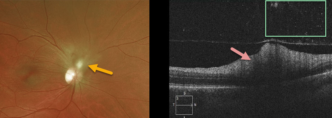
TOXOPLASMOSIS
Originally posted on @retina.rocks June 23, 2020
Our patient had active toxoplasmosis inflammation directly adjacent to the optic nerve and was placed on Bactrim double-strength (DS) and prednisone.
The OCT line scans show hyperreflective retinitis with overlying vitreous cells.
Learning Points:
Toxoplasmosis, the most common cause of infectious chorioretinitis in humans, is caused by the protozoan parasite, Toxoplasma gondii. It is most commonly transmitted through infected felines or by eating raw meat.
The majority of infections are acquired, although they can also be transmitted congenitally. The body’s immune system is unable to completely kill the organism, which often lies dormant within a chorioretinal scar for years.
When the immune balance favors the encysted organism, active chorioretinitis develops. This usually resolves spontaneously within 6 weeks.
If the optic nerve or macula is threatened, a six-week course of Bactrim DS is recommended (see Ophthalmology 2004;112;1876-1882). Oral prednisone can be added as well to help with severe inflammation.
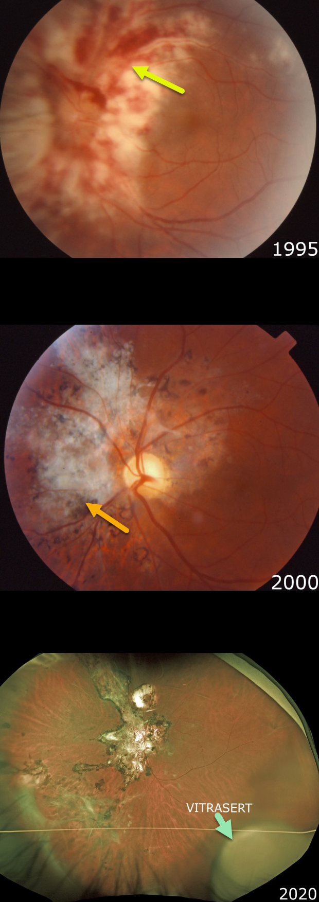
CMV RETINITIS
Originally posted on @retina.rocks June 8, 2020
Our patient presented in 1995 with classic active CMV retinitis involving the peripapillary and macular regions of his left eye. We immediately referred him to the University of Kentucky for implantation of a then-investigational sustained-release ganciclovir implant, Vitrasert.
Note the hemorrhagic, whitish retinal necrosis, which often follows a vascular distribution. The retinitis spreads outwards, leaving behind a thinned necrotic retina and RPE.
Following Vitrasert implantation, the retinitis resolved with secondary scarring. He was then started on highly active antiretroviral therapy (HAART).
In 2007, a rhegmatogenous retinal detachment, a common late sequela of CMV infection, was successfully repaired with vitrectomy (not pictured).
As of 2020, he continues to do well with an undetectable viral load and a normal CD4 count. Vision is 20/20 in his normal right eye and 20/400 OS. The ganciclovir Vitrasert implant remains visible in the inferotemporal periphery.
Learning Points:
Cytomegalovirus (CMV) retinitis develops as a reactivation of latent CMV in immunosuppressed individuals. Before effective antiretroviral treatment emerged in the mid to late 1990’s, CMV retinitis developed in up to 40% of HIV/AIDS patients, often within the last 6 months of life.
Treatment includes a combination of intravenous and intravitreal medications, including ganciclovir, foscarnet, and cidofovir.
Vitrasert was approved by the FDA in 1996 and discontinued in 2013 due to patent expiration. The demise of Vitrasert is directly linked to HAART.
As occurred in our patient, pharmacologic restoration of immune function allows the patient’s own immune system to control CMV.
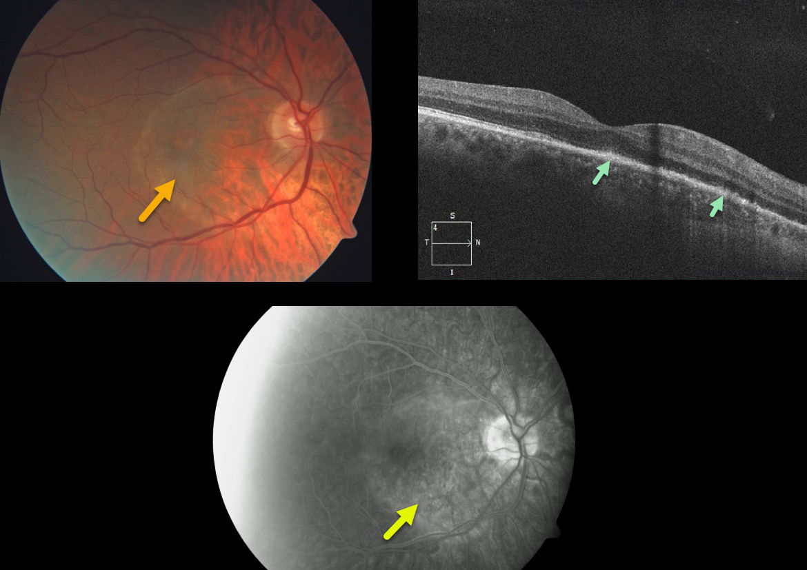
SYPHILIS
Originally posted on @retina.rocks May 27, 2020
This patient has classic findings for acute placoid syphilis in the right eye.
The fundus photo demonstrates an orange-yellow subretinal lesion. OCT shows attenuation of the outer retinal layers (including the ellipsoid zone) and a thickening of the RPE.
Angiographically, this inflammatory lesion blocks early and stains late. The patient was referred to the infectious disease specialist for parenteral antibiotics.
Learning Points:
In internal medicine, syphilis has been called “the great imitator” since it can masquerade as so many systemic disorders. The ocular manifestations are legion as well.
Originally described by Gass (Ophthalmology 1990;97;1288-1297), placoid syphilis affects the outer retina, RPE, and choriocapillaris and clinically manifests as an orange-yellow subretinal lesion.
Placoid syphilis has a fairly good visual prognosis with treatment. Patients also need to be evaluated for HIV since syphilis and HIV infections often coexist.
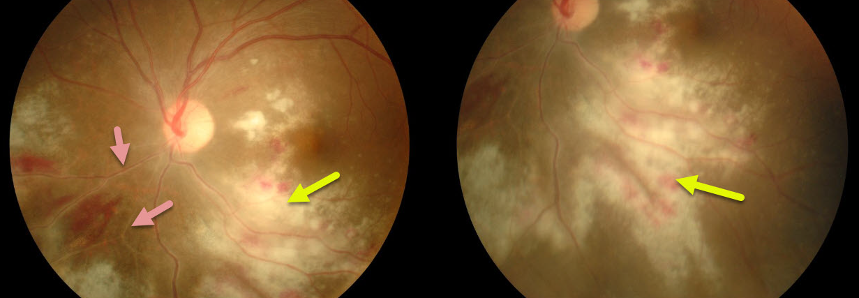
CMV RETINITIS
Originally posted on @retina.rocks March 12, 2020
Our patient shows typical CMV retinitis with active fluffy white hemorrhagic retinitis and vasculitis.
This patient is at high risk for severe vision loss due to both the optic nerve and macular center being threatened.
Immediate intravitreal antiviral injections followed by systemic treatment were recommended.
Learning Points:
Cytomegalovirus retinitis (CMV) is a usually devastating infection most commonly seen in end-stage HIV/AIDS.
Fortunately, this is now rarely seen due to the advent of highly active antiretroviral therapy (HAART) in the mid-1990’s.
CMV retinitis is characterized by confluent hemorrhagic necrosis often following a vascular distribution. Retinal detachments develop in about three-quarters of eyes, especially with more extensive areas of retinitis.
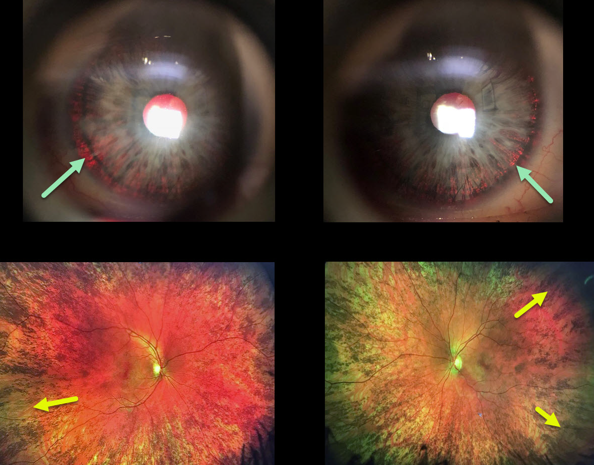
OCULAR ALBINISM
Originally posted on @retina.rocks March 9, 2020
This woman has a history for both a congenital rubella infection and a positive family history for albinism in both an uncle and cousin.
We think that she is likely a carrier for OA1 (ocular albinism type 1) given the presence of iris transillumination defects and pigmentary changes of the peripheral retina, although the fundus changes may also be due to congenital rubella.
Learning Points:
Ocular albinism is a X-linked recessive disorder that affects only the eyes and is caused by a mutation in the OA1 gene. Findings seen in ocular albinism include strabismus, nystagmus iris transillumination defects, blonde fundus, and absent or blunted foveal reflex (fovea plana). There is often a higher number of crossed nerve fibers at the optic chiasm.
OA1 carriers will often have partial iris transillumination defects and up to 90% of OAT1 female carriers exhibit a salt and pepper (or mud-splattered) fundus due to the patches of amelanotic RPE.
This salt and pepper appearance can also be seen in congenital rubella, Leber’s congenital amaurosis, congenital syphilis, and phenothiazine toxicity.
Oculocutanoeus albinism involves both eye and skin findings and is caused by mutations in the tyrosine gene. There is a complete tyrosinase-negative form in which no melanin is produced and a tyrosinase-positive form in which only partial amounts of melanin are produced,
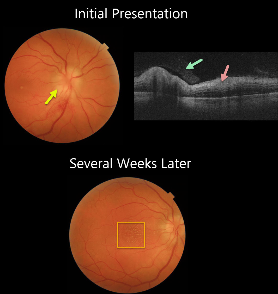
NEURORETINITIS
Originally posted on @retina.rocks January 16, 2020
At initial presentation, our patient showed optic nerve edema clinically with inner retinal hyperreflective retinitis and an overlying vitritis on OCT.
A few weeks later the optic nerve and retinal inflammation improved with the development of a classic lipid star.
The lipid, which leaks out of inflamed optic nerve vessels, accumulates in the outer plexiform layer. Since it originates from the nerve, the star is usually most prominent in the nasal macula.
Learning Points:
Neuroretinitis can be infectious or idiopathic. The most common infectious cause is secondary to Bartonella Henselae (Cat-Scratch Disease).
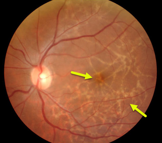
OPHTHALMOMYIASIS
Originally posted on @retina.rocks January 13, 2020
This patient was totally asymptomatic with normal vision when she was referred to us for retinal changes.
The meandering subretinal scarring was caused by a fly larva crawling thru the subretinal space. These eyes show no inflammation.
Learning Points:
Larvae can be found in different parts of the eye including the conjunctiva, cornea, anterior chamber, vitreous, and subretinal space.


