

10 Inflammatory Disorders
<< Back to Cases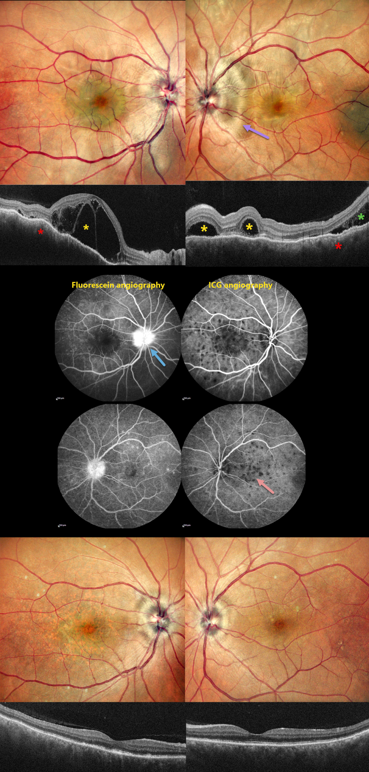
HARADA DISEASE
Rohan Jain and Manish Nagpal
Originally posted on @retina.rocks August 20, 2026
This 46YO male presented with 10 days of bilateral blurred vision with a left-sided headache. Vision was 20/200 OD and 20/20 OS. Anterior segments were normal.
Pseudocolor SLO imaging shows bilateral swollen nerves and irregular macular chorioretinal folds. OCT scanning shows multifocal bacillary layer detachments, subretinal fluid, and an undulating, thickened choroid. Fluorescein angiography shows bilateral multifocal areas of subretinal leakage and disc leakage, and indocyanine green angiography shows multiple hypofluorescent choroidal spots.
Based on the clinical and multimodal imaging findings, a diagnosis of acute Harada disease was established. He received a 3-day course of intravenous methylprednisolone followed by oral corticosteroids.
At the 2-month follow-up, the patient’s symptoms had completely resolved. Vision was 20/30 OD and 20/20 OS. The exudative findings are completely resolved on photography and OCT, and the choroidal thickening with overlying chorioretinal folds is also resolved.
Learning Points:
Harada disease is characterized by relatively acute bilateral panuveitis, optic nerve swelling, thickened choroid, chorioretinal folds, multifocal serous exudative retinal detachments, and bacillary layer detachments. Patients with only ocular findings have Harada disease (as in our patient), whereas those with additional systemic findings (including vitiligo, poliosis, headache, vertigo, and hearing loss) have Vogt-Koyanagi-Harada disease.
TUBERCULOUS POSTERIOR SCLERITIS
Aniruddh Soni
Originally posted on @retina.rocks July 31, 2026
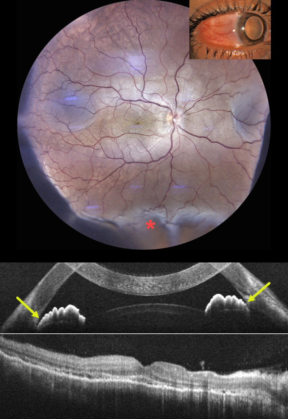
This previously healthy 35YO male presented with 1 week of pain, redness, and decreased vision in his right eye. He had been treated by an outside doctor for conjunctivitis. Vision 20/200 OD and 20/20 in his normal OS. Slit lamp examination showed temporal scleral injection. Intraocular pressure was 23 mm Hg.
Color photography shows radiating chorioretinal folds throughout the macula with an inferior exudative retinal detachment. OCT scanning of the macula shows chorioretinal folds, with anterior chamber OCT showing a narrow-to-closed angle. B-scan ultrasonography showed subtenon’s fluid (not shown).
Following a 3-week course of tapering oral steroids and glaucoma drops, vision improved to 20/40 with reduced chorioretinal folds and complete resolution of the exudative detachment. Rheumatologic workup was negative, but Mantoux skin testing and QuantiFERON-TB GOLD were positive, with a subsequent diagnosis of latent tuberculosis (TB). Antituberculous therapy was recommended, but the patient refused treatment.
Learning Points:
Tuberculous posterior scleritis is a rare manifestation of ocular TB (Agarwal and Majumder, Indian J Ophthalmol 2019;67:1362-1365), which itself occurs in approximately 1.5% of patients with confirmed systemic TB. Ocular findings include optic disc edema, choroiditis, and sclerochoroidal thickening with widening of the sub-Tenon space. It may occur via direct hematogenous dissemination of Mycobacterium tuberculosis or through a hypersensitivity-mediated immune response to the organism and can develop even in the absence of clinically active pulmonary disease.
Diagnosis, as in our patient, is largely presumptive, requiring a high index of suspicion supported by bloodwork, chest imaging to assess pulmonary involvement, and exclusion of mimics such as sarcoidosis, syphilis, and metastatic disease.
TUBERCULAR SERPIGINOUS-LIKE CHOROIDITIS
Aniruddh Soni
Originally posted on @retina.rocks July 6, 2026
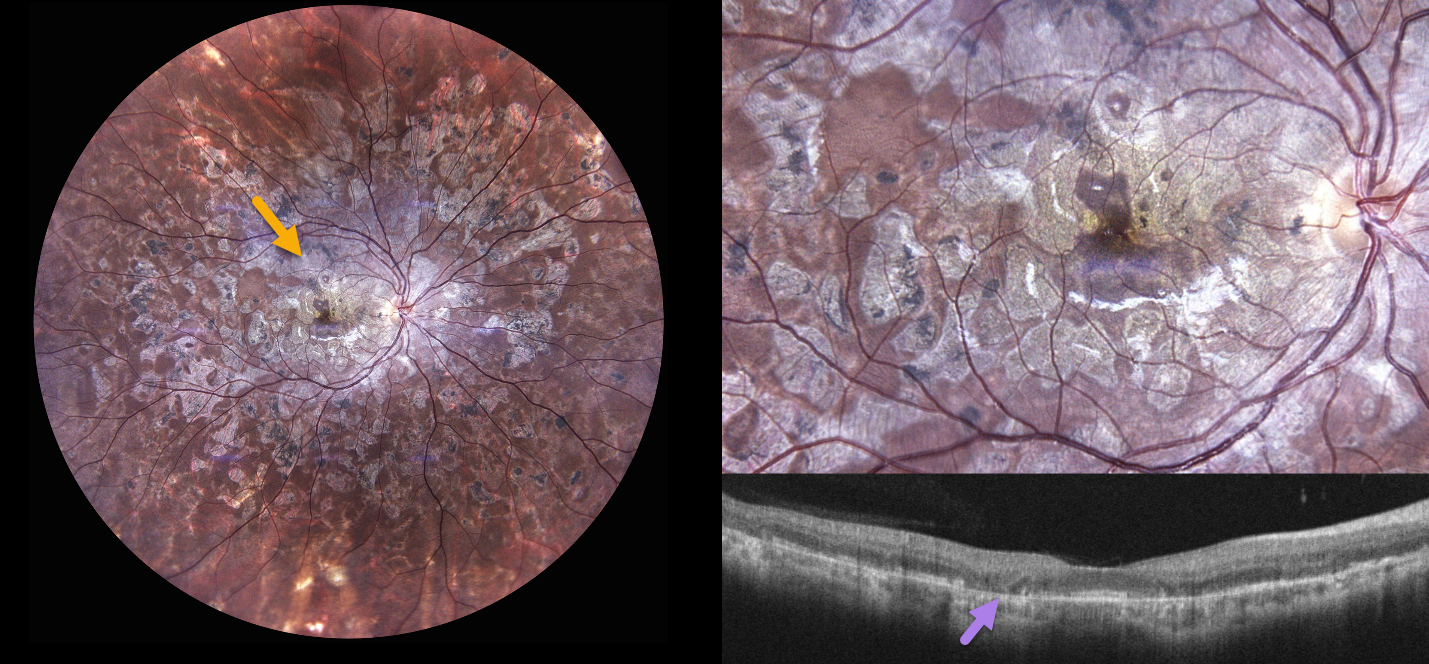
A 30YO female presented with a 1.5-year history of bilateral floaters and blurred vision. She was diagnosed elsewhere with ‘choroiditis’ and treated with oral steroids and azathioprine, with 2 recurrences. When seen in our office, vision was 20/30 OU.
Color photography OD shows extensive, variably confluent, and meandering chorioretinal scarring extending from the disc and macula to the retinal periphery. The foveal center is just spared. OCT scanning shows variable outer retinal and RPE atrophy. Identical findings were noted in her left eye (not shown).
Tuberculin skin testing and QuantiFERON-TB Gold were positive. She was referred to pulmonary and infectious disease specialists, was diagnosed with latent TB, and started on anti-tuberculosis treatment (ATT) with tapering oral steroids.
Learning Points:
Although TB most commonly presents with pulmonary involvement, extrapulmonary sites can involve the gastrointestinal, skin, cardiovascular, genitourinary, and central nervous systems, as well as the eyes. Ocular involvement, like syphilis, can mimic virtually any type of uveitis, including anterior, intermediate, posterior, and panuveitis, retinitis, and retinal vasculitis, neuroretinitis, optic neuropathy, choroidal granuloma, choroiditis, and scleritis.
Tubercular serpiginous-like choroiditis mimics serpiginous but affects younger patients with more multifocal and peripheral recurrences, foveal sparing, vitritis, and progression if untreated (Agarwal et al, AJO 2020;220:160-169). Although the exact mechanism is not yet clear, a direct or indirect infectious trigger by the Mycobacterium tuberculosis is thought to cause choroiditis. Its origin is probably immunogenic, as it appears to respond to corticosteroids and other immunosuppressants. Our patient was a bit unusual in that he required systemic steroids, ATT, and adalimumab to finally control his disease.
FROSTED BRANCH ANGIITIS
The European VitreoRetinal Society (EVRS) and Nitin Ramanujam
Originally posted on @retina.rocks July 3, 2026
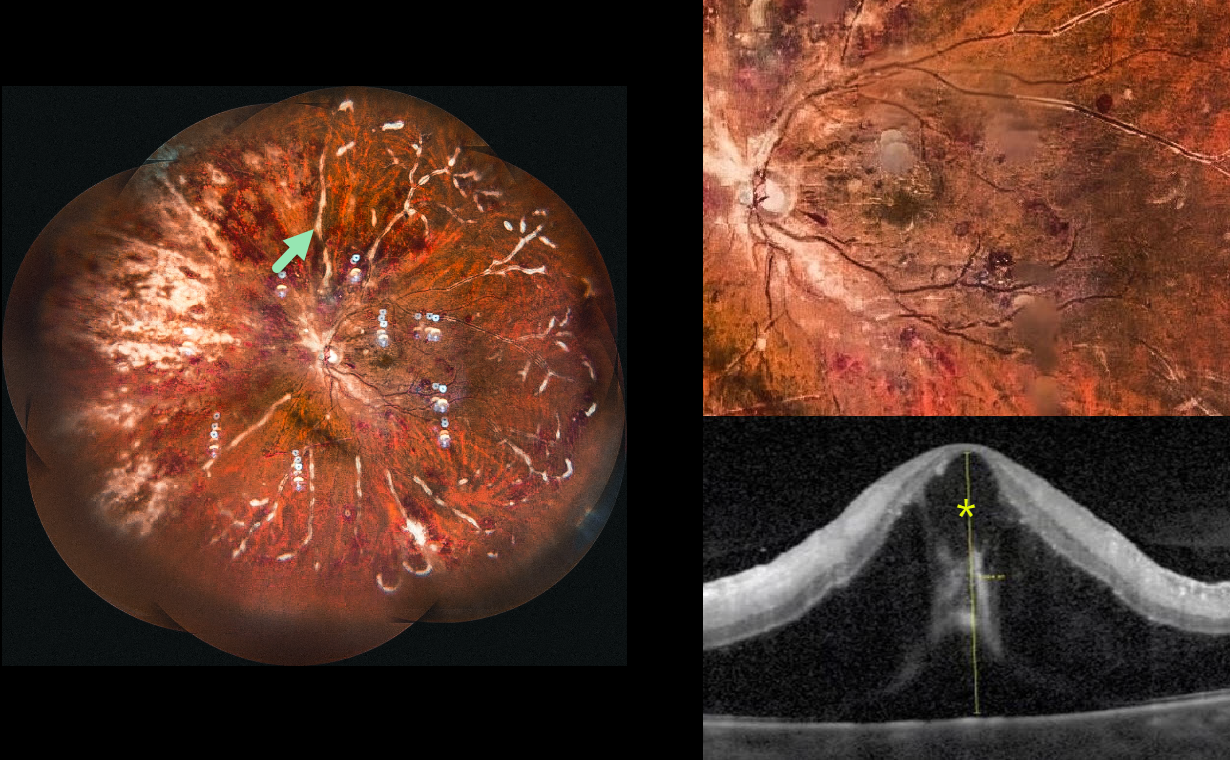
This 45YO female with a known history of poorly controlled systemic lupus erythematosus (SLE) presented to us for a third opinion with 10 days of bilateral vision loss. Vision was 20/100 OD and 20/200 OS.
Ultrawidefield imaging of her left eye shows extensive retinal hemorrhages with a frosted branch appearance to the retinal veins. Marked cystoid edema and subfoveal fluid are noted on OCT. Identical findings were seen in her right eye (not shown). She was immediately lost to follow-up.
Learning Points:
Frosted branch angiitis is a retinal vasculitis that gets its name from the involved vessels resembling branches of a tree that are “frosted” with snow (Kleiner et al, AJO 1988;106:27-34). Although these findings may be isolated, they are often considered a phenotype found in many inflammatory conditions, most commonly CMV retinitis. Inflammation is usually highly responsive to steroids, and the visual prognosis is generally good, although some may develop secondary neovascularization in the anterior and posterior segments due to widespread ischemia.
Although frosted branch angiitis has not been specifically linked to SLE, retinal vasculitis (arteriolar vasculitis with cotton wool spots, retinal hemorrhages, and vascular occlusions) is a known lupus finding (Gao et al, Lupus 2017;26:1182-1189).
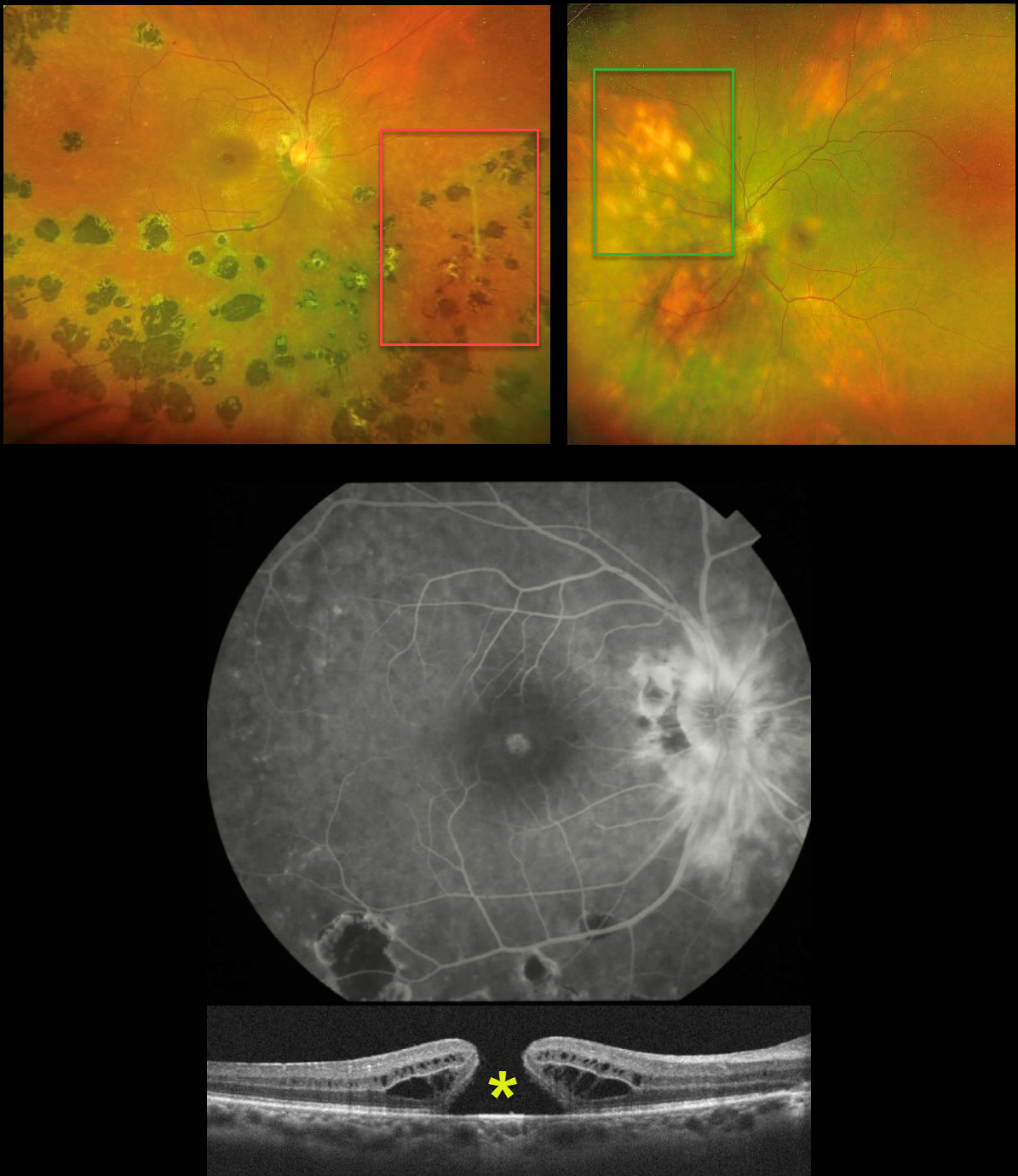
MULTIFOCAL CHOROIDITIS VS BIRDSHOT
Seif Anwar
Originally posted on @retina.rocks June 17, 2026
This healthy 41YO female presented with 6 months of bilateral vision loss. Vision was 20/240 OD and 20/200 OS.
Optos color RG imaging OD shows peripapillary scarring, multifocal large hyperpigmented chorioretinal scars scattered inferiorly and temporally, and some vascular sheathing. A full-thickness macular hole is noted and confirmed on OCT. Fluorescein angiography shows peripapillary leakage.
Her left eye’s Optos shows markedly different findings, with multifocal, creamy-white, depigmented lesions in a pattern radiating away from the disc.
Medical workup was negative, including HLA-A29, QuantiFERON TB Gold, chest X-ray, and serologies for toxoplasmosis and CMV.
Learning Points:
Birdshot chorioretinopathy was appropriately described and named by Drs. Ryan and Maumenee (Am J Ophthlamol 1980;89:31-45) since the lesions look like shotgun birdshot scattered throughout the fundus. Birdshot is typically chronic and bilateral, with about 90-95% of patients being HLA-A29 positive and usually affects women in their 40s to 60s.
Multifocal choroiditis (MFC) is the current preferred term for a group of historically heterogeneous disorders, including multifocal choroiditis, recurrent multifocal choroiditis, multifocal choroiditis and panuveitis, punctate inner choroidopathy, progressive subretinal fibrosis, and pseudo-histoplasmosis (Essex et al, Retina 2013;33:1-4 and Spaide et al, Retina 2013;33:1315-1324). MFC occurs in otherwise healthy adults, often myopic and female. Findings include variably pigmented multifocal scars throughout the fundus, panuveitis, subretinal fibrosis, and macular neovascularization (MNV).
Our patient’s diagnosis is unclear because her right eye shows features suggestive of MFC, while her left eye is fairly pathognomonic for birdshot. In our experience, birdshot lesions never become hyperpigmented, as in our patient’s right eye, although the lesions in her left eye are virtually pathognomic for birdshot.
Following several Remicade treatments, vision remained stable at 20/240 OD and improved to 20/50 OS. We continue to follow her closely.
What’s your diagnosis???
SERPIGINOUS CHORIORETINOPATHY
César Adrián Gómez Valdivia
Originally posted on @retina.rocks June 2, 2026
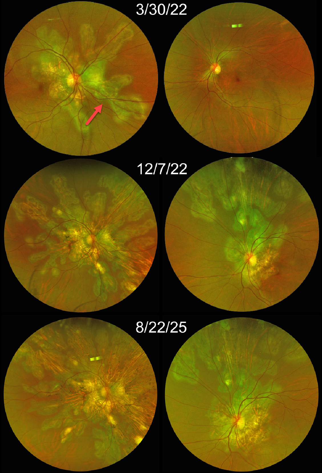
This 41YO female presented on 3/30/22 with a paracentral scotoma and metamorphopsia in her right eye. Vision was 20/800 OD and 20/20 in his initially normal OS.
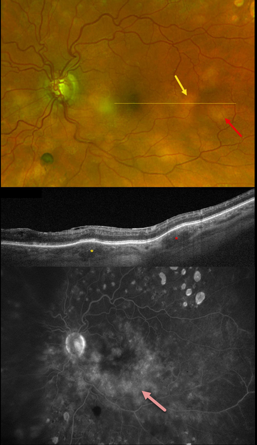
Optos color RG imaging at that time shows multifocal meandering scars radiating outwards from the disc and macula, consistent with serpiginous chorioretinopathy. All scarring appears inactive except for a possible active lesion inferonasally. General laboratory testing and chest CT were all negative. The PPD was positive; however, given that she lived in a TB-endemic area, this was further evaluated with QuantiFERON, which was negative.
Over the following months, the disease evolved in a chronic relapsing pattern, with recurrent and aggressive activity despite intravenous methylprednisolone pulses along with cyclophosphamide.
When last examined on 8/22/25, vision was hand motion OD and 20/150 OS. Current systemic therapy includes azathioprine 100 mg and prednisone 60 mg daily. She is currently awaiting initiation of adalimumab as biologic immunomodulatory therapy, with the goal of achieving better inflammatory control and reducing cumulative corticosteroid exposure.
Learning Points:
Serpiginous choroiditis is an idiopathic, usually bilateral chorioretinal inflammatory condition, most commonly found in middle-aged men (Khanamiri and Rao, Surv Ophthalmology 2013;58:203-232). Recurrent areas of inflammation develop along the edge of a previous scar, producing scarring that meanders from the optic nerve outward into the macula. Vision is good unless scarring or macular neovascularization extends through the macular center. There is no consensus on the optimal systemic treatment, including corticosteroids and steroid-sparing agents.
MULTIFOCAL CHOROIDITIS WITH MACULAR NEOVASCULARIZATION
César Adrián Gómez Valdivia
Originally posted on @retina.rocks May 25, 2026
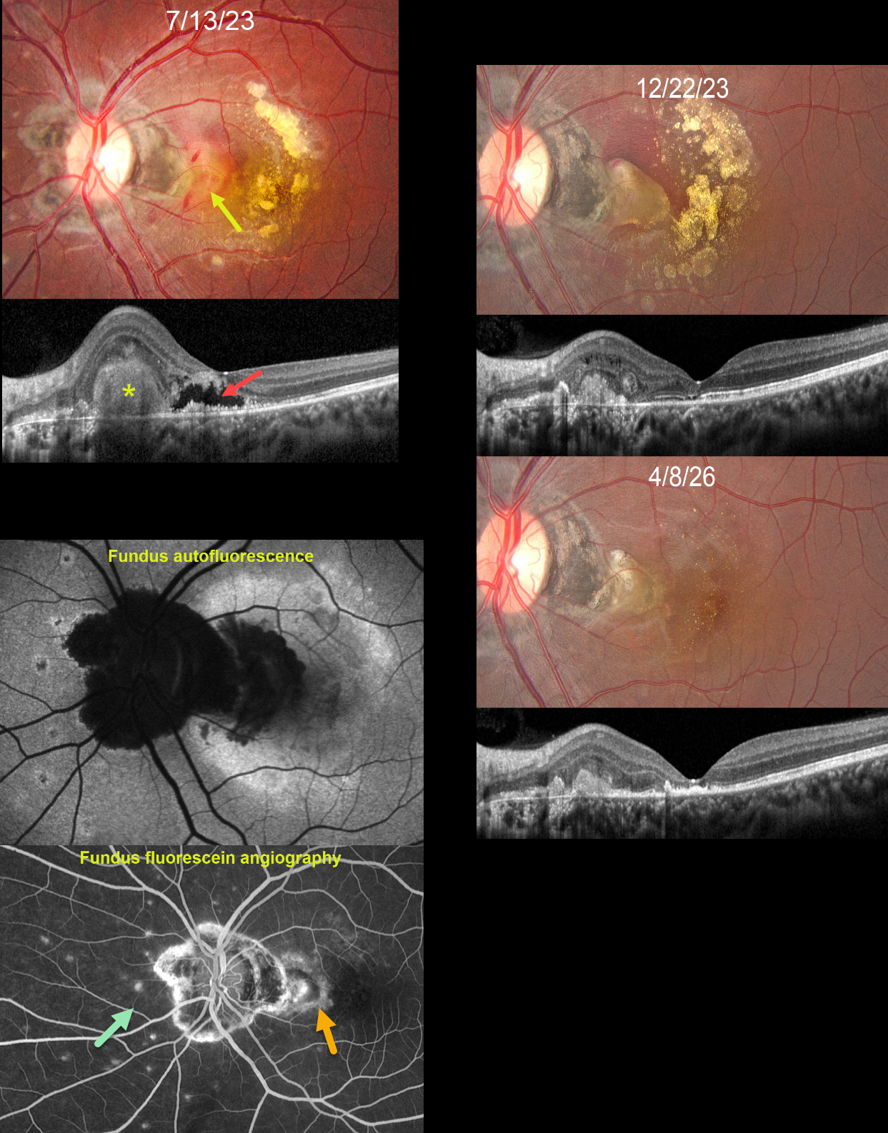
This 21YO female presented with a gradually progressive central scotoma in her left eye for the prior 2 months. Vision was 20/20 in her normal OD and 20/1600 OS.
Fundus photography shows a foveal subretinal fibrotic macular neovascularization (MNV) with surrounding subretinal blood and variably confluent subretinal lipid noted more temporally. Some inner retinal hemorrhages are also noted. Variably pigmented peripapillary scarring is present with multifocal small, depigmented scars nasally. OCT scanning shows a hyperreflective subretinal mound corresponding to the area of subretinal fibrosis. More centrally, there is a fluid-filled pocket of outer retinal hyporeflectivity surrounded by a ragged, variably thickened border of hyperreflective material. Fundus autofluorescence (FAF) shows peripapillary hypo-FAF as does the subretinal fibrosis with surrounding blood. The macula otherwise shows variable hyper-FAF, likely due to unmasking from outer retinal atrophy. The MNV stains angiographically. Multifocal scars stain superiorly, nasally, and inferiorly.
Following 3 monthly injections of Eylea, vision improved to 20/50. The MNV is contracted into an oval-shaped subretinal lesion with surrounding fading subretinal blood and absorbing lipid. She was then lost to follow-up for over 2 years. Thankfully, the MNV showed further contraction, with faint, scattered residual lipid dots. Despite outer retinal atrophy on OCT, vision improved to 20/25.
Learning Points:
Multifocal choroiditis (MFC) is the current preferred term for a group of historically heterogeneous disorders, including multifocal choroiditis, recurrent multifocal choroiditis, multifocal choroiditis and panuveitis, punctate inner choroidopathy, progressive subretinal fibrosis, and pseudo-histoplasmosis (Essex et al, Retina 2013;33:1-4 and Spaide et al, Retina 2013;33:1315-1324).
MFC occurs in otherwise healthy adults, often myopic and female. Findings include variably pigmented multifocal scars throughout the fundus, panuveitis, subretinal fibrosis, and MNV. Unlike ocular histoplasmosis, new scars and uveitis can be seen. MNV can be successfully managed with anti-VEGF therapy.
Our patients’ MNV and peripheral scars most likely represent MFC. We continue to follow her closely
TUBERCULAR SERPIGINOUS-LIKE CHOROIDITIS
Akansha Sharma
Originally posted on @retina.rocks March 9, 2026
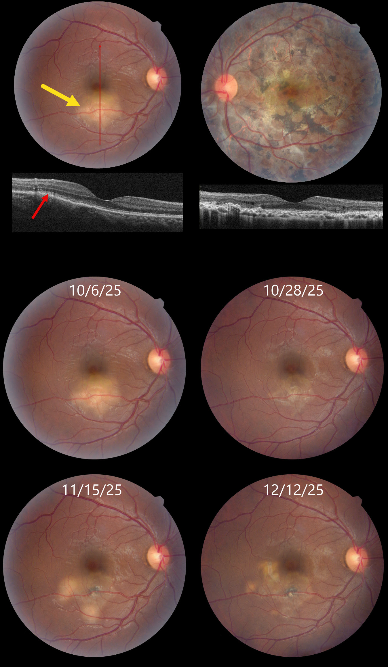
This 32YO male presented on 10/6/25 with a 6-day history of decreased vision in his right eye. About 2 years earlier, he was diagnosed elsewhere with tubercular serpiginous-like choroiditis in his left eye. Extensive workup at the time, including a chest X-ray, was negative except for a positive QuantiFERON TB Gold result. He was lost to follow-up without treatment for his latent tuberculosis (TB). Vision in our office was 20/30 OU.
Color photography shows a deep, creamy-white retinal inflammatory lesion in the inferior macula. OCT shows outer retinal hyperreflectivity with loss of the outer retinal band details. Extensive scarring is noted throughout the left posterior pole, with a small island of preserved foveal tissue.
Tapering systemic corticosteroid therapy was begun, followed by anti-tuberculosis treatment. Over the ensuing 2 months, he experienced waxing and waning inflammation necessitating intravitreal Ozurdex, oral steroids, and adalimumab while vision remained stable at 20/30. We continue to follow him closely.
Learning Points:
Although TB most commonly presents with pulmonary involvement, extrapulmonary sites can include the gastrointestinal, skin, cardiovascular, genitourinary, and central nervous systems, as well as the eyes. Ocular involvement, like syphilis, can mimic virtually any type of uveitis, including anterior, intermediate, posterior, and panuveitis, retinitis, and retinal vasculitis, neuroretinitis, optic neuropathy, choroidal granuloma, choroiditis, and scleritis.
Tubercular serpiginous-like choroiditis mimics serpiginous but affects younger patients with more multifocal and peripheral recurrences and progression if untreated (Agarwal et al, AJO 2020;220:160-169). Although the exact mechanism is not yet clear, a direct or indirect infectious trigger by the Mycobacterium tuberculosis is thought to cause choroiditis. Its origin is probably immunogenic since it seems to respond to corticosteroids and other immunosuppressants. Our patient was a bit unusual in that he required systemic steroids, ATT, and adalimumab to finally control his disease.
SYMPATHETIC OPHTHALMIA
The European VitreoRetinal Society (EVRS), Deepanshu Agrawal and Arun Bhargava
Originally posted on @retina.rocks February 20, 2026
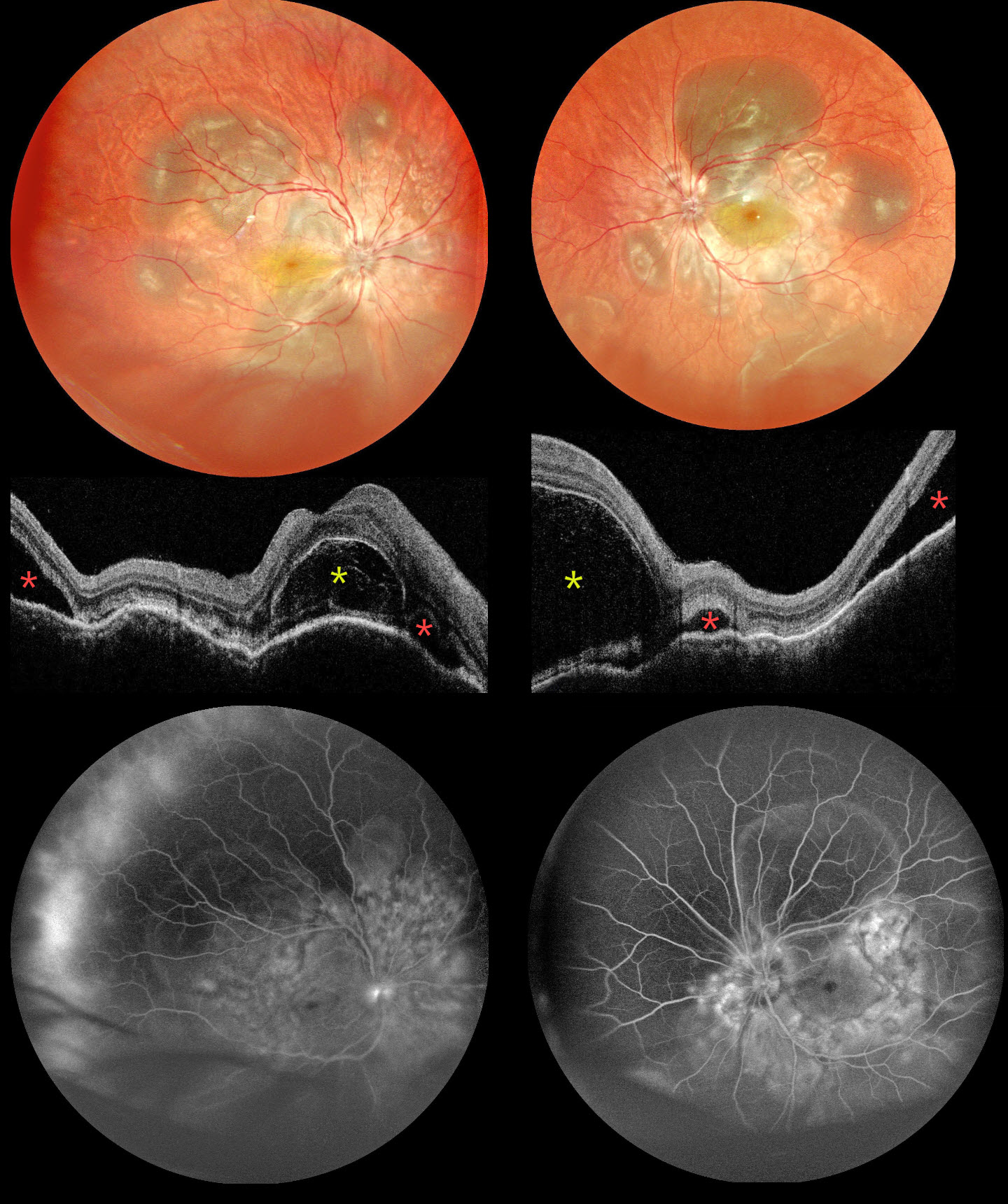
This 30YO male suffered an open globe injury from an iron nail OD on 11/2/25. Three days later, the corneo-limbal laceration was repaired elsewhere. He presented to us on 11/25 with 1 week of bilateral vision loss. Vision was hand motion OD and 20/400 OS. Mild anterior chamber cells were noted OU.
Multicolor SLO imaging shows bilateral multifocal pockets of subretinal fluid (SRF) along with inferior exudate retinal detachments. OCT scanning shows bilateral bacillary layer detachments (BALADs), SRF, and choroidal thickening and undulation. Fluorescein angiography shows multifocal subretinal leakage with disc leakage.
Learning Points:
Sympathetic ophthalmia is a rare, bilateral granulomatous panuveitis that develops following penetrating trauma or intraocular surgery to one eye, with an incidence of approximately 0.19% after open-globe injury (He et al, AJO 2022;234:117-125). It typically presents within the first year after the inciting event but can occur days to years later, presenting with decreased vision, pain, and photophobia. Findings mirror those seen in Vogt-Koyanagi-Harada disease, including granulomatous anterior uveitis, vitritis, exudative retinal detachment with BALAD, choroidal thickening, and Dalen-Fuchs nodules. Systemic corticosteroids remain the first-line treatment, with most patients requiring long-term corticosteroid-sparing immunosuppressive agents to prevent recurrences. Visual prognosis is generally good with treatment. See Parchand et al for a recent comprehensive update (Ind J Ophthalmol 2022;70:1931-1944).
Our patient was started on intravenous methylprednisolone 1g for 5 days, followed by oral steroids and azathioprine. All fluid resolved 3 weeks after starting treatment (not shown). Vision improved to 20/63 OU. A slow steroid taper was planned.
HARADA DISEASE
The European VitreoRetina Society (EVRS) and Ridham Nanda
Originally posted on @retina.rocks January 23, 2026
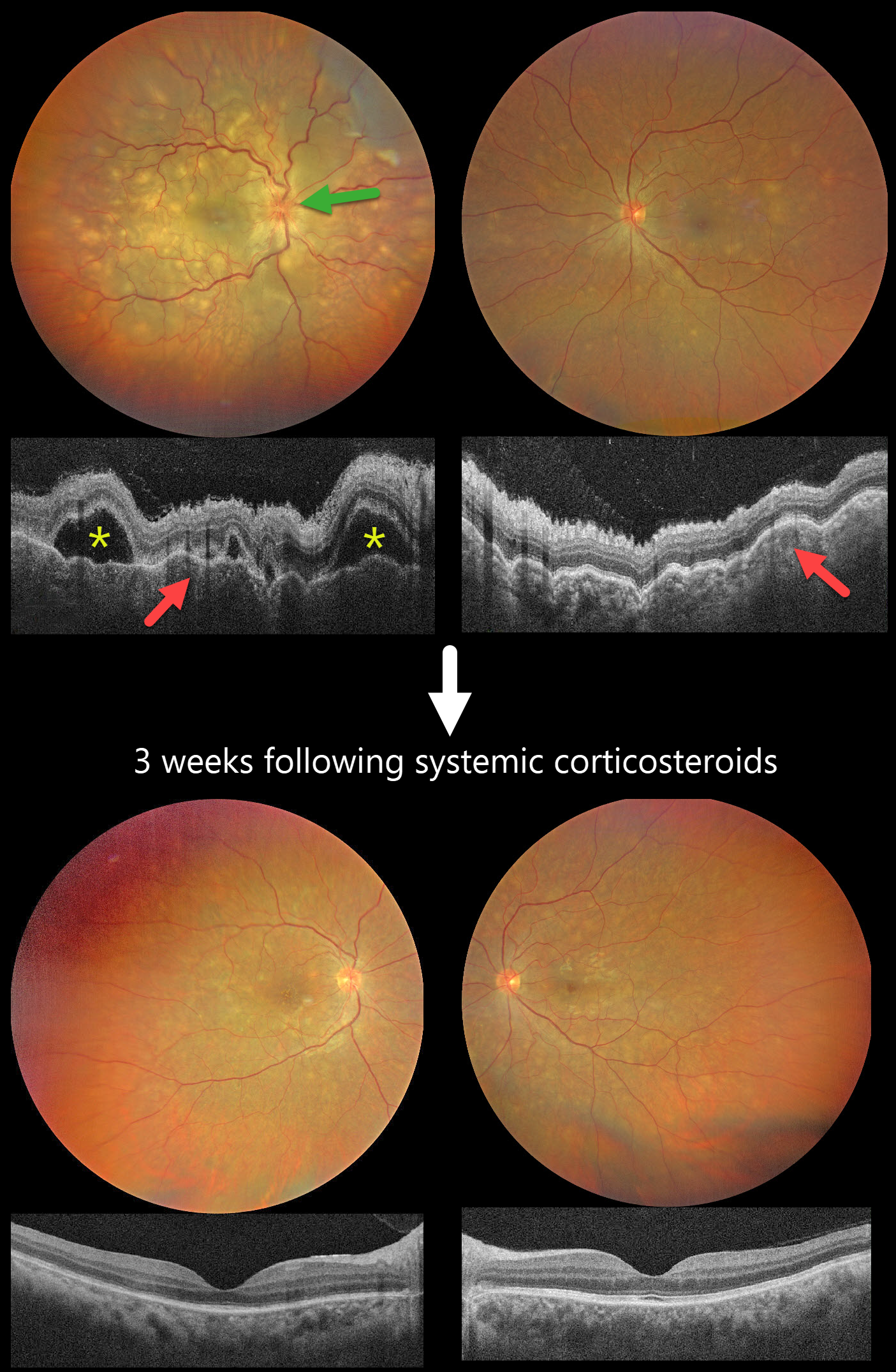
This previously healthy 40YO female presented with 2 days of blurred vision. She denied any systemic symptoms. Vision was 20/200 OD and 20/60 OS.
Color photography shows multifocal yellow-white subretinal lesions scattered throughout each posterior pole, with the right eye greater than the left, associated with overlying multifocal exudative retinal detachments. The right nerve is also edematous. OCT scanning shows a bilateral thickened choroid with overlying chorioretinal folds and multifocal serous detachments OD.
Intravenous methylprednisolone (1000mg/day) was administered for five days, followed by a 2-week course of oral prednisone (1mg/kg). Three weeks later, the funduscopic and OCT findings normalized. Vision improved to 20/30 OD and 20/20 OS.
Learning Points:
Harada disease is characterized by relatively acute bilateral panuveitis, optic nerve swelling, thickened choroid, chorioretinal folds, multifocal serous exudative retinal detachments, and bacillary layer detachments. Patients with just ocular findings have Harada disease (our patient), whereas those with additional systemic findings (including vitiligo, poliosis, headache, vertigo, and hearing loss) have Vogt-Koyanagi-Harada disease.
TUBERCULAR SERPIGINOUS-LIKE CHOROIDITIS
The European VitreoRetinal Society (EVRS) and Vaibhav Sethi
Originally posted on @retina.rocks January 2, 2026
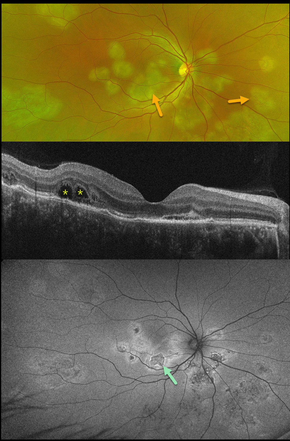
This previously healthy 41YO male presented with 4 days of blurred vision in his right eye. Vision was 20/80 OD and 20/20 in his normal OS.
Optos color RG imaging shows multifocal subretinal creamy white placoid lesions in the macula, peripapillary retina, and midperiphery. OCT scanning shows variable disruption of the outer retinal bands, as well as bacillary layer detachments (BALAD) temporally. The placoid lesions on fundus autofluorescence (FAF) show variable central hypo-FAF with more hyper-FAF borders.
Mantoux skin testing was positive at 24mm, and chest CT revealed left lower lobe ground glass opacities with hilar adenopathy. He was started on anti-tubercular therapy and oral prednisone.
Although tuberculosis most commonly presents with pulmonary involvement, extrapulmonary sites can include the gastrointestinal, skin, cardiovascular, genitourinary, and central nervous systems, as well as the eyes. Ocular involvement, like syphilis, can mimic virtually any type of uveitis, including anterior, intermediate, posterior, and panuveitis, retinitis, and retinal vasculitis, neuroretinitis, optic neuropathy, choroidal granuloma, choroiditis, and scleritis.
Learning Points:
Tubercular serpiginous-like choroiditis mimics serpiginous but affects younger patients with more multifocal and peripheral recurrences and progression if untreated (Agarwal et al, AJO 2020;220:160-169). Although the exact mechanism is not yet clear, a direct or indirect infectious trigger by the Mycobacterium tuberculosis is thought to cause choroiditis. Its origin is probably immunogenic since it seems to respond to corticosteroids and other immunosuppressants.
BALAD appears as a unique dome-shaped collection of intraretinal fluid from photoreceptor splitting. These detachments can be observed in an increasing number of conditions, most classically Vogt-Koyanagi-Harada disease (Cicinelli et al, Ophthalmology Retina 2020;4:454-456).
HARADA DISEASE
Originally posted on @retina.rocks December 16, 2025
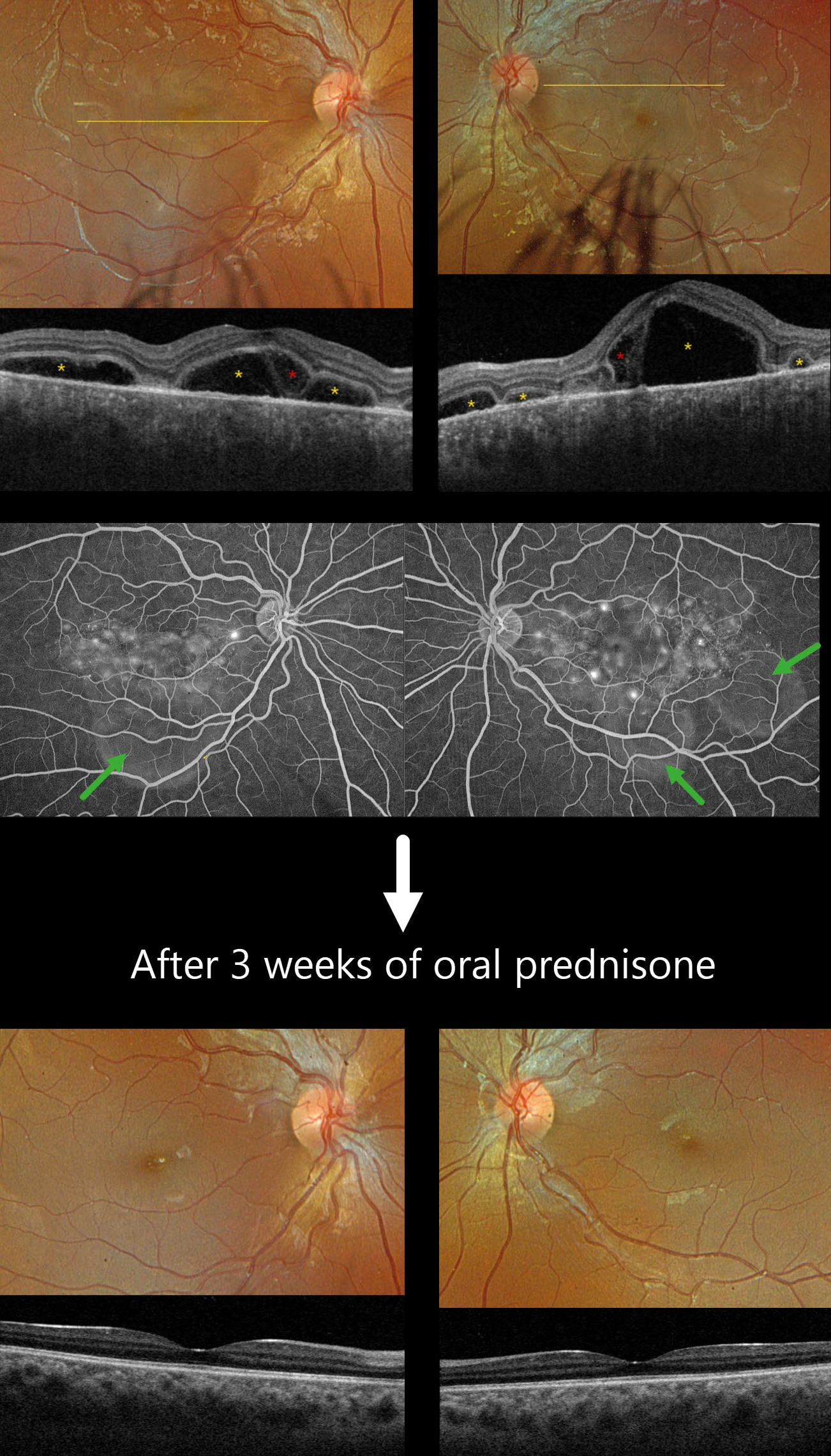
This previously healthy 13YO girl presented with several days of bilateral vision loss. Vision was 20/100 OD and 20/400 OS.
Optos color RGB imaging shows bilateral multifocal serous exudative retinal detachments. Triton swept-source OCT shows a complex pattern of multifocal pockets of subretinal fluid and bacillary layer detachments (BALAD). The choroid is markedly thickened. Fluorescein angiography shows multifocal pinpoint areas of subretinal leakage with pooling in the subretinal space and BALADs.
Learning Points:
Harada disease is characterized by relatively acute bilateral panuveitis, optic nerve swelling, thickened choroid, chorioretinal folds, multifocal serous exudative retinal detachments, and bacillary layer detachments. Patients with just ocular findings have Harada disease, whereas those with additional systemic findings (including vitiligo, poliosis, headache, vertigo, and hearing loss) have Vogt-Koyanagi-Harada disease.
Our patient was started on oral prednisone 40mg daily, and one week later, vision improved to 20/60 OU with resolution of all exudation (not shown). Three weeks later, the fundus appeared normal, and the choroid was approaching normal thickness. A slow steroid taper was started.
HARADA DISEASE
The European VitreoRetina Society (EVRS), Malvika Singh and Manish Nagpal
Originally posted on @retina.rocks October 3, 2025
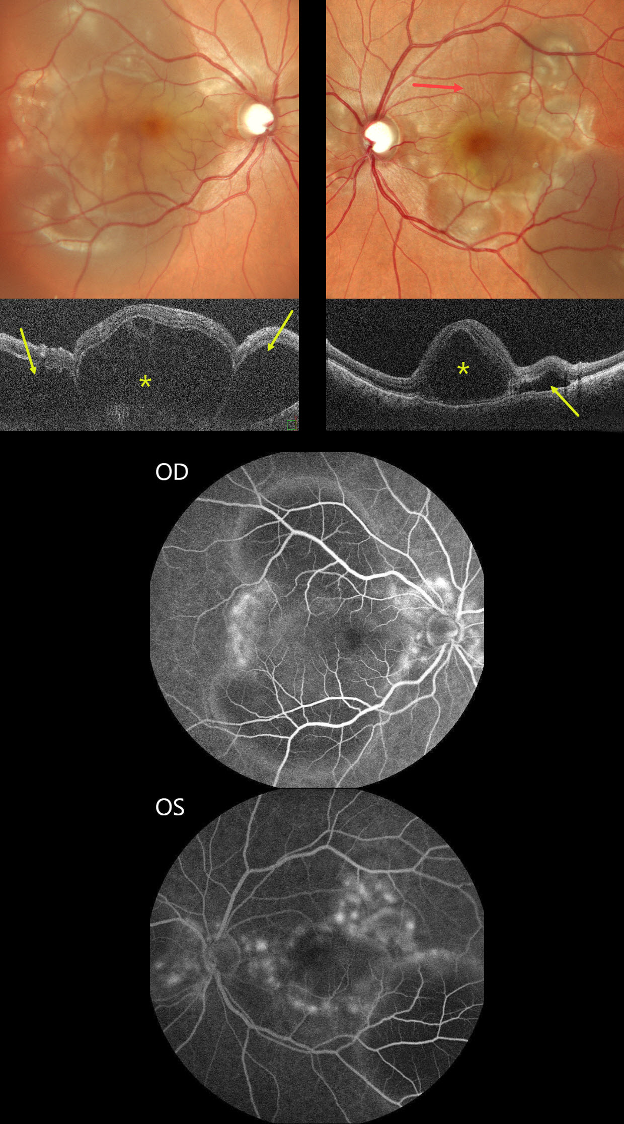
This previously healthy 30YO female presented with sudden bilateral vision loss for 5 days. Vision was counting fingers OU.
Color SLO imaging shows bilateral variably confluent serous detachments involving the posterior poles. Retinal folds are also noted in the left superior macula. OCT scanning shows a complex pattern of bacillary layer detachments (asterisks) and subretinal fluid (arrows). Fluorescein angiography shows multifocal subretinal leaks with pooling in the pockets of subretinal and intraretinal spaces.
She was started on oral steroids and immunomodulators in conjunction with a rheumatologist. One month later, vision returned to 20/20 OU with normalization of the funduscopic findings (not shown).
Learning Points:
Harada disease is characterized by relatively acute bilateral panuveitis, optic nerve swelling, thickened choroid, chorioretinal folds, multifocal serous exudative retinal detachments, and bacillary layer detachments. Patients with only ocular findings (as in our patient) have Harada disease, whereas those with additional systemic findings (including vitiligo, poliosis, headache, vertigo, and hearing loss) have Vogt-Koyanagi-Harada disease.
HARADA DISEASE
Nilesh Kumar
Originally posted on @retina.rocks August 20, 2025
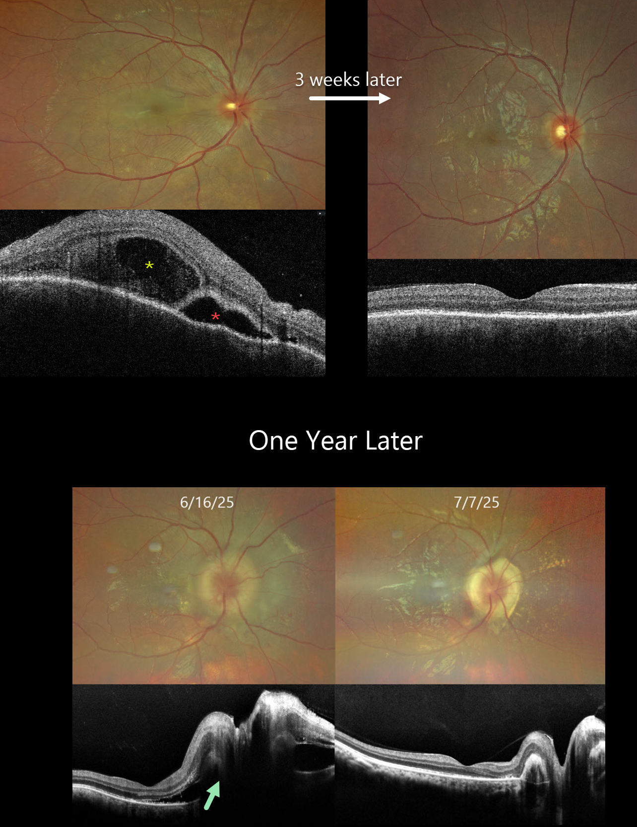
This previously healthy 24YO male presented with acute vision loss in his right eye. Vision was 20/200 OD and 20/20 in his normal OS. There were no neurologic or systemic symptoms.
Color photography shows an exudative retinal detachment involving the posterior pole. OCT shows a bacillary layer detachment, subretinal fluid with thickened retina and choroid. He was diagnosed with unilateral Harada disease and started on oral prednisone. Three weeks later, vision improved to 20/25 OD with marked clinical improvement. The oral prednisolone was tapered weekly over the next 6 weeks and stopped.
He returned one year later with recurrent vision loss. Vision was 20/200 OD. Exudative fluid with distal lipid flecks and a few small retinal hemorrhages surrounded the right nerve. OCT confirmed marked disk and peripapillary thickening with subretinal fluid. Oral prednisone was restarted, and oral azathioprine therapy was added. Three weeks later, the vision improved to 20/30. The fluid completely resolved with a ring of subretinal fibrosis surrounding the disc. The oral prednisone was tapered weekly over the next 6 weeks, and the oral azathioprine is planned to continue for the next year with regular monitoring.
Learning Points:
Harada disease is characterized by relatively acute bilateral panuveitis, optic nerve swelling, thickened choroid, chorioretinal folds, multifocal serous exudative retinal detachments, and bacillary layer detachments. Patients with just ocular findings have Harada disease, whereas those with additional systemic findings (including vitiligo, poliosis, headache, vertigo, and hearing loss) have Vogt-Koyanagi-Harada (VKH) disease. Although classically bilateral, early stages may present unilaterally before involving the fellow eye.
The disease generally progresses through four clinical stages: Prodromal, resembling a viral illness with systemic symptoms (fever, headache, meningismus); Uveitic, characterized by blurred vision, photophobia, optic disc edema, posterior choroidal thickening, and multifocal serous retinal detachments; Chronic/Convalescent, where depigmentation of the choroid leads to a “sunset-glow fundus” along with cutaneous signs like vitiligo, poliosis, and alopecia; and Recurrent, marked by repeated bouts of anterior uveitis and complications such as cataract, glaucoma, or macular neovascularization.
Management centers on high-dose systemic corticosteroids, often followed by steroid-sparing immunosuppressive agents to control inflammation and prevent recurrences. Early, aggressive therapy improves visual outcomes.
PUNCTATE INNER CHOROIDOPATHY WITH MACULAR NEOVASCULARIZATION AND PITCHFORK SIGN
Originally posted on @retina.rocks July 29, 2025
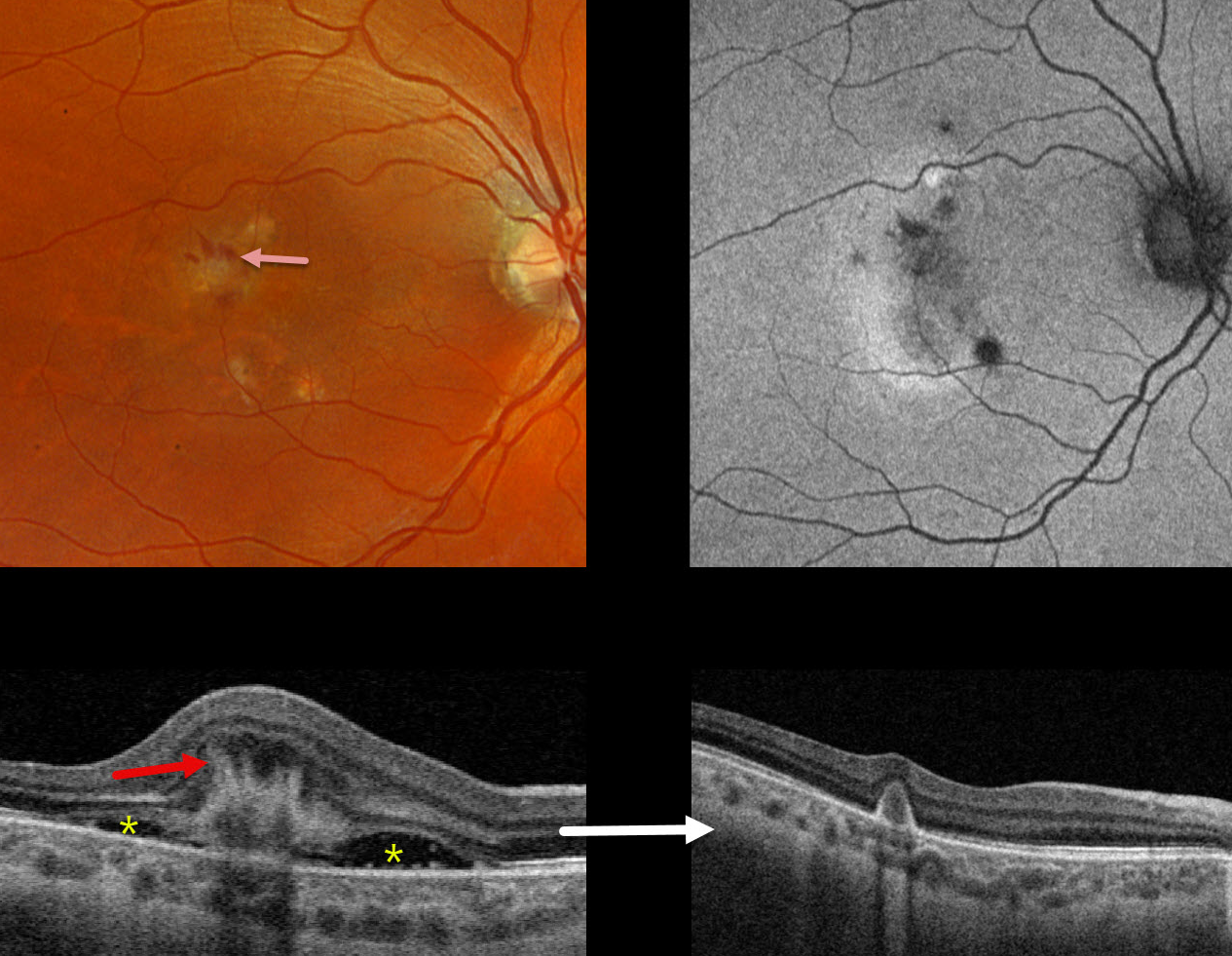
This 36YO female presented with 2 days of blurred vision in her right eye. There was no prior ocular history. Vision was counting fingers OD.
Triton color photography shows a whitish subretinal discoloration with some associated inferior subretinal fluid. A few dots of retinal blood are noted superiorly. Swept-source OCT shows a hyperreflective type 2 (above the RPE) macular neovascularization (MNV) with hyperreflective spikes radiating inwards from its inner surface. There is subretinal fluid on either edge of the MNV. Optos color RG imaging shows some small multifocal depigmented macular scars bilaterally. These scars are hypoautofluorescent.
Following an initial intravitreal Eylea HD injection, vision improved to 20/30 with complete resolution of all fluid and blood.
Learning Points:
The pitchfork sign was originally described by Hoang et al (Retina 2013;33:1049-1055) as a unique OCT finding in inflammatory MNV. Falavarajani et al more recently noted that this finding is also associated with non-inflammatory MNV (Ophthalmic Surg Lasers Imaging Retina 2019;50:719-725). The pathogenesis of the avascular pitchfork spikes is unknown but may be related to outer retinal traction or Muller cell activation.
Punctate inner choroidopathy (PIC) is part of the multifocal choroiditis (MFC) and panuveitis spectrum. MNV, the main cause of vision loss, can be successfully treated with anti-VEGF therapy, as in our patient.
ACUTE IDIOPATHIC MACULOPATHY (AIM)
Mattie Adams
Originally posted on @retina.rocks July 17, 2025
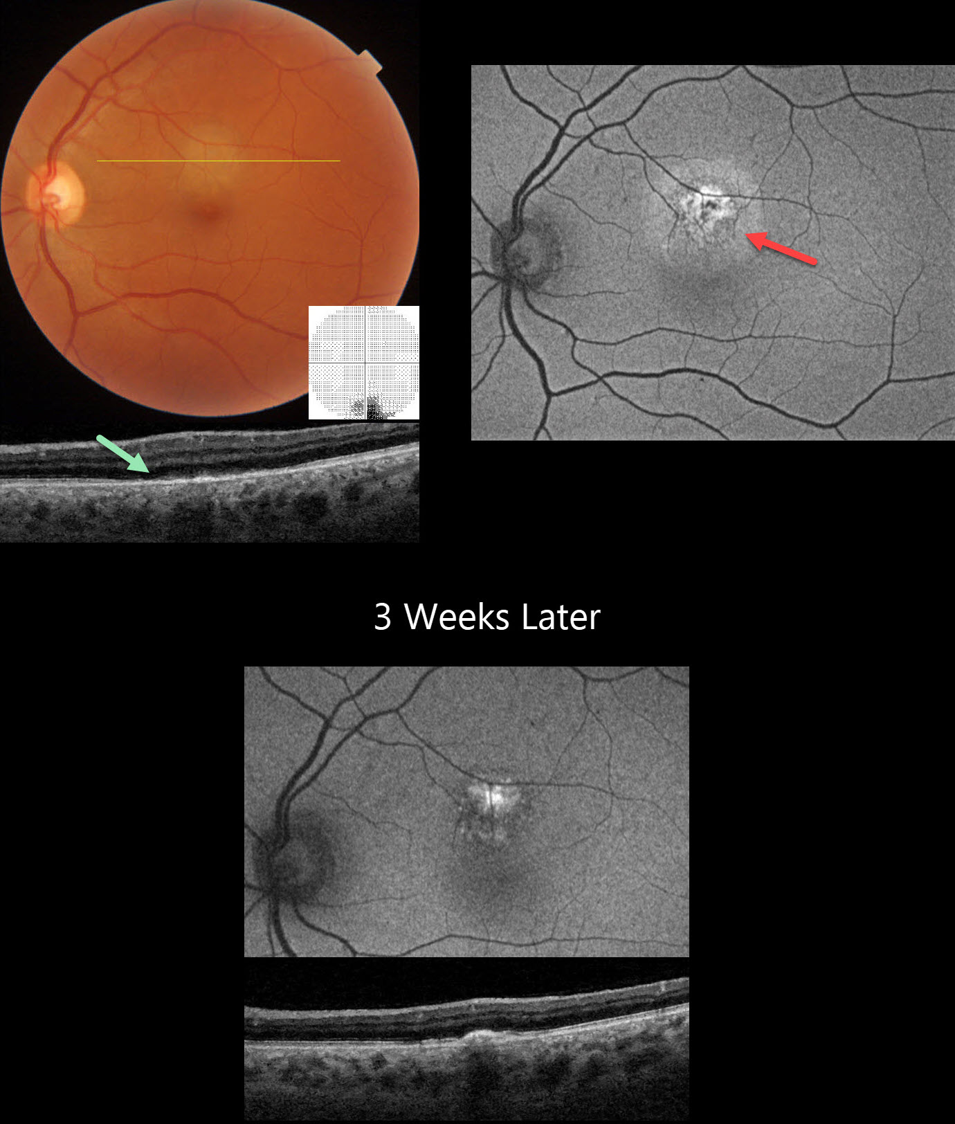
This healthy 46YO male presented with 5 days of an inferior paracentral scotoma in his left eye. Vision was 20/15 in his normal OD and 20/20 OS.
Triton color imaging shows a round, deep retinal amelanotic lesion just superior to the macular center. Swept-source OCT shows loss of the outer retinal band detail with ill-defined hyperreflective material overlying the RPE. Central 10-2 visual field shows inferior loss corresponding to the subretinal lesion. Optos fundus autofluorescence (FAF) of this lesion shows central hyper-FAF with a surrounding rim of less-intense hyper-FAF.
Coxsackie B virus antibody titers were markedly positive at >= 1:640, and observation was recommended. He returned 3 weeks later with stable symptoms and vision, although the OCT and FAF had improved.
Learning Points:
Acute idiopathic maculopathy (AIM) is an idiopathic inflammation of the outer retina, RPE, and inner choroid. There is often a preceding viral prodrome (lacking in our patient) caused by the Coxsackie virus. Healthy young patients typically present with acute unilateral moderately severe central vision loss.
The initial funduscopic changes include macular fluid and, sometimes, deep retinal blood that can mimic macular neovascularization. The OCT shows heterogeneous hyperreflective thickening of the outer retinal/RPE and fluid. Fluorescein angiography usually shows deep retinal leakage. The lesion spontaneously resolves with significant visual improvement after several weeks, usually leaving a pathognomonic bull’s-eye lesion as seen in our patient.
MULTIFOCAL TUBERCULOSIS CHOROIDAL GRANULOMAS
Will Gibson
Originally posted on @retina.rocks July 3, 2025
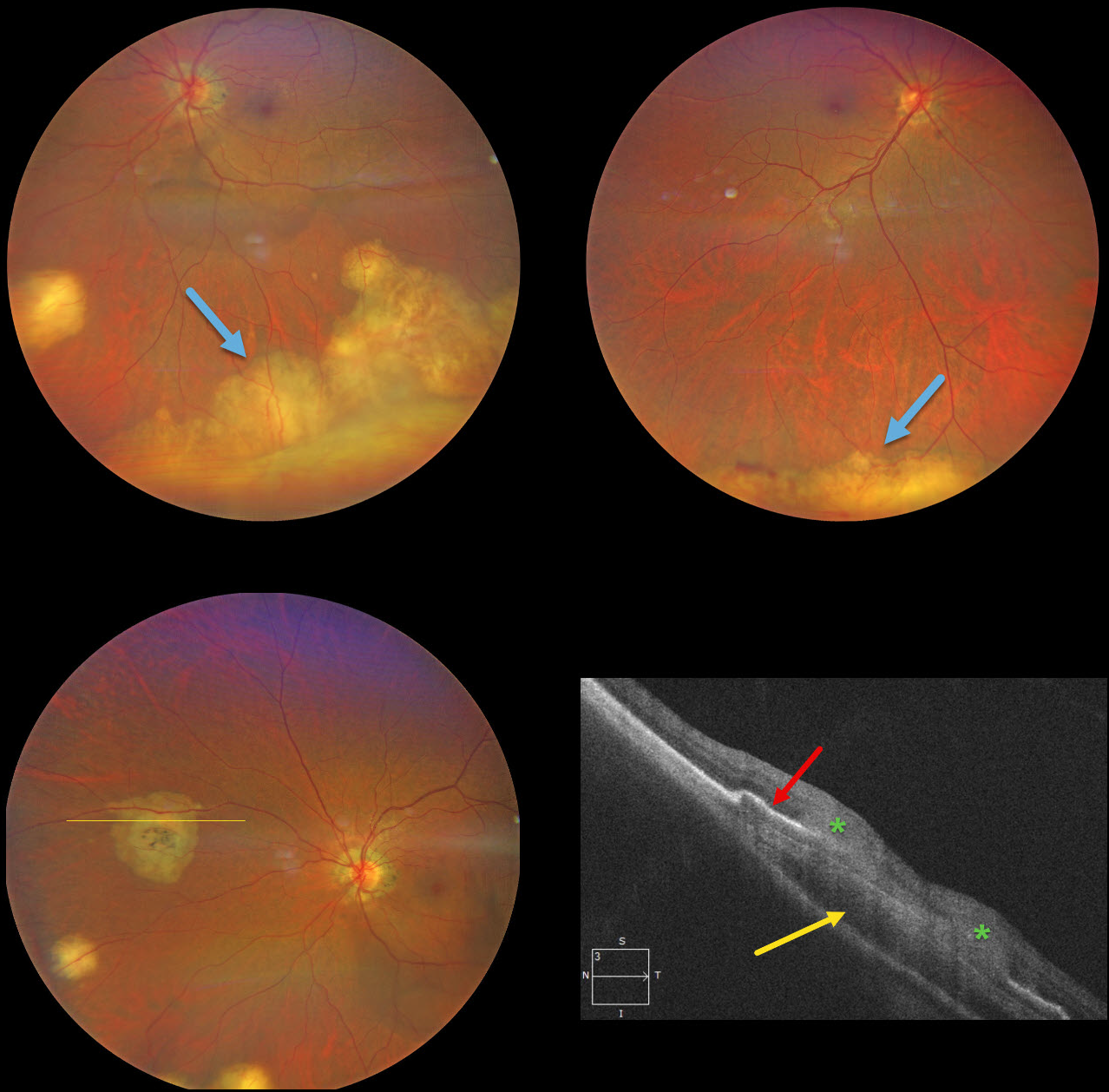
This previously healthy 49YO Indian female was visiting the United States and presented with new floaters in her left eye. Vision was 20/25 OD and 20/40 OS. There was no anterior uveitis, and the vitreous was clear bilaterally.
Color photography shows multifocal bilateral amelanotic choroidal lesions, some isolated and others confluent in the inferior periphery. OCT scanning through a nasal lesion OS shows a relatively hyporeflective choroidal lesion, irregular overlying RPE elevation, and probable infiltration of the overlying retina.
QuantiFERON-TB testing was positive with a negative chest X-ray. The patient was referred to the infectious disease department and started on anti-tuberculosis treatment (ATT).
Learning Points:
Although TB most commonly presents with pulmonary involvement, extrapulmonary sites can include the gastrointestinal, skin, cardiovascular, genitourinary, and central nervous systems, as well as the eyes. Ocular involvement, like syphilis, can mimic virtually any type of uveitis, including anterior, intermediate, posterior, and panuveitis, retinitis and retinal vasculitis, neuroretinitis, optic neuropathy, choroidal granuloma (our patient), choroiditis, and scleritis. For an excellent review of intraocular tuberculosis, see Bupta et al, Survey Ophthalmology 2007;52:561-587.
Tubercular granulomas can be distinguished from sarcoid lesions by being solitary, yellow, larger, and vascularized (Agarwal et al, AJO 2021;226:42-55). Our patient is therefore somewhat unusual with bilateral multifocal lesions. Sarcoid workup was negative.
POSSIBLE EPIDEMIC RETINITIS
Sharat Hegde
Originally posted on @retina.rocks June 2, 2025
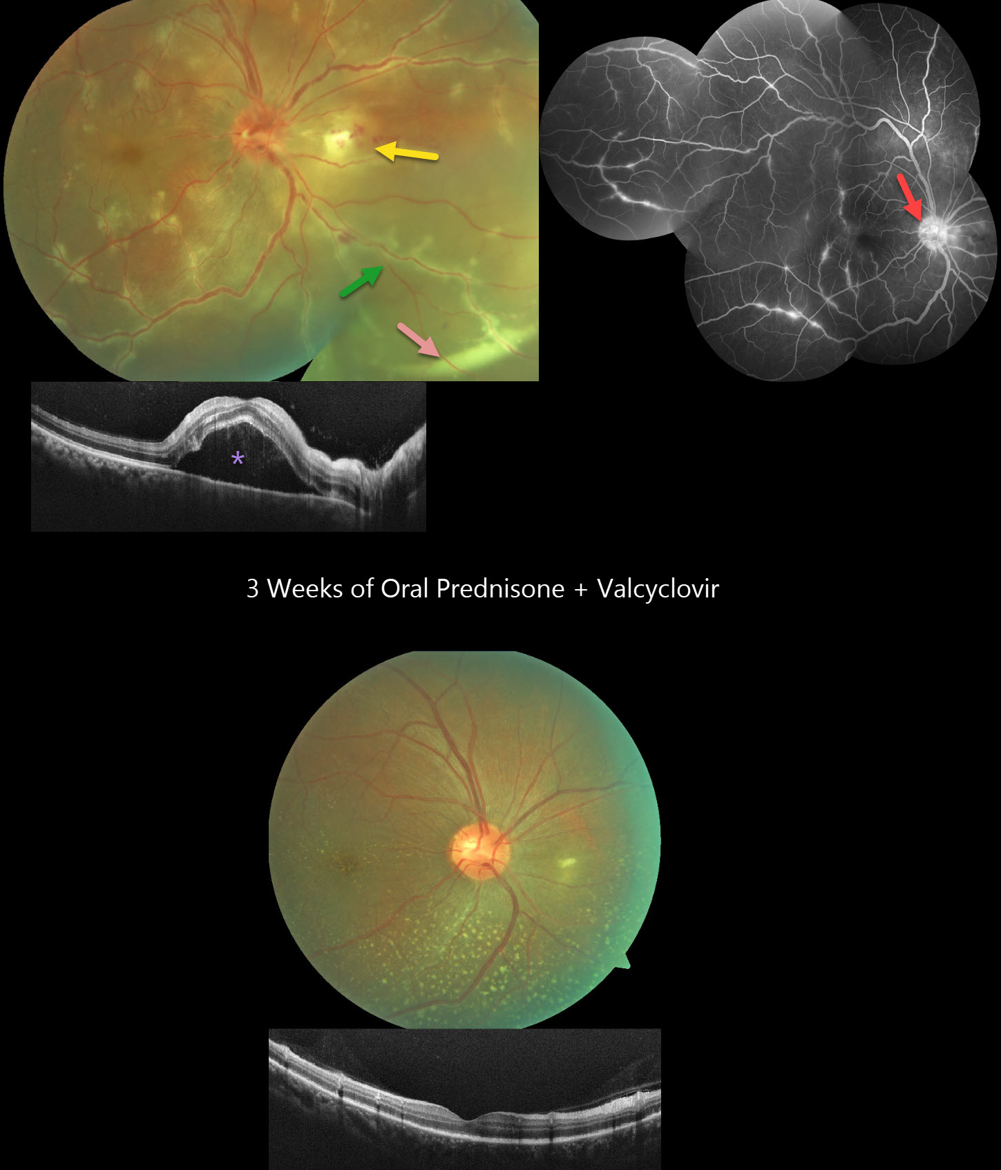
This healthy 36YO male presented with 3 days of floaters and decreased vision in his right eye. Vision was 20/60 OD and 20/20 in his healthy OS. The right eye contained mild anterior chamber cells and moderate vitreous cells.
Fundus photography shows a swollen nerve, a frosted branch pattern of vasculitis involving the retinal veins, an area of retinitis just nasal to the nerve, and a subretinal hypopyon in the inferonasal midperiphery. Some multifocal areas of outer retinal inflammation are noted, along with some retinal hemorrhages. OCT scanning shows variable hyperreflective foveal subretinal fluid. Fluorescein angiography shows variable venous staining and leakage, including from the disc and in nasal retinitis.
The workup included elevated ESR and CRP levels, along with an increased IgG titer for Varicella zoster virus. Mantoux testing, syphilis, Bartonella, typhus, HIV, and chest X-ray were all negative. Although very atypical for viral uveitis, we started oral prednisone and valaciclovir. After one week, the hypopyon resolved, with decreased vasculitis and new scattered flecks of subretinal lipid throughout the posterior pole. By three weeks, vision improved to 20/20 with continued funduscopic improvement.
Learning Points:
Epidemic (post-fever) retinitis is an infectious or para-infectious entity that develops in immunocompetent individuals several weeks following a bacterial, viral, or protozoal infection. Causes include Dengue, Rickettsiosis, West Nile, Leptospirosis, Chikungunya, Typhoid, and Ebola. Patients present with sudden, painless vision loss.
Fundus findings include unilateral or bilateral unifocal or multifocal patches of retinitis with possible nerve involvement, serous macular detachment, and vasculitis or frosted branch angiitis. There is usually a favorable response to steroids and treating the underlying infectious cause with appropriate systemic therapy. See Mahendradas et al (Indian J Ophthalmol 2020;68:1775-1786) for an excellent post-fever retinitis review.
Frosted branch angiitis is a retinal vasculitis that gets its name from the involved vessels resembling branches of a tree that are “frosted” with snow (Kleiner et al, AJO 1988;106:27-34). Although these findings may be isolated, they are often considered a phenotype found in many inflammatory conditions, most commonly CMV retinitis. The inflammation is usually highly responsive to steroids, and the visual prognosis is generally good, although some may develop secondary neovascularization in the anterior and posterior segments due to widespread ischemia.
Subretinal hypopyon is characterized by yellowish inflammatory material settling inferiorly in the subretinal space. It has been documented in infectious (bacterial and fungal endophthalmitis, acute retinal necrosis, tuberculosis, syphilis), inflammatory (sympathetic ophthalmia), and
CHOROIDAL GRANULOMA
Evgenii Chernov
Originally posted on @retina.rocks April 28, 2025
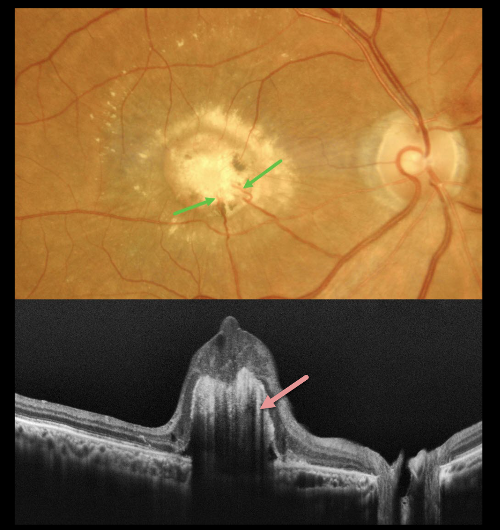
This healthy 64YO male presented with gradual vision loss in his right eye. Vision was 20/2000 OD and 20/20 in his normal OS.
Color imaging shows a yellow-white subfoveal nodular lesion with surrounding subretinal lipid. Several retinal choroidal anastomoses are noted within the lesion inferiorly. OCT scanning shows a hyperreflective elevated deep retinal lesion with overlying disorganized retina. There is marked posterior shadowing obscuring all deep retinal and choroidal detail.
Learning Points:
Choroidal granulomas usually appear as solitary, creamy white, round subretinal lesions. They are generally hyporeflective on OCT imaging (Invernizzi et al, Retina 2015;35:525-531) and can extend into the subretinal space, as in our patient. Causes include sarcoidosis, tuberculosis, Bartonella, and VKH.
Under normal conditions, the retinal and choroidal circulations remain separate and distinct. Rarely, the two circulations directly connect via a chorioretinal anastomosis. These can occur in chorioretinal scars, in macular telangiectasia, and in disciform scars.
Our best guess is that this lesion represents a vascularized choroidal granuloma. Due to its chronicity and lack of significant overlying retinal exudation, observation was recommended.
MULTIPLE EVANESCENT WHITE DOT SYNDROME (MEWDS )+ MFC OR POHS
Mattie Adams
Originally posted on @retina.rocks April 16, 2025
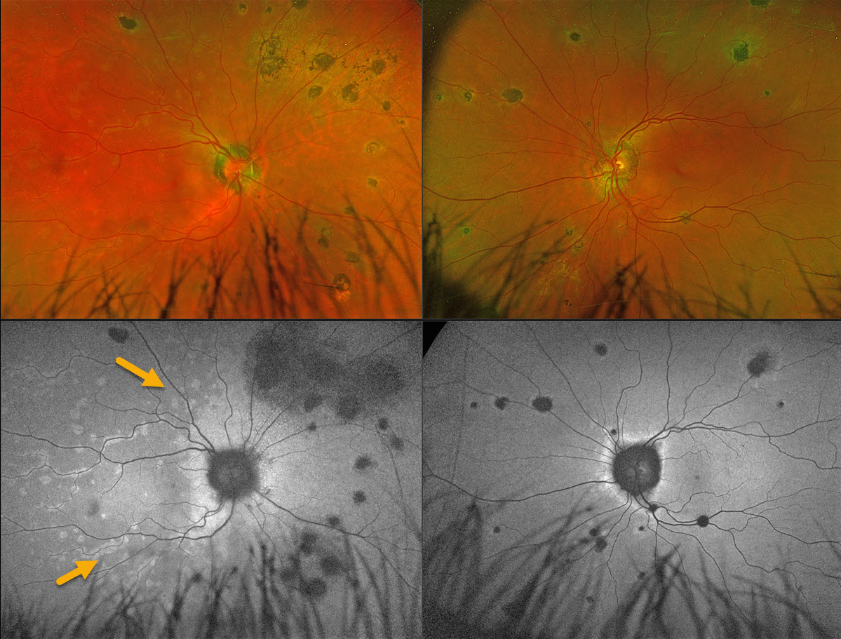
This 56YO female presented with 1 week of flashes and blurred vision in her right eye. Vision was 20/25 OD and 20/20 OS.
Coarse, large pigmented multifocal scars with peripapillary scarring are noted OU. Multiple deep white retinal spots are scattered throughout the right posterior pole. These spots on fundus autofluorescence (FAF) hyper-autofluoresce. Confluent hyper-FAF extends outwards from the optic nerve.
Learning Points:
Multiple evanescent white dot syndrome (MEWDS) is a usually unilateral, idiopathic inflammation of the outer retina that typically occurs in young, healthy myopic females. Acutely, patients will also have an enlarged blind spot, often complaining of temporal visual field loss, due to the peripapillary inflammation. The white dots disappear within a week or two, which is why the lesions are called “evanescent”.
Multifocal choroiditis (MFC) is the current preferred term for a group of historically heterogeneous disorders, including multifocal choroiditis, recurrent multifocal choroiditis, multifocal choroiditis and panuveitis, punctate inner choroidopathy, progressive subretinal fibrosis, and pseudo-histoplasmosis (Essex et al, Retina 2013;33:1-4 and Spaide et al, Retina 2013;33:1315-1324). MFC occurs in otherwise healthy adults, often myopic and female. Findings include variably pigmented multifocal scars throughout the fundus, panuveitis, subretinal fibrosis, and macular neovascularization (MNV). Unlike ocular histoplasmosis, new scars and uveitis can be seen. MNV can be successfully managed with anti-VEGF therapy.
Our patient’s scars represent either preexisting MFC or ocular histoplasmosis, and MEWDS is sometimes found in some of these eyes. Distinct from primary MEWDS, secondary MEWDS may be an epiphenomenon in inflammatory conditions such as MFC that disrupt the choriocapillaris-Bruch membrane-RPE complex (Essilfie et al, Retina 2022;42:227-235).
POST-VIRAL FROSTED BRANCH ANGIITIS
Ayushi Gupta and Vishal Agrawal
Originally posted on @retina.rocks April 7, 2025
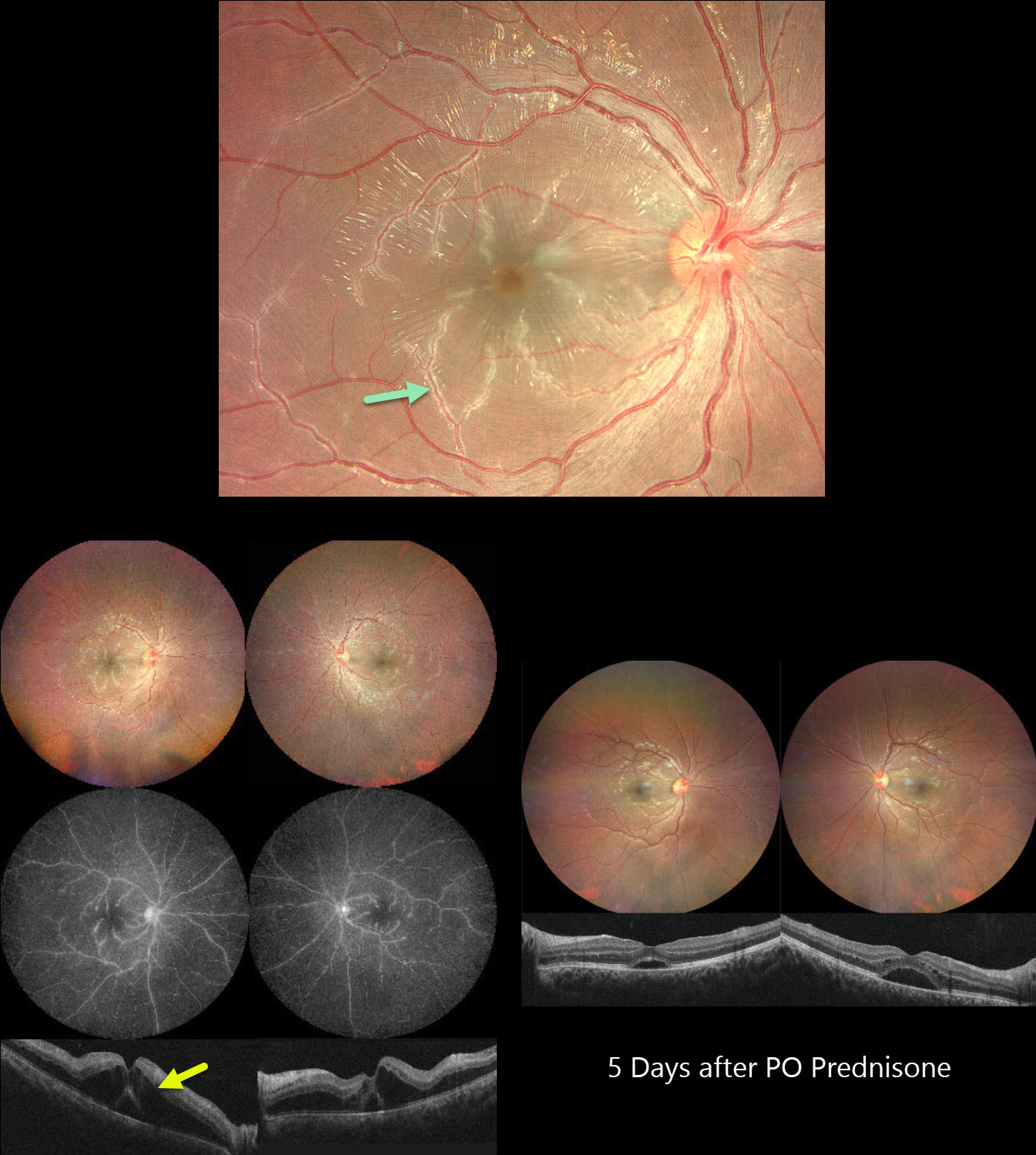
This previously healthy 21YO male developed bilateral vision loss 1 week following a viral illness. Vision was 20/100 OU.
Color photography shows a bilateral frosted-branch appearance of the retinal veins, most prominent in the maculas. Fluorescein angiography shows diffuse leakage and staining of the venous vascular tree. OCT scanning shows marked bilateral outer nuclear layer edema with foveal subretinal fluid. He was started on oral prednisone 60mg daily. Five days later vision improved to 20/40 OU. The venous changes are dramatically better with decreasing macular fluid. A slow steroid taper began.
Learning Points:
Frosted branch angiitis is a retinal vasculitis that gets its name from the involved vessels resembling branches of a tree that are “frosted” with snow (Kleiner et al, AJO 1988;106:27-34). Although these findings may be isolated, they are often considered a phenotype found in many inflammatory conditions, most commonly CMV retinitis. The inflammation is usually highly responsive to steroids, and the visual prognosis is generally good, although some may develop secondary neovascularization in the anterior and posterior segments due to widespread ischemia.
TUBERCULAR SERPIGINOUS-LIKE CHOROIDITIS
Originally posted on @retina.rocks March 11, 2025
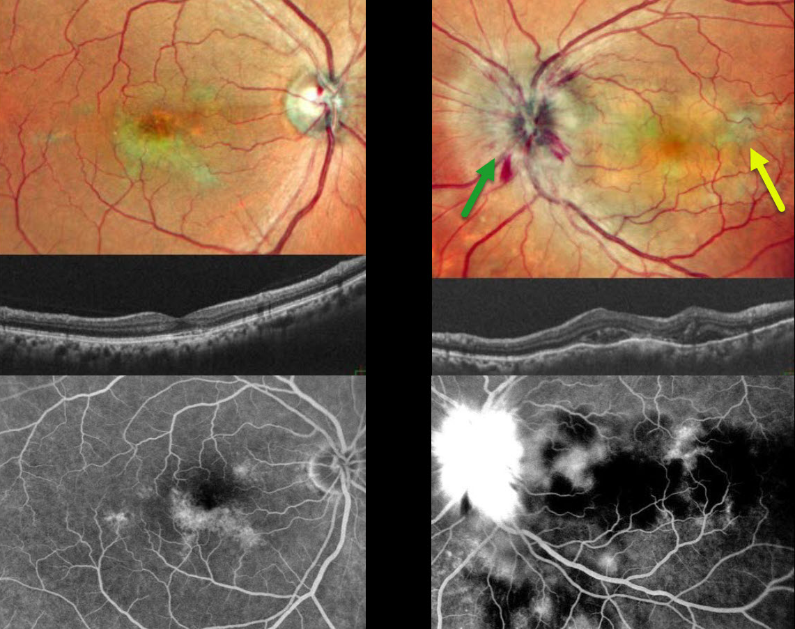
This 54YO male presented with 2 weeks of vision loss in his left eye. He was diagnosed elsewhere with papilledema, and brain MRI scanning was normal. Vision was 20/20 in his asymptomatic OD and counting fingers OS. Pseudocolor SLO imaging of the OS shows marked disc swelling with radiating hemorrhages in the nerve fiber layer. Some deep, hypopigmented retinal lesions are also seen.
OCT scanning shows irregular choroidal thickening vs choroidal folds with overlying pockets of variably reflective subretinal fluid. The right eye shows multifocal serpiginous-like areas of chorioretinal scarring with variable outer retinal thinning on OCT.
On fluorescein angiography, the macular lesions stain OD. The left nerve shows profound leakage, and the choroidal lesions show variable blockage and staining.
We suspected tubercular serpiginous-like choroiditis OD with acute tubercular papillitis and choroiditis OS. Chest X-ray showed a mass pressing on the trachea. Chest CT confirmed the mass, and subsequent biopsy revealed granulomatous thyroiditis. His Mantoux skin test was positive. We advised emergent Infectious disease consultation regarding starting anti-tubercular therapy. Unfortunately, he was immediately lost to follow-up.
Learning Points:
Although tuberculosis most commonly presents with pulmonary involvement, extrapulmonary sites can include the gastrointestinal, skin, cardiovascular, genitourinary, and central nervous systems, as well as the eyes. Ocular involvement, like syphilis, can mimic virtually any type of uveitis, including anterior, intermediate, posterior, and panuveitis, retinitis, and retinal vasculitis, neuroretinitis, optic neuropathy, choroidal granuloma, choroiditis, and scleritis.
Tubercular serpiginous-like choroiditis mimics serpiginous but affects younger patients with more multifocal and peripheral recurrences and progression if untreated (Agarwal et al, AJO 2020;220:160-169). Although the exact mechanism is not yet clear, a direct or indirect infectious trigger by the Mycobacterium tuberculosis is thought to cause the choroiditis. Its origin is probably immunogenic since it seems to respond to corticosteroids and other immunosuppressants.
MULTIPLE EVANESCENT WHITE DOT SYNDROME (MEWDS)
Yuenpang Cheung
Originally posted on @retina.rocks February 26, 2025
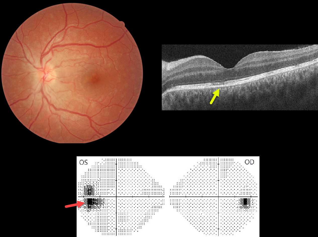
This healthy 27YO female presented with 2 weeks of photopsias OS. Vision was 20/20 in her normal OD and 20/25 OS.
Color photography shows multiple faint subretinal white dots scattered throughout the posterior pole. The nerve is slightly edematous, and the fovea has a granular appearance. OCT scanning shows scattered focal areas of ellipsoid zone and outer retinal band loss. Visual field testing shows an enlarged blind spot OS.
Learning Points:
Multiple evanescent white dot syndrome (MEWDS) is a usually unilateral, idiopathic inflammation of the outer retina that typically occurs in young, healthy myopic females. The granular fovea (Jampol spots, named in honor of Dr. Lee Jampol, who first described this syndrome in 1984) is a classic pathognomonic finding. Acutely, patients will also have an enlarged blind spot, often complaining of temporal visual field loss, due to the peripapillary inflammation.
The white dots disappear within a week or two, which is why the lesions are called “evanescent”. However, the blind spot may persist for many months, well after the fundus appearance has normalized. This stage of MEWDS was originally called the Acute idiopathic blind spot enlargement (AIBSE) syndrome before Gass recognized that the two disorders were the same disease but with distinct findings at different points in time.
HARADA DISEASE
Navneet Mehrotra and Tamanna Patel
Originally posted on @retina.rocks January 23, 2025
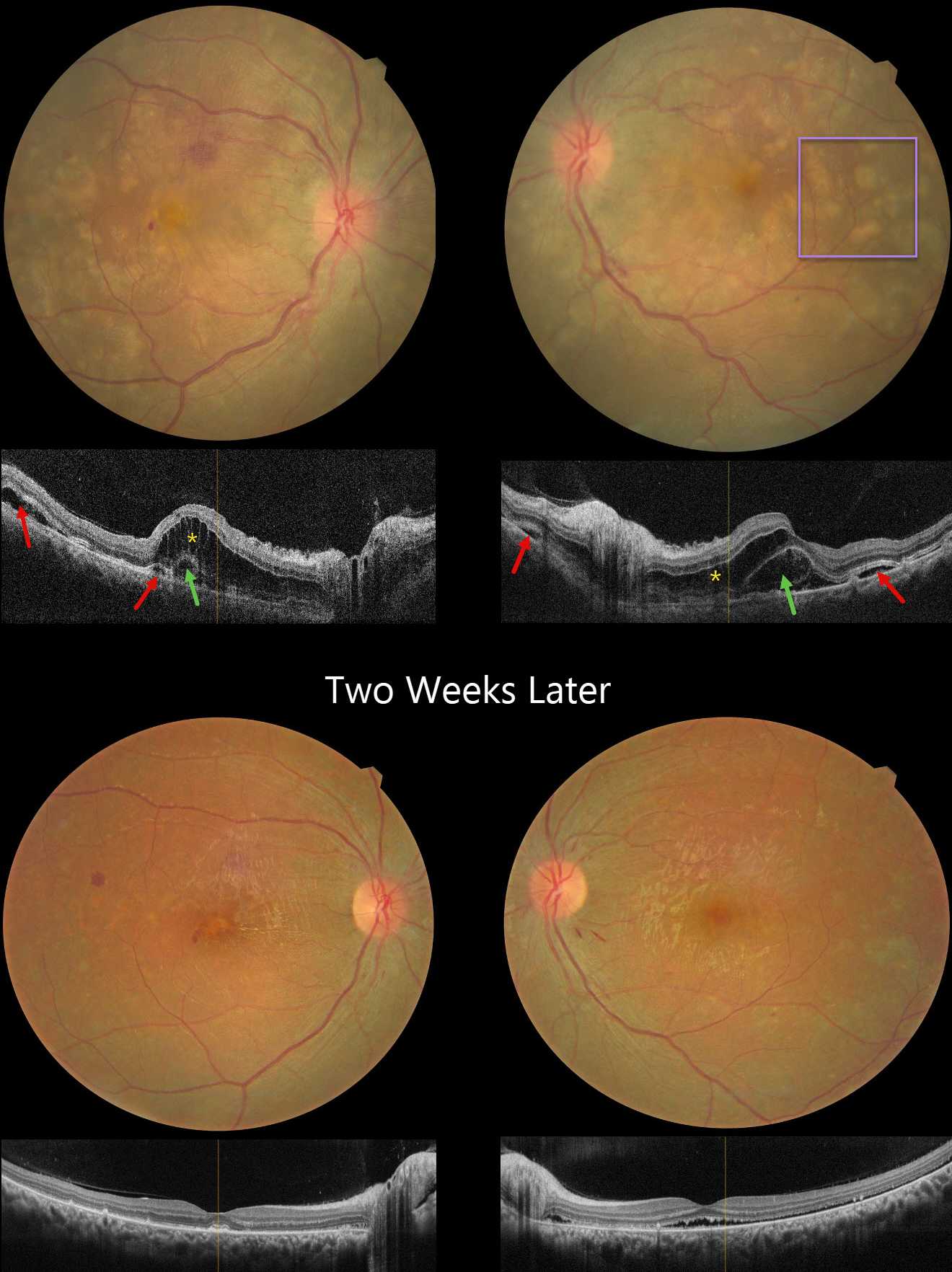
This 24YO female presented with several days of bilateral vision loss. She has a history of anti-glomerular basement membrane disease (anti-GBM) and is on hemodialysis. Her renal disease was quiescent, and there were no acute systemic complaints. There was no anterior segment inflammation, although mild vitreous cells were present bilaterally. Vision was 20/200 OD and 20/80 OS.
Fundus photography shows bilateral multifocal yellow subretinal lesions, inner retinal folds, some retinal hemorrhages, and mild disc edema. OCT scanning shows pockets of subretinal fluid, outer nuclear layer edema, and bacillary layer detachments. The choroid was thickened in both eyes on ultrasonography (not shown).
Oral prednisone 60mg/day was started in a tapering dose. Two weeks later, vision improved to 20/40 OD and 20/80 OS. The acute fundus findings were mostly resolved bilaterally.
Learning Points:
Harada disease is characterized by relatively acute bilateral panuveitis, optic nerve swelling, thickened choroid, chorioretinal folds, multifocal serous exudative retinal detachments, and bacillary layer detachments. Patients with just ocular findings have Harada disease, whereas those with additional systemic findings (including vitiligo, poliosis, headache, vertigo, and hearing loss) have Vogt-Koyanagi-Harada disease.
Anti-GBM disease is a rare autoimmune disorder characterized by autoantibodies that primarily target the basement membranes of the capillaries in the kidneys and lungs. Retinal findings include drusen occurring at an early age (Mansour et al, AJO Case Reports 2017;7:83-90). Acute fundus findings are rarely reported and consist of possibly steroid-induced central serous-like changes with multifocal retinal pigment epithelial detachments and exudative retinal detachments.
BIRDSHOT CHORIORETINOPATHY WITH MNV
Yuenpang Cheung
Originally posted on @retina.rocks January 22, 2025
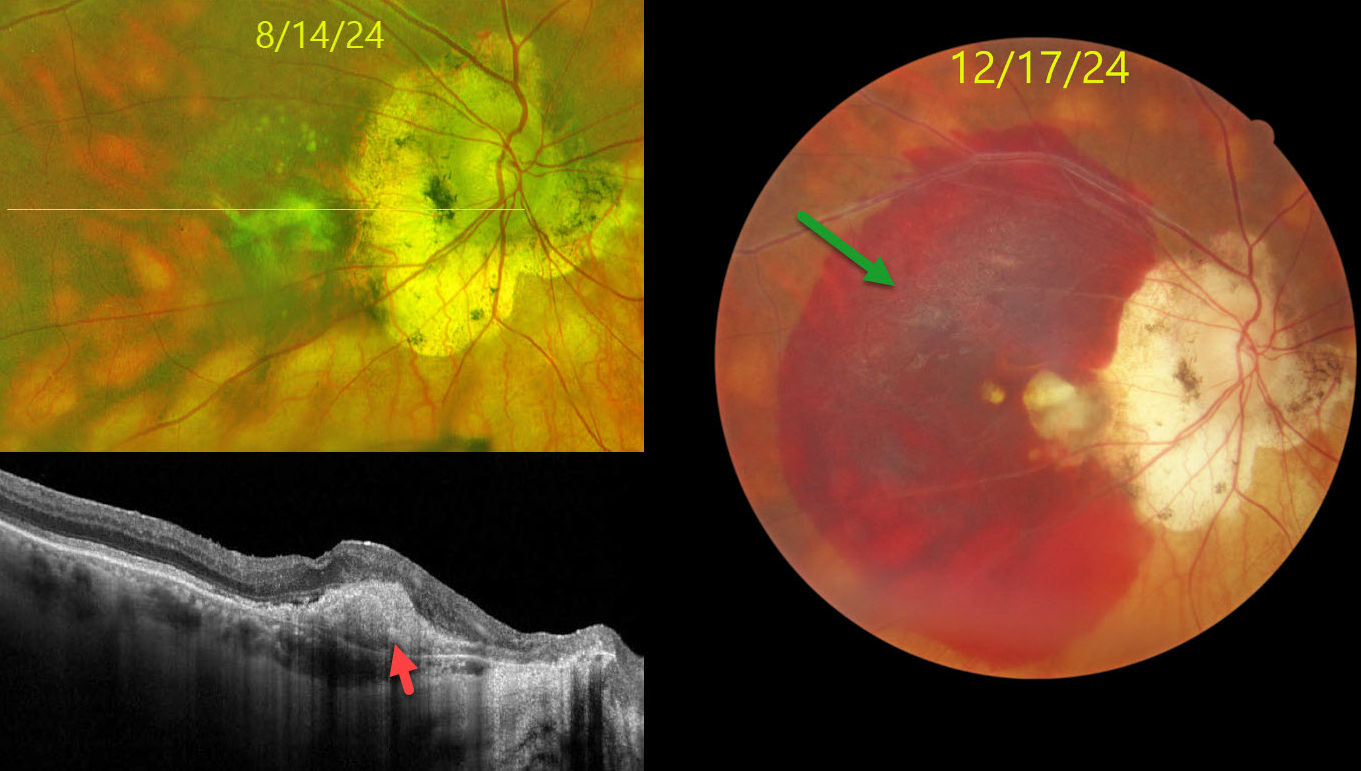
This 73YO male has a 40-year history of birdshot chorioretinopathy which has been managed most recently with PRN intravitreal Ozurdex and Yutiq. He has also been treated with q4 month Vabysmo injections for macular neovascularization (MNV) OD. He presented with recent decreased vision OD. Vision was counting fingers OD and 20/20 OS.
Color photography shows a large submacular hemorrhage, peripapillary scarring, and multiple creamy birdshot lesions more peripherally. Optos RG imaging 4 months earlier shows bilateral peripapillary scarring and scattered birdshot lesions. OCT scanning shows a hyperreflective type 1 (below the RPE) MNV OD.
Learning Points:
Birdshot chorioretinopathy was appropriately described and named by Drs. Ryan and Maumenee (Am J Ophthlamol 1980;89:31-45) since the lesions look like shotgun birdshot scattered throughout the fundus. Birdshot is typically chronic and bilateral with a very high HLA-A29 association. It usually affects women in their 40s to 60s. Vision loss is usually caused by cystoid macular edema (CME) and retinal atrophy, although MNV, as in our patient, can rarely occur. Asymptomatic patients can be observed. CME and symptomatic uveitis can be treated with oral steroids although many will still require immunomodulatory therapy.
HODGKIN LYMPHOMA MIMICKING MULTIFOCAL CHOROIDITIS
Originally posted on @retina.rocks December 4, 2024
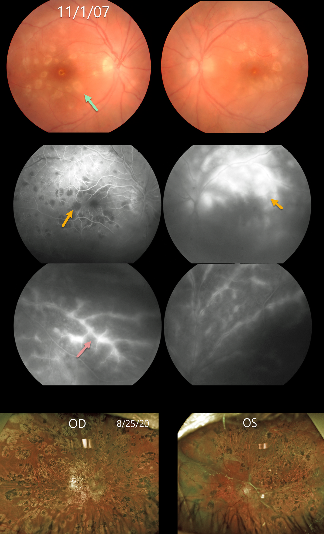
This 18YO female presented on 11/1/07 with acute bilateral panuveitis. Moderate vitreous cells were present along with subretinal multifocal yellow-white choroidal infiltrates. Angiographically, these choroidal lesions blocked early and stained late. A frosted-branch pattern of vascular leakage was noted bilaterally. Following a course of oral prednisone, the acute uveitic findings resolved, leaving behind inactive multifocal scarring. She developed severe recurrent panuveitis a year or two later, leaving her with 20/400 vision bilaterally (images not shown).
Shortly thereafter, she was diagnosed with Hodgkin lymphoma. At almost the exact date of this diagnosis, Baillif et al reported a patient with bilateral angle-closure glaucoma and multifocal choroiditis as the initial presentation for Hodgkin lymphoma (Retinal Cases 2011;5:201-205). The posterior segment findings in this report were virtually identical to those in our patient. The authors felt that the uveitis was a paraneoplastic phenomenon.
She subsequently underwent successful bone marrow transplantation. Her lymphoma and uveitis have both remained in remission since. A rhegmatogenous retinal detachment later developed in her left eye, which was repaired with vitrectomy and silicone oil. Despite successful reattachment, vision subsequently decreased to no light perception. On 8/25/20, vision was 20/400 OD with severe stable bilateral chorioretinal scarring bilaterally.
Learning Points:
Multifocal choroiditis (MFC) is the current preferred term for a group of historically heterogeneous disorders, including multifocal choroiditis, recurrent multifocal choroiditis, multifocal choroiditis and panuveitis, punctate inner choroidopathy, progressive subretinal fibrosis, and pseudo-histoplasmosis (Essex et al, Retina 2013;33:1-4 and Spaide et al, Retina 2013;33:1315-1324). MFC occurs in otherwise healthy adults, often myopic and female. Findings can include variably pigmented multifocal scars throughout the fundus, panuveitis, subretinal fibrosis, and macular neovascularization (MNV). Unlike ocular histoplasmosis, new scars and uveitis can be seen. MNV can be successfully managed with anti-VEGF therapy.
As with any uveitis, the clinician must always include the possibility of a masquerade syndrome as occurred in our patient.
SERPIGINOUS CHORIORETINITIS
Janelle Adeniran
Originally posted on @retina.rocks November 25, 2024
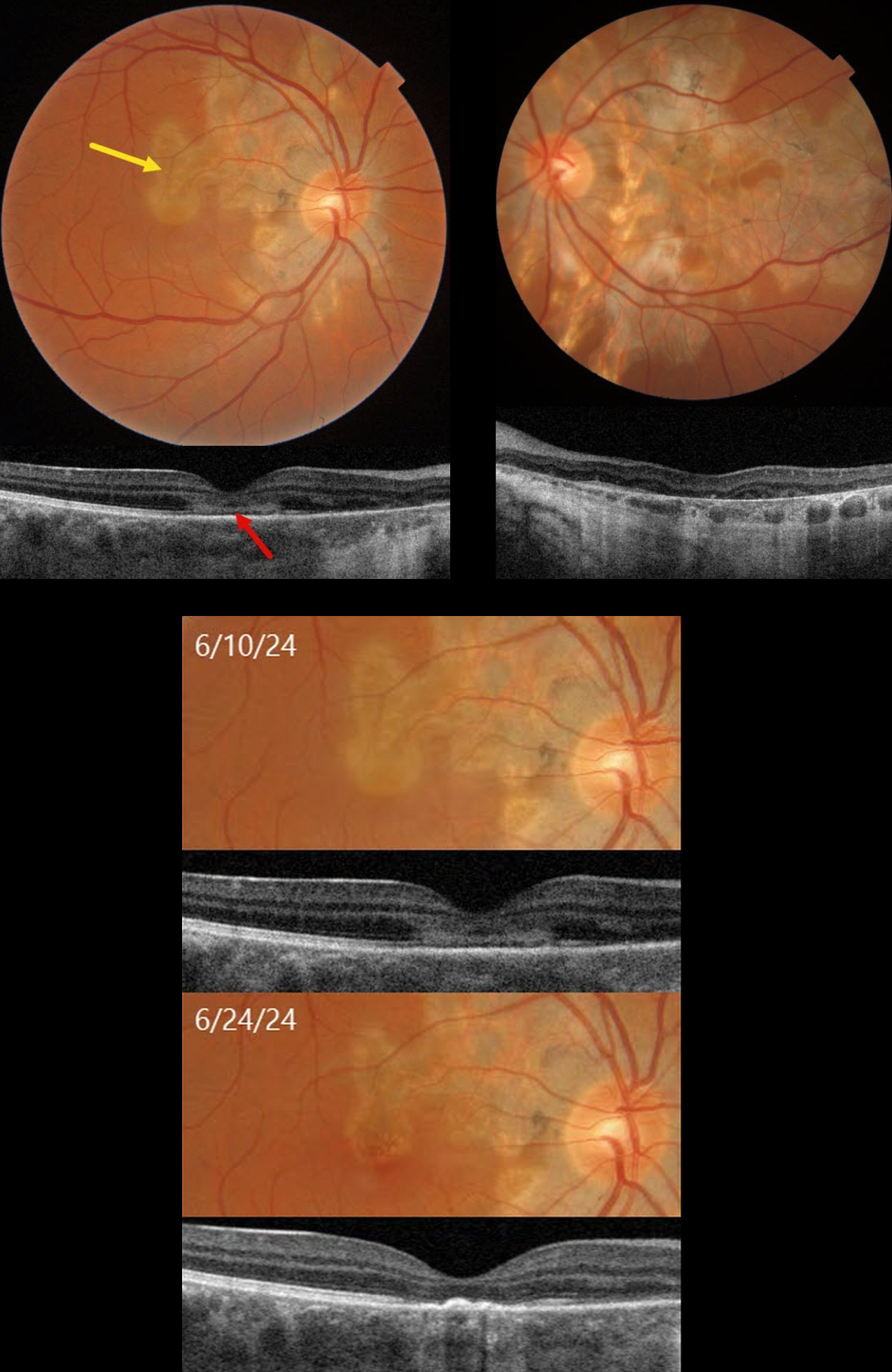
This healthy 37YO male with a known history of serpiginous chorioretinopathy presented with a few weeks of decreased vision in his left eye. Vision was 20/30 OD and 20/400 OS.
Triton color imaging shows bilateral chorioretinal scarring that radiates outwards from each optic nerve. An area of somewhat opaque active outer retinal inflammation just spares the right foveal center, with inactive scarring extending through the left fovea. The active lesion is hyperreflective on Triton swept-source OCT.
Tuberculosis testing was negative. Oral prednisone was started, and 2 weeks later, vision remained at 20/30, but there was marked regression with secondary scarring of the new lesion.
Learning Points:
Serpiginous choroiditis is an idiopathic, usually bilateral chorioretinal inflammatory condition, most commonly found in middle-aged men (Khanamiri and Rao, Surv Ophthalmology 2013;58:203-232). Recurrent areas of inflammation develop along the edge of a previous scar, producing scarring that meanders from the optic nerve outward into the macula. Vision is good unless scarring or macular neovascularization extends through the macular center. There is no consensus on the optimal systemic treatment, including corticosteroids and systemic steroid-sparing agents.
BRVO AND PERIPHLEBITIS FROM LIKELY SARCOIDOSIS
Originally posted on @retina.rocks October 8, 2024
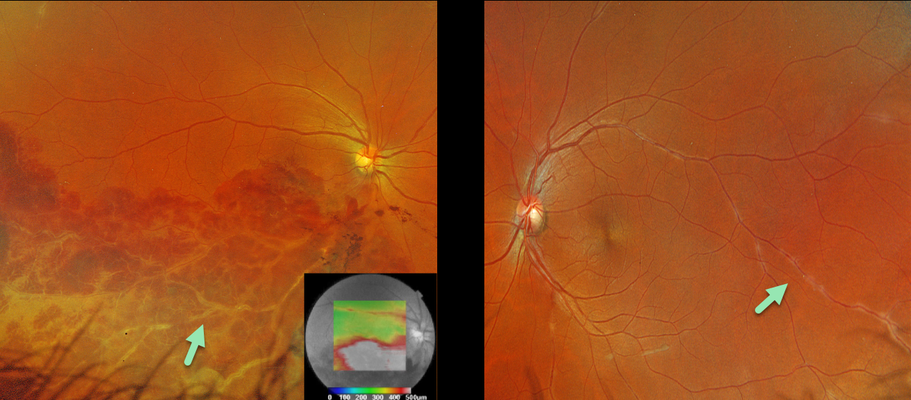
This healthy 36YO male presented with 1 week of unilateral vision loss. Vision was 20/400 OD and 20/20 OS.
Optos color RGB imaging OD shows a major inferotemporal branch retinal vein occlusion (BRVO) with severe confluent retinal hemorrhages and a frosted appearance to the major vessels. The occlusion does not seem to arise from a more proximal arteriovenous (AV) crossing. A small area of peripheral superotemporal periphlebitis is noted OD, with more extensive periphlebitis OS. Triton swept-source OCT shows inferior macular fluid.
BRVOs nearly always arise at an AV crossing, where a thickened arterial vessel compresses the underlying vein. Sarcoidosis is the classic exception where a BRVO is not caused by an AV crossing, but by an inflammatory narrowing of the involved vein. In addition, our patients’ segmental periphlebitis (‘candle wax drippings’) is virtually pathognomonic for sarcoidosis as well.
A sarcoidosis workup was recommended, although the patient failed to show for his testing and was immediately lost to follow-up.
SARCOIDOSIS
Mattie Adams
Originally posted on @retina.rocks August 21, 2024
This 51YO male has a history of bilateral central retinal vein occlusions for which he has been receiving intravitreal injections. On examination on 12/21/23, vision was stable at 20/70 OD and 20/200 OS, with new elevated multifocal choroidal lesions noted bilaterally. Looking back through his prior imaging, these lesions were faintly evident 3 months earlier, but had rapidly grown since.
Optos color RG imaging of his left eye shows multifocal, elevated, creamy white temporal macula choroidal lesions. Triton swept-source OCT reveals these lesions to be hyporeflective. They show variable ill-defined staining vs leakage on fluorescein angiography.
The differential for these amelanotic choroidal lesions initially included choroidal metastasis, lymphoma, and granulomas. Extensive bloodwork, including ACE and lysozyme, was negative. Metastatic workup revealed mild splenomegaly and axial and mediastinal lymphadenopathy with a 5mm pulmonary nodule. Pulmonary biopsy showed noncaseating granulomas. Oral prednisone 60mg/day was started. When last examined on 4/12/24, vision remained at counting fingers, and his findings were relatively unchanged.
Learning Points:
Sarcoidosis, like syphilis, has a myriad of ocular presentations, including anterior, intermediate, and posterior uveitis, vasculitis, papillitis, and choroidal granulomas. Sarcoid granulomas are usually found as unilateral lesions without associated uveitis and appear as yellow choroidal lesions distinguished by an irregular, vermiform margin (Bas et al, Retina 2023;43:1842-1851). Granulomas are hyporeflective on enhanced-depth OCT imaging (Invernizzi et al, Retina 2015;35:525-531).
Establishing the diagnosis of ocular sarcoid in the absence of a known systemic diagnosis is often difficult. In our experience, chest CT is often positive despite negative bloodwork.
Given the negative metastatic workup and biopsy results, we assume the choroidal lesions represent sarcoid granulomas. These lesions have not significantly responded to prednisone. Pulmonary consultation agreed with our sarcoidosis diagnosis but suggested referring to rheumatology for help with getting the patient on a systemic steroid-sparing agent.
TUBERCULOSIS CHOROIDAL GRANULOMA
Divya Nair
Originally posted on @retina.rocks July 22, 2024
This 36YO male presented with 10 days of vision loss in his left eye. One month earlier, he was diagnosed with disseminated tuberculosis (TB), peritonitis, and pleural effusion, and was placed on systemic antitubercular therapy (ATT). Vision was 20/20 in his normal right eye and counting fingers in his left eye.
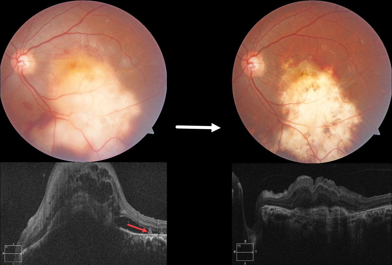
Fundus photography shows a large elevated yellowish submacular lesion that extends into the inferior midperiphery. Subretinal blood is noted along its superior and inferior margins. Faint lipid is present superotemporally, and some retinal striae are seen temporally. Spectral domain OCT shows a large dome-shaped hyporeflective choroidal lesion with overlying cystic retinal thickening, subretinal fluid, and subretinal hyperreflective material. A few hyperreflective pyramidal lesions are noted above the RPE temporally.
We started him on oral corticosteroids. In view of the vascularized lesion with subretinal blood, an intravitreal injection of Avastin was also given. Six weeks later, there is a relatively flat scar with near complete resolution of the blood. The choroidal lesion has resolved with an overlying hyperreflective subretinal scar and decreased retinal thickening. Unfortunately, vision remained at counting fingers.
Learning Points:
Although TB most commonly presents with pulmonary involvement, extrapulmonary sites can include the gastrointestinal, skin, cardiovascular, genitourinary, and central nervous systems, including the eyes. Ocular involvement, like syphilis, can mimic virtually any type of uveitis, including anterior, intermediate, posterior, and panuveitis, retinitis and retinal vasculitis, neuroretinitis, optic neuropathy, choroidal granuloma, choroiditis, and scleritis.
For an excellent review of intraocular tuberculosis, see Bupta et al, Survey Ophthalmology 2007;52:561-587.
Tubercular granulomas can be distinguished from sarcoid lesions by their solitary nature, yellow color, larger size, and vascularization (Agarwal et al., AJO 2021;226:42-55). All these features were found in our patient. Vascularization is thought to arise from localized hypoxia, leading to increased levels of vascular endothelial growth factor. Anti-VEGF injections and systemic corticosteroids, in addition to ATT, are helpful in hastening granuloma resolution and reducing secondary exudation (Lee et al, Graefe’s 2022;260:1641-1650).
SARCOIDOSIS
Mattie Adams
Originally posted on @retina.rocks May 27, 2024
This 58YO female was referred for asymptomatic fundus changes. She gave a history of hypertension and COPD. Vision was 20/40 OU. Anterior segments were normal.
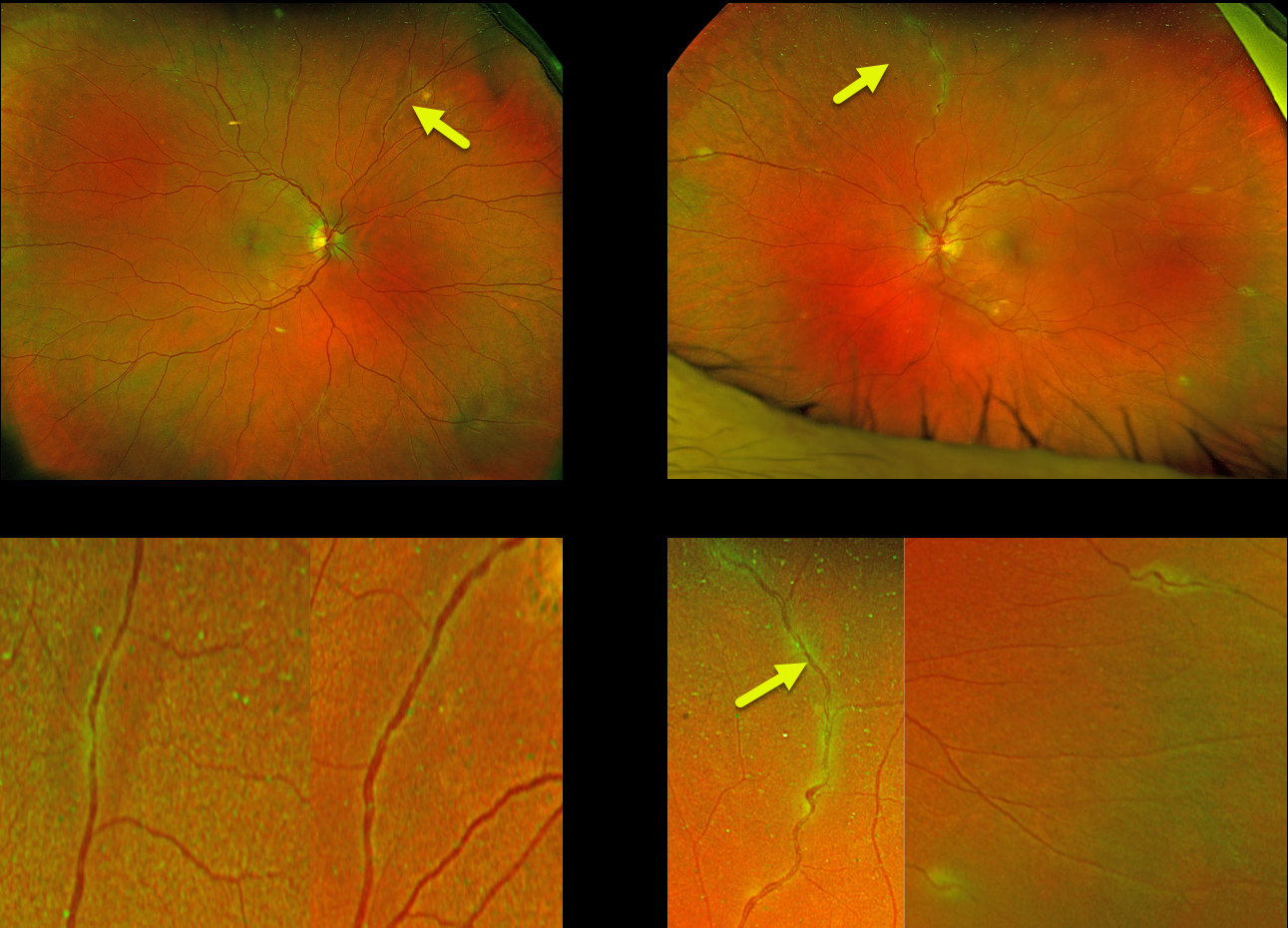
Optos color RG imaging shows bilateral multifocal areas of periphlebitis. Although sarcoid-specific bloodwork was negative (angiotensin converting enzyme, serum lysozyme, and soluble interleukin-2 receptor), chest CT showed multifocal, bilateral calcified and noncalcified pulmonary nodules throughout the lung parenchyma in addition to calcified mediastinal and hilar nodes. She was referred to a pulmonologist who agreed with our diagnosis of sarcoidosis. However, since there were no functional pulmonary deficits, he felt local treatment for the eye findings would be best. The patient will therefore be initially treated with a subtenon triamcinolone injection.
Learning Points:
Sarcoidosis, like syphilis, has a myriad of ocular presentations, including anterior, intermediate, and posterior uveitis, vasculitis, papillitis, and choroidal granuloma. Our patient’s segmental periphlebitis (‘candle wax drippings’) is virtually pathognomonic for sarcoidosis.
In our personal experience, as in our patient, there doesn’t appear to be a strong association between active systemic disease and ocular inflammation. Establishing the diagnosis of ocular sarcoid in the absence of a known systemic diagnosis is often difficult.
Sensitivity and specificity are highest for chest CT, and combined with elevated serum ACE and lymphopenia, are strongly suggestive of systemic sarcoidosis (Nieder and Sims, AJO 1019;206:149-153). Another recent publication found bilateral hilar lymphadenopathy on chest CT and sIL-2R as the most sensitive biomarkers for diagnosing systemic sarcoidosis (Japanese J Ophthalmology 2021;65:191-198).
CHOROIDAL GRANULOMA
Originally posted on @retina.rocks May 20, 2024
This healthy 26YO male presented with a 4-month history of decreased vision in his right eye. Vision was 20/200 OD and 20/25 in his normal OS.
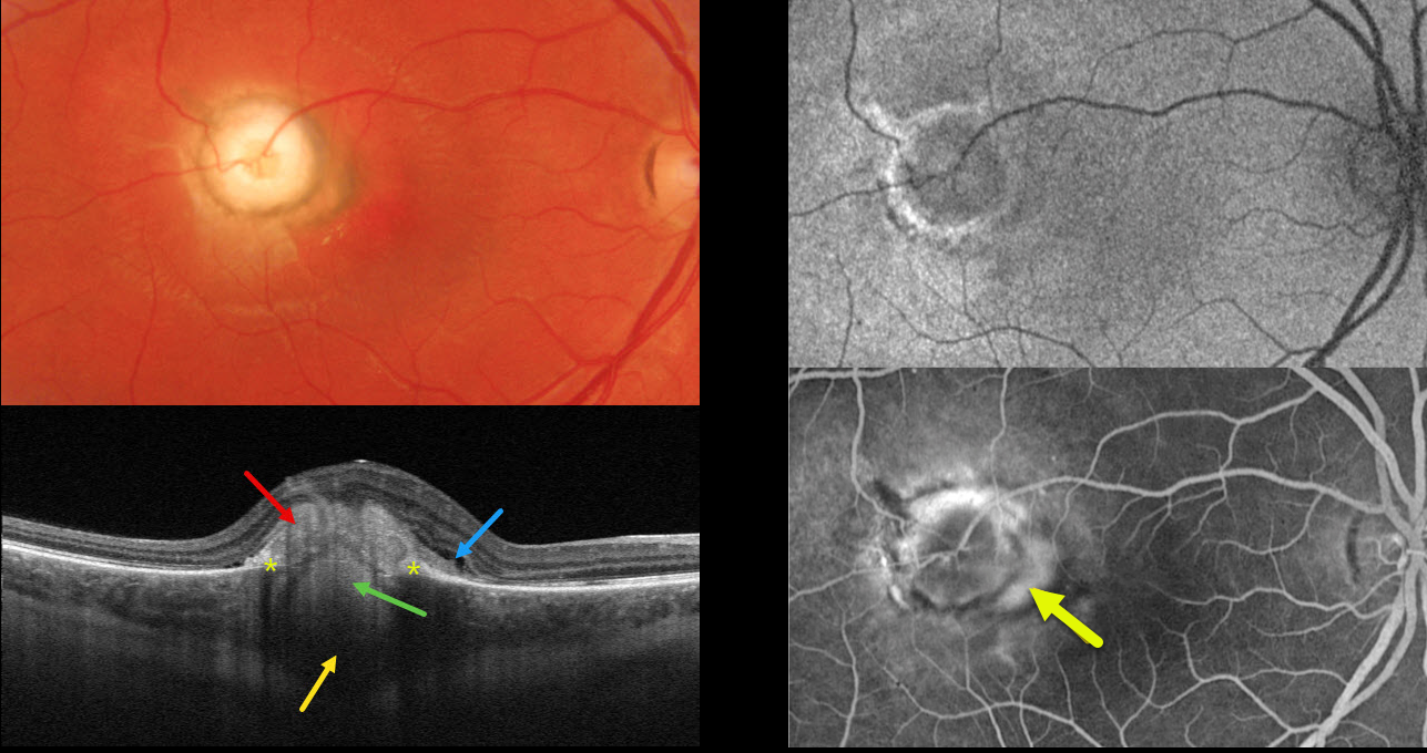
Optos color RG imaging shows a well-defined, 1800-micron, pale, subretinal nodular lesion with surrounding pigment. Triton swept-source OCT shows a hyperreflective subretinal mound, a hyperreflective lesion extending from the choroid through a break in the RPE, posterior choroidal thickening and shadowing, and trace outer retinal fluid.
Optos fundus autofluorescence (FAF) shows the lesion to be hypo-FAF with a rim of hyper-FAF.
On fluorescein angiography, the nodule stains along its edges, with a leaking macular neovascularization along its nasal edge. Anti-VEGF therapy was begun.
Learning Points:
Choroidal granulomas usually appear as solitary, creamy white, round subretinal lesions. They are generally hyporeflective on OCT imaging (Invernizzi et al, Retina 2015;35:525-531) and can extend into the subretinal space, as in our patient. Causes include sarcoidosis, tuberculosis, and VKH. Testing results for our patient were completely negative, including sarcoidosis, tuberculosis, Bartonella, and syphilis.
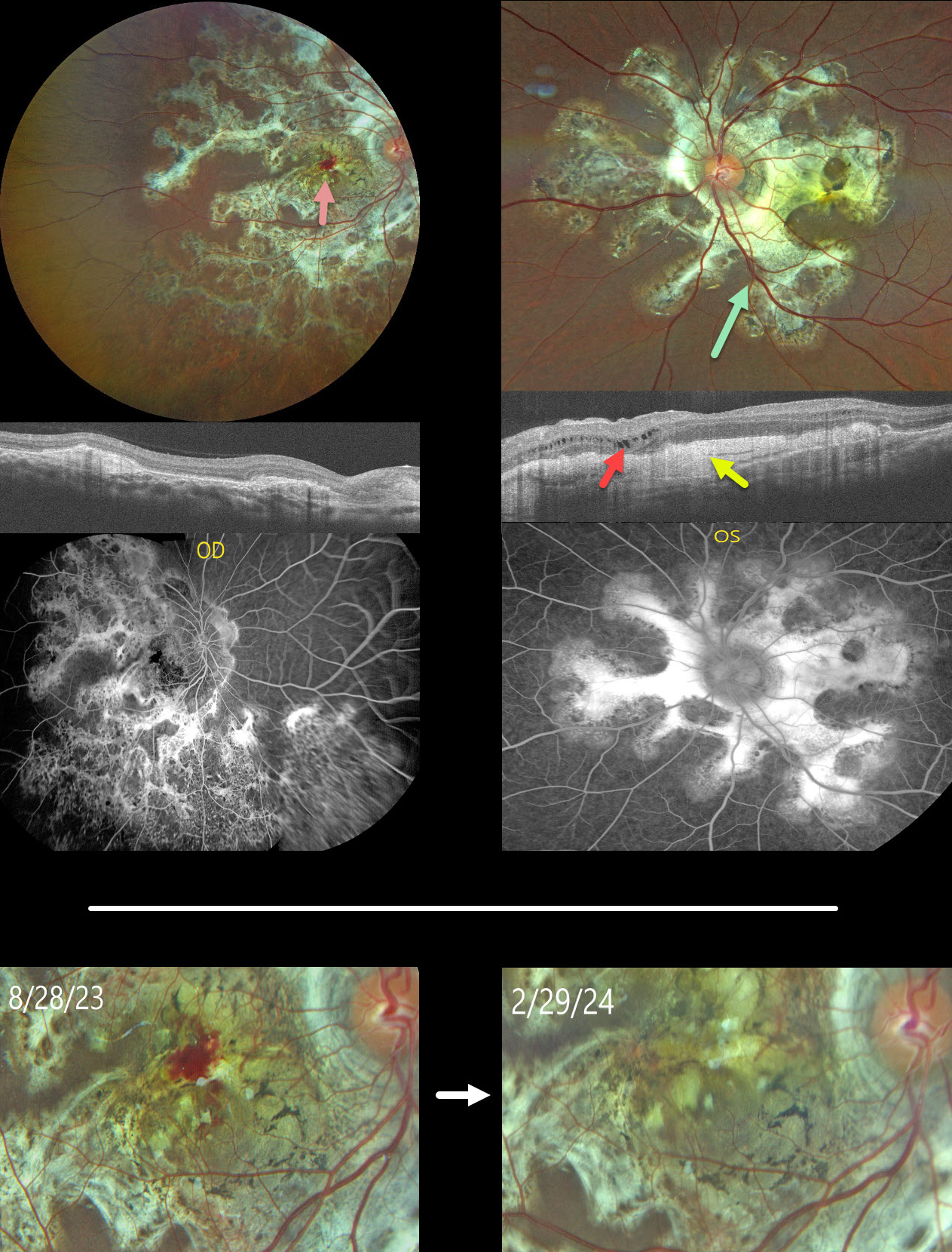
TUBERCULAR SERPIGINOUS-LIKE CHORIORETINITIS
Ogugua Okonkwo, Adekunle Olubola Hassan, Ayodele Harriman, Ogochukwu Sibeudu, and Idris Akintayo Oyekunle
Originally posted on @retina.rocks May 1, 2024
This healthy 13YO male presented with a 5-month history of bilateral vision loss. Vision was 20/200 OU.
Fundus photography shows bilateral subretinal, variably fibrotic peripapillary scarring that extends outwards with finger-like projections. A foveal hemorrhage is noted in the right macula. More mottled pigmented scarring extends into the right inferior periphery.
OCT scanning shows variable bilateral outer retinal atrophy and hyperreflective subretinal fibrosis with mild outer nuclear cysts in the nasal left macula. This scarring stains angiographically.
We were hesitant to start anti-VEGF therapy in a young boy and felt that the macular neovascularization causing the right macular blood might be inflammatory. He was therefore placed on a rapidly tapering course of oral prednisone.
At the 6-month follow-up, vision remained stable at 20/200 in this eye, with resolution of the blood.
Mantoux skin testing was negative. We have made multiple attempts to have the patient get QuantiFERON-TB Gold testing, but to date have been unsuccessful.
Learning Points:
Serpiginous choroiditis is an idiopathic, usually bilateral chorioretinal inflammatory condition, most commonly found in middle-aged men (Khanamiri and Rao, Surv Ophthalmology 2013;58:203-232). Recurrent areas of inflammation develop along the edge of a previous scar, producing scarring that meanders from the optic nerve outwards. Vision is good unless scarring or macular neovascularization extends through the macular center.
Tubercular serpiginous-like choroiditis mimics serpiginous but affects younger patients with more multifocal and peripheral recurrences and progression if untreated (Agarwal et al, AJO 2020;220:160-169). Although the exact mechanism is not yet clear, a direct or indirect infectious trigger by the Mycobacterium tuberculosis is thought to cause the choroiditis. We believe our patient has this diagnosis and will continue to confirm it with further testing.
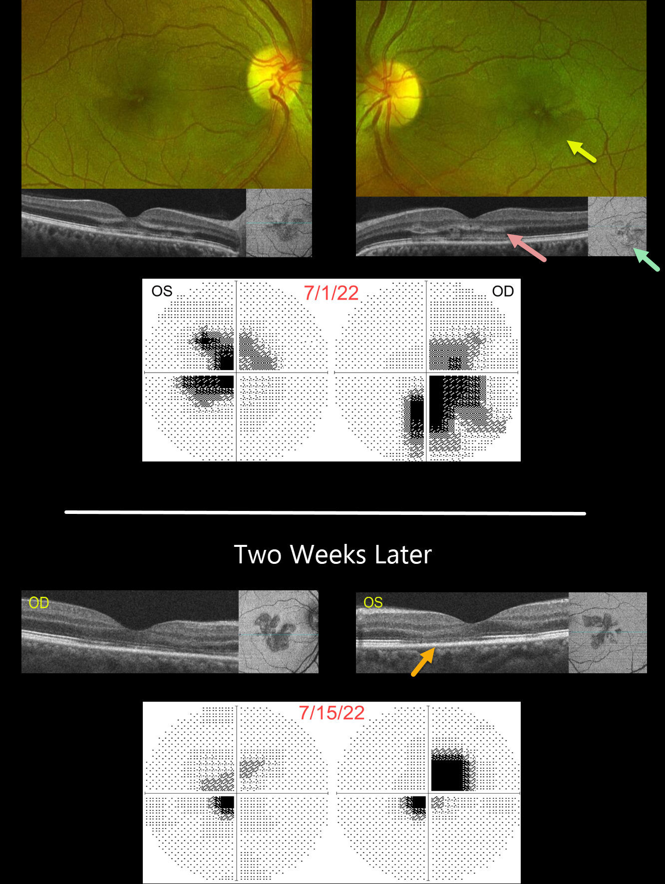
ACUTE MACULAR NEURORETINOPATHY (AMN)
Will Gibson
Originally posted on @retina.rocks February 19, 2024
This 41YO female presented with recent bilateral paracentral scotomas and blurred vision. There was no prior ocular or past medical history. Vision was 20/50 OD and 20/100 OS.
Optos color RG imaging shows faint wedge-shaped lesions radiating outwards from each macular center. OCT B-scanning shows variable patchy hyperreflectivity extending from the outer plexiform layer into the interdigitation zone. OCT en face imaging through the outer retina shows hyporeflective wedge lesions. Central 10-2 visual field testing shows bilateral paracentral scotomas.
Two weeks later, vision spontaneously improved to 20/60 OD and 20/25 OS. On OCT B-scanning, the outer retinal hyperreflective lesions are now replaced by secondary thinning with more prominent en face wedge lesions. The visual field scotomas are also improving.
Learning Points:
This case highlights many characteristic features of acute macular neuroretinopathy (AMN), an idiopathic disorder likely caused by compromise of the deep retinal capillary plexus.
Traditionally, the pathognomonic wedge-shaped AMN lesions are best seen with near-infrared reflectance, although we also find them easily imaged with en face OCT. With resolution, the acute lesions cause secondary outer retinal thinning.
Most patients are white, non-Latino females with a median age of 30 years. The most common associations are a flu-like prodrome and oral contraceptive use. Presenting acuity is usually good, coinciding with sparing of the sub-foveal areas. Roughly half of AMN cases are bilateral. There is typically moderate visual improvement over several months without treatment.
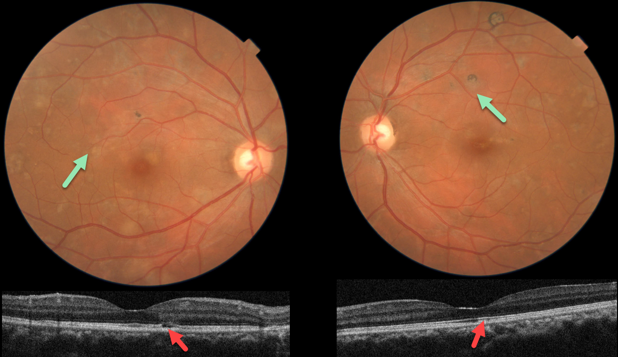
TUBERCULAR SERPIGINOUS-LIKE CHORIORETINITIS
Asma Samsudeen and Ashish Sharma
Originally posted on @retina.rocks January 18, 2024
This 38YO male presented with floaters in each eye. He denied any past medical history. Vision was 20/20 OU.
Fundus photos show multifocal patches of variably pigmented chorioretinal scarring throughout each posterior pole.
OCT scanning in each eye shows variable outer retinal atrophy and ellipsoid disruption. Unfortunately, fundus autofluorescence and fluorescein angiography are not available.
Subsequent workup was consistent for active tuberculosis, including a positive QuantiFERON-TB Gold and chest x-ray findings. Systemic anti-tuberculous therapy was begun.
Learning Points:
Serpiginous choroiditis is an idiopathic, usually bilateral chorioretinal inflammatory condition most commonly found in middle-aged men (Khanamiri and Rao, Surv Ophthalmology 2013;58:203-232).
Recurrent areas of inflammation develop along the edge of a previous scar, producing scarring that meanders from the optic nerve outwards. Vision is good unless scarring or macular neovascularization extends through the macular center.
Tubercular serpiginous-like choroiditis mimics serpiginous, but affects younger patients with more multifocal and peripheral recurrences and progression if untreated (Agarwal et al, AJO 2020;220:160-169).
Although the exact mechanism remains unclear, a direct or indirect infectious trigger by Mycobacterium tuberculosis is believed to cause choroiditis.
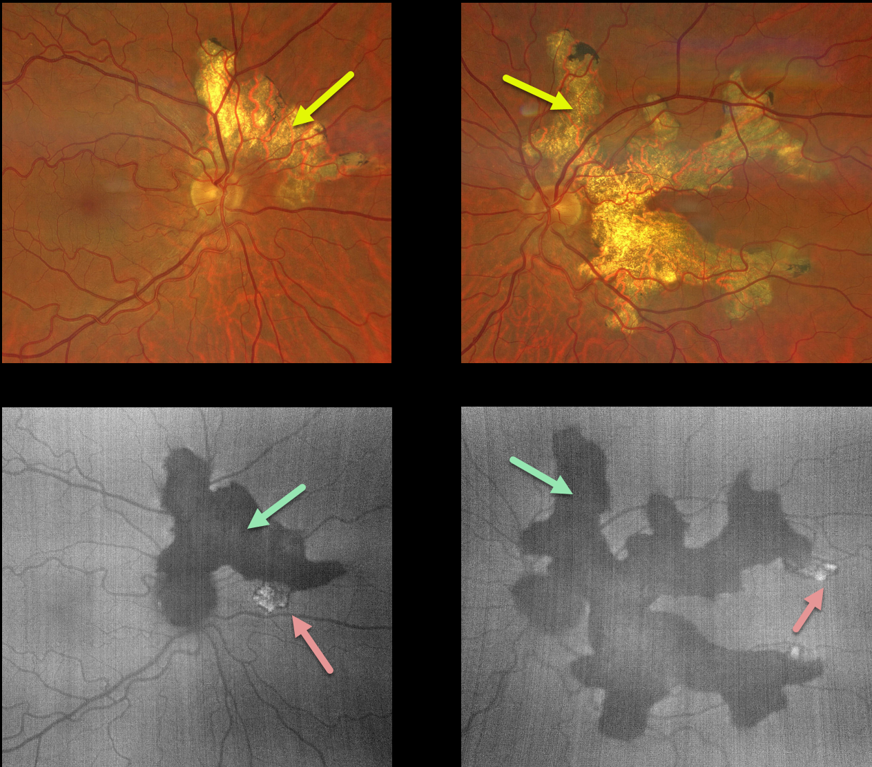
SERPIGINOUS CHORIORETINITIS
Borivoje Ivezić, Nataša Drača, and Emma Oreškovič
Originally posted on @retina.rocks January 2, 2024
This healthy 54YO male presented for a second opinion for untreated serpiginous chorioretinitis that was diagnosed 2 years earlier. Vision was 20/20 OD and 20/200 OS.
Color imaging shows bilateral chorioretinal scarring that extends outwards from the optic nerves. This scarring is more extensive OS and spares the macular center. On fundus autofluorescence (FAF), this scarring is diffusely hypo-FAF except for a few margins bilaterally that are hyper-FAF.
Learning Points:
Serpiginous choroiditis is an idiopathic, usually bilateral chorioretinal inflammatory condition most commonly found in middle-aged men (Khanamiri and Rao, Surv Ophthalmology 2013;58:203-232). Recurrent areas of inflammation develop along the edge of a previous scar, producing scarring that meanders from the optic nerve outward into the macula.
Vision is good unless scarring or macular neovascularization extends through the macular center. There is no consensus on the optimal systemic treatment, including corticosteroids and systemic steroid-sparing agents.
Tubercular serpiginous-like choroiditis mimics serpiginous, but affects younger patients with more multifocal and peripheral recurrences and progression if untreated (Agarwal et al, AJO 2020;220:160-169).
We felt the hyper-FAF lesions in our patient possibly indicated active disease, so prednisone 40mg PO for 10 days was prescribed. Tuberculosis workup was negative.
HARADA DISEASE
Anjana Mirajkar, Manish Nagpal and Navneet Mehrotra
Originally posted on @retina.rocks October 18, 2023
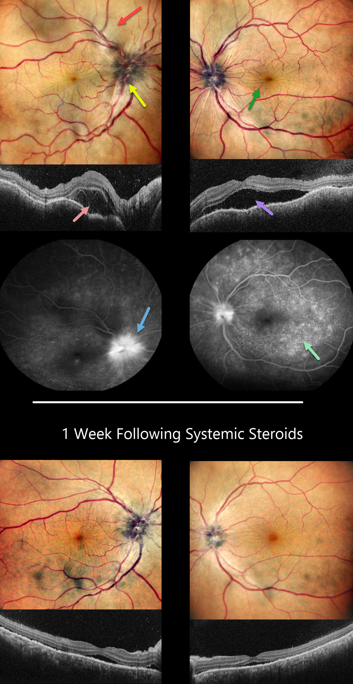
This 41YO female presented with headaches, redness, and pain OD for 1.5 months and OS for 2 weeks. She was diagnosed elsewhere with angle closure glaucoma, received a YAG peripheral iridotomy, and started on glaucoma drops. She then saw multiple other doctors who diagnosed her with papilledema.
Her CSF opening pressure was 21, and she was started on oral acetazolamide for a presumed diagnosis of idiopathic intracranial hypertension (IIH). In our office, vision was 20/100 OD and 20/60 OS. Intraocular pressure (IOP) was 30mmHG.
Anterior segments showed shallow anterior chambers with retrolental vitreous cells. Multicolor imaging shows bilateral irregular chorioretinal folds, multifocal serous detachments, and disc hyperemia.
OCT scanning shows a bacillary layer detachment OD, subretinal fluid OS, and a bilateral undulating thickened choroid. Fluorescein angiography shows bilateral optic nerve involvement and pinpoint subretinal leakage.
She was diagnosed with Harada’s disease and started on intravenous methylprednisolone for 3 days, followed by 60mg prednisone PO daily. She was also referred to rheumatology to start immunosuppressants. The acetazolamide was tapered and discontinued for the misdiagnosed IIH.
One week later, vision improved to 20/40 OU. IOP was 10mmHG OU with marked bilateral improvement in the chorioretinal folds, macular fluid, and choroidal thickening. A slow steroid taper was initiated pending initiation of immunosuppressants.
Learning Points:
This case has many classic findings for Harada’s disease, including bilateral panuveitis, optic nerve swelling, thickened choroid, chorioretinal folds, and multifocal serous exudative retinal detachments.
Patients with just ocular findings have Harada’s disease, whereas those with additional systemic findings (including vitiligo, poliosis, headache, vertigo, and hearing loss) have Vogt-Koyanagi-Harada (VKH) disease.
This case also reminds us that it is easy to misdiagnose these complex uveitis cases, especially when not all ocular findings are taken into full consideration in an attempt to give the patient a single, unifying diagnosis.
POSTERIOR SCLERITIS
Will Gibson
Originally posted on @retina.rocks September 26, 2023
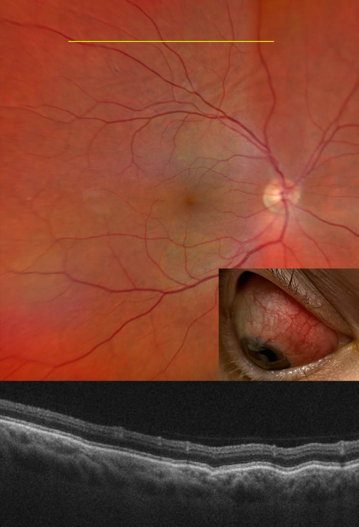
This 48YO female with a history of psoriatic arthritis presented on 10/25/22 with painful anterior scleritis. Vision was 20/20. The patient was started on 50mg of prednisone and slowly tapered off by rheumatology.
She returned on 7/19/23 with pain behind her right eye. Vision was 20/20. Color imaging shows subtle chorioretinal folds in the superior midperiphery.
OCT B-scan through this region confirmed the chorioretinal folds. B-scan ultrasonography revealed bilateral T signs.
Learning Points:
Posterior scleritis can present either as a diffuse thickening of the entire posterior sclera or as a nodular type with localized scleral thickening (see Agrawal et al, Retina 2016;36:392-401).
Nodular inflammation appears as a posterior pole subretinal yellowish mass lesion, often with overlying serous fluid and chorioretinal folds. B-scan ultrasonography may show fluid in the Tenon space (“T” sign).
Unlike anterior scleritis, the eyes are usually quiet and pain-free. Initial treatment is usually with oral non-steroidal anti-inflammatories or corticosteroids, although patients may also require immunosuppressive therapy. For a large case series of posterior scleritis, see McCluskey et al, Ophthalmology 1999;106:2380-2386.
Oral prednisone was restarted and slowly tapered. Further labs and chest X-ray were performed to rule out other possible causes of scleritis, and she was started on Humira.
ACUTE POSTERIOR MULTIFOCAL PLACOID PIGMENT EPITHELIOPATHY (APMPPE)
Mattie Adams
Originally posted on @retina.rocks September 12, 2023
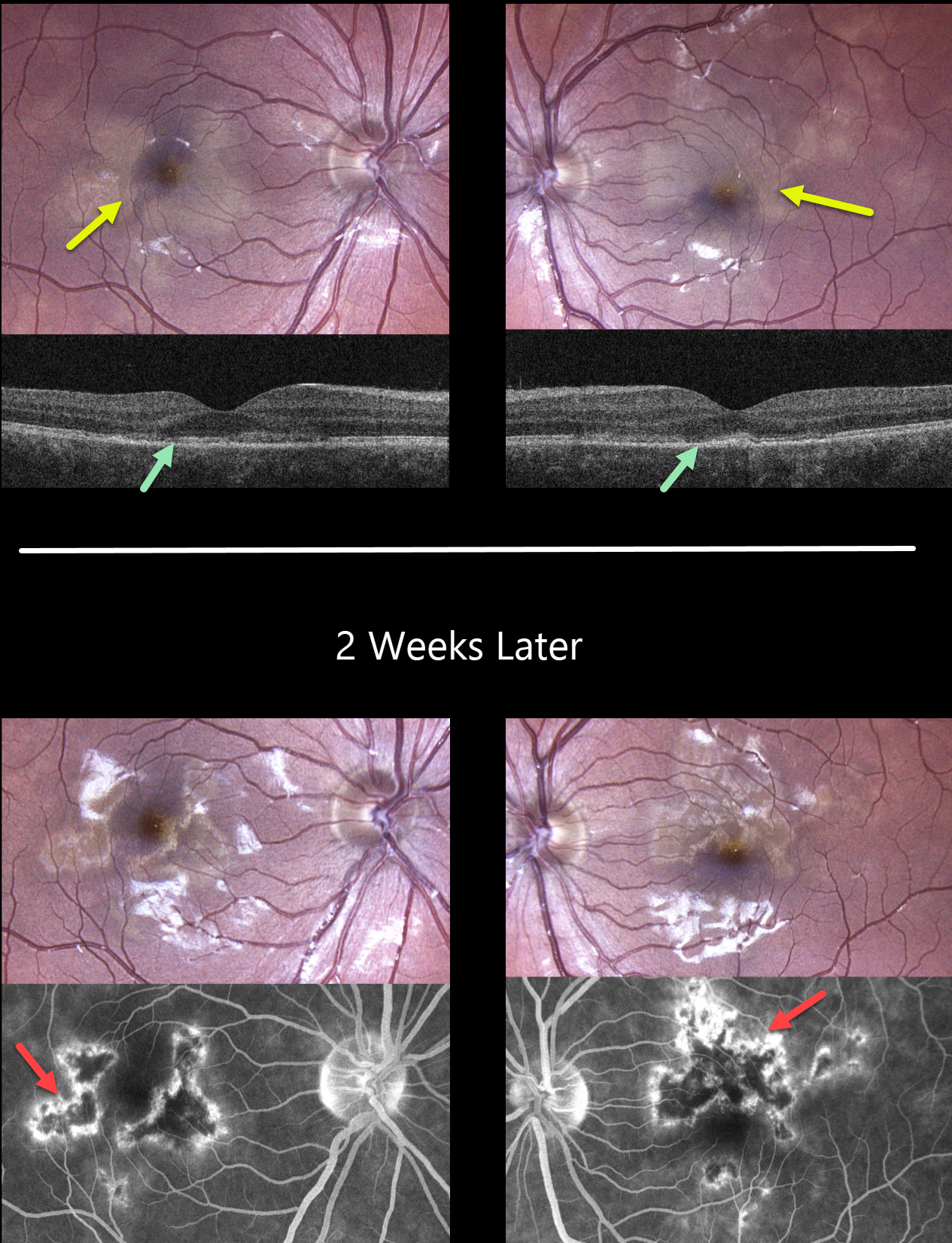
This healthy 25YO female presented with 3 weeks of painless vision loss, especially in her left eye. Vision was 20/20 OD and 20/100 OS. There was a moderate bilateral granulomatous anterior uveitis. The vitreous was clinically free of cells.
Color photography shows bilateral creamy subretinal macular placoid lesions. OCT scanning shows variable loss of the ellipsoid and interdigitation zone layers. Some vitreous cells are noted bilaterally.
Laboratory testing was all negative, including CBC, ESR, C-reactive protein, ACE, syphilis, and QuantiFERON-TB Gold Plus. The patient was started on topical prednisolone acetate QID with a weekly taper.
When examined 2 weeks later, her symptoms were much improved with vision of 20/20 OD and 20/30 OS. The placoid lesions were resolving with angiographic staining.
Learning Points:
Acute posterior multifocal placoid pigment epitheliopathy (APMPPE) is a usually bilateral inflammatory vs ischemic disorder of the inner choroid, RPE, and outer retina. Blurry vision and paracentral/central scotomas often follow a viral illness.
Acutely multifocal creamy yellow or grayish-white subretinal placoid lesions are scattered throughout the posterior poles. The lesions and visual symptoms usually resolve within 4 to 8 weeks with secondary chorioretinal changes. Recurrent lesions are rare, and vision is usually fairly good.
The debate over whether APMPPE is primarily an inflammatory disorder of the outer retina and RPE or an ischemic disease of the inner choroid has raged since Dr. Gass first described this entity in 1968.
Our case supports the current theory that APMPPE is an inflammation of the inner choroid with secondary outer retinal damage.
Anterior uveitis, as seen in our patient, is rarely seen in APMPPE. This was one reason we performed blood work, which fortunately came back negative. Her spontaneous and rapidly resolving posterior segment findings also support our diagnosis of APMPPE.
FROSTED BRANCH ANGIITIS
Originally posted on @retina.rocks June 15, 2023
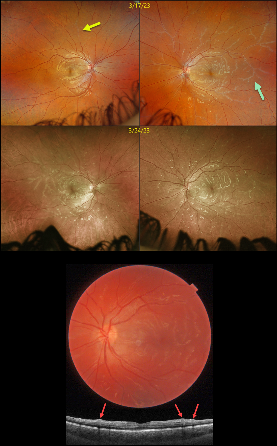
This healthy 16YO female presented with several days of bilateral blurred vision. There was no past ocular or medical history. Vision was 20/25 OD and 20/30 OS. Anterior segments were normal.
Optos imaging of her right eye shows a frosted branch appearance to a retinal vein in the superior midperiphery, with more diffuse vasculitis seen in her left eye.
Swept-source OCT through the left macula shows some hyperreflective inner retinal thickening around the inflamed veins. Bloodwork for Bartonella, syphilis, and sarcoid was ordered.
She returned one week later with new blurred vision in her left eye. Vision was 20/25 OU. Although findings were stable OS, new diffuse frosted branch changes were present in the OD. Her bloodwork returned negative, and she was started on oral prednisone 60mg daily.
Learning Points:
Frosted branch angiitis is a retinal vasculitis that gets its name from the involved vessels resembling branches of a tree that are “frosted” with snow (Kleiner et al, AJO 1988;106:27-34).
Although these findings may be isolated, they are often considered a phenotype found in a multitude of inflammatory conditions, most commonly CMV retinitis. The inflammation is usually very responsive to steroids, and the visual prognosis is generally good, although some patients may develop secondary anterior and posterior segment neovascularization due to widespread ischemia.
RELENTLESS PLACOID CHORIORETINITIS
Ryan Kern
Originally posted on @retina.rocks April 5, 2023
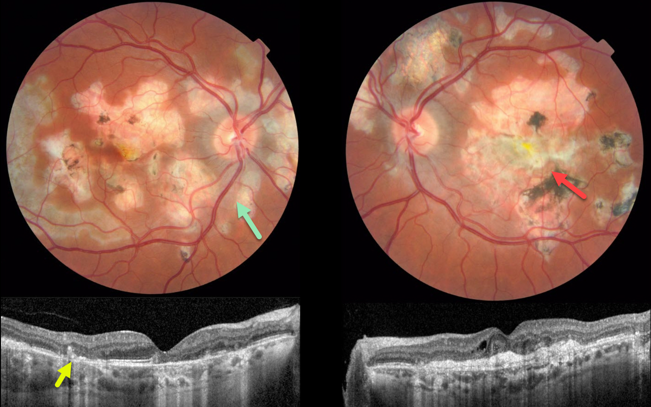
This 34YO male presented with a one-day history of severe vision loss in his left eye. Vision was 20/30 OD and 20/400 OS.
Color imaging shows bilateral areas of centrifugal pseudopod-like projections of atrophic scarring. The peripapillary scarring in the right eye seems to radiate outward from the optic nerve. There is a large, mostly atrophic disciform scar in the left eye, along with bilateral patches of variably pigmented scarring.
OCT scanning shows hyperreflective subfoveal lesions likely representing spontaneously involuted type 2 macular neovascularization (MNV) with overlying disorganized outer retinal layers.
Our patient’s vision in his left eye is remarkably good, given the macular and OCT appearance. It is unclear why he presented with acute vision loss, given the chronic appearance of his findings.
Learning Points:
Relentless placoid chorioretinitis has clinical and angiographic features resembling both serpiginous and APMPPE. Relentless placoid demonstrates the multifocal lesions of APMPPE, with more meandering, less patchy, serpiginous lesions.
Additionally, these patients will have recurrent inflammation typical of serpiginous but not APMPPE. They may also develop MNV that is more typical of the serpiginous form.
ACUTE IDIOPATHIC MACULOPATHY (AIM)
Emily Shepherd
Originally posted on @retina.rocks March 3, 2023
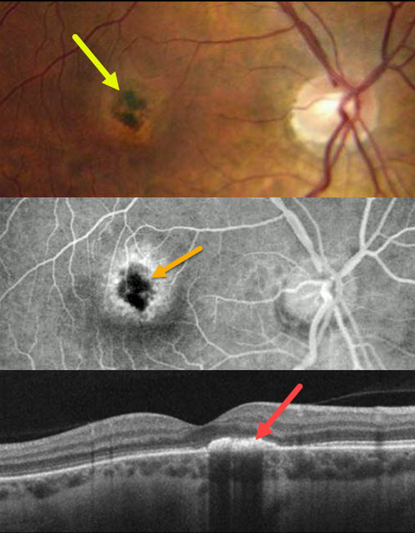
This 33YO healthy female complained of 1 to 2 weeks of a blind spot and ring of light in her right eye. She had a recent non-COVID viral illness when her symptoms started. Vision was 20/60 OD and 20/20 OS.
Color imaging shows a subfoveal area of central hyperpigmentation surrounded by a ring of pigment loss. Angiographically, the pigmentation blocks with a surrounding rim of staining.
OCT B-scan shows a hyperreflective plaque in the outer retina.
Learning Points:
Acute idiopathic maculopathy (AIM) is an idiopathic inflammation of the outer retina, RPE, and inner choroid. There is often a preceding viral prodrome caused by the Coxsackie virus. Healthy young patients typically present with acute unilateral moderately severe central vision loss.
The initial funduscopic changes show macular fluid and, sometimes, deep retinal blood, which can simulate macular neovascularization. The OCT shows heterogeneous hyperreflective thickening of the outer retina/RPE and fluid.
Fluorescein angiography usually shows deep retinal leakage. The lesion spontaneously resolves with significant visual improvement after several weeks, usually leaving a pathognomonic bull’s-eye lesion as seen in our patient.
ACUTE IDIOPATHIC BLIND SPOT ENLARGEMENT SYNDROME
Originally posted on @retina.rocks February 20, 2023
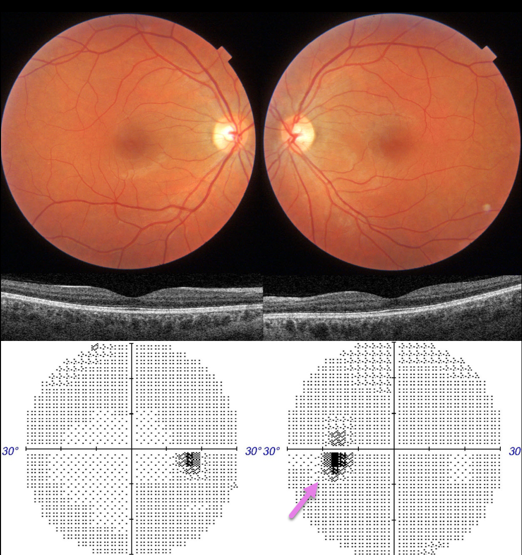
This 36YO female presented with 1 month of a paracentral scotoma in her left eye. There was no prior ocular or past medical history. Vision was 20/20 OU.
Ocular examinations were completely normal OU. Color imaging and OCT were normal. Visual fields were normal OD with a slightly enlarged blind spot OS.
Learning Points:
Multiple evanescent white dot syndrome (MEWDS) presents with usually unilateral idiopathic inflammation of the outer retina that typically occurs in young, healthy myopic females.
The granular fovea (Jampol spots, named after Dr. Lee Jampol, who first described this syndrome in 1984) is a classic pathognomonic finding that was not seen in this patient.
Acute cases will also have an enlarged blind spot, often with temporal visual field loss, due to peripapillary inflammation. The white dots disappear within a week or two, which is why the lesions are called “evanescent”. However, the blind spot may persist for many months, well after the fundus appearance has normalized.
This stage of MEWDS was originally called the Acute idiopathic blind spot enlargement (AIBSE) syndrome before Gass recognized that the two disorders were the same disease but with distinct findings at different points in time.
Observation for our patient was recommended. We expect her symptomatic enlarged blind spot will gradually resolve over the coming months.
MULTIPLE EVANESCENT WHITE DOT SYNDROME (MEWDS)
Originally posted on @retina.rocks January 13, 2023
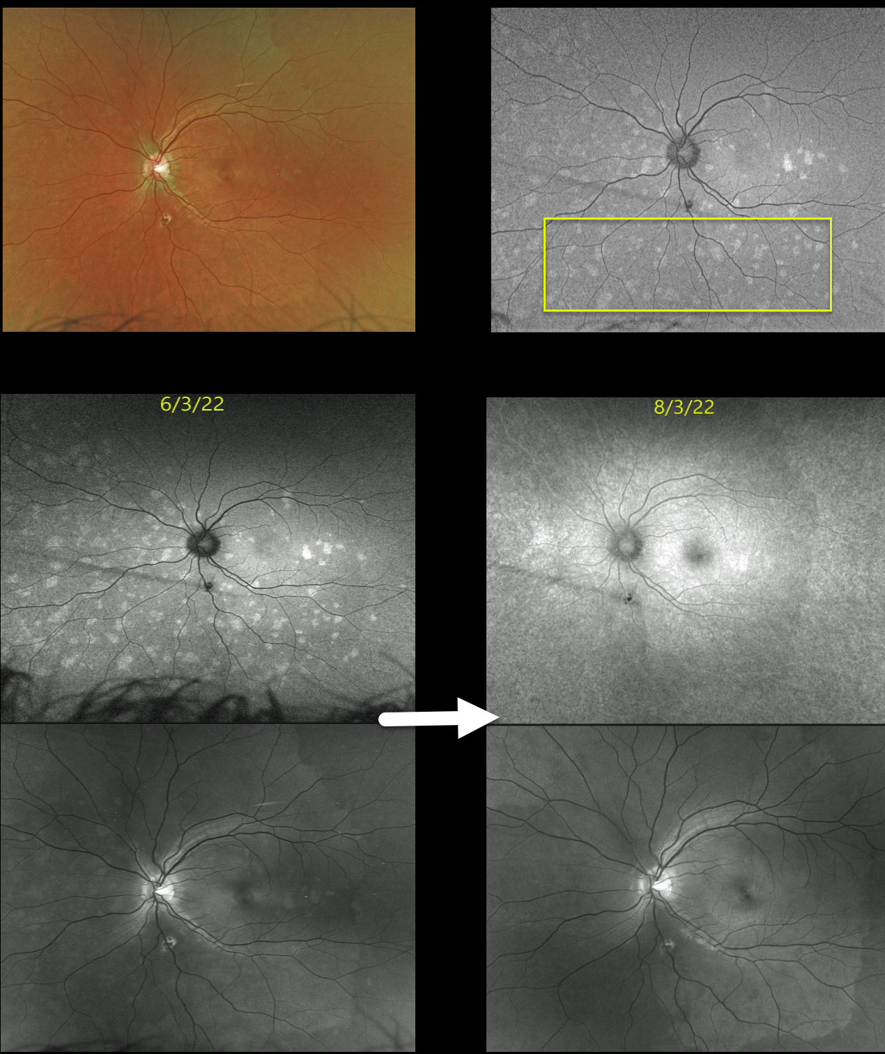
This 22YO female presented with blurred vision in her left eye for 2 weeks. Vision was 20/20 in her normal right eye and 20/70 in her left eye.
Optos color imaging of her left eye shows multiple faint, deep retinal white dots scattered throughout, which are hyper-autofluorescent. Macular OCT scanning was virtually normal (image not shown).
Two months later, the fundoscopic changes resolved, although fundus autofluorescence and Optos green channel imaging show more subtle residual findings.
Learning Points:
Multiple evanescent white dot syndrome (MEWDS) presents with usually unilateral idiopathic inflammation of the outer retina that typically occurs in young, healthy myopic females.
The granular fovea (Jampol spots, named after Dr. Lee Jampol, who first described this syndrome in 1984) is a classic pathognomonic finding that was not seen in this patient. Acute cases will also have an enlarged blind spot, often with temporal visual field loss, due to peripapillary inflammation.
The white dots disappear within a week or two, which is why the lesions are called “evanescent”. However, the blind spot may persist for many months, well after the fundus appearance has normalized.
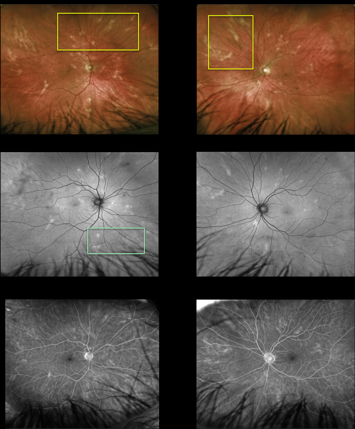
POSSIBLE BIRDSHOT CHORIORETINOPATHY
Originally posted on @retina.rocks January 11, 2023
This healthy 51YO male presented with asymptomatic lesions in each fundus. Vision was 20/25 OU.
Multimodal imaging findings were symmetrical bilaterally. Optos color imaging shows amelanotic subretinal lesions that seem to radiate outwards from each disc. These lesions are mostly hyperautofluorescent with window-type defects on fluorescein angiography. OCT scanning was normal bilaterally (not shown). HLA-A29 was negative.
Learning Points:
Birdshot chorioretinopathy was appropriately named and described by Drs. Ryan and Maumenee (AJO 1980;89:31-45), since the lesions look like shotgun birdshot scattered throughout the fundus. Birdshot is typically chronic and bilateral, with a very high HLA-A29 association.
We are not certain that our patient has birdshot. The pattern of lesions and less creamy coloration is somewhat atypical of what we’ve normally seen in our practice.
Although HLA-A29 is positive in about 90% of cases, it is not required for the diagnosis in the setting of other clinical findings (SUN Working Group AJO 2021;228:65-71). Finally, birdshot lesions are typically hypofluorescent (Koizumi et al., Ophthalmology 2008;115:e15-e20), unlike the hyperfluorescence observed in our patient.
Since he is asymptomatic, has no signs of active uveitis, and no macular edema, we will initially follow him twice yearly.
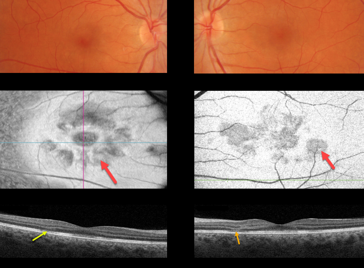
ACUTE MACULAR NEURORETINOPATHY (AMN)
Originally posted on @retina.rocks December 21, 2022
This 41YO female presented with acute bilateral central vision loss. Fundus photography shows very faint reddish-brown wedge-shaped lesions radiating from each fovea.
Swept-source en face imaging of the outer retina shows multiple hyporeflective wedge-shaped lesions radiating from the macular centers.
OCT B-scanning shows bilateral areas of outer nuclear layer thinning with hyperreflectivity associated with disruption of the ellipsoid zone and outer segment layers.
Learning Points:
This case highlights many characteristic features of acute macular neuroretinopathy (AMN), an idiopathic disorder likely caused by compromise of the deep retinal capillary plexus.
Multicolor and infrared imaging often highlight the typical wedge-shaped AMN lesions more effectively than color fundus photos, although the en face OCT images in our patient beautifully capture these pathognomonic findings.
Most patients are white, non-Latino females with a median age of 30 years. The most common associations are a flu-like prodrome and oral contraceptive use.
Presenting acuity is usually good, coinciding with sparing of the sub-foveal areas. Roughly half of AMN cases are bilateral. There is typically moderate visual improvement over several months without treatment.
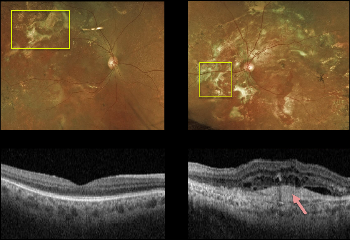
MULTIFOCAL CHOROIDITIS
Originally posted on @retina.rocks August 26, 2022
This 58YO male has stable vision of 20/20 OD and light perception OS. Extensive areas of subretinal fibrosis are scattered throughout each fundus, left greater than right.
A hyperreflective subfoveal scar on OCT in his left eye accounts for the poor vision. Fortunately, the right macula is relatively normal, and we are observing him twice yearly.
Learning Points:
Multifocal choroiditis (MFC) is the current preferred term for a group of historically heterogeneous disorders, including multifocal choroiditis, recurrent multifocal choroiditis, multifocal choroiditis and panuveitis, punctate inner choroidopathy, progressive subretinal fibrosis, and pseudo-histoplasmosis (Essex et al, Retina 2013;33:1-4 and Spaide et al, Retina 2013;33:1315-1324).
MFC occurs in otherwise healthy adults, often myopic and female. Findings include variably pigmented multifocal scars throughout the fundus, panuveitis, subretinal fibrosis, and macular neovascularization (MNV). Unlike ocular histoplasmosis, new scars and uveitis can be seen.
MNV can be successfully managed with anti-VEGF therapy.
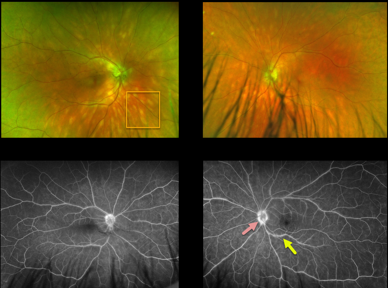
BIRDSHOT CHORIORETINOPATHY
Will Gibson
Originally posted on @retina.rocks August 3, 2022
This 48YO male presented with these creamy yellow-white choroidal lesions scattered throughout each posterior pole. Vision was 20/25 OU.
Fluorescein angiography shows mild optic nerve leakage, particularly in the left eye, and mild scattered venous staining/leakage in the left eye.
Learning Points:
This disease was appropriately described and named by Drs. Ryan and Maumenee (Am J Ophthlamol 1980;89:31-45) since the lesions look like shotgun birdshot scattered throughout the fundus.
Birdshot is typically chronic and bilateral, with a very high association with HLA-A29. It usually affects women in their 40s to 60s. Vision loss is usually caused by cystoid macular edema (CME) and retinal atrophy, although macular neovascularization can rarely occur. Asymptomatic patients can be observed. CME and symptomatic uveitis can be treated with oral steroids, although many will still require immunomodulatory therapy.
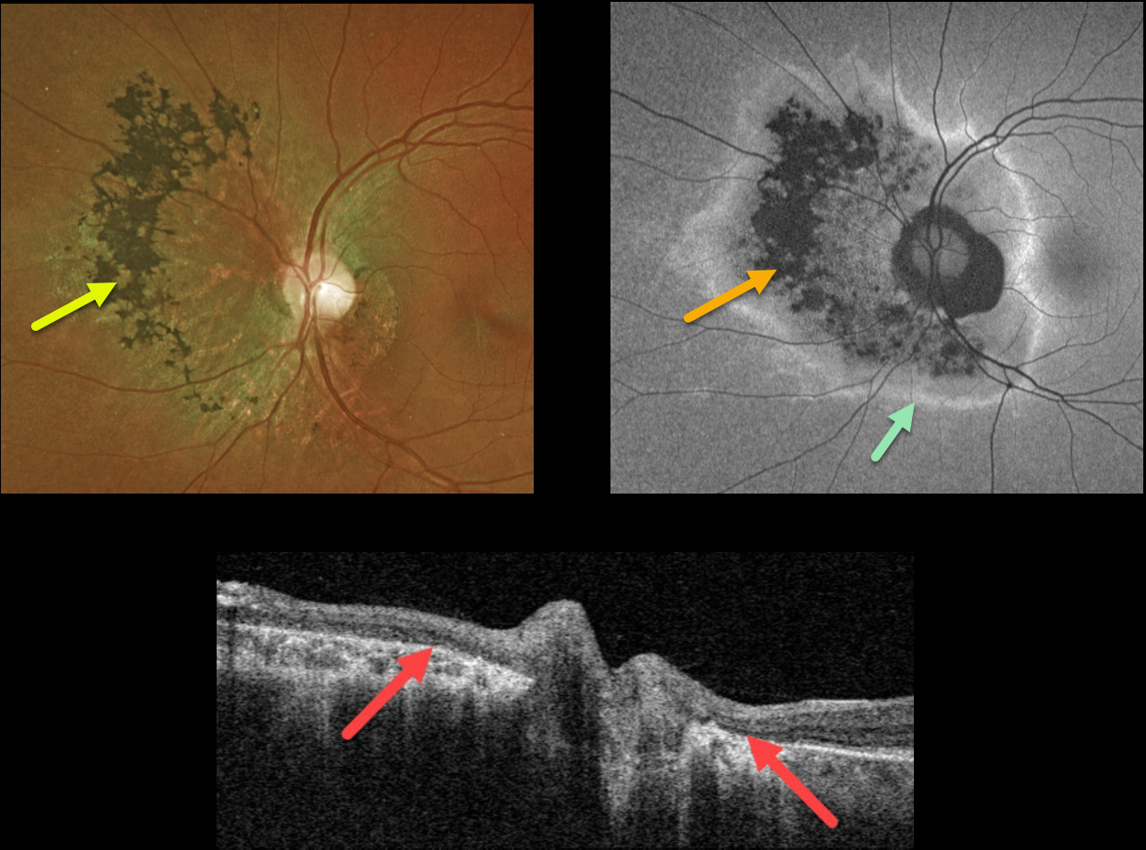
ACUTE ZONAL OCCULT OUTER RETINOPATHY (AZOOR)
Originally posted on @retina.rocks July 13, 2022
This 44YO female was referred for asymptomatic retinal changes in her right eye. Vision was 20/20 in her normal right eye and 20/30 OS.
Optos color imaging shows chorioretinal scarring surrounding the left optic nerve, extending more nasally with intraretinal pigment migration. Fundus autofluorescence (FAF) shows hypo-FAF corresponding to the clinical chorioretinal scarring, with a surrounding rim of hyper-FAF. Swept source OCT through the optic nerve shows loss of the outer retinal bands with RPE atrophy.
We made a presumed diagnosis of resolved acute zonal occult outer retinopathy (AZOOR)
Learning Points:
First described by Gass in 1992, AZOOR initially presents unilaterally or bilaterally in young women with photopsias, central visual changes, and minimal fundoscopic changes. Zones of visual field loss correspond to the locations of the photopsias. Electroretinography is often abnormal. Some may later develop areas of outer retinal, RPE, and choroidal atrophy in the initially affected retina.
The pathogenesis of the condition remains unclear, but it appears to initially involve inflammation of the photoreceptors. AZOOR is believed to be a part of the spectrum of other ‘photoreceptoritis’ conditions, including multiple evanescent white dot syndrome (MEWDS), acute macular neuroretinitis (AMN), and acute retinal pigment epitheliitis.
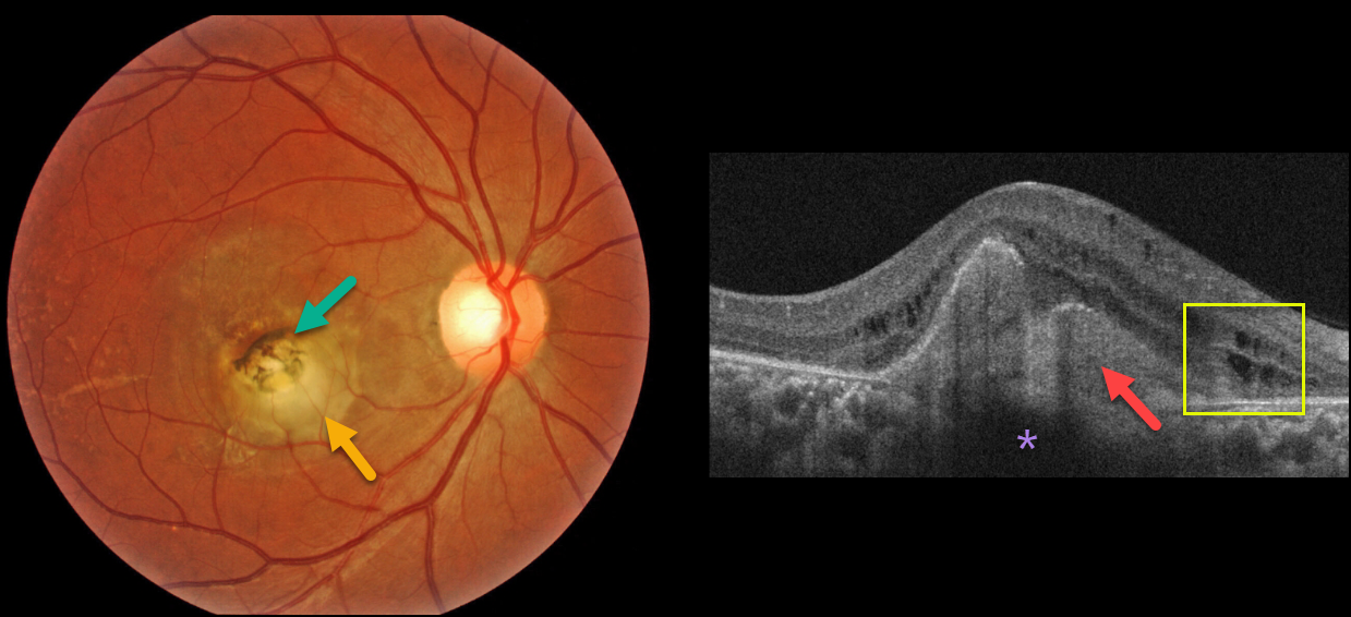
CHOROIDAL GRANULOMA
Originally posted on @retina.rocks June 3, 2022
A healthy 32YO female presented with a 6-month history of vision loss in her right eye. Vision was counting fingers OD and 20/20 in her healthy left eye.
Fundus imaging shows a slightly elevated, creamy white, subfoveal lesion surrounded by slightly hyperpigmented macular scarring.
OCT shows a hyperreflective subretinal lesion that shadows the underlying choroid. There are some mild cystic changes in the inner and outer nuclear layers.
Learning Points:
Choroidal granulomas usually appear as solitary, creamy white, round subretinal lesions. They are hyporeflective on OCT imaging (Invernizzi et al, Retina 2015;35:525-531). They can extend into the subretinal space, as in our patient. Causes include sarcoidosis, tuberculosis, and VKH. Testing results for sarcoidosis and tuberculosis are still pending.
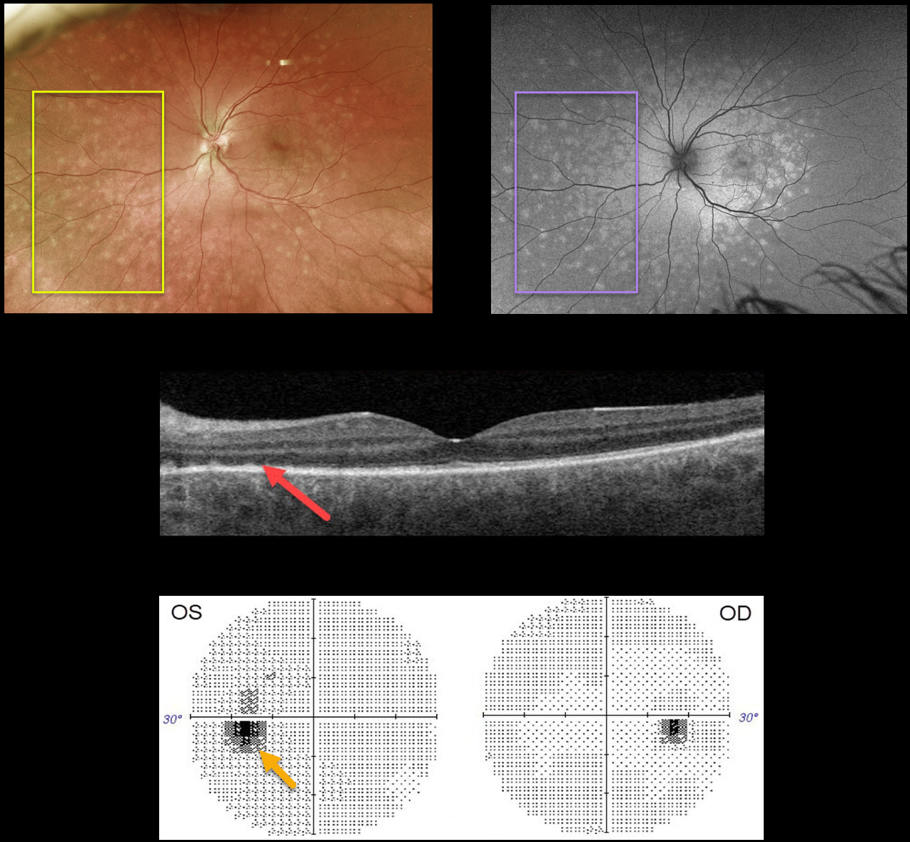
MULTIPLE EVANESCENT WHITE DOT SYNDROME (MEWDS)
Originally posted on @retina.rocks April 13, 2022
This 36YO male presented with flashes and a temporal scotoma in his left eye. Vision was 20/20 OD and 20/25 OS.
Optos color imaging shows multiple deep retinal white dots scattered throughout the posterior pole, which are hyper-autofluorescent.
Swept-source OCT shows nasal loss of the outer retinal bands with some hyperreflective spots. Visual fields show an enlarged left blind spot, consistent with the peripapillary hyperautofluorescence.
Learning Points:
Multiple evanescent white dot syndrome (MEWDS) presents with usually unilateral idiopathic inflammation of the outer retina that typically occurs in young, healthy myopic females.
The granular fovea (Jampol spots, named in honor of Dr. Lee Jampol, who first described this syndrome in 1984), noted in our patient, is a classic pathognomonic finding.
Acutely, patients will also have an enlarged blind spot, often complaining of temporal visual field loss, due to the peripapillary inflammation. The white dots disappear within a week or two, which is why the lesions are called “evanescent”.
However, the blind spot may persist for many months, well after the fundus appearance has normalized. This stage of MEWDS was originally called the Acute idiopathic blind spot enlargement (AIBSE) syndrome before Gass recognized that the two disorders were the same disease but with distinct findings at different points in time.
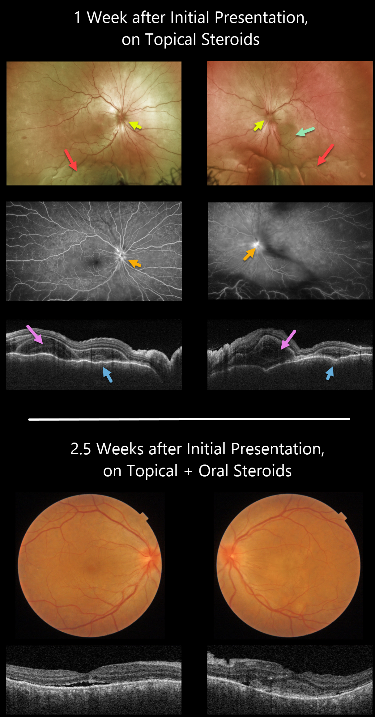
HARADA DISEASE
Originally posted on @retina.rocks February 7, 2022
This 35YO female presented with a 2-week history of severe bilateral vision loss, photophobia, and red eyes. There was no significant past medical history.
Vision was counting fingers OU. There was moderate bilateral conjunctival injection, severe cells with moderate flare, moderate fibrin, and 360-degree posterior synechiae. Topical cycloplegia and steroids were prescribed.
One week later, she was referred to our retinal service. There was marked improvement of her anterior uveitis and the posterior synechiae were mostly broken throughout. There were moderate bilateral vitreous cells, markedly swollen optic nerves, irregular diffuse macular exudative fluid with inferior exudative retinal detachments.
Fluorescein angiography showed bilateral optic nerve leakage. Triton swept-source OCT showed bilateral markedly thickened and rippled choroid with multiple bacillary pockets of outer retinal thickening and fluid. She started oral prednisone 80mg daily.
Around 10 days later, vision improved to 20/200 OU, with improvement in optic nerve edema, macular fluid, bacillary detachments, and choroidal thickening. The inferior exudative detachments were also resolving (not imaged).
Learning Points:
This case has many classic findings for Harada disease, including bilateral panuveitis, optic nerve swelling, thickened choroid, chorioretinal folds, and multifocal serous exudative retinal detachments.
Given the severity of the panuveitis and exudative subretinal fluid, we were surprised that angiography did not show the more classic multifocal subretinal leaks with pooling in the overlying serous detachments and bacillary cavities.
Patients with just ocular findings have Harada disease, whereas those with additional systemic findings (including vitiligo, poliosis, headache, vertigo, and hearing loss) have Vogt-Koyanagi-Harada disease.
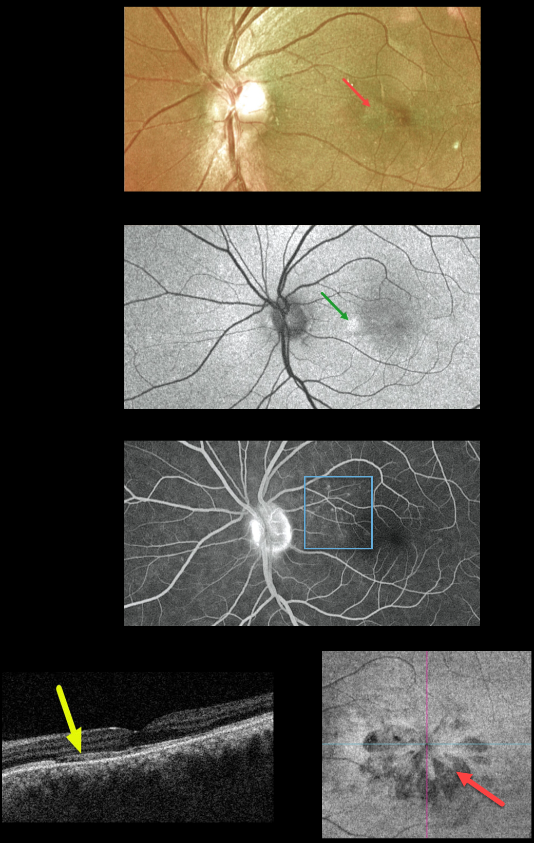
ACUTE MACULAR NEURORETINOPATHY
Originally posted on @retina.rocks January 21, 2022
This 39YO male presented with 1 day of blurred vision in his left eye. Vision was 20/30 OU. Optos color imaging shows a wedge-shaped lesion radiating into the nasal fovea. Optos fundus autofluorescence (FAF) shows hyper-FAF in this area, and fluorescein angiography shows subtle areas of increased nasal macular hyperfluorescence.
Swept-source OCT shows attenuation of the ellipsoid zone and outer segment layers. En face imaging of the outer retina shows multiple hyporeflective wedge-shaped lesions radiating from the macular center. Findings in the right eye were completely normal.
Learning Points:
This case highlights many characteristic features of acute macular neuroretinopathy (AMN), an idiopathic disorder likely caused by compromise of the deep retinal capillary plexus.
Multicolor and infrared imaging often highlight the typical wedge-shaped AMN lesions more effectively than color fundus photos, although the Triton en face OCT image in our patient beautifully imaged these.
Most patients are white, non-Latino females with a median age of 30 years. Roughly half of AMN cases are bilateral. The most common associations are a flu-like prodrome and oral contraceptive use.
Presenting acuity is usually good, coinciding with sparing of the sub-foveal areas. There is typically moderate visual improvement over several months without treatment.
For a great AMN review, see Bhavsar et al, Surv Ophthalmology 2016;61:538-565.
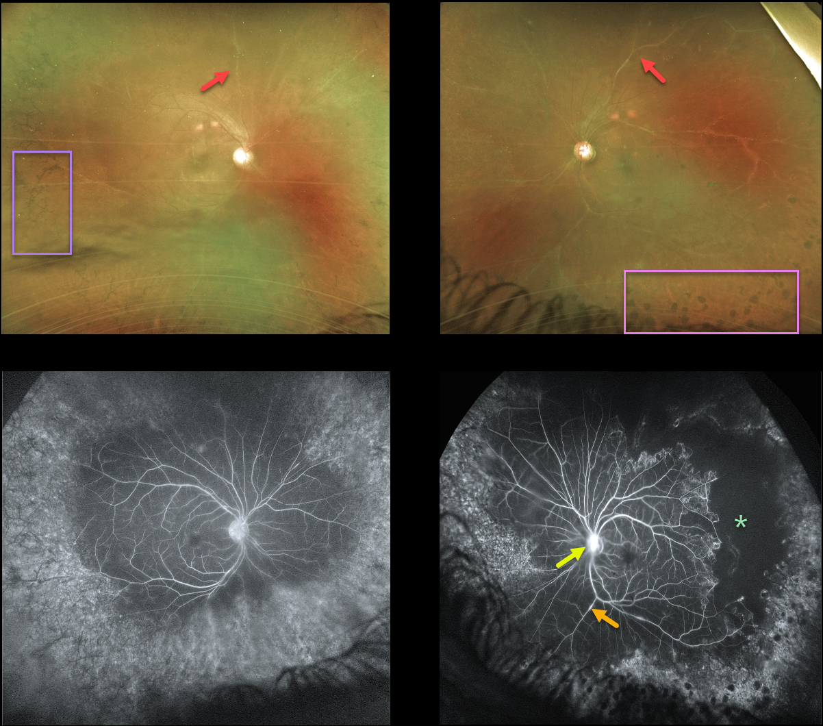
SARCOIDOSIS
Originally posted on @retina.rocks December 30, 2021
This 43YO male has a long history of panuveitis from sarcoidosis, and was previously seen 4 years earlier before being lost to follow-up.
He presented recently with a few weeks of symptomatic panuveitis. Vision was 20/100 OD and 20/200 OS with active granulomatous anterior uveitis.
Optos imaging shows a hazy view of each fundus, with diffusely white retinal vessels. Inferior vitreous puff balls were noted clinically but not imaged on Optos.
Fluorescein angiography shows focal areas of vascular staining/leakage, severe peripheral ischemia, and late optic nerve staining/leakage (especially OS). There is encircling peripheral intraretinal pigment migration OD with apparent peripheral scatter laser scarring OS.
He is being treated with topical steroids since the majority of active inflammation is anterior.
Further scatter laser to the untreated ischemic retina will be recommended if posterior segment neovascularization develops.
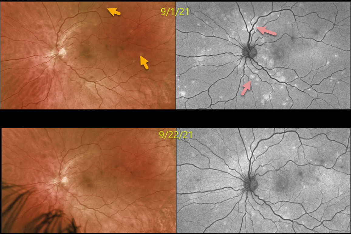
MULTIPLE EVANESCENT WHITE DOT SYNDROME (MEWDS)
Originally posted on @retina.rocks December 28, 2021
This 35YO male presented with 1 week of bilateral vision loss. Vision was 20/30 OD and 20/60 OS. Optos color imaging shows multiple deep retinal white dots scattered throughout the posterior pole (left eye only). Fundus autofluorescence (FAF) more dramatically shows these hyper-FAF lesions.
Three weeks later, vision improved to 20/30 OD and 20/25 OS as the lesions started to regress.
Learning Points:
Multiple evanescent white dot syndrome (MEWDS) presents with usually unilateral idiopathic inflammation of the outer retina that typically occurs in young, healthy myopic females.
The granular fovea (Jampol spots, named after Dr. Lee Jampol, who first described this syndrome in 1984) is a classic pathognomonic finding.
Acute cases will also have an enlarged blind spot, often with temporal visual field loss, due to peripapillary inflammation.
The white dots disappear within a week or two, which is why the lesions are called “evanescent”. However, the blind spot may persist for many months, well after the fundus appearance has normalized.
This stage of MEWDS was originally called the Acute Idiopathic Blind Spot Enlargement (AIBSE) syndrome before Gass recognized that the 2 disorders were the same disease but with distinct findings at different points in time.
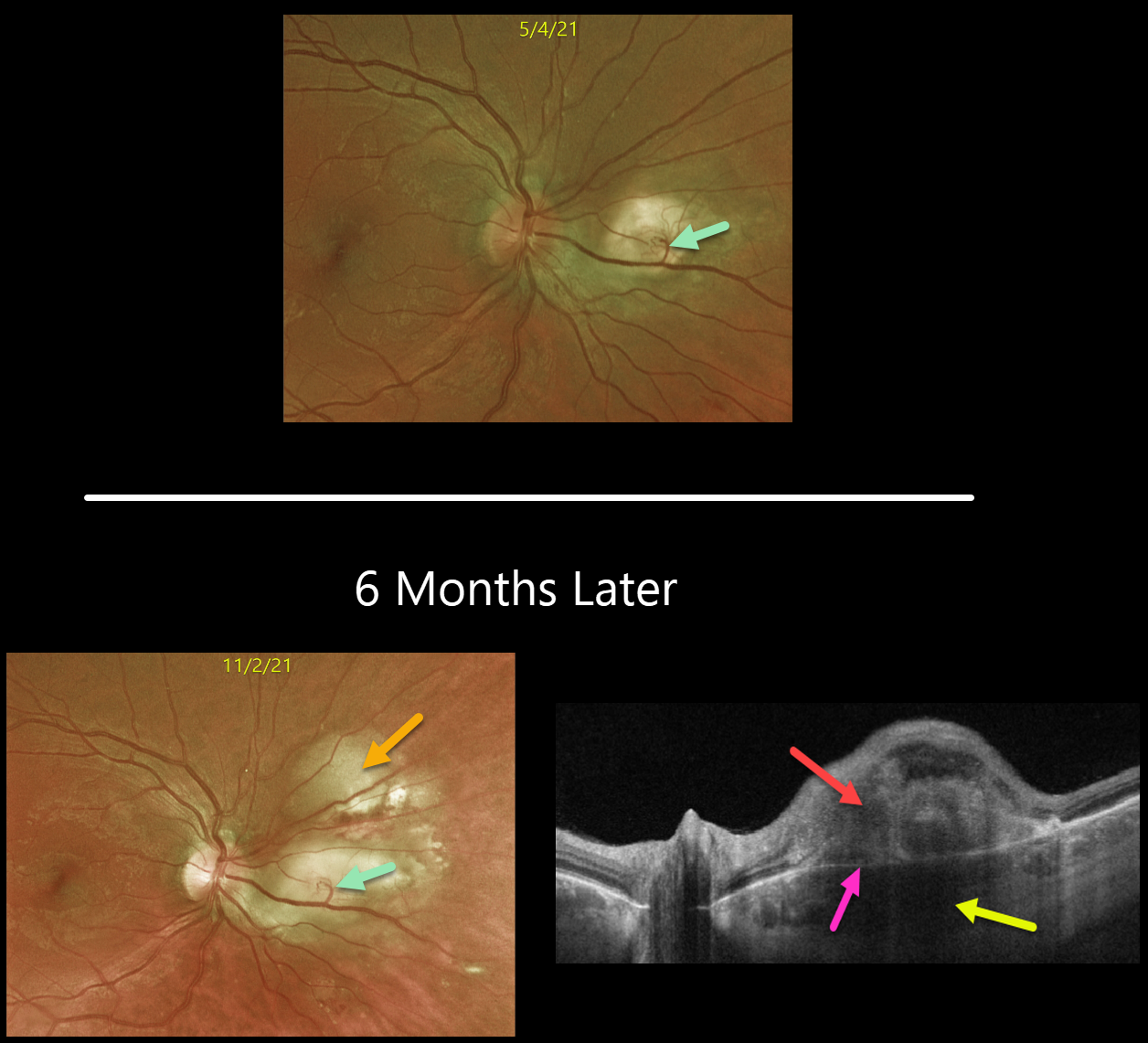
CHOROIDAL GRANULOMA
Originally posted on @retina.rocks December 8, 2021
This is a follow-up from our 6/7/21 post of a choroidal granuloma, likely representing the initial manifestation of systemic sarcoidosis. The patient was sent to pulmonology to confirm the diagnosis, but he did not keep the appointment.
Six months later, the choroidal granuloma had significantly enlarged.
A retinochoroidal anastomosis is noted between the overlying retinal vein and the lesion. Triton swept-source OCT shows a thickened, hyporeflective choroidal lesion, a hyperreflective outer retinal lesion with overlying inner retinal disorganization, and visible Bruch’s membrane separating the choroidal and retinal components.
Learning Points:
Choroidal granulomas are aggregations of inflammatory cells that appear clinically as a solitary, creamy-white, round subretinal lesion. Causes include sarcoidosis, tuberculosis, and VKH.
They are hyporeflective with increased transmission on OCT imaging (see Invernizzi et al, Retina 2015;35:525-531).
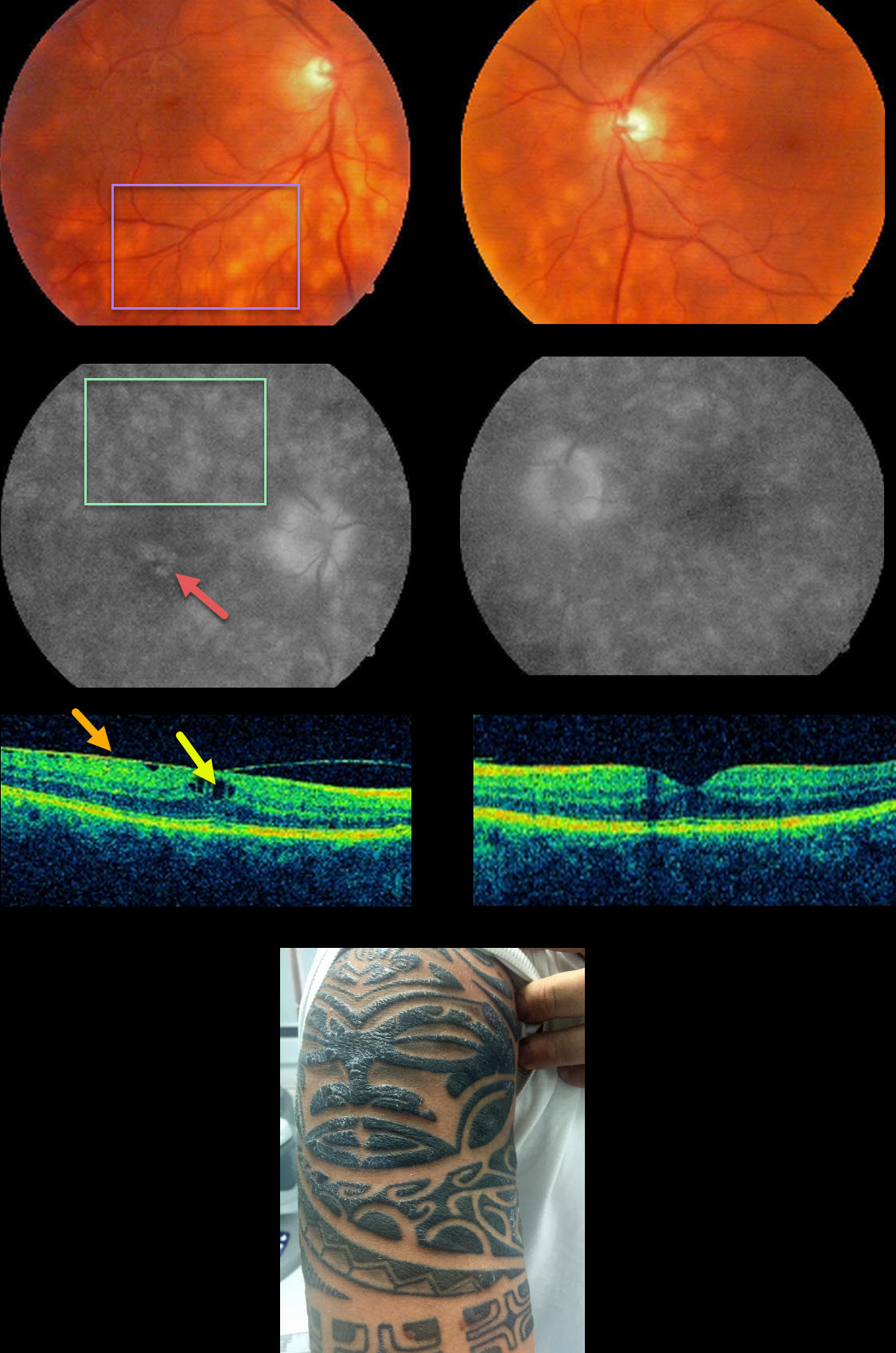
TATTOO-INDUCED UVEITIS
Natasa Draca
Originally posted on @retina.rocks December 7, 2021
This 39YO male presented with bilateral floaters and a several-year history of intermittent panuveitis. He had a history of treated hepatitis C.
Vision was 20/25 OD and 20/20 OS. Anterior segments were normal. There was mild bilateral vitritis and scattered amelanotic subretinal lesions in each posterior pole.
Fluorescein angiography shows staining of these lesions with cystoid leakage in his right macula. OCT scanning shows a mild epiretinal membrane with cystoid edema OD. The initial differential diagnosis included birdshot chorioretinopathy and sarcoidosis.
HLA-A29 was negative, but on further questioning, he told us that the tattoos on his arms always became swollen and painful prior to the recurrent uveitis.
A biopsy of one of the inflamed tattoos revealed inflammation granulomatosa. The granulomas were classified as foreign-body or sarcoid-type reactions.
However, the sarcoidosis workup, including ACE, chest X-ray, and chest CT, was all normal. He is being treated as needed with oral and topical steroids.
Learning Points:
Tattoo pigments are known to produce mast cell activation and to provoke cellular lysis after phagocytosis. Intradermal injection of melanin, interphotoreceptor retinoid-binding protein, and S-antigen is known to produce a granulomatous anterior or pan-uveitis (see Ostheimer et al, AJO 2014;158:637-643). The uveitis can be controlled with systemic steroids and/or immunosuppressants.
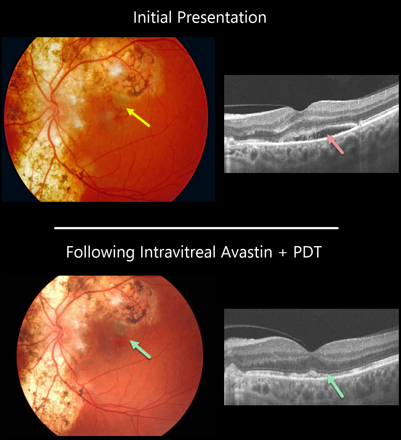
SERPIGINOUS CHOROIDITIS
Originally posted on @retina.rocks November 19, 2021
This patient presented with bilateral serpiginous chorioretinopathy scarring (only the left eye pictured). A pigmented macular neovascularization (MNV) with associated subretinal pigment and blood was noted in the superior left fovea. Subretinal fluid is noted on OCT scanning. The MNV was treated with a combination of photodynamic therapy and intravitreal Avaston.
Vision improved from a preoperative 20/400 to 20/30 with resolution of all fluid and blood. The patient continues to need PRN anti-VEGF injections in his left eye.
Learning Points:
Serpiginous choroiditis is an idiopathic, usually bilateral chorioretinal inflammatory condition most commonly found in middle-aged men.
Recurrent areas of inflammation develop along the edge of a previous scar, producing scarring that meanders from the optic nerve outward into the macula.
Vision is good unless scarring (as in this patient) or macular neovascularization extends through the macular center.
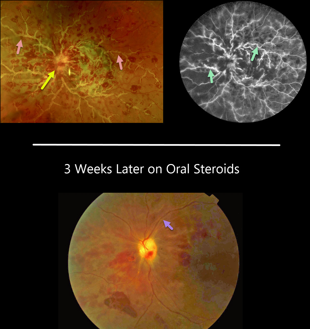
FROSTED BRANCH ANGIITIS FROM SARCOIDOSIS
David Kilpatrick
Originally posted on @retina.rocks October 11, 2021
This 34YO African American female presented with several days of painless vision loss in her left eye. Vision was 20/30 OD and HM OS with an afferent defect OS. Anterior segments were normal.
There was mild disc edema OD (not shown) and florid disc edema OS with retinal vasculitis predominantly involving the veins.
On fluorescein angiography, the retinal veins in the left eye were diffusely stained.
On further review of systems, she had a new skin rash on her right forearm, but was otherwise asymptomatic. Testing revealed elevated angiotensin-converting enzyme and lysozyme levels, and a chest X-ray showed bilateral perihilar fullness.
With a working diagnosis of sarcoidosis, she is currently being managed with oral steroids and has been evaluated by both rheumatology and pulmonology.
While her retinal vasculitis has responded well to treatment, her vision remains HM three weeks later.
Learning Points:
Frosted branch angiitis is a retinal vasculitis that gets its name due to the involved vessels resembling the branches of a tree that are “frosted” with snow. Although these findings may be isolated, they are often considered a phenotype found in a multitude of inflammatory conditions, most commonly CMV retinitis.
The inflammation is usually highly responsive to steroids, and the visual prognosis is generally good, although some may develop secondary neovascularization in the anterior and posterior segments due to widespread ischemia.
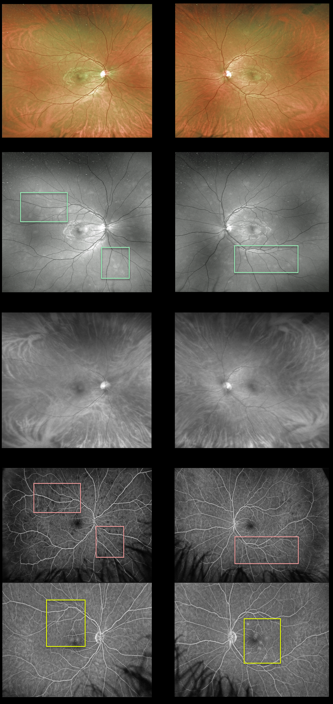
MULTIPLE EVANESCENT WHITE DOT SYNDROME (MEWDS)
Originally posted on @retina.rocks September 17, 2021
This 30YO male presented with a one-day history of seeing “dots” in his vision. Vision was 20/25 OD and 20/20 OS.
There are multiple deep retinal white dots scattered throughout the posterior pole of each eye. The Optos green channel best shows these white dots compared to the color images. The spots disappear with the Optos red channel, confirming their retinal localization.
Fluorescein angiography shows variable early hypofluorescence with late staining of the lesions.
Learning Points:
Multiple evanescent white dot syndrome (MEWDS) presents with usually unilateral idiopathic inflammation of the outer retina that typically occurs in young, healthy myopic females.
The granular fovea (Jampol spots, named in honor of Dr. Lee Jampol, who first described this syndrome in 1984), not well imaged in our case, is a classic pathognomonic finding.
Acute cases will also have an enlarged blind spot, often with temporal visual field loss, due to peripapillary inflammation.
The white dots disappear within a week or two, which is why the lesions are called “evanescent”. However, the blind spot may persist for many months, well after the fundus appearance has normalized.
This stage of MEWDS was originally called the Acute idiopathic blind spot enlargement (AIBSE) syndrome before Gass recognized that the 2 disorders were the same disease but with distinct findings at different points in time.
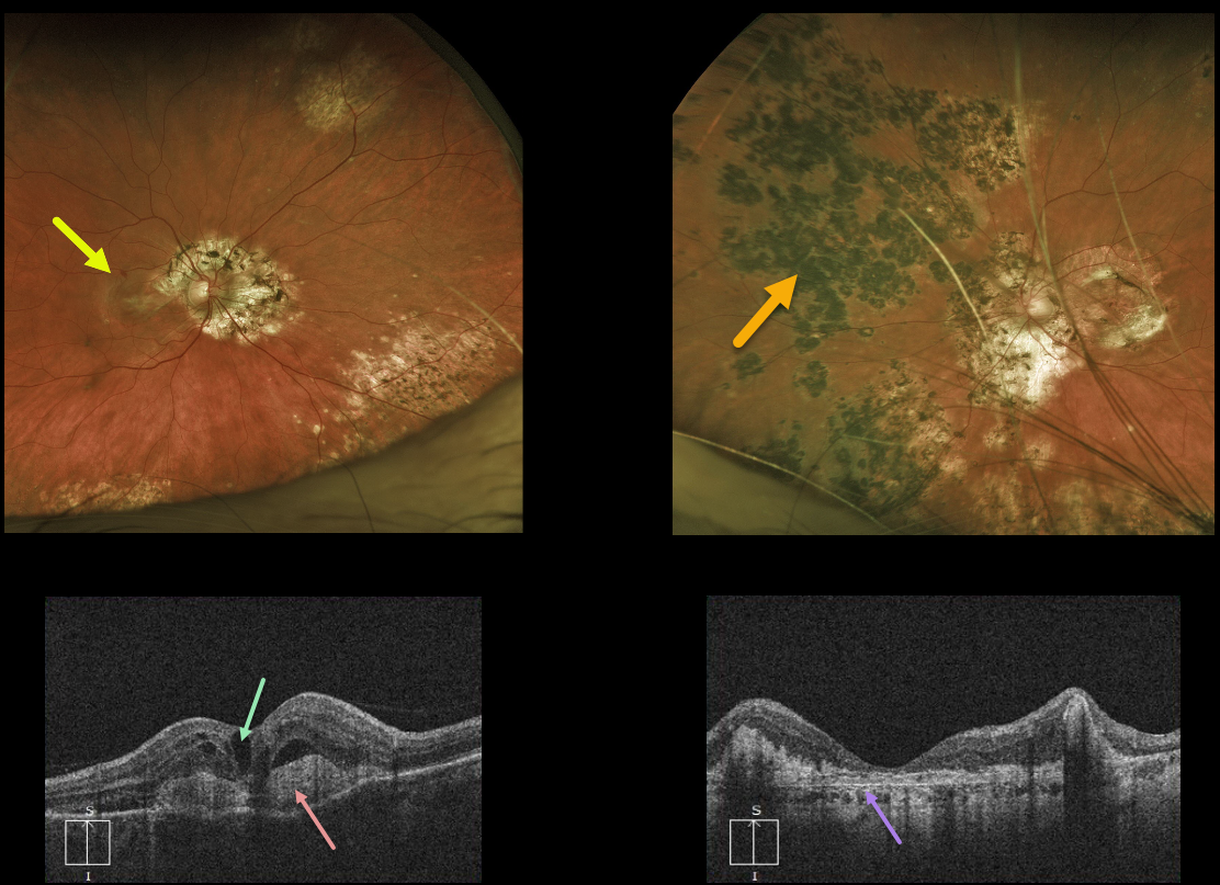
MULTIFOCAL CHOROIDITIS
Originally posted on @retina.rocks August 6, 2021
This 57YO female presented with 20/200 vision OD and counting fingers vision OS from bilateral multifocal choroiditis (MFC).
There is a newly symptomatic active macular neovascularization (MNV) in her right eye and an inactive disciform scar in her left eye.
OCT scanning shows intraretinal fluid overlying a hyperreflective MNV in the right macula with variable thickened and atrophic retina in the left macula. The MNV in the right eye is gradually improving with monthly anti-VEGF injections.
Learning Points:
MFC occurs in otherwise healthy adults, often myopic and female. Findings include multifocal variably pigmented scars throughout the posterior pole and periphery.
Unlike ocular histoplasmosis, new scars and uveitis can be seen. MNV can be successfully managed with anti-VEGF therapy.
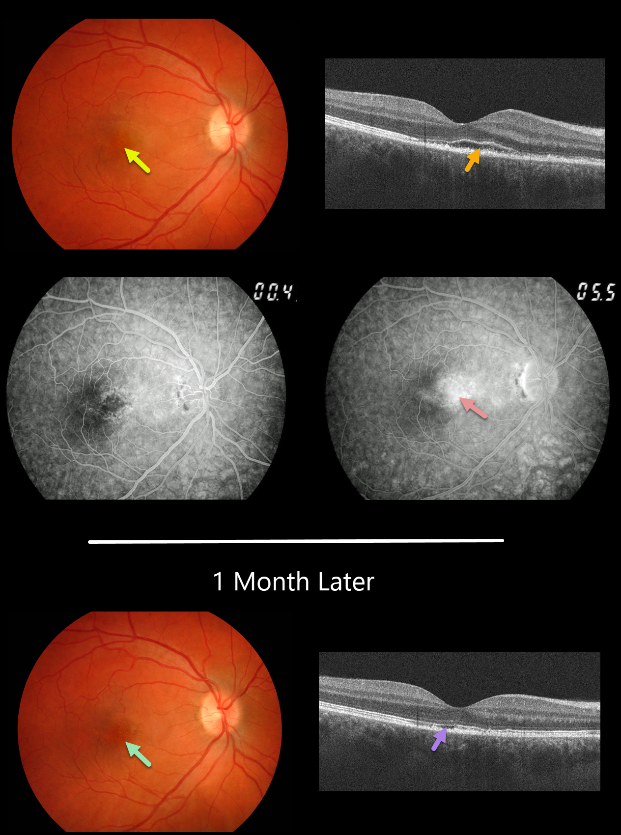
ACUTE IDIOPATHIC MACULOPATHY (AIM)
Originally posted on @retina.rocks August 3, 2021
This 29YO male presented with a 1-week history of acute blurred vision in his right eye. Vision was 20/40 OD.
Fundus photography and fluorescein angiography reveal pigment mottling and late-stage leakage. OCT scanning shows an irregularly elevated ellipsoid zone with underlying outer segment disruption vs subretinal fluid and a thickened RPE.
We felt the patient had acute idiopathic maculopathy (AIM) and observed him without treatment.
One month later, the outer fluid resolved with residual irregularities in the outer retinal and RPE bands on OCT and an area of macular hyperpigmentation surrounded by a rim of pigment loss.
Learning Points:
Originally described by Yannuzzi et al. in 1991 (Arch Ophthalmology 1991;109:1411-1416), AIM is an idiopathic inflammation of the outer retina, RPE, and inner choroid, usually found in young people and often following a viral illness. It is usually self-limited, resolving within several weeks with significant visual improvement. The appearance of a bull’s-eye lesion, as occurred in our patient, is classic for this disease.
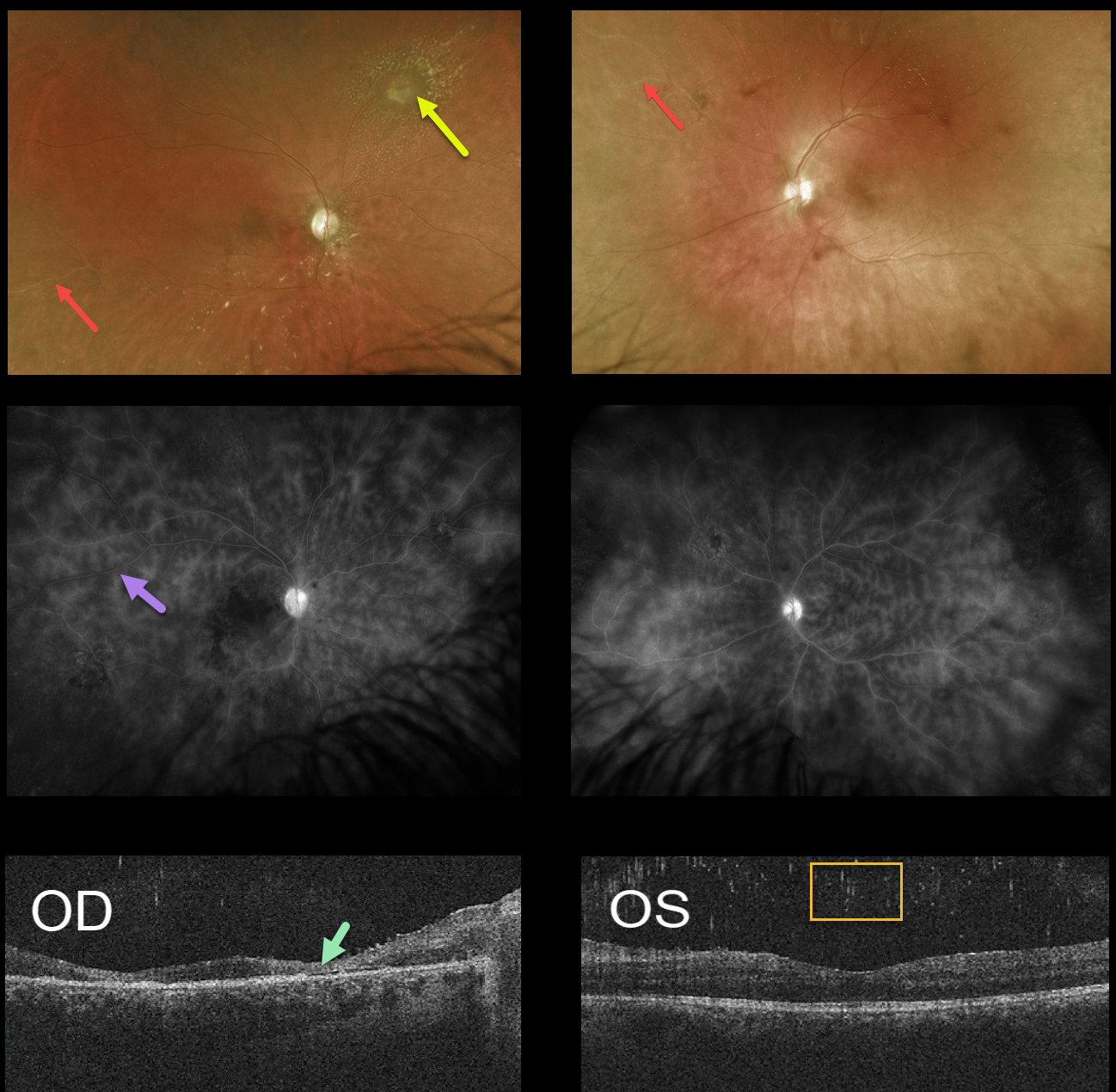
BEHCET’S DISEASE
Originally posted on @retina.rocks July 16, 2021
This 23YO male initially presented with 5 days of bilateral blurred vision and stabbing pain in his right eye. Vision was 20/400 OU.
Panuveitis was noted bilaterally, with moderate granulomatous anterior uveitis, moderate vitreous cells, macular vascular occlusion, and possible vasculitis (not shown).
Review of symptoms revealed occasional headache, lack of genital ulcers, but was positive for an occasional oral ulcer. Subsequent laboratory workup was negative, including ANCA, ANA, rheumatoid factor, ESR, FTA-ABS, RPR, QuantiFERON-TB Gold, Lyme, HLA-B27, and lumbosacral spine x-ray.
He was started on oral prednisone and topical steroid drops. He was then lost to follow-up.
When seen nearly a year later, vision was CF OD and 20/400 OS with significant panuveitis as before.
Optos imaging shows bilateral peripheral ischemia along with a whitish area of possible retinitis with surrounding lipid in the superonasal midperiphery of the right eye.
Fluorescein angiography shows diffuse vasculitis. OCT scanning shows severe retinal atrophy OD with bilateral vitreous cells.
Brain MRI revealed an enhancing lesion within the brainstem, and neurosurgery ruled out a malignancy. He was then referred to a neuro-immunologist. Further testing for infectious etiologies such as toxoplasmosis, toxocariasis, and Whipple’s disease was all negative. He then developed a 6th nerve palsy along with ataxia and was admitted to the hospital.
A combination of specialists, including neuro-immunology, rheumatology, and gastroenterology, felt there was a high probability of Behcet disease. The patient improved with a long course of oral prednisone and was slowly tapered as he was started on azathioprine.
Due to his severe peripheral ischemia and poor compliance with follow-up, he was treated with prophylactic scatter laser photocoagulation to prevent proliferative complications.
Learning Points:
Behcet disease is a systemic vasculitis that includes the classic triad of oral ulcers, genital ulcers, and ocular disease. There are no specific tests to confirm the diagnosis of Behcet’s.
The diagnosis is clinical and includes 1) recurrent oral ulcerations (aphthous or herpetiform) at least three times in one year AND 2) any two of the following: recurrent genital ulcerations, eye lesions (uveitis or retinal vasculitis), skin lesions (erythema nodosum, pseudofolliculitis, papulopustular lesions, acneiform nodules) found in adult patients not being treated with corticosteroids, and a positive “pathergy test” read by a physician within 24-48 hours of testing.
Anterior segment findings include uveitis and a sterile hypopyon. Posterior segment findings can be blinding and include an ischemic hemorrhagic retinal vasculitis. Vascular occlusions can cause optic atrophy, retinal neovascularization, and vitreous hemorrhage.
Treatment for the underlying uveitis and vasculitis is complex and includes topical/systemic corticosteroids, as well as immunomodulatory medications. Ocular neovascularization can be treated with anti-VEGF injections and scatter laser photocoagulation.
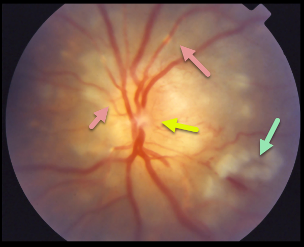
SARCOIDOSIS
Originally posted on @retina.rocks July 9, 2021
This patient with a known history of sarcoidosis presented with a swollen optic nerve, vasculitis, and patches of inner retinal opacification.
Learning Points:
Sarcoidosis, similar to syphilis, has a myriad of ocular presentations, including anterior, intermediate, and posterior uveitis, vasculitis, papillitis, and choroidal granuloma.
In our experience, there appears to be little association between active systemic disease and ocular inflammation. Establishing the diagnosis of ocular sarcoid in the absence of a known systemic diagnosis is often difficult.
Sensitivity and specificity are highest for chest CT and, when combined with elevated serum ACE and lymphopenia, are strongly suggestive of systemic sarcoidosis (see Nieder and Sims, AJO 1019;206:149-153).
Another recent publication identified soluble interleukin-2 receptor (sIL-2R) and chest CT evidence of bilateral hilar lymphadenopathy as the most sensitive biomarkers for diagnosing systemic sarcoidosis (Japanese J Ophthalmology 2021;65:191-198).
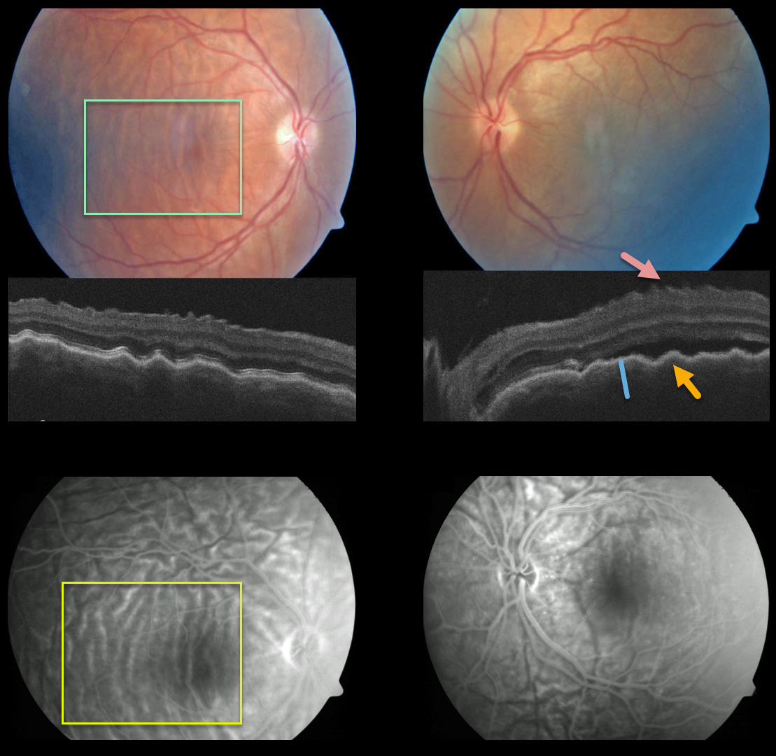
POSTERIOR SCLERITIS
Originally posted on @retina.rocks June 17, 2021
This patient presented with bilateral posterior pole chorioretinal folds, confirmed on OCT as inner retinal folds, outer retinal/RPE folds, and thickened choroid, and on fluorescein angiography.
Our patient most likely had the diffuse type of posterior scleritis. Unfortunately, we don’t have any further images or clinical information.
Learning Points:
Posterior scleritis can present either as a diffuse thickening of the entire posterior sclera or as a nodular type with localized scleral thickening (see Agrawal et al, Retina 2016;36:392-401).
Nodular inflammation appears as a posterior pole subretinal yellowish mass lesion, often with overlying serous fluid and chorioretinal folds.
B-scan ultrasonography may show fluid in the Tenon space (“T” sign). Unlike anterior scleritis, the eyes are usually quiet and pain-free.
Initial treatment is usually with oral non-steroidal anti-inflammatories or corticosteroids, although patients may also require immunosuppressive therapy.
For a large case series of posterior scleritis, see McCluskey et al, Ophthalmology 1999;106:2380-2386.
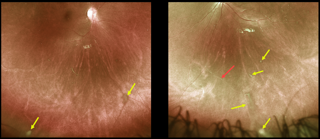
PARS PLANITIS
Originally posted on @retina.rocks June 15, 2021
This healthy 11YO boy was referred for these asymptomatic posterior segment findings.
Optos imaging shows bilateral inferior clumps of vitreous debris (“puff balls”) and vasculitis in the left eye. Not imaged was the bilateral vitreous base inflammation with white pars plana exudate.
Since our patient was completely asymptomatic, we are observing him several times a year.
Learning Points:
One of the more common classification systems for uveitis is based on its primary location within the eye (i.e., anterior vs posterior).
Intermediate uveitis is centered within the retinal periphery and vitreous base, and has a relatively short differential, including sarcoidosis, syphilis, inflammatory bowel disease, multiple sclerosis, and idiopathic (pars planitis).
Pars planitis is unique in the presence of a white vitreous base and pars plana inflammatory material. It is most common in young females, has a waxing-and-waning clinical course over many years, and usually becomes less active with age.
Patients can develop visual symptoms from floaters or cystoid macular edema (CME), and can also develop spill-over anterior uveitis.
Symptomatic vitritis and CME are usually treated with periocular and rarely systemic steroids. Cryotherapy to the inflamed vitreous base is sometimes recommended for more severe and chronic disease (see Sohn et al, AJO 2016;162:35-42).
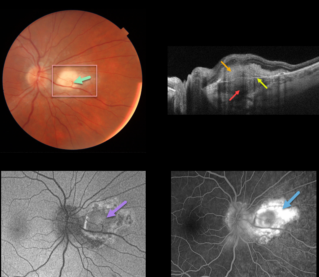
CHOROIDAL GRANULOMA
Originally posted on @retina.rocks June 7, 2021
This 30YO male was referred for an asymptomatic lesion in his right eye. Vision was 20/20 OU, and the left fundus was healthy.
There is a nodular, creamy white, deep retinal/subretinal lesion just nasal to the right optic nerve. A retinochoroidal anastomosis was noted between the overlying retinal vein and the lesion.
Triton swept-source OCT shows a thickened hyporeflective choroidal lesion, a hyperreflective outer retinal lesion with overlying inner retinal disorganization, and visible Bruch’s membrane separating the choroidal and retinal components.
Optos fundus autofluorescence (FAF) shows relatively diffuse hypo-FAF, and fluorescein angiography shows diffuse staining of the lesion.
Our patient’s bloodwork and chest x-ray were suggestive of sarcoid. Quantiferon gold, ACE, and toxoplasmosis IgG/IgM were all negative. His primary care provider was alerted to the probable diagnosis of sarcoidosis.
Learning Points:
Choroidal granulomas usually appear as solitary, creamy white, round subretinal lesions. They are hyporeflective on OCT imaging (Invernizzi et al, Retina 2015;35:525-531). Causes include sarcoidosis, tuberculosis, and VKH.
The choroidal and retinal circulations normally coexist without connecting to each other. A chorioretinal anastomosis breaks this rule when a retinal vessel directly connects with the choroid.
Clinically obvious chorioretinal anastomosis is most common in disciform lesions and toxoplasmosis scars. They are also a subclinical finding noted in 65% of eyes with MacTel 2 using OCT and OCT angiography (see Spaide et al, Retina 2018;38:1920-1929).
A laser-induced chorioretinal anastomosis was originally proposed as a possible treatment for central retinal vein occlusion in 1995, and a recent randomized trial showed it could reduce the anti-VEGF treatment burden (see McAllister et al., JAMA Ophthalmol 2018;136;1391-1397). However, it is still rarely used in current clinical practice.
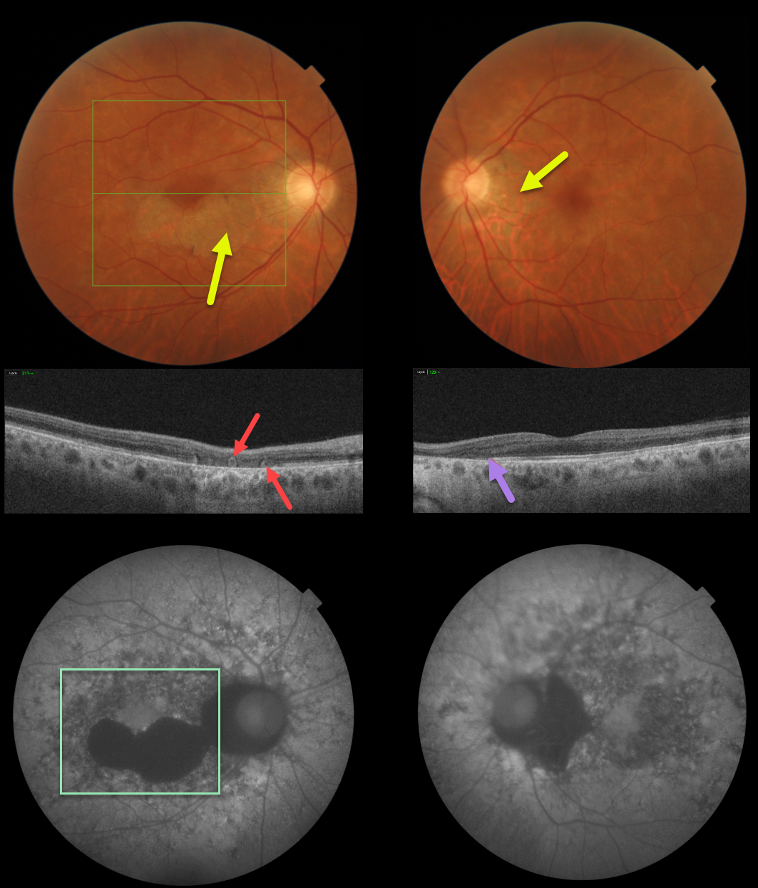
ACUTE POSTERIOR MULTIFOCAL PLACOID PIGMENT EPITHELIOPATHY (APMPPE) WITH OUTER RETINAL TUBULATION
Originally posted on @retina.rocks June 4, 2021
This patient has bilateral fundus findings consistent with resolved acute posterior multifocal placoid pigment epitheliopathy (APMPPE).
Triton fundus photos show placoid areas of atrophic pigmentary changes, which are variably hypo-autofluorescent.
Triton swept-source OCT in the right eye shows outer retinal atrophy with outer retinal tubulation (ORT) and nasal outer retinal atrophy in the left eye.
Learning Points:
ORT, initially described by Zweifel et al (Arch Ophthalmol 2009;127;1596-1602), is a neurodegenerative condition of the photoreceptors and Muller cells associated with atrophy affecting the outer retina and retinal pigment epithelium, including advanced AMD and inherited retinal diseases.
ORT is often seen overlying inactive macular neovascularization in the setting of ongoing anti-VEGF therapy and should not be confused with exudative fluid or cysts, which lack a hyperreflective border. The outer hyperreflective band likely represents inner segment mitochondria undergoing fission and translocation toward the nucleus (Litts et al, Retina 2018;38;445-461).
Acute posterior multifocal placoid pigment epitheliopathy (APMPPE) is a usually bilateral inflammatory vs ischemic disorder of the inner choroid, RPE, and outer retina.
Blurry vision and paracentral/central scotomas often follow a viral illness. Acutely multifocal creamy yellow or grayish-white subretinal placoid lesions are scattered throughout the posterior poles. The lesions and visual symptoms usually resolve within 4 to 8 weeks with secondary chorioretinal changes. Recurrent lesions are rare, and vision is usually fairly good.
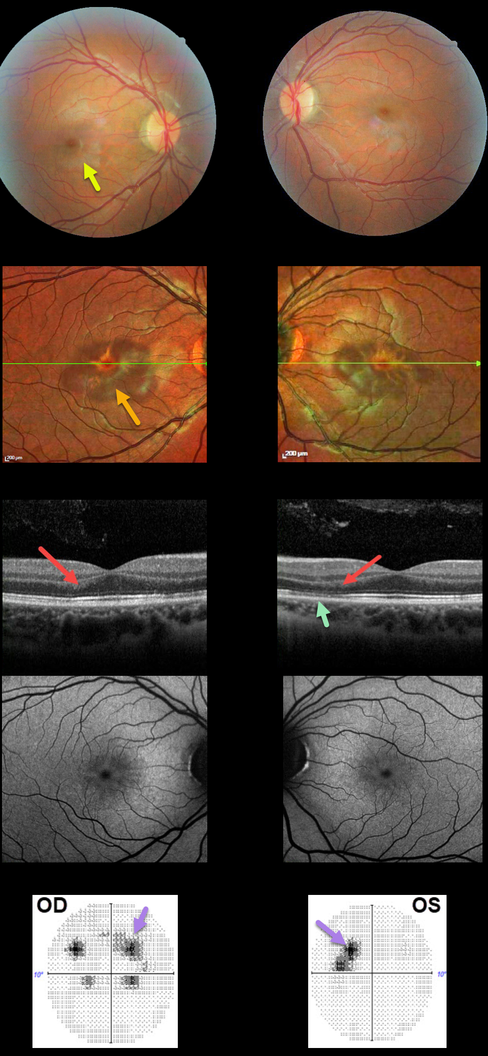
ACUTE MACULAR NEURORETINOPATHY (AMN)
Originally posted on @retina.rocks May 28, 2021
This 21YO female presented with a two-week history of bilateral paracentral relative scotomas. This was preceded by myalgias, sinus pressure, sore throat, and a cough. There was a 4-year history of oral contraceptive use. Vision was 20/25 OU.
There are faint reddish-brown wedge-shaped lesions radiating out of each fovea, seen more easily on multicolor imaging.
OCT scanning shows some focal areas of increased hyperreflectivity in the outer nuclear layer and defects in the ellipsoid zone. Fundus autofluorescence is relatively normal.
10-2 visual fields confirm the paracentral scotomas noticed by the patient. Over the following months, her 10-2 visual field partially improved, coinciding with partial normalization of the OCT findings (not shown).
Learning Points:
This case highlights many characteristic features of acute macular neuroretinopathy (AMN), an idiopathic disorder likely caused by compromise of the deep retinal capillary plexus.
Multicolor and infrared imaging often highlight the typical wedge-shaped AMN lesions more effectively than color fundus photos.
84% of patients are female with a median age of 30 years, and 80% are non-Latino white.
The most common association is a flu-like prodrome (48%), followed by oral contraceptive use (36%).
Presenting acuity is 20/40 or better in 80% of eyes, coinciding with sparing of the sub-foveal areas. Roughly half of AMN cases are bilateral. There is typically moderate visual improvement over the coming months, and no treatment is recommended.
For a great summary of AMN, see Bhavsar KV et al, Surv Ophthalmology 2016; 61:538-565.
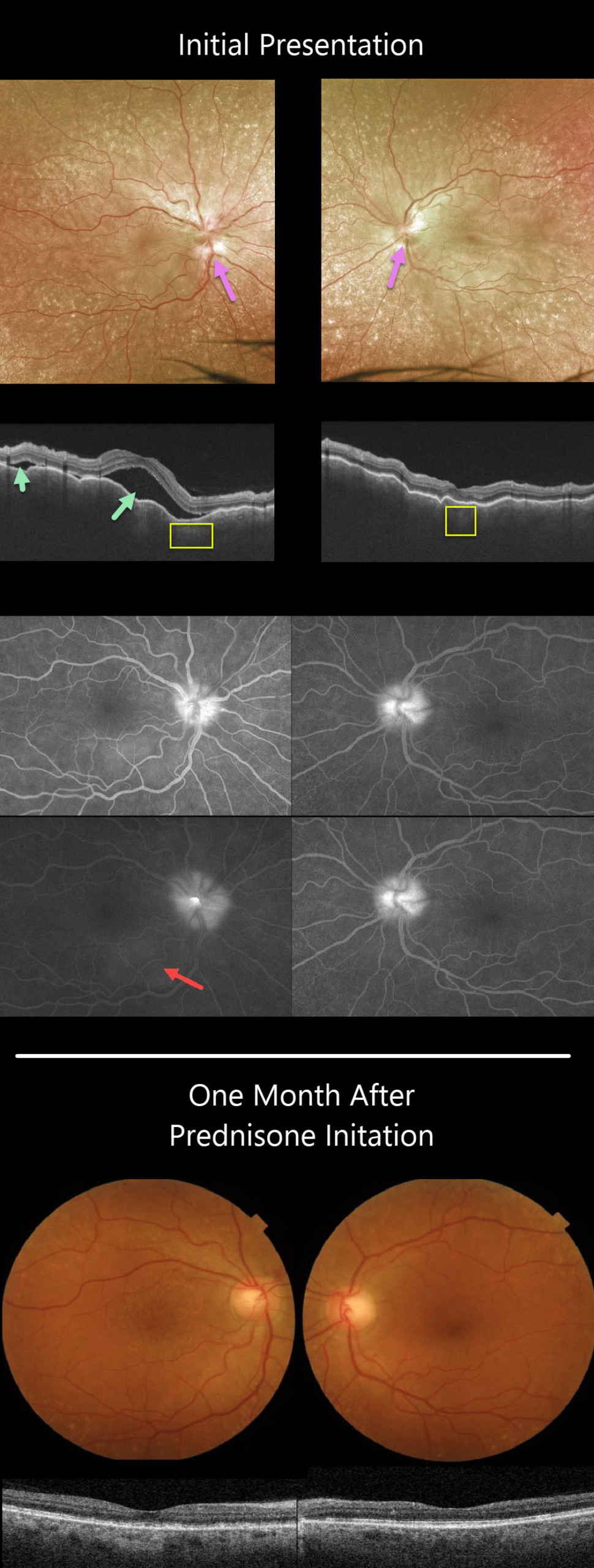
HARADA DISEASE
Originally posted on @retina.rocks April 27, 2021
This 63YO female presented with a 3-week history of bilateral painful vision loss. Vision was 20/400 OD and 20/100 OS.
She had bilateral granulomatous anterior uveitis with a clear vitreous OU. Both nerves were swollen, and irregular chorioretinal folds were noted throughout each macula. Extensive incidental peripheral drusen were also present.
Triton swept-source OCT scanning shows a markedly irregularly thickened choroid OU with multifocal serous detachments in the right eye.
Optos fluorescein angiography (FA) shows bilateral optic nerve leakage and some faint subretinal leakage in the inferonasal right macula.
Oral prednisone was started, and there was near immediate improvement. One month later, vision improved to 20/40 OD and 20/60 OS, and all findings virtually normalized.
Learning Points:
This case has many classic findings for Harada’s disease, including bilateral panuveitis, optic nerve swelling, thickened choroid, chorioretinal folds, and multifocal serous retinal detachments.
We were surprised that the FA showed minimal subretinal leakage, since angiography typically shows extensive multifocal subretinal leaks with pooling in the overlying serous detachments and bacillary cavities.
Patients with just ocular findings have Harada’s disease, whereas those with additional systemic findings (including vitiligo, poliosis, headache, vertigo, and hearing loss) have Vogt-Koyanagi-Harada disease.
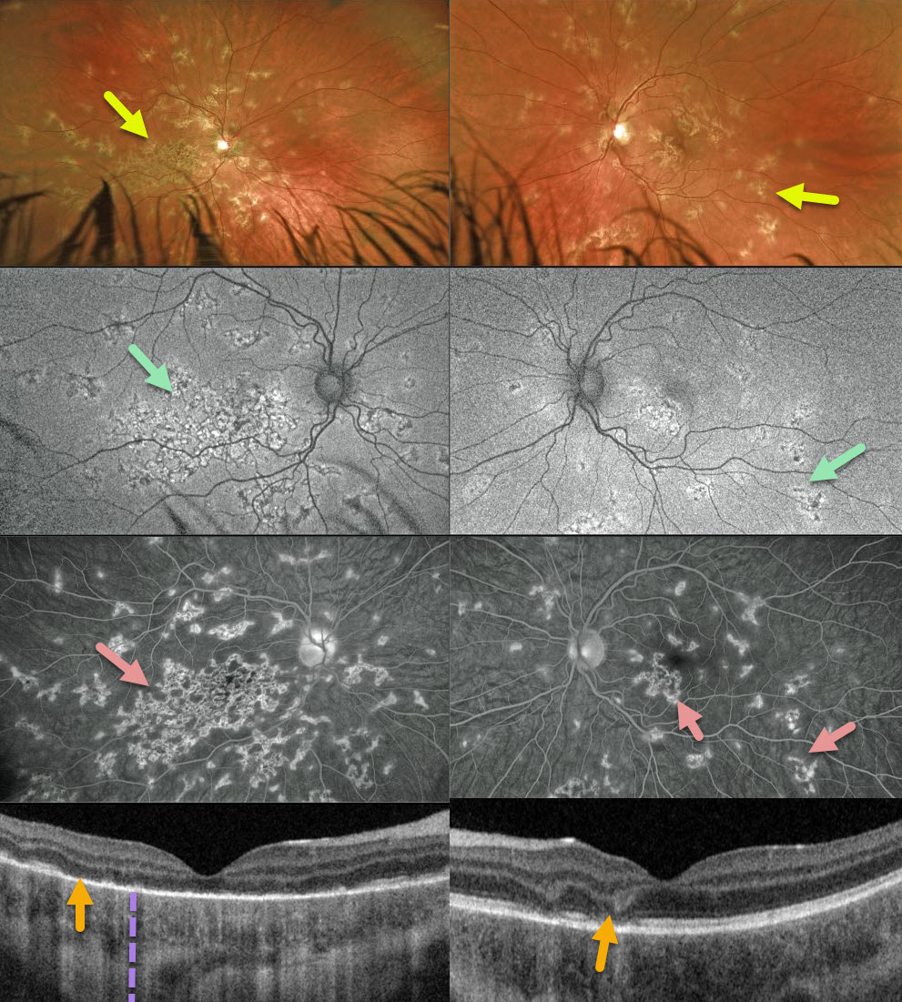
RELENTLESS PLACOID CHORIORETINITIS
Originally posted on @retina.rocks April 20, 2021
This 12YO boy was referred for asymptomatic retinal changes. Vision was counting fingers OD and 20/25 OS.
Optos imaging shows patchy inactive chorioretinal scarring extending from each macula to the mid-periphery, especially confluent in the right macula.
There is variable hyper- and hypo-autofluorescence, and fluorescein angiography shows areas of staining. Triton swept-source OCT shows variable outer retinal loss with fairly intact RPE associated with increased choroidal transmission.
Our presumed diagnosis was relentless placoid chorioretinitis. Since all the funduscopic findings appeared inactive, we are currently observing him without treatment.
Learning Points:
Relentless placoid chorioretinitis has clinical and angiographic features resembling both serpiginous choroiditis and acute posterior multifocal placoid pigment epitheliopathy (APMPPE).
Relentless placoid demonstrates the multifocal lesions of APMPPE, with more meandering, less patchy, serpiginous lesions.
Additionally, these patients will have recurrent inflammation typical of serpiginous but not APMPPE. They may also develop macular neovascularization, which is more typical of serpiginous.
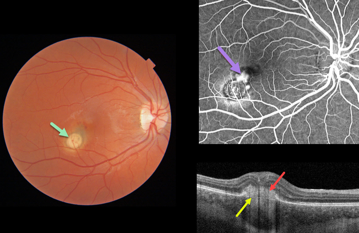
CHOROIDAL GRANULOMA
Originally posted on @retina.rocks April 15, 2021
This 22YO female presented with 20/400 vision from a macular neovascularization (MNV) associated with a choroidal granuloma.
The Triton image shows a focal yellow-white nodular subretinal lesion with adjacent subretinal pigment and blood along its foveal edge.
Swept-source OCT through the MNV shows the hyperreflective MNV above Bruch’s membrane. Fluorescein angiography shows leakage from the MNV.
Learning Points:
Choroidal granulomas usually appear as solitary, creamy white, round subretinal lesions. They are hyporeflective on OCT imaging (Invernizzi et al, Retina 2015;35:525-531). Causes include sarcoidosis, tuberculosis, and VKH. They can occasionally cause exudation that responds to anti-VEGF injections or photodynamic therapy.
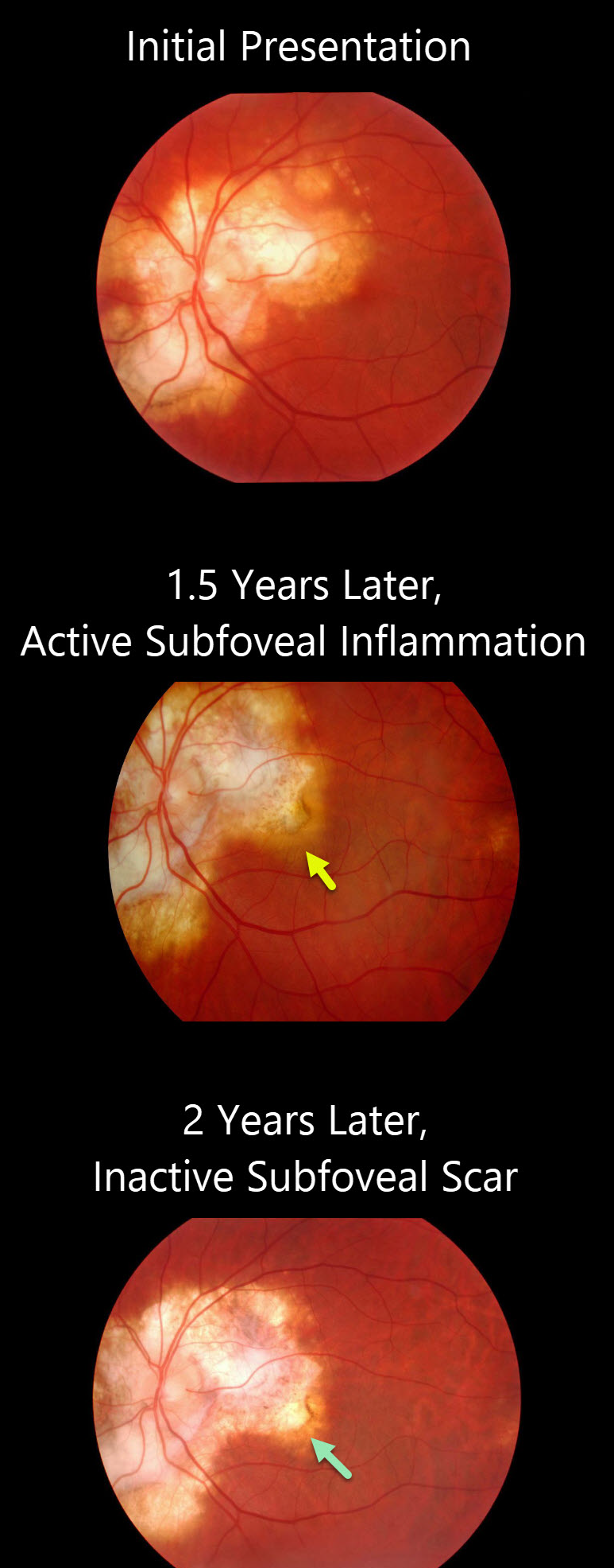
SERPIGINOUS CHORIORETINITIS
Originally posted on @retina.rocks March 16, 2021
This patient was seen initially with inactive peripapillary and macular chorioretinal scarring from serpiginous choroiditis.
A year and a half later, he presented with new subfoveal active inflammation. One year later, there was inactive atrophic subfoveal scarring.
Learning Points:
Serpiginous choroiditis is an idiopathic, usually bilateral chorioretinal inflammatory condition most commonly found in middle-aged men.
Recurrent areas of inflammation develop along the edge of a previous scar, producing scarring that meanders from the optic nerve outward into the macula.
Vision is good unless scarring (as in this patient) or macular neovascularization extends through the macular center.
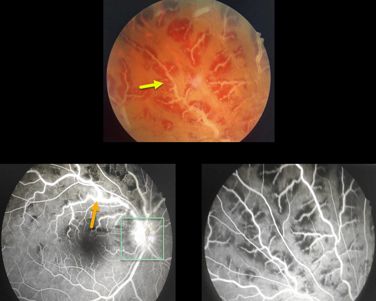
FROSTED BRANCH ANGIITIS
Originally posted on @retina.rocks March 12, 2021
This 19yo presented with counting fingers (CF) vision OD and 20/20 OS.
There is a frosted appearance to the superior retinal veins with surrounding retinal hemorrhages. Fluorescein angiography (FA) shows some vascular staining and optic nerve leakage.
The patient was started on oral steroids, and one week later, the fundus findings had improved significantly (not pictured). Bloodwork came back negative.
Learning Points:
Frosted branch angiitis is a retinal vasculitis that gets its name due to the involved vessels resembling the branches of a tree that are “frosted” with snow.
Although these findings may be isolated, they are often considered a phenotype found in a multitude of inflammatory conditions, most commonly CMV retinitis.
The inflammation is usually highly responsive to steroids, and the visual prognosis is generally good, although some patients may develop secondary neovascularization in the anterior and posterior segments due to widespread ischemia.
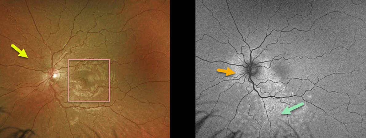
MULTIPLE EVANESCENT WHITE DOT SYNDROME (MEWDS)
Originally posted on @retina.rocks February 11, 2021
This 25YO female presented with 20/25 vision OU and a four-day history of temporal field loss in her left eye. Examination of the right eye was normal.
There was a granular appearance to the left fovea with some deep retinal white dots, most visible just nasal to the nerve. Fundus autofluorescence (FAF) more dramatically showed a ring of hyper-FAF encircling the nerve with multiple hyper-FAF spots extending inferiorly.
Learning Points:
Multiple evanescent white dot syndrome (MEWDS) presents with unilateral idiopathic inflammation of the outer retina, typically occurring in young, healthy myopic females.
The granular fovea (Jampol spots, named in honor of Dr. Lee Jampol, who first described this syndrome in 1984) noted in our case is a classic pathognomonic finding.
Acute cases will also have an enlarged blind spot, often with temporal visual field loss, due to peripapillary inflammation.
The white dots disappear within a week or two, which is why the lesions are called “evanescent”. However, the blind spot may persist for many months, well after the fundus appearance has normalized.
This stage of MEWDS was originally called the acute idiopathic blind spot enlargement (AIBSE) syndrome before Dr. Gass recognized that the 2 disorders were the same disease but with distinct findings at different points in time.
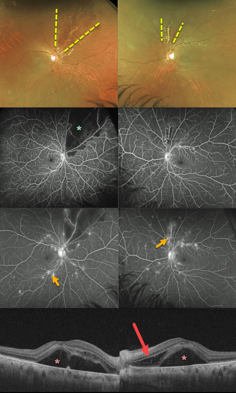
SYSTEMIC LUPUS ERYTHEMATOSUS (SLE)
Originally posted on @retina.rocks December 29, 2020
This 52YO female presented with severe bilateral vision loss (20/400 OD, counting fingers OS) following a several-week gastrointestinal illness with 60-pound weight loss.
She has bilateral areas of hemorrhagic retinal vasculitis, with areas of ischemia and leakage seen on fluorescein angiography. OCT shows macular subretinal fluid and a bacillary detachment in the left macula.
She had been hospitalized recently for hypertension and kidney failure and was diagnosed with a previously asymptomatic stroke. Extensive blood work was negative, and she denied symptoms of Behcet’s.
We suspected an underlying rheumatologic cause, and she was subsequently diagnosed with severe systemic lupus erythematosus (SLE) vasculitis.
She was started on high-dose oral prednisone followed by Cytoxan and hydroxychloroquine. Her ocular findings rapidly improved, and at her last examination 4 months later, vision had improved to 20/40 OD and 20/100 OS.
Learning Points:
SLE-associated retinopathy more typically appears as hypertensive retinopathy (bilateral nerve fiber layer infarcts and retinal hemorrhages), often in patients with cerebral vasculitis. Purtscher-like retinopathy can also be seen.
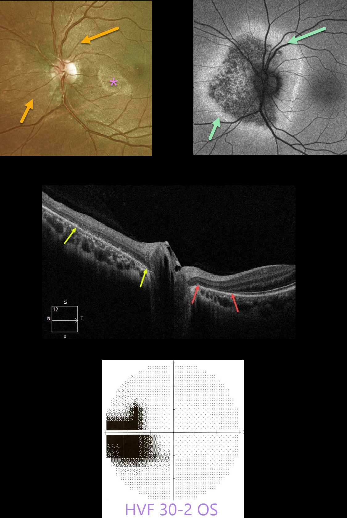
ACUTE ZONAL OCCULT OUTER RETINOPATHY (AZOOR)
Originally posted on @retina.rocks November 23, 2020
This 20YO female presented with a 5-year history of photopsias in her left eye. Vision was 20/20 bilaterally.
There were subtle pigmentary changes surrounding the left optic nerve (a brighter semicircular lesion in the left nasal macula is an artifact). Fundus autofluorescence shows more dramatic peripapillary changes with outer borders of hyperautofluorescence.
Optical coherence tomography (OCT) shows loss of the photoreceptor bands temporal to the optic nerve. Nasal to the optic nerve, both the photoreceptor bands and retinal pigment epithelium (RPE) are variably disturbed.
An enlarged blind spot is consistent with these peripapillary changes.
Learning Points:
First described by Gass in 1992, acute zonal occult outer retinopathy (AZOOR) initially presents unilaterally or bilaterally in young women with photopsias, central visual changes, and minimal fundoscopic changes.
Zones of visual field loss correspond to the locations of the photopsias. Electroretinography is often abnormal. Some may later develop areas of outer retinal, RPE, and choroidal atrophy in the initially affected retina.
The pathogenesis of the condition remains unclear, but it appears to initially involve inflammation of the photoreceptors. AZOOR is believed to be a part of the spectrum of other ‘photoreceptoritis’ conditions, including multiple evanescent white dot syndrome (MEWDS), acute macular neuroretinitis (AMN), and acute retinal pigment epitheliitis.
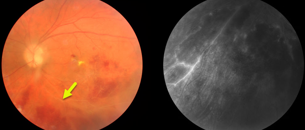
BEHCET DISEASE
Originally posted on @retina.rocks November 17, 2020
This patient presented with hemorrhagic retinal vasculitis, one of the posterior findings of Behcet’s disease. Vascular occlusions can cause optic atrophy, retinal neovascularization, and vitreous hemorrhage.
Anterior segment findings include uveitis and a sterile hypopyon (not present in this patient).
Learning Points:
Behcet disease is a systemic vasculitis that includes the classic triad of oral ulcers, genital ulcers, and ocular disease. There are no specific tests to confirm the diagnosis of Behcet.
The diagnosis is clinical and includes 1) recurrent oral ulcerations (aphthous or herpetiform) at least three times in one year AND 2) any two of the following: recurrent genital ulcerations, eye lesions (uveitis or retinal vasculitis), skin lesions (erythema nodosum, pseudofolliculitis, papulopustular lesions, acneiform nodules) found in adult patients not being treated with corticosteroids, and a positive “pathergy test” read by a physician within 24-48 hours of testing.
Treatment for the underlying uveitis and vasculitis is complex and includes topical/systemic corticosteroids, as well as immunomodulatory medications. Ocular neovascularization can be treated with anti-VEGF injections and panretinal photocoagulation.
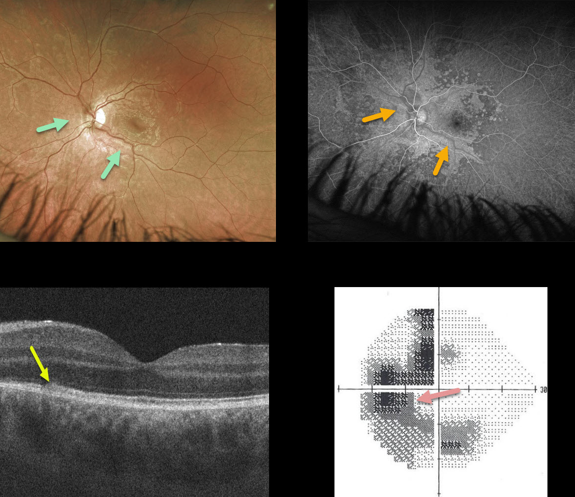
MULTIPLE EVANESCENT WHITE DOT SYNDROME (MEWDS)
Originally posted on @retina.rocks November 11, 2020
This 23YO patient presented with a few days of a temporal blurred spot in his left eye. Vision was 20/20 OD and 20/25 OS.
The left fundus has confluent outer retinal white spots centered around the optic nerve and extending into the macula. These lesions also had an unusual perivascular distribution more peripherally.
Fluorescein angiography shows hyperfluorescence staining in these lesions. Although this appearance suggested a possible vasculitis, angiography revealed absolutely no vascular changes. We are not sure why the outer retinal spots appeared in this vascular distribution.
OCT shows some hyperreflectivity and loss of detail of the ellipsoid zone and outer retina, and visual field testing shows an enlarged blind spot with temporal and inferior loss.
Learning Points:
We initially considered the possibility of syphilis given the pseudo-vasculitis and placoid confluence of the outer retinal lesions. However, the patient is in a long-term monogamous relationship and declined initial RPR/FTA testing.
Visual symptoms improved without any treatment. When the patient returned 6 weeks later, the white spots had disappeared clinically and on fundus autofluorescence, and his scotoma and symptoms had improved.
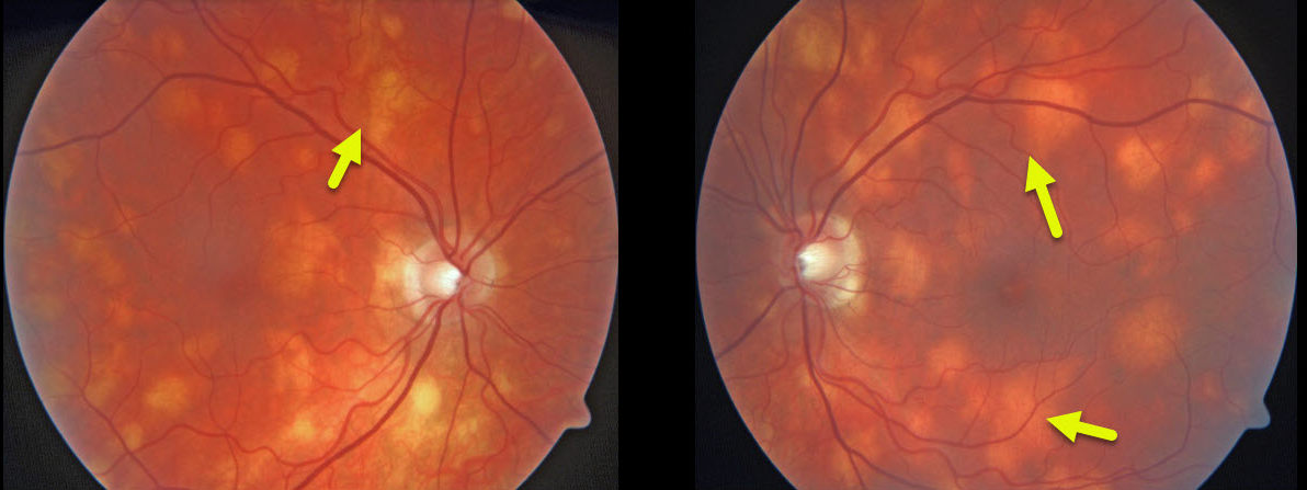
BIRDSHOT CHORIORETINOPATHY
Originally posted on @retina.rocks November 3, 2020
These creamy yellow-white choroidal lesions scattered throughout the posterior pole are characteristic of birdshot chorioretinopathy.
Learning Points:
This disease was appropriately described and named by Drs. Ryan and Maumenee (Am J Ophthalmol 1980;89:31-45), since the lesions look like shotgun birdshot scattered throughout the fundus.
Birdshot is typically chronic and bilateral, with a very high HLA-A29 association. It usually affects women in their 40s to 60s. Vision loss is usually caused by cystoid macular edema (CME) and retinal atrophy, although macular neovascularization can rarely occur.
Asymptomatic patients can be observed. CME and symptomatic uveitis can be treated with oral steroids, although many will still require immunomodulatory therapy.
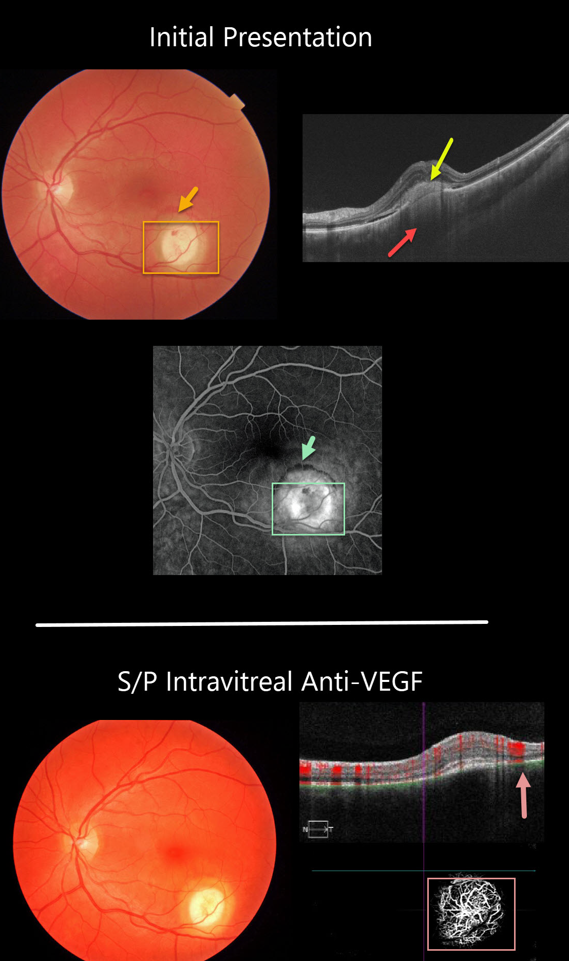
CHOROIDAL GRANULOMA
Originally posted on @retina.rocks October 16, 2020
The initial photo in this series shows a focal yellow-white nodular lesion with adjacent subretinal blood. The OCT shows subretinal hyperreflective material, some shallow subretinal fluid, and an amelanotic choroidal granuloma.
Fluorescein angiography shows staining of the lesion, with some blood blockage along its superior edge.
A later fundus photo shows complete resolution of the hemorrhage following treatment with a single intravitreal anti-VEGF injection.
Despite the resolved exudation, OCTA angiography still shows flow signals within the lesion. We continue to follow her closely for recurrent exudation.
Learning Points:
Choroidal granulomas usually appear as solitary, creamy white, round subretinal lesions. They are hyporeflective on OCT imaging (Invernizzi et al, Retina 2015;35:525-531). Causes include sarcoidosis, tuberculosis and VKH. They can occasionally cause exudation that responds to anti-VEGF injections or photodynamic therapy.
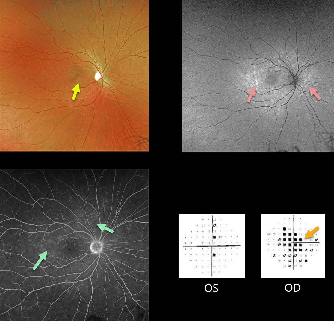
MULTIPLE EVANESCENT WHITE DOT SYNDROME (MEWDS)
Originally posted on @retina.rocks September 25, 2020
Our patient is a 31YO female with acute vision loss of 20/60 in her right eye with classic MEWDS findings.
Granular foveal changes (Jampol spots) and scattered white spots are seen in the outer retina. The spots appear hyper-autofluorescent on fundus autofluorescence imaging.
Fluorescein angiography shows multifocal wreath-like hyperfluorescent staining lesions.
Acute cases will also have an enlarged blind spot, often with temporal visual field loss.
Learning Points:
The MEWDS white dots usually disappear within a week or two, which is why the lesions are called evanescent. However, the blind spot may persist for many months, well after the fundus appearance has normalized.
This stage of MEWDS was originally called the Acute idiopathic blind spot enlargement (AIBSE) syndrome before Gass recognized that the 2 disorders were the same disease but with distinct findings at different points in time.
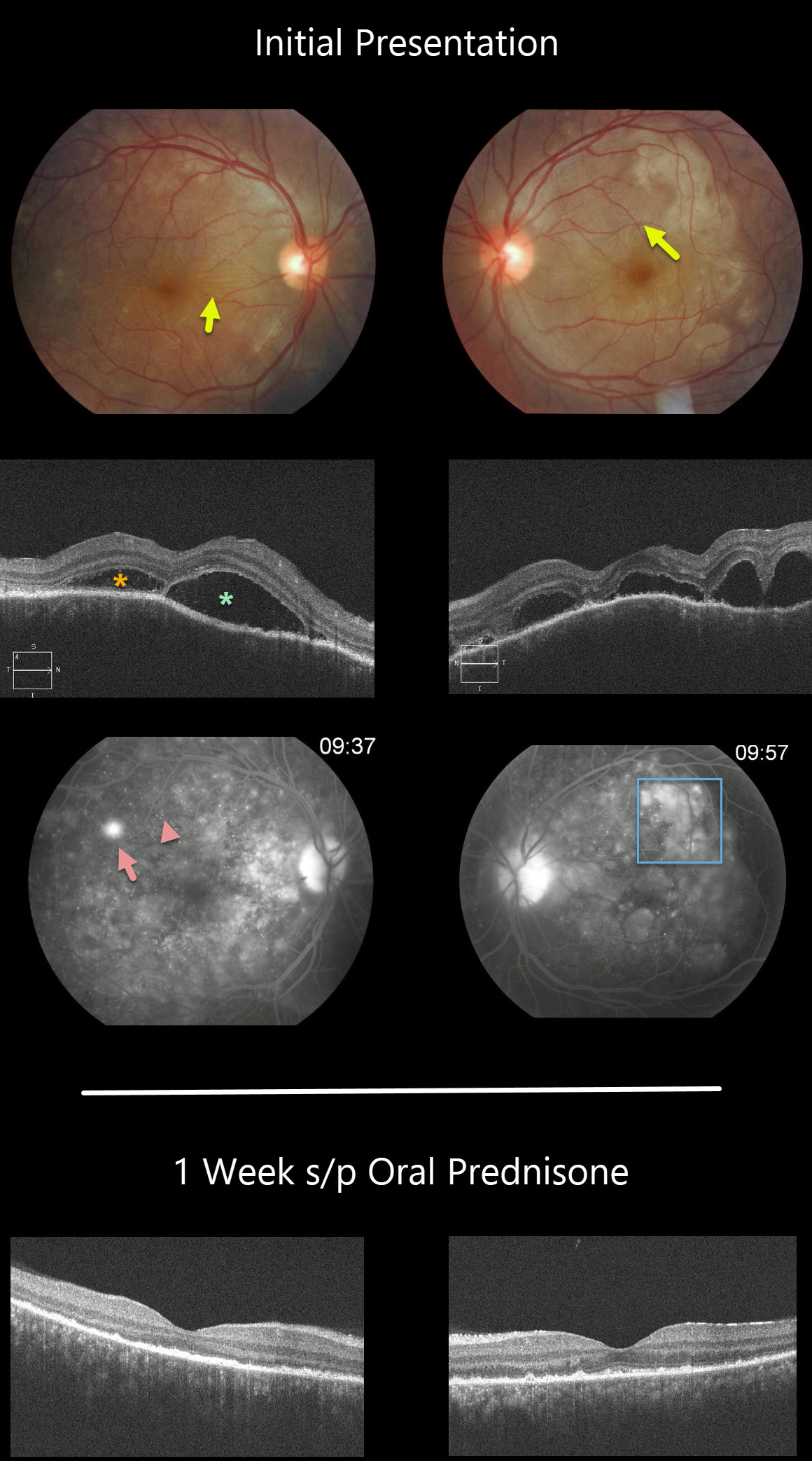
HARADA DISEASE
Originally posted on @retina.rocks September 23, 2020
This patient has classic posterior segment findings of Harada disease, including multifocal exudative serous retinal detachments.
Choroidal inflammation produces a markedly thickened choroid with overlying chorioretinal folds (seen in both eyes as variably radiating outer retinal folds).
The OCT scans show multifocal serous detachments with bacillary detachments. Fluorescein angiography shows multiple subretinal leaks with pooling of fluorescein within the various subretinal and bacillary spaces.
The patient started 60mg of oral prednisone daily. One week later, there was a dramatic improvement, with complete resolution of all intra- and subretinal fluid and normalization of choroidal thickness.
Learning Points:
Harada disease is a bilateral granulomatous panuveitis. These patients often need extended treatment with oral prednisone. Most can be successfully weaned off of steroids, although chronic treatment is sometimes necessary.
Choroidal thickening is a good biomarker for disease activity and thins with controlled disease activity.
Remember that in Harada disease, there are only ocular findings. Vogt-Koyanagi-Harada disease occurs when there are associated auditory, integumentary or neurologic symptoms.
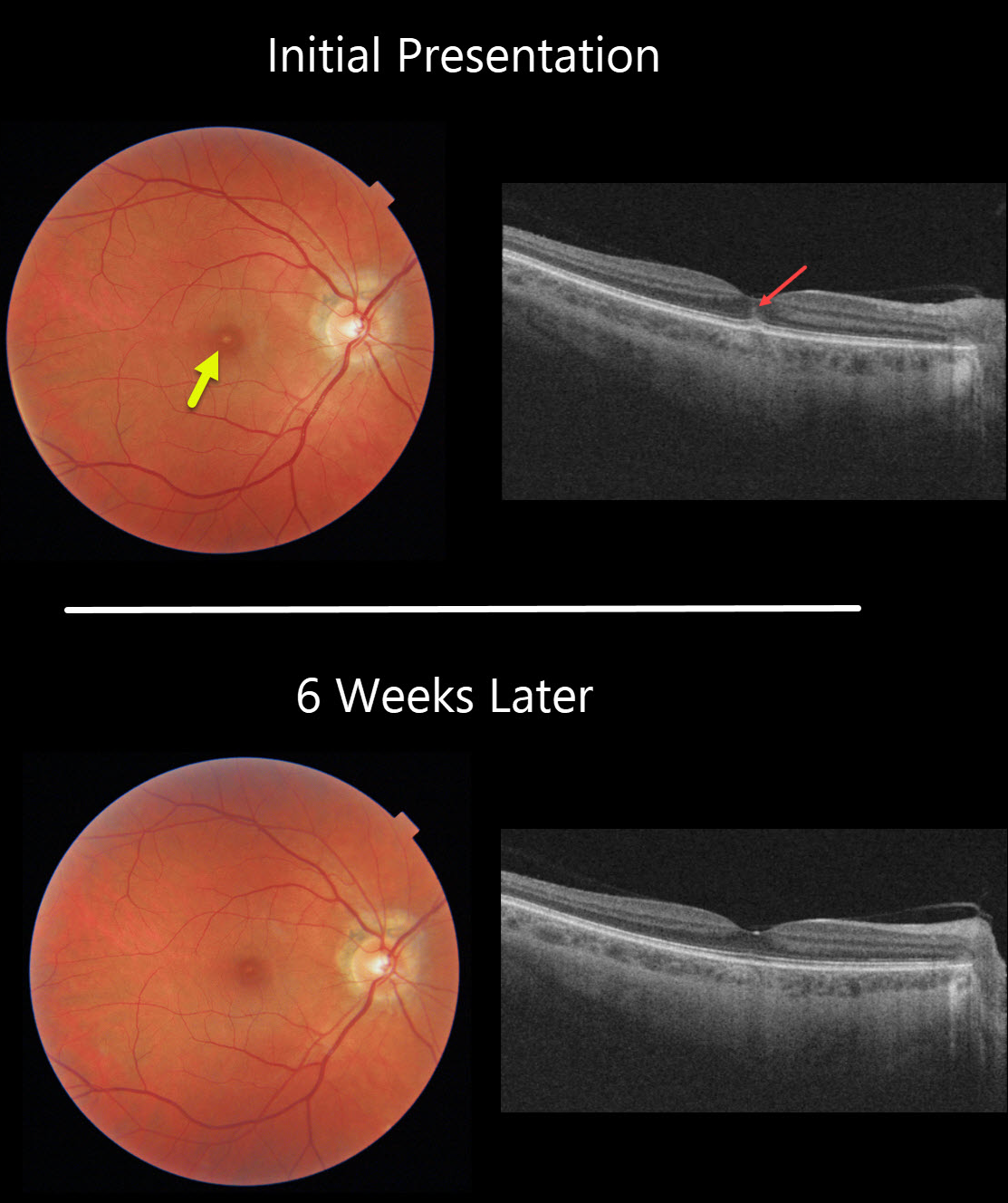
ACUTE RETINAL PIGMENT EPITHELIIITIS
Originally posted on @retina.rocks September 22, 2020
This healthy 31YO presented with sudden vision loss in her right eye.
The fundus image shows a single yellow subfoveal lesion, which appears as a hyperreflective pillar extending into the outer nuclear layer on OCT.
The differential includes solar retinopathy, laser pointer maculopathy, whiplash maculopathy, acute retinal pigment epitheliitis (RPEitis), and central serous retinopathy. There was no history of sungazing or trauma.
She returned 6 weeks later with resolved symptoms and 20/20 vision. We think this was a case of acute RPEitis (Krill’s Disease).
Learning Points:
Acute RPEitis is a self-limited inflammatory disease that affects young adults. Initially felt to be an inflammation of the RPE, it is now thought to be an idiopathic inflammation of the photoreceptors. It presents with one or more small yellow-white macular lesions.
With resolution, the macular appearance can normalize, or patients can be left with a small outer segment defect similar to solar retinopathy.
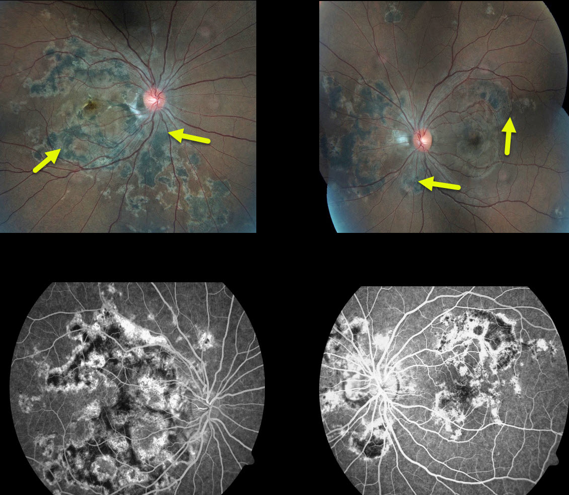
RELENTLESS PLACOID CHORIORETINITIS
Originally posted on @retina.rocks August 17, 2020
Our patient has relentless placoid chorioretinitis with the multifocal lesions of acute posterior multifocal placoid pigment epitheliopathy (APMPPE) and the peripapillary changes of serpiginous choroiditis.
This patient demonstrates chorioretinal scars surrounding the optic nerve but also throughout the posterior pole. Additionally, he has recurrent inflammation typical of serpiginous but not APMPPE.
His vision is 20/20 OU, which worsens when steroid or anti-inflammatory treatment is tapered. Infectious and inflammatory blood work was negative. He is currently treated and stable with methotrexate.
Learning Points:
Relentless placoid chorioretinitis has clinical and angiographic features resembling both serpiginous chorioretinopathy and acute posterior multifocal placoid pigment epitheliopathy (APMPPE).
Patients with relentless placoid choroiditis can develop recurrent inflammation and macular neovascularization that is more typical of serpiginous than APMPPE.
APMPPE is a bilateral inflammatory vs ischemic disorder of the inner choroid, RPE, and outer retina. These patients present with blurred vision and paracentral/central scotomas often following a viral illness.
Acutely, there are multifocal creamy yellow or grayish-white subretinal placoid lesions scattered throughout the posterior pole.
The lesions and visual symptoms usually resolve within 4 to 8 weeks with secondary chorioretinal changes. Recurrent lesions are rare, and vision is usually fairly good.
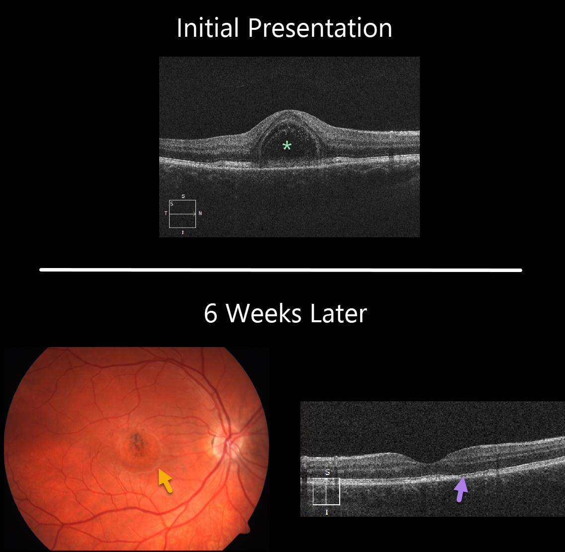
ACUTE IDIOPATHIC MACULOPATHY (AIM)
Originally posted on @retina.rocks July 23, 2020
This 22YO female presented with 1 week of unilateral vision loss and 20/200 vision in her right eye.
The macular OCT line scan demonstrates a bacillary layer (cone and rod inner and outer segments) detachment. This appears as a unique dome shaped collection of intraretinal fluid from photoreceptor myoid splitting.
Six weeks later vision improved to 20/40. The macular fluid resolved, with the development of a dry area of macular hyperpigmentation surrounded by a rim of pigment loss. OCT shows some subtle outer retinal foveal thinning with ellipsoid zone irregularities.
Learning Points:
Acute idiopathic maculopathy (AIM) is a usually unilateral idiopathic condition usually found in young people, often following a viral illness. It is usually self-limited, resolving within several weeks with significant visual improvement. The appearance of a bulls-eye lesion, as occurred in our patient, is classic for this disease.
Bacillary detachments can be observed in multiple conditions including Vogt-Koyanagi-Harada (VKH) disease, AIM, blunt trauma, or APMPPE.
See Fernandez-Avellaneda et al Retinal Cases & Brief Reports 2019 (published ahead of print) for a discussion of bacillary layer detachment in AIM.
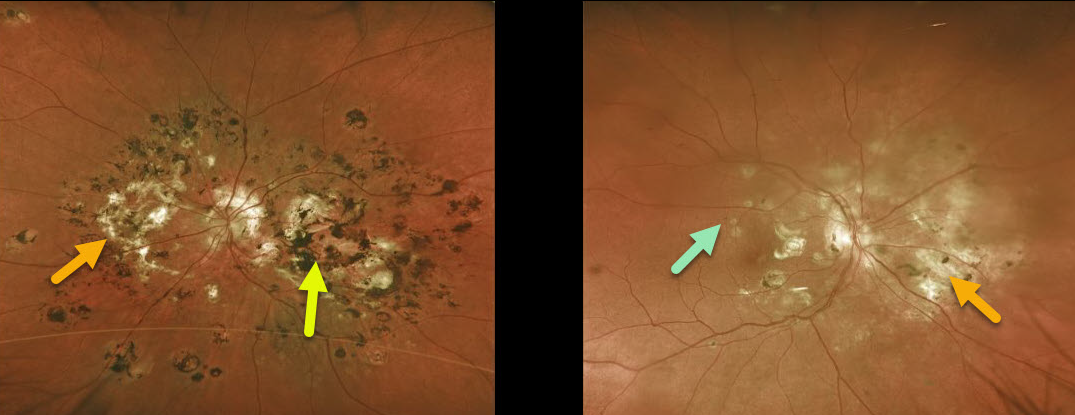
MULTIFOCAL CHOROIDITIS
Originally posted on @retina.rocks July 16, 2020
This 59YO female initially had unilateral multifocal choroiditis (MFC) in her left eye. When we started seeing her in the mid-1990s, predating the anti-VEGF era, we battled multiple inflammatory macular neovascularizations (MNV).
Eventually, she developed subfoveal MNV and nasal subretinal fibrosis despite multiple thermal laser treatments, and her vision remains counting fingers OS.
Fourteen years later, she developed asymptomatic hypo and hyperpigmented lesions in her right eye similar to those of her left eye, and she remained 20/20 OD.
Initially treated with intravitreal Avastin for MNV, intravitreal triamcinolone was also added for new macular lesions.
Learning Points:
MFC occurs in otherwise healthy adults, often myopic and female. Findings include multifocal variably pigmented multifocal scars throughout the posterior pole and periphery.
Unlike ocular histoplasmosis, new scars and uveitis can be seen. Punctate inner choroidopathy most likely represents a continuum of MFC rather than a separate disease. MNV can be successfully managed with anti-VEGF therapy.
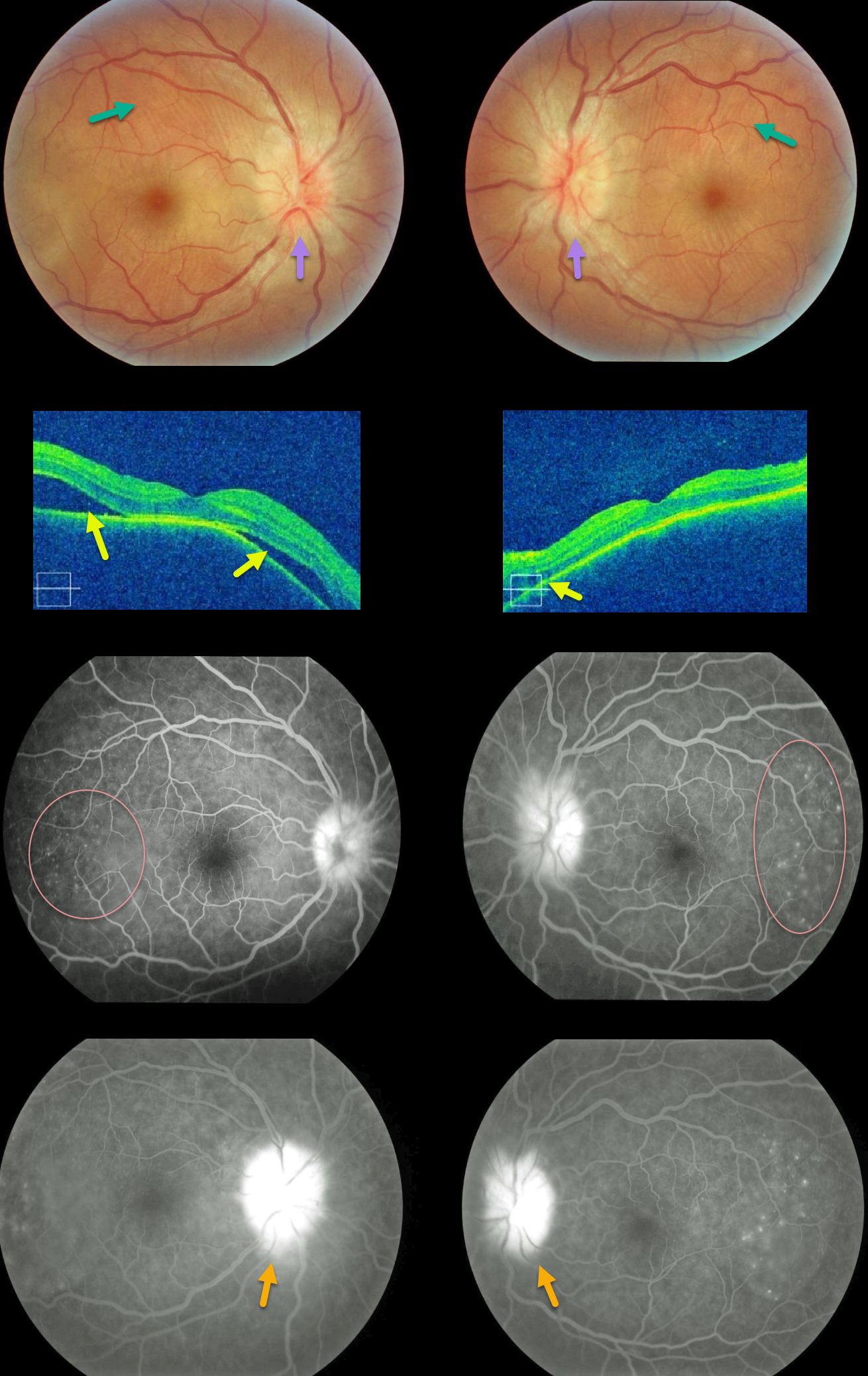
HARADA DISEASE
Originally posted on @retina.rocks May 19, 2020
Our patient shows classic findings for acute Harada’s. The fundus photos and OCT images show bilateral multifocal subretinal exudative detachments with irregular, scattered overlying chorioretinal folds and bilateral optic disc swelling.
Fluorescein angiography (FA) shows multifocal subretinal leaks with pooling of fluid below the retina and within outer retinal cystoid spaces, along with leaking “hot” nerves.
These findings dramatically resolved after just 1 week of oral prednisone (not pictured).
Learning Points:
Bilateral, multifocal yellow-white, serous retinal detachments are the hallmark sign of acute Vogt-Koyanagi-Harada (VKH) disease.
The choroid is markedly thickened with overlying chorioretinal folds. The multiple sets and various orientations of these folds are quite different from those seen with idiopathic chorioretinal folds.
The optic nerves are usually inflamed and swollen. Patients often have a panuveitis with anterior chamber and vitreous cells.
Those with just ocular findings have Harada disease, whereas patients with additional systemic findings (including vitiligo, poliosis, headache, vertigo, and hearing loss) have Vogt-Koyanagi-Harada disease.
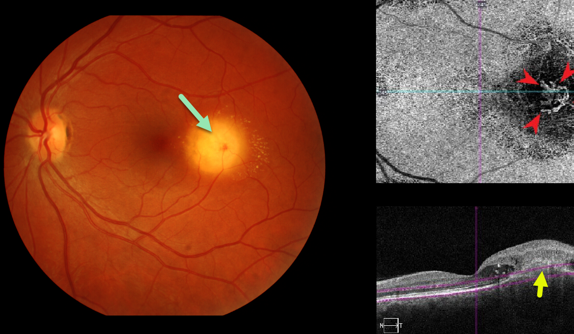
CHOROIDAL GRANULOMA
Originally posted on @retina.rocks May 13, 2020
Our patient was seen recently with asymptomatic macular neovascularization (MNV) associated with a choroidal granuloma. Clinically there was new blood, fluid and lipid.
OCT angiography confirms an active MNV. This lesion is deep to the RPE which is consistent with a focal scleral nodule.
Learning Points:
Choroidal granulomas usually appear as solitary creamy white, round subretinal lesions. They are hyporeflective on OCT imaging (Invernizzi et al, Retina 2015;35:525-531). Causes include sarcoidosis, tuberculosis and VKH. They can occasionally cause exudation that responds to anti-VEGF injections or photodynamic therapy.
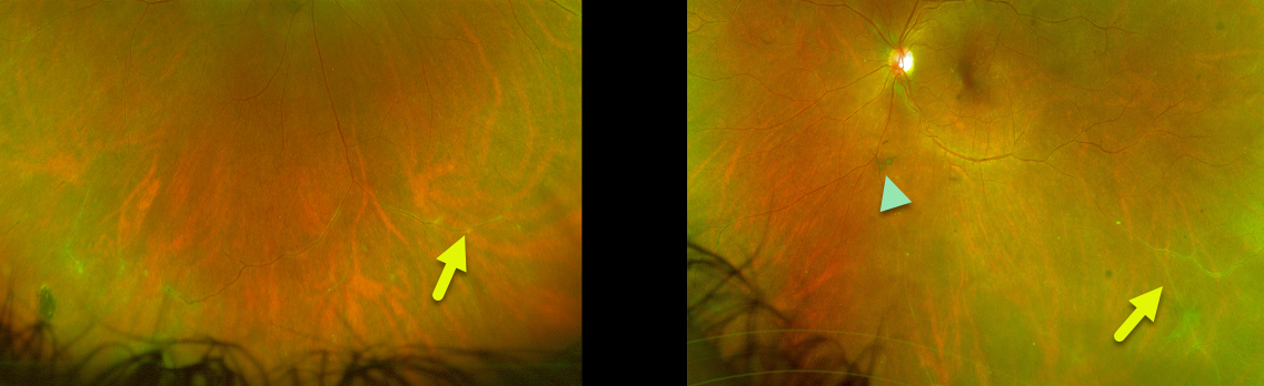
MULTIPLE SCLEROSIS
Originally posted on @retina.rocks April 21, 2020
Our patient with history of multiple sclerosis (MS) has intermediate uveitis with peripheral vasculitis and overlying clumps of vitreous floaters. These posterior segment findings have been stable for decades and her MS remains in total remission.
Learning Points:
Although optic neuritis is the most frequent ophthalmic manifestation of multiple sclerosis (MS), patients can also rarely develop intermediate uveitis.
Although the literature often lists MS as a cause for pars planitis, in our experience, this is not the case. Pars planitis has very characteristic white inflammatory material within the vitreous base (pars plana exudate), with “puff balls” of inflammatory debris breaking free into the vitreous. We have never seen this pars plana exudate with MS.
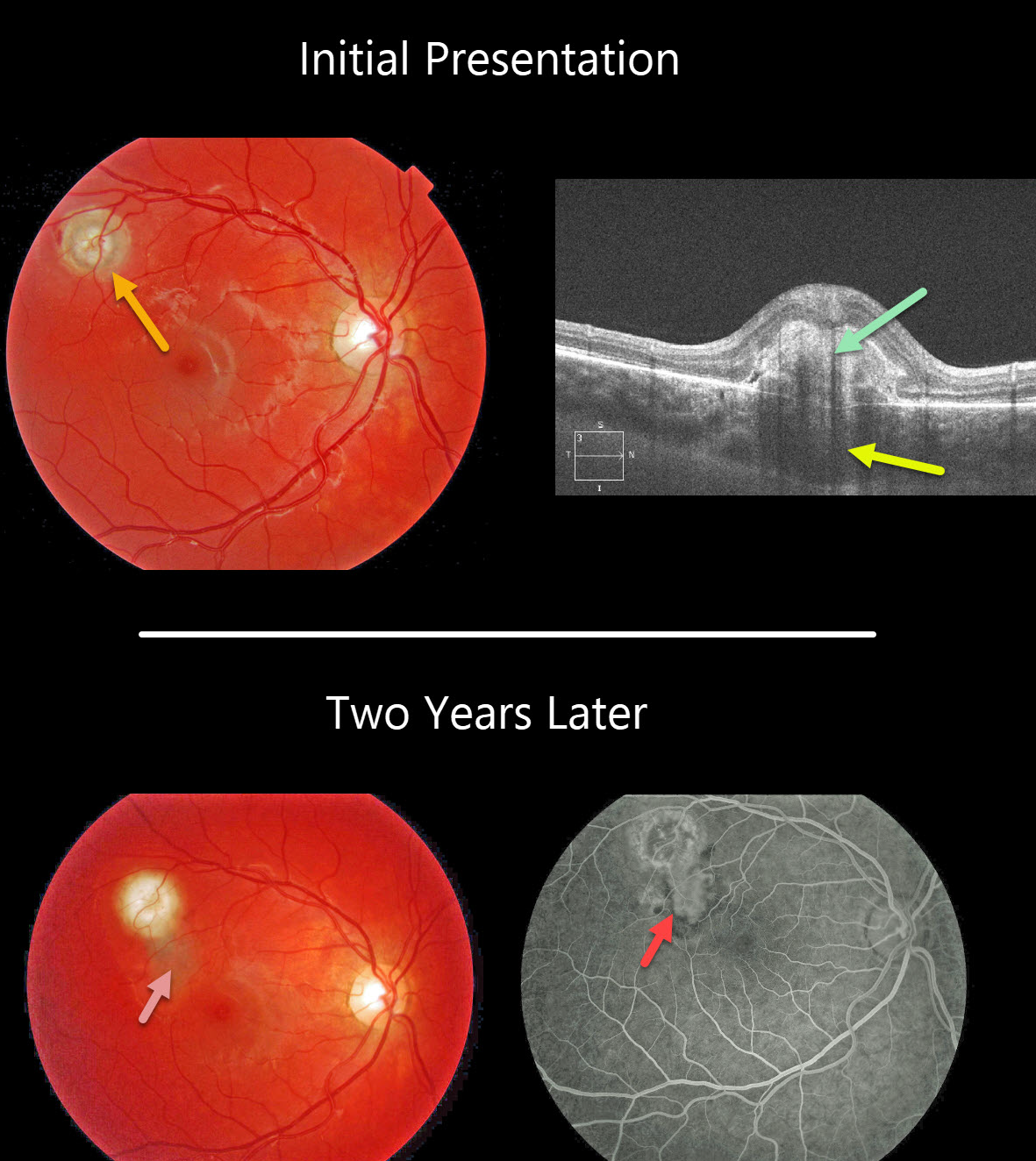
CHOROIDAL GRANULOMA
Originally posted on @retina.rocks April 20, 2020
This patient’s fundus photo shows a focal yellow-white nodular lesion of the right eye. OCT shows the subretinal hyperreflective material and an underlying choroidal granuloma.
Two years later, the patient subsequently developed an active macular neovascularization along the inferior edge of the original lesion, seen better on fluorescein angiography.
Learning Points:
Choroidal granulomas usually appear as solitary creamy white, round subretinal lesions. They are hyporeflective on OCT imaging (Invernizzi et al, Retina 2015;35:525-531). Causes include sarcoidosis, tuberculosis and VKH. They can occasionally cause exudation that responds to anti-VEGF injections or photodynamic therapy.
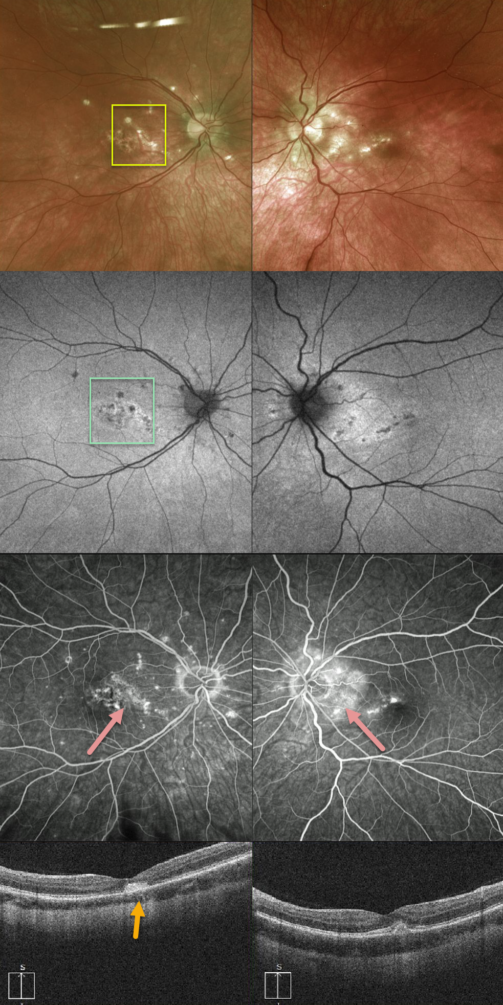
PUNCTATE INNER CHOROIDOPATHY (PIC)
Originally posted on @retina.rocks February 19, 2020
This 28-year-old patient has multifocal small bilateral macular scars with peripapillary scarring in her left eye.
The Optos photos show scattered small well-defined punctate lesions in the nasal macula with corresponding hypoautofluoresence.
Fluoroscein angiogram demonstrates early hypofluorescence of the punctate lesions with late leakage. OCT shows hump-shaped hyperreflective material with disruption of the ellipsoid zone and external limiting membrane (ELM).
Vision is counting fingers OD with inactive macular neovascularization (MNV) on chronic Eylea therapy.
Learning Points:
Punctate inner choroidopathy (PIC) is part of the multifocal choroiditis (MFC) spectrum and typically presents in young myopic females. It is never associated with vitritis.
MNV, the main cause of vision loss, can be successfully treated with anti-VEGF therapy.
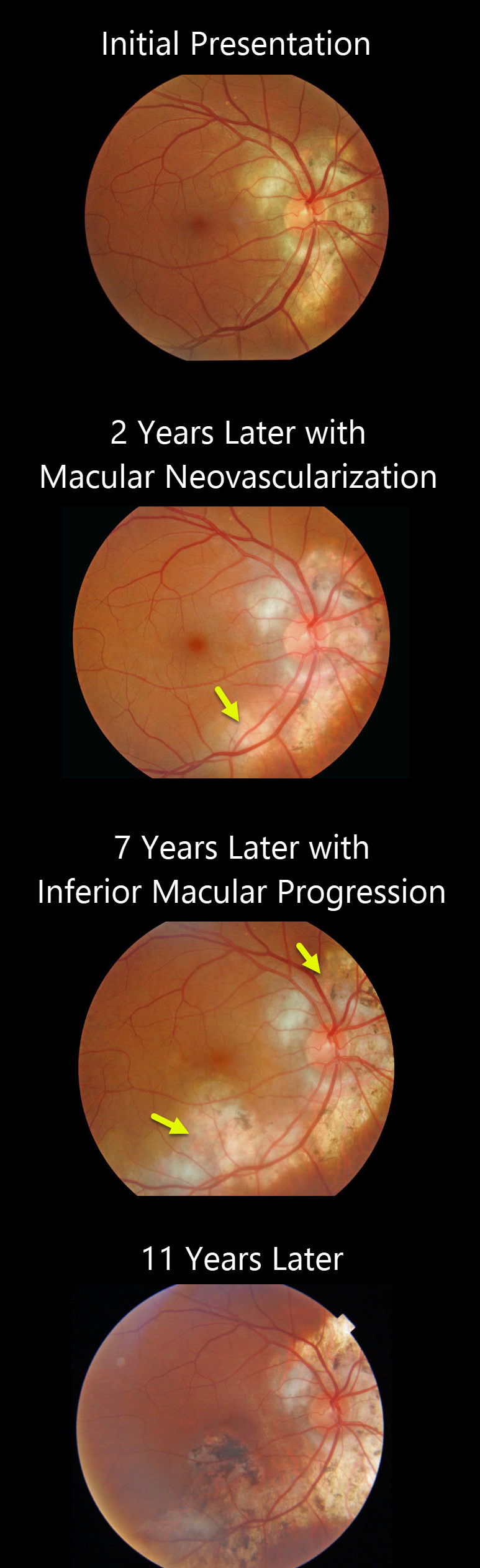
SERPIGINOUS CHORIORETINITIS
Originally posted on @retina.rocks January 22, 2020
This patient with serpiginous choroiditis shows typical progression of the inflammatory and neovascular process over an 11-year period. Ongoing intravitreal Avastin and Ozurdex injections so far have kept him with good central vision.
Learning Points:
Serpiginous choroiditis is an idiopathic, usually bilateral chorioretinal inflammatory condition most commonly found in middle-aged men.
Recurrent areas of inflammation develop along the edge of a previous scar, producing scarring that meanders from the optic nerve outward into the macula.
Vision is good unless scarring or macular neovascularization extends through the macular center.
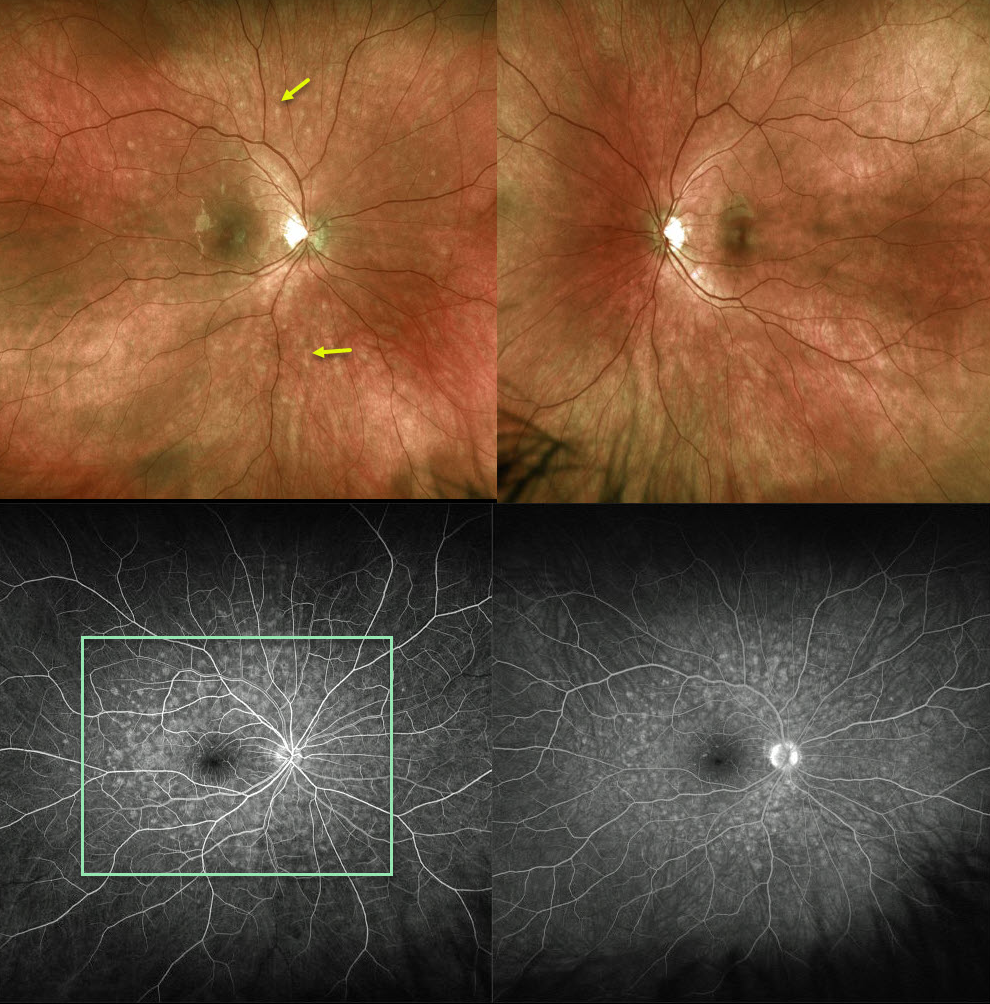
MULTIPLE EVANESCENT WHITE DOT SYNDROME (MEWDS)
Originally posted on @retina.rocks January 15, 2020
The Optos fundus photo of this 20YO female shows 100-200 micron outer retinal white spots scattered around the posterior pole, characteristic of multiple evanescent white dot syndrome (MEWDS).
Fluorescein angiography shows a wreath-like pattern of hyperfluorescence.
Learning Points:
MEWDS is an idiopathic, usually unilateral inflammation of the outer retina.
Most common in young women, patients often have a preceding viral syndrome and complain of blurred vision, photopsias, and temporal visual field loss.
Initially presenting with scatttered outer retinal white spots, the spots resolve within a few weeks making the diagnosis elusive. However, the enlarged blind spot often remains for months.
The condition is self-limited and usually resolves within several months without treatment.


