

12 Neoplastic Retina Disorders
<< Back to Cases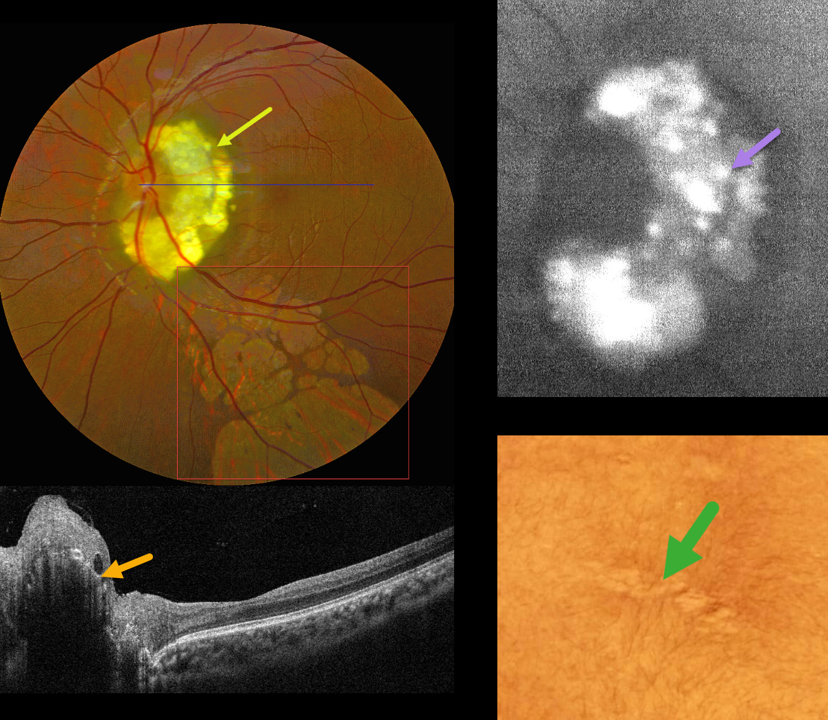
TUBEROUS SCLEROSIS WITH RETINAL ACHROMIC PATCHES AND RETINAL ASTROCYTIC HAMARTOMA
Kanwaljeet Harjot Madan
Originally posted on @retina.rocks September 4, 2026
This 17YO male presented with a known history of tuberous sclerosis, childhood seizures, and decreased vision in his left eye. Vision was 20/20 in his normal OD and 20/40 OS.
Color photography shows a glistening yellow-white inner peripapillary retinal lesion surrounding the superior, temporal, and inferior edge of the optic nerve, composed of innumerable semi-transparent spherules which hyper-autofluoresce. OCT scanning shows an elevated, hyperreflective inner retinal lesion with a hyporeflective intralesional cavity. An area of skin depigmentation (ash leaf spot) was noted on his lower back. There are numerous variably sized and variably confluent lobules of subretinal depigmentation extending from the macula inferiorly.
Learning Points:
The best-known retinal finding in tuberous sclerosis is retinal astrocytic hamartoma. Early in life, these inner retinal lesions have a fleshy white appearance and are often multifocal and bilateral. With time, as in this patient, they show a more classic “mulberry appearance” with significant calcification. The calcified lesions, as in our patient, hyper-autofluoresce while the flat, semitransparent lesions found earlier in life are hypoautofluorescent. Rarely, the vascular component can cause intraretinal exudation requiring treatment.
Retinal achromic patches, although much less described in the literature, are found in up to 39% of tuberous sclerosis patients (Rowley et al, Br J Ophthalmology 2001;85:420-423). They are thought to be analogous to the hypopigmented skin lesions and can appear in one or both eyes, singly or in multiples, and are typically located in the retinal periphery. Unliked astrocytomas, these lesions are flat and subretinal.
RETINOBLASTOMA
Malvika Singh, Akansha Sharma, Shraddha Raj Shrivastava, Anand Gandhi and Manish Nagpal
Originally posted on @retina.rocks August 17, 2026
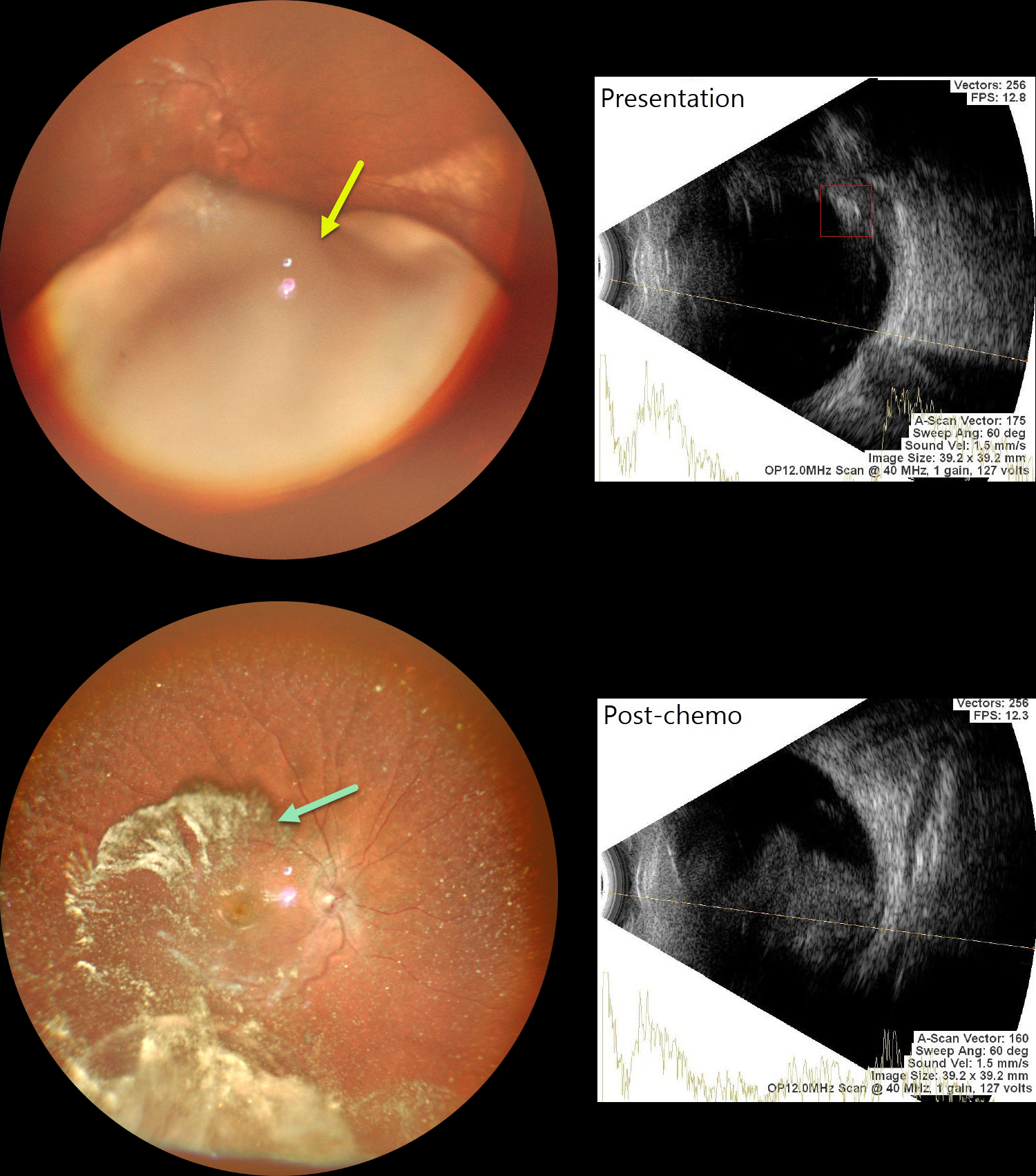
This previously healthy 2YO girl presented with leukocoria in her right eye. Family history was negative.
Examination under anesthesia revealed a large smooth endophytic lesion filling more than half of the vitreous cavity, extending into the inferior macula. B-scan ultrasonography revealed intralesional calcifications. The left eye was normal.
MRI showed no extra-ocular or CNS lesions. We diagnosed her with ICRB Group D retinoblastoma in the right eye. She underwent 3 cycles of chemotherapy.
Repeat examination under anesthesia, 9 weeks following initial presentation, showed marked tumor regression, although there was widespread significant vitreous seeding. MRI did not show any optic nerve infiltration or CNS spread. Enucleation of this eye was scheduled
Learning Points:
Retinoblastoma is the most common primary intraocular malignancy of childhood, with a peak incidence before age five. Presenting signs frequently include leukocoria and strabismus. The disease may be heritable or nonheritable, and delayed diagnosis can result in orbital extension, metastatic spread, and decreased survival. Early identification and intervention with chemotherapy, focal therapies such as laser photocoagulation or cryotherapy, and, in select cases, intra-arterial or intravitreal chemotherapy maximize the chances of ocular salvage and life preservation.
VASOPROLIFERATIVE TUMOR
Rohan Jain and Manish Nagpal
Originally posted on @retina.rocks July 20, 2026
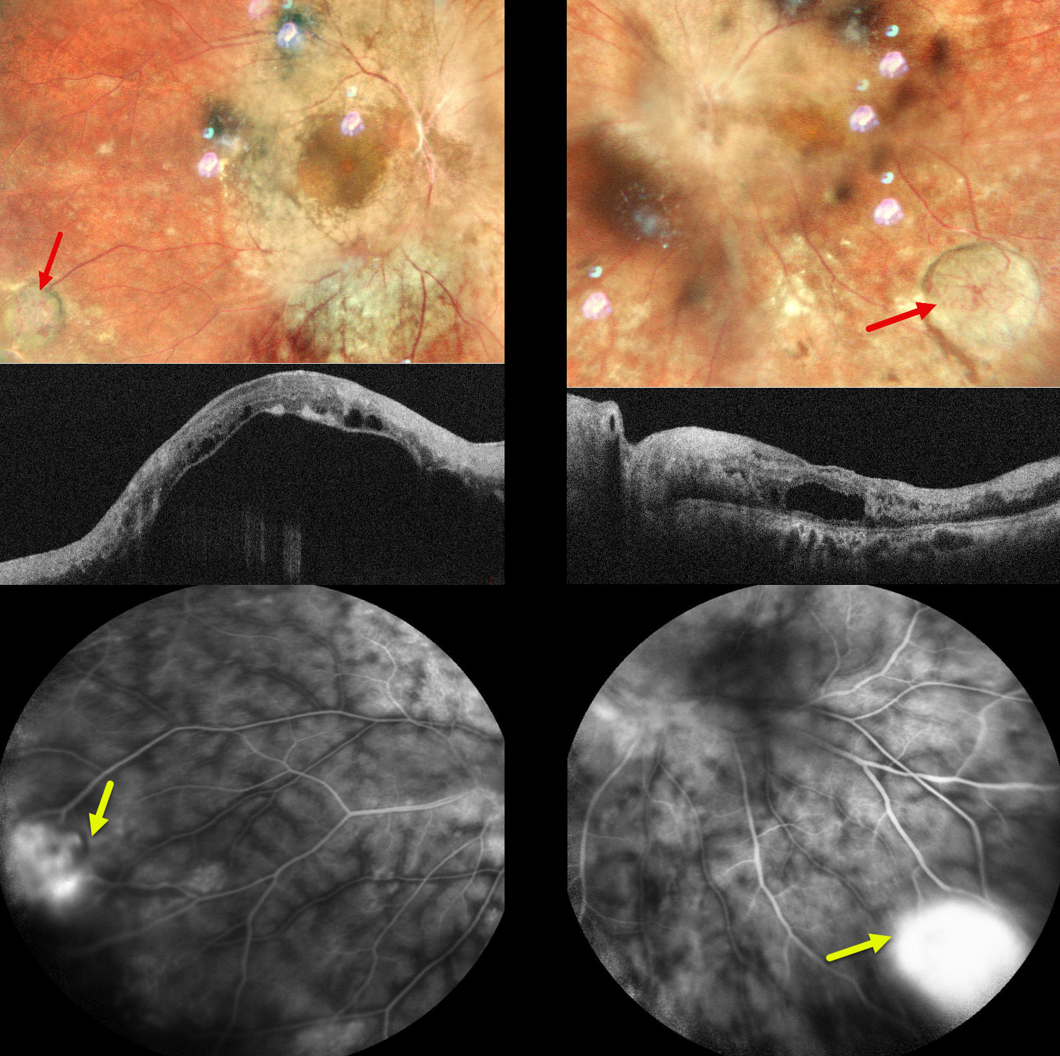
This 37YO female presented with a 10-day history of pain in both eyes and 9 years of poor vision OU. She was diagnosed elsewhere 10 years earlier with chronic vitritis, which was treated with oral steroids. Vision was counting fingers OU. There were bilateral posterior subscapular cataracts that were not considered visually significant.
Pseudocolor SLO imaging shows a yellow-white vascularized lesion in each inferotemporal periphery associated with non-dilated feeding and draining vessels. The central retinal detail is blurred due to the cataracts. However, the major vessels overlying the discs appear sheathed with possible disc swelling OS. Deep retinal whitening/opacification is noted in each posterior pole, which seems to spare the macular centers.
OCT shows an opaque, hyperreflective retina bilaterally. There is marked subretinal fluid OD with a large hyporeflective pocket of fluid in the outer left macula. These lesions leak on fluorescein angiography.
Learning Points:
Vasoproliferative tumors (VPTs) are reactive lesions characterized by vascular and glial proliferation, often triggered by intraocular inflammation. They appear yellow or red and can cause subretinal exudation and detachment, anterior or posterior segment neovascularization, and vitreous hemorrhage. VPTs may arise as primary idiopathic lesions or as a vascular response to various conditions, including intermediate uveitis, retinitis pigmentosa, Coats disease, and prior retinal detachment (Shields et al, JAMA Ophthalmology 2013;131:328–334). In uveitic VPTs, breakdown of the blood-retinal barrier and cytokine-mediated angiogenesis contribute to their development.
Despite their benign nature, VPTs can cause significant visual loss through complications such as macular exudation, cystoid macular edema, vitreous hemorrhage, and epiretinal membrane formation. Treatment options include thermal laser, cryotherapy, and/or anti-VEGF injections. Bilateral intravitreal anti-VEGF injections followed by direct thermal laser photocoagulation were recommended, but the patient was immediately lost to follow-up.
RETINAL CAPILLARY HEMANGIOMA
Ayushi Gupta and Vishal Agrawal
Originally posted on @retina.rocks June 29, 2026
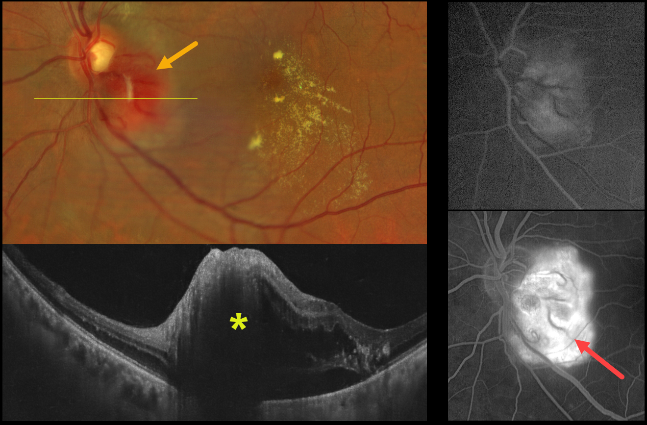
This healthy 51YO male presented with one month of vision loss in his left eye. Vision was 20/20 in his normal OD and 20/200 OS.
Color fundus photography shows an elevated orange-red juxtapapillary tumor along the inferotemporal edge of the optic nerve with associated macular lipid. OCT scanning reveals the lesion to be hyperreflective with associated outer retinal and subretinal fluid. The lesion leaked on fluorescein angiography.
Family history was negative for von Hippel–Lindau disease, and systemic evaluation, including brain MRI and renal ultrasonography, was unremarkable. Anti-VEGF therapy was recommended.
Learning Points:
Retinal capillary hemangiomas appear clinically as orange-red lesions, most commonly peripherally, and are associated with prominent, dilated, tortuous paired feeding and draining vessels. They can also appear on or around the optic nerve lesions, either on the surface (endophytic) as in our patient, or buried within the substance of the nerve (exophytic). Unlike peripheral lesions, juxtapapillary tumors often lack prominent feeder vessels. These lesions can cause severe vision loss or blindness from exudative retinal detachment and thus usually require aggressive treatment. Treatment options include thermal laser, photodynamic therapy, cryotherapy, and anti-VEGF injections.
More recently, oral belzutifan, an inhibitor of hypoxia-inducible factor 2 alpha, has emerged as a highly effective treatment for these lesions. In the LITESPARK-004, a Phase 2 trial, all 16 eyes improved without new lesions after a mean follow-up of 37 months (Wiley et al., Ophthalmology 2024;131;1324-1332).
Retinal capillary hemangioma is also a predominant feature of von Hippel-Lindau disease (VHL), an autosomal dominant condition caused by mutations in the VHL tumor suppressor gene. Patients develop benign and malignant tumors, including central nervous system hemangioblastoma, pheochromocytoma, and renal cell carcinoma. Rarely, sporadic, non-hereditary tumors may be found, as in our patient.
RETINAL ASTROCYTIC HAMARTOMA
European VitreoRetinal Society (EVRS) Dhaivat Shah, Nida Khan and Choithram Netralaya
Originally posted on @retina.rocks January 30, 2026
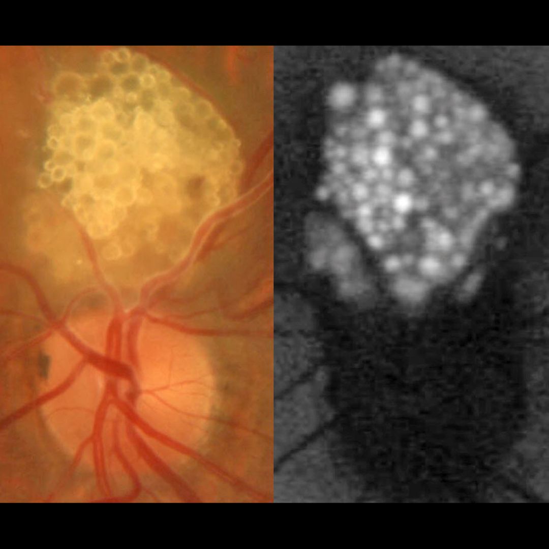
This healthy 43 YO male presented with an asymptomatic lesion in his left eye. Family history was negative. Vision was 20/20 in his normal OD and 20/20 OS.
Color photography shows a glistening yellow-white inner retinal lesion just superior to the optic nerve composed of innumerable semi-transparent spherules which hyper-autofluoresce.
Learning Points:
A retinal astrocytic hamartoma is a benign tumor composed of spindle-shaped fibrous astrocytes located in the nerve fiber layer, most commonly associated with tuberous sclerosis. Tuberous sclerosis includes the classic triad of seizures, mental deficiency and fibroangiomas, but patients can have lesions throughout the body including the eyes, skin (ash-leaf spots), bone, nails, teeth, brain, heart, lungs and kidneys. Early in life the inner retinal lesions have a fleshy white appearance and are often multifocal and bilateral. With time, as in this patient, they show a more classic “mulberry appearance” with significant calcification. Rarely, the vascular component can cause intraretinal exudation requiring treatment.
Clinically retinocytomas can resemble astrocytic hamartomas. However, retinocytomas often have underlying RPE changes and chorioretinal scarring (lacking in our patient), which are absent in astrocytic hamartomas.
Since the patient was self-paying, neurologic imaging was not performed and observation was recommended. A neurology reference was given.
RETINOBLASTOMA
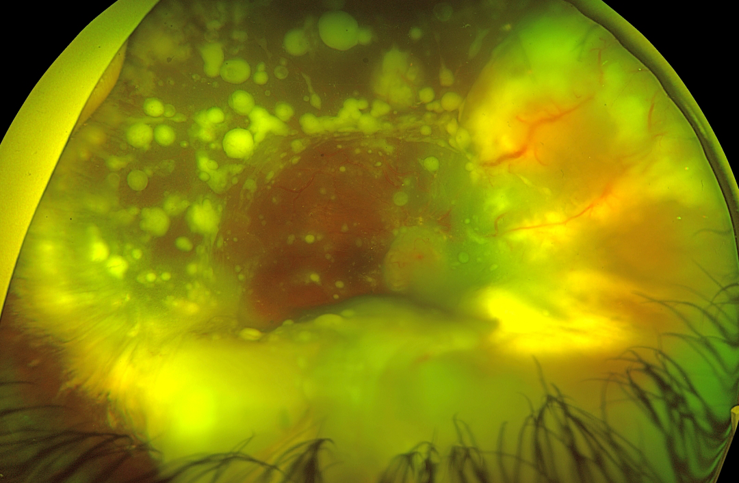
Victor Ribeiro de Sant'Ana, Thailor Dartora, Laísa Ferraz de Arruda, Gabriela Mousse de Carvalho and Rodrigo Jorge
Originally posted on @retina.rocks December 8, 2025
RETINOCYTOMA
The European VitreoRetinal Society and Dibya Prabha
Originally posted on @retina.rocks November 7, 2025
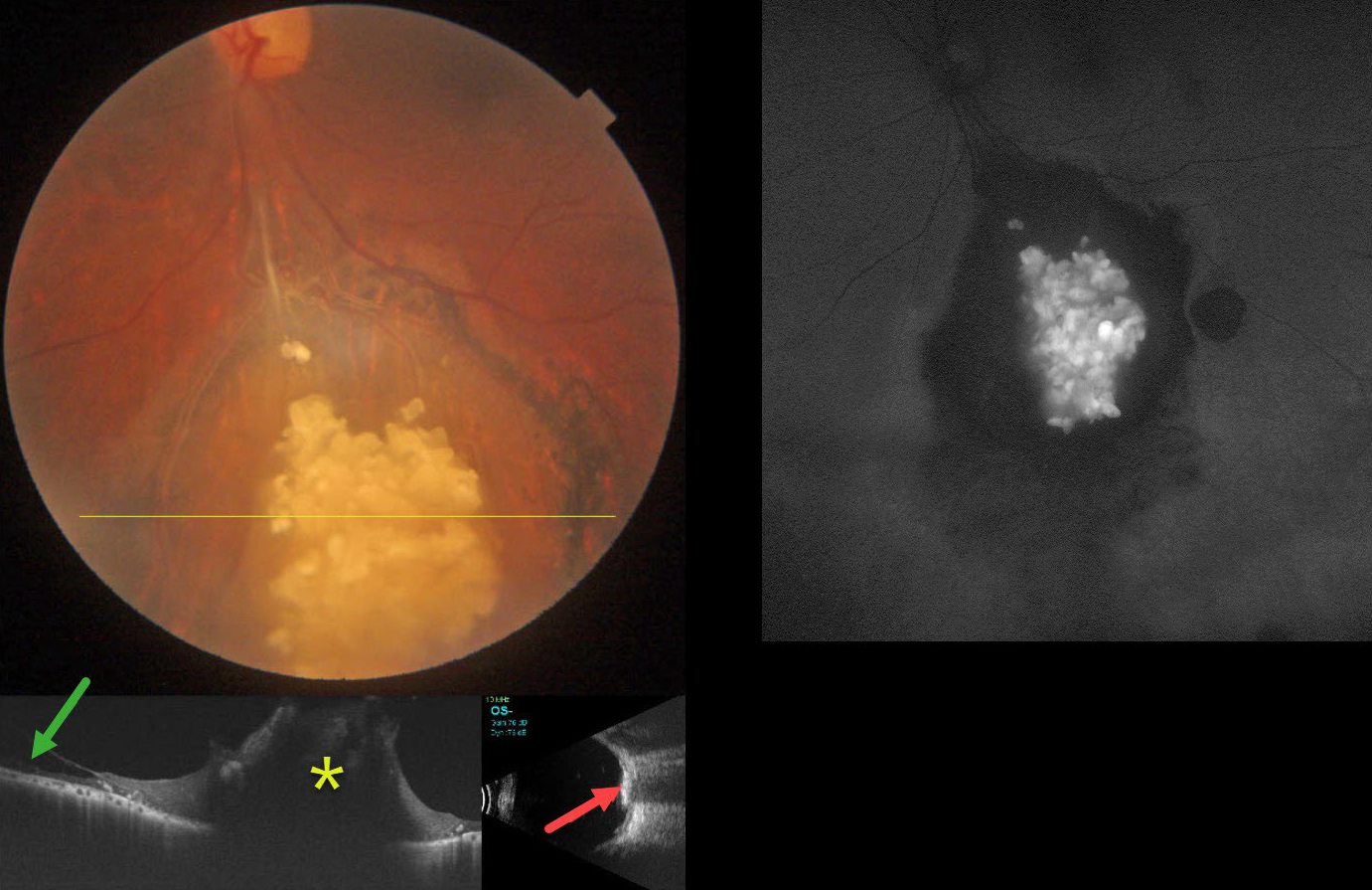
This healthy 43YO male presented with gradual bilateral blurring. Vision was 20/20 in his normal OD and 20/30 OS.
Color photography shows an elevated, glistening, inner retinal yellow-white mulberry-like lesion in the inferior midperiphery. Surrounding subretinal pigmentary changes are noted. OCT through this lesion shows a markedly thickened, disorganized, hyperreflective retina. Retinoschisis is noted nasally. The lesion is markedly hyperreflective on B-scan ultrasonography and hyper-autofluorescent on fundus autofluorescence.
Learning Points:
Retinocytomas are fleshy, opalescent, benign tumors that are felt to represent spontaneously regressed retinoblastoma (RB). They appear identical to previously radiated RB tumors. The risk of a germline RB1 mutation in sporadic unilateral retinoblastoma or retinocytoma is about 10-20%.
Clinically, retinocytomas can resemble astrocytic hamartomas. However, retinocytomas often have underlying RPE changes and chorioretinal scarring (as in our patient), which would not be present with astrocytic hamartomas.
The brain MRI was normal, and observation was recommended.
RETINAL CAVERNOUS HEMANGIOMA
Addy Adkisson
Originally posted on @retina.rocks October 20, 2025
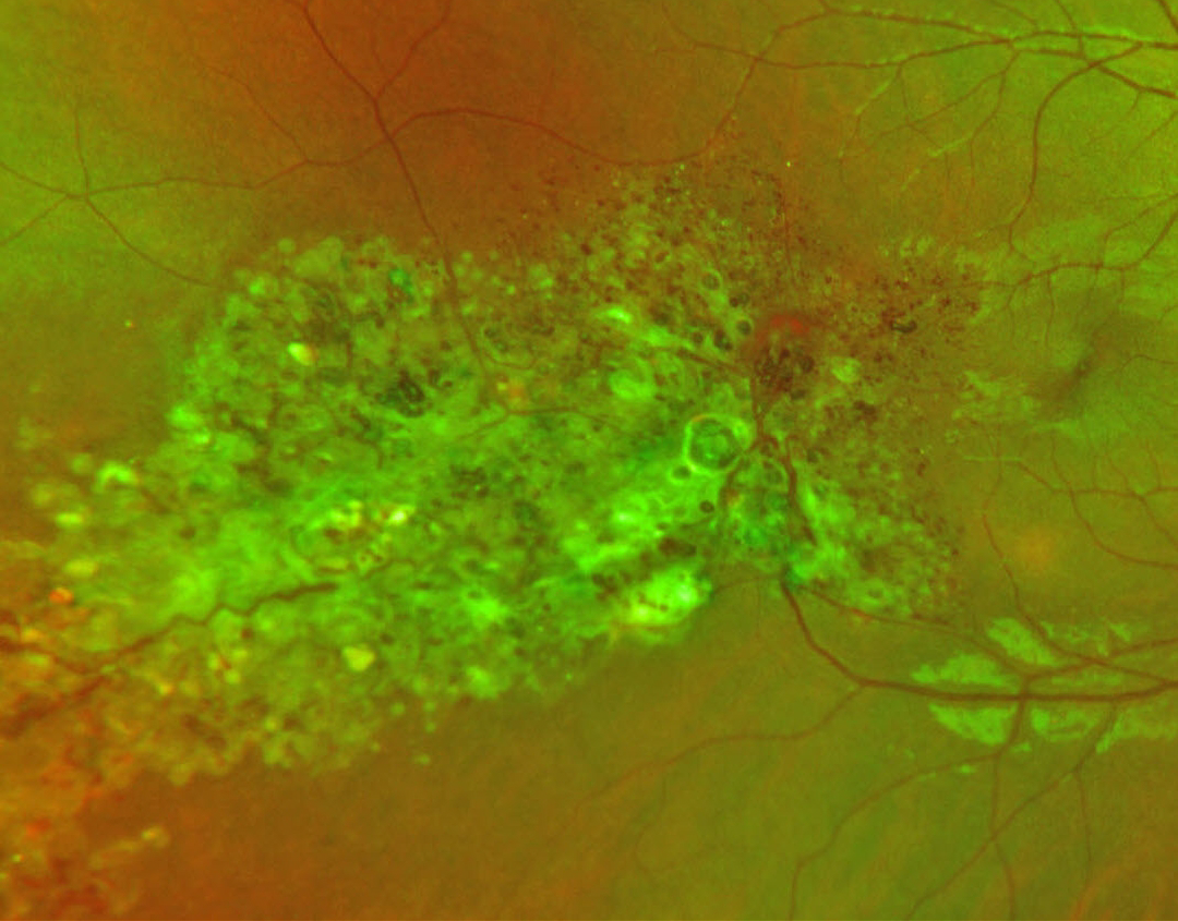
This healthy 25YO female presented with an asymptomatic retinal lesion in her right eye. She recalled being diagnosed with a retinal finding in the past and reported negative prior neurologic imaging. Vision was 20/20 bilaterally.
Optos RG color imaging shows extensive confluent variably sized grape-like clusters of aneurysms filled with dark venous blood. Some scattered gray fibrotic membranes cover its superficial surface.
Learning Points:
Retinal cavernous hemangiomas are usually unilateral, benign vascular abnormalities. Fluorescein angiography shows plasma-erythrocyte layering within these saccular lesions caused by sluggish blood flow. Vision is usually normal, and no treatment is required. Rarely, vision loss can occur from vitreous hemorrhage. Although most occur sporadically, some may be associated with an autosomal dominant neurocutaneous syndrome, including cutaneous and central nervous system hemangiomas. Neurologic imaging is therefore recommended.
Unfortunately, our patient was immediately lost to follow-up.
RETINAL CAVERNOUS HEMANGIOMA
The European VitreoRetina Society (EVRS), Nassim Abreu Carolina Galvá and Patricia Pérez
Originally posted on @retina.rocks September 12, 2025
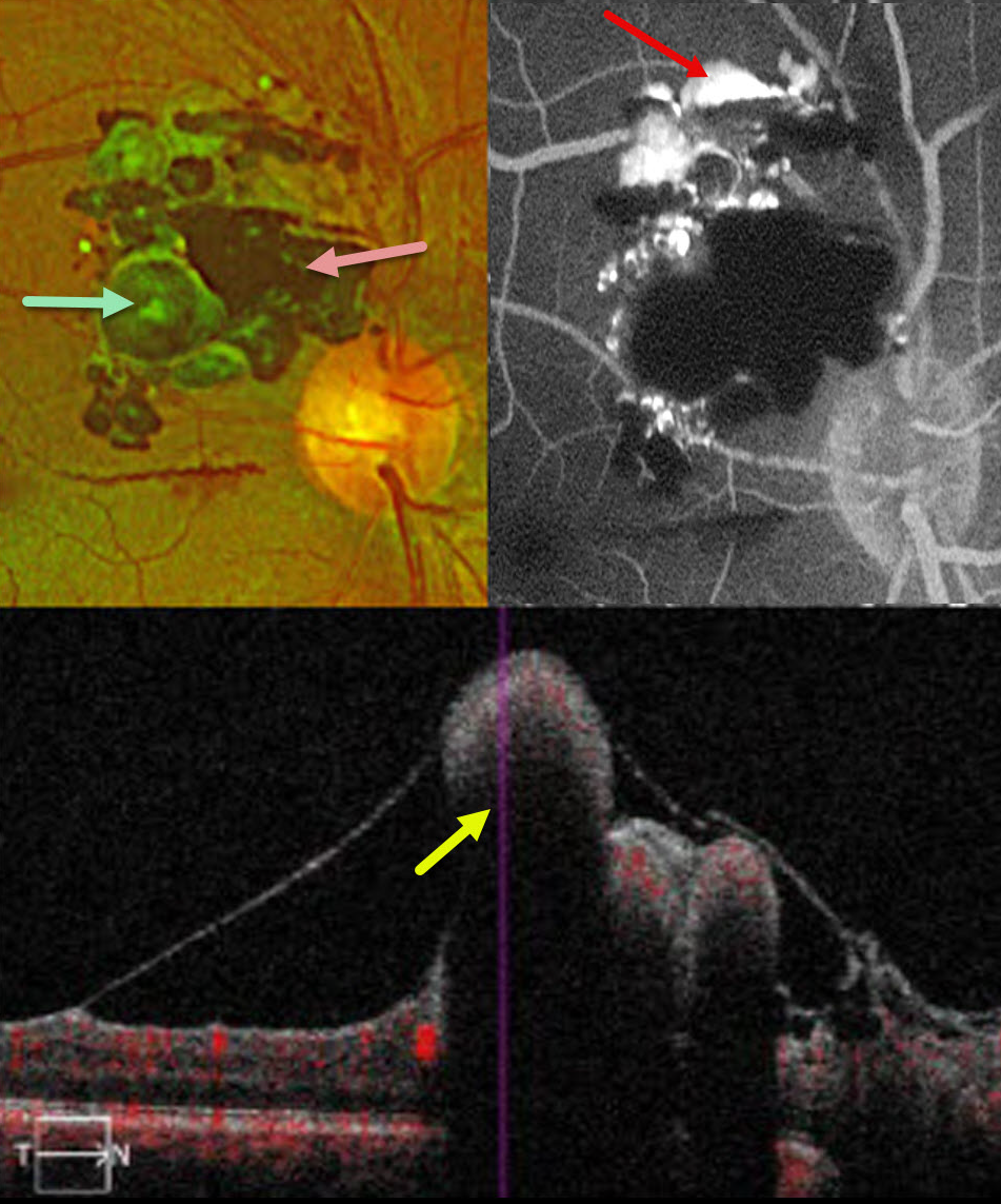
This 32YO male presented with several weeks of decreased vision in the right eye. Vision was 20/200 OD and 20/20 in his normal OS. Examination revealed a dense vitreous hemorrhage, initially attributed to Valsalva retinopathy. He was immediately lost to follow-up and came back six years later with complaints of recent blurred vision with floaters. Vision was 20/50.
Optos color RG imaging shows a multilobulated mass superotemporal to the optic nerve, consisting of large saccular aneurysms with some overlying gliosis and preretinal blood. Fluorescein angiography shows superior layering of fluorescein within the vascular lesions, with blockage by blood. On OCT, the vascular abnormalities protrude into the vitreous with posterior shadowing.
Learning Points:
Retinal cavernous hemangiomas are rare, benign vascular lesions composed of thin-walled saccular venous aneurysms resembling clusters of grapes. Although most are asymptomatic and discovered incidentally, vision loss can occur from macular involvement or vitreous hemorrhage. Fluorescein angiography reflects the sluggish blood flow, classically showing plasma-erythrocyte layering as in our patient.
Although most are sporadic, some may be associated with an autosomal dominant neurocutaneous syndrome, including cutaneous and central nervous system hemangiomas. Neurologic imaging is therefore recommended, and our patient’s MRI results are pending.
RETINAL CAPILLARY HEMANGIOMA
The European VitreoRetina Society (EVRS), Ece Ozdemir Zeydanli and Mehmet Onen
Originally posted on @retina.rocks August 22, 2025
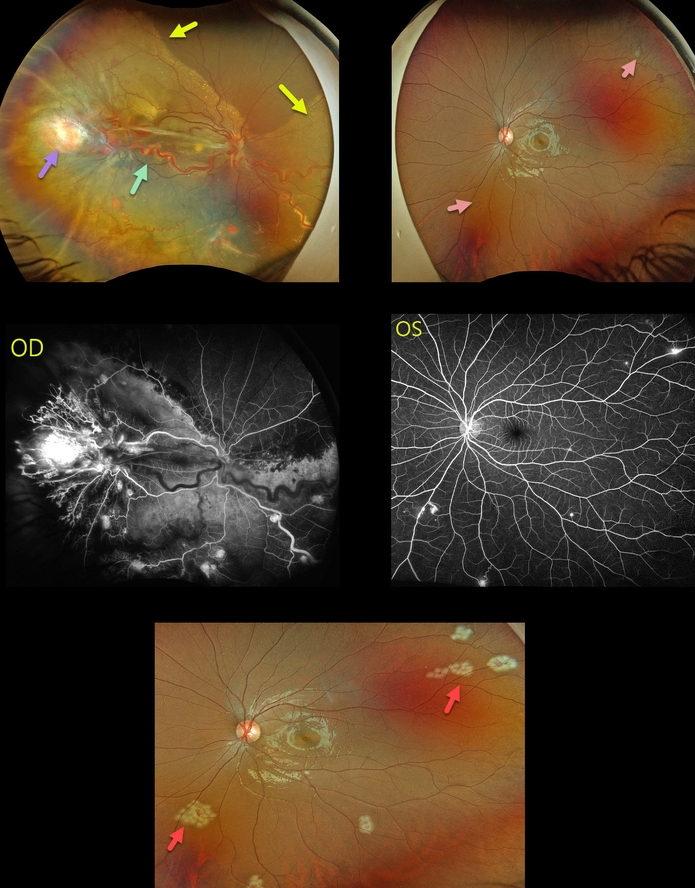
This 17YO female presented with a 3-month history of vision loss in her right eye. Vision was hand motion OD and 20/20 OS.
Optos color RGB imaging OD shows a large yellow-red angioma in the temporal periphery, associated with large dilated and draining vessels. There is another large angioma nasally, near the ora serrata, which is not well imaged. Multiple smaller angiomas are scattered throughout the temporal and inferior retina. A near-total exudative retinal detachment (RD) spares the superonasal retina. A few small peripheral angiomas are noted in her left eye. All tumors leaked angiographically.
The angiomas in the left eye were lasered during her initial examination. She then underwent lens-sparing vitrectomy in the right eye with membrane removal, endophotocoagulation, feeder vessel endodiathermy, and endoresection of the large temporal and nasal tumors. Mitomycin C was applied outside the posterior pole to reduce the risk of PVR, and silicone oil tamponade was used.
Our patient’s family history was negative. Genetic testing was subsequently positive for the von Hippel-Lindau (VHL) gene, which likely represents a de novo mutation.
Learning Points:
Retinal capillary hemangiomas are orange-red lesions associated with a prominent paired feeding and draining vessel. These benign vascular tumors need to be aggressively treated since they can cause severe vision loss or blindness from exudative RD, as in our patient. Patients require lifelong screening examinations for the development of new tumors even after successful treatment. Ultrawidefield imaging is helpful in detecting and monitoring these lesions.
Retinal capillary hemangioma is a predominant feature of VHL, an autosomal dominant condition caused by mutations in the VHL tumor suppressor gene. Patients develop benign and malignant tumors, including central nervous system hemangioblastoma, pheochromocytoma, and renal cell carcinoma.
BENIGN LOBULAR INNER NUCLEAR LAYER PROLIFERATIONS (BLIP) + BEAR TRACKS
Sharat Hegde
Originally posted on @retina.rocks April 30, 2025
RETINOBLASTOMA
Ankit Jain and Manish Nagpal
Originally posted on @retina.rocks March 24, 2025
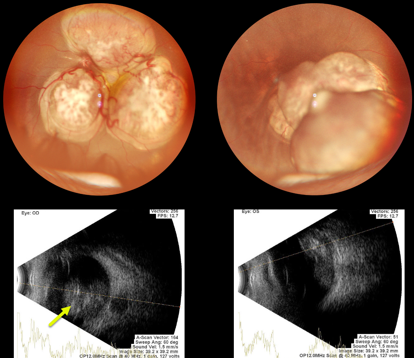
This 8-month-old girl presented with bilateral nystagmus, leukocoria, and inability to focus on objects. Family history was completely negative.
Examination under anesthesia revealed bilateral lobular white retinal masses projecting into each vitreous. B-scan ultrasonography shows intralesional hyperreflectivity suggestive of calcification. She was immediately referred to ocular oncology.
Learning Points:
Retinoblastoma (RB) is the most common primary intraocular tumor of childhood. The RB1 gene, located on chromosome 13 (13q14.2), encodes a tumor suppressor protein. The vast majority of cases have no family history. Bilateral and multifocal RB occurs in 20-35% of patients and is virtually always due to a germinal mutation. These patients are also at risk for secondary tumors, including pinealoblastoma.
Treatment options include systemic, intra-arterial, and intravitreal chemotherapy; focal therapy (laser photocoagulation, cryotherapy, plaque, or external beam radiotherapy); and enucleation. We assume our patient has a new germline mutation given the negative family history.
RETINAL CAPILLARY HEMANGIOMA
Rohan Jain and Manish Nagpal
Originally posted on @retina.rocks January 20, 2025
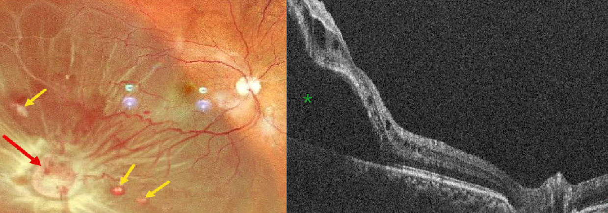
This 52YO male presented with 5 days of vision loss OD. There was a history of bilateral vision loss following facial trauma from a motor vehicle accident 20 years earlier. He denied any family history of eye disease or cancer. Vision was counting fingers OD and 20/200 OS.
Pseudocolor SLO imaging shows an orange-yellow mass consistent with a retinal capillary hemangioma in the inferotemporal midperiphery, causing an exudative retinal detachment. Several smaller hemangiomas are noted. The macula-off detachment is confirmed on OCT scanning.
A recent MRI showed bilateral optic atrophy, a partially empty sella, and an old, healed fracture of the right lateral wall of the orbit. Although emergent vitrectomy surgery was recommended, he was immediately lost to follow-up.
Learning Points:
Retinal capillary hemangiomas are orange-red lesions associated with a prominent paired feeding and draining vessel. These benign vascular tumors need to be aggressively treated since they can cause severe vision loss or blindness from exudative retinal detachment, as in our patient. The presence of hydration outer retinal folds is one of the key findings that help differentiate rhegmatogenous from exudative and traction retinal detachments (Oquendo et al, AJO 2024;268:212-221). The absence of these folds in our patient is typical of an exudative detachment. Common treatment options include thermal laser, cryotherapy, and anti-VEGF injections.
Retinal capillary hemangioma is a predominant feature of VHL, an autosomal dominant condition caused by mutations in the VHL tumor suppressor gene. Patients develop benign and malignant tumors, including central nervous system hemangioblastoma, pheochromocytoma, and renal cell carcinoma.
RETINOCYTOMA
Originally posted on @retina.rocks December 9, 2024
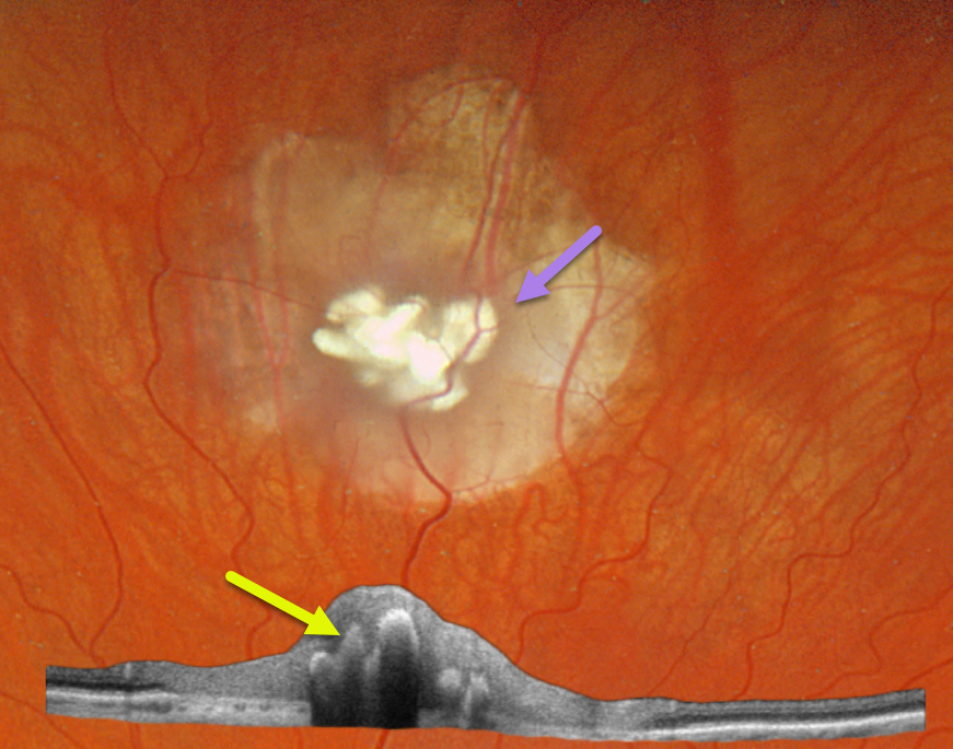
This healthy 30YO male was referred for an asymptomatic retinal lesion in his left eye. There was no family history of eye disease. Vision was 20/25 OU.
Optos color RGB imaging shows a nodular, calcified retinal lesion with surrounding atrophic chorioretinal scarring. Topcon swept-source OCT scanning shows a disorganized retina overlying the hyperreflective calcifications, which shadow posteriorly.
Learning Points:
Retinocytomas are fleshy, opalescent, benign tumors that are felt to represent spontaneously regressed retinoblastoma (RB). They appear identical to previously radiated RB tumors. As with typical RB, these patients’ offspring are at 50% risk of developing RB and thus require genetic counseling.
Clinically, retinocytomas can resemble astrocytic hamartomas. However, retinocytomas often have underlying RPE changes and chorioretinal scarring (as in our patient), which are not present in astrocytic hamartomas.
Brain MRI scanning was normal. Genetic testing for the retinoblastoma (RB1) and tuberous sclerosis (TSC1/TSC2) genes was suggested, but the patient denied this for now due to cost concerns.
BEAR TRACKS WITH BENIGN LOBULAR INNER NUCLEAR LAYER PROLIFERATIONS
Sharat Hegde
Originally posted on @retina.rocks November 13, 2024
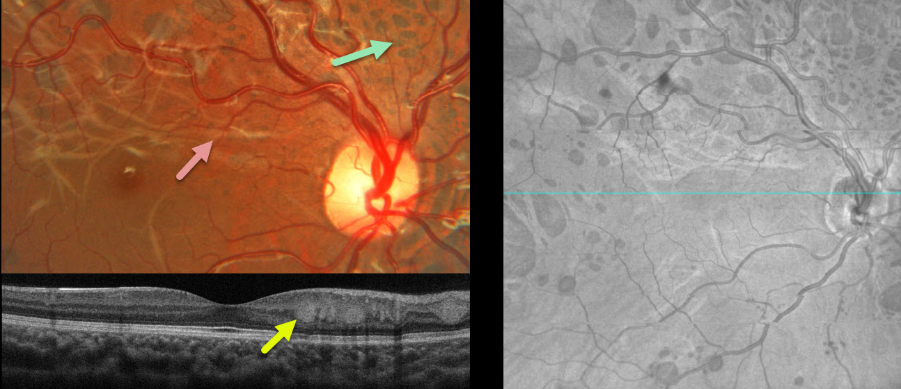
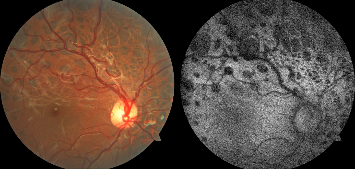
This healthy 10YO female presented with these asymptomatic bilateral lesions. Vision was 20/20 OU.
Fundus photography of her right eye shows multiple bear track lesions scattered throughout the posterior pole. An interlacing pattern of whitish deep retinal lines courses through the macula. The pigmented lesions are hypo-autofluorescent. Similar findings were noted in the left eye (not shown).
Learning Points:
Group-type congenital pigmented nevi of the RPE (bear tracks) is a relatively rare congenital condition characterized by well-demarcated, hyperpigmented, flat, variably sized RPE lesions that resemble bear footprints. Histopathologically, these lesions are similar to congenital hypertrophy of the RPE (CHRPE), with increased numbers of pigment granules in normal-sized RPE cells. These lesions are benign and cause no visual symptoms.
The major fluorophore in fundus autofluorescence (FAF) is lipofuscin, which is intraliposomal RPE material generated as byproducts of outer segment metabolism. The outer retina overlying these bear track lesions shows varying degrees of atrophy. This leads to less lipofuscin production with hypo-FAF.
Although bear tracks and the typical CHRPE lesions (large, round, unilateral, with depigmented lacunae with age) are not associated with systemic abnormalities, the multifocal, often bilateral, and small comet-shaped lesions with familial adenomatosis are associated with colonic polyps and cancer (Romania et al Ophthalmology 1992;99:911-913).
Benign lobular inner nuclear layer proliferations were described by Sanfilippo et al in 2023 as a unique benign lesion often associated with CHRPE lesions (Ophthalmology 2023;130:265-273). They appear as white, multifocal, often lobular tumors centered in the posterior pole. On OCT, they appear in the inner nuclear layer at the border of the outer plexiform layer.
PSCRAP (PRESUMED SOLITARY CIRCUMSCRIBED RETINAL ASTROCYTIC PROLIFERATION)
Mattie Adams
Originally posted on @retina.rocks October 7, 2024
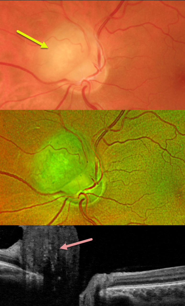
This healthy 41YO female was referred for an asymptomatic lesion in her left eye. Vision was 20/25 in her normal right eye and 20/25 in her left eye. Ocular examinations were normal except for a round nodular amelanotic elevated lesion abutting (and possibly involving) the left superonasal optic nerve. Swept-source OCT shows diffuse hyperreflectivity of the lesion with posterior shadowing.
Learning Points:
pSCRAP (presumed solitary circumscribed retinal astrocytic proliferation) was originally described by Shields et al as a unique lesion found in adulthood that resembles a retinal astrocytoma or retinoma (Arch Ophthalmology 2011;129:1189-1194). Although originally felt to arise from the inner retina, it is now thought to be either a deep retinal glial lesion or a form of RPE fibrous metaplasia (Shields et al, Retinal Cases & Brief Reports 2017;11:18-23).
RETINAL CAPILLARY HEMANGIOMA
Originally posted on @retina.rocks October 1, 2024
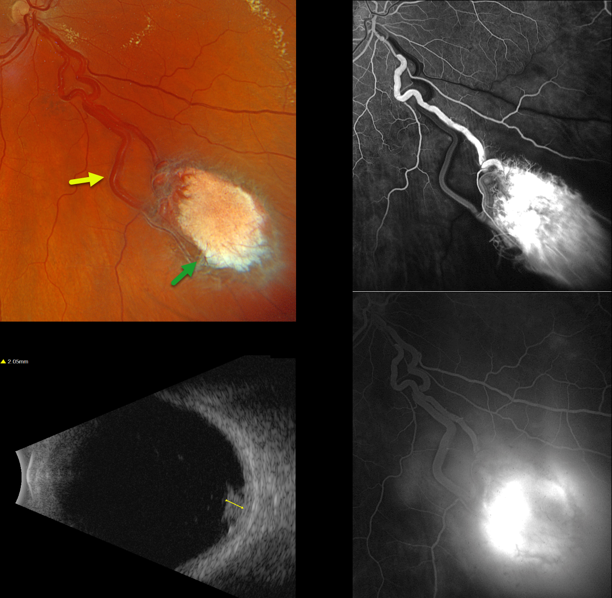
This healthy 42YO male was referred for an asymptomatic retinal lesion in his right eye. There was no family history of cancer or eye disease, although he had no knowledge about his birth father. Vision was 20/30 OD and 20/20 in his normal left eye.
Optos color RGB imaging shows an active endophytic retinal capillary hemangioma (RCH) in the inferonasal midperiphery. The tumor is supplied and drained by dilated, irregular vessels. The lesion shows profound leakage on fluorescein angiography. It measured about 2mm thick on B-scan ultrasonography. Genetic testing for the von Hippel-Lindau syndrome (VHL) gene was negative, as was MRI scanning of the brain, abdomen, and pelvis. Monthly intravitreal Avastin injections are planned to shrink the tumor, followed by thermal laser photocoagulation.
Learning Points:
Retinal capillary hemangiomas are orange-red lesions associated with a prominent paired feeding and draining vessel and can cause severe vision loss or blindness from exudative retinal detachment. These benign vascular tumors need to be aggressively treated. Common treatment options include thermal laser, cryotherapy, and anti-VEGF injections.
They are a predominant feature of VHL, an autosomal dominant condition caused by mutations in the VHL tumor suppressor gene. This prevents the degradation of hypoxia-inducible factor 1a (HIF-1a), which, in turn, causes the production of numerous growth factors, including VEGF (Kaelin, Drug Discoveries Today Disease Mechanisms 2005;2:225-231). Patients develop benign and malignant tumors, including central nervous system hemangioblastoma, pheochromocytoma, and renal cell carcinoma. Sporadic RCHs in the absence of the VHL syndrome are a rare occurrence (Singh et al, Ophthalmology 2001;108:1907-1911). The negative genetic testing fortunately ruled out VHL in our patient. Our patient’s lesion most likely resulted from a localized double-hit retinal VHL mutation.
RETINAL CAPILLARY HEMANGIOMA
Originally posted on @retina.rocks August 19, 2024
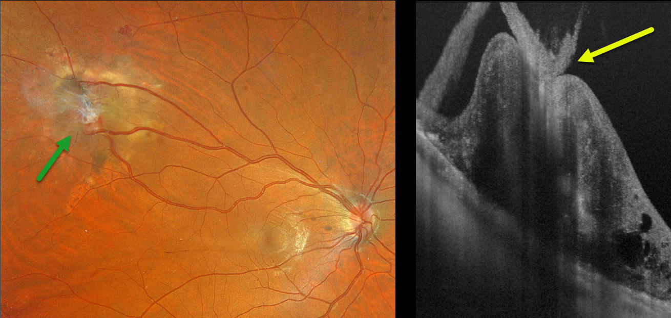
This 28YO male with a known history of Von Hippel-Lindau (VHL) recently moved to our area. He received prior anti-VEGF injections and thermal laser for a retinal capillary hemangioma in his right eye.
Optos color RGB imaging shows a regressed endophytic capillary hemangioma in the superotemporal midperiphery. The tumor is supplied and drained by dilated, irregular vessels. On Silverstone swept-source OCT, the angioma appears as a thickened, hyperreflective lesion with overlying vitreous traction.
Learning Points:
Retinal capillary hemangiomas are orange-red lesions associated with a prominent paired feeding and draining vessel and can cause severe vision loss or blindness from exudative retinal detachment. These benign vascular tumors need to be aggressively treated. Common treatment options include thermal laser, cryotherapy, and anti-VEGF injections.
Retinal capillary hemangioma is a predominant feature of VHL, an autosomal dominant condition caused by mutations in the VHL tumor suppressor gene. Patients develop benign and malignant tumors, including central nervous system hemangioblastoma, pheochromocytoma, and renal cell carcinoma.
We plan on following our patient several times per year to monitor for new lesions or recurrent exudation.
RETINAL CAPILLARY HEMANGIOMA
Yuenpang Cheung and Stephanie Choi
Originally posted on @retina.rocks December 19, 2023
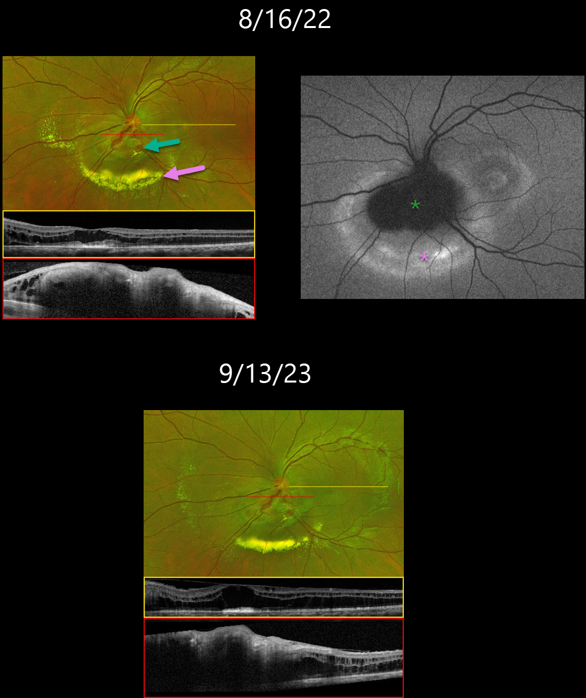
This 23YO female was noted by her optometrist in 2019 to have vision loss from a retinal lesion. At presentation, vision was 20/400 in her left eye and 20/20 in the right eye.
She was referred to an outside retina specialist, who gave a single anti-VEGF injection and then referred her to Columbia University. Our initial examination on 8/16/22 revealed an inferior peripapillary retinal capillary hemangioma with surrounding exudation. OCT of the macula revealed mostly outer retinal fluid. The angioma was diffusely thickened and hyperreflective.
On fundus autofluorescence (FAF), the angioma was hypo-FAF with a surrounding rim of hyper-FAF. Genetic testing for the VHL gene was negative, and thus further systemic work-up of the brain and kidneys was deferred.
She underwent subsequent monthly anti-VEGF injections with resolving exudates. On 9/13/23, the lipid decreased, but she continues to have persistent fluid and thickening on OCT.
Learning Points:
Retinal capillary hemangiomas appear clinically as orange-red lesions, most commonly peripherally, and are associated with prominent, dilated, tortuous paired feeding and draining vessels.
They can also appear on or around the optic nerve, either on the surface (endophytic), as in our patient, or buried within the substance of the nerve (exophytic).
These lesions can cause severe vision loss or blindness from exudative retinal detachment and thus usually require aggressive treatment. Treatment options include thermal laser, photodynamic therapy, cryotherapy, and anti-VEGF injections.
Retinal capillary hemangioma is also a predominant feature of von Hippel-Lindau disease (VHL), an autosomal dominant condition caused by mutations in the VHL tumor suppressor gene. Patients develop benign and malignant tumors, including central nervous system hemangioblastoma, pheochromocytoma, and renal cell carcinoma.
ASTROCYTIC HAMARTOMA
Yuenpang Cheung, Stephanie Choi and Brian Marr
Originally posted on @retina.rocks December 5, 2023
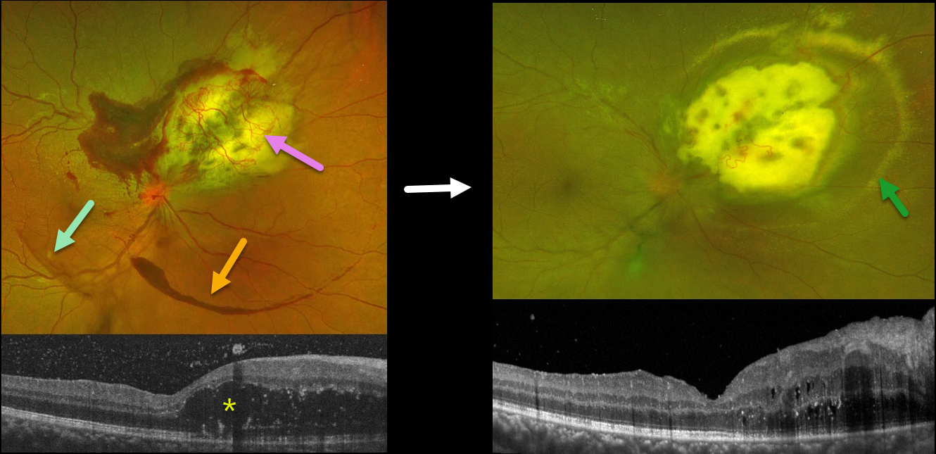
This 27YO female has been followed for an exudative astrocytic hamartoma in her right eye. Vision is 20/20 bilaterally. She was diagnosed with tuberous sclerosis at age 5 and has been followed closely by neurology.
Optos color RG imaging shows a superonasal yellow-and-white lesion with overlying neovascularization and exudates, along with areas of vitreous and subhyaloid blood.
OCT shows significant mostly outer retinal cystoid edema nasal to the fovea, as well as some vitreous blood.
She received a series of intravitreal Avastin injections with modest response and was more recently switched to Vabysmo injections. Two weeks following her third Vabysmo injection, the overlying neovascular network and blood are nearly resolved, but the surrounding subretinal lipid is increasing. The cystoid macular edema has mildly improved.
This case was submitted by Yuenpang Cheung, Stephanie Choi, and Brian Marr.
Learning Points:
Tuberous sclerosis is an autosomal dominant condition caused by mutations in the tumor suppressor genes TSC1 or TSC2. Tuberous sclerosis has multiple systemic manifestations affecting the brain (cortical tubers, subependymal nodules, epilepsy), skin (facial angiofibromas, shagreen patch, ash leaf spots), heart (cardiac rhabdomyoma), and the kidneys (angiomyolipomas).
The primary ocular manifestation is a retinal hamartoma arising from glial astrocytes. Retinal hamartomas can present as subtle gray areas in the nerve fiber layer without calcification or as prominent calcified nodules arising from the inner surface of the retina with a mulberry or fish egg-like appearance.
Although patients usually remain asymptomatic, these benign tumors can enlarge and cause macular edema, serous retinal detachment, or vitreous hemorrhage.
Exudative treatment options include anti-VEGF injections, photodynamic therapy, transpupillary thermotherapy, and radiation therapy. In cases with suboptimal response, reports of systemic mTOR inhibitors, such as Sirolimus, have shown some benefit.
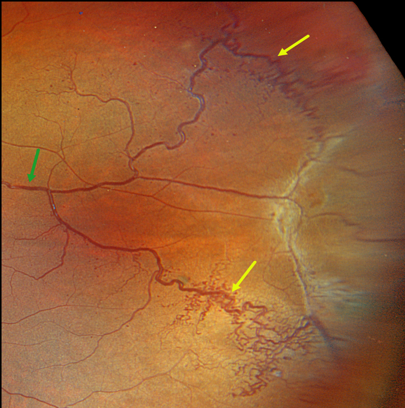
ATYPICAL RETINAL CAVERNOUS HEMANGIOMA
Originally posted on @retina.rocks June 13, 2023
This healthy 5YO boy was referred for an asymptomatic retinal lesion in his left eye. Vision was 20/25 OU.
Optos color RGB imaging shows unusual temporal changes involving the venous vascular tree. Prominent dilated and tortuous retinal veins drain the supero- and inferotemporal periphery before merging into a single vessel more posteriorly.
Surrounding these larger veins, especially inferiorly, are networks of small, corkscrew venules, along with more scattered tiny red microaneurysmal-like structures. Temporally, within the area drained by the larger dilated veins, there is a broad area of variable preretinal fibrosis.
Learning Points:
Retinal cavernous hemangiomas are usually unilateral, inner retinal grape-like clusters of aneurysms filled with dark venous blood. A gray fibrotic membrane often covers its superficial surface.
Fluorescein angiography shows plasma-erythrocyte layering within these saccular lesions caused by sluggish blood flow. Vision is usually normal, and no treatment is required. Rarely can vision loss occur from vitreous hemorrhage.
Although most occur sporadically, some may be associated with an autosomal dominant neurocutaneous syndrome, including cutaneous and central nervous system hemangiomas. Neurologic imaging is therefore recommended.
We believe our patient most likely has an atypical retinal cavernous hemangioma, given the scattered microaneurysmal-like changes (likely small ‘grapes’) and preretinal fibrotic component. Fluorescein angiography would help establish this diagnosis, but this was not possible given our patient’s age. Genetic testing was negative.
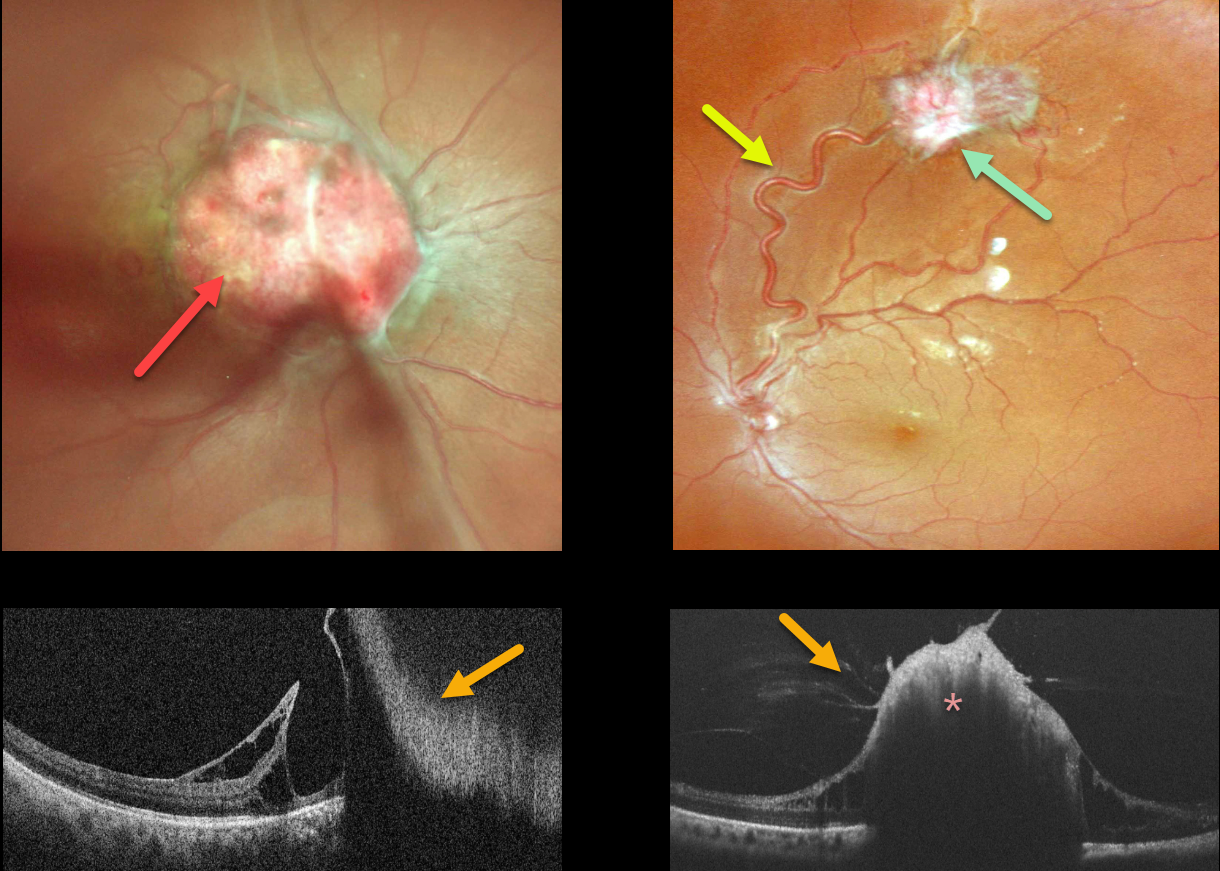
RETINAL CAPILLARY HEMANGIOMA
Sayali Bawankule, Prashant Bawankule, Shilpi Narnaware, and Anju Bansal
Originally posted on @retina.rocks April 6, 2023
This 31YO healthy female presented with a 3-day history of sudden vision loss in her right eye. Other than a recent history of hypertension, past medical and family history were negative. Her twins both died at 6 months of age from complications related to prematurity. Vision was counting fingers OD and 20/20 OS.
There was a moderate diffuse vitreous hemorrhage in her right eye. Color imaging shows an endophytic capillary hemangioma overlying the right optic nerve. Color imaging of her left eye shows a superotemporal peripheral variably fibrosed retinal capillary hemangioma fed and drained by dilated and irregular vessels.
On OCT, the angiomas appear as thickened, hyperreflective lesions with overlying vitreous traction.
A provisional diagnosis of Von Hippel-Lindau (VHL) disease was made, presumably from a new mutation, and she was referred to neurology and nephrology for further systemic evaluation. Unfortunately, she was subsequently lost to follow-up.
Learning Points:
Retinal capillary hemangiomas are orange-red lesions associated with a prominent pair of feeding and draining vessels and can cause severe vision loss or blindness due to exudative retinal detachment. These benign vascular tumors need to be aggressively treated. Common treatment options include thermal laser, cryotherapy, and anti-VEGF injections.
Retinal capillary hemangioma is a predominant feature of VHL, an autosomal dominant condition caused by mutations in the VHL tumor suppressor gene. Patients develop benign and malignant tumors, including central nervous system hemangioblastoma, pheochromocytoma, and renal cell carcinoma.
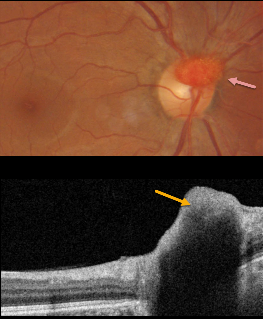
RETINAL CAPILLARY HEMANGIOMA
Originally posted on @retina.rocks February 1, 2023
This 39YO healthy male was referred for an asymptomatic lesion in his right eye.
Fundus photography shows an elevated, vascularized lesion covering the superior optic nerve. The lesion was markedly hyperreflective on OCT.
On further questioning, there was a strong family history of Von Hippel-Lindau (VHL) disease, including his sister and maternal nephew. Genetic testing was ordered.
Learning Points:
Retinal capillary hemangioma is a predominant feature of VHL, an autosomal dominant condition caused by mutations in the VHL tumor suppressor gene. Patients develop benign and malignant tumors including central nervous system hemangioblastoma, pheochromocytoma and renal cell carcinoma.
Retinal capillary hemangiomas appear clinically as orange-red lesions, most commonly seen peripherally associated with a prominent paired feeding and draining vessel. They can also appear on or around the optic nerve lesions, either on the surface (endophytic) as in our patient, or buried within the substance of the nerve (exophytic).
These lesions can cause severe vision loss or blindness from exudative retinal detachment and thus usually require aggressive treatment. Treatment options include thermal laser, photodynamic therapy, cryotherapy, and anti-VEGF injections.
Since our patient is completely asymptomatic with no exudation, we will observe without treatment for now.
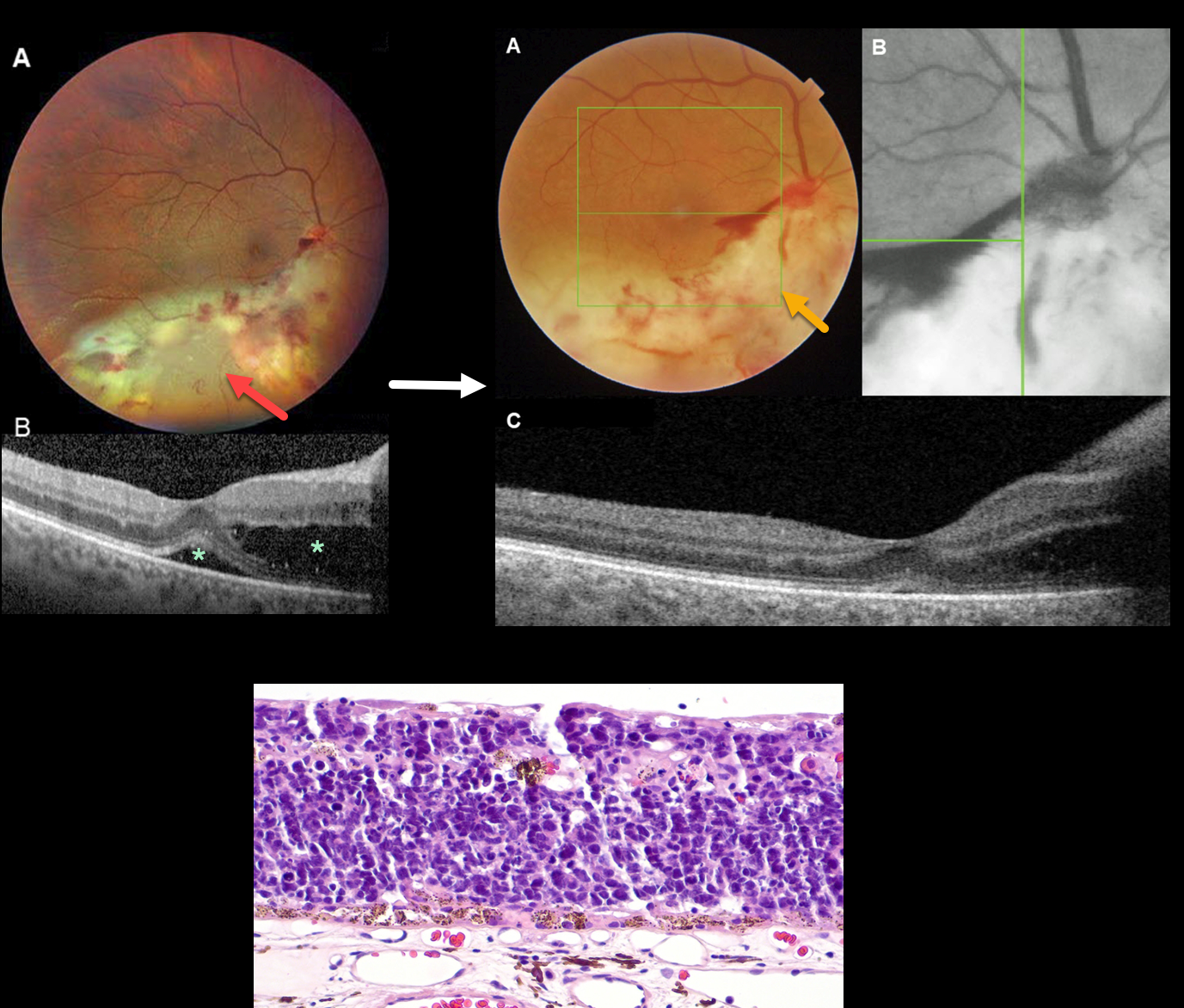
RETINAL METASTASES
Originally posted on @retina.rocks November 4, 2022
This 48YOF had a history of extensive-stage small-cell lung cancer (SCLC) with metastases to the thoracic and abdominal lymph nodes. She presented to an outside practice with a four-week history of a gradually worsening visual field defect and was diagnosed with cytomegalovirus (CMV) retinitis and started on oral ganciclovir.
She was then referred to a second practice for another opinion. Vision was 20/30. Color photography showed a yellow-white retinal lesion with associated retinal hemorrhages that extended from the inferior disc and macula to the inferior and inferotemporal periphery. The remaining peripheral retina and retinal vessels were unremarkable.
Although the fovea appeared spared clinically, optical coherence tomography (OCT) showed outer retinal fluid extending from the optic disc towards the fovea with mild subfoveal fluid. Intravenous and intravitreal Ganciclovir therapy was begun. Aqueous viral PCR testing was negative.
She was then referred to our practice. Vision was 20/40. The white retinal lesions now encompassed the entire optic nerve and nasal peripapillary region. Unlike what would be expected from treated CMV retinitis, the lesion mostly clinically spared the inner retina, had smooth borders, and lacked a central area of chorioretinal atrophy. Red-free imaging was suggestive of disc neovascularization. OCT scanning showed decreased outer retinal and subretinal fluid.
Vitrectomy was performed to obtain vitreous and retinal biopsies. Cytologic assessment of the retinal biopsy was consistent with the patient’s known history of SCLC.
After the discussion of treatment options, the patient chose to undergo systemic chemotherapy and enucleation of the right eye. Microscopic examination of the enucleated eye showed extensive areas of retinal replacement by a mitotically active small blue cell neuroendocrine tumor consistent with metastasis from the patient’s known extensive-stage SCLC.
Learning Points:
Lung cancer is the second most common worldwide cancer in both genders, with SCLC accounting for 13% of all newly diagnosed lung cancers. The prognosis is poor, and up to 50% of patients with extensive-stage SCLC have brain metastases (van Meerbeeck et al, Lancet 2011;378:1741-1755).
The retina is a very rare location for ocular metastases compared to the uvea and orbit. During a 40-year period, the oncology service at the Wills Eye Hospital observed only 8 (0.325%) retinal metastases compared to 2458 uveal and orbital metastases (Sheilds et al, JAMA Ophthalmol 2014:132:1303-1308).
The initial retinal findings can mimic viral retinitis, particularly CMV retinitis. Retinal metastasis should be considered in patients initially diagnosed with viral retinitis who fail to improve on adequate antiviral therapy, particularly with a known history of malignancy.
RETINOBLASTOMA
Originally posted on @retina.rocks May 2, 2022
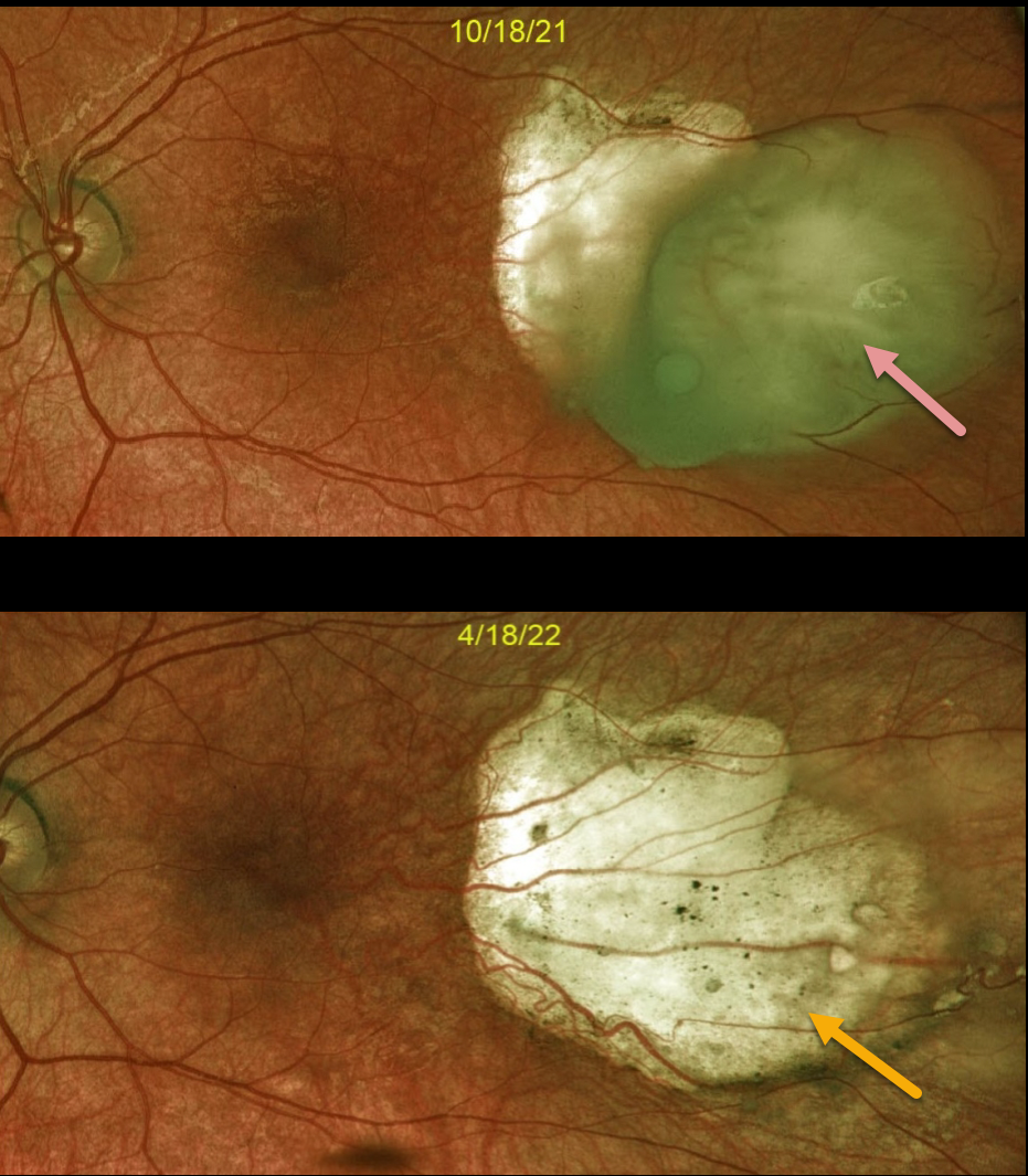
This is a 3YO female with a history of bilateral retinoblastoma and prior enucleation of the right eye. It is a follow-up of a previously posted case (11/7/21).
Optos imaging of the left eye shows an active retinoblastoma with complete regression following 6 months of treatment, which included intra-arterial chemotherapy and cryotherapy.
Learning Points:
Small tumors can be treated with focal therapies such as laser therapy, cryotherapy, or radioactive plaque therapy.
Larger tumors are treated with chemotherapy (most commonly intra-arterial through the ophthalmic artery and occasionally systemic). Chemotherapy has dramatically decreased the need for enucleation.
RETINAL ASTROCYTIC HAMARTOMA
Originally posted on @retina.rocks January 10, 2022
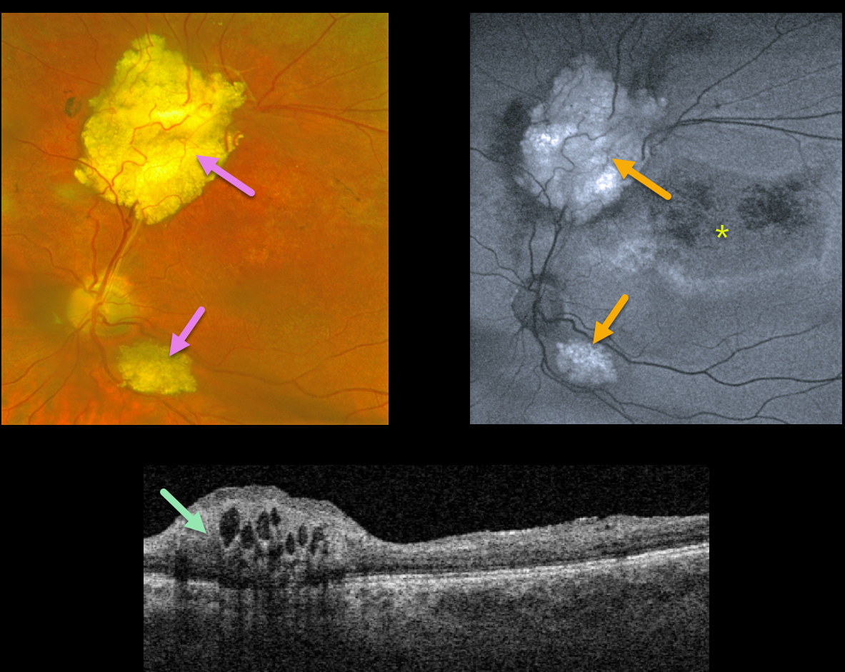
This 21YO male patient with a known history of tuberous sclerosis was referred for multiple, asymptomatic, thickened whitish inner retinal lesions.
On fundus autofluorescence (FAF), these lesions are hyper-FAF. Surrounding hypo-FAF changes that extend into the macula are a sign of prior exudative fluid.
OCT scanning shows an elevated, hyperreflective inner retinal lesion with multiple hyporeflective intralesional cavities.
Learning Points:
A retinal astrocytic hamartoma is a benign tumor composed of spindle-shaped fibrous astrocytes located in the nerve fiber layer, most commonly associated with tuberous sclerosis.
Tuberous sclerosis includes the classic triad of seizures, intellectual disability, and fibroangiomas. Still, patients can have lesions throughout the body, including the eyes, skin (ash-leaf spots), bone, nails, teeth, brain, heart, lungs, and kidneys.
Early in life, the inner retinal lesions appear fleshy white and are often multifocal and bilateral. With time, as in this patient, they show a more classic “mulberry appearance” with significant calcification. Rarely, the vascular component can cause intraretinal exudation requiring treatment.
RETINAL CAVERNOUS HEMANGIOMA
Originally posted on @retina.rocks December 29, 2021
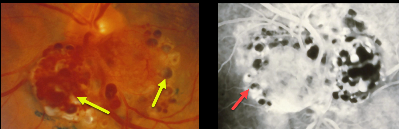
Retinal cavernous hemangiomas are usually unilateral, inner retinal grape-like clusters of aneurysms filled with dark venous blood. A gray fibrotic membrane often covers its superficial surface.
Fluorescein angiogram shows plasma-erythrocyte layering within these saccular lesions, attributable to sluggish blood flow.
Vision is usually normal, and no treatment is required. Rarely, vision loss can occur from vitreous hemorrhage.
Learning Points:
Although most occur sporadically, some may be associated with an autosomal dominant neurocutaneous syndrome, including cutaneous and central nervous system hemangiomas. Neurologic imaging is therefore recommended.
RETINOBLASTOMA
Originally posted on @retina.rocks November 17, 2021
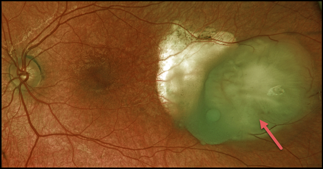
This 3YO female has a history of bilateral retinoblastoma and prior enucleation of the right eye. Optos color RG imaging of her left eye shows a still active retinoblastoma, despite prior treatment, which included intra-arterial chemotherapy and cryotherapy.
Learning Points:
Small tumors can be treated with focal therapies such as laser therapy, cryotherapy, or radioactive plaque therapy.
Larger tumors are treated with chemotherapy (most commonly intra-arterial through the ophthalmic artery and occasionally systemic). Chemotherapy has dramatically decreased the need for enucleation.
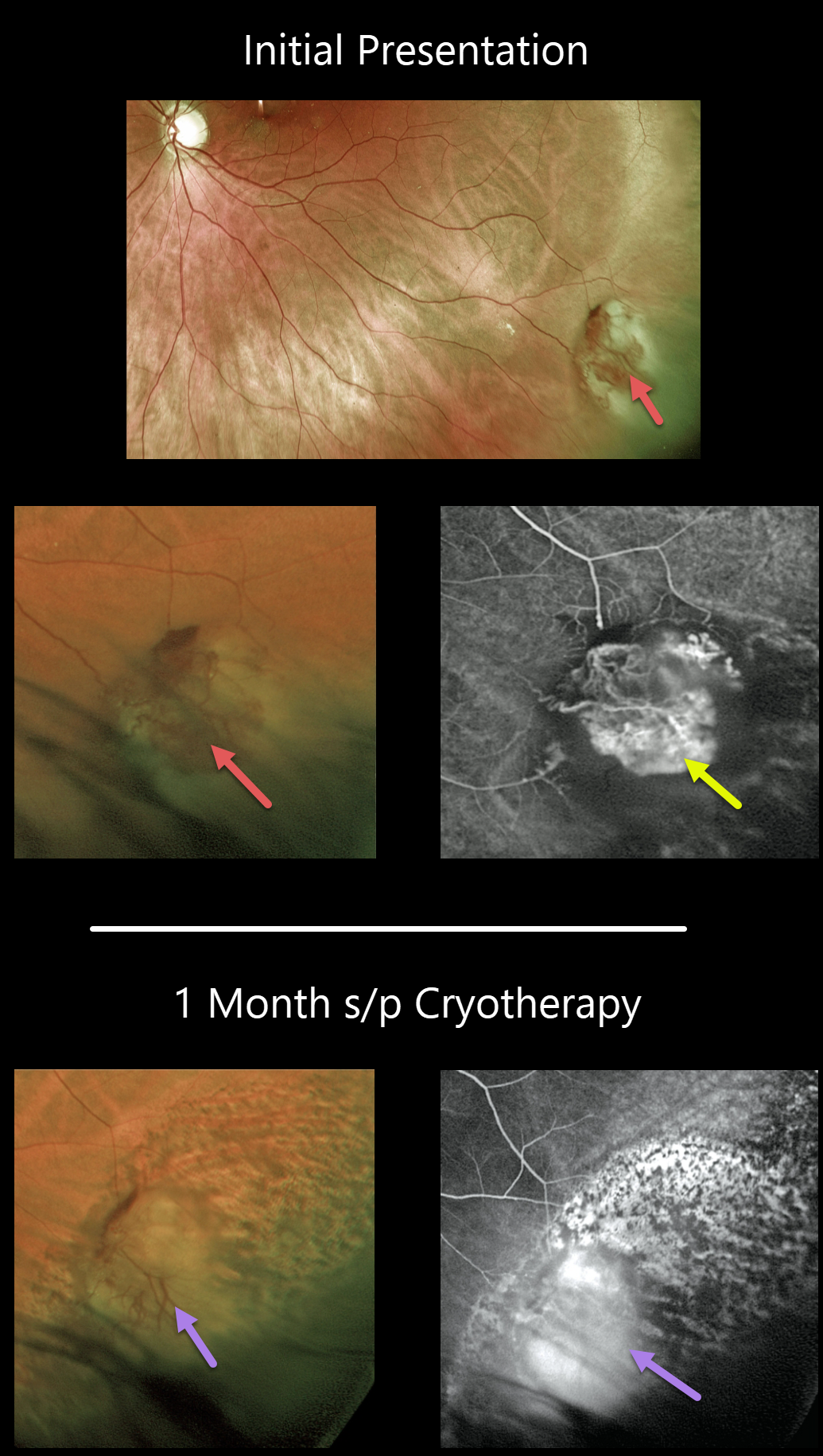
VASOPROLIFERATIVE TUMOR
Originally posted on @retina.rocks November 15, 2021
This 39YO female presented with an asymptomatic vasoproliferative tumor (VPT). The VPT initially appeared as a vascularized, reddish, elevated, peripheral mass.
Fluorescein angiography revealed the vascular nature of the lesion. The lesion was treated with cryotherapy and already showed signs of regression at the 1-month postoperative exam.
Learning Points:
Vasoproliferative tumors are benign globular, dome-shaped lesions arising in the peripheral retina. They can be yellow or red and cause subretinal exudation and detachment, anterior or posterior segment neovascularization, and vitreous hemorrhage.
They can be primary idiopathic lesions (as in our patient) or a vascular response to various entities, including intermediate uveitis, retinitis pigmentosa, Coats disease, and prior retinal detachment (see Shields et al, JAMA Ophthalmology 2013;131;328-334).
The lesions can be treated with thermal laser, cryotherapy, and/or anti-VEGF injections.
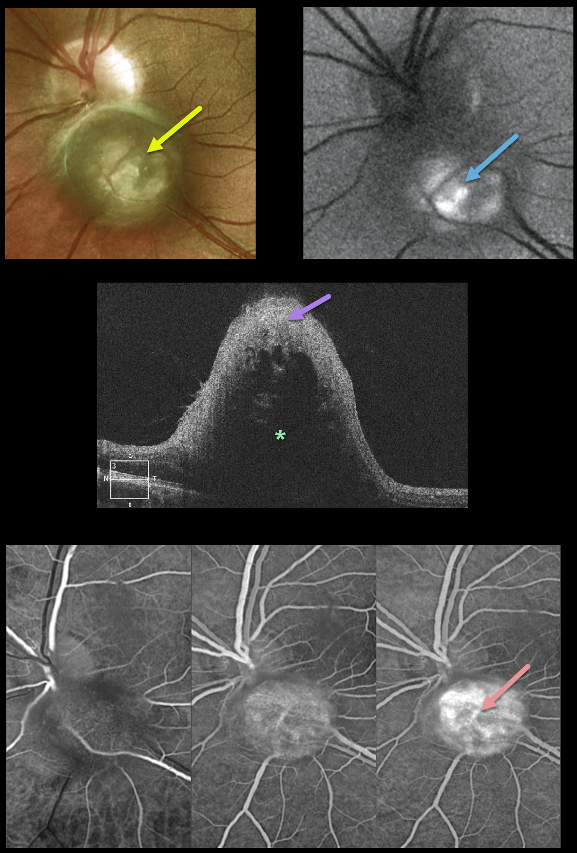
pSCRAP (PRESUMED SOLITARY CIRCUMSCRIBED RETINAL ASTROCYTIC PROLIFERATION)
Originally posted on @retina.rocks July 26, 2021
This healthy 28YO male was referred for an asymptomatic lesion in his left eye. Vision was 20/25 OU. Ocular examination was normal bilaterally except for a round nodular amelanotic elevated lesion abutting (and possibly involving) the left inferior optic nerve.
The lesion shows some central hyperautofluorescence. OCT shows marked reflectivity involving the inner retina with posterior shadowing. Optos fluorescein angiography shows probable intrinsic vascularity of the lesion.
Learning Points:
PSCRAP (presumed solitary circumscribed retinal astrocytic proliferation was originally described by Shields et al as a unique lesion found in adulthood that resembles a retinal astrocytoma or retinoma (Arch Ophthalmology 2011;129;1189-1194).
Although originally felt to arise from the inner retina, it is now felt to be either a deep retinal glial lesion or a form of RPE fibrous metaplasia (Shields et al, Retinal Cases & Brief Reports 2017;11:18-23).
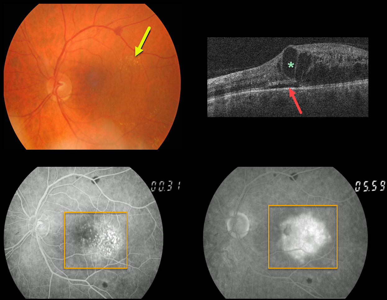
RETINAL CAPILLARY HAMARTOMA
Originally posted on @retina.rocks July 7, 2021
This patient presented with very subtle retinal vascular telangiectasia in the temporal macula, some scattered temporal macular lipid, and moderate cystic retinal edema with trace subretinal fluid on OCT. There were also a few larger scattered deep retinal hemorrhages.
Fluorescein angiography shows much more dramatic intraretinal vascular abnormalities centered in the temporal macula with moderate leakage.
Learning Points:
We believe this patient has a retinal capillary hamartoma. This is an extremely rare finding; to the best of our knowledge, only been previously described in a strikingly similar case in the 5th edition of Gass’ classic atlas (Gass’ Atlas of Macular Diseases 5th Edition, Agarwal A, Elsevier 2011, pages 446-447).
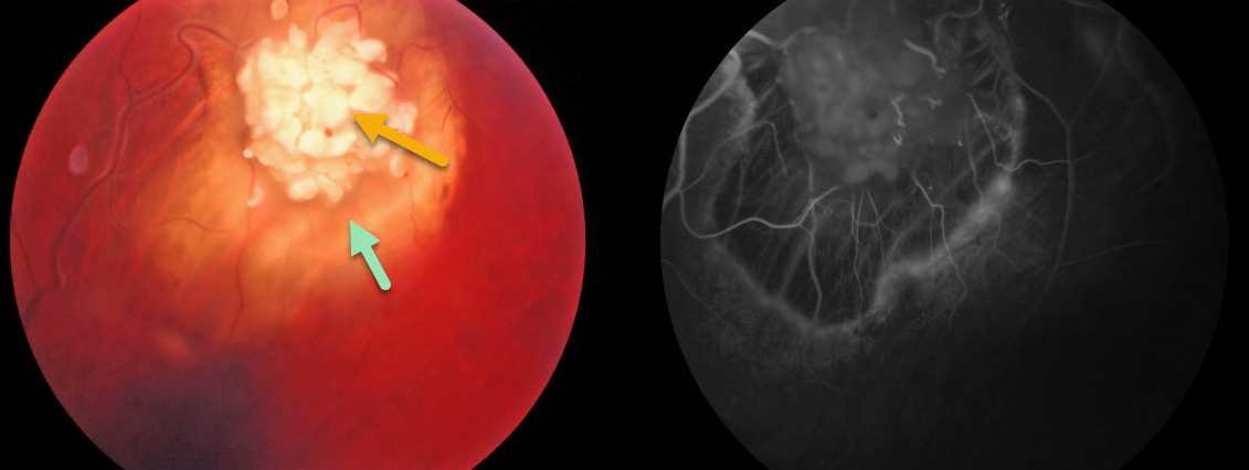
RETINOCYTOMA
Originally posted on @retina.rocks April 16, 2021
This 57YO male presented with a retinocytoma without any history of retinoblastoma (RB) or treatment for RB.
Clinically, retinocytomas can resemble astrocytic hamartomas. However, retinocytomas often have underlying RPE changes and chorioretinal scarring, which are not present in astrocytic hamartomas.
Learning Points:
Retinocytomas are fleshy, opalescent, benign tumors that are thought to spontaneously regress in Rb. They appear identical to previously radiated Rb tumors.
Similar to typical RB, these patients’ offspring are at a 50% risk for developing Rb and thus require genetic counseling.
RETINAL ASTROCYTIC HAMARTOMA
Originally posted on @retina.rocks February 18, 2021
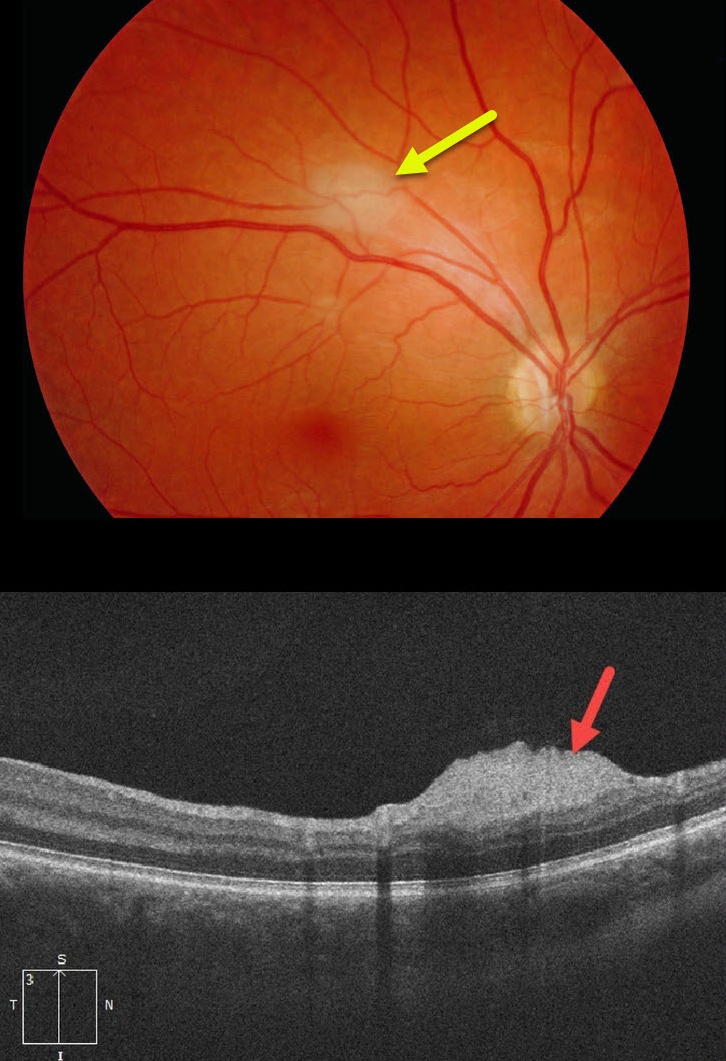
This patient with a known history of tuberous sclerosis was referred for an asymptomatic, slightly thickened whitish inner retinal lesion. OCT scanning showed an elevated, hyperreflective nerve fiber layer lesion.
Learning Points:
A retinal astrocytic hamartoma is a benign tumor composed of spindle-shaped fibrous astrocytes located in the nerve fiber layer, most commonly associated with tuberous sclerosis.
Tuberous sclerosis includes the classic triad of seizures, intellectual disability, and fibroangiomas, but patients can have lesions throughout the body, including the eyes, skin (ash-leaf spots), bones, nails, teeth, brain, heart, lungs, and kidneys.
Early in life, the inner retinal lesions appear fleshy white and are often multifocal and bilateral. Later in life, they show a more classic “mulberry appearance” with significant calcification. Rarely, the vascular component can cause intraretinal exudation requiring treatment.
RETINAL CAVERNOUS HEMANGIOMA
Originally posted on @retina.rocks January 21, 2021
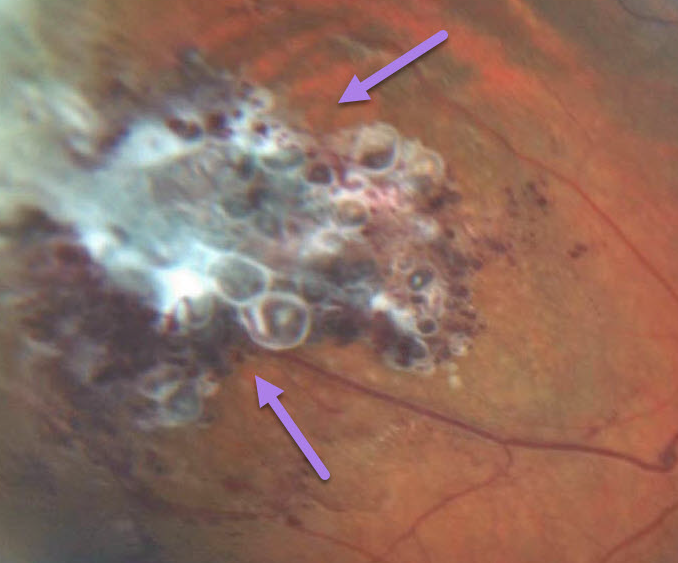
Retinal cavernous hemangiomas are usually unilateral, inner retinal grape-like clusters of aneurysms filled with dark venous blood. A gray fibrotic membrane often covers its superficial surface.
Vision is usually normal, and no treatment is required. Rarely, vision loss can occur from vitreous hemorrhage.
Learning Points:
Although most occur sporadically, some may be associated with an autosomal dominant neurocutaneous syndrome, including cutaneous and central nervous system hemangiomas. Neurologic imaging is therefore recommended.
RETINAL CAPILLARY HEMANGIOMA
Originally posted on @retina.rocks January 20, 2021
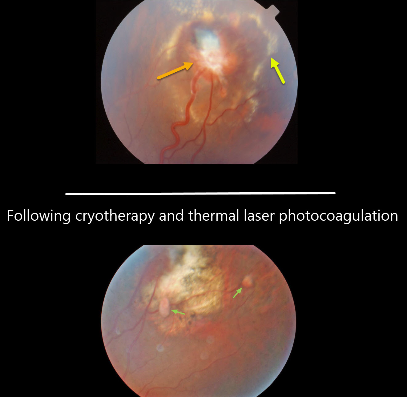
We have been caring for this patient and his family with Von Hippel-Lindau (VHL) disease for over 20 years.
We initially treated the retinal capillary hemangioma in his left eye with cryotherapy. Although the tumor initially regressed, thermal laser photocoagulation was applied due to some new exudation. The exudation eventually resolved. A few smaller involuted angiomas are well-visualized in his most recent photo.
Our patient had a partial nephrectomy for renal cell carcinoma, and his mother died 2 years ago from renal cell carcinoma. His sister required enucleation of her right eye as a teenager for a total exudative detachment.
Learning Points:
Retinal capillary hemangioma is a predominant feature of VHL, an autosomal dominant condition caused by mutations in the VHL tumor suppressor gene. Patients develop benign and malignant tumors, including central nervous system hemangioblastoma, pheochromocytoma, and renal cell carcinoma.
Retinal capillary hemangiomas are orange-red lesions associated with prominent paired feeding and draining vessels and can cause severe vision loss or blindness due to exudative retinal detachment. These benign vascular tumors need to be aggressively treated. Common treatment options include thermal laser, cryotherapy, and anti-VEGF injections.
RETINOCYTOMA
Originally posted on @retina.rocks July 20, 2020
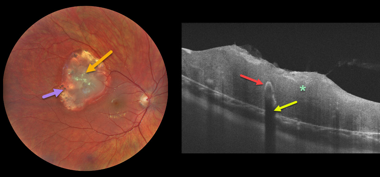
This patient presented with a retinocytoma of the right eye. The OCT line scan shows a disorganized, thickened retina. Typical intralesional calcification is noted. This calcium is highly reflective, causing shadowing of the underlying retina.
Clinically, retinocytomas can resemble astrocytic hamartomas. However, retinocytomas often have underlying RPE changes and chorioretinal scarring, which are not present in astrocytic hamartomas.
Learning Points:
Retinocytomas are fleshy, opalescent, benign tumors that are felt to be spontaneously regressed retinoblastoma (RB). They appear identical to previously radiated RB tumors.
Similar to typical RB, these patients’ offspring are at a 50% risk for developing RB and thus require genetic counseling.
VASOPROLIFERATIVE TUMOR
Originally posted on @retina.rocks June 29, 2020
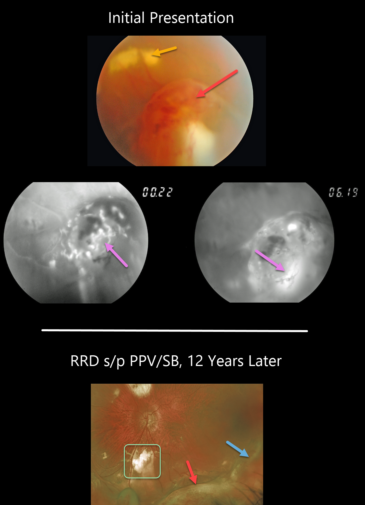
This 46YO presented with an asymptomatic vasoproliferative tumor (VPT). The VPT initially appeared as a tongue-like, reddish, elevated, peripheral mass with more posterior subretinal lipid. Fluorescein angiography (FA) revealed the vascular nature of the lesion.
He then developed a rhegmatogenous retinal detachment that was repaired with a combined vitrectomy and scleral buckle. The indentation from the buckle is visible more temporally and distinct from the complex enlarged VPT more inferiorly. The subretinal lipid resolved with more extensive subretinal scarring.
Learning Points:
Vasoproliferative tumors are benign globular, dome-shaped lesions arising in the peripheral retina. They can be yellow or red and cause subretinal exudation and detachment, anterior or posterior segment neovascularization, and vitreous hemorrhage.
They can be primary idiopathic lesions or a vascular response to various entities, including intermediate uveitis, retinitis pigmentosa, Coats disease, and prior retinal detachment.
For lesions causing symptomatic exudation or neovascularization, treatment with thermal laser, cryotherapy, and/or anti-VEGF injections is indicated.
RETINAL CAVERNOUS HEMANGIOMA
Originally posted on @retina.rocks May 11, 2020
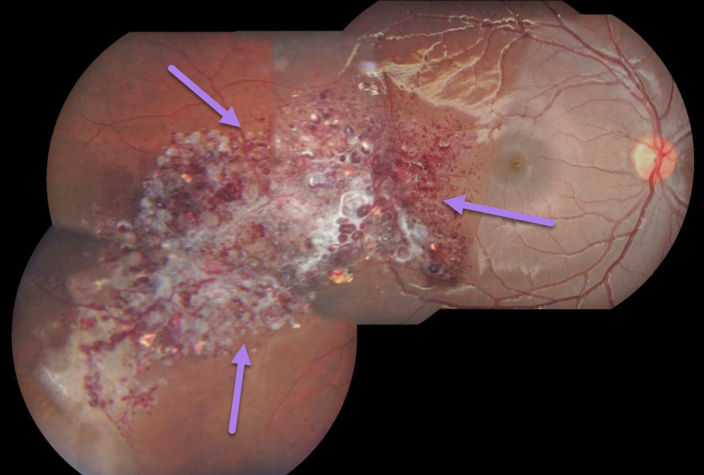
Retinal cavernous hemangiomas are usually unilateral, grape-like clusters of aneurysms filled with dark venous blood. A gray membrane often covers its superficial surface.
Vision is usually normal and no treatment is required. Rarely vision loss can occur from vitreous hemorrhage.
Learning Points:
Although most occur sporadically, some may be caused by an autosomal dominant neurocutaneous syndrome, including cutaneous and central nervous system hemangiomas. Neurologic imaging is therefore recommended.
RETINOBLASTOMA
Originally posted on @retina.rocks May 7, 2020
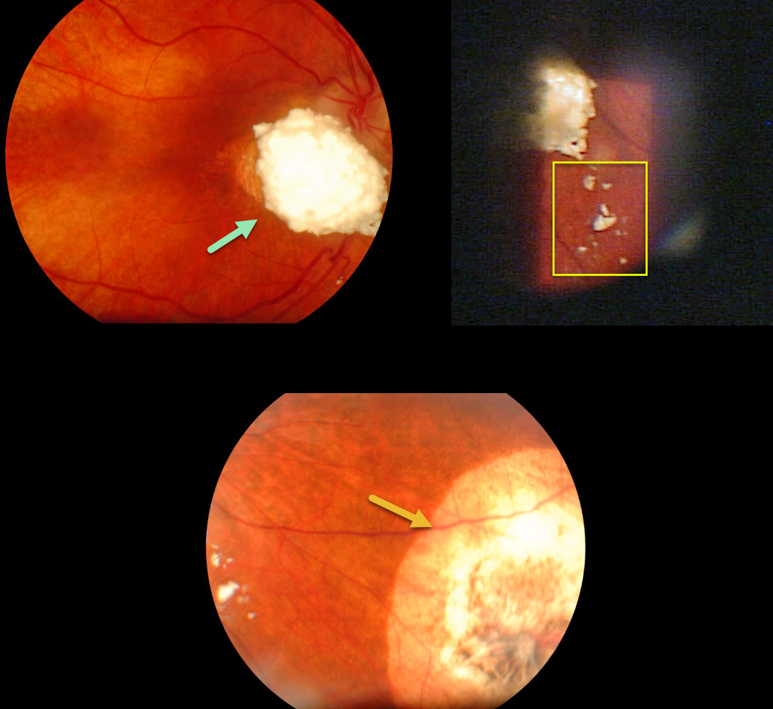
This patient was treated as a child for retinoblastoma, although unfortunately we don’t know the details of her prior treatment.
She has a calcified, elevated peripapillary lesion with overlying vitreous seeds. There is also a scar in the inferonasal midperiphery from another destroyed tumor. The multifocal nature of her tumors indicates that this was a genetic mutation, and that she likely has the retinoblastoma gene.
Learning Points:
Small tumors can be treated with focal treatments such as thermal laser, cryotherapy or radioactive plaque therapy. Larger tumors are treated with chemotherapy (most commonly intra-arterial through the ophthalmic artery and occasionally systemic). Chemotherapy has dramatically decreased the need for enucleation.
RETINAL ASTROCYTIC HAMARTOMA
Originally posted on @retina.rocks April 27, 2020
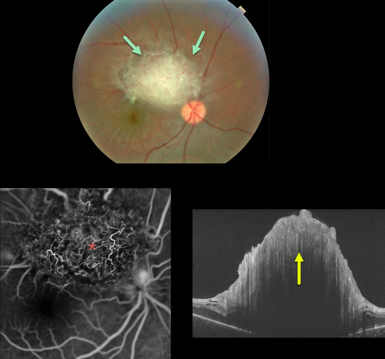
Our patient (courtesy of Tufts) has the classic mulberry appearance of a retinal astrocytic hamartoma. Early in life the inner retinal lesions have a fleshy white appearance and are often multifocal and bilateral. Later in life they develop the mulberry or fish egg-like appearance with significant calcification.
Fluorescein angiography (FA) shows the often angiomatous nature of these lesions. OCT shows the elevated, hyperreflective inner retinal lesion, with marked shadowing more posteriorly.
These tumors are generally are just observed. Rarely, the vascular component can cause intraretinal exudation requiring treatment.
Learning Points:
A retinal astrocytic hamartoma is a benign tumor composed of spindle-shaped fibrous astrocytes located in the nerve fiber layer, most commonly associated with tuberous sclerosis.
Tuberous sclerosis includes the classic triad of seizures, mental deficiency and fibroangiomas, but patients can have lesions throughout the body including the eyes, skin (ash-leaf spots), bone, nails, teeth, brain, heart, lungs and kidneys.


