

13 Neoplastic Choroid Disorders
<< Back to Cases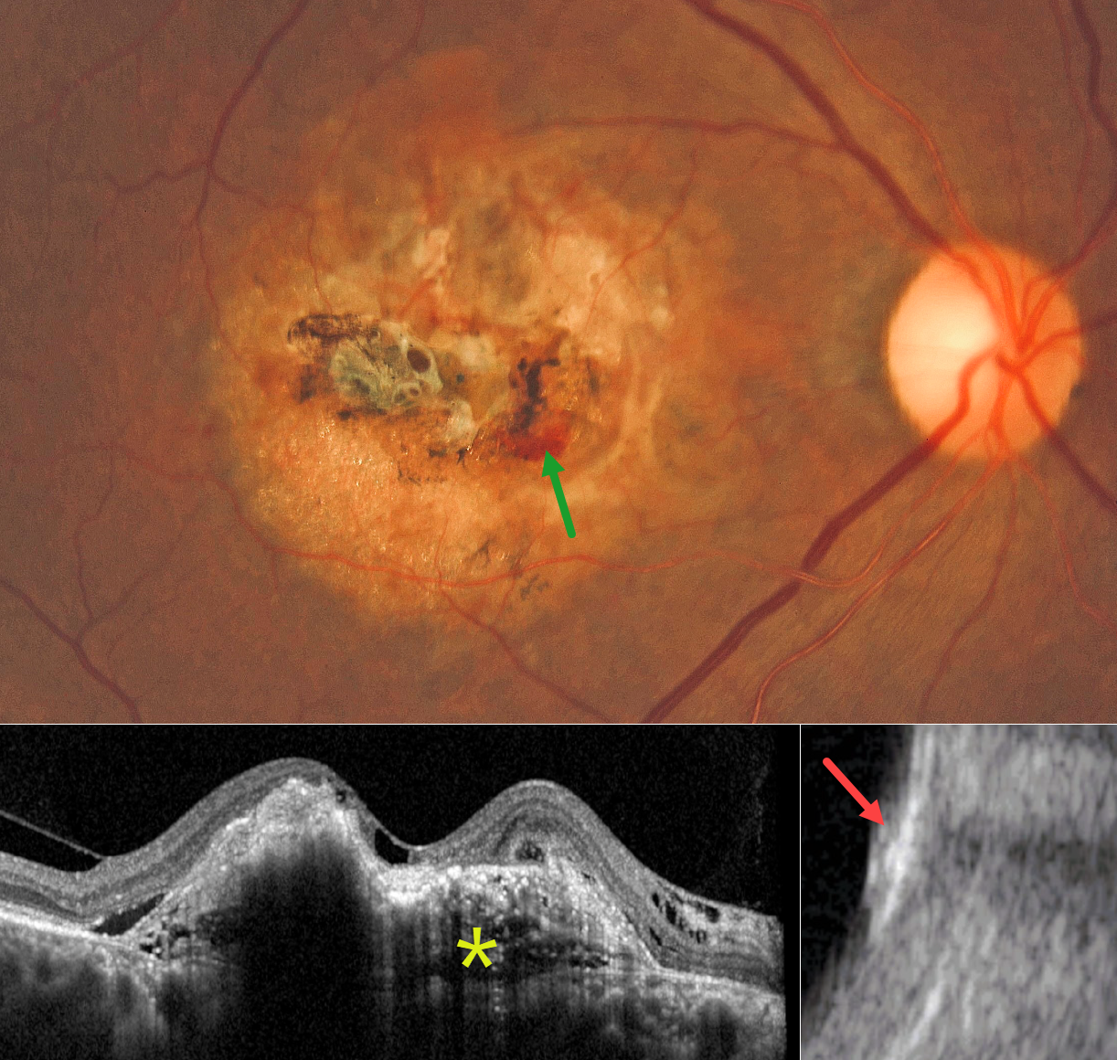
CHOROIDAL OSTEOMA
César Adrián Gómez Valdivia
Originally posted on @retina.rocks July 27, 2026
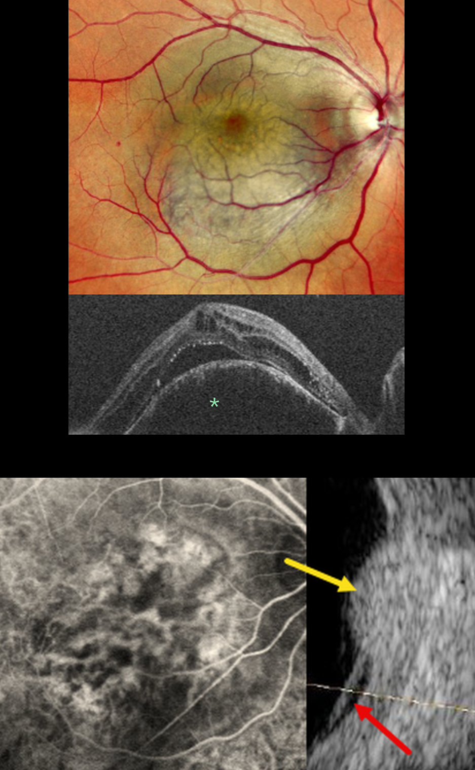
This 73YO male presented with 8 months of gradual vision loss in his right eye. Vision was 20/200 OD and 20/25 in his normal OS.
Color photography shows a yellowish, refractile amelanotic occupying nearly the entire macula. Some areas of increased pigment are noted centrally, along with some blood inferonasally. On OCT, the lesion has an internal lamellar, sponge-like architecture, characterized by horizontal hyporeflective lines and some intraretinal and subretinal fluid. The lesion is markedly hyperreflective on B-scan ultrasonography with posterior acoustic shadowing.
Learning Points:
Choroidal osteoma is a rare, benign ossifying choroidal tumor that typically affects young women, often in the peripapillary or macular region. Typical features include a clinical yellowish-orange lesion, a hyperreflective choroidal mass with a lamellar or trabecular internal architecture on OCT, and, most importantly, marked posterior acoustic shadowing on B-scan ultrasonography due to calcification. These imaging characteristics are distinct from those of other choroidal lesions, such as melanoma or hemangioma, which lack dense calcification and exhibit different reflectivity patterns.
Although benign, choroidal osteoma may cause vision loss due to macular neovascularization (MNV), subretinal fluid, and progressive decalcification, with atrophy of the retinal pigment epithelium and outer retinal layers. MNV can be successfully treated with anti-VEGF therapy (see Seong et al, Graefe’s 2022;250:1713-1721). Intravitreal Eylea was started for our patient.
CHOROIDAL METASTASES
Ayushi Gupta and Vishal Agrawal
Originally posted on @retina.rocks July 13, 2026
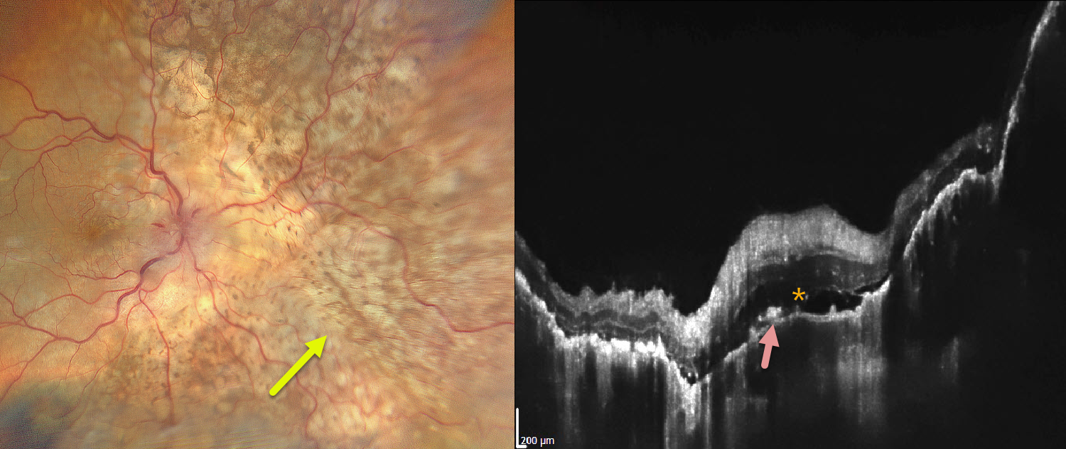
This 68YO male presented with severe vision loss in his right eye for several weeks. There was a known history of metastatic lung adenocarcinoma. Vision was light perception OD and 20/20 in his normal OS.
Color photography shows variably confluent, creamy-yellow subretinal lesions scattered within the macula and extending into the midperiphery. OCT scanning shows an undulating thickened choroid with shallow overlying subretinal fluid and scattered hyperreflective material above the RPE, likely representing lipofuscin.
Learning Points:
Although uveal melanoma is the most common primary intraocular malignancy, metastatic choroidal tumors are the most common intraocular malignancies. Lung and breast cancer are the most common sources.
CHOROIDAL OSTEOMA
Manuel Bande
Originally posted on @retina.rocks June 8, 2026
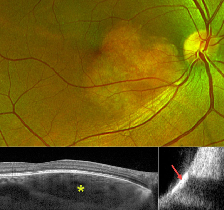
Optos color RG imaging shows a yellowish, well-circumscribed amelanotic lesion centered in the inferonasal macula. On OCT, the lesion has an internal lamellar, sponge-like architecture, characterized by horizontal hyporeflective lines corresponding to intralesional vascular channels and bony trabeculae. The overlying RPE and retina appear fairly normal. The lesion is markedly hyperreflective on B-scan ultrasonography with posterior acoustic shadowing.
Learning Points:
Choroidal osteoma is a rare, benign ossifying choroidal tumor that typically affects young women, often in the peripapillary or macular region. Typical features include a clinical yellowish-orange lesion, a hyperreflective choroidal mass with a lamellar or trabecular internal architecture on OCT, and, most importantly, marked posterior acoustic shadowing on B-scan ultrasonography due to calcification. These imaging characteristics are distinct from those of other choroidal lesions, such as melanoma or hemangioma, which lack dense calcification and exhibit different reflectivity patterns.
Although benign, choroidal osteoma may cause vision loss from macular neovascularization (MNV), subretinal fluid, and progressive decalcification with atrophy of the retinal pigment epithelium and outer retinal layers. MNV can be successfully treated with anti-VEGF therapy (see Seong et al, Graefe’s 2022;250:1713-1721).
SUBCLINICAL CHOROIDAL NEVUS VS HEMANGIOMA
Shraddha Raj Shrivastava, Steven Bloom, and Manish Nagpal
Originally posted on @retina.rocks May 13, 2026
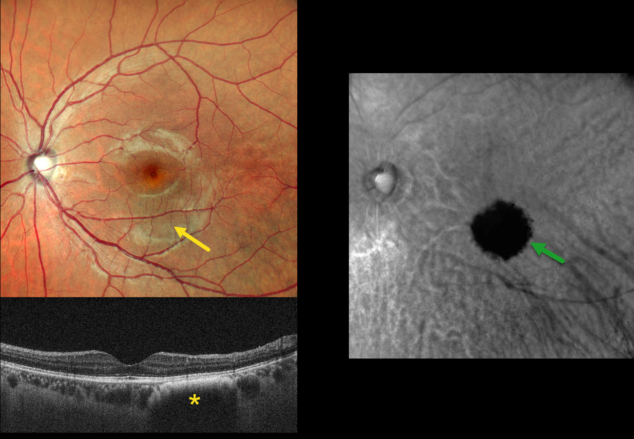
This asymptomatic 26YO male presented for a routine eye examination. Vision was 20/20.
Clinical biomicroscopy and MultiColor scanning laser ophthalmoscope (SLO) imaging are normal except for faint hyperpigmentation in the inferior macula. OCT in this region shows a dome-shaped hyporeflective lesion with an anterior hyperreflective border. Retromode imaging shows a well-defined hyporetroreflective lesion.
Learning Points:
Traditional color photography uses white light to image the posterior segment. Newer imaging devices utilize LED or SLO technology to illuminate the fundus with blue, green, and infrared light, which best image the vitreoretinal interface/inner retina, mid-retina to RPE, and choroid, respectively. Many of these technologies create a ‘color’ fundus image that is not true to life yet contains clinically useful depth information in a flat image. In our patient’s case, Nidek Mirante Retromode imaging best detected this subclinical lesion using a 790 nm SLO. For a recent review of Retromode imaging, see Sukkarieh et al, Surv Ophthalmology 2023;68:1027-1037.
So does our patient have a choroidal nevus or hemangioma? Internally, the authors are split between a nevus (SRS and MN) and a hemangioma (SB). Findings supporting a choroidal hemangioma include its orange color blending with the surrounding RPE/choroid and round shape on Retromode imaging. However, we have not seen a hemangioma with an anterior hyperreflective margin on OCT. Jonna et al described 5 OCT patterns of flat choroidal nevi (Ophthalmology Retina 2019;3:270-277), including a subtype with “anteriorly bowed hyperreflectivity with discrete borders and cascading edges,” which appears identical to our patient’s OCT.
METASTASES
The European VitreoRetinal Society, Aysegul Mavi Yildiz and Hikmet Ozcetin
Originally posted on @retina.rocks March 6, 2026
This 67YO male presented with progressive decreased vision in his right eye. He had a known history of metastatic lung cancer. Vision was hand motion OD.
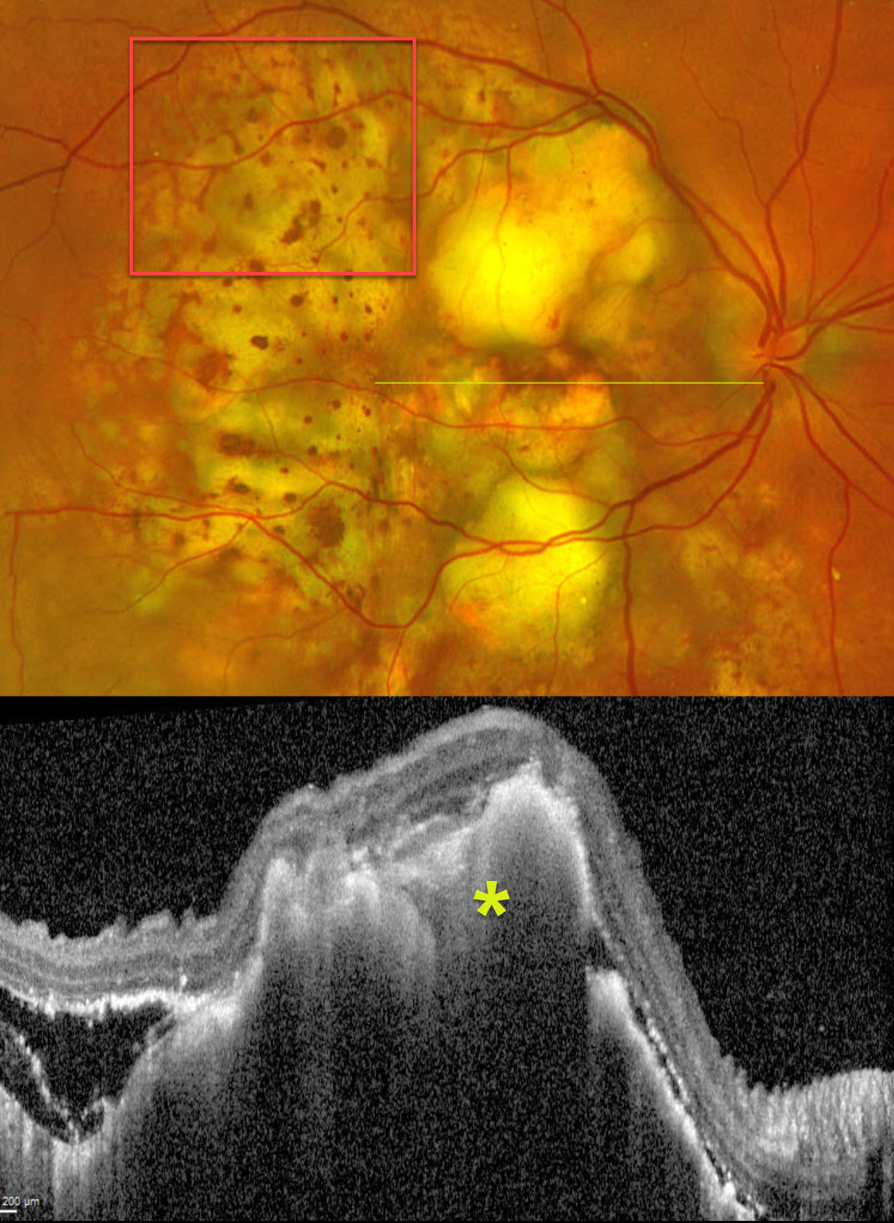
Optos color RG imaging shows multifocal, variably confluent, creamy-yellow subretinal lesions scattered within the macula and extending into the midperiphery. A leopard spot pattern of pigmentary changes is noted temporally. OCT scanning shows marked hyperreflective lumpy choroidal thickening with posterior shadowing. There is temporal subretinal fluid containing variable hyperreflective material.
Learning Points:
Although uveal melanoma is the most common primary intraocular malignancy, metastatic choroidal tumors are the most common intraocular malignancies. Lung and breast cancer are the most common sources.
SUSPECT CHOROIDAL NEVUS
Originally posted on @retina.rocks January 29, 2026
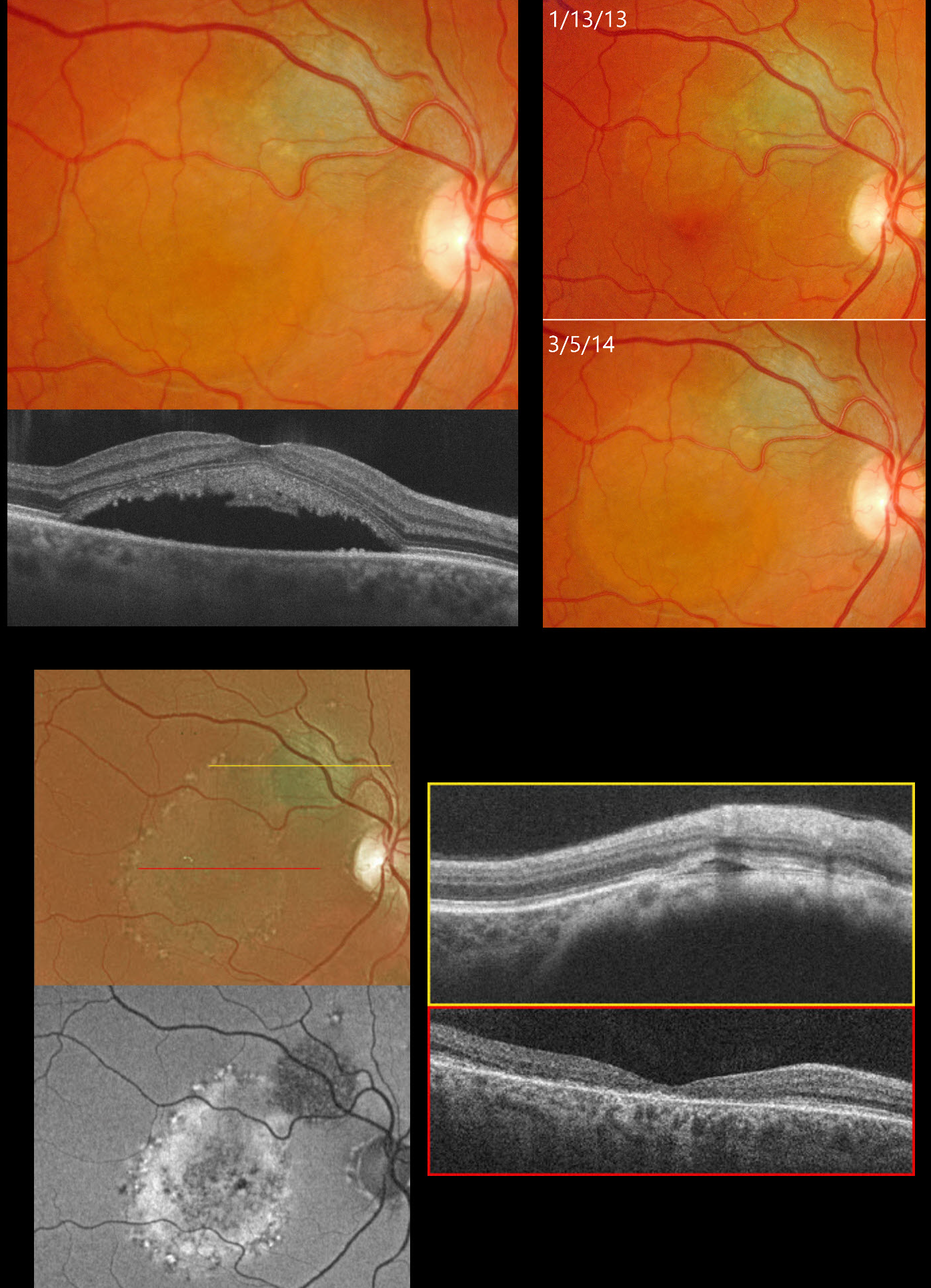
This 35YO female was examined for a suspect choroidal nevus on 1/13/13. Color photography shows about a 2x2mm minimally thickened choroidal nevus superotemporal to the right optic nerve (image 2). Subretinal fluid extends into the superior macular and some orange pigment overlies the nevus.
She returned on 3/15/14, now with fluid extending through the fovea (image 1). OCT scanning confirms the foveal fluid and hyperreflective material, likely representing shed outer segments, lines the outer surface of the detached macula.
She was referred to an ocular oncologist and immediately lost to follow-up until she presented on 4/2/25 without new complaints. She told us she had received two photodynamic therapies elsewhere shortly after we last saw her over 10 years earlier. Vision was 20/200. Optos color RG imaging shows the nevus to be stable in size but with new pigmentary changes throughout the macula within the area of prior fluid (image 3). On fundus autofluorescence (FAF), the nevus is hypo-FAF, and the region of prior fluid shows variable hyper-FAF. OCT scanning shows no fluid overlying the nevus with foveal atrophy. Continued observation was recommended.
Learning Points:
Clinical characteristics of choroidal nevi at risk for progressing to a uveal MM were initially described by Shields et al (Ophthalmology 1995;102:1351-1361). The most recent version of this famous acronym (Shields et al, Retina 2019;39:1840-1851) is To Find Small Ocular Melanoma Doing Imaging (TFSOM-DIM), which stands for To (Thickness >2mm) Find (subretinal Fluid) Small (Symptoms, vision <= 20/50) Ocular (Orange pigment) Melanoma (Melanoma hollow on ultrasonography) DIM (DIaMeter > 5mm).
CHOROIDAL MELANOCYTOSIS
Originally posted on @retina.rocks January 22, 2026
This healthy 15YO male was referred for an asymptomatic pigmented lesion in his right eye. Family history was negative. Vision was 20/40 OD and 20/30 in his normal OS.
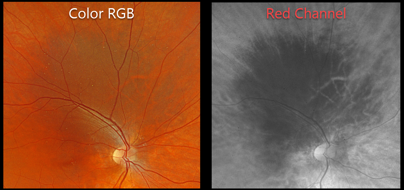
Optos color RGB imaging shows a region of increased choroidal pigmentation extending from the superonasal macula into the superior midperiphery. This lesion is best seen with the red channel.
Learning Points:
Choroidal melanocytosis is part of the oculodermal spectrum which occasionally includes pigmentation of the globe or periocular skin (Nevus of Ota, melanosis oculi). There are increased dendritic melanocytes in the affected tissues following the distribution of the first and second branches of the trigeminal nerve. Patients need ongoing monitoring since about 1 in 400 will develop uveal melanoma. Of those that develop melanoma, they are twice as likely to undergo metastasis when compared to patients with uveal melanoma and no pre-existing melanocytosis.
Although pretty color images that match what we see on biomicroscopy are most intuitive clinically, newer imaging modalities that distort the normal fundus colors often contain very clinically relevant information. Standard Optos color RG images are generated from a red (635nm) and green (532nm) laser. Although these images have a greenish tint, they essentially contain two images in one: the RPE/neurosensory retina is best captured with the green channel, and the choroid is best seen with the red channel. The newer Optos true color RGB unit maintains the ability to view the separate RG channels but also generates a true to life color image by adding a blue (488nm) laser. In our patient’s case, the red channel from the RG image best shows the melanocytosis.
METASTATIC MELANOMA OF UNKNOWN ORIGIN, SUSPECT CHOROIDAL NEVUS OD AND PROBABLE MELANOMA OS
Mattie Adams
Originally posted on @retina.rocks January 19, 2026
This 67YO female presented with a 3-week history of floaters in her left eye. She has a known history of metastatic melanoma of unknown origin, including numerous brain lesions. There was no family history of ocular or cutaneous melanoma. Vision was 20/20 OD and 20/40 OS.
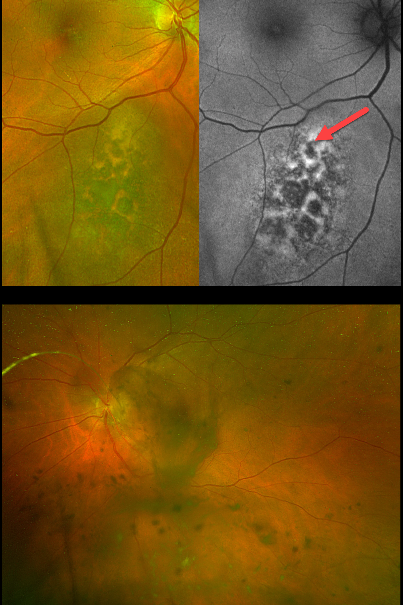
Optos color RG imaging OD shows a suspect choroidal nevus just inferonasal to the macula. There is overlying orange lipofuscin pigment, which is hyperautofluorescent. Optos imaging OS shows extensive pigmented vitreous debris variably obscuring the posterior pole.
Learning Points:
Uveal malignant melanoma (MM) and cutaneous melanoma are rarely associated with each other. The exception is the presence of a mutated tumor suppressor gene, BRCA1-associated protein 1 (BAP1), which is associated with autosomal-dominant uveal MM and other primary cancers, including cutaneous melanoma.
Our patient did not have a primary for her metastatic disease. The presence of a suspect nevus in her right eye and possible metastatic melanoma to her left eye argues for a pathogenic BAP1 mutation. We discussed this possibility with her oncologist, who felt this was highly unlikely given the negative family history. Given her relatively good vision and poor systemic prognosis, observation was recommended as the initial management for her ocular findings. Unfortunately, she died 3 months later.
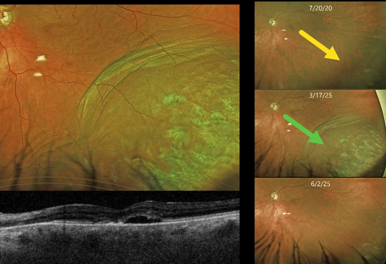
PSEUDO-UVEAL MELANOMA FROM CHOROIDAL DETACHMENT
Originally posted on @retina.rocks December 25, 2025
This 85YO female presented with an elevated pigmented choroidal mass in her inferotemporal periphery, which extended into her distal macula (Optos color RG). This mass was absent 5 years earlier. Circumferential choroidal folds extend along the posterior edge of the lesion. Triton swept-source OCT shows a blister of subretinal fluid and a thickened choroid.
She has a history of uncontrolled glaucoma and underwent a XEN Gel Stent procedure 8 days earlier. Intraocular pressure (IOP) was 8 mmHg.
Learning Points:
A choroidal detachment is defined by the abnormal presence of fluid or blood in the suprachoroidal space. Serous choroidal detachments, also known as choroidal effusions, are a frequent complication of glaucoma surgery. Several causative mechanisms are believed to be at play: hypotony allows fluid to accumulate in the suprachoroidal space, and inflammation increases the choroidal permeability. The detached ciliary body may also produce less aqueous, thus creating a self-perpetuating cycle. Often, observation is sufficient, but topical steroids and cycloplegics can be helpful in promoting resolution.
Other than the obvious appearance of this lesion immediately following glaucoma surgery, clues to this not being a uveal MM include the choroidal folds along the posterior extent of the choroidal detachment and an OCT consistent with hypotony maculopathy. About 10 weeks later, the IOP was 9 mmHg, and the choroidal effusion had completely resolved.
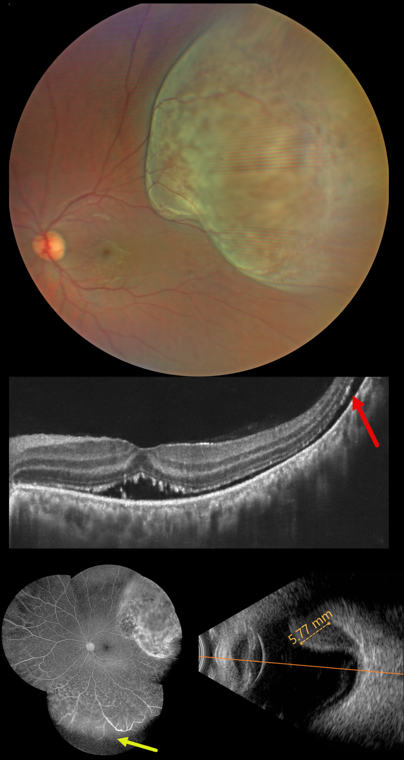
UVEAL MELANOMA
by Ayushi Gupta and Vishal Agrawal
Originally posted on @retina.rocks December 22, 2025
This healthy 36YO male presented with two weeks of vision loss in his left eye. Vision was 20/20 in his normal OD and 20/40 OS.
Fundus photography shows an elevated, pigmented, solid choroidal tumor, which spares the macula but occupies nearly the entire superotemporal quadrant. OCT scanning shows shallow subretinal fluid tracking from the tumor into the fovea with hyperreflective shaggy outer segments. Fluorescein angiography shows leaking intrinsic tumor vascularity with inferior peripheral retinal capillary loss. The tumor, which measures 5.77mm in axial thickness, shows moderate internal reflectivity on B-scan ultrasonography.
Learning Points:
Uveal melanoma is a malignant tumor arising from melanocytes in the uveal tract (iris, ciliary body, or choroid). The choroid is the most common site, accounting for 90% of uveal melanomas. Uveal melanoma is the most common primary intraocular cancer in adults, whereas metastatic disease is the most common intraocular malignancy. The liver is the most common metastatic site, although metastatic disease is rare on presentation. Metastases are more common in large tumors, with BAP1 mutations, and with certain tumor-related genetic markers (including positive PRAME and Class 2).
He was immediately referred to an ocular oncologist for further evaluation and treatment, including genetic testing, metastatic workup, and plaque brachytherapy. One month postoperatively, the tumor regressed to 4.6mm in thickness with decreased exudative fluid (not shown).
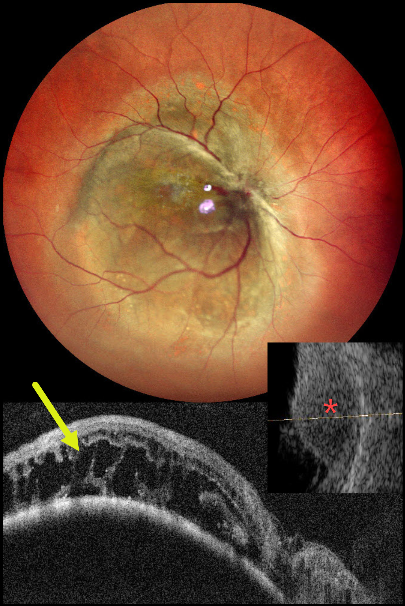
UVEAL MELANOMA
Malvika Singh and Manish Nagpal
Originally posted on @retina.rocks November 11, 2025
This healthy 42YO female presented with one and a half years of progressive vision in her right eye. Vision was counting fingers OD and 20/20 in her normal OS.
Pseudocolor SLO imaging shows a large, elevated pigmented choroidal lesion occupying the entire macula and extending into the midperiphery. OCT shows marked cystic outer retinal edema overlying the tumor. The lesion, which measures 6.9mm in thickness, has moderate homogenous internal reflectivity on B-scan ultrasonography. She was referred to ocular oncology for further evaluation and treatment.
Learning points:
Uveal malignant melanoma is a malignant tumor arising from melanocytes in the uveal tract (iris, ciliary body, or choroid). The choroid is the most common site, accounting for 90% of uveal melanomas. Uveal melanoma is the most common primary intraocular cancer in adults, whereas metastatic disease is the most common intraocular malignancy. The liver is the most common metastatic site, although metastatic disease is rare on presentation. Metastases are more common in large tumors, with BAP1 mutations, and with certain tumor-related genetic markers (including positive PRAME and Class 2).
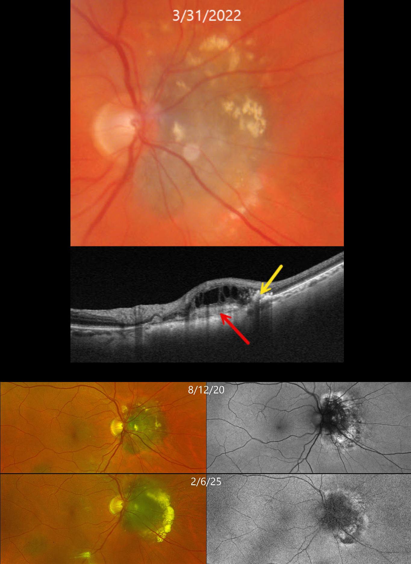
CHOROIDAL NEVUS WITH CNV
Originally posted on @retina.rocks October 15, 2025
This 71YO female has been followed for several years with a choroidal nevus and chronic overlying choroidal neovascularization (CNV).
On March 31, 2022, Triton color photography shows a nasal peripapillary choroidal nevus that touches the optic nerve. Lipid is noted mostly along its superior and temporal edges. Swept-source OCT shows a hyperreflective type 2 (above the RPE) CNV, hyperreflective lipid, and overlying cystic retinal thickening.
The CNV has been observed due to her complete lack of symptoms and extramacular location of the lesion. When last examined on February 6, 2025, the nevus looks fairly stable on Optos RG imaging, although there is increased subretinal lipid along its temporal edge. On fundus autofluorescence (FAF), the CN is hypo-FAF with hyper-FAF along its periphery.
Learning Points:
Choroidal neovascularization is a rare finding in choroidal nevi, and associated exudation can be misinterpreted as exudation from possible malignant transformation. OCT angiography is helpful in diagnosing these lesions, which respond well to either intravitreal anti-VEGF monotherapy or combination treatment with photodynamic therapy (Munie and Demirci, Ophthalmology Retina 2018;2:53-58).
UVEAL MELANOMA
Ankit Jain and Manish Nagpal
Originally posted on @retina.rocks September 3, 2025
This 47YO female presented with 1 month of decreased vision in her right eye. Vision was 20/400 OD and 20/20 in her normal OS.
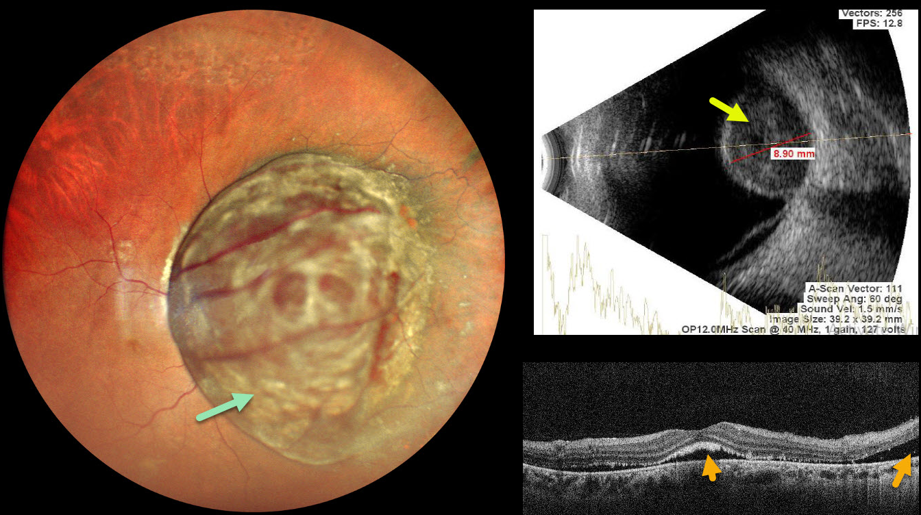
Pseudocolor SLO imaging shows a large, well-circumscribed pigmented choroidal lesion that obscures the entire posterior pole. The tumor vessels are markedly dilated and tortuous. OCT shows foveal and nasal subretinal fluid with ragged shed outer segments along the posterior retinal surface. The lesion, which measures 8.9mm in thickness, shows moderate homogenous internal reflectivity on B-scan ultrasonography. She was referred to ocular oncology for further evaluation and treatment.
Learning Points:
Uveal melanoma is a malignant tumor arising from melanocytes in the uveal tract (iris, ciliary body, or choroid). The choroid is the most common site, accounting for 90% of uveal melanomas. Uveal melanoma is the most common primary intraocular cancer in adults, whereas metastatic disease is the most common intraocular malignancy. The liver is the most common metastatic site, although metastatic disease is rare on presentation. Metastases are more common with large tumors, BAP1 mutations, and certain tumor-related genetic markers (including positive PRAME and Class 2).
CHOROIDAL METASTASTES
Keissy Sousa
Originally posted on @retina.rocks August 13, 2025
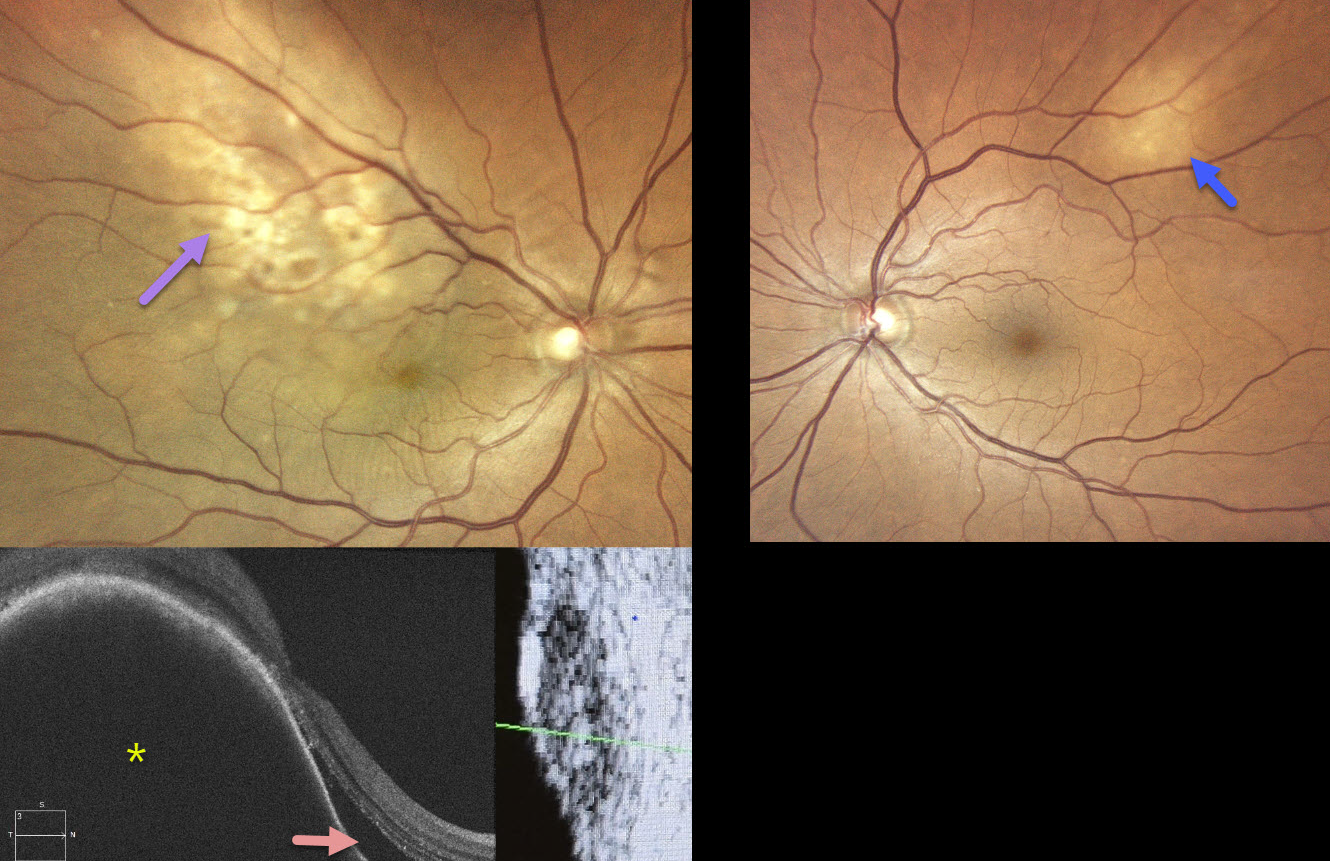
This healthy 70YO female recently underwent cataract surgery elsewhere for vision loss in her right eye. Vision failed to improve postoperatively. Vision was 20/40 OD and 20/20 OS.
Color photography OD shows an ill-defined amelanotic macular choroidal lesion extending into the superotemporal midperiphery. There are some macular chorioretinal folds. OCT scanning shows a markedly thickened choroidal lesion with overlying subretinal fluid. B-scan ultrasonography shows moderate homogenous internal reflectivity. The left eye shows a much smaller amelanotic choroidal lesion superotemporally.
MRI scanning showed a right front paramedial metastatic lesion, and she was subsequently diagnosed with a right-sided primary pulmonary neoplasia. Systemic chemotherapy with pulmonary radiotherapy began without local treatment for the choroidal metastases. When examined 5 months later, vision was no light perception OD and 20/20 OS. The right optic nerve was pale, and the metastatic lesions were fairly stable bilaterally (not shown).
Learning Points:
Although uveal melanoma is the most common primary intraocular malignancy, metastatic choroidal tumors are the most common intraocular malignancies. Lung and breast cancer are the most common sources. Although we can’t be certain, we believe that our patient’s ‘cataract’ symptoms were most likely caused by a missed choroidal metastasis. This underscores the importance of a thorough preoperative dilated funduscopic exam before cataract surgery. In our patient’s case, this not only led to an unnecessary cataract procedure but also delayed the diagnosis and treatment of her metastatic disease.
CHOROIDAL HEMANGIOMA
Moazzam Parvez and Krishnendu Nandi
Originally posted on @retina.rocks July 14, 2025
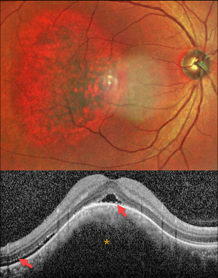
This healthy 42YO male presented with blurred and distorted vision in his right eye. Vision was 20/80 OD and 20/20 in his normal OS.
MultiColor SLO imaging shows an oval orange-red choroidal lesion in the central and temporal macula. OCT scanning shows an elevated choroidal lesion with overlying foveal and distal temporal subretinal fluid. The outer retinal layers are thinned and somewhat disorganized. Shaggy hyperreflective material overlies the central RPE.
Learning Points:
Choroidal hemangioma is a benign vascular choroidal tumor. Circumscribed tumors are isolated and non-syndromic, and appear as orange-red, dome-shaped posterior pole lesions. Diffuse hemangiomas are usually seen in association with the Sturge-Weber syndrome. The lesions are difficult to see funduscopically because the orange tumor blends with the surrounding choroid. In our patient, the MultiColor imaging more dramatically highlights this lesion.
Although there is no malignant potential, it can cause vision loss from exudation with subretinal and intraretinal fluid, which is best treated with photodynamic therapy (Yang et al, Surv Ophthalmology 2025;70:389-400).
UVEAL MELANOMA
Ayushi Gupta and Vishal Agrawal
Originally posted on @retina.rocks July 7, 2025
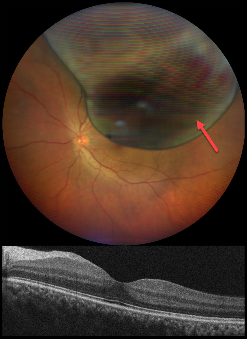
This 32YO male presented with a peripheral shadow of vision in his left eye for several weeks. Vision was 20/20 OU.
Color photography shows a large, elevated, well- defined, pigmented choroidal mass with a few overlying hemorrhages. Despite the lesion nearly draping over the central macula, foveal OCT scanning is remarkably normal. He was immediately referred to an ocular oncologist for a presumed uveal malignant melanoma. He was immediately lost to follow up.
Learning Points:
Uveal melanoma is a malignant tumor arising from melanocytes in the uveal tract (iris, ciliary body, or choroid). The choroid is the most common site, accounting for 90% of uveal melanomas. Uveal melanoma is the most common primary intraocular cancer in adults, whereas metastatic disease is the most common intraocular malignancy. The liver is the most common metastatic site, although metastatic disease is rare on presentation. Metastases are more common with large tumors, BAP1 mutations, and certain tumor-related genetic markers (including positive PRAME and Class 2).
Despite our patient having normal vision, their left eye harbored a blinding and potentially lethal uveal MM. This reminds us why we must always listen to our patient and explain their symptoms even with a normal acuity.
UVEAL LYMPHOMA
Janelle Adeniran
Originally posted on @retina.rocks June 19, 2025
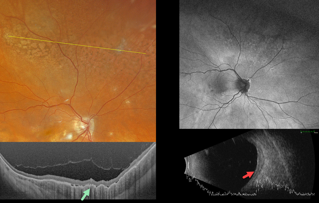
This 69YO female was receiving ongoing bilateral anti-VEGF therapy for neovascular age-related macular degeneration when she presented with new asymptomatic fundus changes in her left eye. There was no significant past medical history.
Optos color RGB imaging shows a leopard spot pattern of superior midperipheral pigmentary changes. Silverstone swept-source OCT shows a markedly thickened choroid with overlying chorioretinal folds. Fundus autofluorescence (FAF) is fairly unremarkable despite these funduscopic findings. The choroid is diffusely thickened on B-scan ultrasonography, with a maximal thickness of 1.90mm.
CT of the chest, abdomen, and pelvis was negative. MRI of the brain and orbits was also negative. We felt this represented primary uveal lymphoma, and this was confirmed with an outside third opinion before beginning serial intravitreal methotrexate injections. After 11 months of treatment, the choroid flattened clinically and on ultrasonography, shrinking to 1.09mm in thickness (not shown).
CHOROIDAL METASTASES WITH EXUDATIVE RETINAL DETACHMENT
Ayushi Gupta and Vishal Agrawal
Originally posted on @retina.rocks June 18, 2025
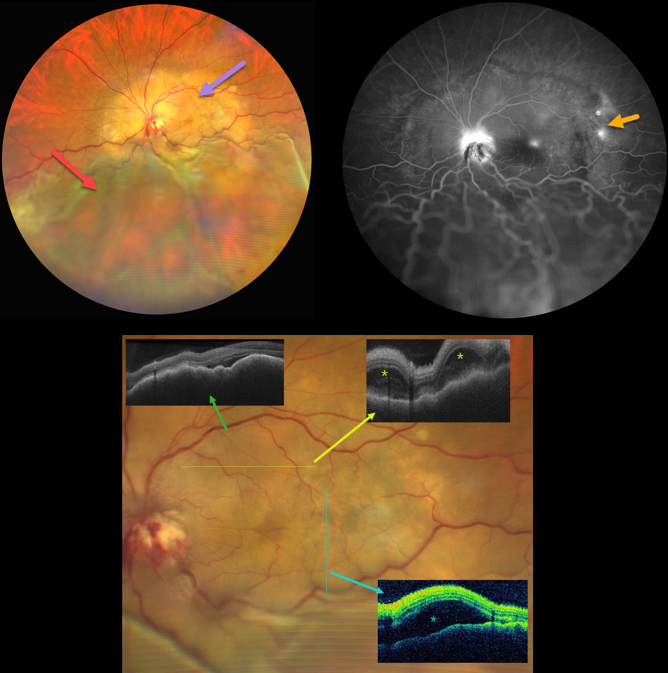
This 47YO female with a known history of metastatic breast carcinoma presented with several weeks of decreased vision in her left eye. She was on active chemotherapy and hormonal therapy. Vision was no light perception OD following trauma 30 years earlier, and hand motion OS.
Ultrawidefield color photography shows a large, amelanotic yellow choroidal lesion involving the entire macula and peripapillary choroid, which extends into the midperiphery. A large secondary inferior exudative retinal detachment involves the entire inferior hemisphere. This fluid shifted when laid supine. The optic disc is hyperemic with indistinct margins and associated blood.
OCT scanning shows a thickened, undulating choroid with variable overlying subretinal fluid and bacillary layer detachments. Fluorescein angiography shows multifocal subretinal leaks, choroidal lesion staining with possible overlying pooling, and intense nerve leakage.
Learning Points:
Although uveal melanoma is the most common primary intraocular malignancy, metastatic choroidal tumors are the most common intraocular malignancies. Lung and breast cancer are the most common sources.
Our patient had a known history of metastatic disease when she presented with this large lesion occupying her entire posterior pole. The extent of the metastasis and the overlying exudation are atypical in our experience. She was immediately referred to her oncologist for external-beam radiation, but was lost to follow-up.
RADIATION RETINOPATHY AND PAPILLOPATHY
Mattie Adams
Originally posted on @retina.rocks May 27, 2025
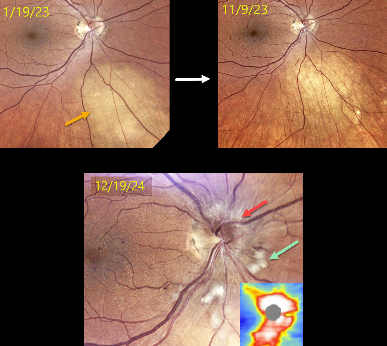
This 52YO female presented on 1/19/23 with a history of metastatic breast cancer and an asymptomatic uveal metastasis in her right eye. The lesion completely regressed following external beam radiation.
When examined on 12/19/24, she remained visually asymptomatic with 20/60 vision. Color photography shows a new, mildly swollen nerve, which is confirmed on OCT. Scattered nerve fiber layer infarcts (cotton-wool spots, CWS) and some nasal macular telangiectasia are also noted.
Learning Points:
Radiation retinopathy usually occurs 3 months to 3 years after external-beam radiation or plaque radiotherapy. The ocular findings closely resemble those seen with diabetic retinopathy. Radiation optic neuropathy (Carey et al, Br J Ophthalmol 2023;107:743-749) can also develop.
Since our patient was completely asymptomatic without macular edema, we recommended close observation. We will begin intravitreal anti-VEGF therapy if she develops macular edema or worsening nerve findings.
CHOROIDAL METASTASES
Mattie Adams
Originally posted on @retina.rocks May 20, 2025
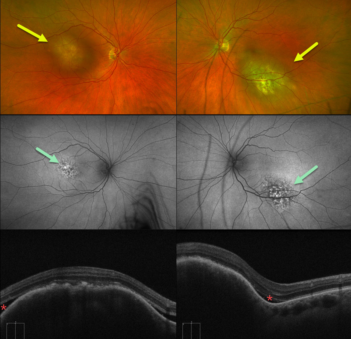
This 62YO female presented with 1 week of bilateral vision loss. She had a known history of metastatic urothelial bladder carcinoma. Vision was counting fingers OD and 20/100 OS.
Optos color RG imaging shows bilateral elevated submacular choroidal lesions. These lesions were clinically creamy in coloration, although they appeared variably amelanotic and pigmented on Optos pseudocolor imaging. Fundus autofluorescence (FAF) shows speckled hyper- and hypo-FAF overlying the superonasal portion of the lesion OD and the inferonasal portion of the lesion OS. The remainder of each lesion is mildly hyper-FAF. Some localized overlying subretinal fluid is present along the edges of the choroidal tumors.
Although uveal melanoma is the most common primary intraocular malignancy, metastatic choroidal tumors are the most common intraocular malignancies. Lung and breast cancer are the most common sources.
We immediately spoke with the patient’s oncologist and suggested referral to a radiation oncologist for external beam radiation to the choroidal metastases. Unfortunately, she died within several months of seeing us.
RECURRENT UVEAL MELANOMA
Originally posted on @retina.rocks May 19, 2025
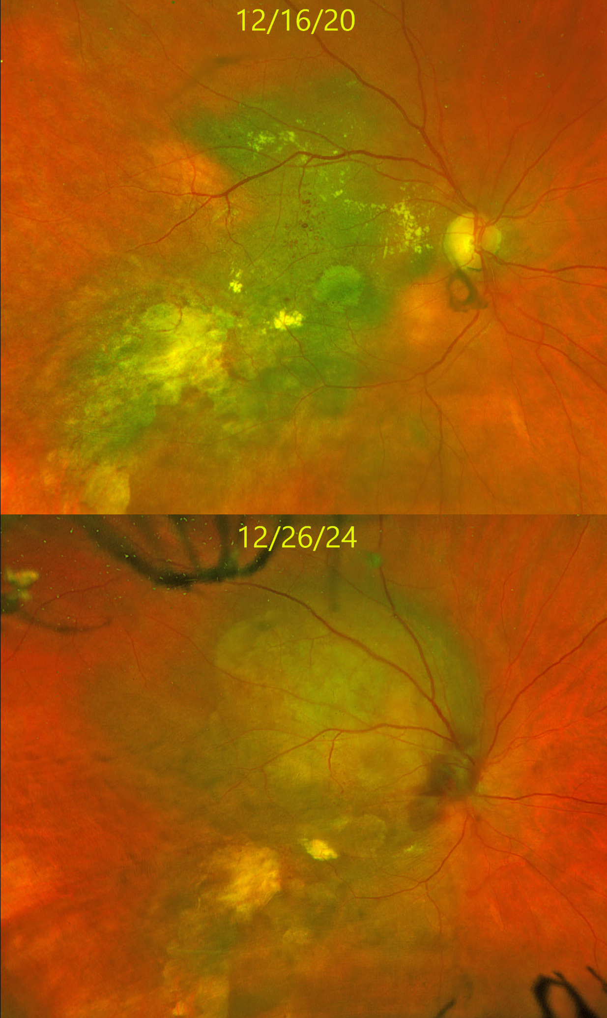
This 66YO female underwent plaque radiotherapy for a uveal malignant melanoma (MM) seventeen years earlier in 2007. The tumor responded well to this single treatment, and she has been followed twice yearly since. On 12/16/24, she had no new symptoms, and vision was stable at counting-fingers.
When examined about 8 months earlier, on 3/13/24, the tumor was stable compared with the prior Optos color RG image from 12/16/20. On the 12/26/24 visit, there is marked tumor growth with apparent optic nerve involvement/
Metastatic workup, including brain MRI and CT of the chest, abdomen, and pelvis, showed no metastases. She was referred to ocular oncology for further management.
Learning Points:
It is extremely rare for a previously dormant tumor to recur so many years following initially successful treatment. The Collaborative Ocular Melanoma Study (COMS) found that local treatment failure occurred in 10% of eyes, most commonly within 3 years of treatment (Jampol et al, Ophthalmology 2002;109:2197-2206). Our case highlights the need for continued long-term monitoring of these patients.
UVEAL MELANOMA
Ankit Jain and Manish Nagpal
Originally posted on @retina.rocks May 8, 2025
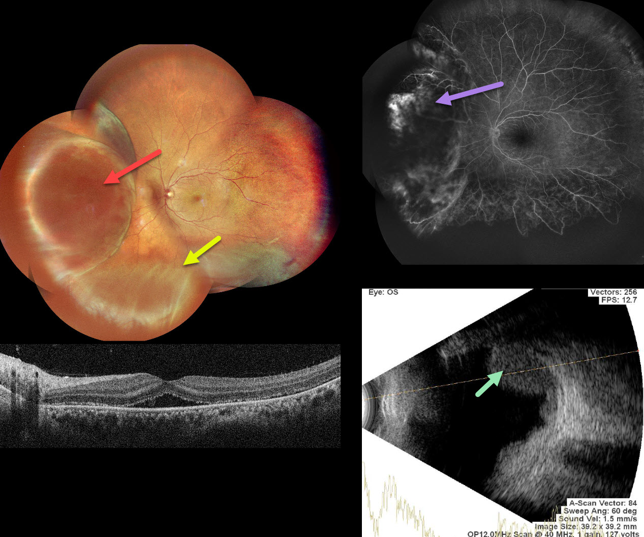
This 39 YO female presented with 1 month of decreased vision in her left eye. Vision was 20/20 in his normal OD and 20/60 OS.
Pseudocolor SLO imaging shows a large, nasal, well-circumscribed amelanotic choroidal lesion with an inferior exudative retinal detachment. OCT shows foveal subretinal fluid with likely shed outer segments along the posterior retinal surface.
Fluorescein angiography shows some leakage within the choroidal tumor. Severe peripheral retinal ischemia is noted temporally and inferiorly. The lesion shows moderate homogenous internal reflectivity on B-scan ultrasonography. MRI (not shown) was suggestive of a uveal malignant melanoma (MM), measuring 11×7 mm in size.
She was referred to ocular oncology for further treatment and evaluation.
CHOROIDAL NEVUS WITH MNV
Originally posted on @retina.rocks April 24, 2025
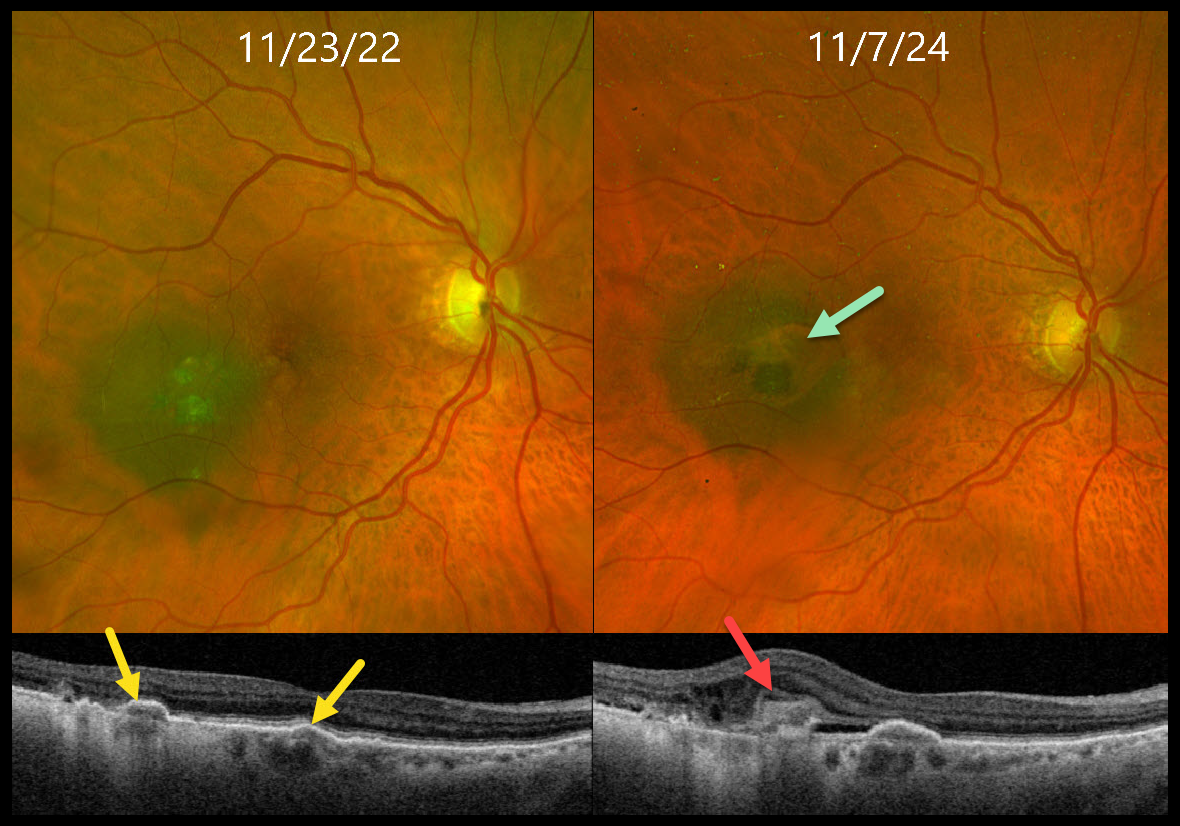
This 85YO female presented for her annual choroidal nevus examination, noticing worsening reading vision. Vision had decreased to 20/200 from 20/40 one year earlier.
When seen 2 years earlier, Triton swept-source OCT showed a few areas of drusen-like changes overlying a temporal macular nevus. At the current visit, although the margins of the nevus were unchanged, Optos color RGB imaging shows a new area of relative depigmentation with a nodular area of pigmented subretinal thickening. OCT scanning shows a hyperreflective type 2 (above the RPE) macular neovascularization (MNV) with mild associated fluid.
Monthly intravitreal Avastin therapy was started. The lesion regressed after several monthly injections, vision improved to 20/60, and a treat-and-extend regimen was started.
Learning Points:
Macular neovascularization is a rare complication of choroidal nevi. Intravitreal Avastin monotherapy is usually successful, although photodynamic therapy may be needed in some cases (Munie and Demirci, Ophthalmology Retina 2018;2:53-58).
We were initially somewhat concerned that the OCT appearance of the type 2 MNV might represent early tumor proliferation through Bruch’s membrane, but the absence of underlying choroidal thickening and the prompt response to anti-VEGF treatment argued against this.
CHOROIDAL HEMANGIOMA
Ayushi Gupta and Vishal Agrawal
Originally posted on @retina.rocks March 25, 2025
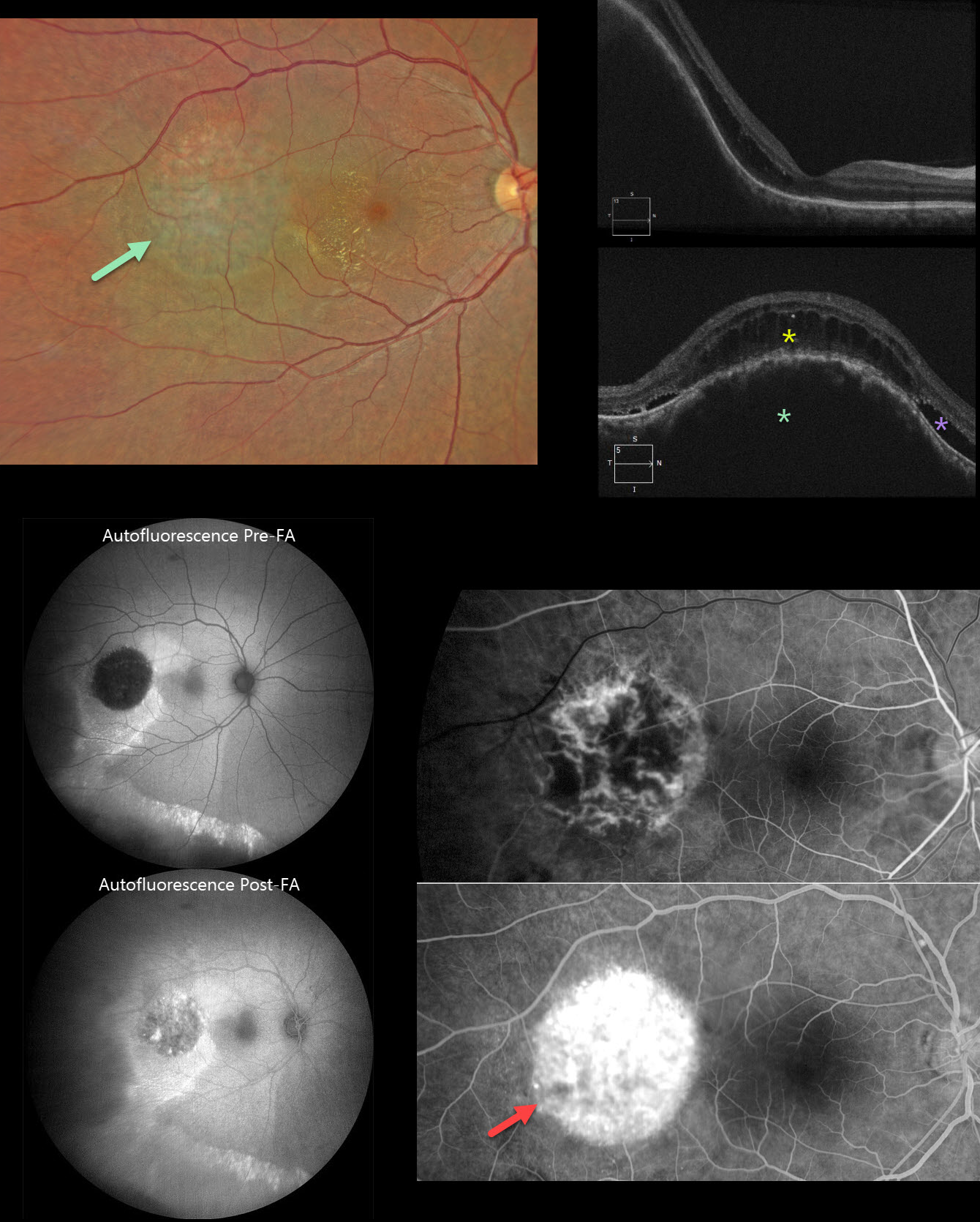
This 37YO male presented with a 3-week history of decreased vision in his right eye. Vision was 20/70 OD and 20/20 in his normal OS.
Color photography shows a subretinal lesion with a somewhat orange hue in the temporal macula. Lipid extends into the temporal fovea. Some subtle pigmentary changes extend inferiorly. OCT scanning shows marked choroidal thickening within the lesion with overlying cystic retinal edema and some subretinal fluid.
On fundus autofluorescence (FAF), the lesion is hypo-FAF with surrounding hyper-FAF and a hyper-FAF gutter extending inferiorly. The lesion becomes hyperfluorescent following fundus fluorescein angiography (FFA). The lesion shows intrinsic vasculature with late leakage on FFA. Unfortunately, our patient was lost to follow-up immediately.
Learning Points:
Choroidal hemangioma is an uncommon benign hamartomatous disorder that presents as either a circumscribed, orange-red, round choroidal lesion (our patient), which is almost always isolated and nonsyndromic, or a diffuse form, which is usually part of Sturge-Weber syndrome. Although there is no malignant potential, it can cause vision loss due to exudation of subretinal and intraretinal fluid, which is best treated with photodynamic therapy (see Yang et al, Surv Ophthalmology 2025;70:389-400). Lipid exudate is a rare finding.
Our case also reminds us why fundus imaging is performed in a specific order: color photography, followed by FAF, then FFA. In FAF, lipofuscin within the RPE is the intrinsic pigment that generates the FAF signal. In FFA, fluorescence is generated by sodium fluorescein that is injected intravenously. Our patient’s hemangioma is hypo-FAF, but when fluorescein stains the lesion, it no longer generates an ‘autofluorescent’ signal due to the fluorescein dye.
CHOROIDAL HEMANGIOMA
Anand Temkar, Navneet Mehrotra, and Manish Nagpal
Originally posted on @retina.rocks December 31, 2024
This 42YO male presented with two months of vision loss in his right eye. Vision was 20/200 OD and 20/20 in his normal left eye.
Mirante pseudocolor SLO imaging shows a pigmented subretinal lesion occupying the entire macula. OCT shows a markedly thickened and elevated choroid with overlying cystic retinal edema and subretinal fluid. The lesion is hyperfluorescent on angiography. B-scan ultrasonography shows it to have homogeneous high internal reflectivity with localized subretinal fluid inferiorly.
Learning Points:
Choroidal hemangioma is an uncommon benign hamartomatous disorder that presents as either a circumscribed, orange-red, round choroidal lesion (as in our patient), which is almost always isolated and nonsyndromic, or a diffuse form that is usually part of Sturge-Weber syndrome.
Although it has no malignant potential, it can cause vision loss due to exudation of subretinal and intraretinal fluid, which can be treated with photodynamic therapy (see Tsipursky et al., Surv Ophthalmology 2011;56:68-85).
The Mirante SLO uses blue (488nm), green (532nm), and red (670nm) confocal lasers to generate a fundus image. The blue laser best images the vitreoretinal interface and inner retina, the green images the mid retina, and the infrared images the outer retina, RPE, and choroid (Roy et al, Surv Ophthalmology 2024;69:378-402). Combining these 3 wavelengths into a single file produces a pseudocolor image that often provides the clinician with more information than a standard color photo. However, in some disorders, including choroidal hemangioma, this technology creates a false hyperpigmented appearance, which can lead to the misdiagnosis of uveal melanoma (Becker et al, OSLIR 2023;54:292-296).
HODGKIN LYMPHOMA MIMICKING MULTIFOCAL CHOROIDITIS
Originally posted on @retina.rocks December 4, 2024
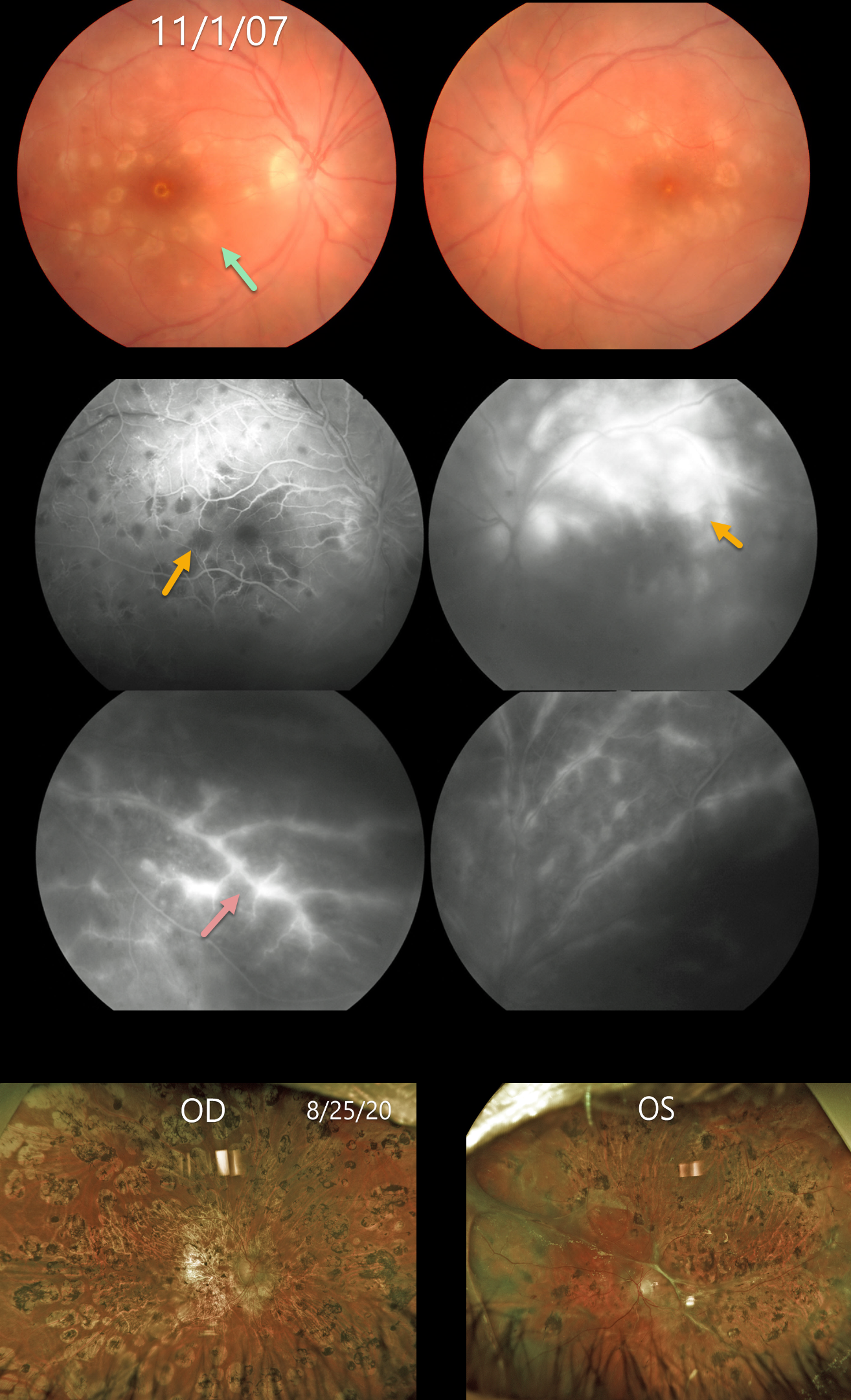
This 18YO female presented on 11/1/07 with acute bilateral panuveitis. Moderate vitreous cells were present along with subretinal multifocal yellow-white choroidal infiltrates. Angiographically, these choroidal lesions blocked early and stained late. A frosted-branch pattern of vascular leakage was noted bilaterally. Following a course of oral prednisone, the acute uveitic findings resolved, leaving behind inactive multifocal scarring. She developed severe recurrent panuveitis a year or two later, leaving her with 20/400 vision bilaterally (images not shown).
Shortly thereafter, she was diagnosed with Hodgkin lymphoma. At almost the exact date of this diagnosis, Baillif et al reported a patient with bilateral angle-closure glaucoma and multifocal choroiditis as the initial presentation for Hodgkin lymphoma (Retinal Cases 2011;5:201-205). The posterior segment findings in this report were virtually identical to those in our patient. The authors felt that the uveitis was a paraneoplastic phenomenon.
She subsequently underwent successful bone marrow transplantation. Her lymphoma and uveitis have both remained in remission since. A rhegmatogenous retinal detachment later developed in her left eye, which was repaired with vitrectomy and silicone oil. Despite successful reattachment, vision subsequently decreased to no light perception. On 8/25/20, vision was 20/400 OD with severe stable bilateral chorioretinal scarring bilaterally.
Learning Points:
Multifocal choroiditis (MFC) is the current preferred term for a group of historically heterogeneous disorders, including multifocal choroiditis, recurrent multifocal choroiditis, multifocal choroiditis and panuveitis, punctate inner choroidopathy, progressive subretinal fibrosis, and pseudo-histoplasmosis (Essex et al, Retina 2013;33:1-4 and Spaide et al, Retina 2013;33:1315-1324). MFC occurs in otherwise healthy adults, often myopic and female. Findings can include variably pigmented multifocal scars throughout the fundus, panuveitis, subretinal fibrosis, and macular neovascularization (MNV). Unlike ocular histoplasmosis, new scars and uveitis can be seen. MNV can be successfully managed with anti-VEGF therapy.
As with any uveitis, the clinician must always include the possibility of a masquerade syndrome as occurred in our patient.
CHOROIDAL HEMANGIOMA MISDIAGNOSED AS UVEAL MELANOMA
Originally posted on @retina.rocks November 19, 2024
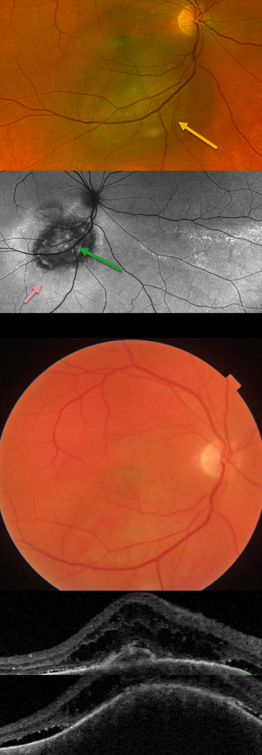
This 44YO male was referred for recent vision loss from a possible uveal melanoma. Vision was 20/150 in his right eye and 20/20 in his healthy left eye.
Optos color RG imaging shows an elevated, pigmented subretinal lesion extending from the inferior macula below the inferotemporal arcade. Fundus autofluorescence (FAF) shows this lesion to be hypo-FAF, with surrounding hyper-FAF and areas of hyper-FAF extending into the inferior and inferotemporal midperiphery, indicating prior extramacular subretinal fluid.
Triton color imaging, however, shows the choroidal lesion as orange. Swept-source OCT shows a thickened choroidal lesion with overlying subretinal fluid and cystic retinal edema. The lesion measured 9.6 x 9.0mm in basal diameter and 3.4mm thick with B-scan ultrasonography (not shown). There was moderate-to-high internal reflectivity, consistent with a choroidal hemangioma. Photodynamic therapy was recommended.
Learning Points:
Isolated choroidal hemangiomas are benign vascular lesions. Although they have no malignant potential, they can cause vision loss due to exudation of subretinal and intraretinal fluid, which can be treated with photodynamic therapy (see Tsipursky et al, Surv Ophthalmology 2011;56:68-85).
This lesion was initially misdiagnosed as a uveal malignant melanoma based on the hyperpigmented appearance on Optos RG imaging. This has also been reported in the literature (Becker et al, OSLIR 2023;54:292-296). These images are generated from a red (635nm) and green (532nm) laser. This creates a greenish tint, which distorts the true fundus color. In our patient, this was quite dramatic, with the hemangioma’s true orange color evident only in the Triton image. A newer Optos true-color RGB unit has recently been introduced, which maintains the ability to view the separate RG channels while also generating a true-to-life color image.
CHOROIDAL METASTASES
Ankit Jain, Navneet Mehrotra and Manish Nagpal
Originally posted on @retina.rocks November 14, 2024
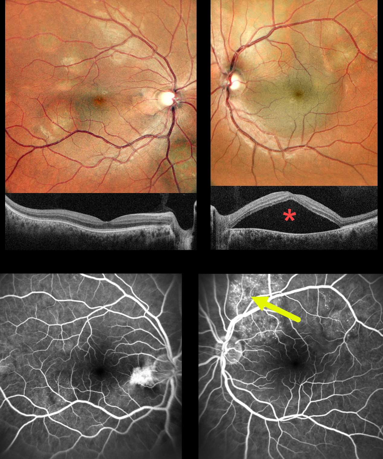
This previously healthy 30YO female presented with several weeks of decreased vision in her left eye. Vision was 20/20 OD and 20/60 OS.
MultiColor imaging shows bilateral multifocal variably sized yellow-white choroidal lesions throughout each posterior pole. A large serous detachment occupies the entire left macula, which is confirmed on SD-OCT. Fluorescein angiography of the right eye shows some speckled subretinal hyperfluorescence overlying a choroidal lesion in the distal inferotemporal macula. Angiography of the left eye shows similar but more extensive hyperfluorescence overlying the superior and superotemporal choroidal lesions, likely responsible for the serous macular fluid.
An extensive medical workup revealed widely metastatic pulmonary adenocarcinoma, and she was subsequently referred to oncology. She was unfortunately lost to follow-up immediately.
Learning Points:
Although uveal melanoma is the most common primary intraocular malignancy, metastatic choroidal tumors are the most common intraocular malignancies. Lung and breast cancer are the most common sources. The extensive multifocal lesions in each of our patient’s eyes are unusual, since most patients have 1-2 choroidal lesions (Shields et al Retina 2020;40:204-213).
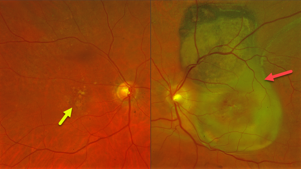
PSEUDO-UVEAL MELANOMA FROM NEOVASCULAR AMD SUBRETINAL FIBROSIS
Fraser McKay
Originally posted on @retina.rocks June 20, 2024
This 78YO female presented with at least several months of vision loss in her left eye. Vision was 20/40 OD and light perception OS. She was examined 2 years earlier with 20/400 vision OS from macular atrophy.
Optos color RG imaging OS shows a variably pigmented and slightly thickened subretinal lesion extending from the superior macula into the superior midperiphery. Subretinal fibrosis extends through the macula, along with retinal blood dots. Intermediate AMD with mixed drusen was noted OD.
Learning Points:
Suprachoroidal, subretinal, or sub-RPE blood can rarely simulate a uveal malignant melanoma. We suggested a trial of monthly anti-VEGF therapy for this treatment naïve neovascular AMD lesion, although the visual prognosis remains quite guarded given the extent of submacular fibrosis.
UVEAL MELANOMA
Janelle Adeniran
Originally posted on @retina.rocks June 4, 2024
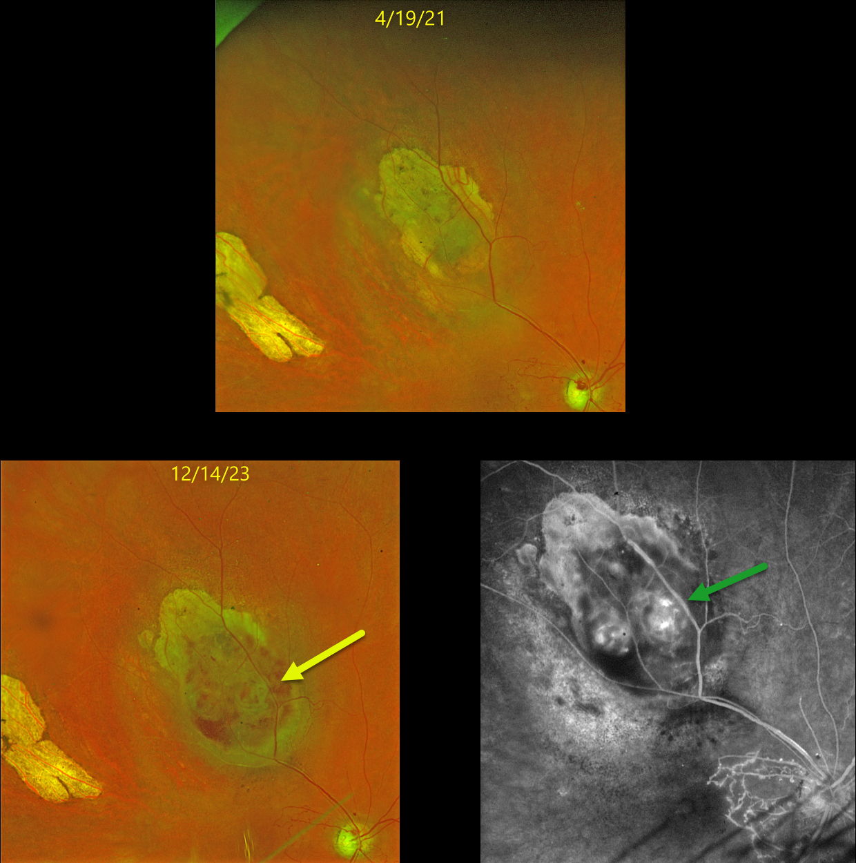
This 59YO female underwent iodine-125 plaque therapy in 2010 for a uveal malignant melanoma (MM) in her right eye. The tumor initially regressed and has remained stable with yearly examinations since. Vision has been stable at counting fingers due to a secondary branch retinal vein occlusion and radiation retinopathy.
In June 2023, she presented with new floaters and vision loss in this eye from a new moderate vitreous hemorrhage coming from the MM. Vision was light perception. Once the blood cleared following an intravitreal Avastin injection, the tumor showed some hemorrhages within or anterior to the lesion on Optos color RGB imaging.
Fluorescein angiography showed intrinsic vascularity and profound macular capillary loss. B-scan ultrasonography revealed a nodular appearance with heterogeneous echogenicity (not shown). There was definite growth compared with the tumor size several years earlier. Metastatic workup was negative. After extensive discussion, the patient elected for enucleation, which was scheduled in the near future.
Learning Points:
The Collaborative Ocular Melanoma Study (COMS) reported a 10% 5-year local treatment failure rate following plaque radiation (Jampol et al., Ophthalmology 2002;109:2197-2206). Our patient’s failure developed about 13 years following brachytherapy and reminds us of the need for long-term surveillance of these treated tumors.
CHOROIDAL CARCINOID METASTASIS
Originally posted on @retina.rocks April 23, 2024
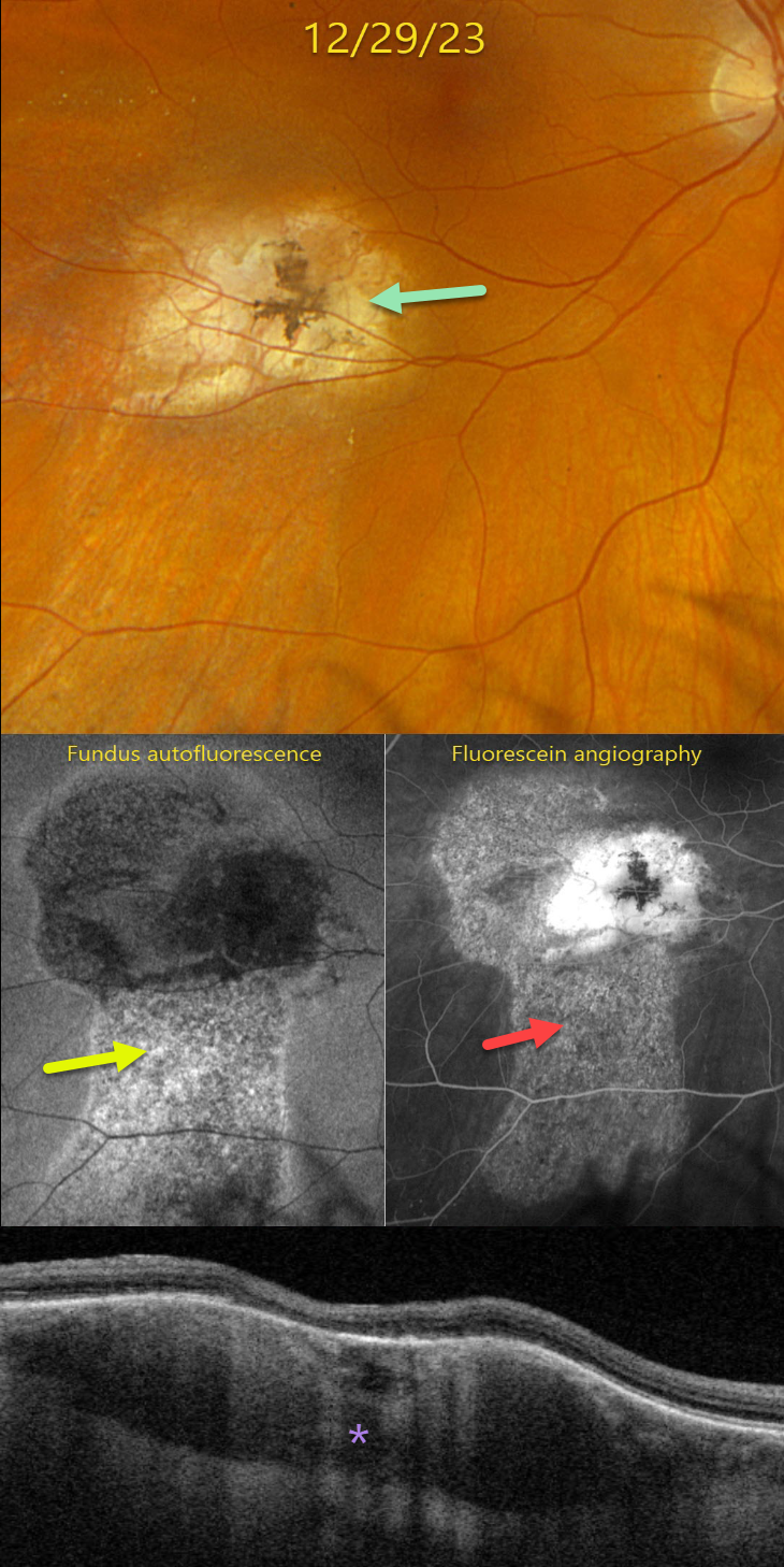
This 43YO female presented to our office with recent vague symptoms of vision loss OD. She was followed elsewhere since 2006 for a carcinoid lesion metastatic to the right choroid. Her right bronchial carcinoid tumor was treated earlier that year with a right pneumonectomy without systemic treatment. Initial observation of the choroidal lesion was recommended, but she was almost immediately lost to follow-up for 13 years.
She returned to the outside practice in 2019, and the choroidal lesion showed slight growth with mild new fluid. She was again lost to follow-up until June 2021, with continued growth and increased fluid. Photodynamic therapy was applied on 6/8/21 and 9/21/21 with subsequent tumor shrinkage and resolution of the fluid.
On 12/29/23, vision was 20/25. Optos color RGB imaging shows an atrophic, variably pigmented, lumpy choroidal lesion in the inferotemporal macula.
Fundus autofluorescence (FAF) shows a hyper-FAF gutter extending inferiorly from the lesion. This gutter is well-visualized on fluorescein angiography, with staining of the inactive tumor.
Triton swept-source OCT shows a variably thickened and mostly hyporeflective choroidal lesion with overlying outer retinal atrophy. These findings were all stable compared to images seen 1 year earlier at the outside practice. We plan to follow her twice a year.
Learning Points:
Although uveal melanoma is the most common primary intraocular malignancy, metastatic choroidal tumors are the most common intraocular malignancies. Lung and breast cancer are the most common sources. These lesions can be treated with systemic chemotherapy and are also usually very responsive to external beam radiation.
Neuroendocrine (carcinoid) tumors are usually low-grade malignancies that arise in the bronchopulmonary system or the gastrointestinal or urogenital tracts. In a recent review of over 1100 patients referred to the Wills ocular oncology service over a 43-year period, pulmonary carcinoid accounted for 2% of cases (Shields et al, Retina 2020;40:204-213). These lesions have a characteristic orange appearance, in contrast to most other choroidal metastases, which are usually creamy yellow (Shields et al, BJO 1996;80:852-853).
SCLEROCHOROIDAL CALCIFICATION
Janelle Adeniran
Originally posted on @retina.rocks October 9, 2023
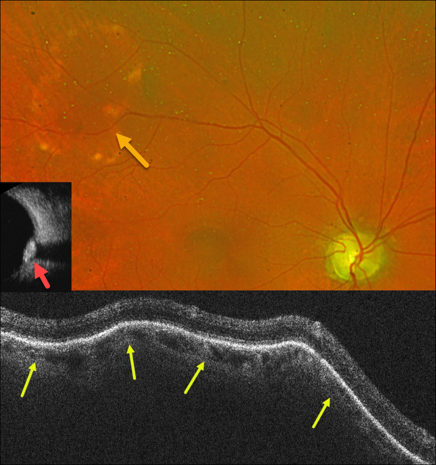
This 76YO male was referred for an asymptomatic lesion in his right fundus. Vision was 20/30.
Optos color RG imaging shows a lumpy, elevated subretinal yellow-white nodular lesion in the superotemporal midperiphery.
OCT scanning through this lesion shows marked lumpy scleral elevation with distortion of the overlying choroid and retina. This lesion was markedly hyperreflective on B-scan ultrasonography, indicating calcification. The left fundus was normal (not shown).
Learning Points:
Sclerochoroidal calcification is a relatively uncommon and benign finding in the elderly population. These yellowish lesions are slightly lumpy and elevated, and usually appear in the mid superior/inferior temporal/nasal quadrants. The lesions are highly reflective on B-scan ultrasonography and are also visible on CT scanning. They require no treatment and are not associated with vision loss.
However, they may be associated with systemic hypercalcemic conditions such as hyperparathyroidism and pseudohypoparathyroidism. Shields et al therefore recommended that all such patients be evaluated for underlying systemic calcium disorders (Retina 2015;35:547-554). Our patient’s bloodwork was fortunately negative.
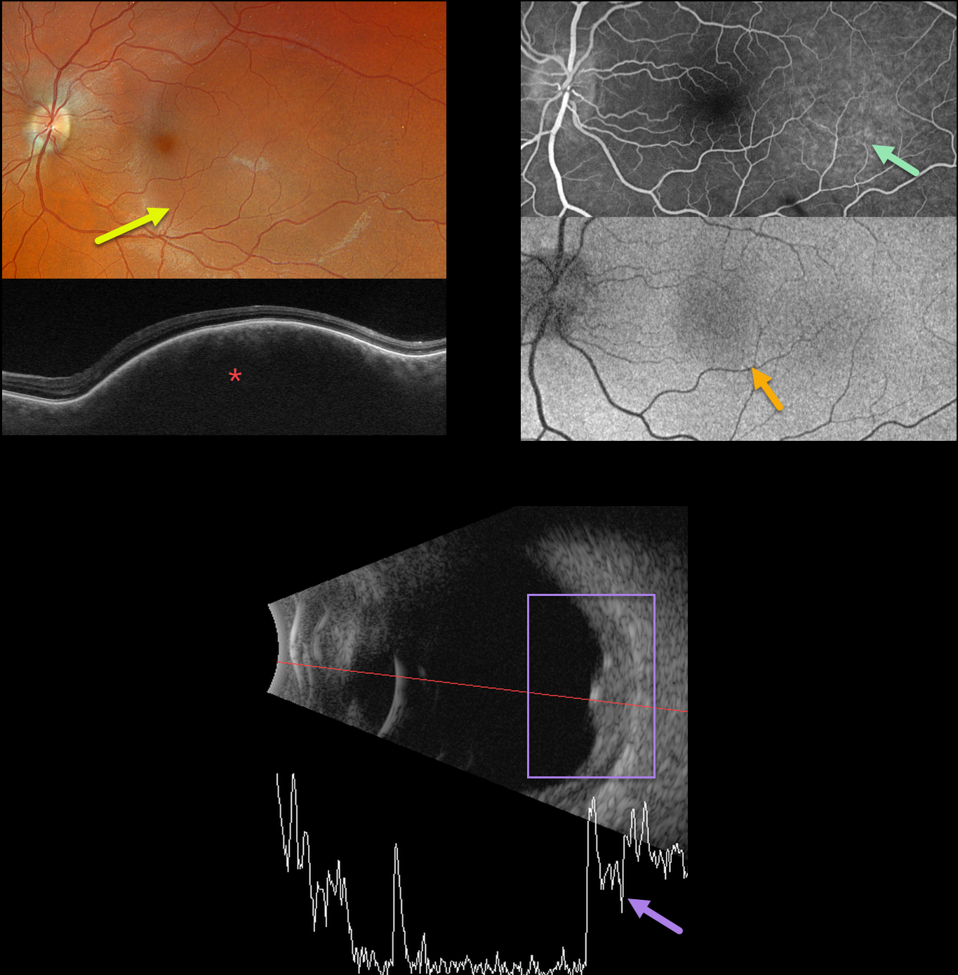
CHOROIDAL HEMANGIOMA
Originally posted on @retina.rocks September 18, 2023
This 35YO female was referred for an asymptomatic left macular lesion. Vision was 20/20 in her normal right eye and 20/30 in her left eye.
Optos color RGB imaging shows a perfectly round subretinal lesion centered in the temporal macula, extending just beneath the macular center.
Swept-source OCT shows a thickened choroidal lesion without overlying subretinal fluid. Fluorescein angiography shows filling of this lesion without leakage, and fundus autofluorescence (FAF) shows faint hypo-FAF.
B-scan ultrasonography shows homogeneous high internal reflectivity.
Learning Points:
Isolated choroidal hemangiomas are benign vascular lesions. Although they have no malignant potential, they can cause vision loss due to exudation of subretinal and intraretinal fluid, which can be treated with photodynamic therapy (see Tsipursky et al, Surv Ophthalmology 2011;56:68-85).
Since our patient’s lesion was asymptomatic and without exudation, we will follow her several times yearly.
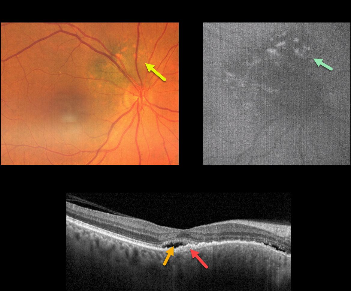
CHOROIDAL NEVUS MISDIAGNOSED AS CENTRAL SEROUS
Emma Oreskovic and Natasa Draca
Originally posted on @retina.rocks September 7, 2023
This 71YO female was referred for further treatment of chronic idiopathic central serous chorioretinopathy (ICSC) of her right eye, for which she had previously received unsuccessful anti-VEGF therapy at another institution. Vision was 20/100 OD and 20/20 in her normal left eye.
Fundus photography shows a variably pigmented, minimally thickened peripapillary choroidal nevus that extends from about 7-1 o’clock around the optic nerve margin. Overlying orange pigmentation is noted, which variably hyper-autofluoresces.
OCT scanning shows shallow foveal subretinal fluid with a subretinal layer of thickened hyperreflective material extending nasally overlying the thickened nevus. Beneath this is a very shallow, irregular elevation of the RPE.
Learning Points:
Clinical characteristics of choroidal nevi at risk of progressing to uveal malignant melanoma (MM) were initially described by Shields et al. (Ophthalmology 1995;102:1351-1361).
The most recent version of this famous acronym (Shields et al, Retina 2019;39:1840-1851) is To Find Small Ocular Melanoma Doing Imaging (TFSOM-DIM), which stands for To (Thickness >2mm) Find (subretinal Fluid) Small (Symptoms, vision <= 20/50) Ocular (Orange pigment) Melanoma (Melanoma hollow on ultrasonography) DIM (DIaMeter > 5mm).
ICSC and pachychoroid disorders, similar to syphilis, are one of the ‘great imitators,’ including uveal MM masquerading as ICSC (Negretti et al, Ophthalmology Retina 2023;7:171-177).
In our patient, on the central macular OCT B-scan, one sees a thickened choroid, subretinal fluid, and hyperreflective shed outer segments/lipofuscin, all of which are suggestive of ICSC.
This case emphasizes the importance of looking beyond the OCT, using both the clinical exam and multimodal imaging, for every patient, including those with chronic ICSC, to arrive at the correct diagnosis and treatment.
Since our patient has several high-risk characteristics (fluid, symptoms, and orange pigment), she was referred to an ocular oncologist for further management.
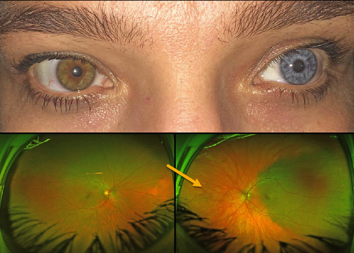
WAARDENBURG SYNDROME
Gil Calvão-Santos and Keissy Sousa
Originally posted on @retina.rocks May 24, 2023
This 43YO female has a constellation of findings that are pathognomonic for Waardenburg syndrome type 1. Vision was 20/20 OU.
External examination shows telecanthus with iris heterochromia. When questioned, she mentioned synophrys (fusion of the eyebrows), which she removed with a laser, poliosis, which she dyed, and a family history of poliosis and heterochromia. She is also congenitally deaf.
Optos imaging shows apparent choroidal melanocytosis temporally in her left eye. But in actuality, this is the only normally pigmented choroid in each eye, with diffuse choroidal hypopigmentation elsewhere.
Learning Points:
Waardenburg syndrome is a group of usually autosomal dominant genetic disorders characterized by achromia of the hair and/or skin, congenital deafness, partial or total iris heterochromia, synophrys, broad and high nasal root, telecanthus, and choroidal hypopigmentation. Vision is usually normal, as was the case in our patient.
See Shields at al for a review of the iris and choroidal abnormalities found in Waardenburg syndrome (JAMA Ophthalmol 2013;131:1167-1173).
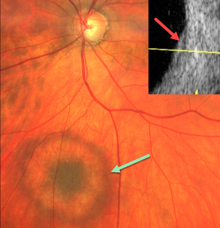
HALO CHOROIDAL NEVUS
Natasa Draca and Emma Oreskovic
Originally posted on @retina.rocks April 26, 2023
During a routine glaucoma examination, this 61YO male was found to have a halo choroidal nevus in his right eye. The nevus was hyperpigmented centrally, then encircled by relative depigmentation, and finally surrounded by a hyperpigmented rim. The lesion was clinically minimally thickened and measured about 2-3 disc diameters. The nevus was barely visible with B-scan ultrasonography.
Learning Points:
Halo choroidal nevi are a relatively rare variant of the more typical diffusely hyperpigmented lesions. A large retrospective series by Shields et al found that these lesions may be associated with cutaneous melanoma (Arch Ophthalmology 2010;128:859-864).
Malignant transformation to uveal melanoma is reduced when a halo is present (Shields et al, Arch Ophthalmol 2009;127:981-987), although this does not preclude malignant transformation.
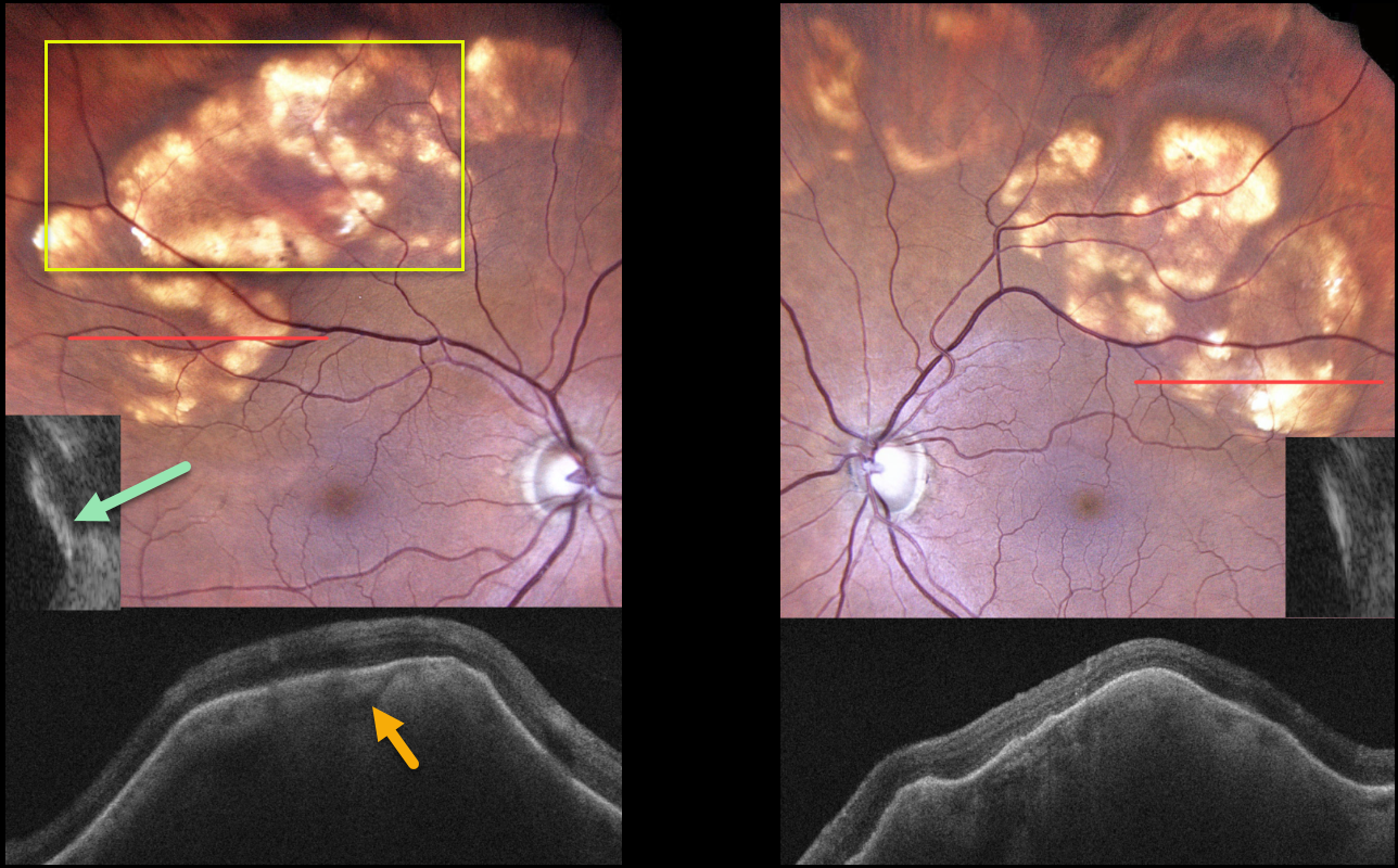
SCLEROCHOROIDAL CALCIFICATION
Originally posted on @retina.rocks April 18, 2023
This 81YO female was referred for asymptomatic retinal changes. Vision was 20/50 OD and 20/30 OS.
Color imaging shows bilateral lumpy, elevated subretinal yellow-white nodules extending from the superotemporal maculas into the midperipheries.
OCT scanning through these lesions shows marked hyperreflective scleral thickening with loss of the overlying choroid. These lesions are markedly hyperreflective on B-scan ultrasonography, indicating calcification.
Learning Points:
Sclerochoroidal calcification is a relatively uncommon and benign finding in the elderly population. These yellowish lesions are slightly lumpy and elevated, and usually appear in the mid superior/inferior temporal/nasal quadrants.
The lesions are highly reflective on B-scan ultrasonography and visible on CT. They require no treatment and are not associated with vision loss.
However, they may be associated with systemic hypercalcemic conditions such as hyperparathyroidism and pseudohypoparathyroidism. Shields et al therefore recommended that all such patients be evaluated for underlying systemic calcium disorders (Retina 2015;35:547-554). Although recommended, our patient refused this blood work.
CHOROIDAL NEVUS
Originally posted on @retina.rocks March 30, 2023
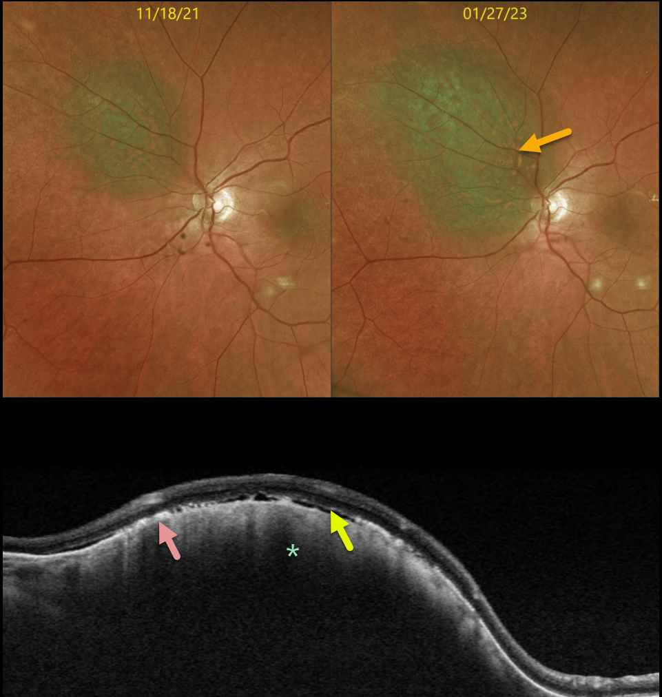
This 71YO female presented on 1/27/23 for follow-up for an asymptomatic, less than 1mm thick choroidal nevus measuring 6.3 x 5.5 mm.
Optos color imaging shows that the nevus has significantly increased in size (3.0 x 10.5 x 7.5 mm) compared with the prior visit on 11/18/21. The nevus enlarged laterally in all dimensions and now touched the nerve margin. There was significantly more overlying orange pigment.
Swept-source OCT shows a thickened choroidal lesion. The clinically noted orange pigment is seen on OCT as hyperreflective outer retinal deposits. Trace variable subretinal fluid vs loss of outer retinal tissue is also seen.
Radioactive plaque placement was performed. Fine needle biopsy revealed the tumor to be Class 1A, PRAME negative.
Learning Points:
Clinical characteristics of choroidal nevi at risk for progressing to a uveal MM were initially described by Shields et al (Ophthalmology 1995;102:1351-1361). The most recent version of this famous acronym (Shields et al, Retina 2019;39:1840-1851) is To Find Small Ocular Melanoma Doing Imaging (TFSOM-DIM), which stands for:
To (Thickness >2mm)
Find (subretinal Fluid)
Small (Symptoms, vision <= 20/50)
Ocular (Orange pigment)
Melanoma (Melanoma hollow on ultrasonography)
DIM (DIaMeter > 5mm).
Although our patient had several of these baseline risk factors (orange pigment, 6.3mm diameter), the growth in just 6 months is unusual.
Patients with Class 1A lesions have a lower 5-year metastatic risk (2%) than those with Class 1B or 2 lesions. PReferentially expressed Antigen in MElanoma (PRAME) is a gene that can be aberrantly expressed in some malignancies. Its positivity can indicate an increased risk for metastatic disease in Class 1 tumors, but its negativity does not tend to alter prognosis.
CHOROIDAL METASTASES
Originally posted on @retina.rocks March 2, 2023
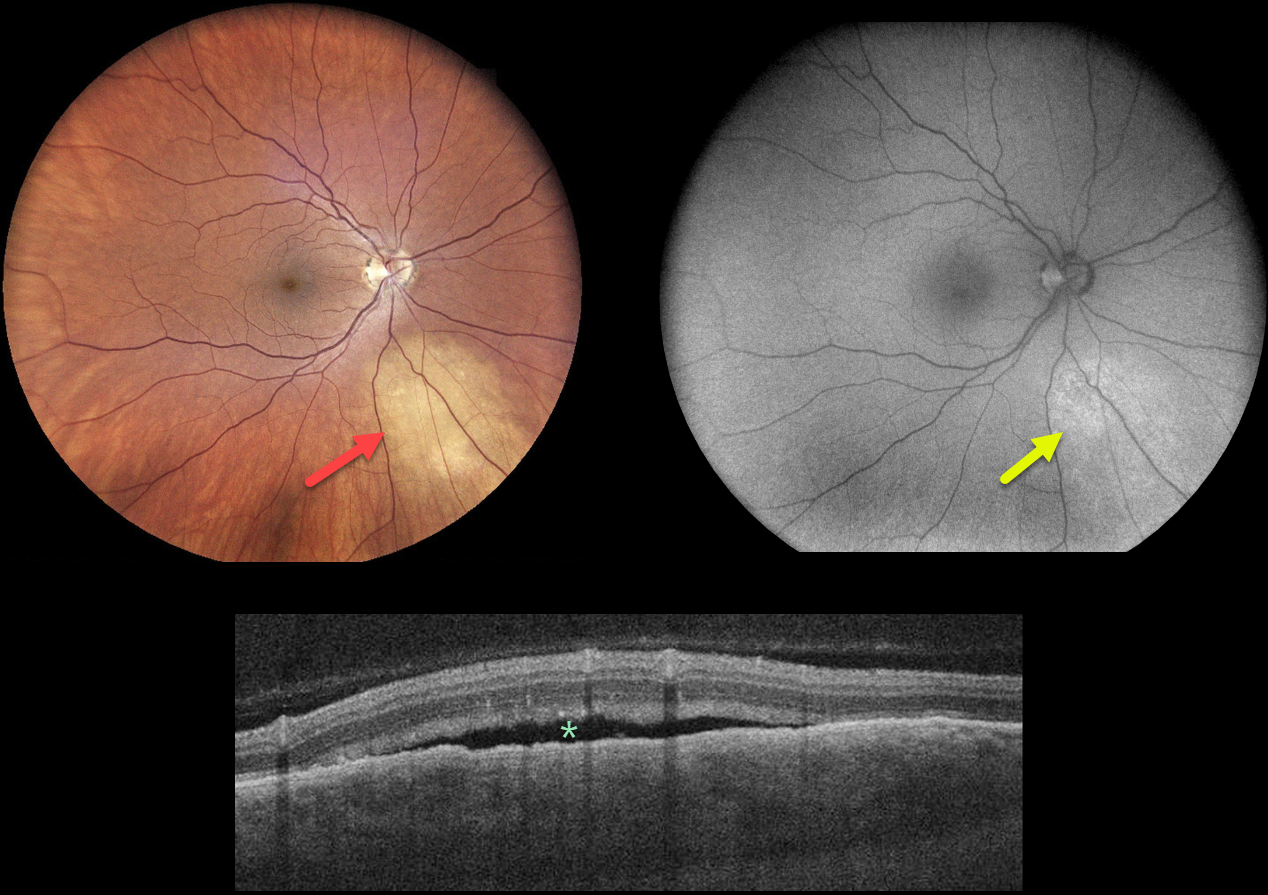
This 52YO female has a 10-year history of breast cancer and a 3-year history of metastatic pulmonary disease, which is controlled with systemic chemotherapy. She was referred for an asymptomatic lesion in her right eye.
Fundus photography shows an amelanotic subretinal lesion inferonasal to the nerve. This lesion faintly hyper-autofluoresces.
OCT scanning through this lesion shows a thickened choroid with a somewhat undulating overlying RPE and shallow subretinal fluid.
Learning Points:
Although uveal melanoma is the most common primary intraocular malignancy, metastatic choroidal tumors are the most common intraocular malignancies. Lung and breast cancer are the most common sources. These lesions can be treated with systemic chemotherapy and are also usually very responsive to external beam radiation.
After discussing the findings with her oncologist, the lesion will be treated with external beam radiation.
UVEAL MELANOMA
Giovanni Cuffaro
Originally posted on @retina.rocks February 6, 2023
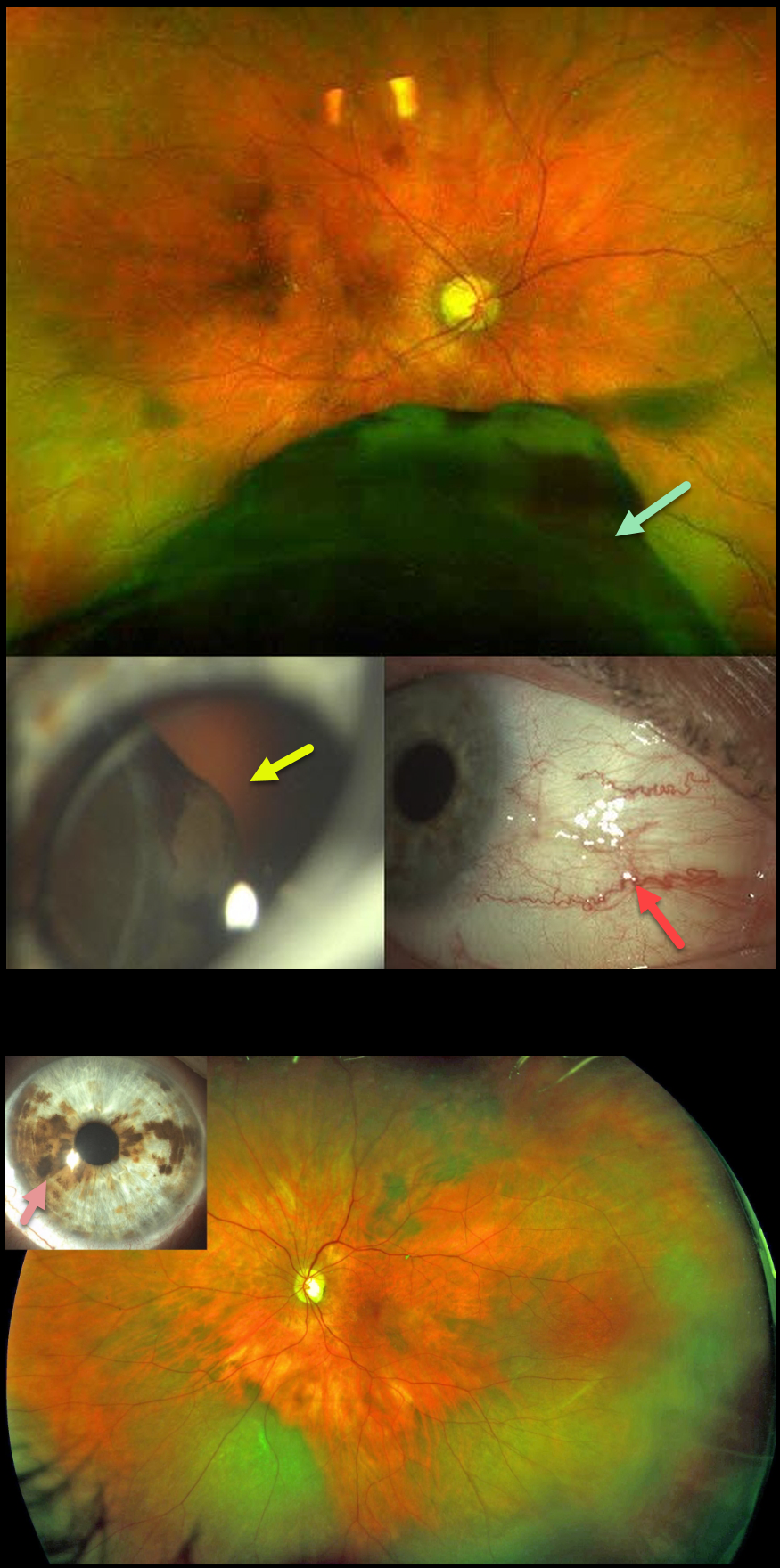
This 71YO female presented in January 2019 with one month of photopsias in her right eye. Optos imaging shows a large inferior malignant melanoma (MM) extending from the inferior retinal periphery into the posterior pole. The tumor was visible through the pupil at the slit lamp, and sentinel vessels were noted. Ocular melanocytosis was noted in the left eye.
She underwent primary enucleation of the right eye one month later. There was complete monosomy 3, and HLA-A*0201 testing was positive.
She subsequently developed a peripheral inferotemporal uveal malignant melanoma in her left eye (axial thickness 5mm, lateral dimensions 13.5 by 13 mm), which was treated with brachytherapy in March 2020. In October 2021, she developed liver metastases.
She was placed on systemic Nivolumab, in addition to external radiotherapy to the liver lesions. Vision is currently 20/20 OS with no signs of local recurrence.
Learning Points:
Our patient had a unique combination of unfortunate multiple risk factors for developing uveal malignant melanoma with metastatic disease (see Kaliki et al, Indian J Ophthalmol 2015;62:93-102).
Monosomy 3 is strongly associated with metastatic disease and melanoma-related mortality. The tumor suppressor gene BRCA1-associated protein 1 (BAP1) is mapped to chromosome 3p21.1, which is associated with autosomal dominant uveal MM and other primary cancers.
Choroidal melanocytosis is part of the oculodermal spectrum, which occasionally includes pigmentation of the globe or periocular skin (Nevus of Ota, melanosis oculi). There are increased dendritic melanocytes in the affected tissues following the distribution of the first and second branches of the trigeminal nerve. Patients need ongoing monitoring since about 1 in 400 will develop uveal melanoma. Those who develop melanoma are twice as likely to undergo metastasis when compared to patients with uveal melanoma and no pre-existing melanocytosis.
For a recent review of oculodermal melanocytosis, see Abdolrahimzadeh et al, Graefe’s 2023;261:291-301.
CHOROIDAL METASTASES
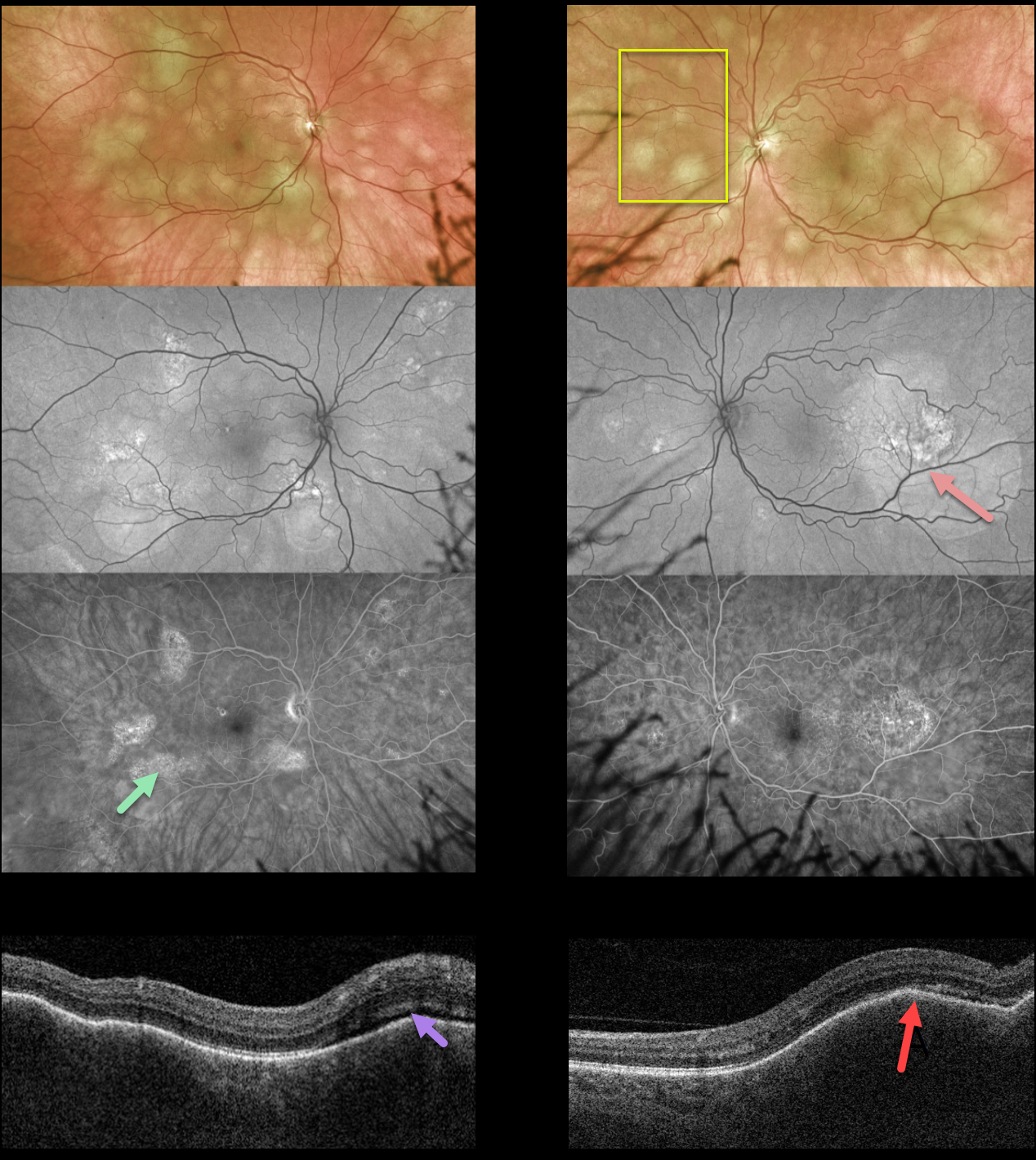
Originally posted on @retina.rocks January 23, 2023
This 58YO female presented with 2 weeks of blurred vision in her left eye. There was no past medical history. Vision was 20/40 OD and 20/200 OS.
Optos color imaging shows multifocal, creamy-yellow subretinal lesions bilaterally. The lesions are more confluent, extending beneath the left fovea.
The smaller of these lesions is fairly silent on fundus autofluorescence, although the larger ones show hyper-FAF. These lesions angiographically show variable late staining with window defects.
Swept-source OCT shows bilateral thickened and undulating choroid with trace overlying subretinal fluid.
She was referred to the emergency room and was subsequently diagnosed with widely metastatic lung adenocarcinoma.
Learning Points:
Although uveal melanoma is the most common primary intraocular malignancy, metastatic choroidal tumors are the most common intraocular malignancies. Lung and breast cancer are the most common sources.
The extensive multifocal lesions in each of our patients’ eyes are unusual, since most patients have 1-2 choroidal lesions (Shields et al Retina 2020;40:204-213).
UVEAL MELANOMA
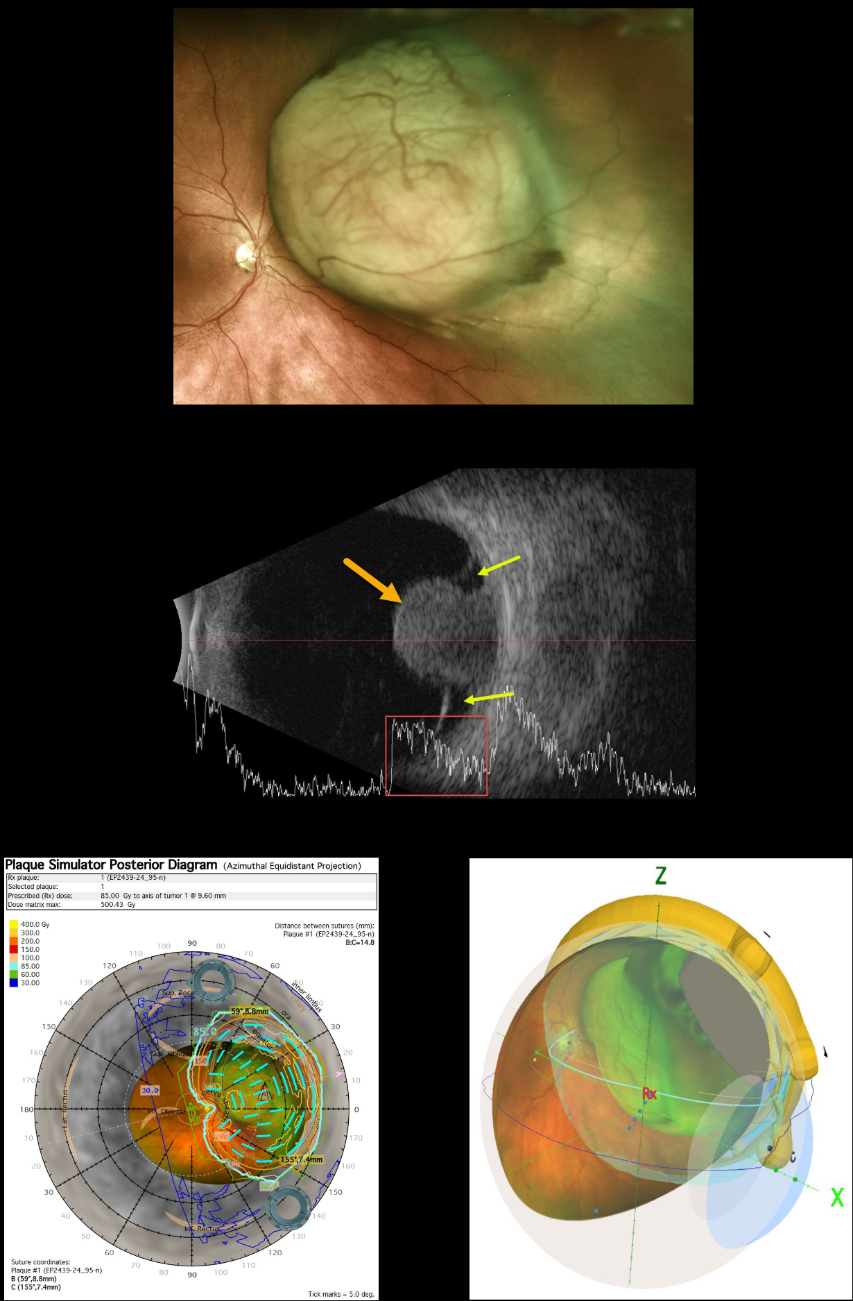
Originally posted on @retina.rocks January 18, 2023
This 63YO female presented with several weeks of peripheral vision loss in her left eye. Vision was 20/40.
Optos color imaging shows a large, mostly amelanotic, mushroom-shaped choroidal lesion extending from just nasal to the nerve into the midperiphery. Intrinsic large vessels are noted. The lesion measured 11.5 × 14.5 mm in lateral dimensions.
Ultrasonography shows a mushroom-shaped choroidal tumor that measured 9.6mm in thickness. Localized subretinal fluid is seen on either edge. The lesion has moderate internal reflectivity.
Plaque radiotherapy of the uveal malignant melanoma (MM) was performed. The plaque simulator images created by the radiation oncologist during the design of the custom plaque with radioactive I-131 seeds are shown.
CHOROIDAL METASTASES
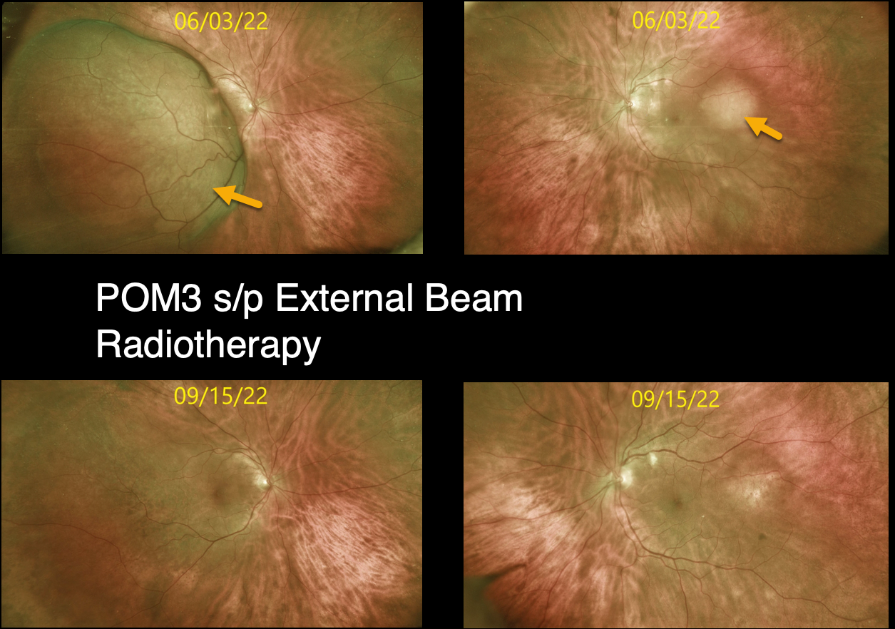
Originally posted on @retina.rocks October 17, 2022
This is a follow-up post from 6/6/22 of a 74YO female who initially presented with a several-week history of progressive vision loss in her right eye. Vision was counting fingers OD and 20/25 OS.
There was a known history of metastatic breast cancer when we diagnosed her with bilateral uveal metastases.
After three months of external beam radiotherapy, the metastatic choroidal lesions dramatically resolved. Vision improved to 20/50 OD and 20/25 OS.
Learning Points:
Although uveal melanoma is the most common primary intraocular malignancy, metastatic choroidal tumors are the most common intraocular malignancies. Lung and breast cancer are the most common sources.
CHOROIDAL METASTASIS
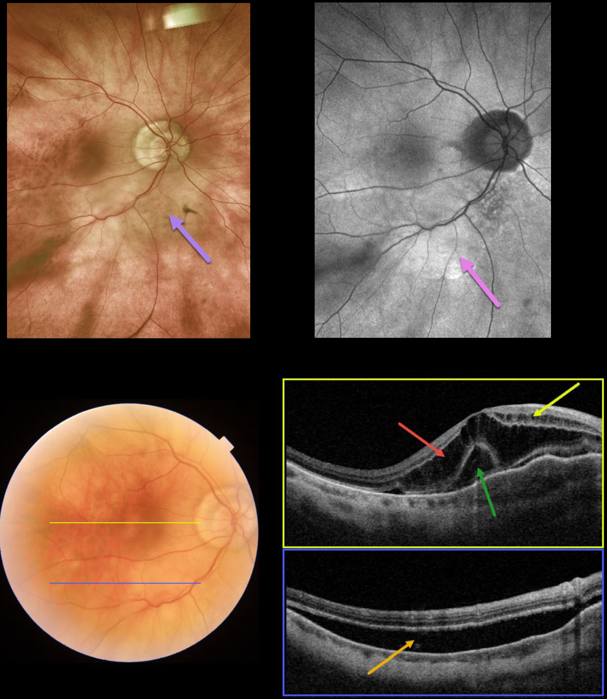
Originally posted on @retina.rocks September 23, 2022
This 87YO male presented with recent blurred vision in his right eye. Vision was 20/200 OD. He has a medical history of hypertension and prostate cancer that was successfully treated with radiation 15 years earlier.
Optos color imaging shows a creamy white subretinal discoloration in the nasal macula extending inferiorly.
Fundus autofluorescence (FAF) shows mostly hyper-FAF within this region. Swept-source OCT through the macular center shows severe cystic fluid in the outer nuclear layers and milder fluid in the inner nuclear layers. An unusual collection of outer vs subfoveal fluid is also seen. A B-scan through the inferior macula shows subretinal fluid.
The patient was referred to his oncologist for a metastatic workup, which disclosed previously undiagnosed colon cancer.
Learning Points:
Although uveal melanoma is the most common primary intraocular malignancy, metastatic choroidal tumors are the most common intraocular malignancies.
Lung and breast cancer are the most common sources. Prostate cancer rarely metastasizes to the choroid, found in only 2% of uveal metastases (Shields et al, Retina 2020;40:204-213).
CHOROIDAL HEMANGIOMA
Barbara Parolini and Veronika Matello
Originally posted on @retina.rocks September 12, 2022
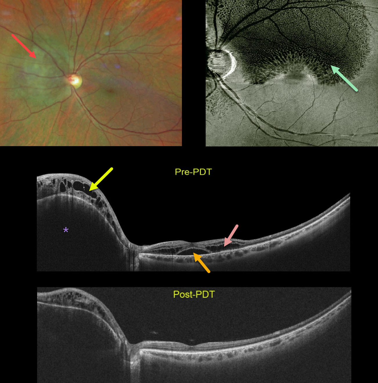
This healthy 45YO female was referred for macular schisis. Vision was 20/50. Color photography shows an orange-red subretinal lesion nasal to the nerve.
Widefield OCT shows an elevated hyporeflective choroidal mass with overlying cystic retinal edema. Subfoveal fluid with cystic fluid in the outer retina is noted in the macula. An en face OCT of the outer retina more dramatically shows the cystic changes.
The choroidal lesion with overlying retinal edema dramatically improved following photodynamic therapy. Vision improved to 20/20.
Learning Points:
Widefield imaging is revolutionizing the way we diagnose and treat retinal disease. This case is a perfect example in which macular imaging alone could lead to an incorrect diagnosis.
In this case, the macular findings in isolation suggest either optic pit maculopathy (Imamura et al, Retina 2010;30:1104-1112) or paraproteinemic maculopathy (Mansour et al, Ophthalmology 2014;121:1925-1932). This widefield technology led to a correct diagnosis, allowing for appropriate treatment with excellent anatomic and visual results.
CHOROIDAL NEVUS
Originally posted on @retina.rocks August 30, 2022
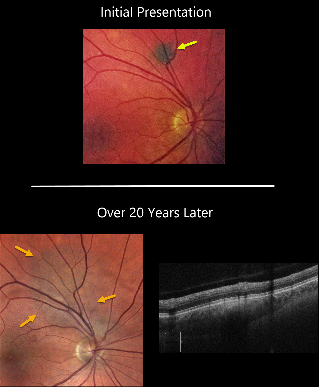
This 58YO male was initially seen in 2001 with a small asymptomatic choroidal nevus superior to the right optic nerve.
He presented to us in 2022 with a still asymptomatic but much enlarged nevus. The lesion now extends to the nerve. There is no overlying orange pigment or subretinal fluid clinically or on OCT. The lesion was 0.4mm thick.
Learning Points:
Clinical characteristics of choroidal nevi at risk for progressing to a uveal MM were initially described by Shields et al (Ophthalmology 1995;102:1351-1361). The most recent version of this famous acronym (Shields et al, Retina 2019;39:1840-1851) is To Find Small Ocular Melanoma Doing Imaging (TFSOM-DIM), which stands for:
To (Thickness >2mm)
Find (subretinal Fluid)
Small (Symptoms, vision <= 20/50)
Ocular (Orange pigment)
Melanoma (Melanoma hollow)
DIM (DIaMeter > 5mm)
Although our patient’s nevus has grown over time, this does not imply malignant transformation, as about one-third will enlarge with long-term follow-up (Shields et al., Ophthalmology 2011;118;382-388).
Shields et al found a marked increased risk for
malignant transformation for choroidal nevi over 2.0mm (Retina 2019;39:1840-1851).
Since our patient’s nevus is still well below this number and has no TFSOM-DIM risk factors other than diameter, we feel safe managing him conservatively with observation twice yearly.
CHRPE + CHOROIDAL NEVUS
Originally posted on @retina.rocks July 19, 2022
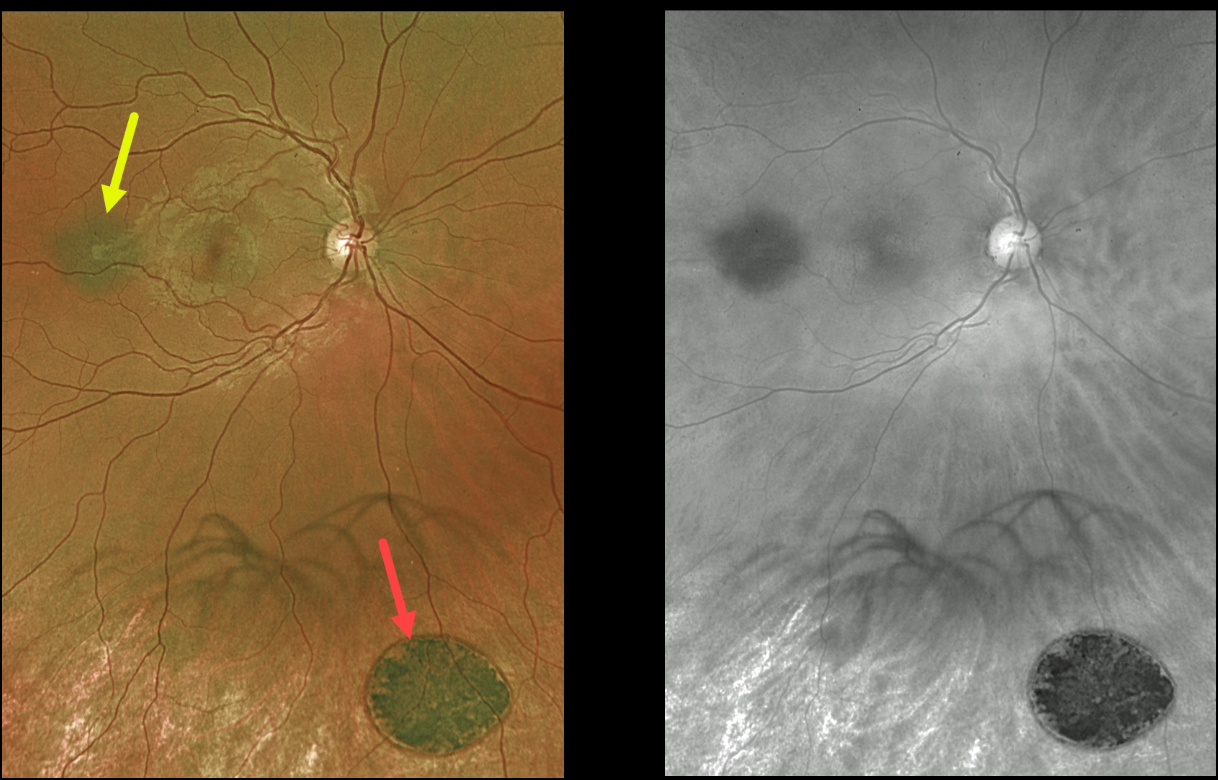
This 27YO male was referred for two distinct and asymptomatic pigmented fundus lesions.
Optos color imaging shows a small, flat choroidal nevus and a larger, flat patch of congenital hypertrophy of the RPE. The color image best shows the color difference between these lesions, with the CHRPE lesion appearing jet-black compared with the nevus.
The red channel best highlights the typical, somewhat indistinct margins of the nevus compared with the very sharp, round margins typical for the CHRPE. The patient is being followed yearly.
Learning Points:
Clinical characteristics of choroidal nevi at risk for progressing to a uveal malignant melanoma were initially described by Shields et al (Ophthalmology 1995;102:1351-1361).
The most recent version of this famous acronym (see Retina 2019;39:1840-1851) is To Find Small Ocular Melanoma Doing Imaging (TFSOM-DIM), which stands for:
To (Thickness >2mm)
Find (subretinal Fluid)
Small (Symptoms, vision <= 20/50)
Ocular (Orange pigment)
Melanoma (Melanoma hollow on ultrasonography)
DIM (DIaMeter > 5mm)
Although CHRPE lesions can enlarge with time (see Shields et al, Ophthalmology 2003;110:1968-1973), they carry virtually no malignant potential.
Histologic RPE hypertrophy presents in several ways. The typical isolated CHPRE lesions and bear tracks are not associated with intestinal polyps or cancer, and thus require no systemic evaluation. Only the multiple, small comet-shaped lesions are part of the familial polyposis spectrum.
CHOROIDAL OSTEOMA
Omar Mulki
Originally posted on @retina.rocks June 21, 2022
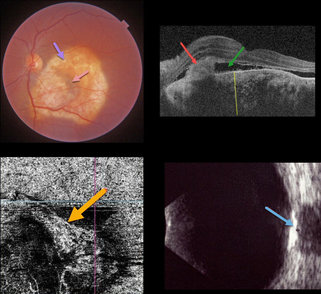
This 40YO female presented with recent vision loss in her left eye. Vision was 20/20 in her normal right eye and counting fingers in her left eye.
Triton color imaging shows an amelanotic choroidal lesion occupying virtually the entire macula. There is a small dot of central blood. Swept-source OCT shows a markedly thickened choroid with variable regions of hyper- and hyporeflectivity. A hyperreflective type 2 macular neovascularization (MNV) is seen along with exudative subretinal fluid. OCT angiography shows flow signals within the MNV. B-scan ultrasonography shows the lesion to be markedly hyperreflective.
An anti-VEGF injection was given, but she was unfortunately subsequently lost to follow-up.
Learning Points:
Choroidal osteomas are benign, calcified amelanotic choroidal tumors. They are more common in women, are almost always unilateral, and are usually located in the peripapillary or macular regions.
Choroidal lesions are best seen on the Optos red channel. Fluorescein angiography (FA) usually shows early patchy hyperfluorescence with late diffuse staining. Ultrasonography is often helpful in differentiating an osteoma (markedly hyperreflective) from other amelanotic choroidal tumors.
Macular neovascularization develops in a significant number of eyes and can be successfully treated with anti-VEGF therapy (see Seong et al, Graefe’s 2022;250:1713-1721).
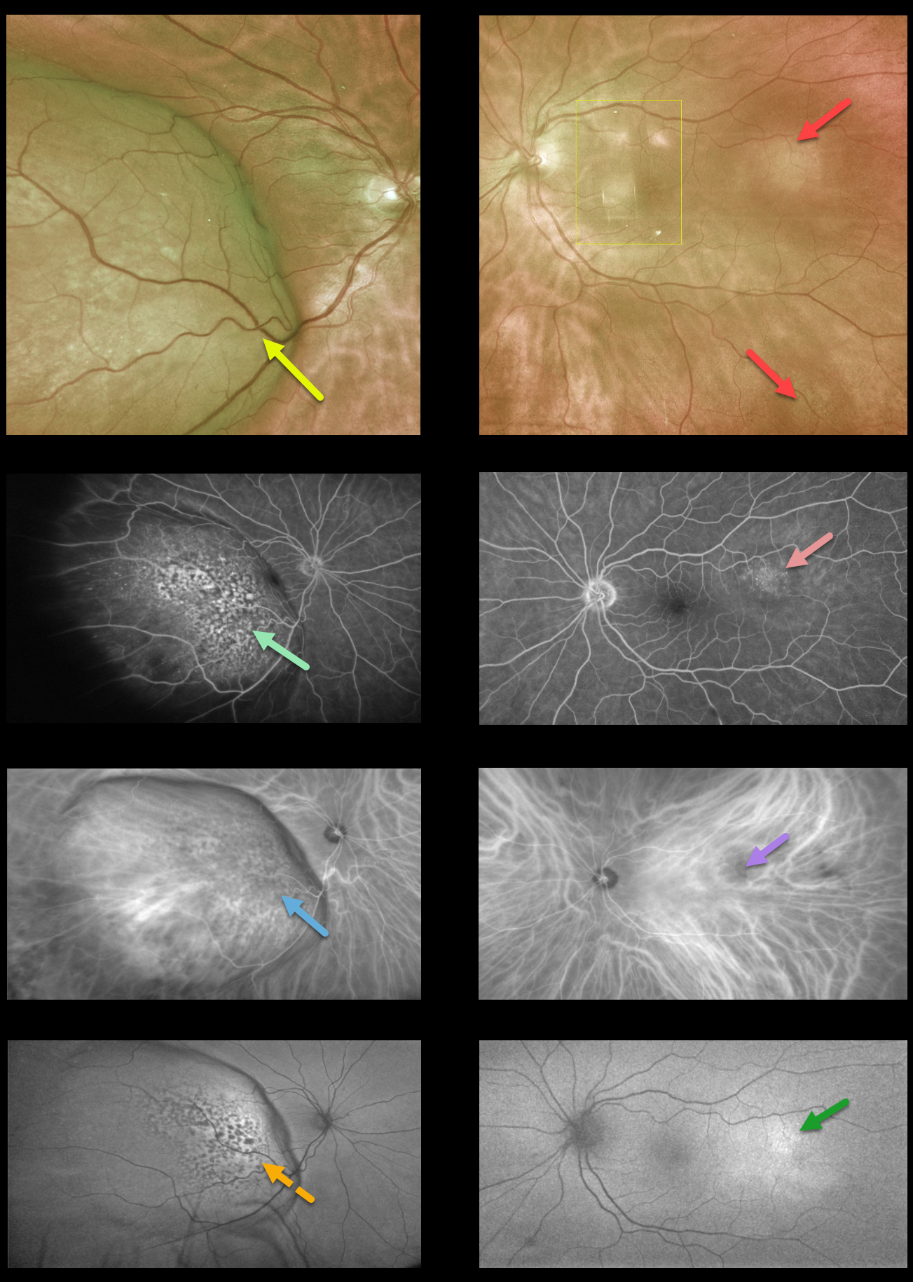
CHOROIDAL METASTASES
Originally posted on @retina.rocks June 6, 2022
This 74YO female presented with a 2-week history of progressive vision loss in her right eye. Vision was 20/200 OD and 20/20 OS.
Optos imaging shows a large, creamy-colored temporal choroidal lesion extending into the right temporal macula. The fovea is markedly elevated and tilted along the nasal edge of the tumor. Two smaller and fainter amelanotic choroidal lesions are noted in the left eye (the lesions in the left macula are artifacts).
Fundus fluorescein angiography (FFA) shows patchy window defects over the nasal dome of the lesion in her right eye and faint staining from the temporal lesion in the left eye.
Indocyanine green angiography shows mostly hypofluorescence corresponding to the FFA window defects with some increased fluorescence from the intrinsic vasculature more temporally in the right eye, and hypofluorescence from the temporal lesion in the left eye.
Fundus autofluorescence (FAF) shows hypo-FAF corresponding to the FFA window defects in the right eye and hyperfluorescence from the temporal lesion in the left eye.
There was a known history of metastatic breast cancer, and a recent PET scan showed possible new lung metastases.
We are currently working with the patient’s oncologist for the next step forward, which will likely include external beam radiotherapy for a presumed choroidal metastasis in her right eye. Photodynamic therapy or transpupillary thermotherapy will be considered for the left eye.
Learning Points:
Although uveal melanoma is the most common primary intraocular malignancy, metastatic choroidal tumors are the most common intraocular malignancies. Lung and breast cancer are the most common sources.
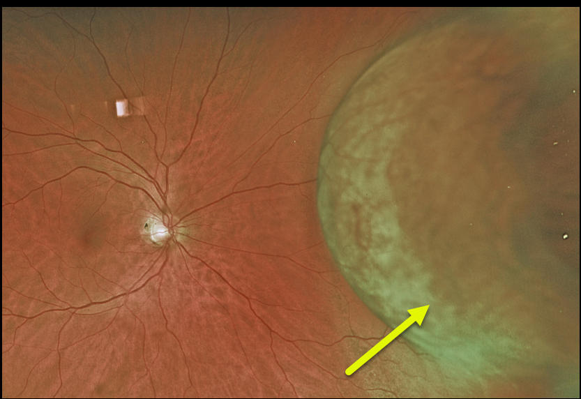
UVEAL MELANOMA
Originally posted on @retina.rocks May 30, 2022
This healthy 51YO female presented with one year of increasing flashes in her right eye. Vision was 20/25. Funduscopic examination revealed a large, variably pigmented nasal choroidal mass measuring at least 9.7mm in the largest basal diameter.
She was referred for further evaluation and treatment for a presumed uveal malignant melanoma.
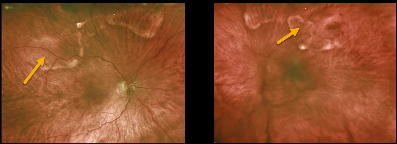
SCLEROCHOROIDAL CALCIFICATION
Originally posted on @retina.rocks May 17, 2022
This 77YO female presented with bilateral idiopathic sclerochoroidal calcification.
Learning Points:
Sclerochoroidal calcification is a relatively uncommon and benign finding in the elderly population. These yellowish lesions are slightly lumpy and elevated, and usually appear in the mid superior/inferior temporal/nasal quadrants.
The lesions are highly reflective on B-scan ultrasonography and visible on CT. These lesions require no treatment and are not associated with vision loss.
However, they may be associated with systemic hypercalcemic conditions such as hyperparathyroidism and pseudohypoparathyroidism. Shields et al therefore recommended that all such patients be evaluated for underlying systemic calcium disorders (Retina 2015;35:547-554).
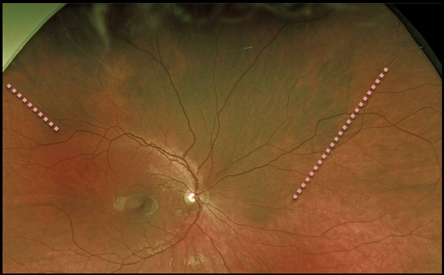
CHOROIDAL MELANOCYTOSIS
Originally posted on @retina.rocks May 6, 2022
This 16YO male presented with asymptomatic, unilateral disseminated patches of choroidal melanocytosis. There were no anterior segment or skin abnormalities.
Learning Points:
Choroidal melanocytosis is part of the oculodermal spectrum, which occasionally includes pigmentation of the globe or periocular skin (Nevus of Ota, melanosis oculi).
There are increased dendritic melanocytes in the affected tissues following the distribution of the first and second branches of the trigeminal nerve.
Patients need ongoing monitoring since about 1 in 400 will develop uveal melanoma. Of those who develop melanoma, they are twice as likely to undergo metastasis when compared to patients with uveal melanoma and no pre-existing melanocytosis.
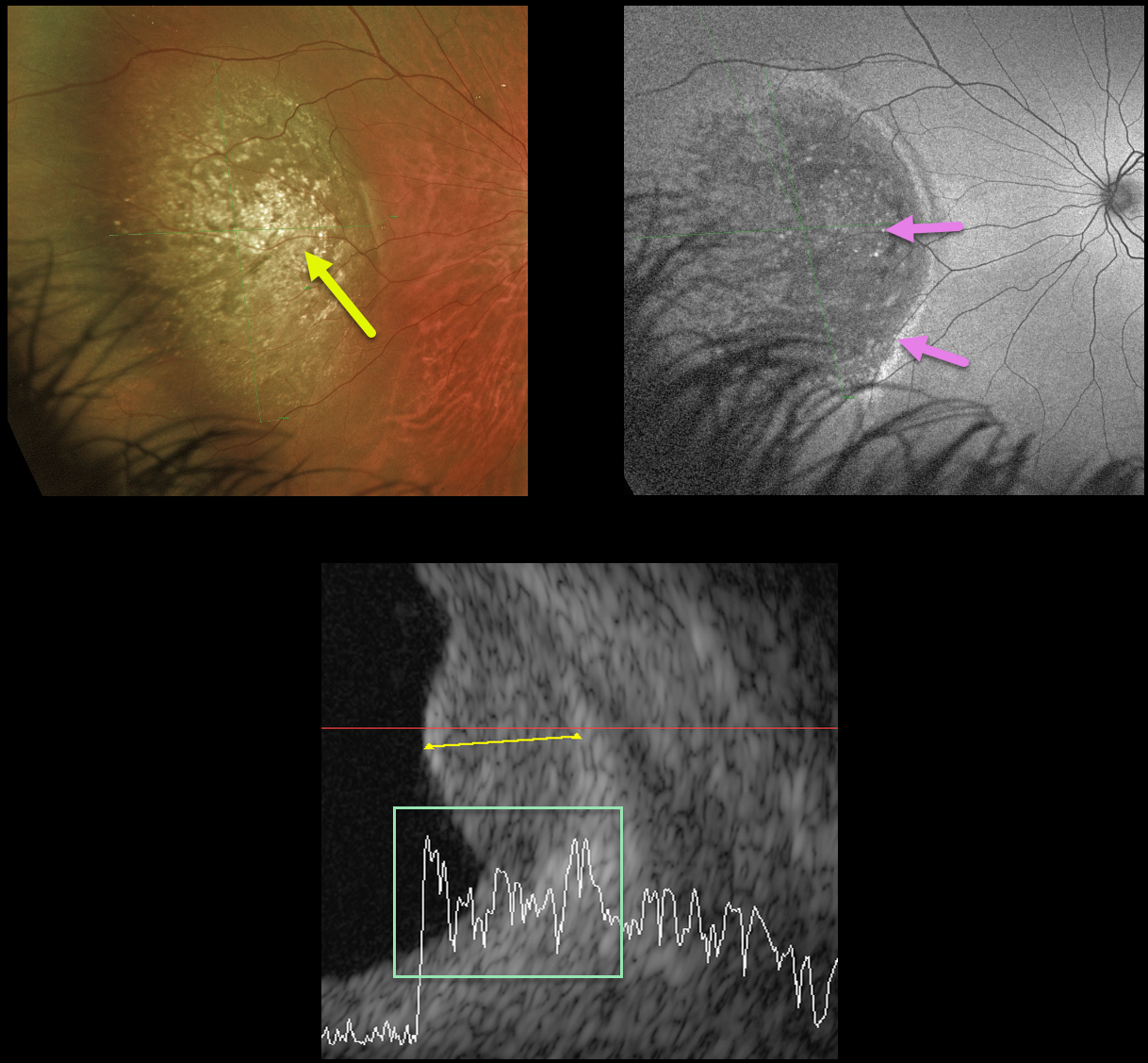
UVEAL MELANOMA
Originally posted on @retina.rocks April 20, 2022
This 25YO male presented with this asymptomatic choroidal mass in his left eye. Vision was 20/20.
Optos color imaging shows a large, well-defined variably pigmented lesion with numerous overlying drusen in the nasal fundus. The lesion shows variable hypo- and hyper-autofluorescence.
B-scan ultrasonography shows moderate internal reflectivity. It measured 4 mm thick and 13.5 mm at its widest diameter.
We felt this lesion was a uveal malignant melanoma and referred him to an ocular oncologist for further evaluation and treatment.
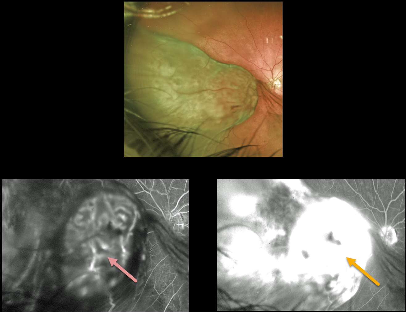
UVEAL MELANOMA
Originally posted on @retina.rocks March 8, 2022
This 81YO male presented with a 9.5mm-thick uveal malignant melanoma with fan-like opacities emanating from the apex of the tumor in his left eye. We suspected early vitreous hemorrhage, pigment shedding, or vitreous seeding.
Early phase angiogram beautifully reveals its internal vascularity, with late leakage from the entire tumor. Vision was 20/70.
The patient underwent diagnostic vitrectomy with plaque radiotherapy. Cytology from the vitreous sample confirmed PRAME, SOX10, and Ki67-positive cells. Genetic expression profiling showed that the tumor was Class 1B, PRAME positive.
Learning Points:
About 20% of patients with a Class 1B lesion will experience clinical metastasis within 5 years. PReferentially expressed Antigen in MElanoma (PRAME) is a gene that can become aberrantly expressed in some malignancies. Its positivity can indicate an increased risk for metastatic disease in Class 1 tumors, but its negativity does not tend to alter prognosis.
Vitreous seeding from a choroidal melanoma before surgical intervention is exceedingly rare. Various approaches have been taken, including immediate enucleation (Cheng et al, Taiwan Journal of Ophthalmology 2017;7:168-171), close observation for recurrence or further seeding (Metz et al, Br J Ophthalmol 2016;100:660-664), or delivery of intravitreal melphalan (Masoomian et al, Retin Cases Brief Rep 2021;15:97-100).
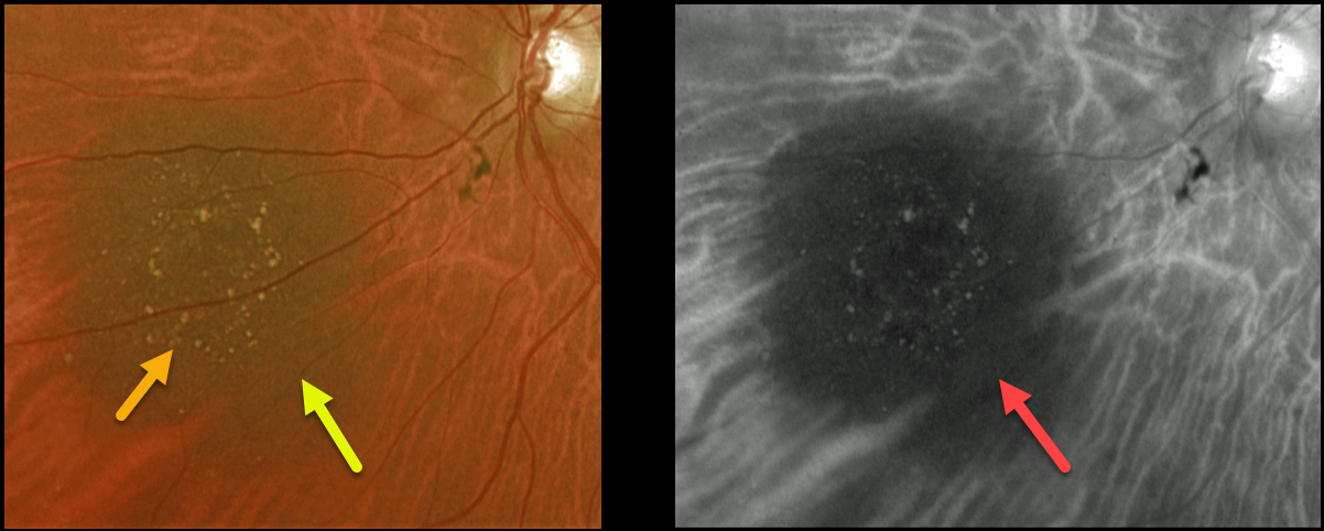
CHOROIDAL NEVUS
Originally posted on @retina.rocks February 11, 2022
This 72YO female presented with a nasal choroidal nevus seen on Optos color and Optos red channel. The lesion is flat, has overlying drusen, and measures 4.5 by 5.1 mm. Is the nevus at risk for progressing to a uveal malignant melanoma (MM)?
Learning Points:
Clinical characteristics of choroidal nevi at risk for progressing to a uveal MM were initially described by Shields et al (Ophthalmology 1995;102:1351-1361).
The most recent version of this famous acronym is To Find Small Ocular Melanoma Doing Imaging (TFSOM-DIM), which stands for To (Thickness >2mm) Find (subretinal Fluid) Small (Symptoms, vision <= 20/50) Ocular (Orange pigment) Melanoma (Melanoma hollow on ultrasonography) DIM (DIaMeter > 5mm).
Other than measuring just over 5mm in diameter, our patient has none of these other risk factors. The presence of drusen also indicates chronicity and low risk for malignant transformation.
We will follow her yearly.
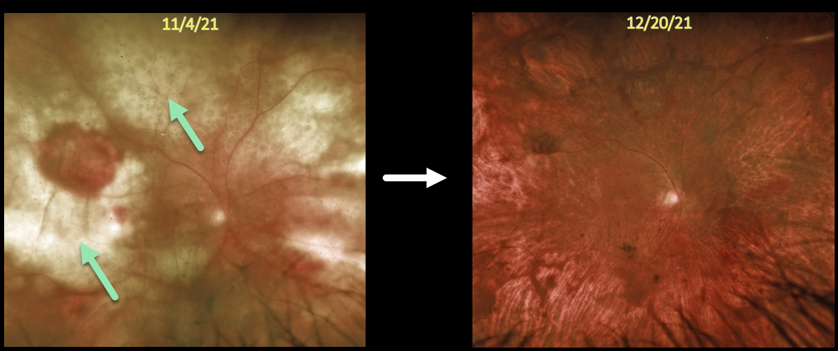
LYMPHOMA
Originally posted on @retina.rocks January 5, 2022
This 73YO man presented with subacute vision loss and these retinal findings. There were variable yellow-white, mostly confluent, lumpy subretinal infiltrates throughout the right posterior pole. The left fundus was normal.
He had a remote history of bladder cancer, which was in remission. He had recently completed chemotherapy for non-Hodgkin lymphoma, including a left retroperitoneal biopsy confirming a diffuse large B-cell lymphoma. We felt the findings in his right eye were consistent with metastatic lymphoma to the choroid.
Systemic chemotherapy supplemented with weekly intravitreal methotrexate was started. About 6 weeks later, vision remained at counting fingers, but there was a dramatic, complete clinical resolution of the tumor.
UVEAL MELANOMA
Originally posted on @retina.rocks November 3, 2021
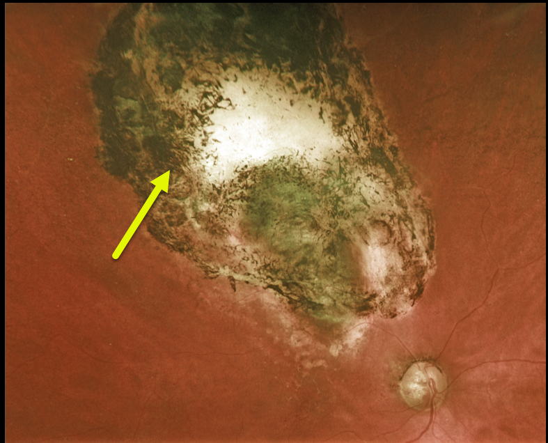
This 75YO female has a history of bilateral uveal malignant melanoma and is now monocular following enucleation of her left eye in 2001.
Plaque therapy in 2011 successfully saved her right eye, and the tumor has completely regressed.
Unfortunately, vision is 20/200 due to prior radiation retinopathy and possible glaucomatous optic neuropathy.
Learning Points:
In the United States, uveal malignant melanoma has an incidence of about 1 in 5 million people per year, and is virtually always unilateral. Primary bilateral uveal melanoma, as in this patient, is exceedingly rare (see Scott et al, Clin Exp Ophthalmology 2018;46:502-510). The BAP1 (BRCA-associated protein 1) germline mutation may be implicated in these cases.
CHOROIDAL OSTEOMA
Originally posted on @retina.rocks October 7, 2021
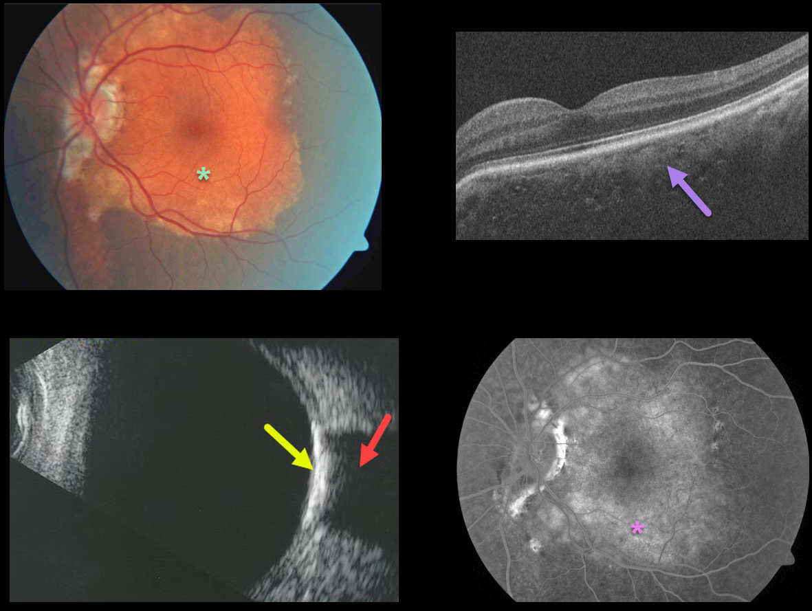
This patient presented with an orange-yellow choroidal lesion occupying the entire left macula.
OCT scanning shows diffuse choroidal thickening with small scattered specks of increased reflectivity.
B-scan ultrasonography shows marked increased reflectivity with marked posterior shadowing. The lesion diffusely stains on fluorescein angiography.
Learning Points:
Choroidal osteomas are benign, calcified amelanotic choroidal tumors. They are more common in women, are almost always unilateral, and are usually located in the peripapillary or macular regions.
Ultrasonography, as in this patient, is often helpful in differentiating an osteoma (markedly hyperreflective) from other amelanotic choroidal tumors. Fluorescein angiography usually shows early patchy hyperfluorescence with late diffuse staining.
Although the tumor itself does not require treatment, patients still need to be followed due to the risk of macular neovascularization.
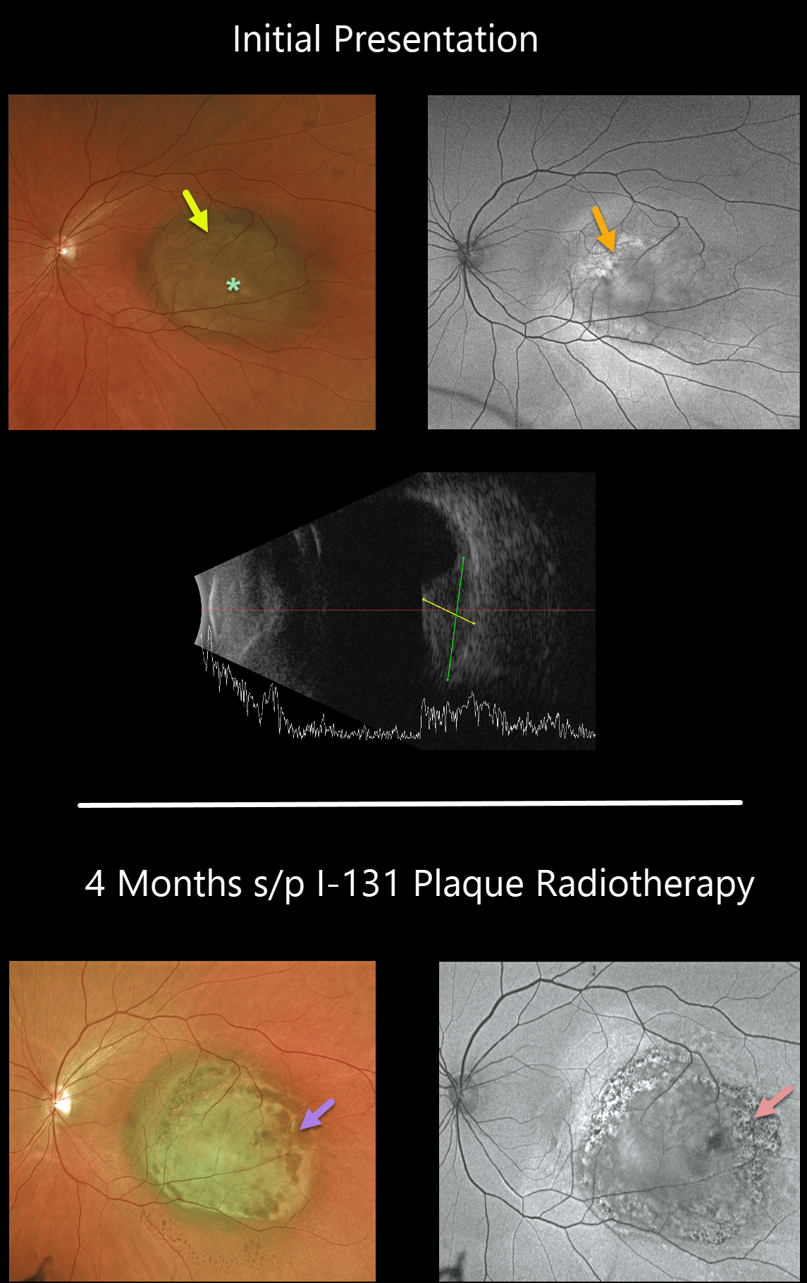
UVEAL MELANOMA
Originally posted on @retina.rocks October 4, 2021
This 53YO male presented on 1/7/21 with a medium-sized posterior pole uveal malignant melanoma. Lipofuscin was noted along the surface of the tumor. Optos fundus autofluorescence (FAF) showed variable hyper- and hypo-FAF. The lesion measured 8 x 11 mm and was 5.5mm thick. Despite its macular location, vision was 20/30.
The patient underwent I-131 plaque radiotherapy. About four months postoperatively, the tumor began to flatten (4.2mm thick), with resolving lipofuscin, coarse pigment clumping, and a generalized decrease in FAF. Vision remained stable.
Genetic testing revealed a Class 2 PRAME-negative lesion.
Learning Points:
Close to 75% of patients with a Class 2 lesion will experience clinical metastasis within 5 years. PReferentially expressed Antigen in MElanoma (PRAME) is a gene that can be aberrantly expressed in some malignancies. Its positivity may indicate a shorter time to metastatic disease, but its negativity does not typically alter prognosis.
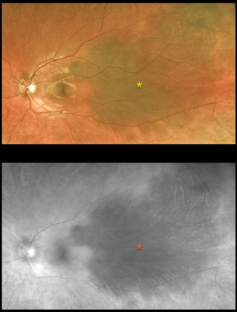
CHOROIDAL MELANOCYTOSIS
Originally posted on @retina.rocks August 27, 2021
This 17YO female presented with asymptomatic, unilateral disseminated patches of choroidal melanocytosis on Optos color and red-channel imaging. There were no anterior segment or skin abnormalities.
Learning Points:
Choroidal melanocytosis is part of the oculodermal spectrum, which occasionally includes pigmentation of the globe or periocular skin (Nevus of Ota, melanosis oculi).
There are increased dendritic melanocytes in the affected tissues following the distribution of the first and second branches of the trigeminal nerve.
Patients need ongoing monitoring since about 1 in 400 will develop uveal melanoma. Of those who develop melanoma, they are twice as likely to undergo metastasis when compared to patients with uveal melanoma and no pre-existing melanocytosis.
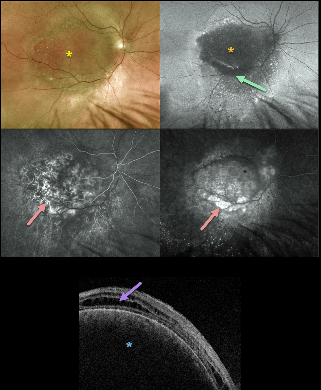
CHOROIDAL HEMANGIOMA
Originally posted on @retina.rocks August 11, 2021
We have been following this 71YO male since 2014 with a choroidal hemangioma. Vision without correction was counting fingers, but improved with a +8.00 correction to 20/70. He tried a trial with a contact lens, but preferred to remain uncorrected due to visually disabling metamorphopsia.
The lesion has remained relatively stable over the years, and he continues to prefer observation over possible photodynamic therapy (PDT).
Multifocal Optos imaging on 8/3/21 shows a fairly stable tumor. Color imaging shows the hemangioma to be mostly orange-red with some more peripheral brownish coloration.
Fundus autofluorescence (FAF) reveals that the tumor is mostly hypo-FAF, indicating damage to the overlying RPE. There is a surrounding variable hyper- and hypo-FAF with a gutter of changes extending more inferiorly due to overlying exudative subretinal fluid.
Fluorescein angiography shows minimal leakage with mostly variable window-type defects. Triton swept-source OCT shows cystic edema overlying the markedly elevated choroidal tumor.
Learning Points:
Isolated choroidal hemangiomas are benign vascular lesions. Although they have no malignant potential, they can cause vision loss from exudation leading to subretinal and intraretinal fluid, which can be treated with PDT.
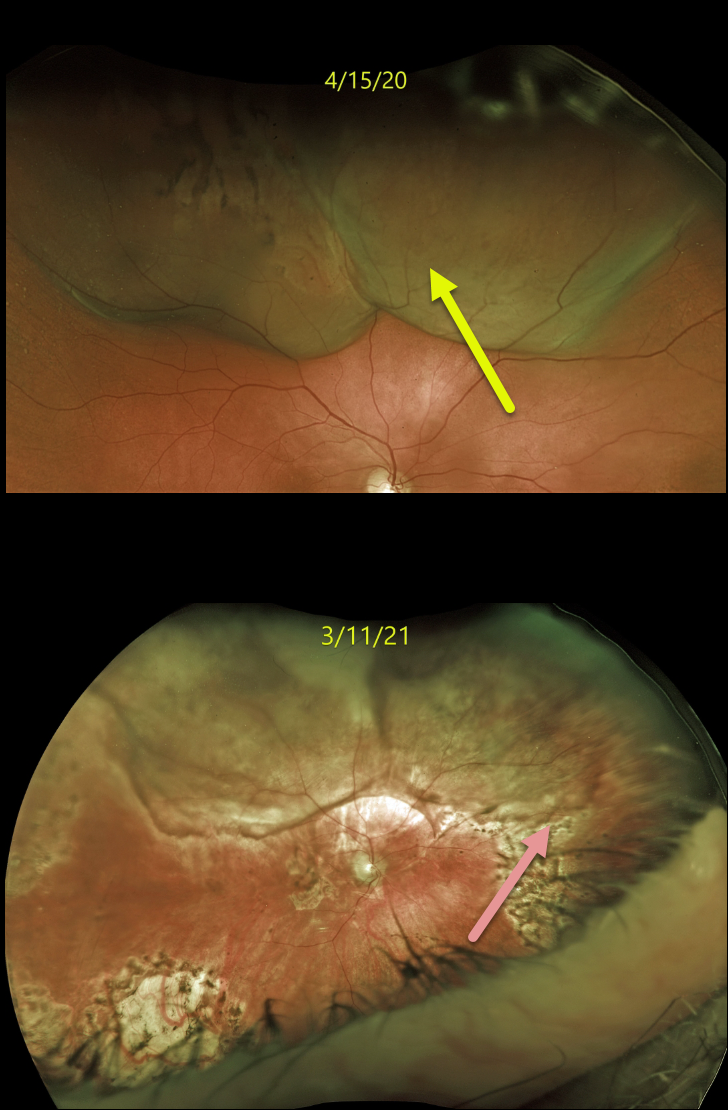
CHOROIDAL METASTASES
Originally posted on @retina.rocks August 2, 2021
This 82YO male with a history of retinal detachment treated with a segmental buckle and cryotherapy presented on 4/15/20 with some pain and new floaters in his right eye. Vision was 20/40.
Optos imaging shows an exudative retinal detachment secondary to a large, elevated, irregular, and amelanotic choroidal mass in the superior hemisphere. B-scan ultrasonography (not shown) revealed the tumor to be 6mm in height and 23 by 13 mm in basal diameter.
There was a prior history of metastatic clear cell renal carcinoma that was treated with radical left nephrectomy and chemotherapy from 2012 to 2014. He remained in remission until 12/2019, when a thoracic lymph node was treated with external beam radiation. This new large choroidal metastatic lesion heralded more widespread metastatic disease.
A fine needle aspiration biopsy was performed, and metastatic renal cell carcinoma was confirmed. The right globe was treated with external beam radiation, and he was started on systemic immunotherapy.
After the lesion showed regression and stability, vitrectomy with silicone oil was performed for retinal detachment. When last examined on 3/11/21, the tumor had regressed. Vision was counting fingers.
Unfortunately, he died shortly after this last exam.
Learning Points:
Although uveal melanoma is the most common primary intraocular malignancy, metastatic choroidal tumors are the most common intraocular malignancy. Lung and breast cancer are the most common sources. Renal cell cancer is an uncommon cause for choroidal metastasis.
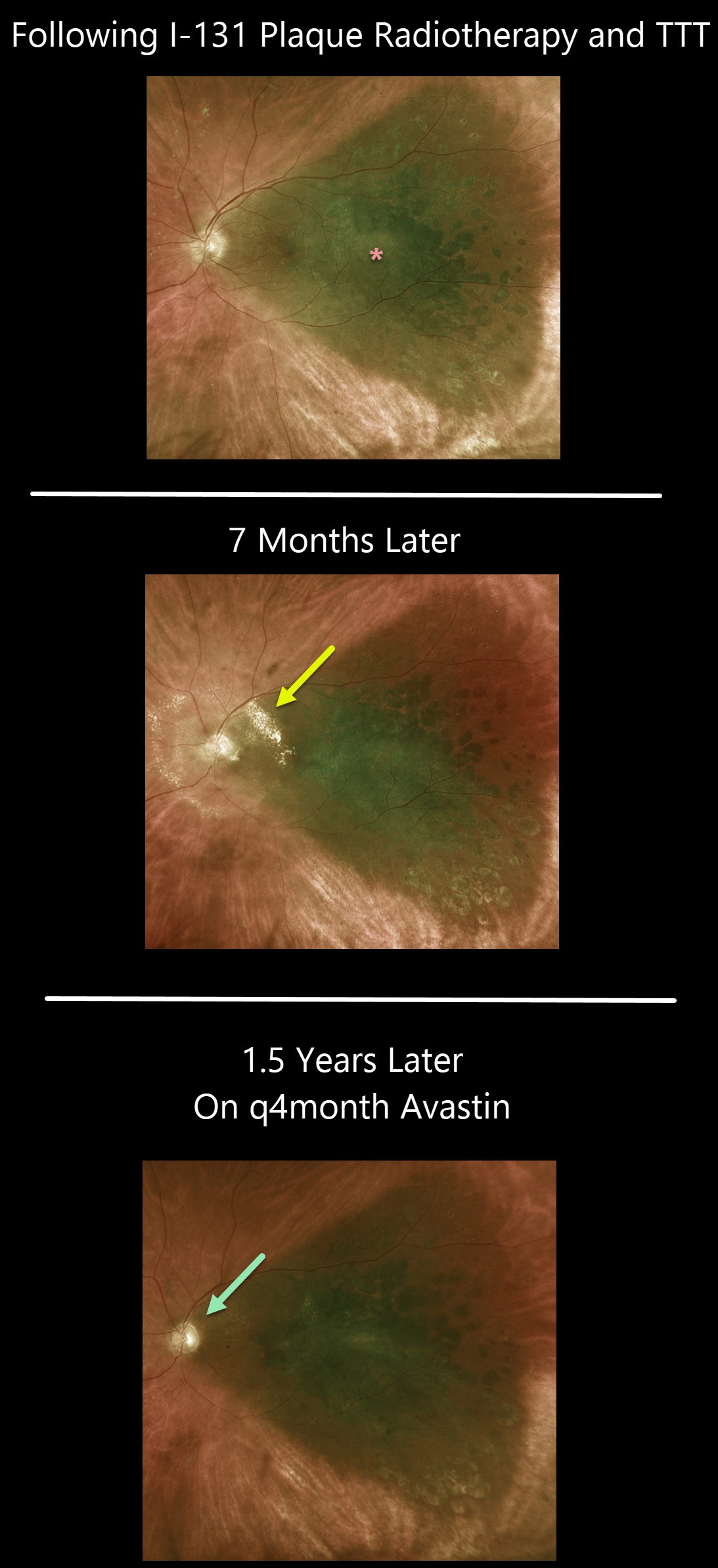
RADIATION PAPILLOPATHY
Originally posted on @retina.rocks July 2, 2021
This 66YO male was examined following I-131 plaque radiotherapy and transpupillary thermotherapy for a type 1A, PRAME+ uveal malignant melanoma.
Vision was 20/80, and the tumor was regressing compared to its preoperative appearance (not shown). He was getting intravitreal injections every 4 months as prophylaxis against radiation retinopathy.
The patient returned 8 months later for an overdue examination with new peripapillary lipid from radiation papillopathy. The intravitreal Avastin injections were restarted, and the papillopathy gradually resolved.
Learning Points:
The incidence of radiation retinopathy is relatively high following plaque therapy for uveal malignant melanoma. Ongoing intravitreal injections greatly reduce this complication (see Fallico et al, Surv Ophthalmology 2021;66;441-460).
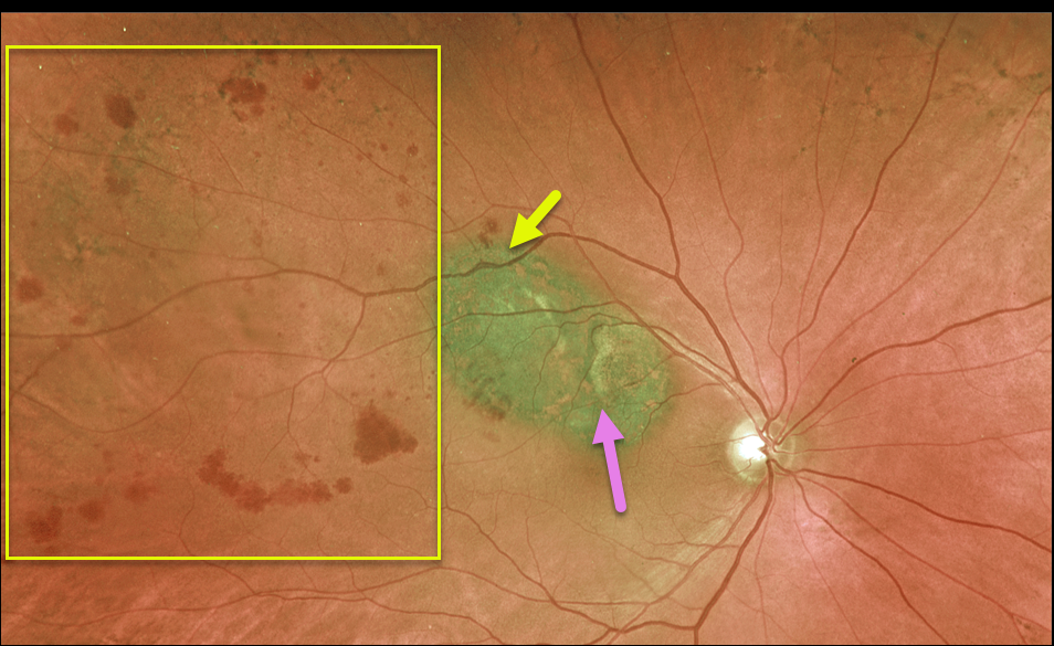
UVEAL MELANOMA + BRVO
Originally posted on @retina.rocks May 18, 2021
This 78YO male has a history of uveal malignant melanoma (MM), which was successfully treated with initial photodynamic therapy (PDT) (due to the presence of subretinal fluid) followed by transpupillary thermotherapy (TTT) once dry.
He developed a secondary branch retinal vein occlusion (BRVO), likely due to thermal damage from the TTT.
Vision is 20/30, and we continue to follow him closely. A scatter laser will be needed if he develops increasing ischemia with neovascularization from the BRVO.
Learning Points:
BRVOs are virtually always caused by an artery compressing a vein at a shared arteriovenous crossing.
However, rarely, they can occur due to other causes, including inflammation (most commonly sarcoidosis) or secondary to interventions such as TTT.
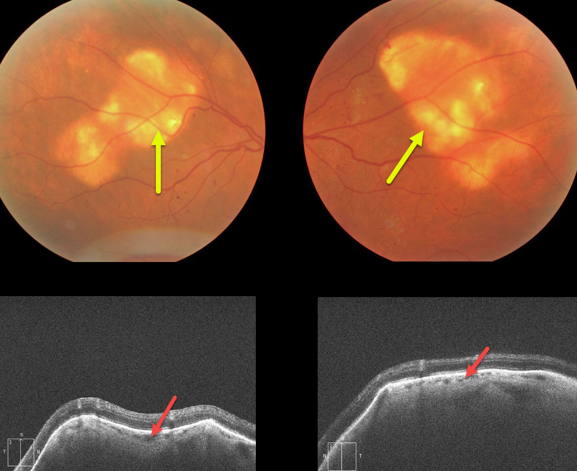
SCLEROCHOROIDAL CALCIFICATION
Originally posted on @retina.rocks April 14, 2021
This patient presented with bilateral idiopathic sclerochoroidal calcification. OCT scanning showed an irregularly thickened sclera compressing the overlying choroid.
Sclerochoroidal calcification is a relatively uncommon and benign finding in the elderly population. These yellowish lesions are slightly lumpy and elevated, and usually appear in the mid superior/inferior temporal/nasal quadrants.
The lesions are highly reflective on B-scan ultrasonography and visible on CT. They require no treatment and are not associated with vision loss.
Learning Points:
Sclerochoroidal calcification may be associated with systemic hypercalcemic conditions such as hyperparathyroidism and pseudohypoparathyroidism. Shields et al. therefore recommended that all such patients be evaluated for underlying systemic calcium disorders (Retina 2015;35:547-554).
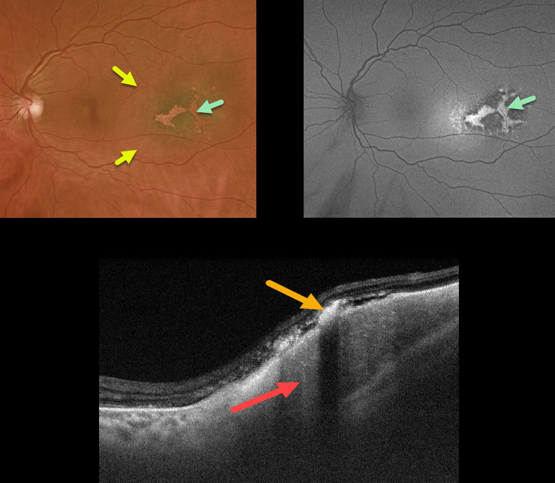
CHOROIDAL METASTASES
Originally posted on @retina.rocks March 31, 2021
This 50YO female with a known history of cutaneous melanoma and renal cell carcinoma presented with a variably pigmented temporal macular lesion.
The lesion had an associated shallow serous detachment and overlying lipofuscin, which was hyperautofluorescent. OCT B-scan showed the choroidal mass with overlying hyperreflective lipofuscin.
Transpupillary thermotherapy was recommended, but external beam radiation will be needed if the lesion further enlarges.
We assume this lesion is a choroidal metastasis, but we are unsure of the primary cancer. We can also not rule out that this represents a primary uveal malignant melanoma (MM). Her oncologist is aware of this choroidal lesion, and she continues to receive systemic treatment for her renal cell carcinoma.
Learning Points:
Although uveal MM is the most common primary intraocular malignancy, metastatic choroidal tumors are the most common intraocular malignancies. Lung and breast cancer are the most common sources.
Uveal MM is usually not familial nor associated with systemic cancers. The BAP1 mutation is a recently recognized autosomal dominant syndrome with predisposition to uveal melanoma and other primary cancers, including cutaneous melanoma, renal cell carcinoma, and mesothelioma (see Singh et al, AJO 2021;224:172-177).
We spoke at length with the patient’s oncologist, and at least for now, he did not want to check for BAP1.
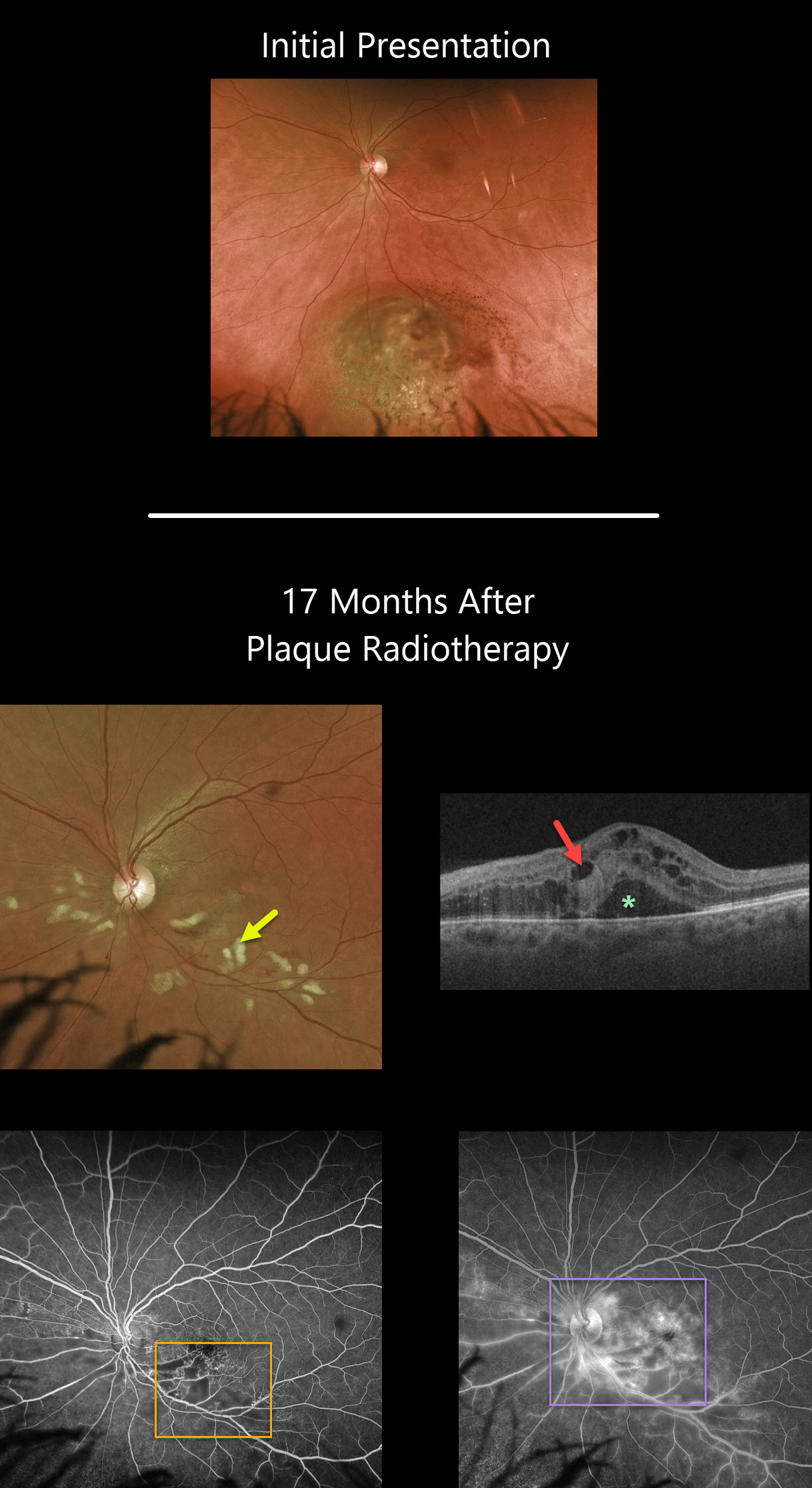
UVEAL MELANOMA
Originally posted on @retina.rocks March 29, 2021
Seventeen months following plaque radiotherapy for a uveal malignant melanoma, this patient developed radiation retinopathy with scattered inner retinal hemorrhages and nerve fiber layer infarcts (cotton wool spots). Vision was counting fingers, although it had been 20/40 ten days following plaque therapy a year and a half earlier.
Cystoid macular edema with subretinal fluid is noted on Triton swept-source OCT. Fluorescein angiography shows areas of capillary dropout, telangiectasias, and hyperfluorescent leakage secondary to cystoid macular edema.
Learning Points:
Radiation retinopathy usually occurs 3 months to 3 years after external beam radiation (e.g., for facial or orbital cancers) or plaque radiotherapy. The ocular findings closely resemble those seen with diabetic retinopathy, as in our patient, who is receiving Avastin injections for the macular edema.
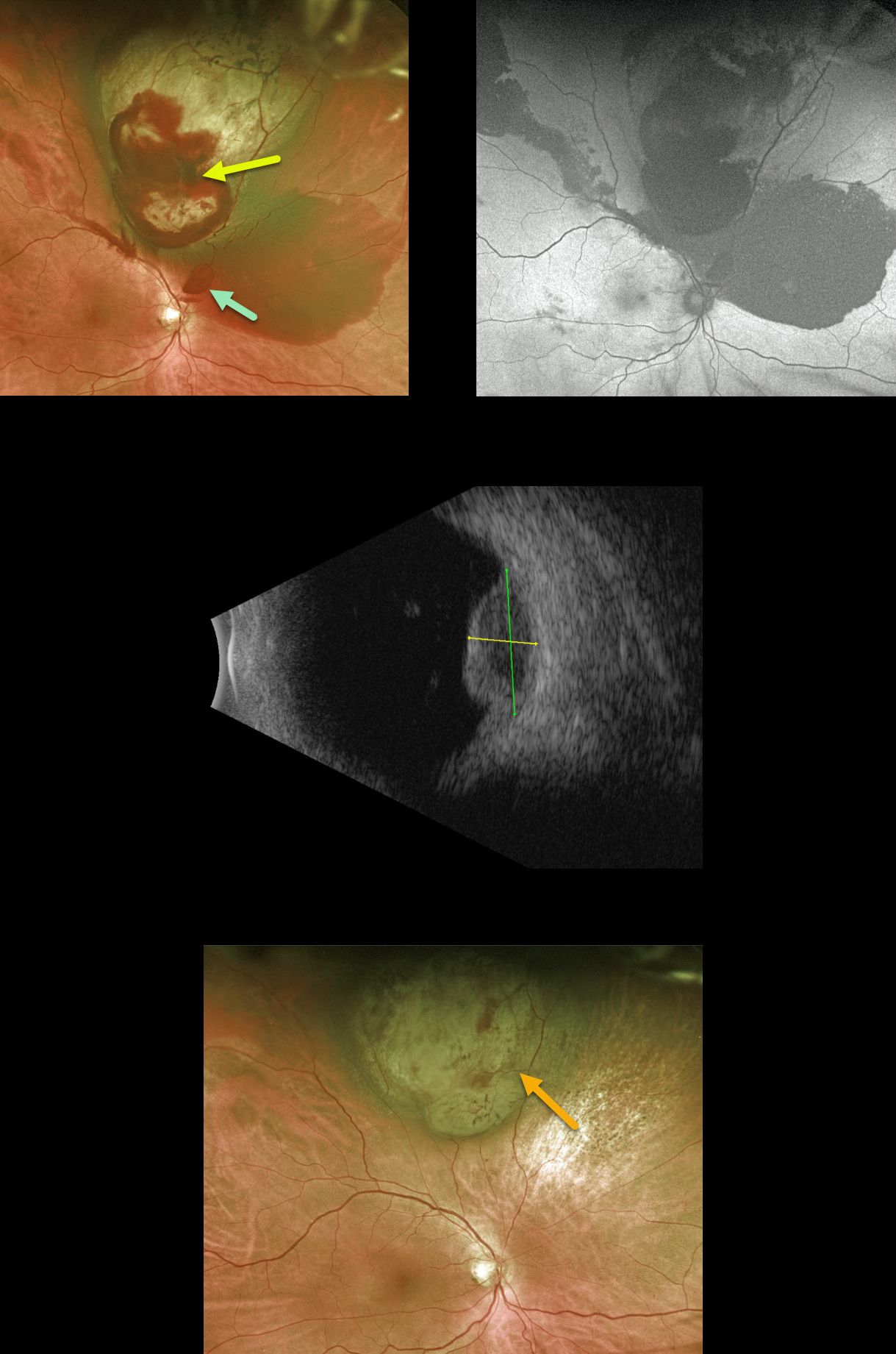
UVEAL MELANOMA
Originally posted on @retina.rocks March 4, 2021
This 60YO male presented with 20/25 vision in his right eye despite a superior uveal malignant melanoma. The tumor was associated with subretinal blood overlying and outside the lesion, as well as a small area of preretinal blood just superior to the optic nerve.
Fundus autofluorescence showed no lipofuscin. The tumor measured 5.55mm in thickness, with a basal diameter of 11.07mm, and appeared hollow on ultrasound, while the overlying subretinal blood was more echogenic.
A few days prior to radioactive plaque therapy, he noted a severe worsening of vision and was found to have a dense vitreous hemorrhage, obscuring the view of the tumor (not pictured).
Pars plana vitrectomy was performed at the time of plaque placement to clear the view and allow for transvitreal fine-needle aspiration.
About 3 months postoperatively, the tumor regressed to a thickness of 3.5mm. The subretinal blood resolved with secondary pigmentary changes, and his vision returned to baseline.
Learning Points:
Genetic expression profiling demonstrated a Class 1B, PRAME-negative tumor.
The patient continues to be followed closely for local recurrence and metastatic disease.
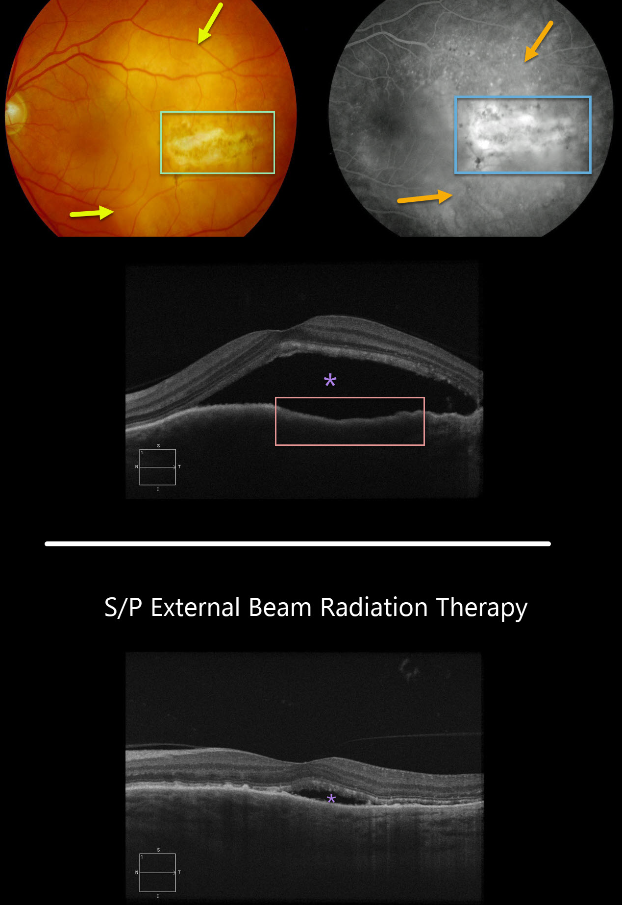
CHOROIDAL METASTASES
Originally posted on @retina.rocks February 9, 2021
This patient with a known history of breast cancer presented with a creamy colored choroidal lesion with central overlying pigmentary changes and scarring in the temporal macula.
Fluorescein angiography showed diffuse subretinal leakage with staining of the overlying scar. OCT scanning showed choroidal thickening with an irregular retinal pigment epithelial contour and overlying subretinal fluid.
The metastatic lesion was treated with localized external beam irradiation, with near immediate tumor regression and decreasing fluid 5 weeks later.
Learning Points:
Although uveal melanoma is the most common primary intraocular malignancy, metastatic choroidal tumors are the most common intraocular malignancies.
Lung and breast cancer are the most common sources.
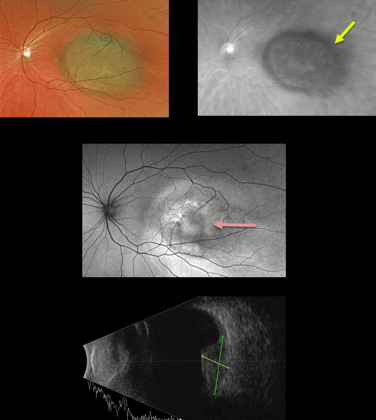
UVEAL MELANOMA
Originally posted on @retina.rocks January 18, 2021
This 52YO female presented with 20/200 vision in her left eye due to a uveal malignant melanoma. Amazingly, she was completely asymptomatic since she was right eye dominant.
The hyperpigmented lesion is well visualized on the Optos red channel and exhibits variable hyper- and hypoautofluorescence. The tumor measured about 5.5mm in thickness and 12mm in basal diameter on B-scan.
Radioactive plaque therapy was recommended.
Learning Points:
Clinical characteristics of choroidal nevi at risk for progressing to a uveal MM were described by Shields et al. (Ophthalmology 1995;102:1351-1361). The most recent version of this famous acronym, To Find Small Ocular Melanoma Doing Imaging (TFSOM-DIM, Retina 2019;39:1840-1851) is:
To (Thickness >2mm)
Find (Subretinal Fluid)
Small (Symptoms, vision <= 20/50)
Ocular (Orange pigment)
Melanoma (Melanoma hollow)
DIM (DIIaMeter >5mm)
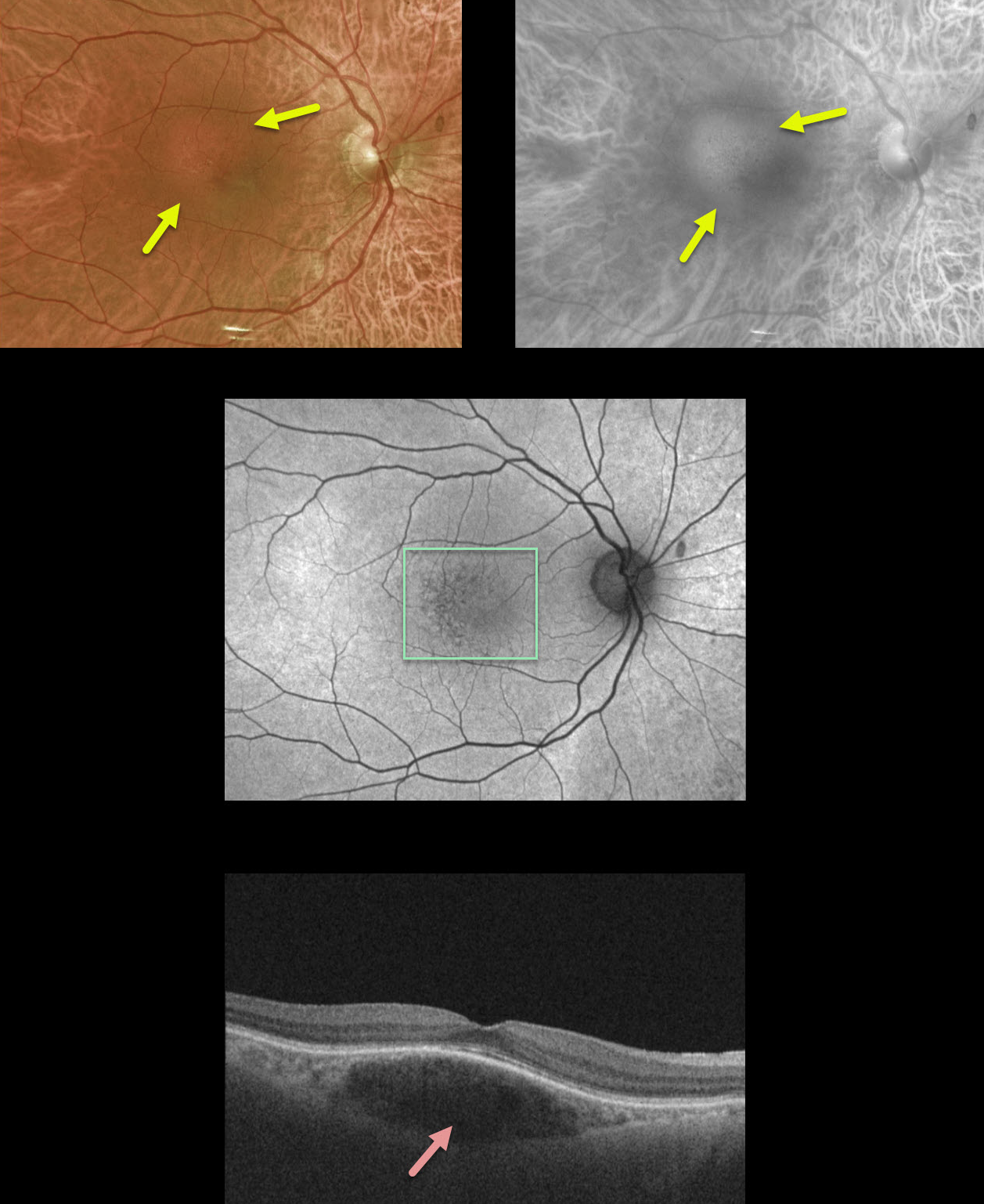
CHOROIDAL HEMANGIOMA
Originally posted on @retina.rocks January 8, 2021
This 44yo female presented with 20/20 vision and no visual complaints.
If you look closely at the color Optos image, you can see a small 1.5 disc diameter subretinal lesion in the superotemporal macula with mild overlying pigmentary changes. The Optos red channel best illuminates the lesion, indicating its choroidal location.
Fundus autofluorescence shows very mild overlying pigmentary changes. OCT shows a somewhat fusiform-shaped hyporeflective choroidal tumor without overlying exudation.
Since our patient was asymptomatic without fluid, we are following her twice yearly and will consider treatment if she develops symptomatic fluid.
Learning Points:
Isolated choroidal hemangiomas are benign vascular lesions. Although they have no malignant potential, they can cause vision loss due to exudation and subretinal and intraretinal fluid, which can be treated with photodynamic therapy.
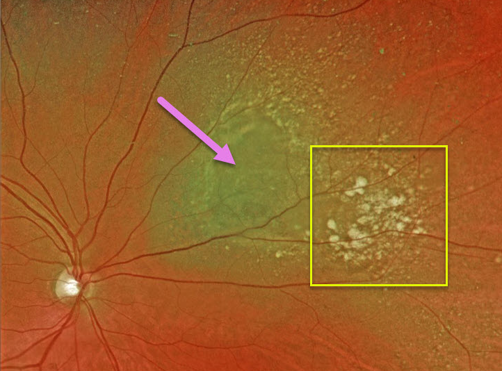
CHOROIDAL NEVUS
Originally posted on @retina.rocks December 16, 2020
This 70YO male presented with this nasal choroidal nevus.
Other than being near the optic nerve, our patient has none of the risk factors for transformation to uveal melanoma. The presence of drusen also indicates chronicity and low risk for malignant transformation. We are following him yearly.
Learning Points:
Clinical characteristics of choroidal nevi at risk for progressing to a uveal MM were described by Shields et al. (Ophthalmology 1995;102:1351-1361). The most recent version of this famous acronym, To Find Small Ocular Melanoma Doing Imaging (TFSOM-DIM, Retina 2019;39:1840-1851), is:
To (Thickness >2mm)
Find (Subretinal Fluid)
Small (Symptoms, vision <= 20/50)
Ocular (Orange pigment)
Melanoma (Melanoma hollow)
DIM (DIIaMeter >5mm)
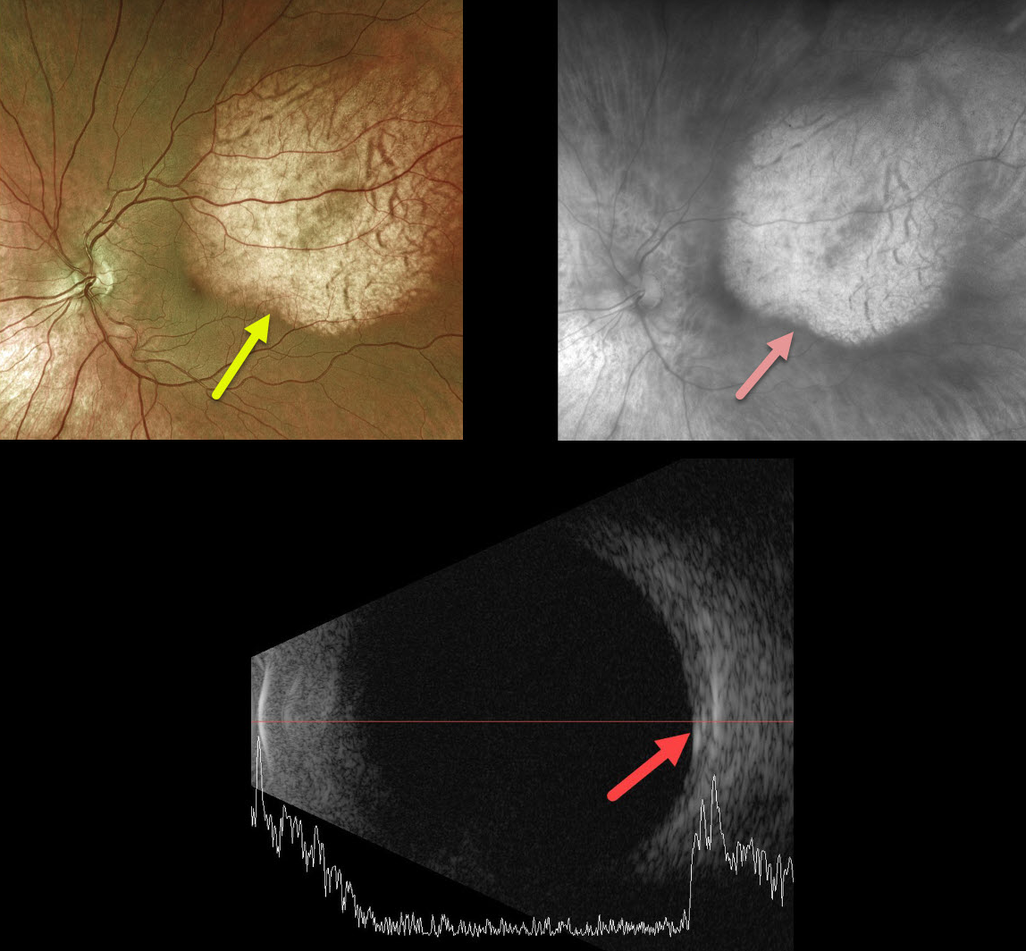
CHOROIDAL OSTEOMA
Originally posted on @retina.rocks December 8, 2020
Our best guess is that this patient has a choroidal osteoma. As choroidal lesions, they are best seen on the Optos red channel.
We would, however, expect more reflectivity on the B-scan as osteomas are typically markedly hyperreflective.
Learning Points:
Choroidal osteomas are benign, calcified amelanotic choroidal tumors. They are more common in women, are almost always unilateral, and usually located in the peripapillary or macular regions.
Ultrasonography is useful in differentiating choroidal osteomas from other amelanotic choroidal tumors.
Fluorescein angiography (FA) usually shows early patchy hyperfluorescence with late diffuse staining.
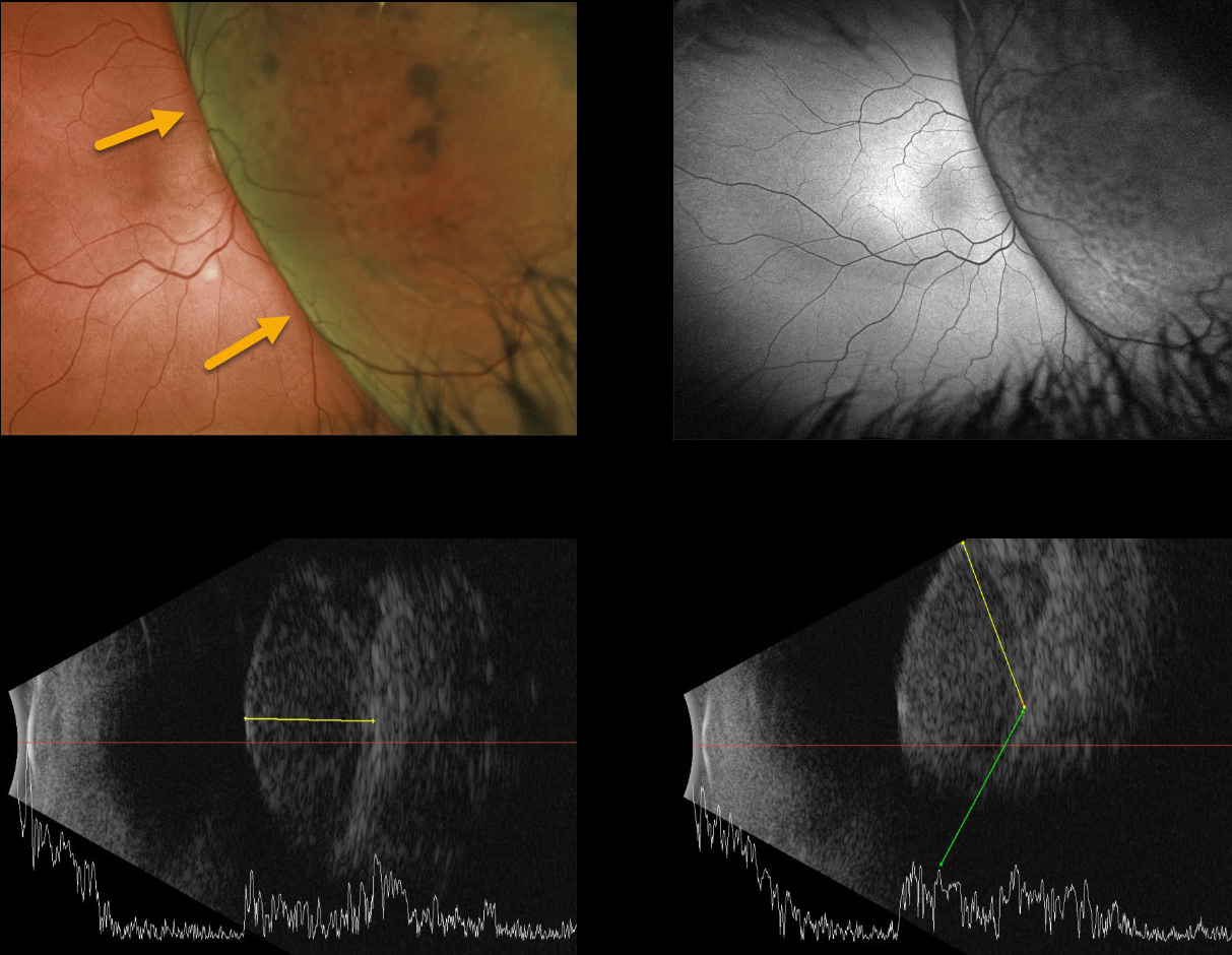
UVEAL MELANOMA
Originally posted on @retina.rocks October 20, 2020
This 81yo presented with peripheral vision loss. Vision was 20/100.
She had a large nasal uveal melanoma draping over the optic nerve. The tumor measured 10 mm in thickness and over 23 mm in basal diameter on B-scan ultrasonography.
Enucleation was recommended.
Learning Points:
Clinical characteristics of choroidal nevi at risk for progressing to a uveal MM were described by Shields et al. (Ophthalmology 1995;102:1351-1361). The most recent version of this famous acronym, To Find Small Ocular Melanoma Doing Imaging (TFSOM-DIM, Retina 2019;39:1840-1851) is:
To (Thickness >2mm)
Find (Subretinal Fluid)
Small (Symptoms, vision <= 20/50)
Ocular (Orange pigment)
Melanoma (Melanoma hollow)
DIM (DIaMeter >5mm)
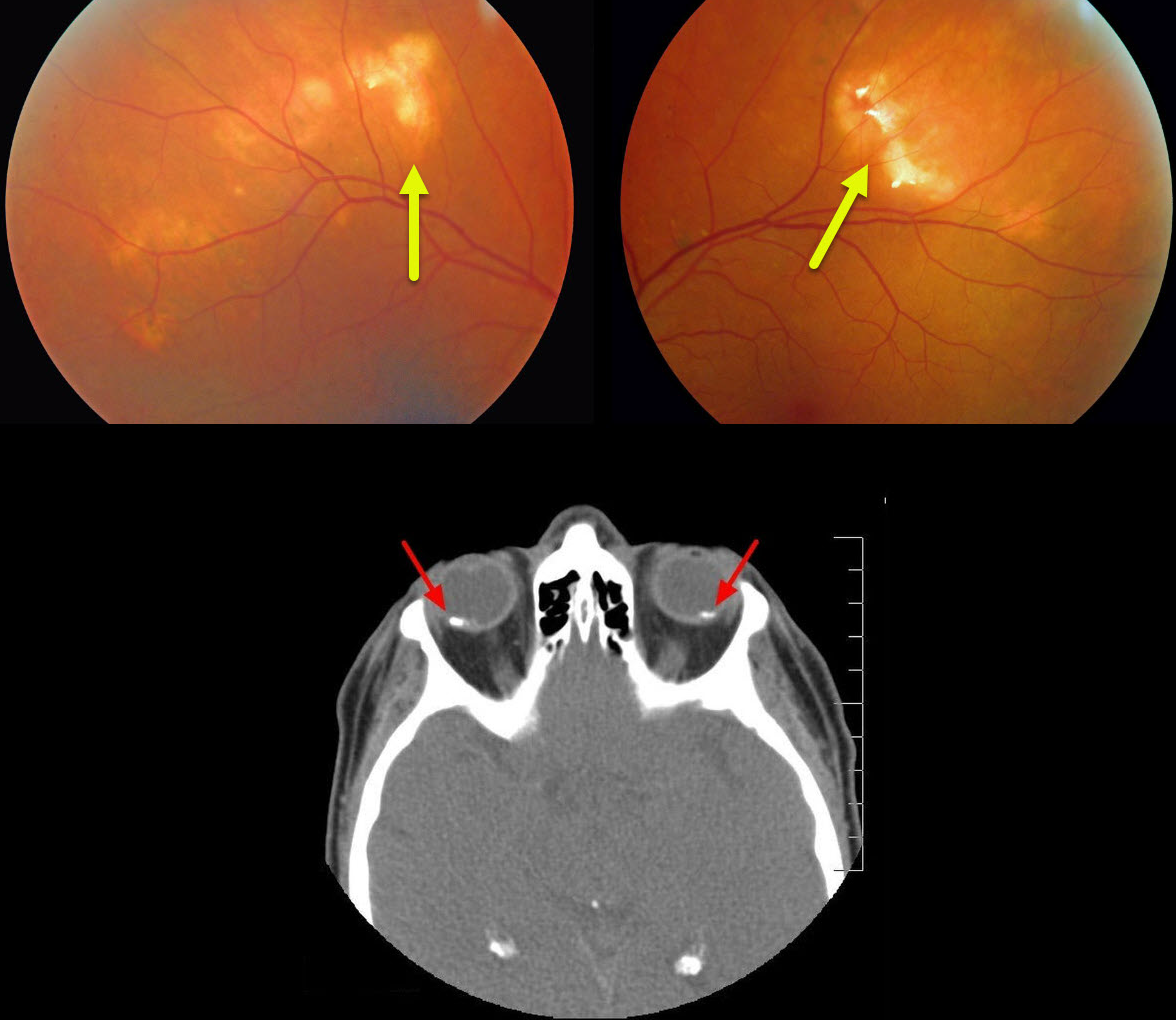
SCLEROCHOROIDAL CALCIFICATION
Originally posted on @retina.rocks October 2, 2020
This patient presented with bilateral idiopathic sclerochoroidal calcification, a relatively uncommon and benign finding in the elderly population. These yellowish choroidal lesions are slightly lumpy and elevated, and usually appear in the mid superior/inferior temporal/nasal quadrants.
The lesions are highly reflective on B-scan ultrasonography and visible on CT. They require no treatment and are not associated with vision loss.
Learning Points:
Sclerochoroidal calcification may be associated with systemic hypercalcemic conditions such as hyperparathyroidism and pseudohypoparathyroidism. Shields et al. therefore recommended that all such patients be evaluated for underlying systemic calcium disorders (Retina 2015;35:547-554).
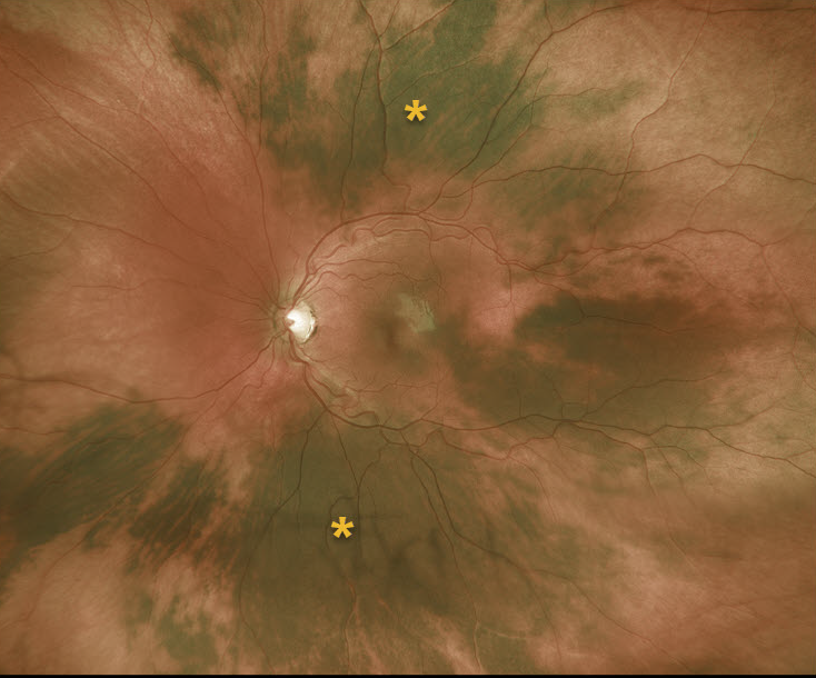
CHOROIDAL MELANOCYTOSIS
Originally posted on @retina.rocks August 10, 2020
This 13YO female has unilateral disseminated patches of choroidal melanocytosis, but without anterior segment or skin abnormalities.
Learning Points:
Choroidal melanocytosis is part of the oculodermal spectrum, which occasionally includes pigmentation of the globe or periocular skin (Nevus of Ota, melanosis oculi).
There are increased dendritic melanocytes in the affected tissues following the distribution of the first and second branches of the trigeminal nerve.
Patients need ongoing monitoring since about 1 in 400 will develop uveal melanoma. Those who develop melanoma are twice as likely to undergo metastasis when compared to patients with uveal melanoma and no pre-existing melanocytosis.
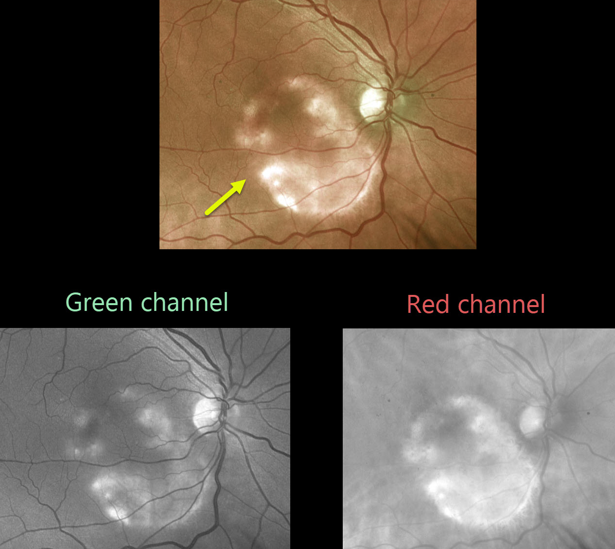
CHOROIDAL OSTEOMA
Originally posted on @retina.rocks July 15, 2020
This asymptomatic patient was noted to have a choroidal osteoma.
As choroidal lesions, osteomas are best seen on the red channel compared with the green Optos channel. Ultrasonography is often helpful in differentiating an osteoma (markedly hyperreflective) from other amelanotic choroidal tumors.
Learning Points:
Choroidal osteomas are benign, calcified amelanotic choroidal tumors. They are more common in women, are almost always unilateral, and are usually located in the peripapillary or macular region.
Although many patients are asymptomatic, vision loss can occur from overlying subretinal fluid or macular neovascularization.
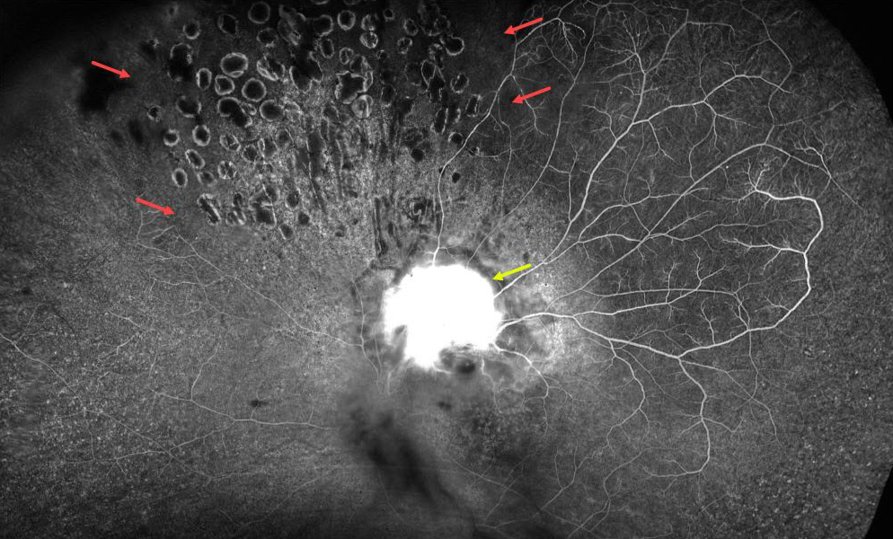
UVEAL MELANOMA
Originally posted on @retina.rocks June 19, 2020
This patient had a uveal melanoma previously treated with plaque radiotherapy.
Although the tumor was destroyed, retinal capillary damage created ischemic regions and secondary retinal neovascularization. The tumor diffusely stains on fluorescein angiography. Although scatter laser successfully caused the neovascularization to regress, residual areas of nonperfusion remain.
Learning Points:
Retinal neovascularization, which develops at the vitreoretinal interface, is caused by retinal ischemia. Remember that neovascularization below the retina is usually caused by subretinal disorders.
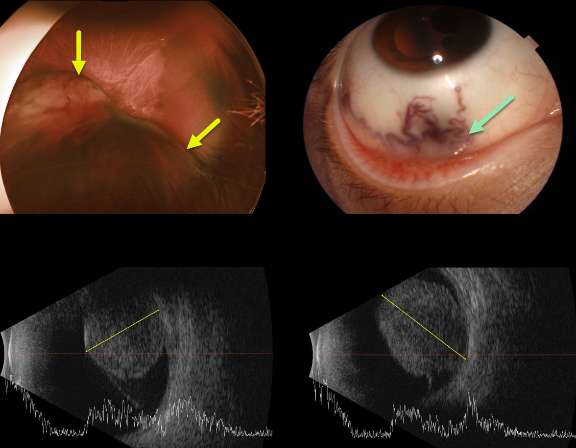
UVEAL MELANOMA
Originally posted on @retina.rocks May 14, 2020
This 22YO white male presented with a large ciliary body uveal malignant melanoma that was about 14mm thick x 18mm in length on B-scan ultrasonography.
Unfortunately, the tumor required enucleation since it was too large for radioactive plaque therapy.
Learning Points:
Large and dilated episcleral (sentinel) vessels are often a tell-tale sign of an underlying tumor.
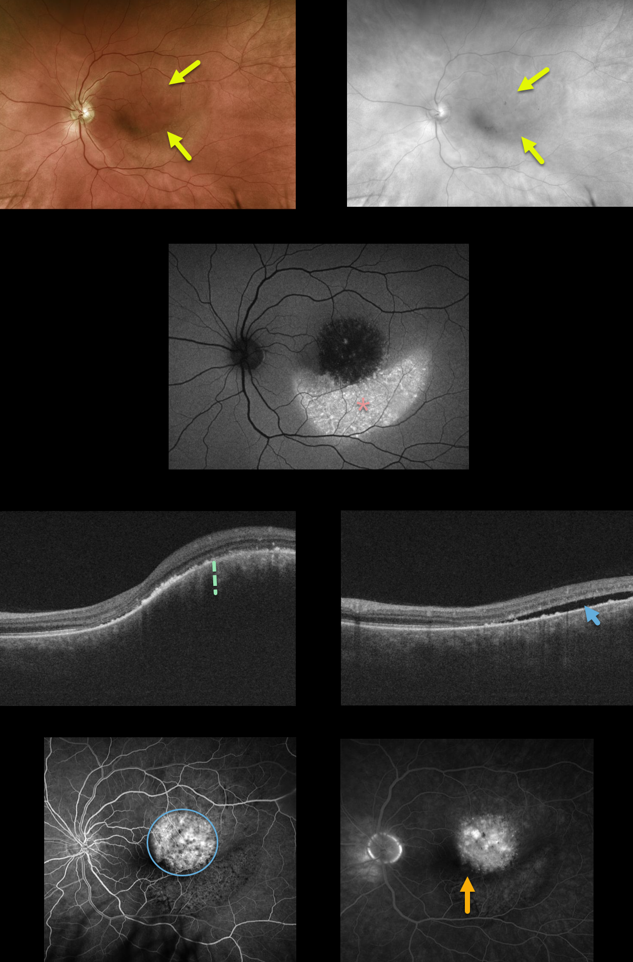
CHOROIDAL HEMANGIOMA
Originally posted on @retina.rocks March 2, 2020
Our patient has a choroidal hemangioma which is difficult to see in the Optos color photograph, but somewhat better defined in the red channel image.
There is marked increased fundus autofluorescence inferior to the tumor, due to migrating subretinal fluid causing RPE alterations. OCT shows marked choroidal thickening within the tumor with subretinal fluid.
Fluoroscein angiography shows mild leakage from the tumor, with faint pigmentary changes inferiorly. Vision is 20/40 and he is minimally symptomatic due to the lack of foveal fluid. We are currently carefully observing this lesion and will consider photodynamic therapy if increased fluid and vision loss develop.
Learning Points:
Choroidal hemangiomas are benign choroidal vascular tumors that can become symptomatic later in life from overlying exudation. The usually round tumors are variably elevated and are often difficult to see clinically since their orange color blends in with the normal surrounding fundus.
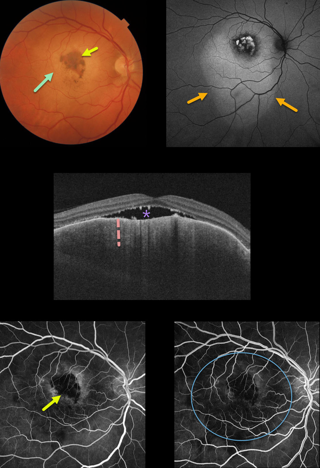
CHOROIDAL METASTASES
Originally posted on @retina.rocks February 10, 2020
This patient has an uncommon pancreatic choroidal metastasis. The lesion is clinically amelanotic with a more central area of increased pigmentation but is much
larger on fundus autofluorescence.
The OCT shows marked choroidal thickening with some subretinal fluid. Fluoroscein angiography (FA) shows no leakage with blockage from the central hyperpigmentation and fainter surrounding blockage from the tumor itself.
Learning Points:
Although uveal melanoma is the most common primary intraocular malignancy, metastatic choroidal tumors are the most common intraocular malignancies.
Lung and breast cancer are the most common sources.
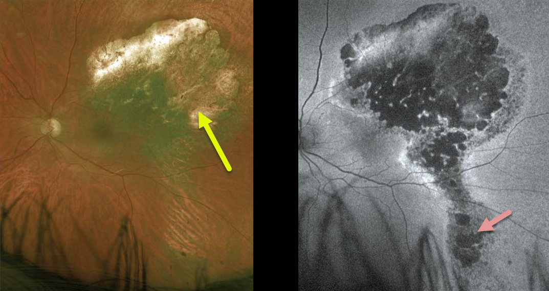
UVEAL MELANOMA
Originally posted on @retina.rocks February 7, 2020
This is a patient with uveal melanoma treated with a radiation plaque therapy 8 years ago.
Widefield fundus autofluorescence helps us image these clinically subtle RPE findings better than ever before.
Learning Points:
When active, these lesions can chronically leak causing disruption in the RPE with a “gutter”. Gutters are commonly seen in pathology with chronic leakage, including central serous retinopathy and overlying choroidal hemangiomas, choroidal nevi and uveal melanoma.
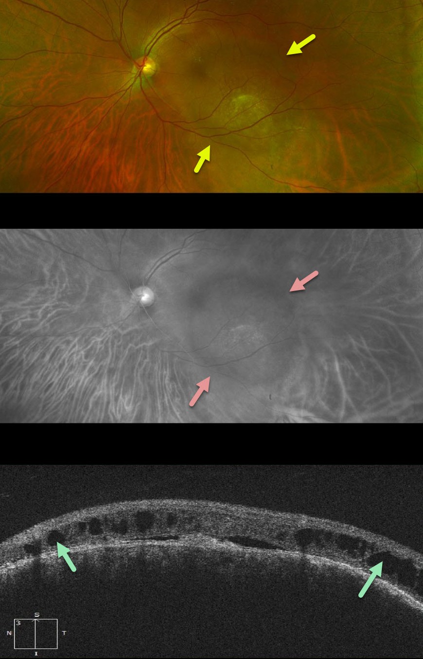
CHOROIDAL HEMANGIOMA
Originally posted on @retina.rocks January 1, 2020
This patient has an isolated choroidal hemangioma. The extent of the lesion is difficult to appreciate on the Optos color photograph, but becomes more obvious on the red Optos channel (633nm) which best visualizes the choroid.
The OCT shows overlying cystic changes which is causing increasing visual symptoms.
Photodynamic therapy was performed in hopes of decreasing the macular leakage.
Learning Points:
Isolated choroidal hemangiomas are benign vascular tumors. The lesion can be difficult to see with the color photograph and is better visualized with the red channel, fundus autofluorescence, and fluoroscein angiography.


