

14 Optic Nerve Disorders
<< Back to Cases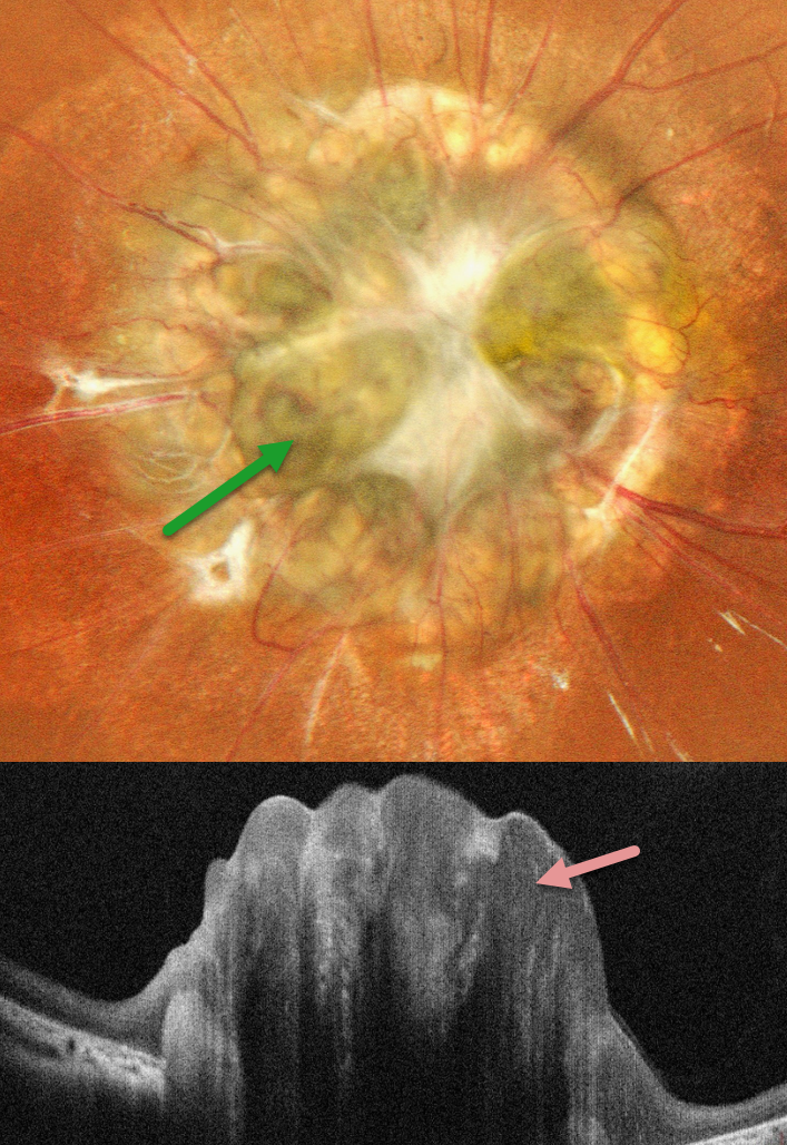
MORNING GLORY DISC WITH EXTENSIVE GLIAL PROLIFERATION OBSCURING DISC EXCAVATION
Shraddha Raj Shrivastava and Manish Nagpal
Originally posted on @retina.rocks September 11, 2026
This healthy 8YO female was born blind in her left eye. Vision was 20/20 in her normal OD and no light perception OS.
Pseudocolor SLO imaging shows a markedly enlarged optic disc with surrounding chorioretinal scarring. Numerous retinal vessels extend outwards from the disc, and extensive fibrotic tissue obscures the disc details. This tissue is markedly elevated and hyperreflective on OCT scanning.
Learning Points:
Morning glory syndrome is a typically unilateral congenital anomalous disc malformation that presents with an enlarged, colobomatous-appearing disc with an increased number of retinal vessels that radiate outwards. The center of the nerve is typically covered by glial tissue. In our patient’s case, this tissue is so extensive that it obscures the disc coloboma.
This abnormality is named for its striking resemblance to the Morning Glory flower. Visual prognosis is usually poor. The condition can be associated with serous macular detachment. Neuroimaging should be considered to rule out transsphenoidal encephalocele and intracranial carotid artery dysgenesis. Fortunately, our patient’s neurologic imaging was negative.
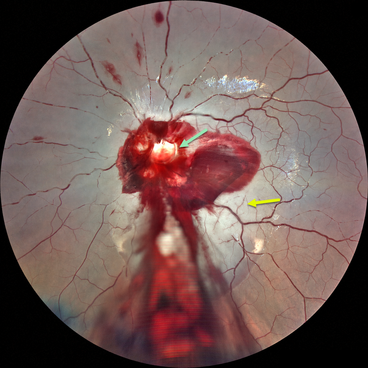
ACUTE OPTIC NERVE AVULSION WITH SECONDARY CRAO
Ayushi Gupta and Vishal Agrawal
Originally posted on @retina.rocks September 8, 2026
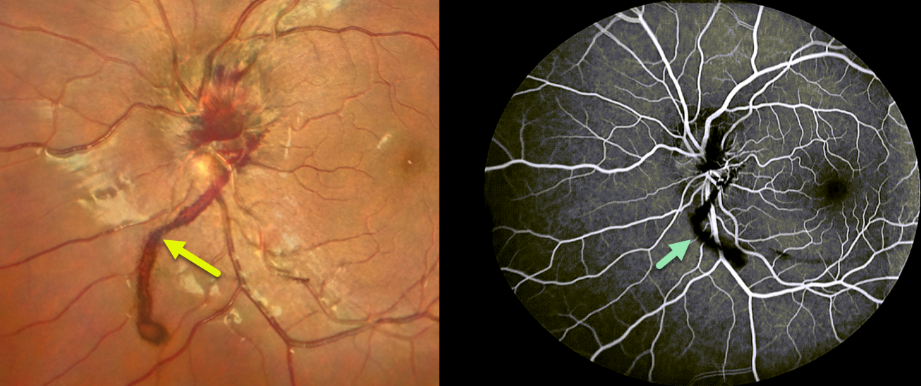
This 6YO boy developed sudden and complete loss of vision in his left eye immediately following blunt trauma. He was riding a bicycle as a pillion passenger when the bike stopped suddenly, throwing his face against the handlebar. Vision was no light perception. Anterior segments were normal.
Fundus photography shows preretinal blood surrounding the nerve, extending through the macula and into the inferior periphery. A pale-yellow cavity is noted where the nerve head should be. A fresh central retinal artery occlusion (CRAO) with a stagnant blood column is noted, along with some scattered inner retinal hemorrhages. There were no other physical or neurologic injuries.
Learning Points:
Traumatic optic nerve head avulsion is a rare and visually devastating traumatic event in which the optic nerve is forcibly separated from the globe at its junction with the sclera, typically resulting in immediate, severe, and irreversible vision loss. The mechanism involves sudden forceful rotation or anterior displacement of the globe following blunt periorbital trauma, including projectiles, falls, or finger-poke injuries, whereby the globe moves anteriorly or rotationally while the optic nerve remains relatively fixed, causing violent separation. Fundoscopic examination may reveal a partial or complete tear of the optic nerve head from the sclera (Foster et al, Arch Ophthalmol 1997;115:623-630). Avulsion can be partial or complete, and may be associated with a CRAO, as was noted in our patient.
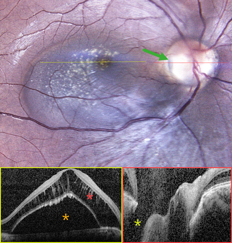
OPTIC NERVE PIT
Rohan Suresh Ninan
Originally posted on @retina.rocks August 28, 2026
This healthy 24YO female presented with asymptomatic retinal changes in her right eye, which was 20/20 one year earlier. Vision was 20/80 OD and 20/20 in her normal OS.
Fundus photography shows a serous macular detachment connected to a yellow-grey optic nerve pit along the temporal edge of the disc. OCT scanning shows an elevated serous detachment with marked overlying cystic schisis in the outer nuclear layer. The pit is noted on the OCT.
Learning Points:
An optic nerve pit is a rare (about 0.1% prevalence) congenital anomaly thought to be caused by incomplete closure of the optic fissure during gestation. Pits are most often located within or along the inferior-temporal disc margin and usually appear as a gray depression. While most optic pits are asymptomatic, they can cause vision loss by forcing fluid into the inner retina, outer retina, and, ultimately, the subretinal space (optic pit maculopathy). The source of the fluid remains a subject of ongoing debate, with opinions ranging from the vitreous to the subarachnoid space.
Given the lack of symptoms, continued observation was recommended for our patient.
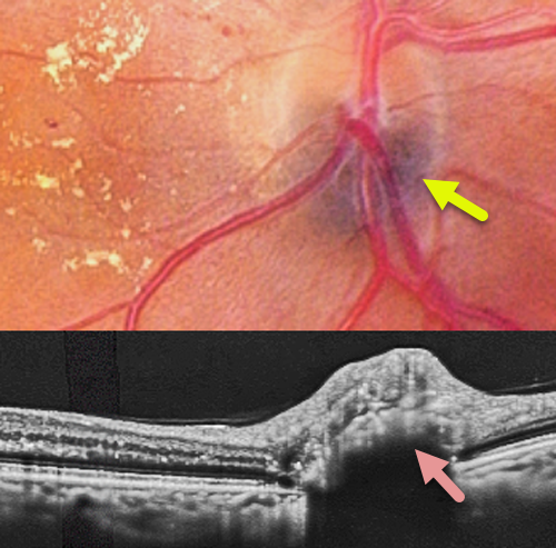
MELANOCYTOMA
Abhishek Karra and Ravindra Karra
Originally posted on @retina.rocks August 24, 2026
This 62YO female with type 2 diabetes was referred to us for diabetic retinopathy screening. Vision was 20/40 OD.
Color photography shows moderate non-proliferative diabetic retinopathy with nasal lipid exudation. The inferior portion of the optic disc has a black discoloration deep to the vessels. OCT scanning through this lesion shows a dome-shaped hyperreflective mass with posterior shadowing.
Learning Points:
Melanocytomas are benign melanocytic tumors intrinsic to the optic nerve. They are intensely pigmented with feathery margins, sometimes spreading into the adjacent retina and choroid. A retinal component, as seen in our patient, is found in about 30% of cases (Shields et al, Ophthalmology 2004;111:1739-1746). Although they may enlarge over time, malignant transformation is rare. Spontaneous necrosis may present with a similar appearance to vitreous seeding from melanoma. Vision and visual field loss can rarely occur from compressive optic neuropathy.
PAPILLEDEMA
Abhishek Karra and Ravindra Karra
Originally posted on @retina.rocks August 5, 2026
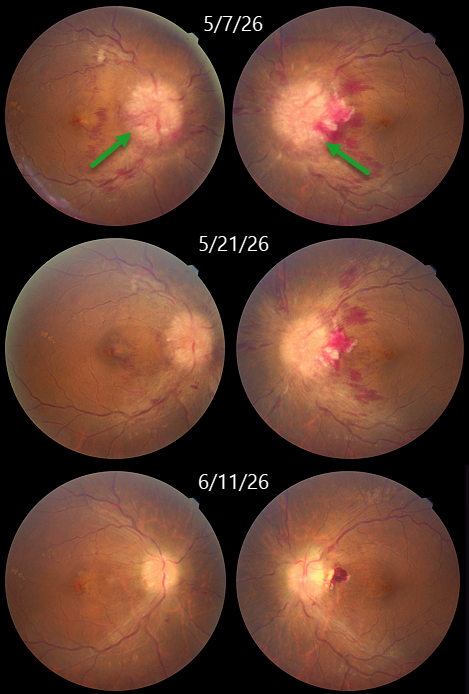
This 44YO obese female presented with several days of decreased vision bilaterally. Vision was counting fingers OU. Fundus photography shows severe bilateral disc swelling.
MRI of the brain and orbit was suggestive of partial empty sella with prominent perioptic CSF spaces and no intracranial mass lesions. All these were suggestive of idiopathic intracranial hypertension (IIH). She was referred to neurology, where a spinal tap established the diagnosis of IIH, and she was started on acetazolamide 1000 mg qday. One month later, vision improved to 20/20 OU with marked improvement in the papilledema.
Learning Points:
IIH, formerly known as pseudotumor cerebri, is caused by elevated intracranial pressure primarily due to decreased cerebrospinal fluid (CSF) drainage caused by elevated cerebral venous pressure. The condition is most often found in obese women of childbearing age. Presenting symptoms include headaches, transient visual obscurations, neck/back pain, and tinnitus/whooshing. Treatment includes weight loss, acetazolamide, and topiramate. Severe cases unresponsive to medical therapy, or those with transverse sinus stenosis, require surgery (CSF diversion procedures, venous sinus stenting, or optic nerve sheath fenestration).
OPTIC NERVE PIT
Shraddha Raj Shrivastava and Manish Nagpal
Originally posted on @retina.rocks July 24, 2026
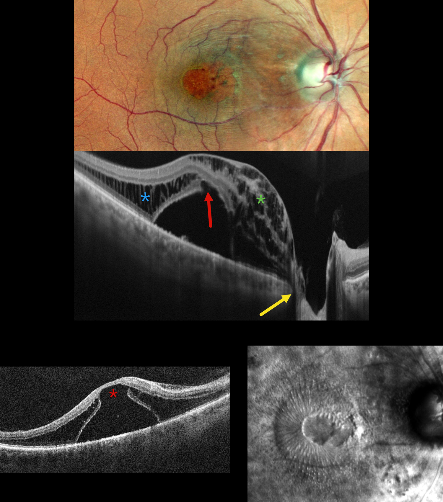
This 50YO male presented with 1 year of decreased vision in his right eye. He had been treated elsewhere with intravitreal injections for cystoid macular edema. Vision was 20/200 OD and 20/20 in his normal OS.
Pseudocolor SLO imaging shows a possible serous macular detachment with a large macular pseudohole. A possible pit is noted along the temporal aspect of the optic nerve. OCT scanning shows an elevated serous detachment. An outer-layer macular hole is noted along the posterior aspect of the fovea, along with full-thickness cystic retinal edema nasally and outer nuclear layer cystic edema temporally. The subretinal fluid directly communicates with the optic nerve pit. Retro mode reveals a unique view of the macular pathology, with the central outer macular hole and radiating fluid.
Learning Points:
An optic nerve pit is a rare (about 0.1% prevalence) congenital anomaly thought to be caused by incomplete closure of the optic fissure during gestation. Pits are most often located within or along the inferior-temporal disc margin and usually appear as a gray depression. While most optic pits are asymptomatic, they can cause vision loss by forcing fluid into the inner retina, outer retina, and, ultimately, the subretinal space (optic pit maculopathy). The source of the fluid remains a subject of ongoing debate, with opinions ranging from the vitreous to the subarachnoid space.
Vitrectomy surgery was discussed, and the patient elected for observation unless his symptoms worsened.

MELANOCYTOMA
Ayushi Gupta and Vishal Agrawal
Originally posted on @retina.rocks July 17, 2026
This 42YO female presented without complaint. Vision was 20/20 in her normal OD and 20/20 OS. Color fundus photography of the left eye reveals a well-defined, densely pigmented, jet-black lesion arising from the optic disc and extending slightly into the peripapillary retina. On OCT, the elevated lesion is hyperreflective with complete posterior shadowing.
Learning Points:
Melanocytomas are benign melanocytic tumors intrinsic to the optic nerve. They are intensely pigmented with feathery margins, sometimes spreading into the adjacent retina and choroid. A retinal component, as seen in our patient, is found in about 30% of cases (Shields et al, Ophthalmology 2004;111:1739-1746). Although they may enlarge over time, malignant transformation is rare. Spontaneous necrosis may present with a similar appearance to vitreous seeding from melanoma. Vision and visual field loss can rarely occur from compressive optic neuropathy.
MORNING GLORY DISC AND CHOROIDAL COLOBOMA WITH PSEUDODUPLICATED OPTIC NERVE
Shraddha Raj Shrivastava and Manish Nagpal
Originally posted on @retina.rocks June 30, 2026
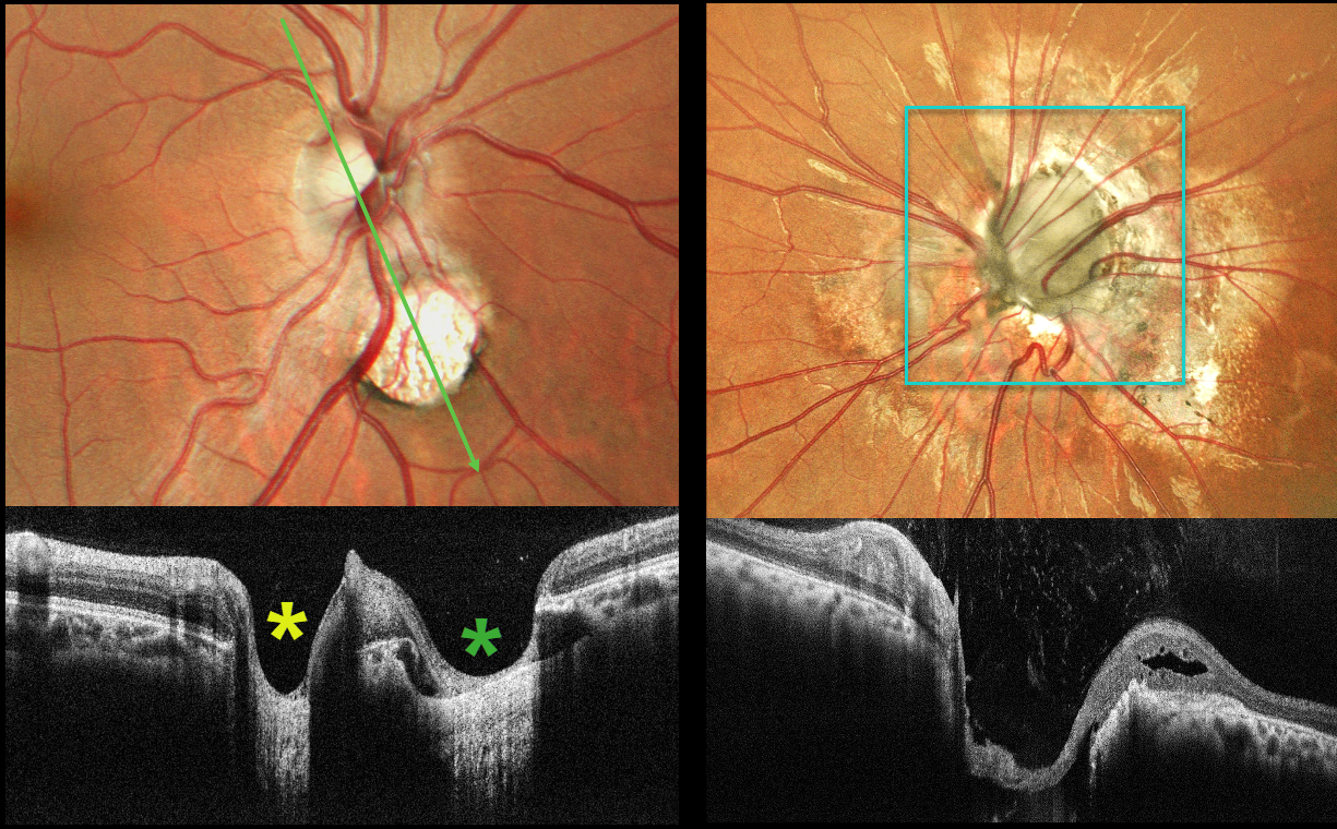
This 26YO male presented with a history of stable poor vision and inward deviation of his left eye since childhood. Vision was 20/20 OD and counting fingers in his esotropic, microphthalmic OS.
Color photography of the right posterior pole shows a round coloboma inferonasal to the nerve, giving the appearance of a pseudo-duplicated disc. OCT scanning through the nerve and coloboma reinforces the illusion of a second nerve head. The anomalous left nerve is enlarged and excavated, with numerous vessels radiating outwards from its inferonasal aspect.
Morning glory syndrome is typically unilateral and shares some similarities with optic disc coloboma and juxtapapillary staphyloma. This abnormality is named for its striking resemblance to the Morning Glory flower. Visual prognosis is usually poor. The condition can be associated with serous macular detachment. Neuroimaging should be considered to rule out transsphenoidal encephalocele and intracranial carotid artery dysgenesis, and fortunately, our patient’s imaging was negative. For a great review on the pathogenesis and treatment of maculopathy associated with cavitary optic disc anomalies, see Jain and Johnson, AJO 2014;158:423-435.
The coexistence of morning glory disc anomaly with microphthalmos, and contralateral chorioretinal coloboma in this case, may represent a continuum of optic fissure developmental defects with asymmetric expression, leading to a spectrum of cavitary optic disc anomalies in the same individual.
Pseudoduplication of the optic nerve occurs when a round choroidal coloboma or chorioretinal scar about the size of the optic nerve is associated with overlying radiating blood vessels (Bloom et al, Retinal Cases 2022;16:174-176). In our experience, this fake-out is most common from toxoplasmosis scars located near the optic nerve.
MFRP RETINITIS PIGMENTOSA
João Pedro Marques
Originally posted on @retina.rocks June 9, 2026
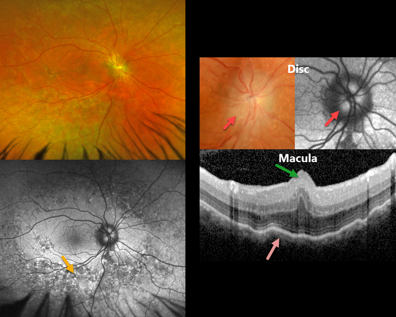
This 49YO male has a history of MFRP-related retinitis pigmentosa (RP). We have followed him for 13 years with stable findings of bilateral nanophthalmos (+18D hyperopia, axial lengths 14.82mm OD and 15.14 OS), optic disc drusen (ODD), and choroidal folds. He has compound heterozygosity for 2 pathogenic variants in the MFRP gene.
Optos color RG imaging in his OD shows pigmentary changes throughout the midperiphery along with an ill-defined disc margin. Fundus autofluorescence (FAF) shows variable hyper- and hypo-FAF in the midperiphery. Disc drusen are noted on a more magnified view, which hyper-FAF. Macular OCT shows a central fold, choroidal thickening, and chorioretinal folds. Identical findings were present in his OD (not shown).
Learning Points:
The MFRP (membrane frizzled-related protein) is expressed in the RPE and ciliary epithelium. MFRP functions as a molecular hub on the RPE apical membrane, coordinating protein trafficking and lipid homeostasis. Loss of MFRP leads to DHA accumulation in the RPE, downregulation of visual cycle genes and phototransduction genes, and progressive photoreceptor degeneration.
Biallelic MFRP mutations cause a well-characterized autosomal recessive retinitis pigmentosa (RP) syndrome with nanophthalmos and ODD (Li et al, BJO 2024;108;1679-1687). These eyes are at high risk of angle-closure glaucoma, usually requiring prophylactic laser peripheral iridotomy.
PAPILLEDEMA WITH SPONTANEOUS VITREOUS HEMORRHAGE
The European VitreoRetinal Society (EVRS) and Gordon Shen
Originally posted on @retina.rocks June 5, 2026
This 20YO female presented with a history of headaches and recent floaters in her right eye. There was a history of idiopathic intracranial hypertension.
Color photography shows a swollen right nerve with overlying preretinal blood extending inferiorly. Fluorescein angiography showed no leakage. Unfortunately, further clinical information and follow-up are not available.
Learning Points:
Vitreous hemorrhage directly from papilledema is exceedingly rare, and the published literature on this specific complication is very limited. Prior reports attribute vitreous hemorrhage to papilledema in eyes with no other pathology. One report suggested that the breakthrough vitreous hemorrhage originated from the optociliary shunt vessels (Fraser et al, J Neuroophthalmology 2012;32:332-334). Our patient offers visual evidence that vitreous blood can arise directly from an edematous nerve.
SCRUB TYPHUS POST-FEVER RETINITIS
Anand Temkar, Surendra Pal, Vasumathi Vijay, and Manorama Baheti
Originally posted on @retina.rocks June 3, 2026
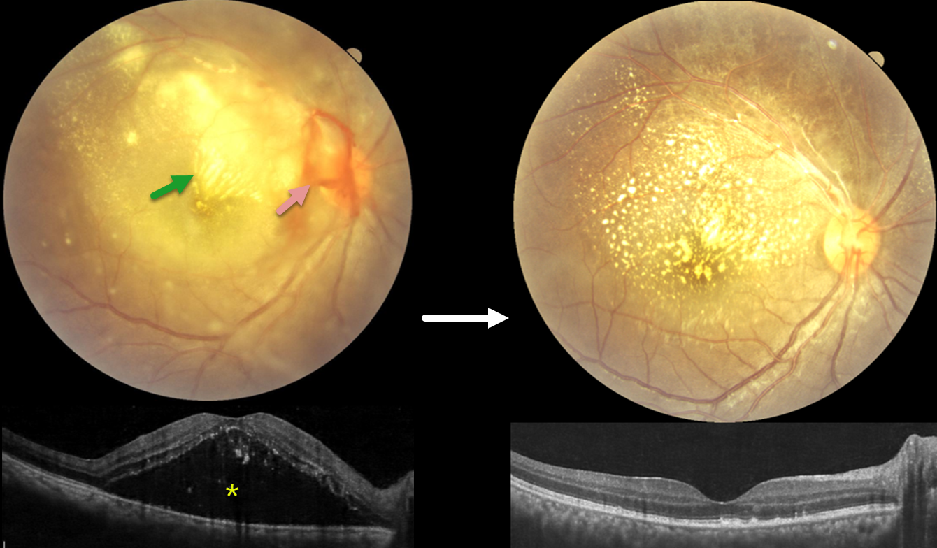
This 28YO female presented with 4 days of blurred vision in her right eye. For the past 2 weeks, she has complained of generalized weakness, malaise, and body pain. Vision was counting fingers OD and 20/20 in her normal OS.
Fundus photography shows variable, dense subretinal lipid, particularly confluent in the superonasal macula. There is some preretinal blood around the nerve, and the retina is somewhat yellow and opaque temporally and inferiorly. Patches of inner retinal opacification are noted outside the macula superiorly. OCT shows hyperreflective dots from lipid exudate in the outer retina, with a large serous detachment. Laboratory testing, including CBC, ESR, syphilis, TB, HIV, TORCH profile, and chest X-ray, was negative. We started a 1-week tapering course of oral prednisone 40mg and doxycycline 100mg BID. Three days after our initial examination, she mentioned that she had 2 weeks of mild intermittent fever. Weil-Felix testing was positive, and she was subsequently diagnosed with scrub typhus and asked to continue the doxycycline for a full 2-week course.
The clinical picture gradually improved, and about 6 weeks following presentation, vision improved to 20/40 despite significant residual lipid. OCT scanning through the macular center was mostly normalized. We continue to follow her closely.
Learning Points:
Rickettsial infections cause posterior segment involvement in a substantial proportion of patients, often asymptomatic despite significant fundoscopic findings. The most common rickettsial organisms causing retinal disease include Rickettsia rickettsii (Rocky Mountain spotted fever), Rickettsia conorii (Mediterranean spotted fever), and Rickettsia typhi (murine typhus).
Scrub typhus, caused by Orientia tsutsugamushi and transmitted by a larval mite (chigger) bite, has ocular involvement in about 20% of patients. Retinal findings include retinal hemorrhages, branch retinal vein occlusion, retinal vasculitis/retinitis, and papillitis (Ganekal et al, Ind J Ophthalmol 2021;69:1167-1171).
The ocular and systemic findings of vasculitis and perivasculitis result from both direct endothelial cell destruction by the organism and exaggerated immune responses. Doxycycline is the drug of choice; treatment should last at least 7 days and continue for at least 3 days after fever has subsided and clinical improvement is evident. Visual prognosis is generally favorable with appropriate treatment.
OPTIC NERVE PIT
Malvika Singh and Manish Nagpal
Originally posted on @retina.rocks May 26, 2026
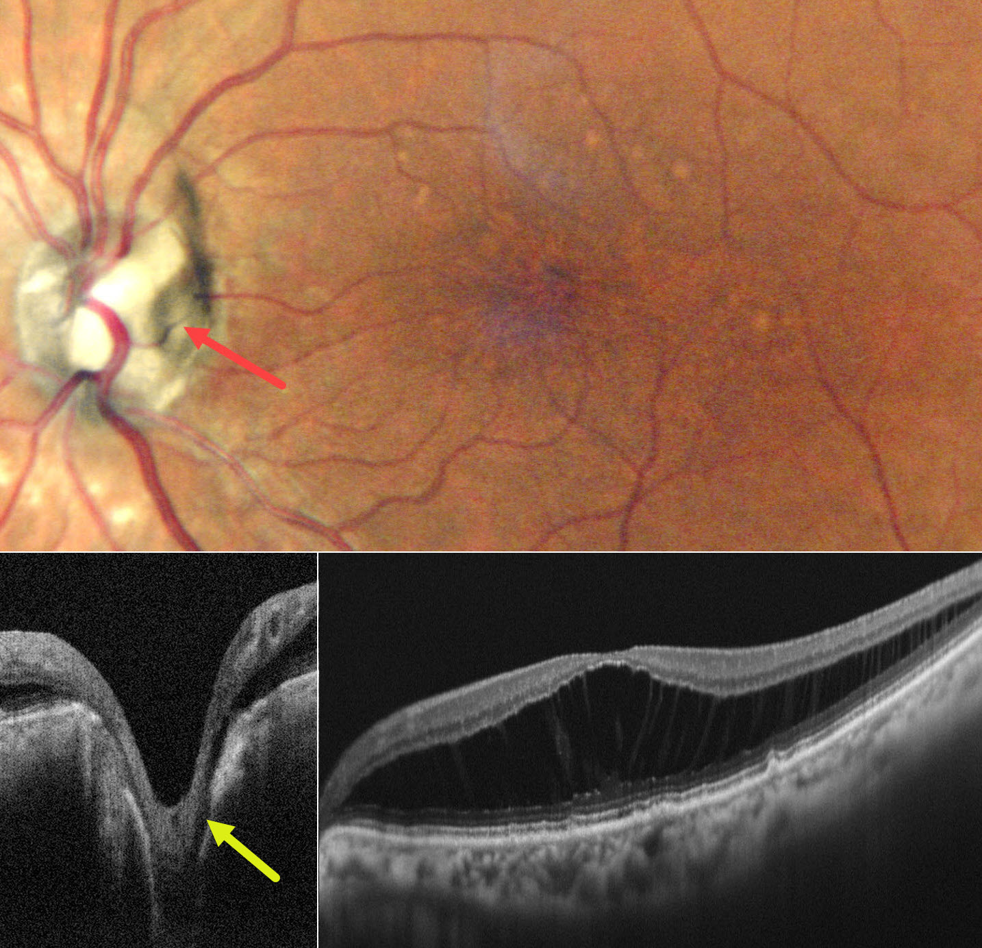
This 72YO female presented with 7 months of stable decreased vision in her left eye. Vision was 20/20 OD and 20/30 OS.
Pseudocolor SLO imaging shows irregular foveal pigment loss with more distal drusen-like changes. There is diffuse outer nuclear layer macular schisis on OCT. An optic pit is noted along the temporal edge of the nerve, which directly communicates with the schisis cavity. Observation was recommended.
Learning Points:
An optic nerve pit is a rare (about 0.1% prevalence) congenital anomaly thought to be caused by incomplete closure of the optic fissure during gestation. Pits are most often located within or along the inferior-temporal disc margin and usually appear as a gray depression. While most optic pits are asymptomatic, they can cause vision loss by forcing fluid into the inner retina, outer retina, and, ultimately, the subretinal space (optic pit maculopathy). The source of the fluid remains a subject of ongoing debate, with opinions ranging from the vitreous to the subarachnoid space.
FULL-THICKNESS EYEWALL TRANSPLANT WITH INADVERTENT GRAFT INVERSION
Originally posted on @retina.rocks April 1, 2026
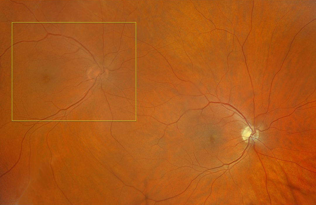
This healthy 43YO male fighter pilot developed a complex corneal laceration in his left eye. Despite multiple surgical procedures, he was left with an opaque cornea and an inability to fly due to the lack of normal binocular vision. Full-thickness eyewall transplantation from his normal left posterior segment into his right temporal posterior segment was therefore performed.
Intraoperatively, the dyslexic surgeon inadvertently inverted the allograft, resulting in the donor graft being inverted 180 degrees vertically. Optos color RGB imaging shows the donor nerve and macula in the right superotemporal midperiphery. Note that the disc should have been placed temporal to the donor macula, not nasal. Although surgically successful, this left the patient with monocular diplopia instead of the desired monocular binocular stereopsis. He remains grounded, and his case is still mired in litigation.
And happy April Fool’s Day!
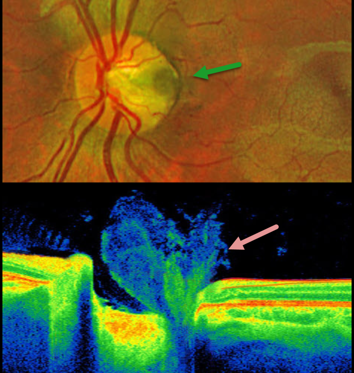
OPTIC NERVE PIT WITH VITREOUS PLUG
The European VitreoRetinal Society (EVRS) and Miguel Rechichi
Originally posted on @retina.rocks January 9, 2026
This healthy 6YO boy presented for a routine examination. Vision was 20/20 in his normal right eye and 20/20 in his left eye. OCT scanning shows a vitreous plug extending into the depth of the pit.
Optos color RG imaging shows a temporal optic nerve pit without associated macular abnormalities.
Learning Points:
An optic nerve pit is a rare (about 0.1% prevalence) congenital anomaly thought to be caused by incomplete closure of the optic fissure during gestation. Pits are most often located within or along the inferior-temporal disc margin and usually appear as a gray depression. While most optic pits are asymptomatic, they can cause vision loss due to fluid being forced into the inner retina, outer retina, and the subretinal space (optic pit maculopathy).
Utilizing high-resolution swept-source OCT, Spaide defined the optic pit complex as a broader means to characterize the structural abnormalities found with optic disc pits (Retina 2026;46:15-24). The spectrum of abnormalities includes lamina cribrosa defects (all eyes) with associated tissue cavitations extending a mean of 1855 microns posterior to a modified Bruch membrane opening. One of his patients had OCT findings similar to our patient’s, with vitreous extending into the pit. Sources of intra- and subretinal fluid included direct communication from a cavitation, perivascular hyporeflective spaces, intraneural channels extending toward cystoid spaces, and isolated retinal cysts. These concepts support the “dynamic and multifactorial process involving abnormal structural anatomy, disrupted tissue boundaries, potential misrouting of glymphatic flow, vitreous or cerebral spinal fluid (CSF)-related gradients, and mechanical forces exerted during eye movements.”
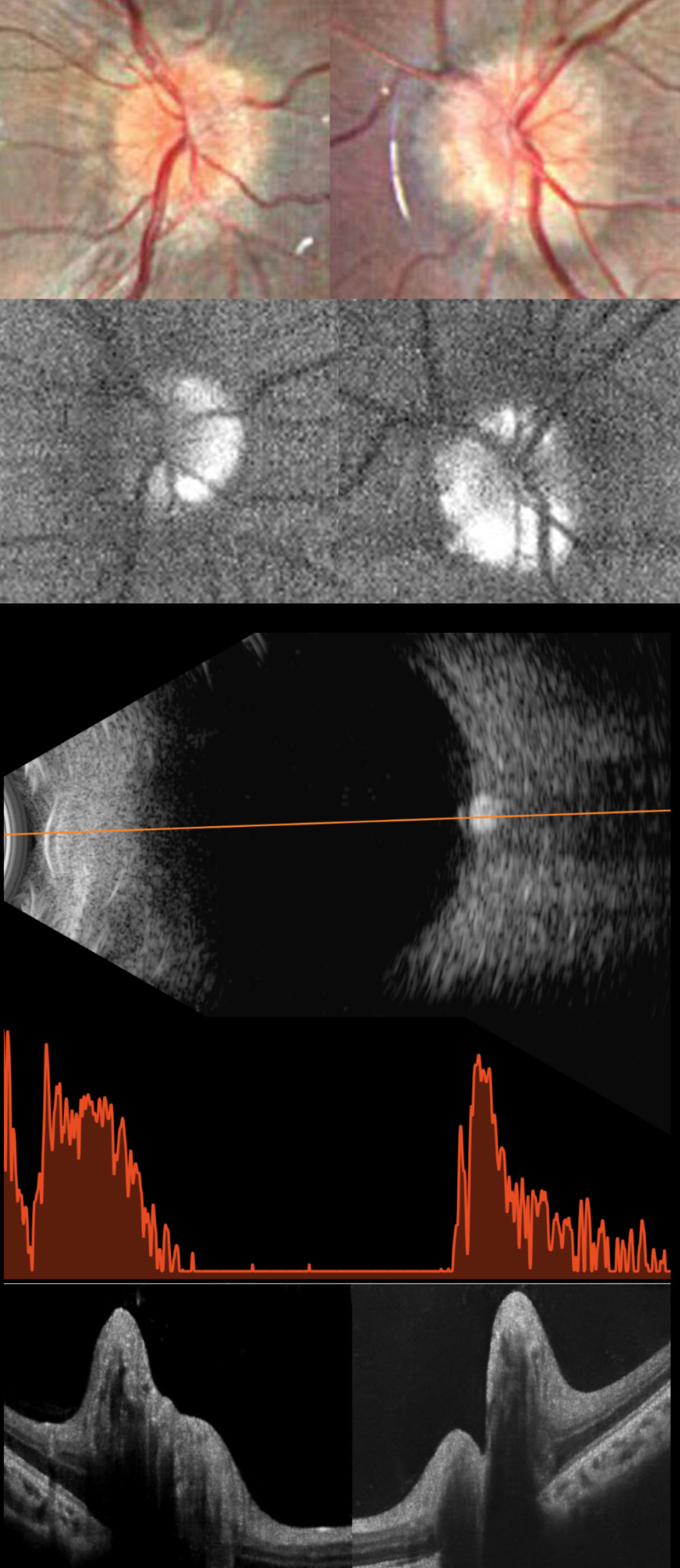
OPTIC NERVE DRUSEN
Ayushi Gupta
Originally posted on @retina.rocks November 25, 2025
This healthy 11YO girl was diagnosed elsewhere 2 months earlier with papilledema and precocious puberty, which was being treated with oral acetazolamide. Workup included a normal brain MRI and lumbar puncture with normal opening pressure and CSF analysis. Vision in our office was 20/20 OU.
Fundus photography shows pseudopapilledema with extensive optic disc drusen (ODD). On fundus autofluorescence (FAF), the drusen are hyper-FAF. On B-scan ultrasonography, the discs are markedly hyperreflective, indicating calcification. OCT scanning shows marked, irregular disc thickening due to drusen.
Learning Points:
Optic disc drusen (ODD) are refractile calcific concretions that form anterior to the lamina cribrosa. They are thought to represent extruded, extracellular, calcified mitochondria and may be inherited as an autosomal dominant trait.
Porphyrins, organic molecules composed of four interconnected pyrrole subunits, are present within mitochondria. When these compounds break down, they become autofluorescent, which is likely why drusen autofluoresce. Porphyrins are also part of the heme molecule in hemoglobin, which explains why devitalized blood autofluoresces (Bloom and Spaide, Retinal Cases 2022;16:401-402).
ODD can be confused with true disc swelling, as occurred in our patient, which led to an erroneous diagnosis of papilledema. In our experience, the pathognomonic B-scan ultrasonographic disc calcification is rarely seen in patients this young, but is extremely helpful in establishing the diagnosis when present. We find FAF to be a more reliable test when the ODD are not obvious clinically. After speaking with her neurologist, the acetazolamide was discontinued, and observation was recommended.
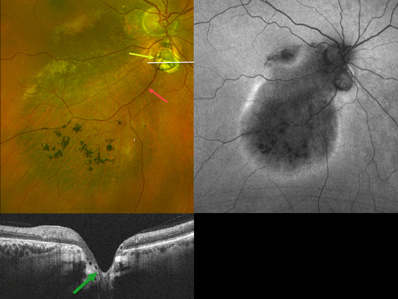
CHOROIDAL COLOBOMA WITH OPTIC NERVE PSEUDODUPLICATION
Originally posted on @retina.rocks October 8, 2025
This 60YO female was referred for asymptomatic unilateral fundus findings in her right eye. Vision was 20/30 OD.
Optos color RG imaging shows a choroidal coloboma just inferior to the disc, giving the appearance of a pseudoduplicated optic nerve. Coarse pigmentary changes with some pigment migration extend from the macula into the inferior midperiphery. An inferotemporal retinal vein drains directly into the choroid through the coloboma. Triton swept-source OCT shows that the coloboma overlies an area of ectatic sclera. Fundus autofluorescence (FAF) shows variable hypo-FAF within the area of clinical pigmentary changes, with a surrounding rim of hyper-FAF.
Learning Points:
Choroidal colobomas are most often due to failure of the optic vesicle and choroidal fissure to close during fetal development. Lesions like this may also be from other causes, including toxoplasmosis. Choroidal coloboma can present as solitary or multiple lesions. Visual prognosis depends on the location and involvement of the optic nerve and macula.
Pseudoduplication of the optic nerve occurs when a round choroidal coloboma or chorioretinal scar about the size of the optic nerve is associated with overlying radiating blood vessels (Bloom et al, Retinal Cases 2022;16:174-176). In our experience, it is most common for toxoplasmosis scars to be located near the optic nerve.
Under normal conditions, the retinal and choroidal circulations remain separate and distinct. Rarely, the two circulations connect directly through a chorioretinal anastomosis. These can occur in chorioretinal scars (typically from inactive toxoplasmosis lesions), in macular telangiectasia, and in disciform scars.
The prominent gutter of inferior pigmentary changes indicates a prior retinal detachment, most likely related to spontaneously resolved fluid associated with the coloboma (Tanaka et al, Ophthalmology Retina 2021;5:702-710). Similar pigmentary changes are seen in central serous retinopathy, although our patient’s macular OCTs showed no pachychoroid features (not shown). Finally, this may represent previously resolved pit-like macular fluid, which is rarely found in the absence of a pit or severe glaucomatous cupping (Fujimoto et al, Ophthalmology Retina 2023;7:811-818).
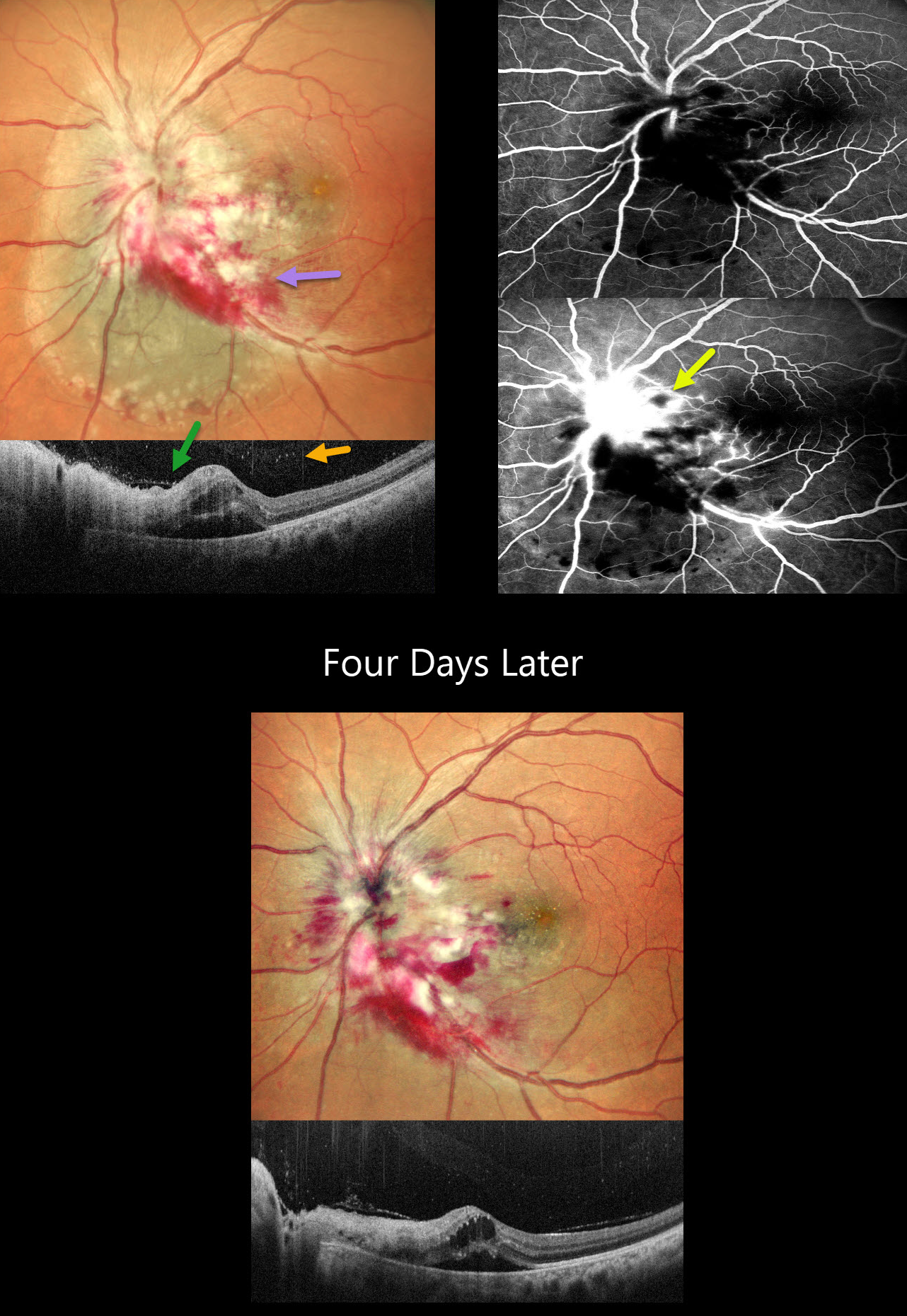
CMV PAPILLITIS AND RETINITIS
Akansha Sharma and Manish Nagpal
Originally posted on @retina.rocks September 18, 2025
This 42YO female with a known history of HIV infection presented with 1 month of vision loss in her left eye. Her last CD4 count was 38 despite being compliant with HAART therapy. Her husband was HIV positive. Vision was 20/20 in her normal OD and 20/400 in OS.
Multicolor SLO imaging shows active cytomegalovirus (CMV) retinitis involving the nerve, which extends nasally and inferotemporally. Subretinal fluid is noted clinically and on OCT. OCT also shows full-thickness disorganization and hyperreflectivity, along with overlying vitreous cells. Fluorescein angiography shows early blockage with late disc and retinal leakage.
Oral valganciclovir and biweekly intravitreal injections were started. Four days later, vision improved to 20/90 with improvement in the papilledema and retinitis.
Learning Points:
CMV retinitis develops as a reactivation of latent CMV in immunosuppressed individuals. Before effective antiretroviral treatment emerged in the mid to late 1990’s, CMV retinitis developed in up to 40% of HIV/AIDS patients, often within the last 6 months of life. Treatment includes a combination of systemic and intravitreal medications, including ganciclovir, foscarnet, and cidofovir. Rhegmatogenous retinal detachment is a common late sequela and requires vitrectomy with silicone oil.
PAPILLEDEMA
Anand Temkar and Surendra Pal
Originally posted on @retina.rocks September 10, 2025
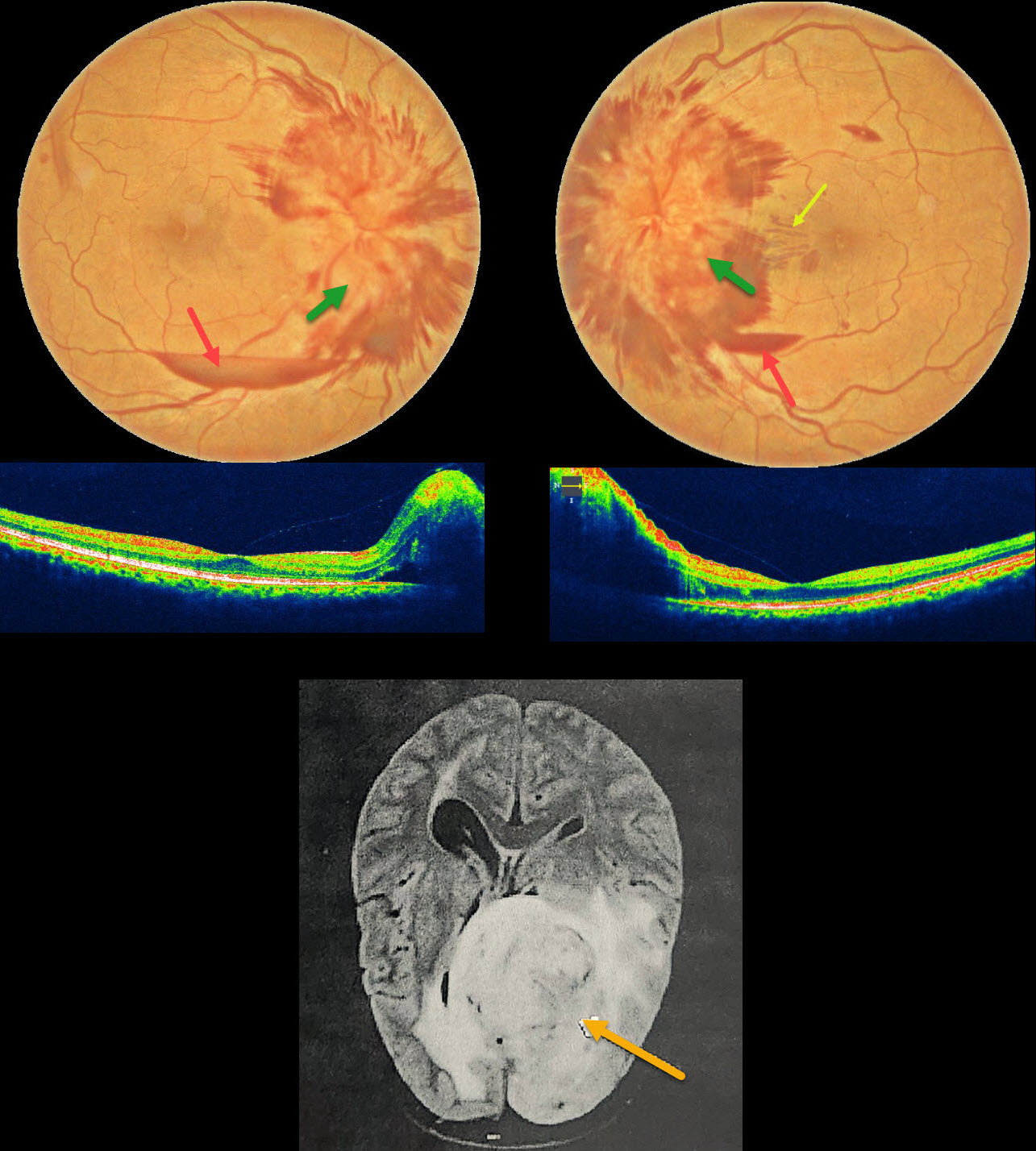
This 50YO female presented with one month of bilateral vision loss. Vision was 20/90 OD and 20/70 OS.
Fundus photography shows severe bilateral optic nerve swelling with surrounding nerve fiber layer hemorrhages. Small inferior subhyaloid hemorrhages are present bilaterally, along with a small outer plexiform layer hemorrhage in the left nasal macula.
MRI scanning showed a high-grade glioma, and she was immediately referred to neurosurgery for further management.
Learning Points:
Glioblastoma multiforme, also known as a grade IV astrocytoma, represents the most common and aggressive primary malignant brain tumor. A variety of neuro-ophthalmic signs and symptoms result from direct involvement of the visual pathway or elevated intracranial pressure, including vision loss or visual field defects, afferent pupillary defect, extraocular muscle motility abnormalities, and papilledema. Although most patients rarely survive beyond 1 year from diagnosis, the prognosis is especially poor with optic nerve involvement.
GLAUCOMATOUS PERIPAPILLARY RETINOSCHISIS
Vaibhav Sethi
Originally posted on @retina.rocks September 5, 2025
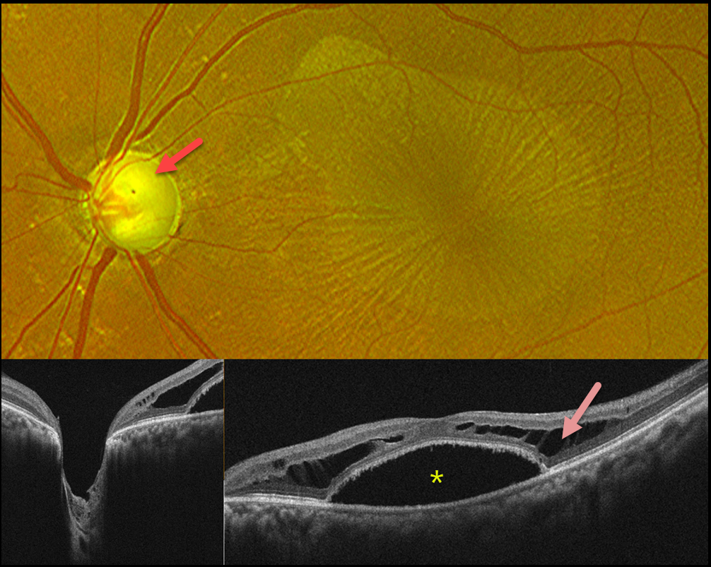
This 45YO male presented with blurred vision in his left eye. The right eye was phthisical due to absolute glaucoma. He was taking topical dorzolamide and travoprost OU. Vision was 20/400 OS. Intraocular pressure was 12 mm Hg.
Optos color RG imaging shows a nearly completely cupped glaucomatous nerve. There is an irregular oval discoloration of the outer macula with overlying radiating retinal striae. OCT scanning shows Henle’s layer schisis with a central macular detachment. An optic pit could not be identified clinically or on OCT.
Learning Points:
While peripapillary retinoschisis (PPRS) secondary to congenital optic disc cavitary abnormalities is a well-known phenomenon, similar findings in patients with glaucomatous cupping but without nerve head cavitation have more recently been described and are less common (Fujimoto et al, Ophthalmology Retina 2023;7:811-818). The retinal nerve fiber layer is most commonly involved in glaucoma, whereas in optic pit maculopathy, outer and inner nuclear layer schisis, foveal involvement, and subretinal fluid are more frequently noted.
Most cases of glaucoma-related PPRS are asymptomatic and spontaneously resolve. However, in fovea-involving or symptomatic cases, surgical intervention may improve vision and decrease time to resolution. There is no consensus regarding the optimal treatment approach, including whether to perform pars plana vitrectomy alone or with the addition of juxtapapillary laser treatment, ILM peeling, inner retinal fenestration, and/or autologous platelet concentrate.
We recommended adding brimonidine and timolol drops to further lower his IOP. We are not planning any surgical intervention.
OPTIC NERVE PIT
Seif Anwar
Originally posted on @retina.rocks July 23, 2025
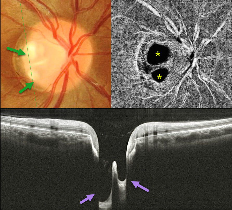
This healthy 20YO female presented with asymptomatic unilateral optic nerve findings in her right eye. Vision was 20/20 OD and 20/20 in her normal OS.
Color photography shows two independent optic nerve pits along the temporal edge of the nerve. OCT angiography dramatically illustrates the pits due to completely absent flow signals. The pits are also well imaged on OCT scans. Observation was recommended.
Learning Points:
An optic nerve pit is a rare (about 0.1% prevalence) congenital anomaly thought to be caused by incomplete closure of the optic fissure during gestation. Pits are most often located within or along the inferior-temporal disc margin and usually appear as a gray depression. Double pits are exceedingly rare (Babu et al, Indian J Ophthalmol 2020;68:663-665).
While most optic pits are asymptomatic, they can cause vision loss by forcing fluid into the inner retina, outer retina, and, ultimately, the subretinal space (optic pit maculopathy). The source of the fluid remains a subject of ongoing debate, with opinions ranging from the vitreous to the subarachnoid space.
RADIATION RETINOPATHY AND PAPILLOPATHY
Mattie Adams
Originally posted on @retina.rocks May 27, 2025
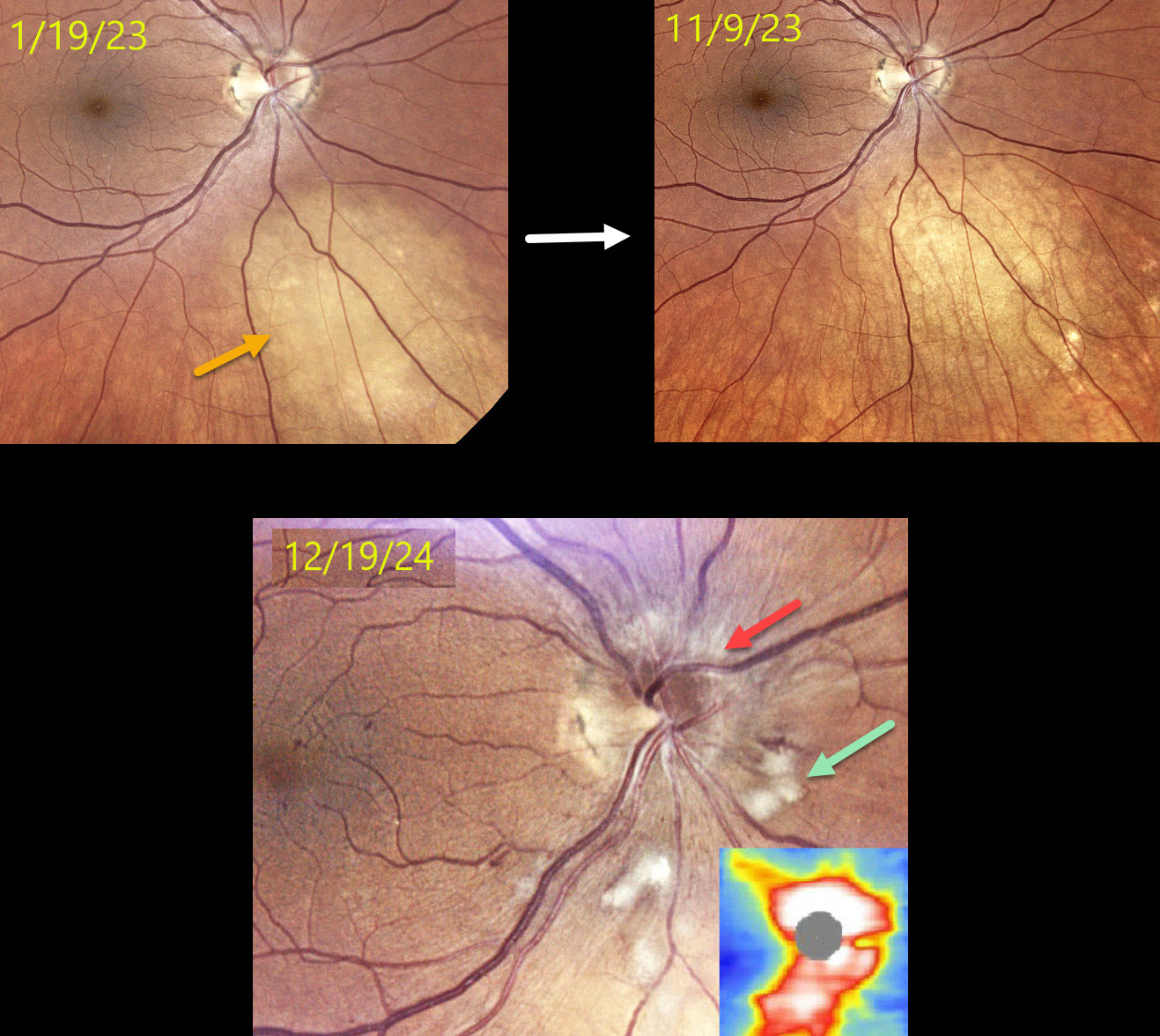
This 52YO female presented on 1/19/23 with a history of metastatic breast cancer and an asymptomatic uveal metastasis in her right eye. The lesion completely regressed following external beam radiation.
When examined on 12/19/24, she remained visually asymptomatic with 20/60 vision. Color photography shows a new, mildly swollen nerve, which is confirmed on OCT. Scattered nerve fiber layer infarcts (cotton-wool spots, CWS) and some nasal macular telangiectasia are also noted.
Learning Points:
Radiation retinopathy usually occurs 3 months to 3 years after external-beam radiation or plaque radiotherapy. The ocular findings closely resemble those seen with diabetic retinopathy. Radiation optic neuropathy (Carey et al, Br J Ophthalmol 2023;107:743-749) can also develop.
Since our patient was completely asymptomatic without macular edema, we recommended close observation. We will begin intravitreal anti-VEGF therapy if she develops macular edema or worsening nerve findings.
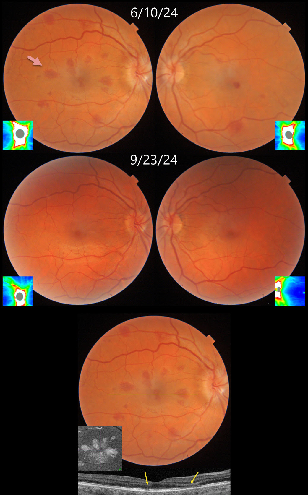
ANEMIA WITH BILATERAL OPTIC NERVE EDEMA
Fraser McKay
Originally posted on @retina.rocks March 27, 2025
This 34YO female underwent uterine ablation 2 weeks earlier for heavy menstruation and severe secondary iron deficiency anemia. Although we don’t know what her hemoglobin level was prior to the procedure, it had risen to 10.7 postoperatively. Vision was 20/30 OD and 20/50 OS.
Triton color photography shows bilateral teardrop-shaped macular outer plexiform layer (OPL) hemorrhages, with greater involvement in her right eye. Numerous more peripheral deep and white-centered hemorrhages are also noted. Swept-source OCT confirms the location of the macular blood. En face OCT shows a more dramatic view of their petaloid shape. There is also mild optic nerve edema, especially in the right eye. The hemorrhages and nerve swelling mostly resolved with iron supplementation several months later.
Learning Points:
Anemia-related retinal findings most commonly include hemorrhages in the retina and sub-internal limiting membrane. Severe anemia is a rare cause for optic nerve swelling and should always be considered in patients with atypical idiopathic intracranial hypertension (Biousse et al, AJO 2003;135:437-446). The underlying pathogenesis of anemia-induced optic nerve swelling remains unknown.
Radiating deep macular hemorrhages are seen in multiple conditions, including Valsalva and trauma (Baumal et al, Br J Ophthalmol 2021;105:374-380). We have not previously seen these hemorrhages in isolated anemia. Their unique radiating appearance is caused by blood accumulating in Henle’s layer. Kon Graversen et al described hemorrhagic unilateral retinopathy, an idiopathic disorder with a similar appearance (Retina 2014;34:483-489).
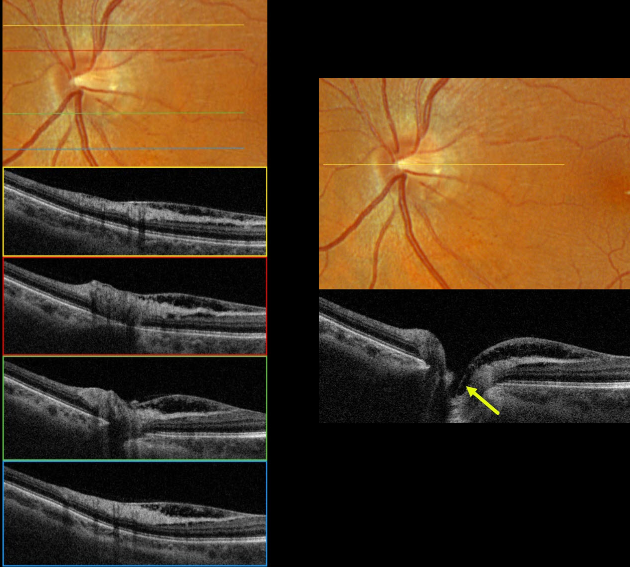
NO OPTIC PIT RETINOSCHISIS
Originally posted on @retina.rocks March 13, 2025
This 55YOF was referred for asymptomatic macular changes in her left eye. Vision was 20/30 bilaterally. The right eye was normal.
Optos color RGB imaging shows a normal appearing optic nerve with a small central cup. Very subtle inner retinal telangiectasia is seen inferotemporal to the disc. Triton swept-source OCT shows nasal macular inner retinoschisis, which extends into the central depression of the optic cup, but with no definite pit. This inner retinal schisis was variably present along the temporal disc margin, extending into the nasal macula. Fluorescein angiography was normal without leakage (not pictured).
Learning Points:
An optic nerve pit is a rare (about 0.1% prevalence) congenital anomaly thought to be caused by incomplete closure of the optic fissure during gestation. Pits are most often located within or along the inferior-temporal disc margin and usually appear as a gray depression. While most optic pits are asymptomatic, they can cause vision loss by forcing fluid into the inner retina, outer retina, and, ultimately, the subretinal space (optic pit maculopathy).
No optic pit retinoschisis (NOPIR) is a rare entity that causes pit-like findings in the absence of a pit or severe glaucomatous cupping (Fujimoto et al, Ophthalmology Retina 2023;7:811-818). As with optic pit maculopathy, eyes with good vision should be observed. Persistent vision loss from foveal involvement can be managed with vitrectomy, peeling of the internal limiting membrane, and fluid-gas exchange.
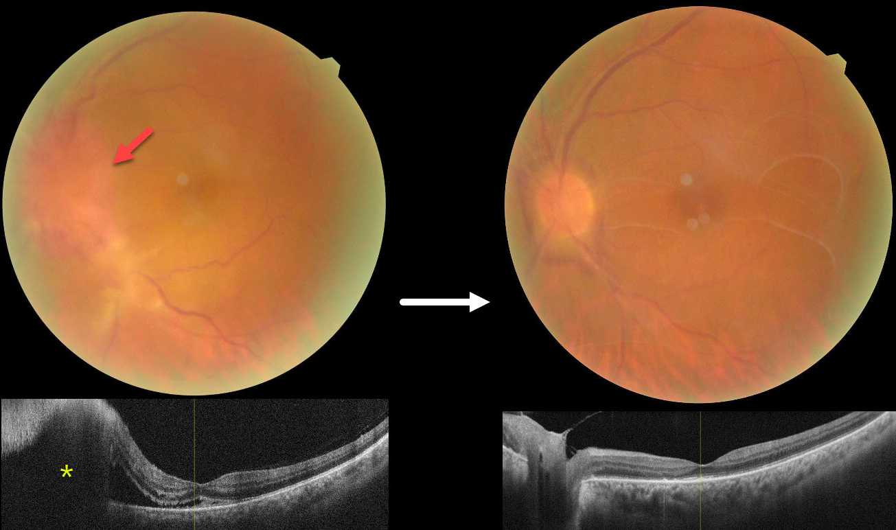
SYPHILITIC PAPILLITIS
Navneet Mehrotra and Tamanna Patel
Originally posted on @retina.rocks March 12, 2025
This 52YO male presented with 3 weeks of vision loss in his left eye. He has a history of diabetes, hypertension, and HIV. He was diagnosed elsewhere with optic neuritis and started on intravenous methylprednisolone with no improvement. Vision was 20/30 in his normal OD and counting fingers OS. There was a mild nongranulomatous anterior uveitis OS on slit lamp examination.
Color photography shows a markedly swollen nerve with possible retinitis extending inferotemporally. OCT scanning confirms the disc elevation with subretinal fluid extending beneath the nasal macula.
On further questioning, there was a recent history of a genital chancre, and the treponema pallidum hemagglutination assay was positive for syphilis. Following a 2-week course of parenteral penicillin, vision improved to 20/120 with marked improvement in the posterior segment findings. Unusual premacular vitreous membranes are noted.
Learning Points:
Ocular manifestations of syphilis vary widely and include virtually all variations of anterior and posterior segment inflammation. In our experience, placoid syphilis is the most common posterior segment presentation, although a recent systematic review and meta-analysis found papillitis to be the most reported finding in syphilitic uveitis (Zhang et al, J of Ophthalmology 2017;1; 6594849). Papillitis can be isolated or associated with other findings.
Any patient who tests positive for syphilis should always be checked for HIV since coinfection, as in our patient, is common.
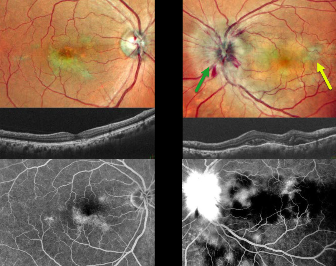
TUBERCULAR SERPIGINOUS-LIKE CHOROIDITIS
Originally posted on @retina.rocks March 11, 2025
This 54YO male presented with 2 weeks of vision loss in his left eye. He was diagnosed elsewhere with papilledema, and brain MRI scanning was normal. Vision was 20/20 in his asymptomatic OD and counting fingers OS. Pseudocolor SLO imaging of the OS shows marked disc swelling with radiating hemorrhages in the nerve fiber layer. Some deep, hypopigmented retinal lesions are also seen.
OCT scanning shows irregular choroidal thickening vs choroidal folds with overlying pockets of variably reflective subretinal fluid. The right eye shows multifocal serpiginous-like areas of chorioretinal scarring with variable outer retinal thinning on OCT.
On fluorescein angiography, the macular lesions stain OD. The left nerve shows profound leakage, and the choroidal lesions show variable blockage and staining.
We suspected tubercular serpiginous-like choroiditis OD with acute tubercular papillitis and choroiditis OS. Chest X-ray showed a mass pressing on the trachea. Chest CT confirmed the mass, and subsequent biopsy revealed granulomatous thyroiditis. His Mantoux skin test was positive. We advised emergent Infectious disease consultation regarding starting anti-tubercular therapy. Unfortunately, he was immediately lost to follow-up.
Learning Points:
Although tuberculosis most commonly presents with pulmonary involvement, extrapulmonary sites can include the gastrointestinal, skin, cardiovascular, genitourinary, and central nervous systems, as well as the eyes. Ocular involvement, like syphilis, can mimic virtually any type of uveitis, including anterior, intermediate, posterior, and panuveitis, retinitis, and retinal vasculitis, neuroretinitis, optic neuropathy, choroidal granuloma, choroiditis, and scleritis.
Tubercular serpiginous-like choroiditis mimics serpiginous but affects younger patients with more multifocal and peripheral recurrences and progression if untreated (Agarwal et al, AJO 2020;220:160-169). Although the exact mechanism is not yet clear, a direct or indirect infectious trigger by the Mycobacterium tuberculosis is thought to cause the choroiditis. Its origin is probably immunogenic since it seems to respond to corticosteroids and other immunosuppressants.
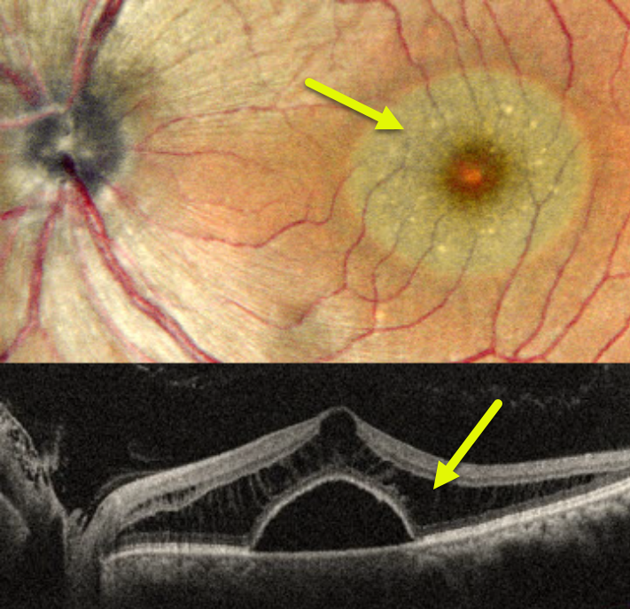
NO OPTIC PIT RETINOSCHISIS
Anand Temkar and Manish Nagpal
Originally posted on @retina.rocks December 12, 2024
This 48YO male presented with one month of vision loss in his left eye. He was diagnosed elsewhere with idiopathic central serous chorioretinopathy. Vision was 20/20 in his normal OD and 20/40 OS.
Pseudocolor SLO imaging shows a round serous macular detachment, which is confirmed on OCT. The outer nuclear layer has a schisis-like thickening. Fluorescein angiography was normal (not shown). There was no optic pit clinically or on OCT.
Learning Points:
No optic pit retinoschisis (NOPIR) is a rare entity that presents with pit-like findings in the absence of a pit or severe glaucomatous cupping (Fujimoto et al, Ophthalmology Retina 2023;7:811-818). Somewhat similar findings can be observed in paraproteinemia (Mansour et al, Ophthalmology 2014;121:1925-1932), but our patient’s serum protein electrophoresis was normal.
As with optic pit maculopathy, eyes with good vision should be observed. Persistent vision loss from foveal involvement can be managed with vitrectomy, internal limiting membrane peeling, and fluid-gas exchange.
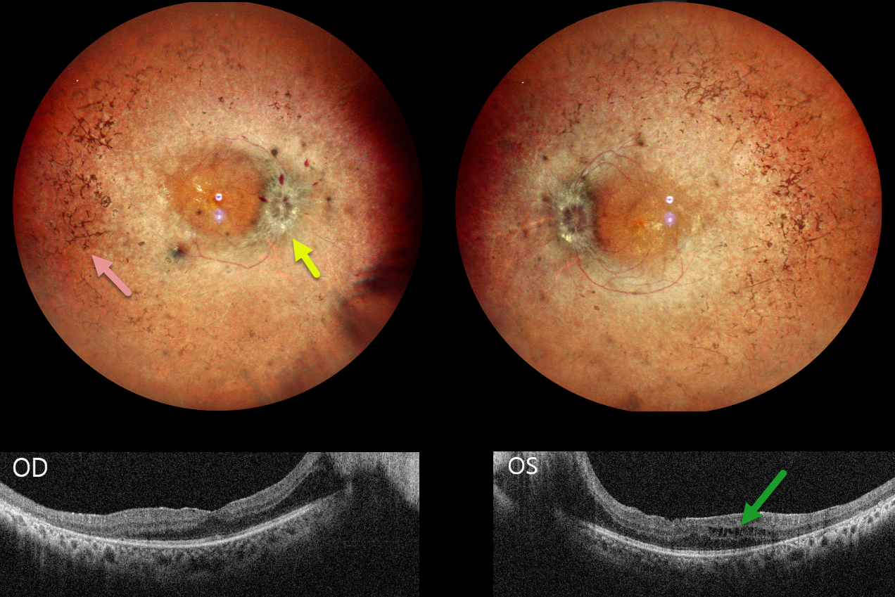
RETINITIS PIGMENTOSA WITH PAPILLEDEMA
Tejaswita Verma and Manish Nagpal
Originally posted on @retina.rocks November 20, 2024
This 30YO male presented with 3 weeks of decreased vision. His father has a history of retinitis pigmentosa (RP). Vision was 20/30 OD and 20/80 OS.
Pseudocolor SLO imaging shows typical bilateral RP scarring with intraretinal pigment migration extending from the peripheral maculas into the retinal peripheries. Bilateral macular puckers and disc swelling with right peripapillary inner retinal fluid are also seen. OCT scanning confirms the epimacular membranes. Bilateral pericentral outer macular atrophy is noted, along with some cystic edema in the left inner and outer nuclear layers.
Learning Points:
RP is a clinically and genetically heterogeneous group of inherited retinal disorders. The disease can present sporadically or with any inheritance pattern (autosomal dominant, autosomal recessive, sex-linked, or mitochondrial). It is characterized by diffuse, progressive dysfunction of predominantly rod photoreceptors, with subsequent RPE degeneration and intraretinal pigment migration. Visual impairment usually presents as night blindness and progressive visual field loss.
Bilateral optic disc edema is not typical for RP. Unfortunately, we were unable to work up our patient for a secondary cause because he was lost to follow-up immediately.
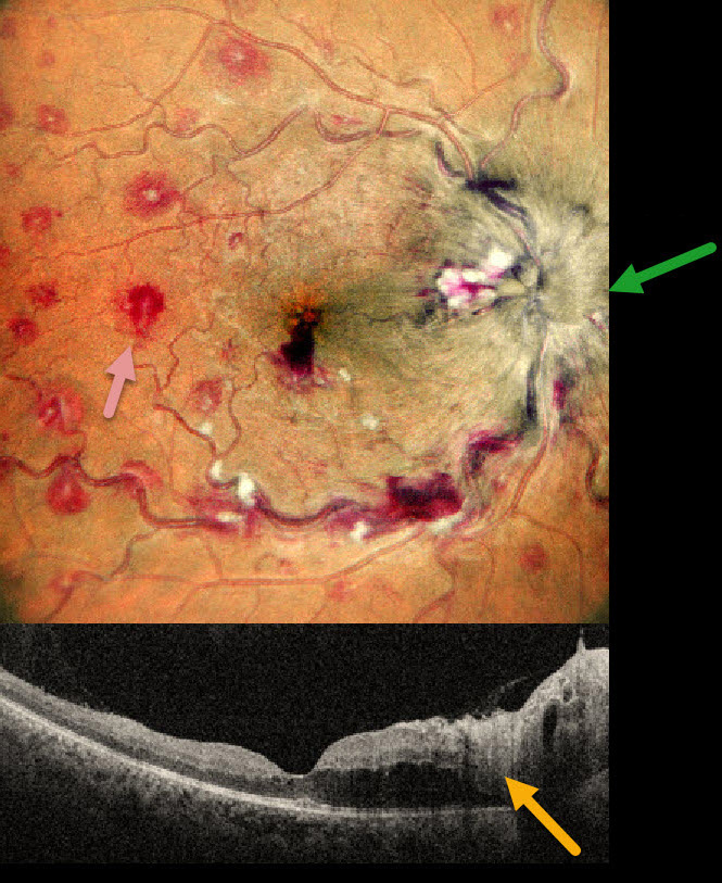
ANEMIA
Anand Temkar and Manish Nagpal
Originally posted on @retina.rocks October 24, 2024
This 35YO male presented with 1 month of vision loss in his right eye. There was no past medical history. Vision was 20/40 OD and 20/20 in his normal OS.
MultiColor imaging shows a markedly swollen optic nerve with extensive multifocal deep, white-centered retinal hemorrhages. OCT shows marked mostly outer retinal edema extending from the nerve towards the macular center.
Extensive laboratory evaluation was negative except for a microcytic anemia with a hemoglobin of 2.9 g/dl. This was felt to be caused by an iron deficiency due to poor diet. Intravitreal anti-VEGF injections were recommended, along with oral prednisone for the optic nerve swelling. Unfortunately, he was immediately lost to follow-up.
Learning Points:
Anemia-related retinal findings most commonly include hemorrhages in the retina and sub-internal limiting membrane. Severe anemia is a rare cause for papilledema and should always be considered in patients with atypical idiopathic intracranial hypertension (Biousse et al, AJO 2003;135:437-446). The underlying pathogenesis of anemia-induced optic nerve swelling remains unknown. It is unclear why our patient had unilateral findings from a systemic disorder.
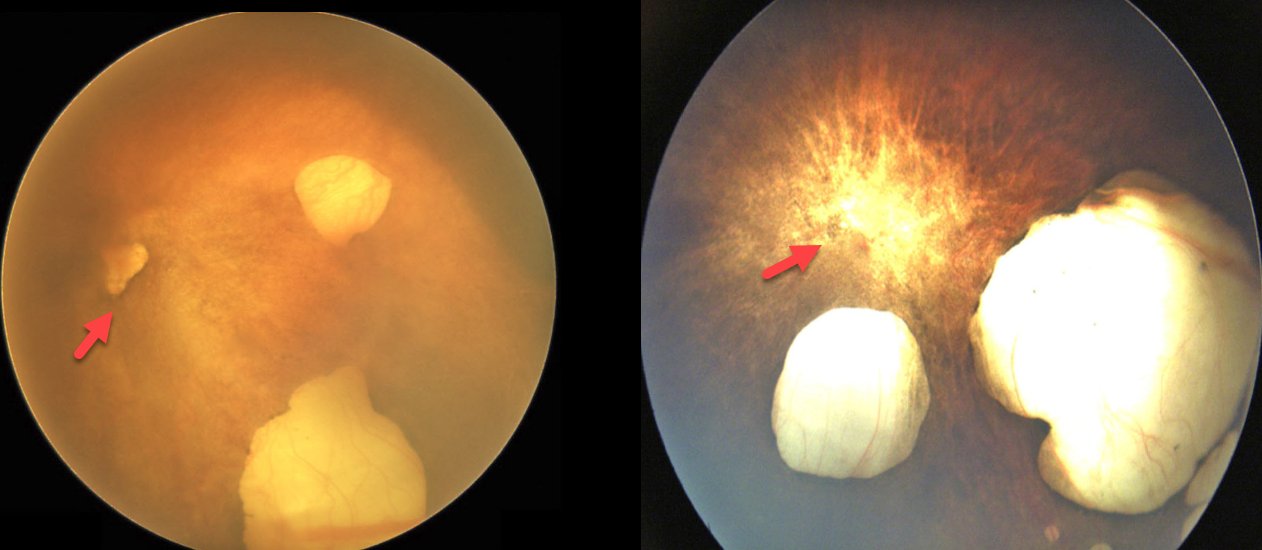
OPTIC NERVE APLASIA
Gokcen Deniz Gulpinar Ikiz and Sengül Özdek
Originally posted on @retina.rocks October 9, 2024
A 5-month-old girl and a 4-month-old boy were referred at different points in time for severe microphthalmia of their left eyes. They were both full-term babies without any systemic or neurological abnormalities. Consanguinity was reported between the parents of both babies (both were first cousins). Their right eyes were normal.
Their left eyes were severely microphthalmic without reaction to light. Color imaging shows multiple choroidal colobomas in each baby, with a small coloboma-like whitish area within the presumed optic disc region. There are no retinal vessels visible with total retinal aplasia. B-scan ultrasonography of the left eye showed short axial length with no visible optic nerve shadow (not shown).
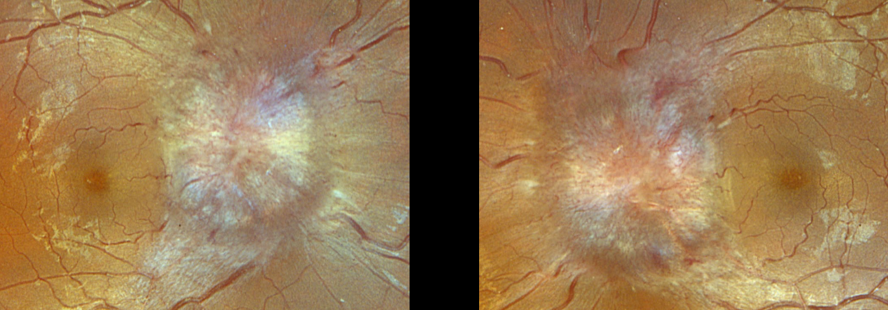
PAPILLEDEMA
Originally posted on @retina.rocks September 9, 2024
This 19YO obese female presented with 2 weeks of headaches, dizziness, and blurred vision. Vision was counting fingers OD and 20/400 OS. Blood pressure was 129/91.
Optos color RGB imaging shows severe bilateral optic disc swelling. The lumbar puncture opening pressure was extremely elevated at 43cm H2O, establishing the diagnosis of idiopathic intracranial hypertension (pseudotumor cerebri), and she was started on Diamox 500mg BID. Unfortunately, she was immediately lost to follow up.
OPTIC NERVE DRUSEN
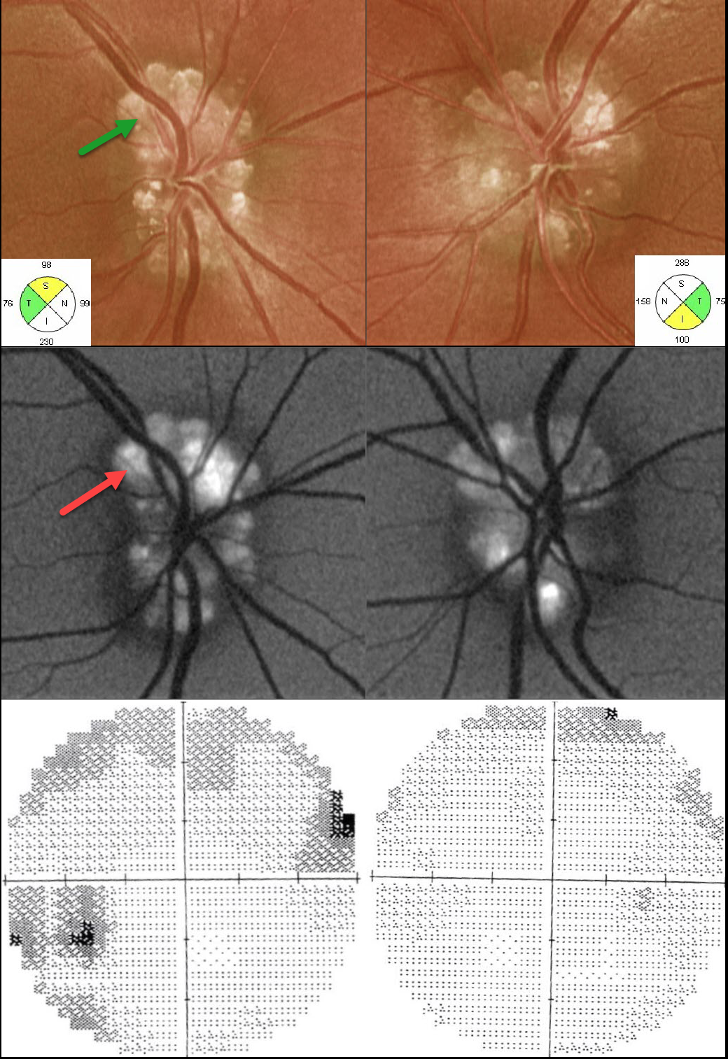
Originally posted on @retina.rocks July 24, 2024
This 29YO male presented with bilateral optic disc drusen (ODD). Vision was 20/40 OD and 20/30 OS. The drusen are markedly hyper-autofluorescent. The retinal nerve fiber layer is variably thickened and thinned. Humphrey visual fields show scattered defects OD with a full field OS. Observation was recommended.
Learning Points:
Optic disc drusen (ODD) are refractile calcific concretions that form anterior to the lamina cribrosa. They are thought to be extruded, extracellular, calcified mitochondria and may be inherited as an autosomal dominant trait.
Porphyrins, organic molecules composed of four interconnected pyrrole subunits, are present within mitochondria. When these compounds break down, they become autofluorescent, which is likely why drusen autofluoresce. Porphyrins are also part of the heme molecule in hemoglobin, accounting for why devitalized blood also autofluoresces (Bloom and Spaide, Retinal Cases 2022;16:401-402).
OPTIC NERVE MORNING GLORY
Larissa Cervera-Perez
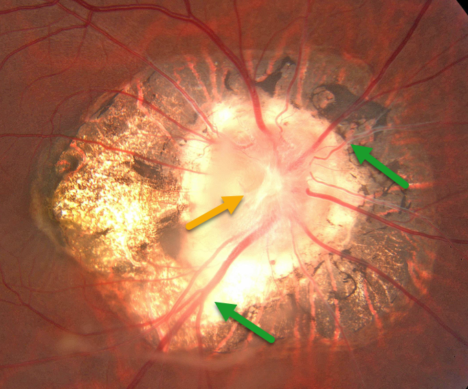
Originally posted on @retina.rocks July 9, 2024
This 43YO female presented for a routine examination. There was a history of pathological myopia. Vision was 20/70 OD and 20/60 OS.
Optos color imaging shows peripapillary scarring with a morning-glory optic disc, including a large disc, a central glial tissue area, and numerous straight, radiating vessels. Observation was recommended.
Learning Points:
Morning glory syndrome is typically unilateral and shares some similarities with optic disc coloboma and juxtapapillary staphyloma. This abnormality gets its name from its striking resemblance to the Morning Glory flower. Visual prognosis is usually poor. The condition can be associated with serous macular detachment. Neuroimaging should be considered to rule out transsphenoidal encephalocele and intracranial carotid artery dysgenesis.
For a great review on the pathogenesis and treatment of maculopathy associated with cavitary optic disc anomalies, see Jain and Johnson, AJO 2014;158:423-435.
LEUKEMIC OPTIC NERVE INFILTRATION WITH PSEUDOPAPILLEDEMA
Anjana Mirajkar and Manish Nagpal
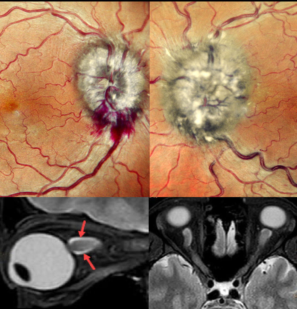
Originally posted on @retina.rocks July 8, 2024
This 40YO male with a known 10-year history of chronic myeloid leukemia complained of vision loss in his left eye for 3 weeks. Vision was 20/20 OD and hand motion OS.
Fundus examination revealed marked bilateral optic nerve swelling, with the left eye greater than the right eye, and some nerve fiber layer hemorrhages along the right inferior disc margin. MRI scanning was consistent with leukemic infiltration of the optic chiasm and optic nerves. She was referred back to her oncologist.
Learning Points:
Leukemia with central nervous system involvement can rarely present with leukemic optic nerve infiltration. This can masquerade as papilledema (bilateral swollen nerves from elevated intracranial pressure) or nerve edema from inflammatory or infectious causes (Miller et al, Graefe’s 2021;259;1315-1322).
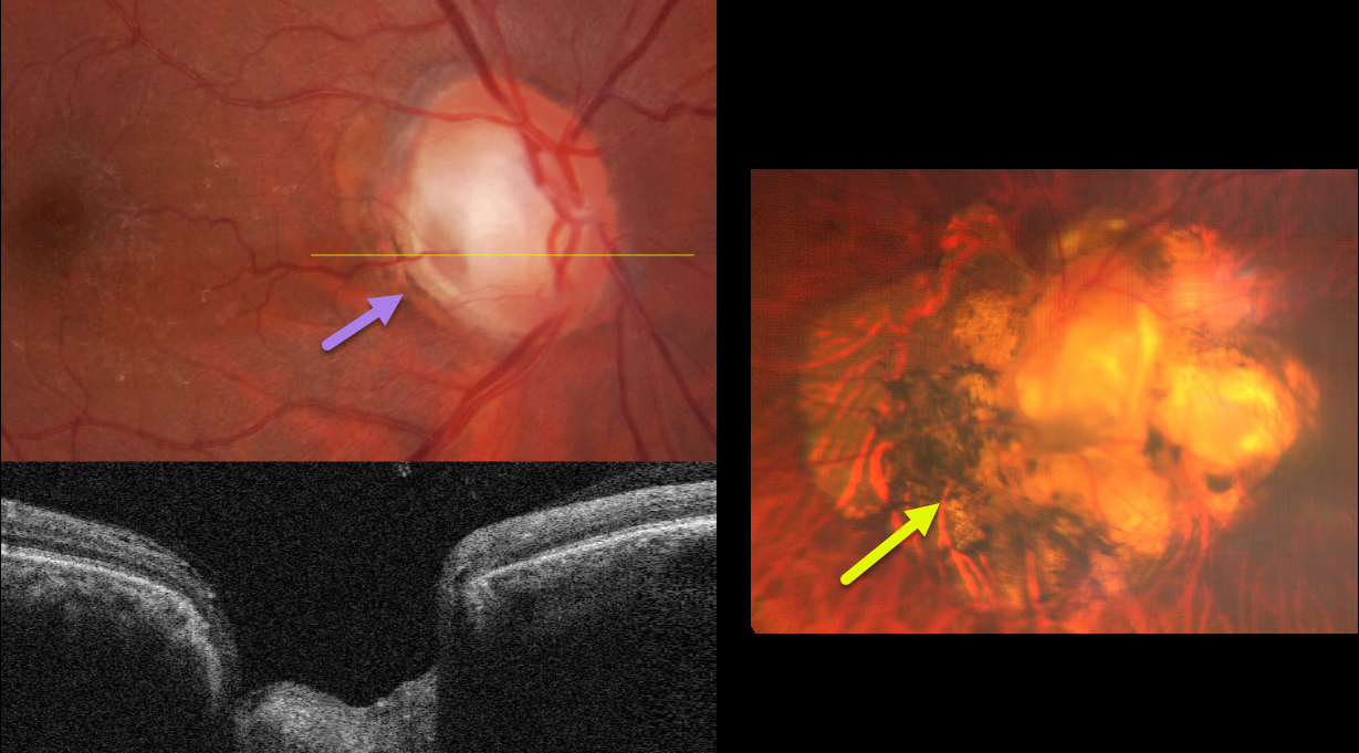
OPTIC NERVE PIT AND COLOBOMA
Kanwaljeet Harjot Madan
Originally posted on @retina.rocks June 13, 2024
This healthy 63YO male was referred for cataract surgery in his right eye. He had a lifelong history of poor vision in his left eye. Vision was 20/60 OD and counting vision OS.
Color fundus imaging and OCT of the right eye show a grey inferotemporal optic nerve pit. Marked peripapillary scarring surrounds the left nerve, which is anomalous with an inferotemporal disc coloboma.
Learning Points:
An optic nerve pit and coloboma are cavitated abnormalities that can be a unilateral or bilateral condition and are caused by incomplete closure of the embryonic fissure. While most lesions are asymptomatic, they can cause vision loss by forcing fluid into the inner and outer retina, and finally into the subretinal space (optic pit maculopathy). Our patient currently shows no evidence of this.
Cataract surgery was performed on his right eye, and vision improved to 20/20. Observation for the disc abnormalities was recommended.
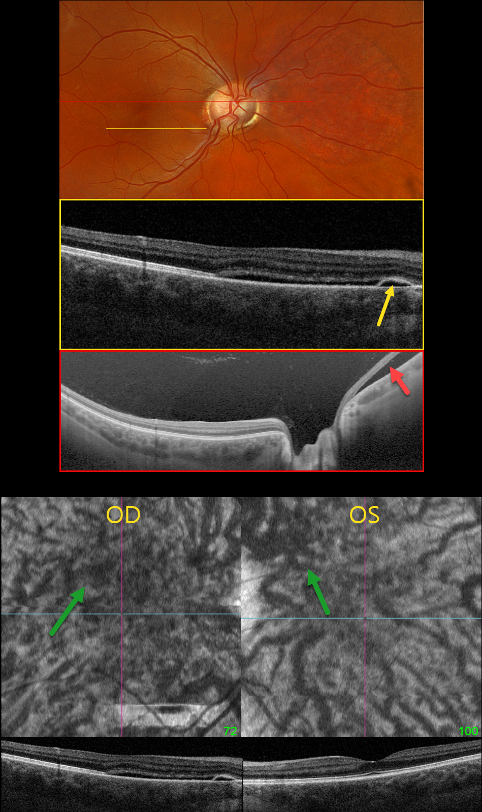
IDIOPATHIC CENTRAL SEROUS CHORIORETINOPATHY WITH INCIDENTAL OPTIC NERVE PIT
Originally posted on @retina.rocks May 30, 2024
This healthy pregnant 32YO female presented with recent mild central vision loss in her right eye. Vision was 20/40 OD and 20/30 OS.
Optos color RGB imaging shows an anomalous right optic disc with an optic pit both nasally and temporally. Triton swept-source OCT shows both pits and subretinal fluid extending from the nerve nasally. OCT through the macula shows shallow nasal subretinal fluid extending into the macular center, along with a tiny nasal RPE detachment. This fluid does not communicate with the nerve or temporal pit. Optos Silverstone swept-source OCT through the posterior pole shows the subretinal fluid nasal to the nerve. Macular en face and B-scans show a thickened choroid with anomalous, thickened, and tortuous vessels.
Our patient’s symptoms are from idiopathic central serous retinopathy (ICSC), most likely related to her pregnancy. The macular fluid does not communicate with the temporal disc pit, and her macular OCT findings are classic for central serous. Fluorescein angiography was not performed due to her pregnancy. Although she has optic pit ‘maculopathy,’ this is in the nasal retina from her temporal disc pit. Observation was recommended.
Learning Points:
An optic nerve pit is a rare (about 0.1% prevalence) congenital anomaly thought to be caused by incomplete closure of the optic fissure during gestation. Pits are most commonly located within or along the inferior-temporal disc margin and usually appear as a gray depression. While most optic pits are asymptomatic, they can cause vision loss by forcing fluid into the inner and outer retina, and ultimately into the subretinal space (optic pit maculopathy). The source of the fluid remains a topic of ongoing debate, with the fluid originating either from the vitreous or the subarachnoid space.
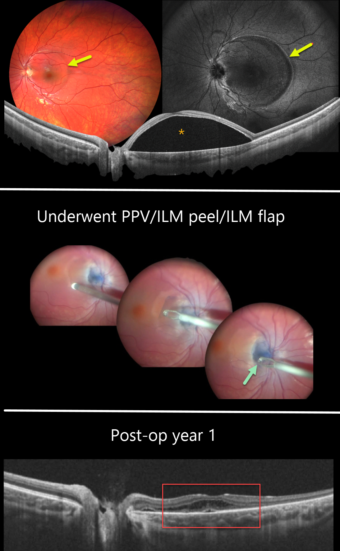
OPTIC NERVE PIT
Barbara Parolini, Veronika Matello, Giulia Freschi, and Roberta Penzani
Originally posted on @retina.rocks May 14, 2024
This 10YO female was referred for vision loss in her left eye from a macular detachment and optic pit. Vision was 20/20 in the normal OD and 20/80 OS.
Color imaging shows a round elevation of the entire macula, which is confirmed on widefield OCT. An optic nerve pit is noted along the inferotemporal edge of the nerve.
The macular detachment was initially treated with micropulse laser, resulting in partial reabsorption. Due to persistent fluid, one year later, pars plana vitrectomy with ILM peeling in the inter-papillomacular area and ILM flap into the pit was performed. One year postoperatively, the macular detachment resolved, and vision improved to 20/50.
Learning Points:
An optic nerve pit is a rare (about 0.1% prevalence) congenital anomaly thought to be caused by incomplete closure of the optic fissure during gestation. Pits are most often located within or along the inferior-temporal disc margin and usually appear as a gray depression.
While most optic pits are asymptomatic, they can cause vision loss by forcing fluid into the inner and outer retina, and ultimately into the subretinal space (optic pit maculopathy). The source of the fluid remains a topic of ongoing debate, with the fluid originating either from the vitreous or the subarachnoid space.
A host of treatments for optic pit maculopathy have been suggested over the years. However, a recent meta-analysis suggested that a plain-vanilla vitrectomy without gas tamponade is as successful as other procedures (Zheng et al, Ophthalmology Retina 2020;4:289-299).
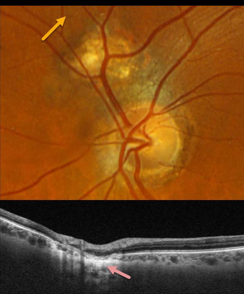
CHOROIDAL COLOBOMA WITH OPTIC NERVE PSEUDODUPLICATION
Originally posted on @retina.rocks March 25, 2024
This 66YO male was referred for an asymptomatic lesion in his left fundus. Vision was 20/25 in his normal right eye and 20/25 in his left eye.
Optos color RGB imaging of the left eye shows a normal optic nerve. Just superonasal to the nerve is a round, colobomatous scar.
Triton swept-source OCT through this lesion shows disorganized neurosensory thinning with an underlying choroidal coloboma.
Learning Points:
Pseudoduplication of the optic nerve occurs when a round choroidal coloboma or chorioretinal scar about the size of the optic nerve is associated with overlying radiating blood vessels (Bloom et al, Retinal Cases 2022;16:174-176). In our experience, it is most common for toxoplasmosis scars to be located near the optic nerve.
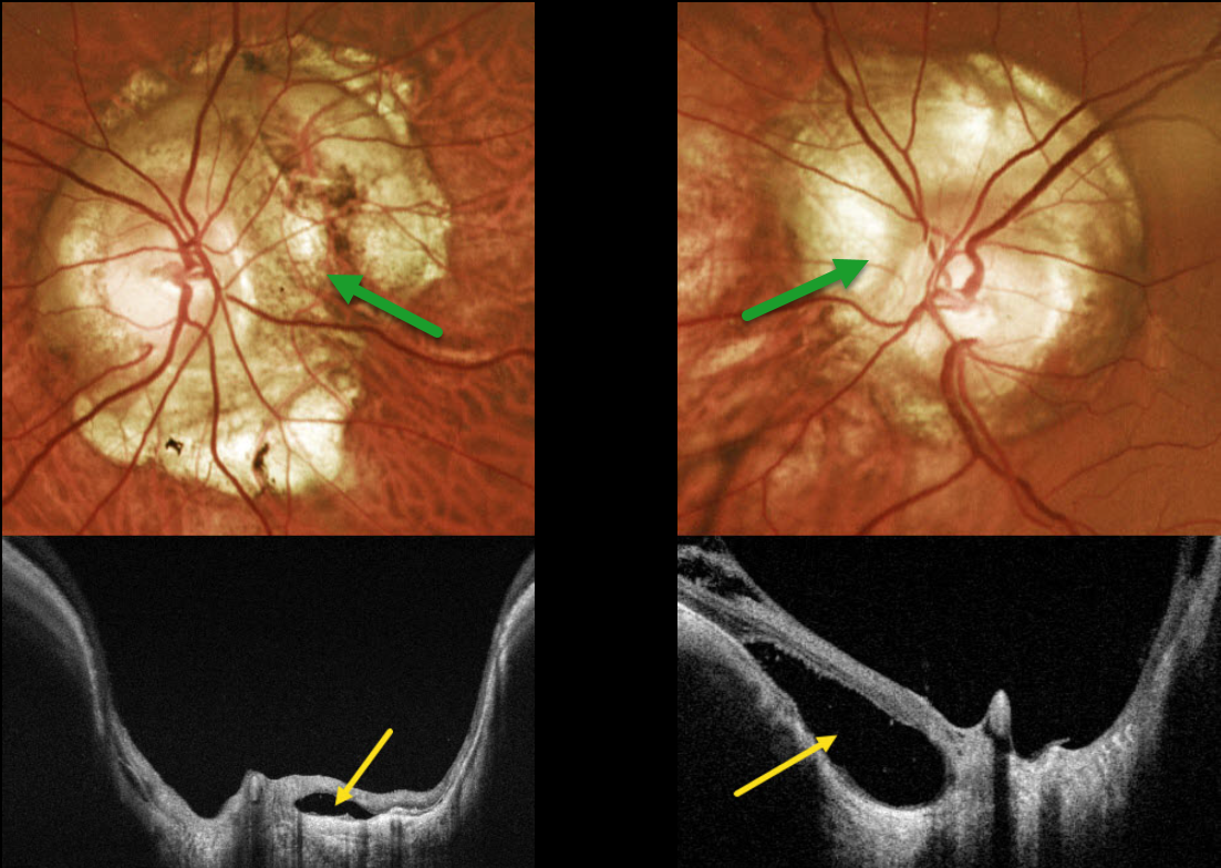
OPTIC NERVE CONUS + PERIPAPILLARY RETINAL DETACHMENT
Originally posted on @retina.rocks March 13, 2024
This 42YO male with high myopia was referred for asymptomatic optic nerve changes. Vision was 20/30 OD and 20/25 OS.
Optos color RG imaging shows bilateral peripapillary atrophy. Clinically, this scarring is markedly excavated from the surrounding retinal plane. Triton swept-source OCT confirms peripapillary staphyloma. Localized retinal detachments are noted bilaterally, particularly within the nasal aspect of atrophy around the left nerve. Within these detachments, there is variable loss of RPE, absence of chorioid, and variable retinal disorganization.
Learning Points:
An optic nerve conus, most commonly found in high myopes, is a staphylomatous-like depression of the optic nerve and surrounding tissue.
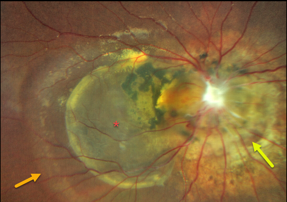
MORNING GLORY + NEUROFIBROMATOSIS TYPE 2
Yuenpang Cheung and Stephanie Choi
Originally posted on @retina.rocks December 12, 2023
This 18YO male, originally from the Dominican Republic, was first seen by an ophthalmologist at 14YO due to strabismus. At that time, he was found to have a significant morning glory anomaly.
Two years later, at age 16, he developed a right-sided facial palsy and, on work-up, was found to have a right optic nerve sheath meningioma. He was then genetically confirmed to have neurofibromatosis 2 (NF-2).
There was an unclear treatment history, and he recently presented to our clinic to establish care. Vision was no light perception in his right eye and 20/25 in his normal left eye.
Optos color RG imaging shows a morning glory disc with peripapillary chorioretinal scarring, an associated chronic retinal detachment, and a large macular cyst.
Learning Points:
Neurofibromatosis type 2 (NF2) can arise spontaneously or be inherited as an autosomal dominant mutation in the NF2 gene located at chromosome 22q12.2.
The hallmark sign of NF2 is bilateral vestibular schwannomas (or acoustic neuromas). Other manifestations of NF2 include cranial meningioma, spinal ependymomas, and peripheral nerve tumors. The most common ocular finding is a posterior subcapsular cataract in young patients.
Less common findings include optic nerve meningiomas, papilledema, strabismus, epiretinal membrane, and combined hamartoma of the retina and RPE.
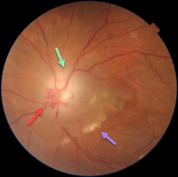
OPTIC NERVE GLIOMA
Asma Samsudeen and Ashish Sharma
Originally posted on @retina.rocks October 12, 2023
This 40YO female presented with vision loss in her left eye for 7 months. Vision was 20/20 in her healthy right eye and hand motion in her left eye.
Fundus photography shows an ill-defined, somewhat pale left nerve with peripapillary fluid, subretinal lipid, and foveal pigmentary changes. Disc collaterals are noted. Externally, the left globe was proptotic and displaced down and out (not shown).
MRI scanning was highly suggestive of an optic nerve glioma, and she was immediately referred for neurosurgical evaluation.
This case was submitted by Asma Samsudeen and Ashish Sharma.
Learning Points:
An optic nerve glioma is a benign, slow-growing tumor most commonly seen in children and associated with neurofibromatosis type 1. This is in contrast to a glioblastoma, an aggressive malignant tumor that is more common in adults. Unfortunately, we do not have a definitive diagnosis since our patient was subsequently lost to follow-up.
Collateral vessels are dilated, pre-existing vessels that bypass an occlusion. These are most commonly seen at the posterior pole following retinal venous occlusions, but can also occur at the optic disc with optic nerve meningiomas or gliomas. The endothelial tight junctions are intact, so they don’t leak on angiography. This helps differentiate them from neovascularization, which does leak.
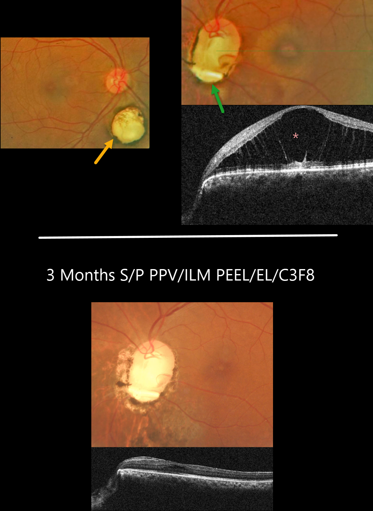
OPTIC PIT MACULOPATHY
Asma Samsudeen and Ashish Sharma
Originally posted on @retina.rocks August 31, 2023
This 51YO female presented with 1 month of blurred vision in her left eye. There was no past ocular history. Vision was 20/30 OD and 20/80 OS. Anterior segments were normal.
Fundus photography of her right eye shows a small choroidal coloboma inferior to the nerve. The left nerve is anomalous with an inferior coloboma and optic pit. The central macula was elevated, with severe foveoschisis noted on OCT.
Vitrectomy with ILM peeling, endolaser along the disc margin, and C3F8 was performed, followed by some additional thermal laser around the nerve postoperatively.
Three months following vitrectomy, vision improved to 20/40 with complete flattening of the macular schisis.
Learning Points:
An optic nerve coloboma can be unilateral or bilateral and is caused by incomplete closure of the embryonic fissure. It has a pathogenesis similar to optic nerve pits, and our patient’s left nerve shows features of both.
While most optic pits are asymptomatic, they can cause vision loss by forcing fluid into the inner retina, outer retina, and, ultimately, the subretinal space (optic pit maculopathy).
The source of the fluid remains a point of ongoing debate, with the fluid originating from either the vitreous or subarachnoid space.
A host of treatments for optic pit maculopathy have been suggested over the years. However, a recent meta-analysis suggested that a plain-vanilla vitrectomy without gas tamponade is as successful as other procedures (Zheng et al., Ophthalmology Retina 2020;4:289-299).
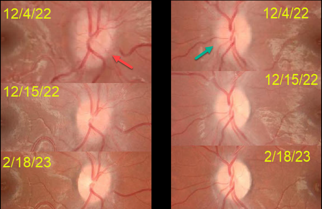
MOG-ASSOCIATED OPTIC NEURITIS
Asma Samsudeen and Ashish Sharma
Originally posted on @retina.rocks April 27, 2023
This 10YO boy presented on 12/4/22 with acute blurred vision in his right eye. He was hospitalized 2 weeks earlier for a viral illness with musculoskeletal pain and fever.
Vision was counting fingers OD and 20/20 OS. An afferent pupillary defect was noted OD. The right disc was moderately swollen, and milder disc swelling was noted OS. MRI was suggestive of bilateral optic neuritis along with a suspicious cervical cord lesion. He was diagnosed with possible parainfectious demyelination and started on high-dose intravenous steroids. Vision improved OD to 20/40 four days later.
On 12/15/22, vision was 20/20 OU, and the nerves were non-edematous. Despite oral mycophenolate mofetil (MMF) immunotherapy, he presented on 1/14/23 with counting vision OS, a new afferent pupillary defect OS, and new left disc edema (not shown).
Anti-NMO (neuromyelitis optica) testing was negative, and Anti-MOG (myelin oligodendrocyte glycoprotein, a protein located on the surface of central nervous system myelin) testing was positive, and was diagnosed with MOG antibody disease (MOG-AD).
Three days of intravenous pulse steroids were given in addition to continuing oral MMF.
Since he developed a relapse in his left eye while on MMF immunotherapy, Rituximab (RTX) will be added. When last examined on 2/18/23, vision was 20/20 OU.
Learning Points:
MOG-AD is an acquired immune-mediated demyelinating syndrome, distinct from multiple sclerosis, that causes inflammation of the optic nerve, spinal cord, and brain. Treatments include intravenous steroids, plasma exchange, and intravenous immunoglobulin. RTX reduces relapse frequency in the majority of patients (Bai et al., Multiple Sclerosis and Related Disorders, 2021;53:103044).
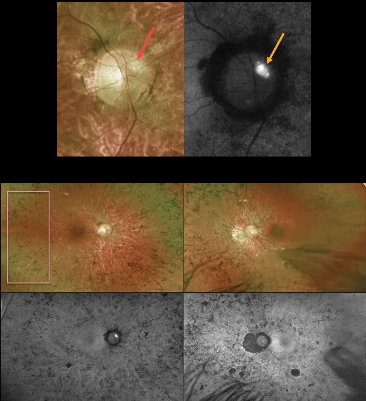
OPTIC NERVE DRUSEN + RETINITIS PIGMENTOSA
Originally posted on @retina.rocks April 17, 2023
This 43YO male with a known history of retinitis pigmentosa (RP) was referred for further evaluation. He noted a gradual worsening of vision bilaterally. Vision was 20/80 OD and 20/200 OS.
Optos color imaging of the right optic nerve shows several large optic disc drusen (ODD) that markedly hyperautofluoresce. Ultrawide-field imaging shows diffuse, classic bilateral RP scarring.
Learning Points:
ODD are refractile calcific concretions that form anterior to the lamina cribrosa. They are thought to be extruded, extracellular, calcified mitochondria and may be inherited as an autosomal dominant trait. They are present in about 3% of RP patients (Russell et al, Eye 2022;36:2213).
Most patients are asymptomatic, but some can develop progressive visual field defects or central vision loss. There is no treatment for the drusen themselves, although symptomatic macular neovascularization can be treated with thermal laser, photodynamic therapy, or anti-VEGF injections.
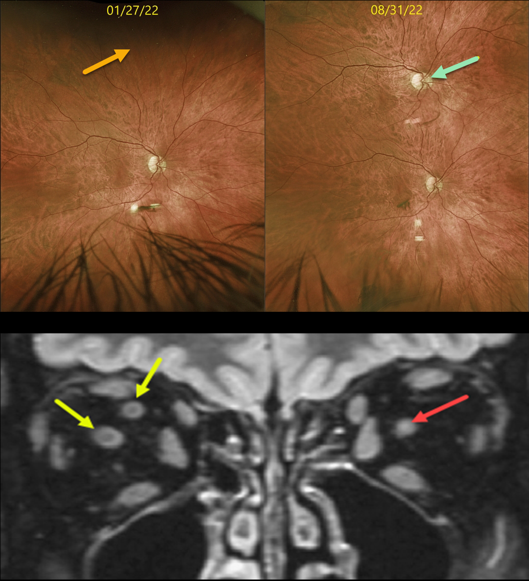
DUPLICATED OPTIC NERVE
Originally posted on @retina.rocks March 31, 2023
This 61YO diabetic male has been followed yearly for routine retinopathy screenings. On 1/27/22, vision was 20/20 with a normal right fundus. Note that the superior periphery is also normal.
On 8/31/22, he returned complaining of a new inferotemporal peripheral scotoma with monocular vertical diplopia in this eye. Vision was 20/20 centrally and 20/20 inferiorly. A new optic nerve and macula had developed in the interim. These findings were confirmed on MRI scanning, which revealed a single nerve OS and two nerves OD in the retro-orbital space.
Learning Points:
Duplication of the optic nerve is a non-heritable developmental abnormality with an extremely low prevalence of about 1 per 23.5 million live births, and is a rare cause of monocular diplopia. To the best of our knowledge, ours is the first such known de novo case.
AND HAPPY APRIL FOOL’S DAY 2023!
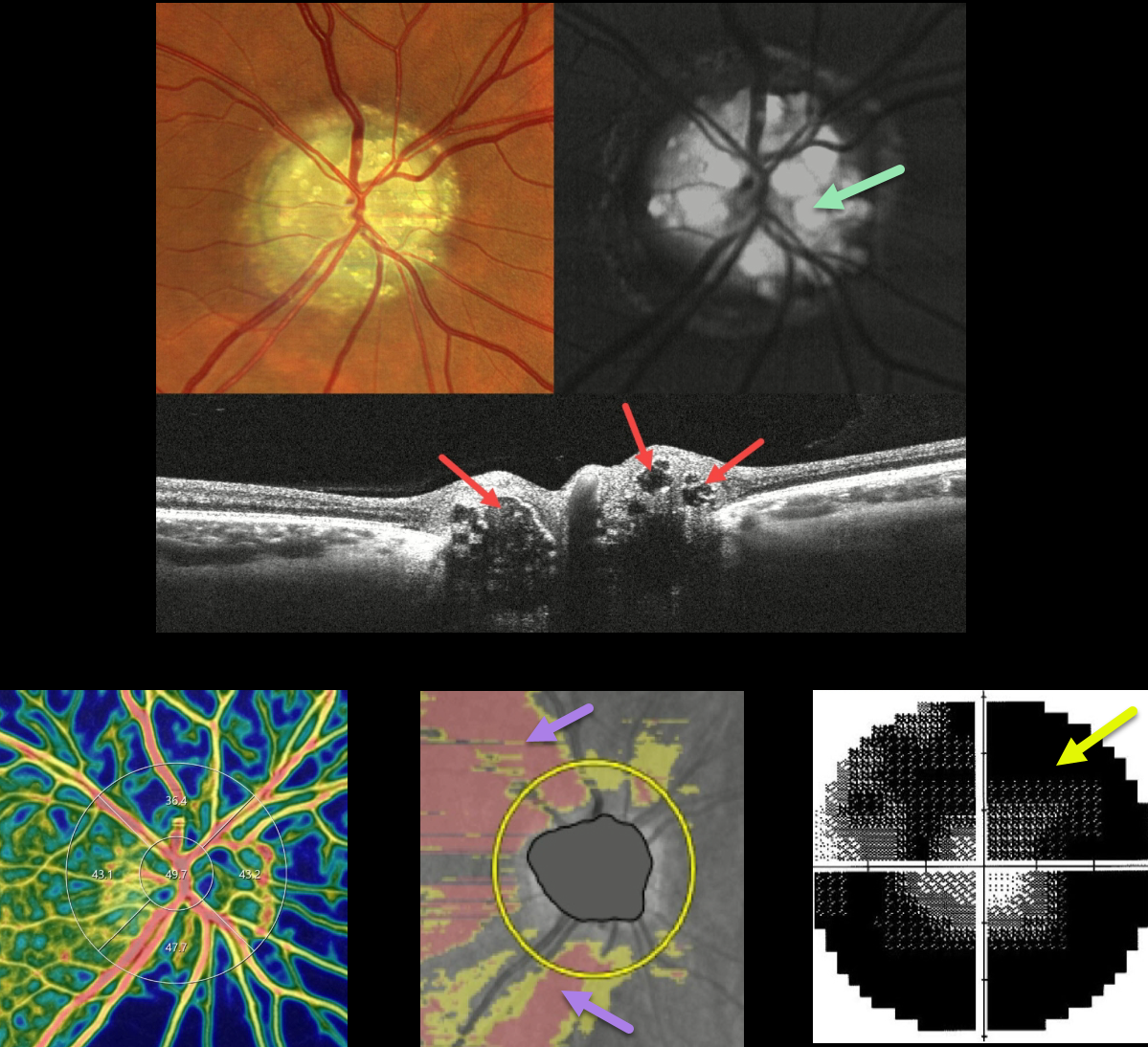
OPTIC NERVE DRUSEN
Barbara Parolini and Veronika Matello
Originally posted on @retina.rocks March 17, 2023
This 47YO female was examined for a routine eye examination. Although she initially denied any visual complaints, further questioning revealed a long history of peripheral vision loss. Vision was 20/20 in each eye, and the following findings were symmetrical and bilateral.
Color imaging of the right nerve shows severe optic disc drusen (ODD), which are hyper-autofluorescent. On OCT B-scan, the drusen appeared as relatively hyporeflective ovoid lesions within the substance of the nerve.
The drusen can be appreciated as blue flow voids on the OCT angiography density map. Diffuse retinal nerve fiber layer thinning is seen, and there is a severely constricted and depressed visual field. Her father was examined the same day and was discovered to also have ODD.
Learning Points:
ODD are refractile calcific concretions that form anterior to the lamina cribrosa. They are thought to be extruded, extracellular, calcified mitochondria and may be inherited as an autosomal dominant trait. Most patients are asymptomatic, but some can develop progressive visual field defects or central vision loss.
There is no treatment for the drusen themselves, although symptomatic macular neovascularization can be treated with thermal laser, photodynamic therapy, or anti-VEGF injections.
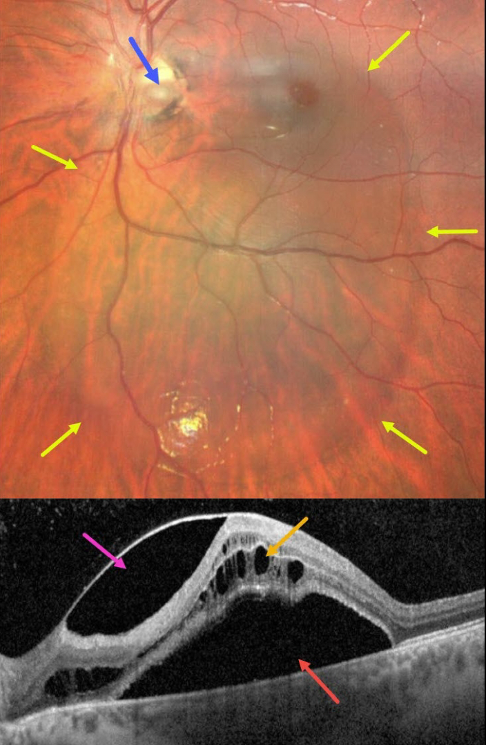
OPTIC NERVE PIT
Emma Oreskovic and Natasha Draca
Originally posted on @retina.rocks January 27, 2023
This 14YO boy presented with these asymptomatic and unilateral findings in his left eye. Vision was 20/50.
Fundus imaging shows a large vertical oval of retinal elevation extending from the superior macula into the inferior midperiphery. This fluid connects to an inferotemporal optic nerve pit.
OCT scanning shows subretinal fluid, cystic fluid mostly in the outer nuclear layer, and fluid below the internal limiting membrane vs within a split nerve fiber layer. He was referred to a retinal specialist for further evaluation and possible treatment.
Learning Points:
An optic nerve pit is a rare (about 0.1% prevalence) congenital anomaly thought to be caused by incomplete closure of the optic fissure during gestation. Pits are most commonly located within or along the inferior-temporal disc margin and usually appear as a gray depression.
While most optic pits are asymptomatic (as in this case), they can cause vision loss by forcing fluid into the inner retina, outer retina, and, ultimately, the subretinal space (optic pit maculopathy). The source of the fluid remains a subject of ongoing debate, with opinions ranging from the vitreous to the subarachnoid space.
A host of treatments for optic pit maculopathy have been suggested over the years. However, a recent meta-analysis suggested that a plain-vanilla vitrectomy without gas tamponade is as successful as other procedures (Zheng et al., Ophthalmology Retina 2020;4:289-299).
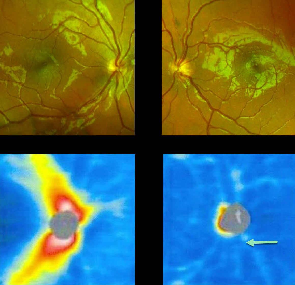
OPTIC NERVE HYPOPLASIA
Originally posted on @retina.rocks December 23, 2022
This 11YO adopted boy presented with a history of poor vision in his left eye since birth. He was referred for an abnormal-appearing left optic nerve.
Vision was 20/20 OD and hand motion OS. Although the left nerve clinically is hypoplastic with a surrounding scleral ring, color imaging failed to capture these disc findings. However, marked loss of the nerve fiber is noted compared to that seen in the right fundus. Optic nerve OCT is normal OD, but shows marked nerve fiber layer thinning OS.
MRI scanning showed an absent septum pellucidum with bilateral hypoplastic nerves, consistent with septo-optic dysplasia. He was referred to pediatric endocrinology.
Learning Points:
Hallmarks of optic disc hypoplasia include a decreased disc diameter, low disc/artery ratio, and the peripapillary double ring sign, a yellow/gray peripapillary halo delineated by an outer ring (the normal junction between the lamina cribrosa and sclera) and an inner pigmented ring (the abnormal extension of the retina and RPE over the outer portion of the lamina cribrosa).
Major risk factors include maternal diabetes and young maternal age. Vision is usually 20/200 or worse. Moderate or severe hypoplasia of the optic disc may
cause visual field defects. The condition may be associated with brain malformations, pituitary disorders, cerebral palsy, and seizures.
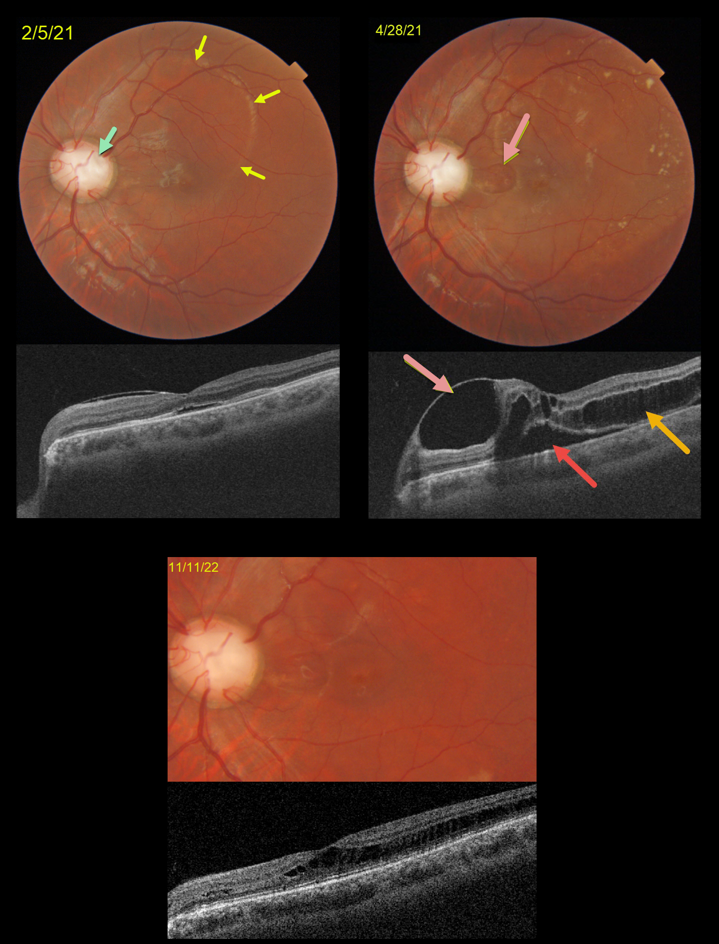
OPTIC NERVE COLOBOMA AND PIT
Originally posted on @retina.rocks December 2, 2022
This is a follow-up to our 2/5/21 and 4/28/21 posts of a patient with bilateral optic nerve coloboma with optic pit maculopathy in his left eye.
On 2/5/21, vision was 20/70 with an optic pit and macular fluid. Despite the mostly peripheral fluid, the macular center was normal except for a trace amount of subretinal fluid (SRF).
Two months later, the peripheral fluid was better, but the central OCT findings were markedly different, with a nasal inner cyst, temporal outer schisis, and increased SRF. Although vision had decreased to 20/200, he subjectively felt his vision was better and elected continued observation.
When examined 1.5 years later on 11/11/22, the OCT findings had spontaneously improved. Temporal outer retinal schisis extends through the macular center, which is now free of SRF. Vision was 20/100, and he remained visually asymptomatic.
Learning Points:
An optic nerve coloboma can be a unilateral or bilateral condition caused by incomplete closure of the embryonic fissure.
While most optic pits are asymptomatic, they can cause vision loss due to fluid being forced into the inner retina, outer retina, and finally the subretinal space (optic pit maculopathy). The source of the fluid continues to be a point of ongoing debate, coming from either the vitreous or subarachnoid space.
A host of treatments for optic pit maculopathy have been suggested over the years. However, a recent meta-analysis suggested that a plain old vanilla vitrectomy without gas tamponade is as successful as any other procedure (Zheng et al Ophthalmology Retina 2020;4:289-299).
Our case illustrates how this fluid can spontaneously wax and wane, and that sometimes simple observation is the best option for some patients.
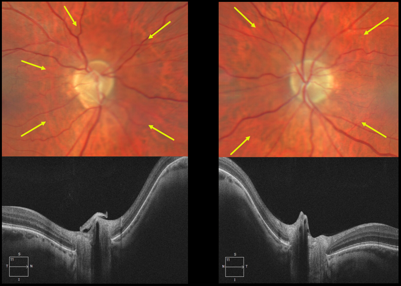
OPTIC NERVE CONUS
Will Gibson
Originally posted on @retina.rocks November 7, 2022
This 54YO male patient presented with asymptomatic bilateral optic nerve conus. Optos color imaging of each eye shows each optic nerve and peripapillary tissue to be sunken from the plane of the surrounding fundus. These findings are captured on OCT.
Learning Points:
An optic nerve conus is a staphylomatous-like depression of the optic nerve and surrounding tissue. This finding is usually seen in high myopia, although our patient had mild bilateral hyperopia.
OPTIC NERVE PIT
Originally posted on @retina.rocks October 20, 2022
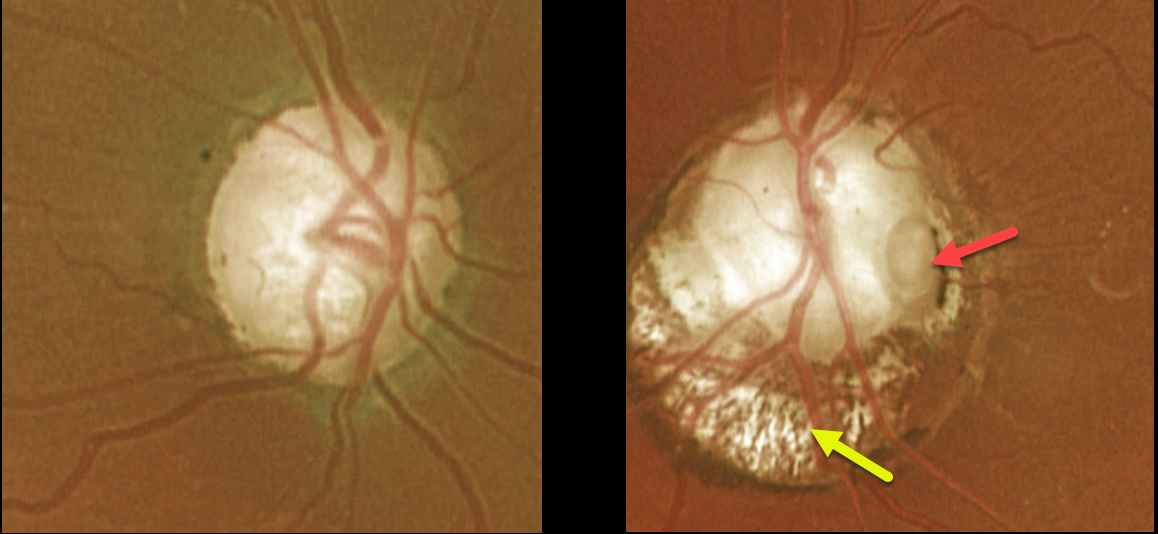
This 66YO female presented with an asymptomatic left optic nerve coloboma with a large temporal pit. The mildly colobomatous portion of the nerve is enlarged and slightly excavated inferiorly. Vision was 20/25 OD and 20/200 OS. Observation was recommended.
Learning Points:
Optic pits are most commonly located within or along the inferior-temporal disc margin and usually appear as a gray depression. They are thought to be caused by incomplete closure of the optic fissure during gestation.
While most optic pits are asymptomatic (as in this case), they can cause vision loss by forcing fluid into the inner and outer retina and, ultimately, the subretinal space (optic pit maculopathy).
The source of the fluid remains a subject of ongoing debate, with origins in either the vitreous or the subarachnoid space.
OPTIC NERVE DRUSEN
Will Gibson
Originally posted on @retina.rocks September 29, 2022
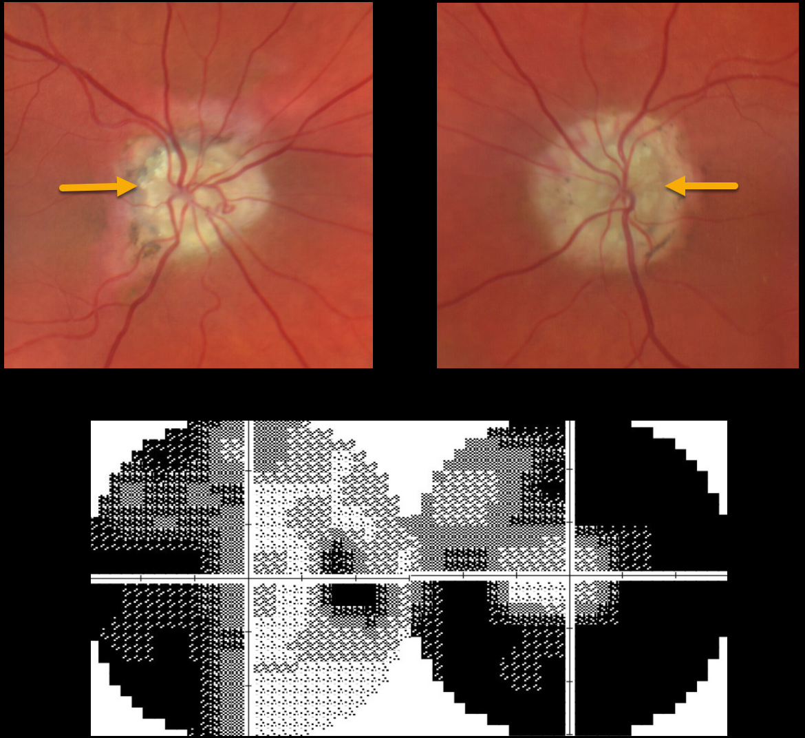
This 65YO female presented with dramatic bilateral optic nerve drusen with severe visual field defects. Vision was 20/20 OU.
Learning Points:
Optic disc drusen are refractile calcific concretions that form anterior to the lamina cribrosa. They are felt to be extruded extracellular calcified mitochondria and may be inherited as an autosomal dominant trait.
Most patients are asymptomatic, but some can develop progressive visual field defects or central vision loss.
There is no treatment for the drusen themselves, although symptomatic macular neovascularization can be treated with thermal laser, photodynamic therapy, or anti-VEGF injections.
PAPILLEDEMA
Originally posted on @retina.rocks September 2, 2022
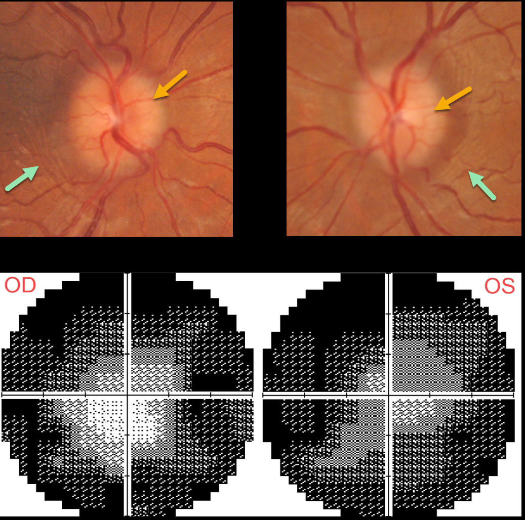
This 30YO female was diagnosed with idiopathic intracranial hypertension (pseudotumor cerebri) in 2020. Her initial lumbar puncture opening pressure was elevated at 25cm H2O.
She was lost to follow-up for about 2 years, when she presented with recurrent headaches, a ‘whooshing’ sound in her ears, and blurred vision. She was off of Topamax for about a year. Vision was 20/30 OD and 20/40 OS.
Both optic nerves are mildly edematous. The retinal nerve fiber layer (not shown) is somewhat thickened bilaterally. Despite the modest nerve edema, prominent temporal circumferential retinal folds (Paton’s folds) surround both nerves. Visual fields are severely constricted.
Learning Points:
In 1911 Paton and Holmes established that papilledema was due to elevated intracranial pressure and not inflammation as was previously thought (Brain 1911;33:389-432).
They also noted that nerve edema was associated with “a series of folds which run concentric with the edge of the disc” which they felt represented retinal folds.
Sibony and Kupersmith, correlating OCT with fundus photography, found that these folds represented a variety of structural stresses on the nerve and peripapillary tissues, including peripapillary wrinkles, inner retinal folds, choroidal folds and outer retinal folds/creases (Ophthalmology 2016;123:1397-1399).
HYPERTENSIVE RETINOPATHY vs AION
Originally posted on @retina.rocks August 16, 2022
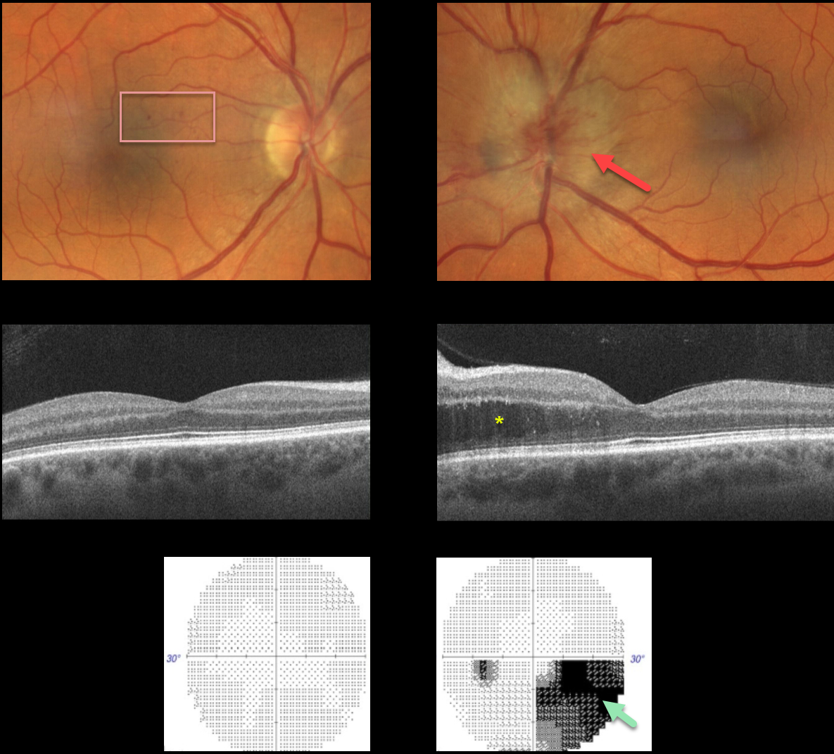
This 57YO male presented with unilateral optic nerve swelling in his left eye and a few faint nasal macular retinal hemorrhages in his right eye. Vision was 20/40 OD and 20/25 OS.
OCT was normal OD and thickened in the nasal outer nuclear layer OS. Visual fields were normal OD and inferiorly depressed, especially inferonasally, OS.
Blood pressure was 190/110, and he was immediately referred to the emergency room.
Learning Points:
Malignant hypertension, defined as blood pressure above 180 systolic and/or 120 diastolic, is a life-threatening medical emergency. Eye doctors are in a unique position to often diagnose this condition.
Patients will often present with bilateral optic nerve swelling, flame-shaped retinal hemorrhages, and, with more chronic disease, lipid precipitates in the nasal macular outer plexiform layer. However, rarely, the ocular findings may be asymmetric or even unilateral.
Since the macular fluid emanates from the optic nerve, the macular thickening is always worse nasally (as in our case), and is an important clinical clue for this diagnosis.
Our patient may also have had an isolated anterior ischemic optic neuropathy in his left eye and mild hypertensive retinopathy in his right eye. The partial altitudinal deffect is more common with this diagnosis than hypertension.
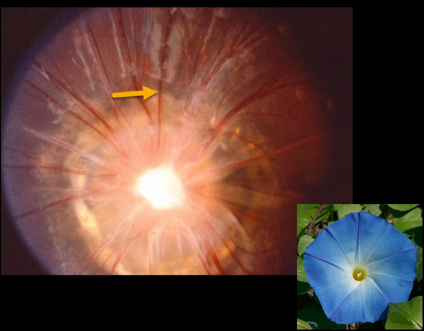
OPTIC NERVE MORNING GLORY
Originally posted on @retina.rocks July 29, 2022
This patient has a classic morning glory optic disc, including a large disc, an area of central glial tissue, and numerous straight radiating vessels.
Learning Points:
Morning glory syndrome is typically unilateral and shares some similarities with optic disc coloboma and juxtapapillary staphyloma. This abnormality gets its name from its striking resemblance to the Morning glory flower. Visual prognosis is usually poor. The condition can be associated with serous macular detachment.
Neuroimaging should be considered to rule out transsphenoidal encephalocele and intracranial carotid artery dysgenesis.
For a great review on the pathogenesis and treatment of maculopathy associated with cavitary optic disc anomalies, see Jain and Johnson, AJO 2014;158:423-435.
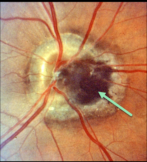
MELANOCYTOMA
Originally posted on @retina.rocks June 22, 2022
This is just a great image of a melanocytoma! Unfortunately, we don’t have any clinical information for this patient.
Learning Points:
Melanocytomas are benign melanocytic tumors intrinsic to the optic nerve. They are intensely pigmented with feathery margins, sometimes spreading into the adjacent retina and choroid.
Although they may enlarge over time, malignant transformation is very rare. Spontaneous necrosis may present with a similar appearance to vitreous seeding from melanoma. Vision and visual field loss can rarely occur from compressive optic neuropathy.
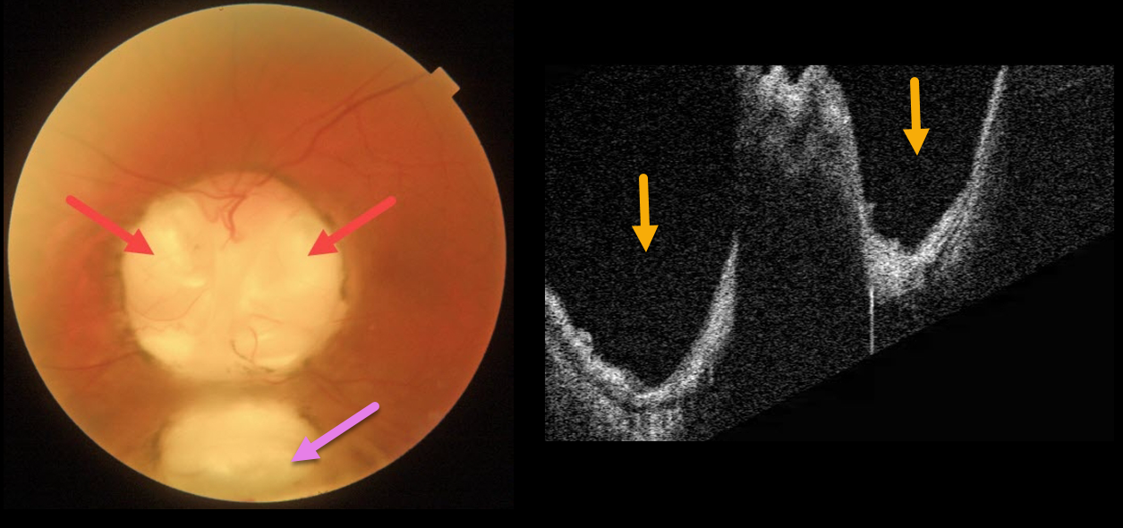
OPTIC NERVE COLOBOMA
Originally posted on @retina.rocks May 9, 2022
This 66YO male has mild lifelong decreased vision in his left eye. Vision was 20/70.
Triton imaging of the optic nerve coloboma shows optic nerve pits temporally and nasally, as well as a more inferior choroidal coloboma. Swept-source OCT dramatically reveals the pits.
We are following him yearly.
Learning Points:
Both optic nerve and choroidal colobomas can be unilateral or bilateral and are caused by incomplete closure of the embryonic fissure.
Patients with choroidal colobomas are at increased lifetime risk of rhegmatogenous retinal detachment due to breaks in the overlying thinned retina.
Optic nerve pits are also associated with serous macular detachment and macular neovascularization.
For a great review of cavitary optic nerve abnormalities, see Jain and Johnson, AJO 2014;158:423-435.
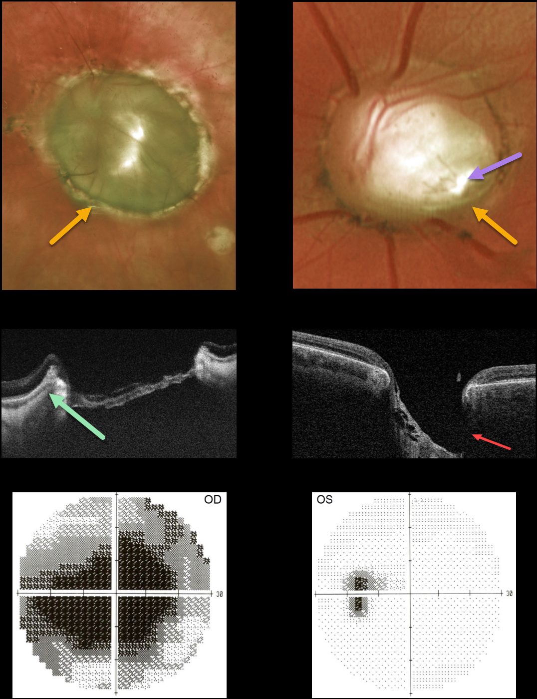
OPTIC NERVE COLOBOMA
Originally posted on @retina.rocks April 4, 2022
This 45YO female presented with bilateral optic nerve colobomas. Vision was hand motion OD and 20/25 OS.
Triton swept-source OCT shows a relatively featureless retina spanning the coloboma. The much smaller left nerve coloboma also contains an inferotemporal optic disc pit. The pit is also quite evident on OCT. The left nerve has a morning glory appearance with multiple vessels radiating outward from the peripheral disc margin, although there is no central glial tissue.
Visual fields show a large central defect OD with an enlarged blind spot OS.
Learning Points:
Optic nerve cavitary abnormalities can be unilateral or bilateral and result from incomplete closure of the embryonic fissure.
Our patient shows a spectrum of these abnormalities, including a huge optic disc coloboma OD, a much smaller disc coloboma OS with morning glory-like features, and an optic pit.
For a great review of cavitary optic nerve abnormalities, see Jain and Johnson, AJO 2014;158:423-435.
PAPILLEDEMA
Originally posted on @retina.rocks March 18, 2022
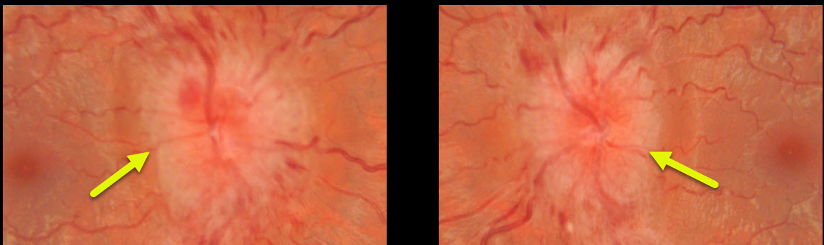
This 22YO female presented with severe bilateral optic nerve edema and enlarged blind spots. She reported recent weight gain following the placement of an IUD.
MRI and MRV scanning were normal. Lumbar puncture opening pressure was extremely elevated at 55cm H2O, establishing the diagnosis of idiopathic intracranial hypertension (pseudotumor cerebri).
She was evaluated by neurosurgery, and a lumboperitoneal shunt was placed, improving the patient’s symptoms and nerve appearance (not shown).
OPTIC NERVE HYPOPLASIA
Originally posted on @retina.rocks December 7, 2021
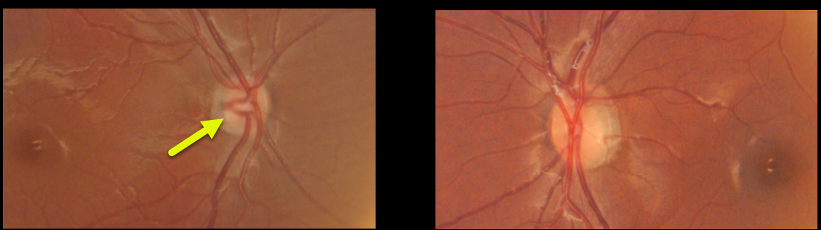
This healthy 20YO female presented with vision of 20/400 OD and 20/20 OS. Vision in her right eye was unchanged and poor since birth. The right nerve was hypoplastic, while the left nerve appeared fairly normal.
Learning Points:
Hallmarks of optic disc hypoplasia include a decrease in disc diameter, low disc/artery ratio, and the peripapillary double ring sign – yellow/gray peripapillary halo delineated by an outer ring (the normal junction between the lamina cribrosa and sclera) and an inner pigmented ring (the abnormal extension of the retina and RPE over the outer portion of the lamina cribrosa).
Major risk factors include maternal diabetes and young maternal age. Vision is usually 20/200 or worse. Moderate or severe hypoplasia of the optic disc may cause visual field defects.
The condition may be associated with brain malformations, pituitary disorders, cerebral palsy, and seizures.
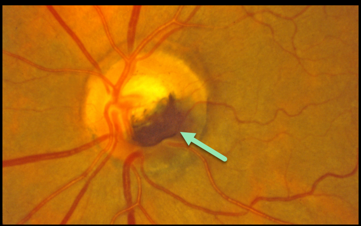
MELANOCYTOMA
Originally posted on @retina.rocks October 15, 2021
This patient has a melanocytoma, which is a benign melanocytic tumor intrinsic to the optic nerve.
They are intensely pigmented with feathery margins, sometimes spreading into the adjacent retina and choroid.
Learning Points:
Although they may enlarge over time, malignant transformation is very rare. Spontaneous necrosis may present with a similar appearance to vitreous seeding from melanoma. Decreased vision and visual field loss can rarely occur from compressive optic neuropathy.
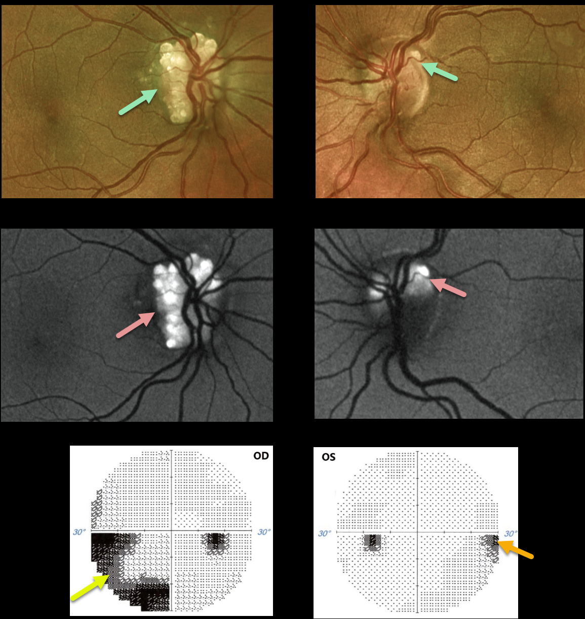
OPTIC NERVE DRUSEN
Originally posted on @retina.rocks October 8, 2021
This 38YO female presented with bilateral optic disc drusen (ODD). Vision was 20/80 OD and 20/40 OS.
The drusen are markedly hyper-autofluorescent. The retinal nerve fiber is thinned, particularly in the right eye, due to the more extensive ODD. Humphrey visual fields show significant visual field defects inferonasally OD and mild nasal depression OS. Observation was recommended.
Learning Points:
Optic disc drusen (ODD) are refractile calcific concretions that form anterior to the lamina cribrosa. They are thought to be extruded, extracellular, calcified mitochondria and may be inherited as an autosomal dominant trait.
Porphyrins, organic molecules composed of four interconnected pyrrole subunits, are present within mitochondria. When these compounds break down, they become autofluorescent, which is likely why drusen autofluoresce.
Porphyrins are also part of the heme molecule in hemoglobin, accounting for why devitalized blood autofluoresces.
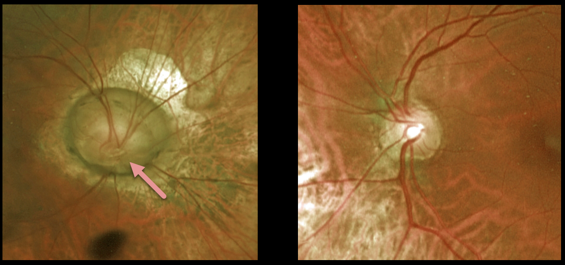
OPTIC NERVE COLOBOMA
Originally posted on @retina.rocks August 25, 2021
This 55YO female presented with a unilateral optic nerve coloboma in her right eye. Vision was a remarkable 20/50 in her right eye and 20/30 in her normal left eye.
Learning Points:
Optic nerve colobomas can be a unilateral or bilateral condition, and, similar to other cavitary optic disc abnormalities, are caused by incomplete closure of the embryonic fissure. They can have an associated optic nerve pit, which was not seen in this patient. For a great review of cavitary optic nerve abnormalities, see Jain and Johnson, AJO 2014;158:423-435.
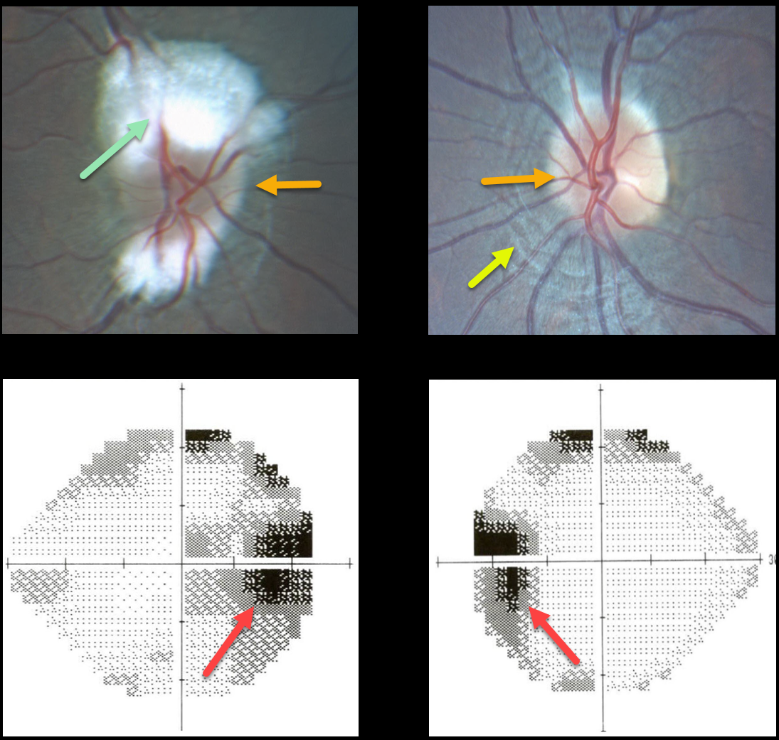
PAPILLEDEMA
Originally posted on @retina.rocks July 28, 2021
This 14YO presented with mild bilateral optic nerve edema and enlarged blind spots. Evaluation of the right nerve was difficult due to the myelinated nerve fiber layer.
There are prominent circumferential retinal lines (Paton’s folds) surrounding the left nerve.
MRI and MRV scanning were normal, and lumbar puncture opening pressure was elevated at 26mm H2O. The patient was diagnosed with idiopathic intracranial hypertension (pseudotumor cerebri).
Learning Points:
In 1911, Paton and Holmes established that papilledema was due to elevated intracranial pressure rather than inflammation, as previously thought (Brain 1911;33:389-432).
They also noted that nerve edema was associated with “a series of folds which run concentric with the edge of the disc,” which they felt represented retinal folds.
Sibony and Kupersmith, correlating OCT with fundus photography, found that these folds represented a variety of structural stresses on the nerve and peripapillary tissues, including peripapillary wrinkles, inner retinal folds, choroidal folds, and outer retinal folds/creases (Ophthalmology 2016;123:1397-1399).
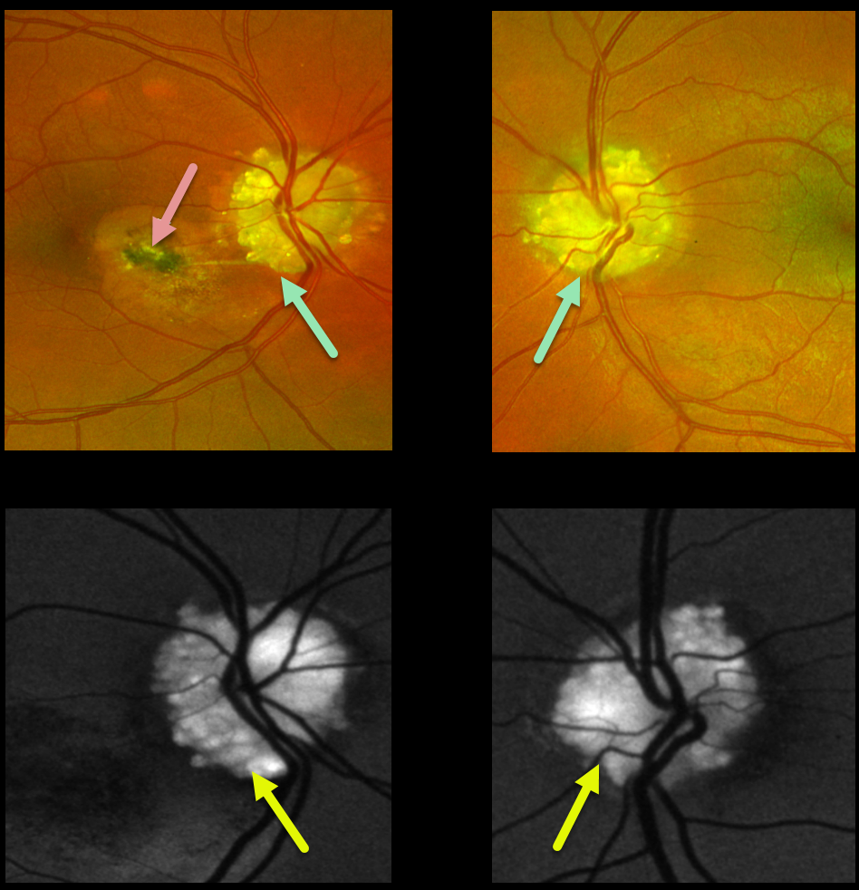
OPTIC NERVE DRUSEN
Originally posted on @retina.rocks July 22, 2021
This 30YO male presented with dramatic bilateral optic nerve drusen. Vision was 20/20 OD and 20/200 in his amblyopic OS.
A spontaneously involuted macular neovascularization (MNV) is noted in the nasal macula. The drusen are markedly hyper-autofluorescent.
Observation was recommended.
Learning Points:
Optic disc drusen (ODD) are refractile calcific concretions that form anterior to the lamina cribrosa. They are thought to be extruded, extracellular, calcified mitochondria and may be inherited as an autosomal dominant trait.
MNV is a known complication of ODD. These can spontaneously involute, as in our patient, or can be treated with thermal laser, photodynamic therapy, or anti-VEGF injections.
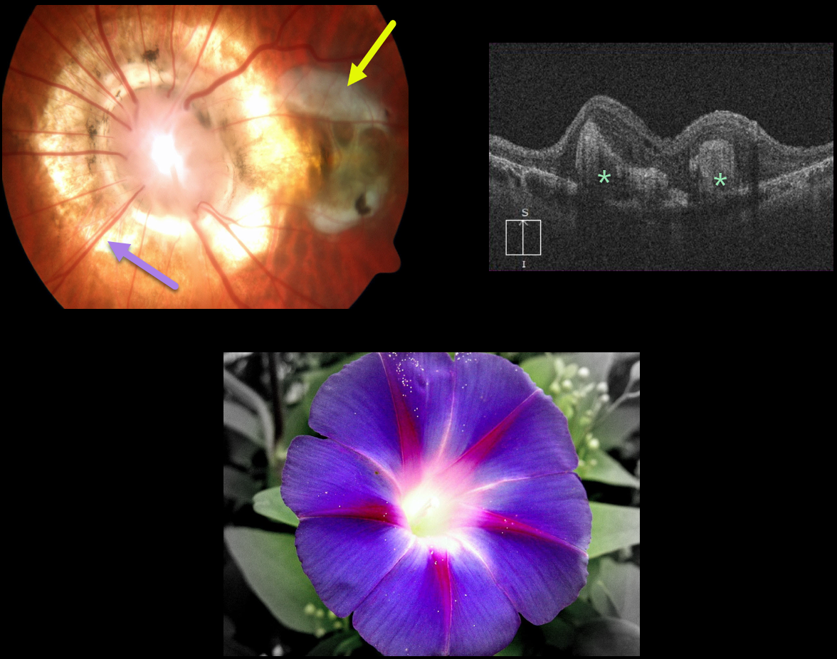
OPTIC NERVE MORNING GLORY
Originally posted on @retina.rocks July 15, 2021
This patient has a unilateral morning glory disc in their left eye with an associated fibrotic disciform scar. OCT scanning shows variable subretinal macular scarring.
Learning Points:
Morning glory syndrome is typically unilateral and shares some similarities with optic disc coloboma and juxtapapillary staphyloma. This abnormality is named for its striking resemblance to the morning glory flower.
Visual prognosis is usually poor. The condition can be associated with serous macular detachment. Neuroimaging should be considered to rule out transsphenoidal encephalocele and intracranial carotid artery dysgenesis.
For a great review on the pathogenesis and treatment of maculopathy associated with cavitary optic disc anomalies, see Jain and Johnson, AJO 2014;158:423-435.
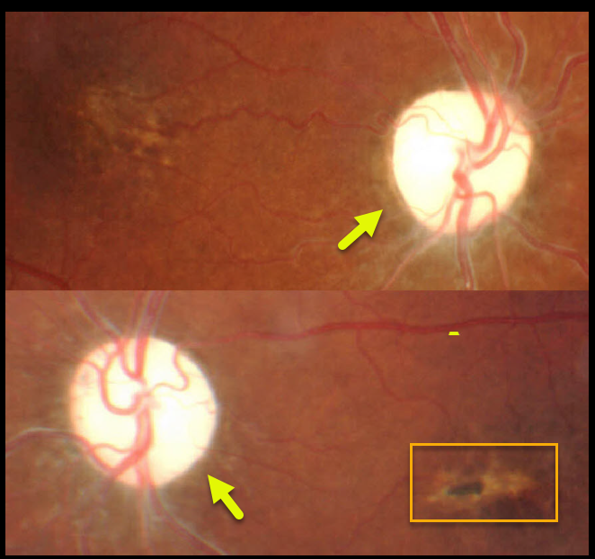
OPTIC NERVE ATROPHY
Originally posted on @retina.rocks July 14, 2021
This 33YO male has no light perception vision bilaterally following a failed shunt for hydrocephalus when he was 13YO. Both optic nerves are severely pale.
There are also bilateral macular pigmentary changes, most likely from resolved secondary macular edema when his nerves were severely swollen before his shunt.
Learning Points:
Secondary optic nerve atrophy and vision loss can occur in cases if increased intracranial pressure is not controlled.
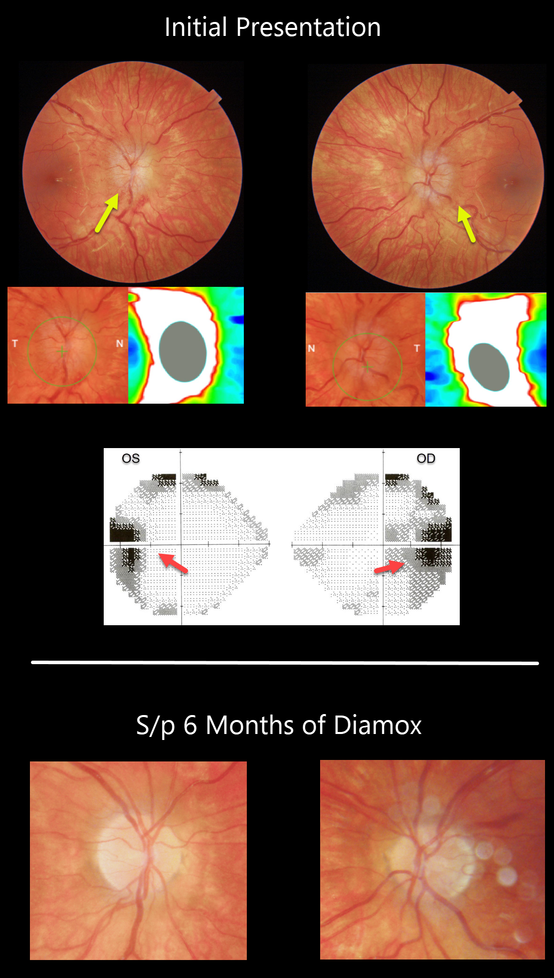
PAPILLEDEMA
Originally posted on @retina.rocks June 29, 2021
This 13YO girl presented with headaches and tinnitus with 20/25 vision OU. There was moderate bilateral optic nerve swelling, beautifully imaged with Triton swept-source OCT. Visual fields showed enlarged blind spots.
MRI scanning was normal, and her lumbar puncture had a severely elevated opening pressure of 49mm H2O, establishing the diagnosis of idiopathic intracranial hypertension (IIH).
After six weeks of oral Diamox, her headaches and tinnitus had improved significantly, and the optic nerve edema had virtually resolved bilaterally.
Learning Points:
Remember that papilledema is not synonymous with optic nerve swelling. Papilledema is defined as bilateral optic nerve swelling due to elevated intracranial pressure.
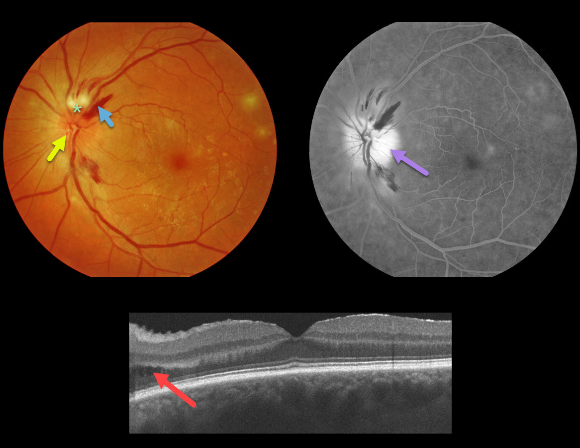
OPTIC NERVE PAPILLITIS
Originally posted on @retina.rocks June 18, 2021
This 66YO diabetic male presented with an asymptomatic unilateral swollen nerve, peripapillary hemorrhages, and nerve fiber layer ischemia. Vision was 20/25, and there was no afferent pupillary defect (APD).
OCT scanning shows some mild cystic fluid in the peripapillary inner and outer nuclear layer. Fluorescein angiography shows diffuse optic nerve leakage.
Blood pressure was normal. He denied exposure to deer ticks or cat scratches, and the MRI was normal. The funduscopic findings spontaneously normalized within a few months.
Learning Points:
Diabetic papillopathy, often a diagnosis of exclusion, is most commonly seen in type 2 diabetics. It has a good visual prognosis with observation alone. A small physiologic cup may predispose to this condition (see Regillo et al, Arch Ophthlamol 1995;113:889-895).
Unlike ischemic optic neuropathy, vision is usually quite good, there is usually no APD, the nerve swelling is more diffuse than sectorial, and there is no altitudinal field defect.
The differential for unilateral acute optic nerve swelling is broad, including diabetes, ischemic (anterior ischemic optic neuropathy), and inflammatory causes (optic neuritis, Bartonella, syphilis, sarcoidosis).
Papillitis must be differentiated from papilledema, which is bilateral optic nerve swelling caused by elevated intracranial pressure.
We have been burned more than once by unilateral nerve edema being the only presenting sign of severe hypertension. Although nerve swelling is usually bilateral in hypertensive retinopathy, we always check blood pressure in all patients presenting with unilateral or bilateral optic nerve swelling.
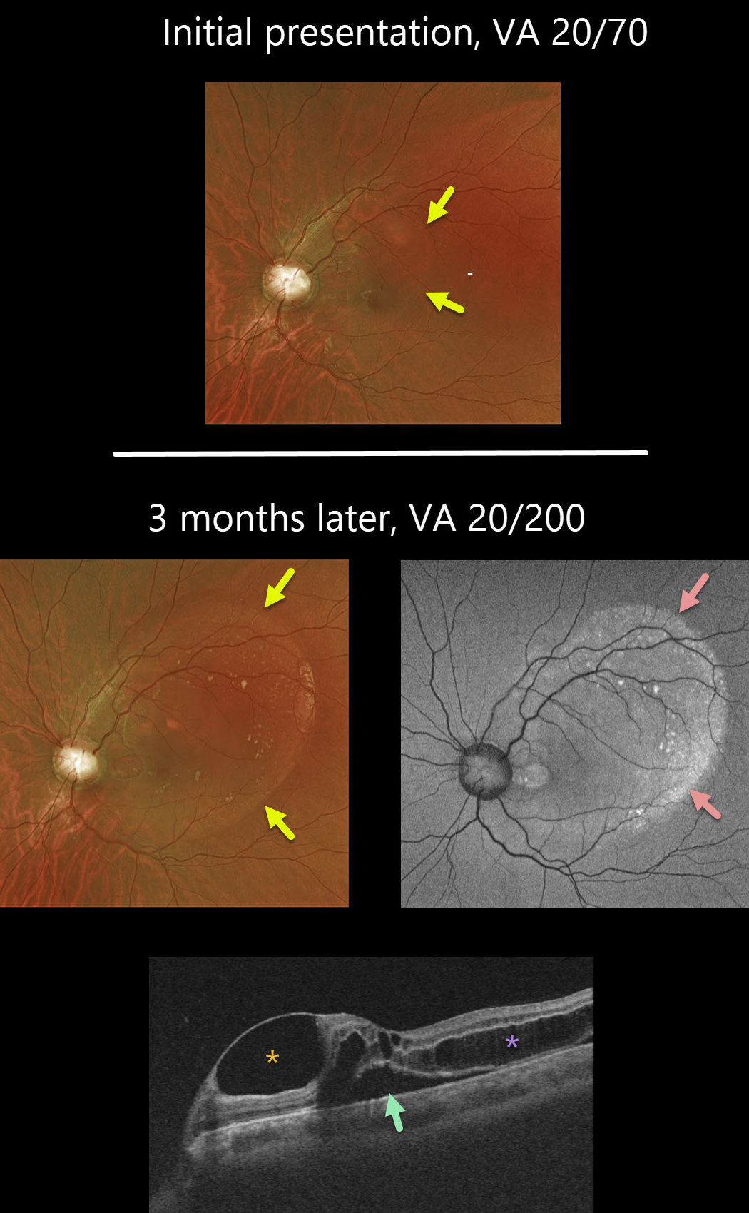
OPTIC NERVE COLOBOMA
Originally posted on @retina.rocks April 28, 2021
This 19YO male has bilateral optic nerve coloboma with optic pit maculopathy in his left eye.
On initial presentation, vision was 20/70, which decreased to 20/200 at 3-month follow-up. The extent of the macular schisis/fluid dramatically enlarged, accounting for the drop in vision.
Optos fundus autofluorescence (FAF) showed variable hyper-FAF along the temporal edge of the fluid.
Triton OCT imaging showed an unusual inner retinal cyst, as well as subretinal fluid and outer macular schisis.
Remarkably, the patient felt his vision had improved subjectively and did not wish to consider the recommended vitrectomy procedure.
Learning Points:
While most optic pits are asymptomatic, they can cause vision loss by causing fluid to accumulate in the inner and outer retina and the subretinal space (optic pit maculopathy). The source of the fluid remains a point of ongoing debate, with potential contributions from the vitreous and/or subarachnoid space.
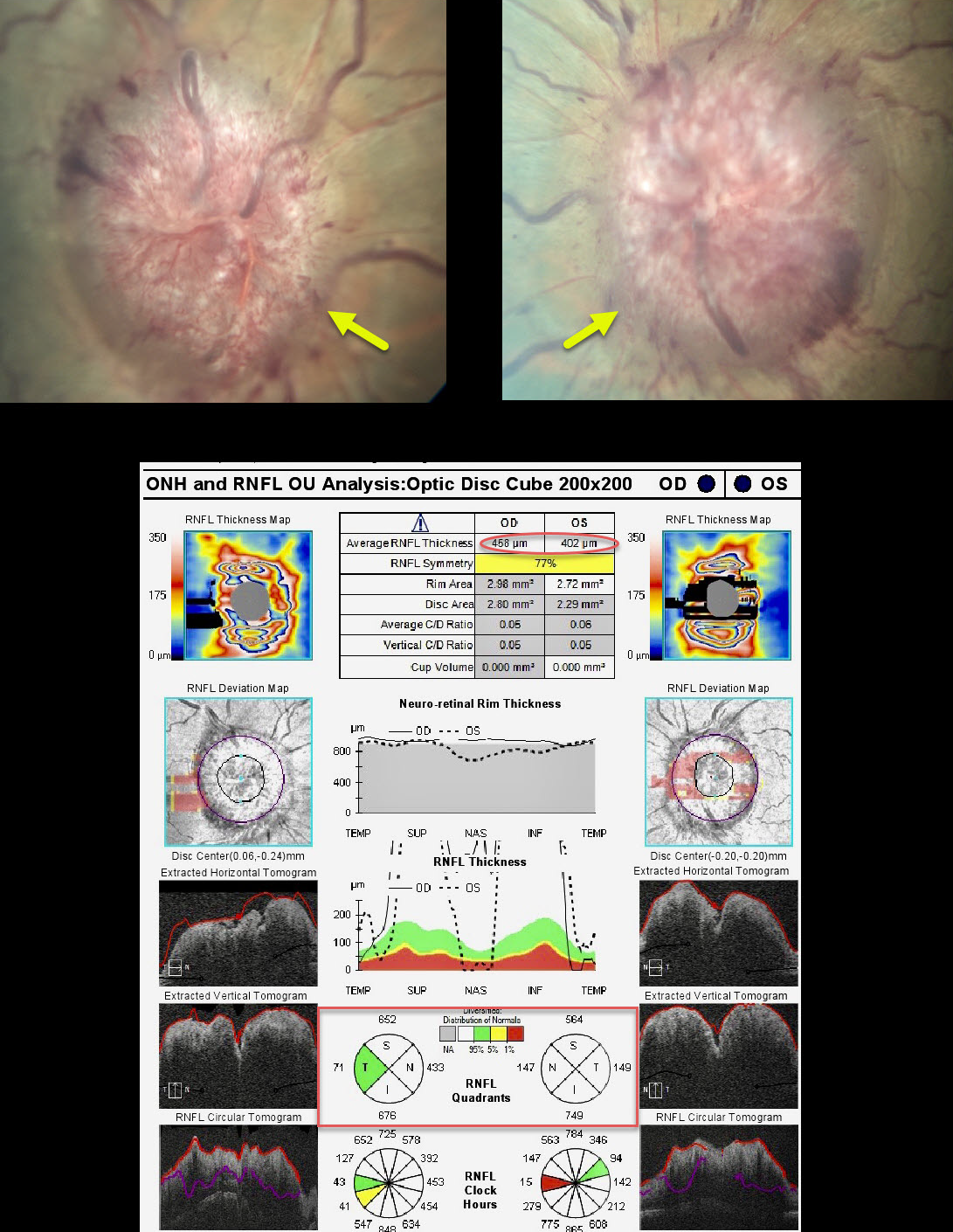
PAPILLEDEMA
Originally posted on @retina.rocks February 23, 2021
This patient presented with diplopia from bilateral esotropia and reduced abduction in each eye due to sixth nerve palsies. There were no headaches or visual disturbances.
Her nerves showed severe papilledema with severe retinal nerve fiber layer thickening on OCT.
An MRI showed no mass lesions, but her lumbar puncture opening pressure was 41 cm H2O.
Despite an aggressive weight loss regimen and oral acetazolamide, her intracranial pressure remained elevated and required a ventriculoperitoneal shunt.
Learning Points:
Idiopathic intracranial hypertension (pseudotumor cerebri) can occasionally cause a downward displacement of the brainstem, stretching the sixth nerve as it exits the pons towards Dorello’s canal at the apex of the petrous bone.
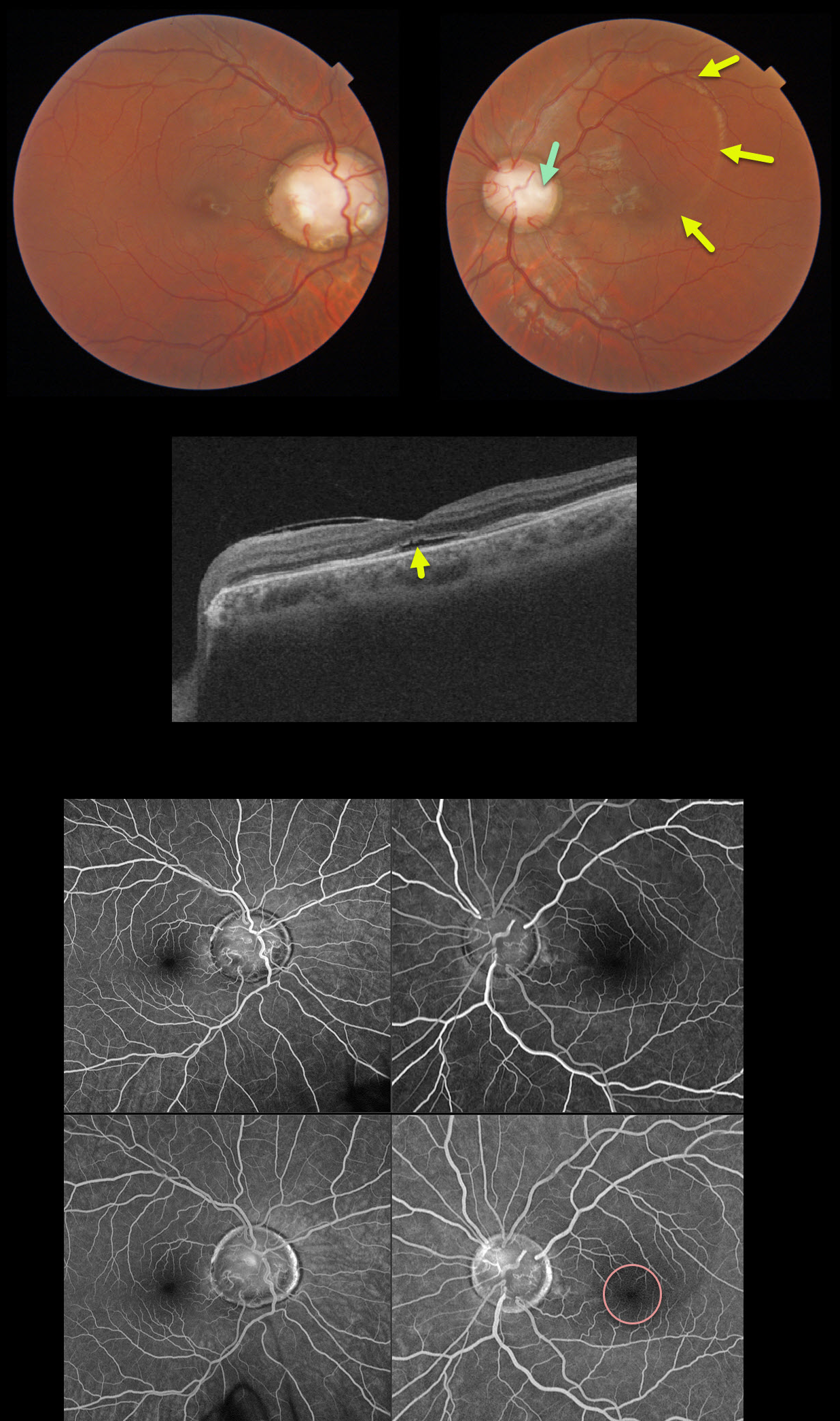
OPTIC NERVE COLOBOMA
Originally posted on @retina.rocks February 5, 2021
Our patient had bilateral optic nerve colobomas with an optic pit in the left eye. Vision was 20/40 OD and 20/70 OS.
The serous fluid from the optic pit is in the superior macula with mild fluid in the subfoveal space, best noted on the macular OCT. Fluorescein angiography shows no leakage, indicating that this fluid is not exudative.
Our patient opted for no treatment due to minimal symptoms and fairly good vision.
Learning Points:
An optic nerve coloboma can be unilateral or bilateral and is caused by incomplete closure of the embryonic fissure.
While most optic pits are asymptomatic, they can cause vision loss by forcing fluid into the inner and outer retina and, ultimately, the subretinal space (optic pit maculopathy). The source of the fluid remains a point of ongoing debate, with the fluid originating either from the vitreous or the subarachnoid space.
A host of treatments for optic pit maculopathy have been suggested over the years. However, a recent meta-analysis suggested that a plain old vanilla vitrectomy without gas tamponade is as successful as any other procedure (Zheng et al Ophthalmology Retina 2020;4;289-299).
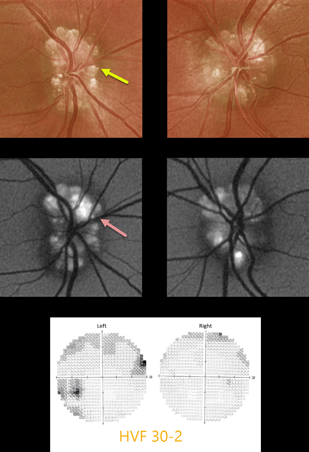
OPTIC NERVE DRUSEN
Originally posted on @retina.rocks January 4, 2021
This 29yo male presented with asymptomatic bilateral optic nerve drusen. Vision was 20/40 OD and 20/30 OS.
The drusen hyperautofluoresce, and visual fields show some non-specific depression and defects.
Learning Points:
Optic disc drusen are small hyaline bodies within the optic nerve that progressively calcify over time. They can be buried or superficial, giving the nerve a “lumpy-bumpy” appearance with normal overlying vessels. These nerves can often be confused with papilledema, especially in younger patients.
We have found ultrasonography to be totally unhelpful in younger patients, since drusen don’t calcify until later in life.
The drusen often hyperautofluoresce on fundus autofluorescence imaging. Swept-source OCT can occasionally visualize buried drusen, which can be either hypo- or hyperreflective.
When autofluorescence is negative, the best clinical test is fluorescein angiography, which will show optic nerve leakage with papilledema. Eyes with drusen will show either no nerve leakage or nodular staining.
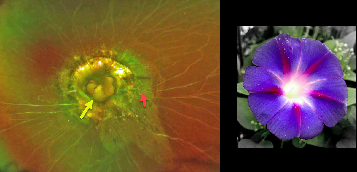
OPTIC NERVE MORNING GLORY
Originally posted on @retina.rocks December 28, 2020
This patient has morning glory syndrome. The left optic nerve appears to have a large optic disc excavation with radial vessels, central glial tissue, and peripapillary atrophy (PPA) and pigmentation.
Visual prognosis is usually poor.
Learning Points:
The morning glory syndrome is typically unilateral and shares some similarities with optic disc coloboma and juxtapapillary staphyloma.
The condition is often associated with serous macular detachment. Neuroimaging should be considered to rule out transsphenoidal encephalocele and intracranial carotid artery dysgenesis.
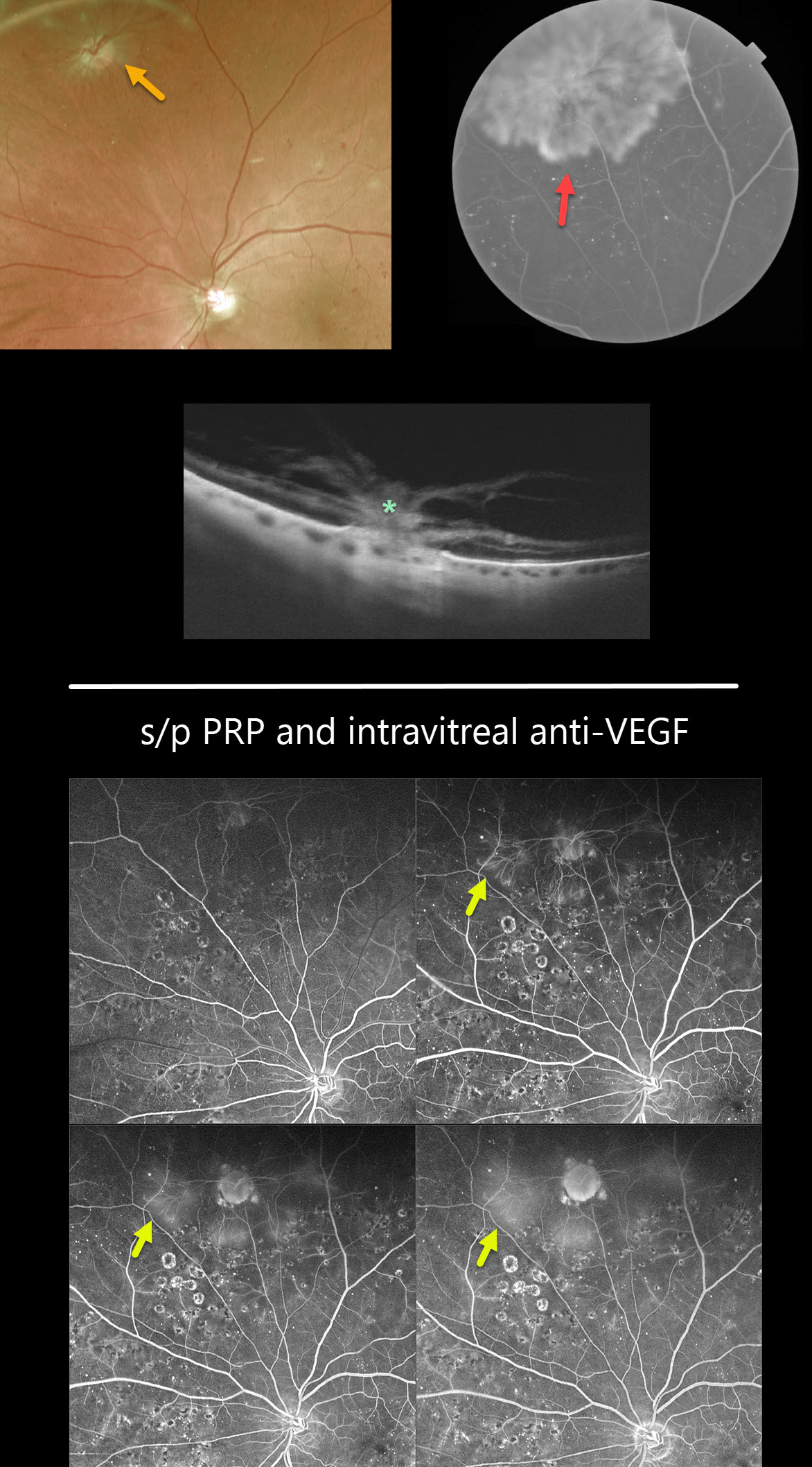
OPTIC NERVE PSEUDO-DUPLICATION WITH NVD ELSEWHERE
Originally posted on @retina.rocks December 18, 2020
Is that another optic nerve?
This is a pseudo-duplicated optic disc from peripheral neovascularization emanating from a disc-sized mid-peripheral chorioretinal scar.
The OCT reveals the area of choriovitreal neovascularization.
Learning Points:
The neovascularization regressed following panretinal photocoagulation (PRP) supplemented with intravitreal Eylea injections.
Fluorescein angiography following PRP and anti-VEGF injections shows virtually no leakage from the neovascularization.
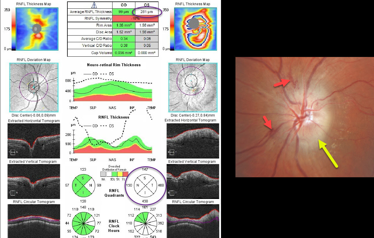
OPTIC NEUROPATHY
Originally posted on @retina.rocks November 20, 2020
This patient presented with unilateral diffuse optic nerve edema with some splinter hemorrhages and possible inferotemporal lipid. Optic nerve OCT confirms markedly increased retinal nerve fiber layer thickness.
The differential diagnosis for a unilaterally acutely swollen nerve is quite long and includes arteritic (AION) and non-arteritic (NAION) anterior ischemic optic neuropathy, infectious causes (including syphilis and neuroretinitis), and inflammatory conditions (including sarcoidosis and optic neuritis). In this case, we diagnosed NAION.
Learning Points:
NAION presents with sudden painless unilateral central vision loss with an altitudinal defect. It is the most common acute optic neuropathy in patients over age 50.
It is thought to be caused by non-embolic occlusion of small branches of the short posterior ciliary arteries that supply the anterior optic nerve. It is associated with congenitally small and crowded nerves, optic disc drusen, medications (amiodarone and possibly phosphodiesterase inhibitors), hypertension, and diabetes. About 75% of patients have sleep apnea. Nocturnal hypotension or taking blood pressure pills at bedtime is also considered a risk factor.
Erythrocyte sedimentation rate and C-reactive protein should be ordered to rule out giant cell arteritis. There is no effective treatment.
Optic nerve edema resolves within a few months, often with secondary optic nerve pallor.
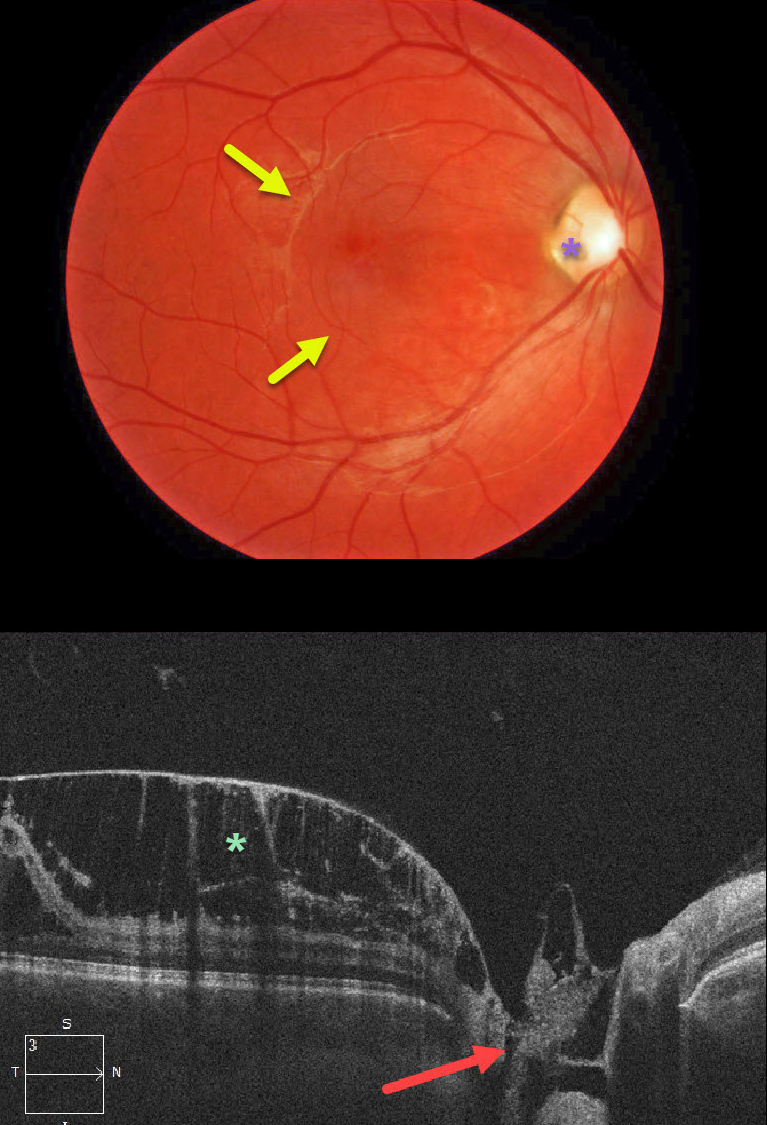
OPTIC NERVE PIT
Originally posted on @retina.rocks November 19, 2020
This patient presented with asymptomatic 20/30 vision OD and subtle macular thickening coming from an optic nerve pit along the temporal aspect of the optic nerve.
OCT shows a direct communication between the pit and the prominent inner macular schisis. Patients can also have subretinal fluid, which our patient did not. The left eye was completely normal.
Our patient opted for no treatment due to the lack of symptoms and fairly good vision.
Learning Points:
Optic pits are most commonly located within or along the inferior-temporal disc margin and usually appear as a gray depression. They are thought to be caused by incomplete closure of the optic fissure during gestation.
While most optic pits are asymptomatic, they can cause vision loss by forcing fluid into the inner and outer retina and, ultimately, the subretinal space (optic pit maculopathy). The source of the fluid remains a subject of ongoing debate, with origins in either the vitreous or the subarachnoid space.
A host of treatments have been suggested over the years. However, a recent meta-analysis suggested that a plain old vanilla vitrectomy without gas tamponade is as successful as any other procedure (Zheng et al Ophthalmology Retina 2020;4;289-299).
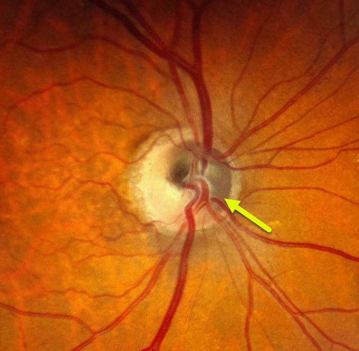
MELANOCYTOMA
Originally posted on @retina.rocks November 12, 2020
This patient has a melanocytoma, which is a benign melanocytic tumor intrinsic to the optic nerve. There is pigmentation noted within the superonasal portion of the optic disc.
Learning Points:
Melanocytomas can sometimes enlarge and spread into the adjacent retina and choroid, and can very rarely undergo malignant transformation.
Most patients are asymptomatic, but some can develop visual field loss from compressive optic neuropathy.
These lesions should be periodically monitored for any signs of growth.
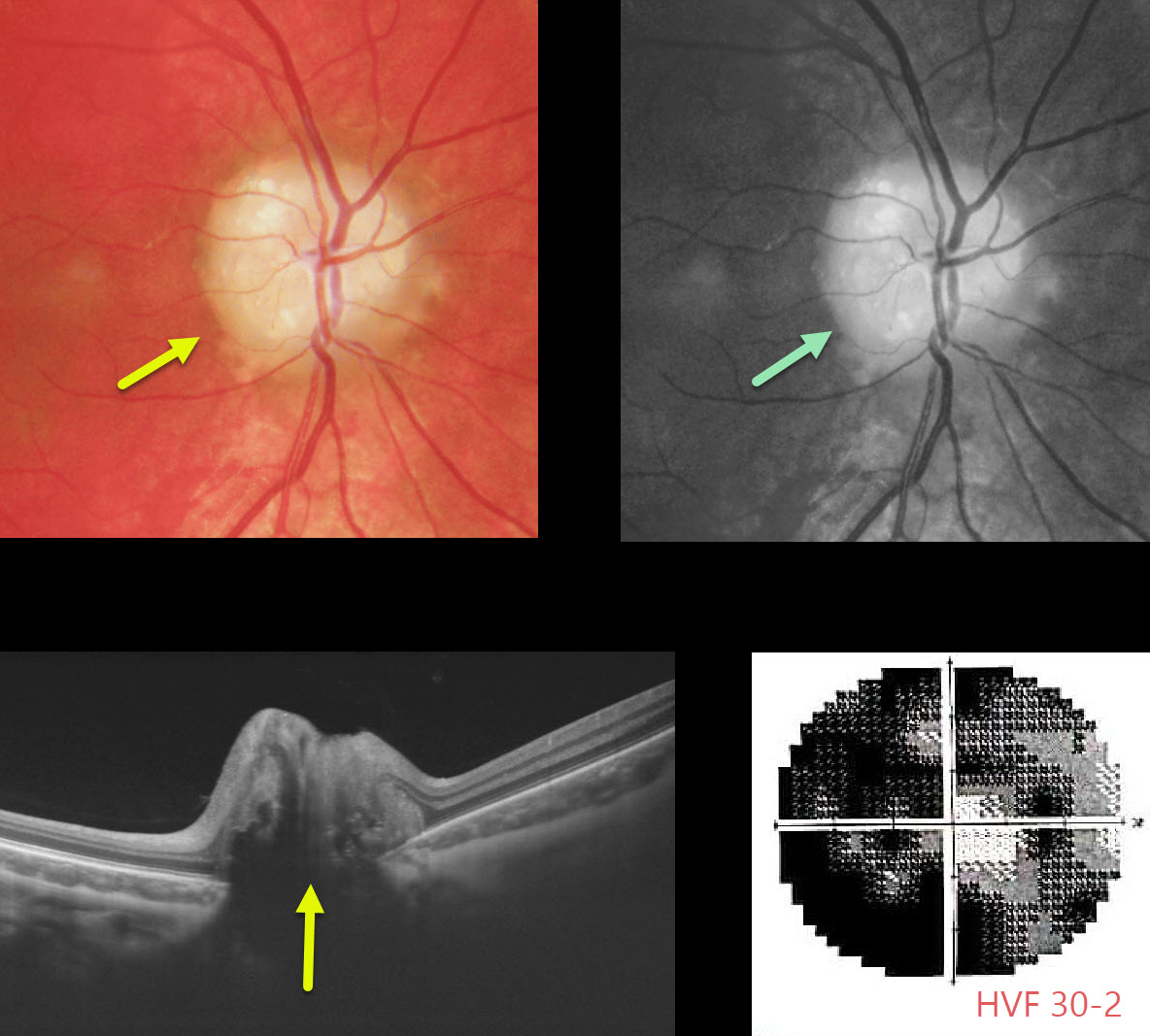
OPTIC NERVE DRUSEN
Originally posted on @retina.rocks September 28, 2020
Our patient is a 15YO boy with quite pronounced bilateral optic nerve drusen visible clinically, as well as on OCT (only right eye pictured). ODD will often hyperautofluoresce on fundus autofluorescence (FAF).
Although central vision was good, he complained of increasing peripheral vision loss due to drusen compressing the surrounding nerve fibers.
Learning Points:
Optic disc drusen (ODD) are refractile calcific concretions that form anterior to the lamina cribrosa. They are thought to be extruded, extracellular, calcified mitochondria and may be inherited as an autosomal dominant trait.
It is often difficult to differentiate ODD from true optic nerve edema. This is often the dilemma when seeing a child with a possible swollen nerve.
We have found ultrasonography to be unhelpful, since ODD don’t calcify until later in life.
B-scan OCT imaging is often unhelpful, although newer swept-source machines can directly visualize drusen. The RNFL will be variably thinned with ODD and thickened with true disc edema.
Fluorescein angiography (FA) is probably the best way to differentiate the two, since a swollen nerve will show late leakage, whereas ODD will not leak and often shows staining of the drusen.
Unfortunately, there is no proven treatment, although some have tried pressure-lowering medications for neuroprotection.
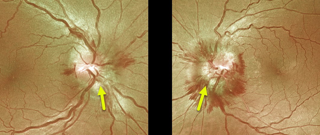
PAPILLEDEMA
Originally posted on @retina.rocks September 17, 2020
This is a beautiful example of papilledema in a patient with idiopathic intracranial hypertension (pseudotumor cerebri).
This patient had an opening lumbar puncture pressure of 28 cm H2O.
Note the absence of lipid, which, for some reason, is much more common in patients with malignant hypertension or neuroretinitis.
Learning Points:
Remember that papilledema is not synonymous with optic nerve swelling. Papilledema is defined as bilateral optic nerve swelling due to elevated intracranial pressure.
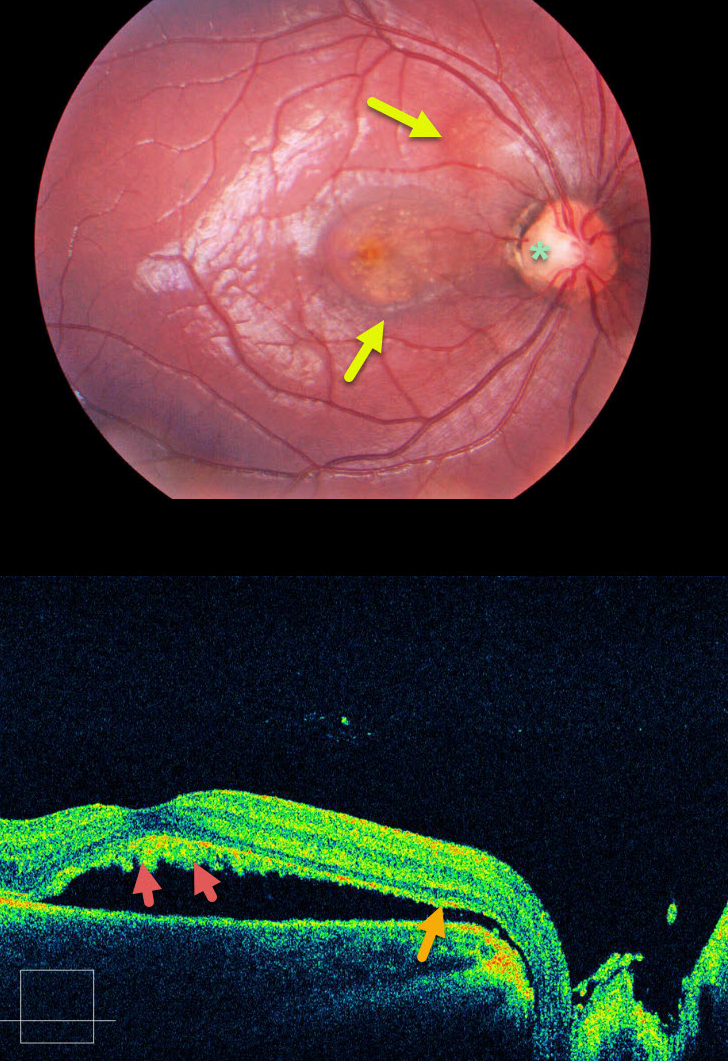
OPTIC NERVE PIT
Originally posted on @retina.rocks September 2, 2020
This patient has optic pit maculopathy, in which areas of subretinal fluid can be traced back to the optic nerve pit via an abnormal communication between the subretinal and subarachnoid spaces.
Shed photoreceptor outer segments accumulate on the back surface of the detached retina since the RPE cells can’t perform their normal function of phagocytosis. This shaggy vitelliform material gives the central macula a yellow appearance.
Learning Points:
Optic pits are congenital defects caused by the incomplete closure of the fetal fissure during embryogenesis. Macular detachment and schisis occur in up to 30-40% of eyes.
When symptomatic, a myriad of surgical procedures have been described. As with any problem with too many solutions, there is usually no best option.
A recent meta-analysis concluded that just a plain, vanilla vitrectomy is probably as successful as anything else (Zheng et al, Ophthalmology Retina 2020;4:389-299).
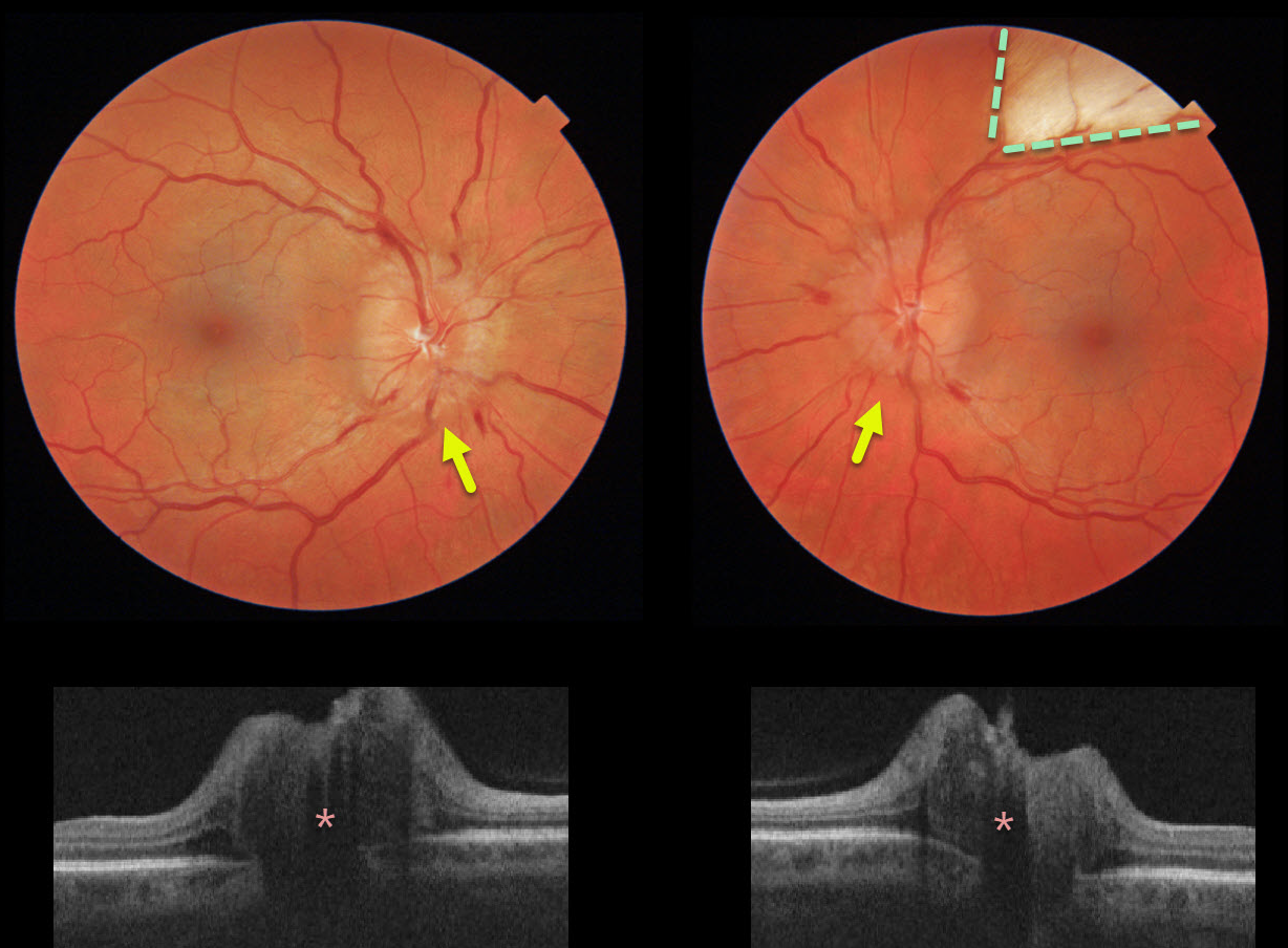
PAPILLEDEMA
Originally posted on @retina.rocks June 26, 2020
This healthy 40yo female presented with chronic headaches. She was on no systemic medications, and her blood pressure was normal. Her nerves were swollen bilaterally, and a myelinated nerve fiber layer was also noted in her left eye.
The retinal nerve fiber layer on OCT was severely thickened bilaterally.
Learning Points:
The diagnosis of idiopathic intracranial hypertension (pseudotumor cerebri) was made after an MRI scan was normal and a lumbar puncture revealed a markedly elevated opening pressure.
She is currently being successfully managed with oral acetazolamide.
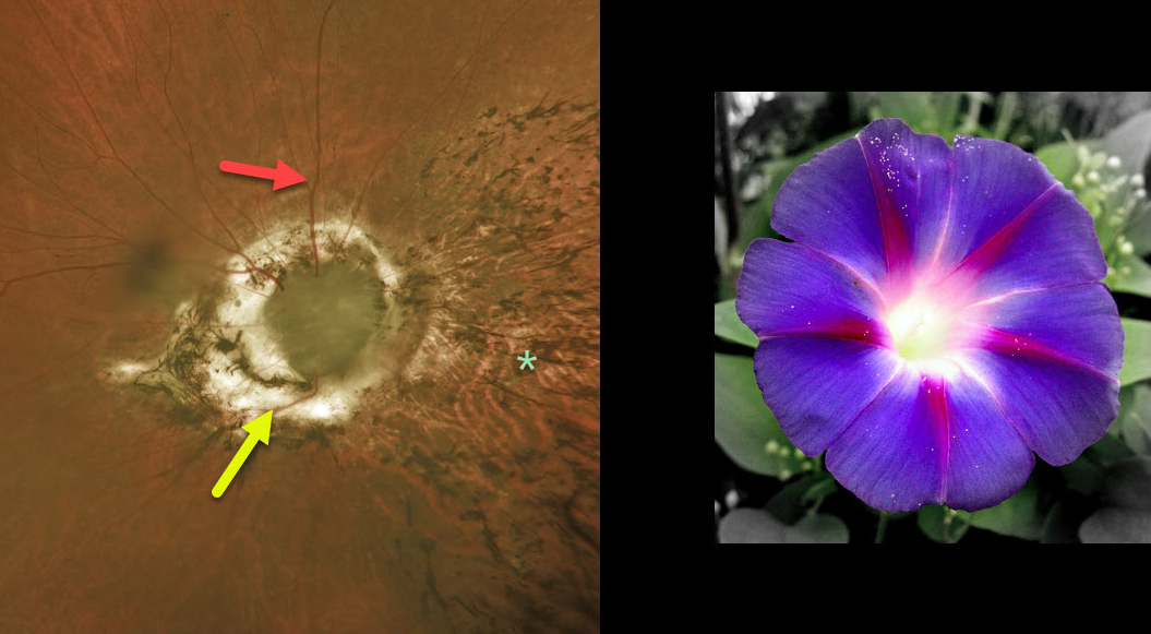
OPTIC NERVE MORNING GLORY
Originally posted on @retina.rocks June 5, 2020
This 66yo male is CF OD and 20/200 OS. He has a history of retinal detachment surgery OD, as well as severe glaucoma.
His right eye has a possible morning glory anomaly (no central vessels with possible overlying glial tissue, radiating numerous retinal vessels) versus severe glaucomatous damage.
The intraretinal pigment migration nasally is from the prior detachment. Although he had prior retinal reattachment surgery (presumed from a retinal break), this may also have been from subretinal fluid that is occasionally seen with these anomalous optic nerves.
Learning Points:
The morning glory syndrome is typically unilateral and shares some similarities with optic disc coloboma and juxtapapillary staphyloma. This syndrome is appropriately named after the Morning Glory flower.
The condition is often associated with serous macular detachment.
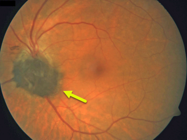
MELANOCYTOMA
Originally posted on @retina.rocks April 15, 2020
This patient has a melanocytoma, which is a benign melanocytic tumor intrinsic to the optic nerve.
They are intensely pigmented with feathery margins, sometimes spreading into the adjacent retina and choroid.
Learning Points:
Although melanocytomas may enlarge over time, malignant transformation is very rare. Spontaneous necrosis may cause a similar appearance to vitreous seeding from a melanoma.
Vision and visual field loss can rarely occur from compressive optic neuropathy.
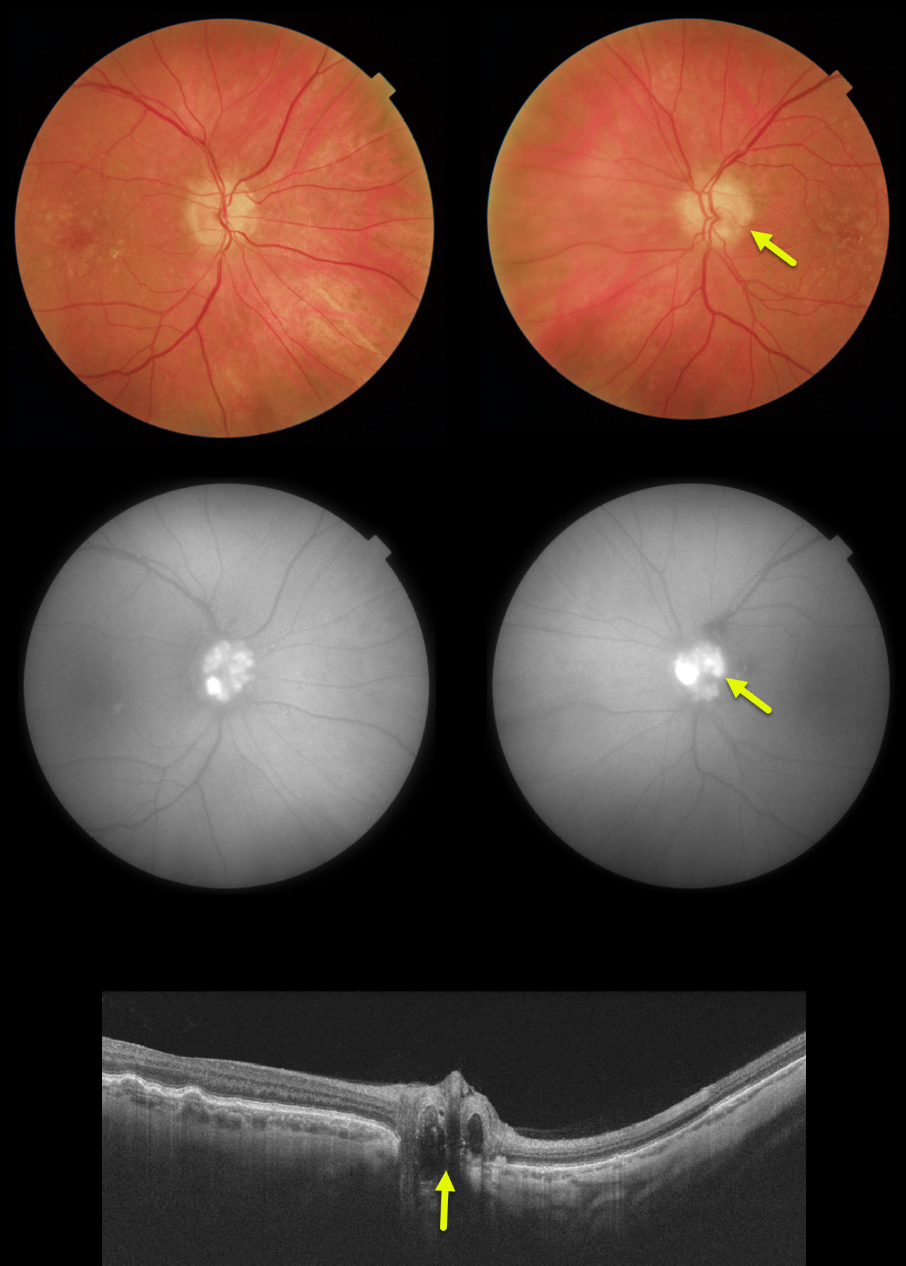
OPTIC NERVE DRUSEN
Originally posted on @retina.rocks March 16, 2020
Our patient’s optic disc drusen were obvious clinically, on FAF and on OCT. In younger patients they are usually much more difficult to see and can be confused with papilledema.
We have found ultrasonography in younger patients cases to be totally unhelpful since drusen don’t calcify until later in life.
If fundus autofluorescence (FAF) is negative, the best clinical test in these cases is fluorescein angiography, which will show optic nerve leakage with papilledema. Eyes with drusen will show either no nerve leakage or nodular staining.
Learning Points:
Optic disc drusen are small hyaline bodies within the optic nerve that progressively calcify over time. They can be buried or superficial giving the appearance of pseudopapilledema. The nerve usually has a “lumpy-bumpy” appearance with normal overlying vessels. Retinal nerve fiber layer will usually show variably thinning.
FAF can show drusen hyperautofluoresence. Swept source OCT can occasionally visualize buried drusen.which can be either hypo- or hyperreflective.
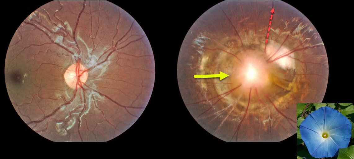
OPTIC NERVE MORNING GLORY
Originally posted on @retina.rocks February 13, 2020
This patient has morning glory syndrome, and the left optic nerve appears to have large optic disc excavation with radial vessels, central glial tissue and peripapillary atrophy and pigmentation.
Visual prognosis is usually poor.
Learning Points:
The morning glory syndrome is typically unilateral and shares some similarities with optic disc coloboma and juxtapapillary staphyloma.
The condition is often associated with serous macular detachment. Neuroimaging should be considered to rule out transsphenoidal encephalocele and intracranial carotid artery dysgenesis.
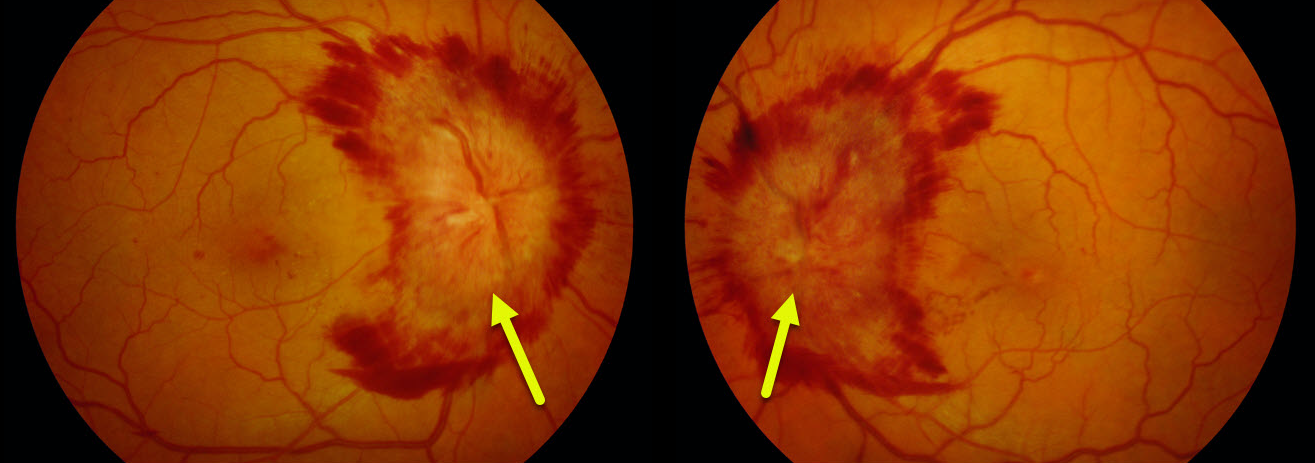
PAPILLEDEMA
Originally posted on @retina.rocks February 3, 2020
This patient had papilledema with severe disc hemorhages. MRI detected a compressive meningioma as the cause for the elevated intracranial pressure.
Learning Points:
Optic nerve swelling has numerous causes, including neurologic (optic neuritis and elevated intracranial pressure) and retinal (retinal vein occlusion, neuroretinitis, sarcoidosis, Harada’s) causes.
Bilateral optic nerve swelling due to elevated intracranial pressure is known as papilledema.
Neurologic imaging is important to establish the correct diagnosis.
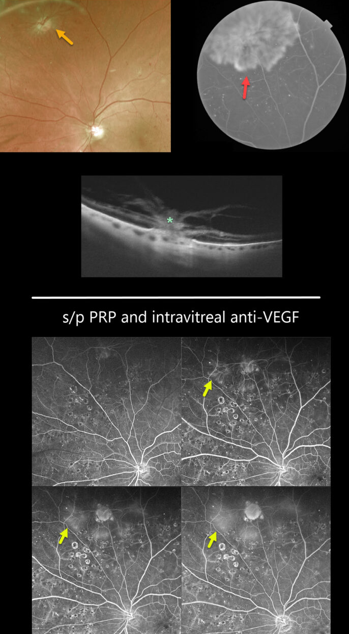
OPTIC NERVE PSEUDO-DUPLICATION
Originally posted on @retina.rocks December 18, 2019
Is that another optic nerve????
Pseudoduplicated optic disc from peripheral neovascularization emanating from disc-sized mid peripheral chorioretinal scar. The neovascularization regressed following panretinal photocoagulation with intravitreal eylea injections. The OCT showing the choriovitreal neovascularization is amazing!


