

15 Miscellaneous
<< Back to Cases
DENGUE RETINITIS
Rohan Jain and Manish Nagpal
Originally posted on @retina.rocks September 9, 2026
This 63YO male presented with 1 month of bilateral decreased vision following dengue fever, with positive Dengue NS1 antigen during the acute illness. Vision was 20/200 OD and counting fingers OS.
Pseudocolor SLO imaging shows extensive peripapillary cotton wool spots, retinal whitening, and significant macular edema. The fundus detail is partially obscured by overlying vitreous debris from vitritis. OCT scanning neurosensory retinal detachment with subretinal hyperreflective material, intraretinal cystoid edema, and subretinal fluid involving the macula in both eyes, consistent with post-dengue inflammatory retinitis. A possible bacillary layer detachment is also noted in the left eye.
Topical steroids and a 9-week course of tapering oral steroids, as well as a 3-week course of oral doxycycline, were started. Four months later, vision improved to 20/20 OD and 20/30 OS. The patches of white inner retinal ischemia are fading, and some lipid flecks have precipitated out in the mid-retina.
Learning Points:
Epidemic (post-fever) retinitis is an infectious or para-infectious entity that develops in immunocompetent individuals 2-6 weeks following a bacterial, viral, or protozoal infection. Rickettsiosis is the most identified cause, though a large proportion of cases remain idiopathic despite serologic testing; dengue, chikungunya, West Nile, Zika, leptospirosis, typhoid, and Ebola have also been implicated. Patients present with sudden painless vision loss. Fundus findings include unilateral or bilateral unifocal or multifocal patches of retinitis with possible nerve involvement, neuroretinitis with macular star, serous macular detachment, cystoid macular edema, and vasculitis or frosted branch angiitis.
Management is stratified by etiology and lacks controlled data: bacterial causes are treated with systemic antibiotics with or without corticosteroids, viral causes with corticosteroids alone, and many authors advocate observation, as the disease often resolves on its own. Corticosteroids should not be used as sole therapy while infectious causes (including TB, syphilis, and toxoplasmosis) remain in the differential. See Mahendradas et al (Indian J Ophthalmol 2020;68:1775-1786) for an excellent post-fever retinitis review.
HARADA DISEASE
Rohan Jain and Manish Nagpal
Originally posted on @retina.rocks August 20, 2026
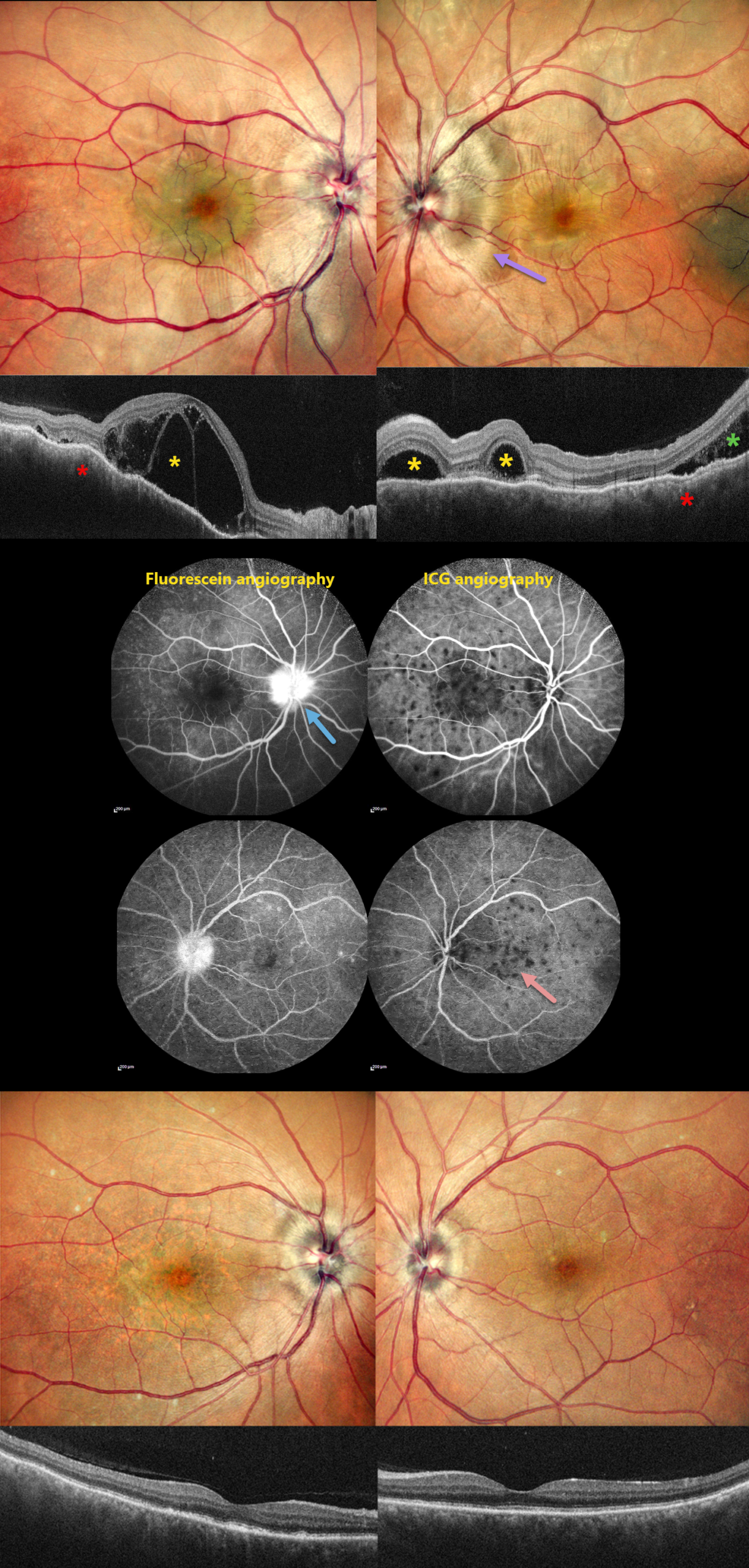
This 46YO male presented with 10 days of bilateral blurred vision with a left-sided headache. Vision was 20/200 OD and 20/20 OS. Anterior segments were normal.
Pseudocolor SLO imaging shows bilateral swollen nerves and irregular macular chorioretinal folds. OCT scanning shows multifocal bacillary layer detachments, subretinal fluid, and an undulating, thickened choroid. Fluorescein angiography shows bilateral multifocal areas of subretinal leakage and disc leakage, and indocyanine green angiography shows multiple hypofluorescent choroidal spots.
Based on the clinical and multimodal imaging findings, a diagnosis of acute Harada disease was established. He received a 3-day course of intravenous methylprednisolone followed by oral corticosteroids.
At the 2-month follow-up, the patient’s symptoms had completely resolved. Vision was 20/30 OD and 20/20 OS. The exudative findings are completely resolved on photography and OCT, and the choroidal thickening with overlying chorioretinal folds is also resolved.
Learning Points:
Harada disease is characterized by relatively acute bilateral panuveitis, optic nerve swelling, thickened choroid, chorioretinal folds, multifocal serous exudative retinal detachments, and bacillary layer detachments. Patients with only ocular findings have Harada disease (as in our patient), whereas those with additional systemic findings (including vitiligo, poliosis, headache, vertigo, and hearing loss) have Vogt-Koyanagi-Harada disease.
SPONTANEOUSLY DISLOCATED CATARACT
Originally posted on @retina.rocks July 23, 2026
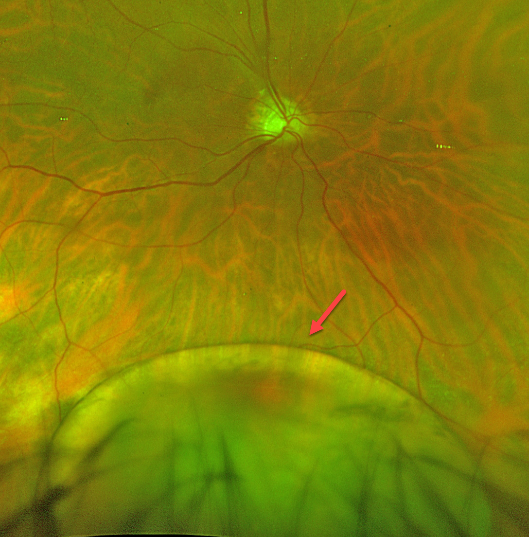
This 56YO developmentally delayed male presented with seeing a “black spot” in his right eye. He was monocular with a phthisical left eye from unclear etiology. Vision in his right eye was decreased to 20/400 from his baseline of 20/70 at his prior visit.
He was largely nonverbal and difficult to examine due to extreme photosensitivity. Optos color RG imaging shows an inferiorly dislocated crystalline lens.
Urgent pars plana vitrectomy and lensectomy were performed. After discussion with the patient’s guardian, the decision was made to leave the patient aphakic rather than to place a secondary scleral-fixated or scleral-sutured lens due to a history of frequent eye rubbing. The patient was fitted with aphakic glasses, which restored his vision to baseline.
Learning Points:
Dislocation or subluxation of the crystalline lens can be caused by trauma, ocular conditions such as ectopia lentis et pupillae, pseudoexfoliation, aniridia, high myopia, or hereditary disorders such as Marfan syndrome, homocystinuria, Weill-Marchesani syndrome, Ehlers-Danlos syndrome, or sulfite oxidase deficiency. Our patient had a known cardiofaciocutaneous (CFC) syndrome secondary to BRAF mutation, but this has not been associated with ectopia lentis.
DISLOCATED IOL
Originally posted on @retina.rocks May 6, 2026
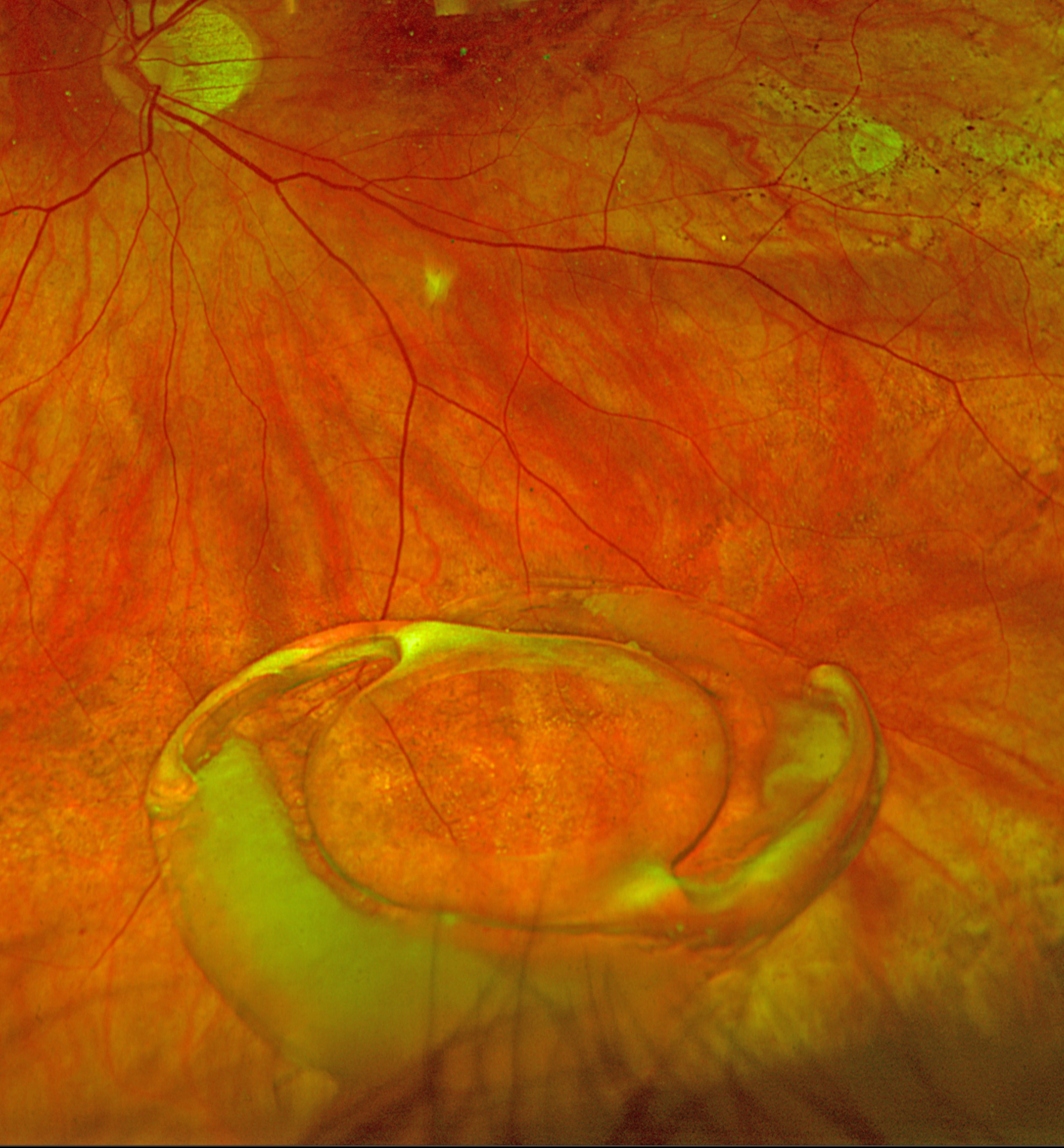
This 80YO female noticed sudden vision loss in her left eye. When examined the next morning, vision was 20/400.
Optos color RG imaging shows an inferiorly dislocated intraocular lens (IOL) with surrounding capsular material settled over the inferior retinal periphery. She underwent successful IOL explantation with a secondary anterior chamber IOL.
SICKLE CELL RETINOPATHY
Originally posted on @retina.rocks May 5, 2026
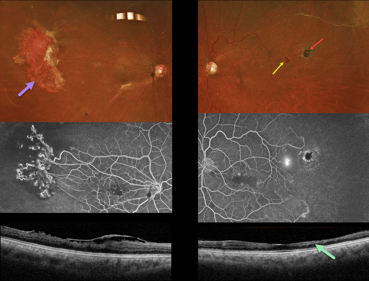
This 44YO male with a known history of sickle cell disease was referred for a possible detached retina. Vision has decreased over the past few years. Vision was 20/200 OU.
Optos color RGB imaging shows a large area of active neovascularization in the right temporal midperiphery associated with extensive angiographic retinal ischemia. Similar ischemia is noted OS with a small patch of leaking neovascularization. There is a small, more distal black sunburst lesion. The foveal avascular zones are enlarged with surrounding telangiectasia. This is reflected in bilateral inner retinal thinning with disorganization of inner retinal layers on OCT scanning (DRIL). An epimacular membrane is also noted OD.
Learning Points:
Patients with sickle cell disease develop symptoms throughout their bodies as stiffened, sickled red blood cells block capillary beds. In the eye, this can cause proliferative sickle cell retinopathy (PSR) with sea-fan-shaped areas of peripheral retinal neovascularization developing at the junction of perfused and ischemic retina. Increasing macular ischemia correlates with peripheral angiographic nonperfusion, as seen in our patient (Han et al., Ophthalmology Retina 2019;2:599-605). Complications include vitreous hemorrhage and tractional retinal detachment. Although patients with SS disease tend to have more systemic complications, SC disease is usually associated with more severe ocular findings.
Treatment with sectoral (Farber et al Arch Ophthalmology 1991;109:363-367) or circumferential (Kimmel et al Ophthalmology 1986;93:1429-1434) scatter laser photocoagulation is somewhat controversial since, unlike more typical proliferative retinopathies (ie from diabetes and branch retinal vein occlusions), these new vessels tend to auto-infarct with a good natural history for untreated disease (Downes et al Ophthalmology 2005;112:1869-1875). Based on the active appearance of the large temporal neovascularization OD, we elected for prophylactic scatter laser throughout the ischemic retina.
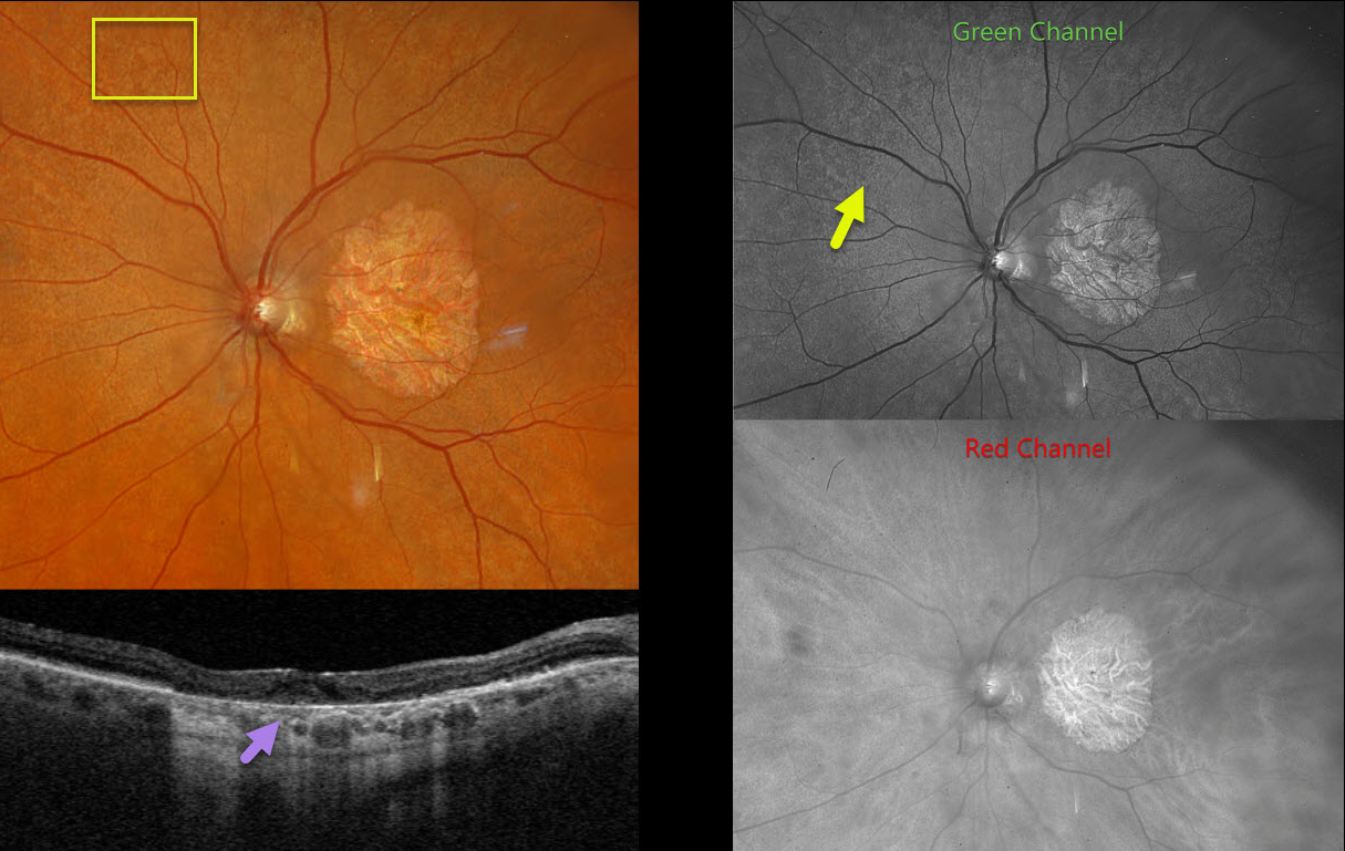
EXTENSIVE MACULAR ATROPHY WITH PSEUDODRUSEN (EMAP)
Originally posted on @retina.rocks March 26, 2026
This 78YO female, previously diagnosed with macular atrophy from age-related macular degeneration, has stable vision of 20/400 OD and counting fingers OS.
Optos color RGB imaging of her left eye shows extensive subretinal drusenoid deposits scattered throughout the posterior pole. These are best seen in the green channel. Triton swept-source OCT shows variable incomplete outer retinal and RPE atrophy. Identical findings were noted in her right eye (not shown).
Learning Points:
Originally described by Hamel et al (AJO 2009;147:609-620), extensive macular atrophy with pseudodrusen (EMAP) is an idiopathic, rapidly progressive macular atrophy. Unlike age-related macular atrophy, EMAP begins at an earlier age (50-55 years) and progresses more rapidly to legal blindness within 5 years. The macular atrophy is typically greater in the vertical meridian. Patients also have extensive subretinal drusenoid deposits (reticular pseudodrusen) with extensive peripheral cobblestone degeneration.
For an excellent recent review, see Antropoli et al, Progress in Retinal and Eye Research 2025;104:101320.
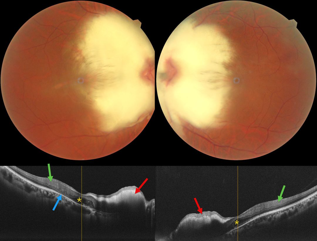
MYELINATED NERVE FIBER LAYER
Aarzoo Juneja and Lalit Singhal
Originally posted on @retina.rocks March 10, 2026
This healthy 36YO female presented with high myopia of -11.50 -1.75 x 50 OD and -11.00 -1.00 x 170 OS. Vision was 20/80 OD and 20/60 OS. Color photography shows a bilaterally symmetric myelinated nerve fiber layer (NFL) covering each nerve and the nasal macula. The discs may also be hypoplastic, although the margins are difficult to discern due to the myelination.
On OCT, the myelination appears as a markedly hyperreflective, thickened inner retina with posterior shadowing. The foveal outer nuclear layers (ONL) appear thickened OU. There is hyperreflective material in the right nasal foveal ONL with disorganization of the underlying outer retinal bands. The inner retinal bands are disorganized more temporally OU, with outer retinal atrophy OD.
Learning Points:
Myelinated NFL occurs when retinal nerve fibers develop a myelin sheath, which usually stops posterior to the lamina cribrosa. The myelination may represent an oligodendrocytic choristoma (Rao et al, Retina 2019;39:1125-1132).
Myelinated NFL is a relatively uncommon, unilateral or bilateral, usually isolated finding present at birth. The myelinated NFL is often connected to the optic nerve. Most patients are asymptomatic, although some may have a corresponding relative scotoma. The myelination can progress in about 10% of cases and can also resolve following retinal ischemic events. Our patient’s findings were consistent with Straatsma syndrome, which consists of the triad of bilateral myopia, amblyopia, and myelinated NFL.
Myelinated NFL is reported to anatomically affect just the NFL (Rao et al, Retina 2019;39:1125-1132). Our patient’s OCT findings showing both inner and outer retinal changes outside the area of myelination has not been previously reported.
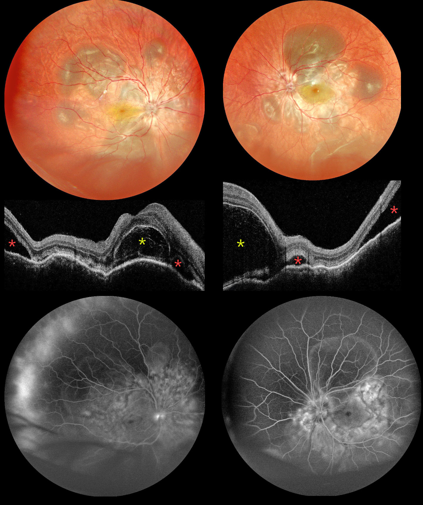
SYMPATHETIC OPHTHALMIA
The European VitreoRetinal Society (EVRS), Deepanshu Agrawal and Arun Bhargava
Originally posted on @retina.rocks February 20, 2026
This 30YO male suffered an open globe injury from an iron nail OD on 11/2/25. Three days later, the corneo-limbal laceration was repaired elsewhere. He presented to us on 11/25 with 1 week of bilateral vision loss. Vision was hand motion OD and 20/400 OS. Mild anterior chamber cells were noted OU.
Multicolor SLO imaging shows bilateral multifocal pockets of subretinal fluid (SRF) along with inferior exudate retinal detachments. OCT scanning shows bilateral bacillary layer detachments (BALADs), SRF, and choroidal thickening and undulation. Fluorescein angiography shows multifocal subretinal leakage with disc leakage.
Learning Points:
Sympathetic ophthalmia is a rare, bilateral granulomatous panuveitis that develops following penetrating trauma or intraocular surgery to one eye, with an incidence of approximately 0.19% after open-globe injury (He et al, AJO 2022;234:117-125). It typically presents within the first year after the inciting event but can occur days to years later, presenting with decreased vision, pain, and photophobia. Findings mirror those seen in Vogt-Koyanagi-Harada disease, including granulomatous anterior uveitis, vitritis, exudative retinal detachment with BALAD, choroidal thickening, and Dalen-Fuchs nodules. Systemic corticosteroids remain the first-line treatment, with most patients requiring long-term corticosteroid-sparing immunosuppressive agents to prevent recurrences. Visual prognosis is generally good with treatment. See Parchand et al for a recent comprehensive update (Ind J Ophthalmol 2022;70:1931-1944).
Our patient was started on intravenous methylprednisolone 1g for 5 days, followed by oral steroids and azathioprine. All fluid resolved 3 weeks after starting treatment (not shown). Vision improved to 20/63 OU. A slow steroid taper was planned.
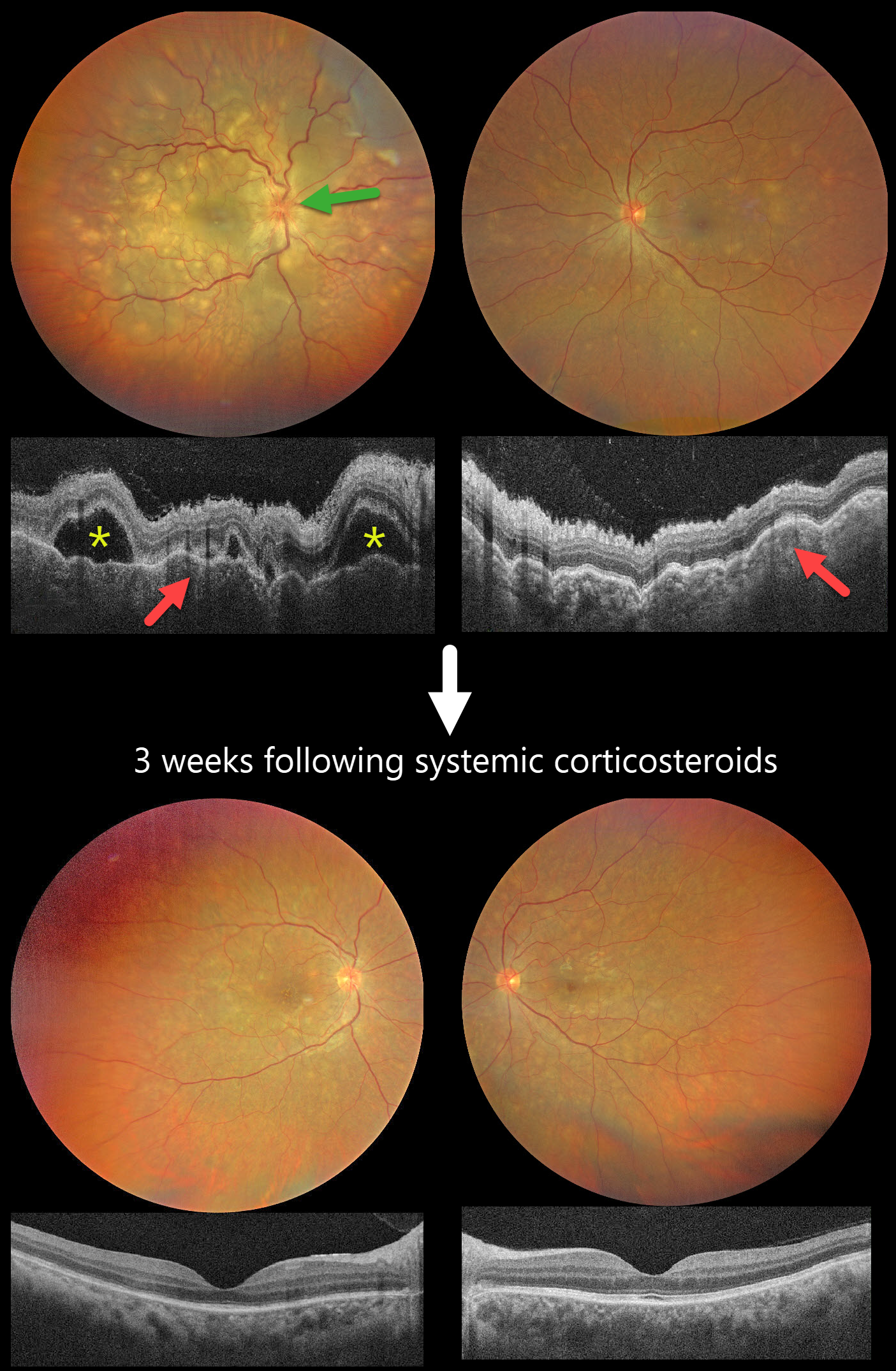
HARADA DISEASE
The European VitreoRetina Society (EVRS) and Ridham Nanda
Originally posted on @retina.rocks January 23, 2026
This previously healthy 40YO female presented with 2 days of blurred vision. She denied any systemic symptoms. Vision was 20/200 OD and 20/60 OS.
Color photography shows multifocal yellow-white subretinal lesions scattered throughout each posterior pole, with the right eye greater than the left, associated with overlying multifocal exudative retinal detachments. The right nerve is also edematous. OCT scanning shows a bilateral thickened choroid with overlying chorioretinal folds and multifocal serous detachments OD.
Intravenous methylprednisolone (1000mg/day) was administered for five days, followed by a 2-week course of oral prednisone (1mg/kg). Three weeks later, the funduscopic and OCT findings normalized. Vision improved to 20/30 OD and 20/20 OS.
Learning Points:
Harada disease is characterized by relatively acute bilateral panuveitis, optic nerve swelling, thickened choroid, chorioretinal folds, multifocal serous exudative retinal detachments, and bacillary layer detachments. Patients with just ocular findings have Harada disease (our patient), whereas those with additional systemic findings (including vitiligo, poliosis, headache, vertigo, and hearing loss) have Vogt-Koyanagi-Harada disease.
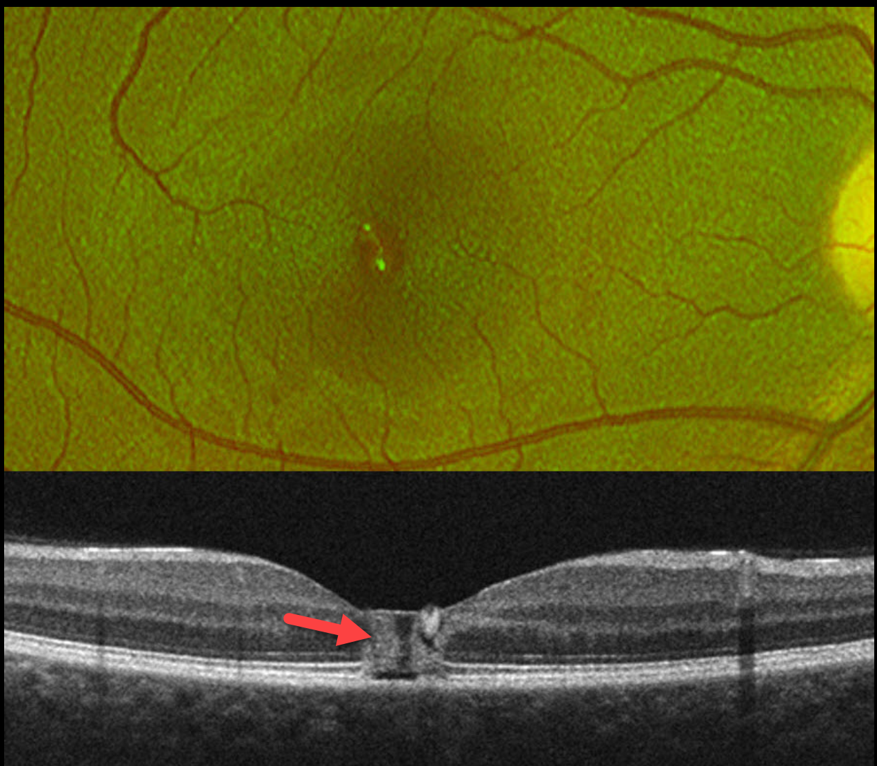
LASER MACULOPATHY
Vaibhav Sethi
Originally posted on @retina.rocks January 20, 2026
This healthy 19YO female noticed immediate blurred vision in her right eye after looking at a laser during a party. Her vision in this eye was 20/60 when she presented to us the following day.
Optos color RG imaging shows a foveal pseudohole. OCT scanning shows a central foveal column of full-thickness hyperreflectivity. There is loss of the outer segment bands overlying the RPE.
Learning Points:
Most cases of laser maculopathy reported in the literature are caused by intentional or inadvertent exposure to laser pointers (Bhavsar et al, Survey Ophthalmology 2021;66:231-260). However, more recently, there have been reports of macular injury from exposure to high-power lasers during concerts or from DJ laser machines (Perz-Montano et al, Doc Ophthalmol 2019;138:71-76). Most eyes recover fairly good vision with conservative management.
Our patient was immediately lost to follow-up.
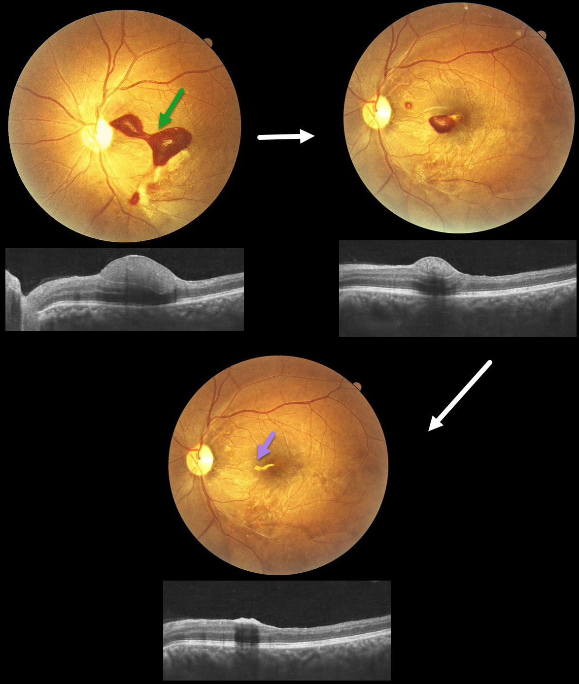
TERSON SYNDROME
Anand Temkar, Surendra Pal, Nagamani Gunasekar
Originally posted on @retina.rocks January 12, 2026
This 32YO male presented with 20 days of vision loss in his left eye. He underwent cardiovascular surgery 3 weeks earlier to remove a clot, followed 1 week later by a subarachnoid hemorrhage. Vision was 20/20 in his normal OD and 20/400 in his OS.
Fundus photography shows a subinternal limiting membrane (ILM) hemorrhage extending from the central macula, superonasally, towards the disc. This blood is hyperreflective on OCT. Observation was recommended.
One month later, vision improved to 20/70, with a marked decrease in sub-ILM blood. One month later, vision improved to 20/30, with near-complete resolution of the blood, except for a small amount of devitalized yellow blood extending nasally from the foveal center.
Learning Points:
Terson syndrome consists of intraocular hemorrhage (vitreous, subhyaloid, or retinal) associated with spontaneous or traumatic subarachnoid hemorrhage. Traditionally, the cause was attributed to elevated intracranial pressure (ICP), which caused cerebrospinal fluid effusion into the optic nerve sheath, compressing the central retinal vein and leading to subsequent rupture of retinal vessels. More recent evidence suggests glymphatic reflux, in which raised ICP causes subarachnoid blood to reflux through glymphatic channels into the globe (Kumaria et al, J Neurology 2022;269:1264-1271).
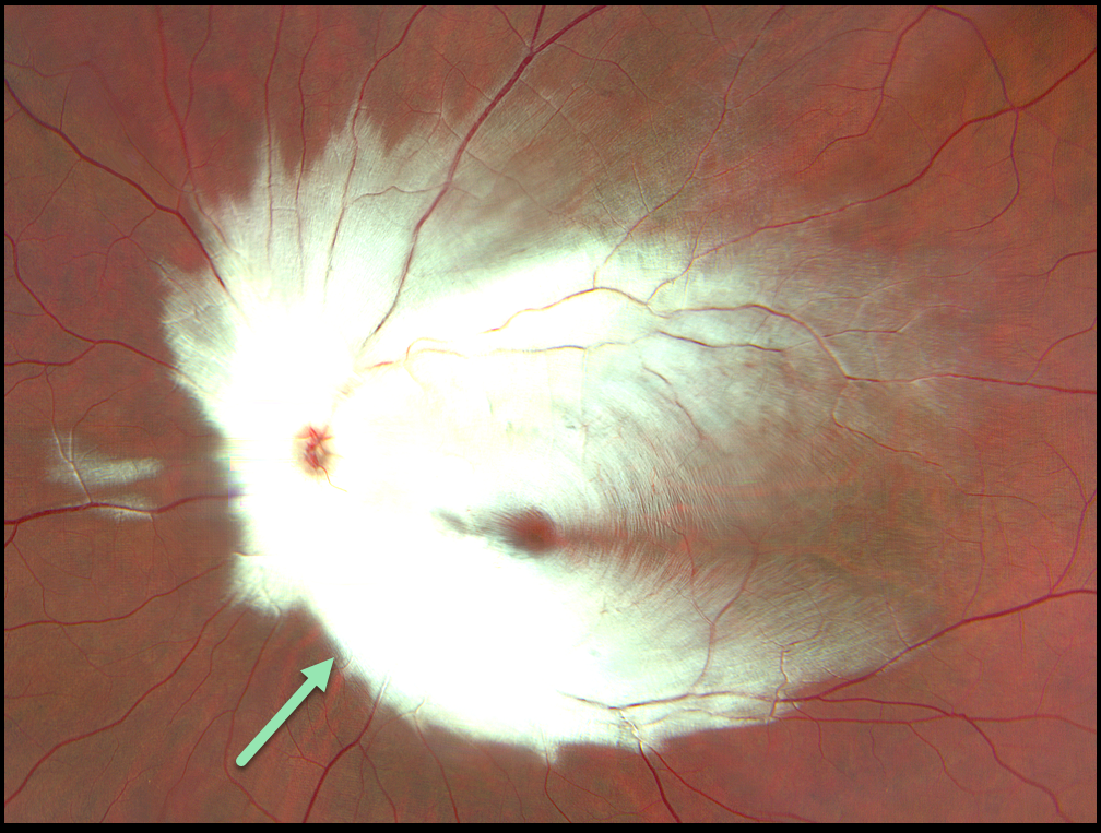
MYELINATED NERVE FIBER LAYER
Originally posted on @retina.rocks January 7, 2026
This healthy 17YO male presented with a history of decreased vision in the left eye since birth. Family and developmental history were unremarkable. Vision was 20/20 in his normal right eye and counting fingers at 10 feet in his left eye.
Color photography shows the myelinated nerve fiber layer (NFL) covering nearly the entire posterior pole and peripapillary region.
Learning Points:
Myelinated NFL occurs when retinal nerve fibers develop a myelin sheath, which usually stops posterior to the lamina cribrosa. The myelination may represent an oligodendrocytic choristoma (Rao et al, Retina 2019;39:1125-1132).
Myelinated NFL is a relatively uncommon, unilateral or bilateral, usually isolated finding present at birth. The myelinated NFL is often connected to the optic nerve. Most patients are asymptomatic, although some may have a corresponding relative scotoma. The myelination can progress in about 10% of cases and can also resolve following retinal ischemic events.
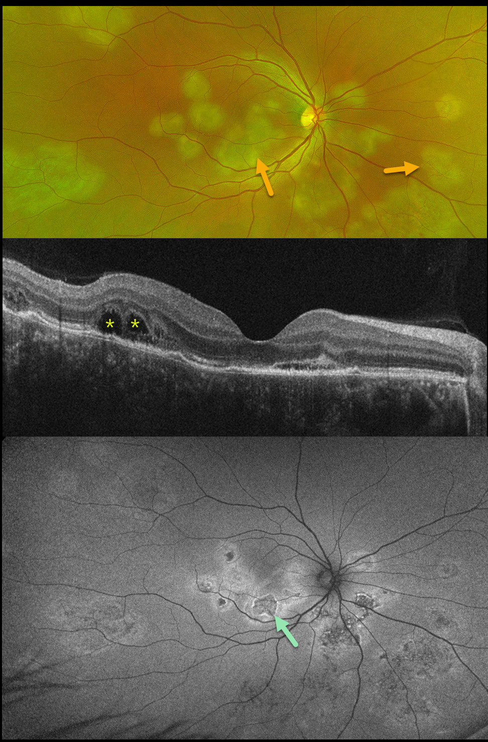
TUBERCULAR SERPIGINOUS-LIKE CHOROIDITIS
The European VitreoRetinal Society (EVRS) and Vaibhav Sethi
Originally posted on @retina.rocks January 2, 2026
This previously healthy 41YO male presented with 4 days of blurred vision in his right eye. Vision was 20/80 OD and 20/20 in his normal OS.
Optos color RG imaging shows multifocal subretinal creamy white placoid lesions in the macula, peripapillary retina, and midperiphery. OCT scanning shows variable disruption of the outer retinal bands, as well as bacillary layer detachments (BALAD) temporally. The placoid lesions on fundus autofluorescence (FAF) show variable central hypo-FAF with more hyper-FAF borders.
Mantoux skin testing was positive at 24mm, and chest CT revealed left lower lobe ground glass opacities with hilar adenopathy. He was started on anti-tubercular therapy and oral prednisone.
Although tuberculosis most commonly presents with pulmonary involvement, extrapulmonary sites can include the gastrointestinal, skin, cardiovascular, genitourinary, and central nervous systems, as well as the eyes. Ocular involvement, like syphilis, can mimic virtually any type of uveitis, including anterior, intermediate, posterior, and panuveitis, retinitis, and retinal vasculitis, neuroretinitis, optic neuropathy, choroidal granuloma, choroiditis, and scleritis.
Learning Points:
Tubercular serpiginous-like choroiditis mimics serpiginous but affects younger patients with more multifocal and peripheral recurrences and progression if untreated (Agarwal et al, AJO 2020;220:160-169). Although the exact mechanism is not yet clear, a direct or indirect infectious trigger by the Mycobacterium tuberculosis is thought to cause choroiditis. Its origin is probably immunogenic since it seems to respond to corticosteroids and other immunosuppressants.
BALAD appears as a unique dome-shaped collection of intraretinal fluid from photoreceptor splitting. These detachments can be observed in an increasing number of conditions, most classically Vogt-Koyanagi-Harada disease (Cicinelli et al, Ophthalmology Retina 2020;4:454-456).
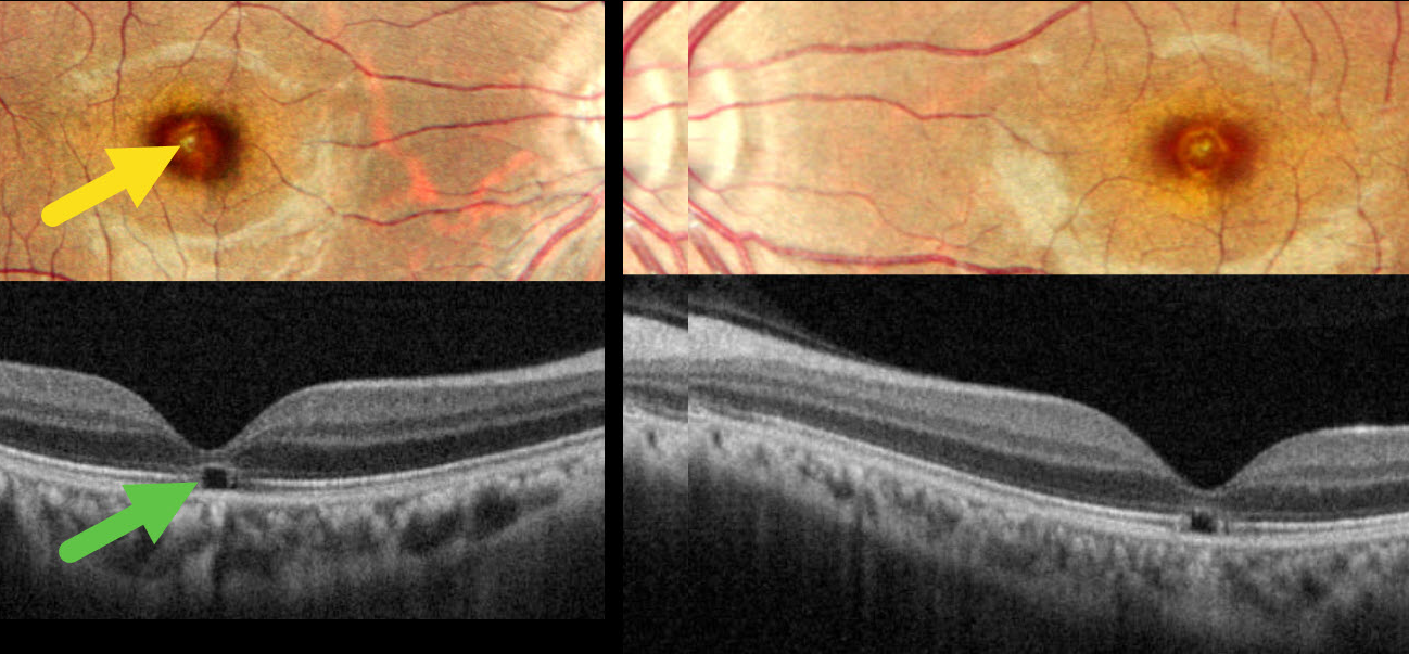
WELDING ARC MACULOPATHY
Malvika Singh and Manish Nagpa
Originally posted on @retina.rocks December 24, 2025
This 31YO male presented with 2 years of bilateral vision loss. He works as a welder and admits to rarely wearing his safety glasses. Vision was 20/30 OU.
Pseudocolor SLO imaging shows symmetrical, tiny yellow foveal scars. OCT shows outer foveal microdefects characterized by hyporeflectivity due to loss of all bands deep to the external limiting membrane.
Learning Points:
The welding process emits optical radiation across various wavelengths and intensities, including infrared, visible, and ultraviolet (UV) light. Exposure to this UV light without proper eye protection can result in keratoconjunctivitis (the most common clinical presentation) and foveal lesions similar to solar retinopathy. Findings include a small yellowish foveal spot or scar with an outer foveal microdefect on OCT (Singh et al, Surv Ophthalmology 2023;68:655-668). Vision is usually good despite the persistent clinical and OCT findings.
Our patient was lucky to have good vision despite his poor work habits. He was advised to use protective eyewear during welding.
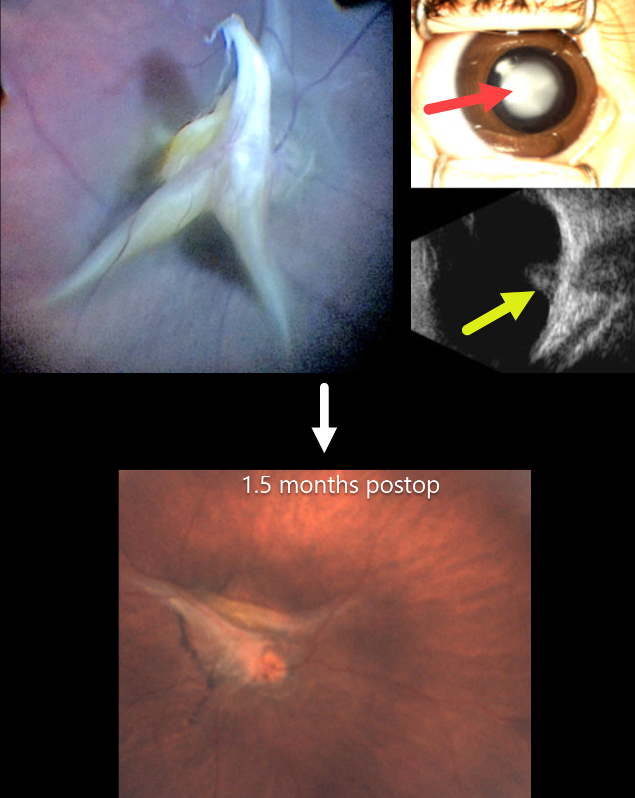
PERSISTENT FETAL VASCULATURE
European VitreoRetina Society (EVRS), Gökçen Deniz and Şengül Özdek
Originally posted on @retina.rocks December 19, 2025
This healthy 4-month-old girl, born full-term, presented with leukocoria in her right eye that her parents recently noticed.
Preoperatively, there was a white central cataract with a clear peripheral zone. B-scan ultrasonography showed a highly reflective stalk that extended from the disc to the periphery with a tent-like elevation over the disc. The left eye was normal.
At surgery, the lens was aspirated with vitrectomy to release all traction. Although the stalk overlying the nerve was trimmed, persistent blood flow was noted as stalk pulsations. The retina remained attached 1.5 months postoperatively.
This case was submitted by the European VitreoRetina Society (EVRS), Gökçen Deniz, and Şengül Özdek .
Learning Points:
Persistent fetal vasculature (PFV), previously recognized as persistent hyperplastic primary vitreous (PHPV), represents a failure of regression of fetal vessels. It is a common cause of leukocoria and subsequent amblyopia. There is no identifiable genetic association.
In PFV, the arterial blood supply from the disc to the anterior segment that is supposed to regress after birth does not. This results in a remnant of the fetal hyaloid artery remaining abnormally patent. The arterial flow from this remnant can cause cataract, intraocular hemorrhages, and retinal detachment, as the abnormal vessel continues to be supplied by systemic arterial pressure.
The main surgical pearl of this case is that, when you have a clear zone around the central opacity of PFV, such eyes usually do not have peripheral retinal extensions through the ciliary body, which allows safe surgical entry through the pars plicata. One still needs to check internally that the incisions are anterior to the peripheral retina, and limbal entry is needed.
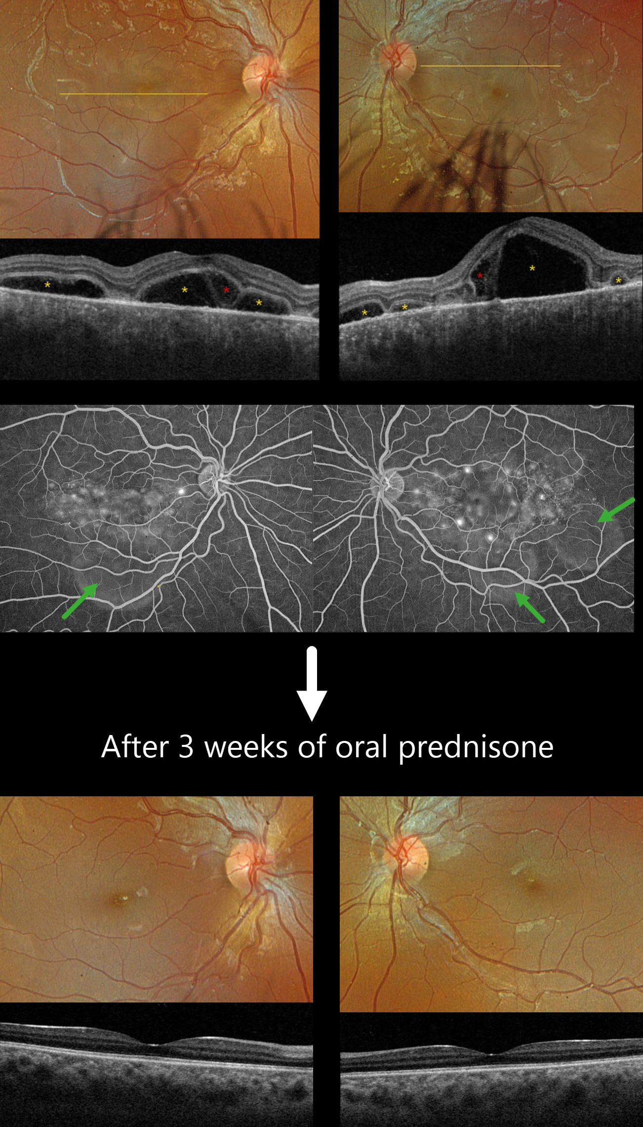
HARADA DISEASE
Originally posted on @retina.rocks December 16, 2025
This previously healthy 13YO girl presented with several days of bilateral vision loss. Vision was 20/100 OD and 20/400 OS.
Optos color RGB imaging shows bilateral multifocal serous exudative retinal detachments. Triton swept-source OCT shows a complex pattern of multifocal pockets of subretinal fluid and bacillary layer detachments (BALAD). The choroid is markedly thickened. Fluorescein angiography shows multifocal pinpoint areas of subretinal leakage with pooling in the subretinal space and BALADs.
Learning Points:
Harada disease is characterized by relatively acute bilateral panuveitis, optic nerve swelling, thickened choroid, chorioretinal folds, multifocal serous exudative retinal detachments, and bacillary layer detachments. Patients with just ocular findings have Harada disease, whereas those with additional systemic findings (including vitiligo, poliosis, headache, vertigo, and hearing loss) have Vogt-Koyanagi-Harada disease.
Our patient was started on oral prednisone 40mg daily, and one week later, vision improved to 20/60 OU with resolution of all exudation (not shown). Three weeks later, the fundus appeared normal, and the choroid was approaching normal thickness. A slow steroid taper was started.
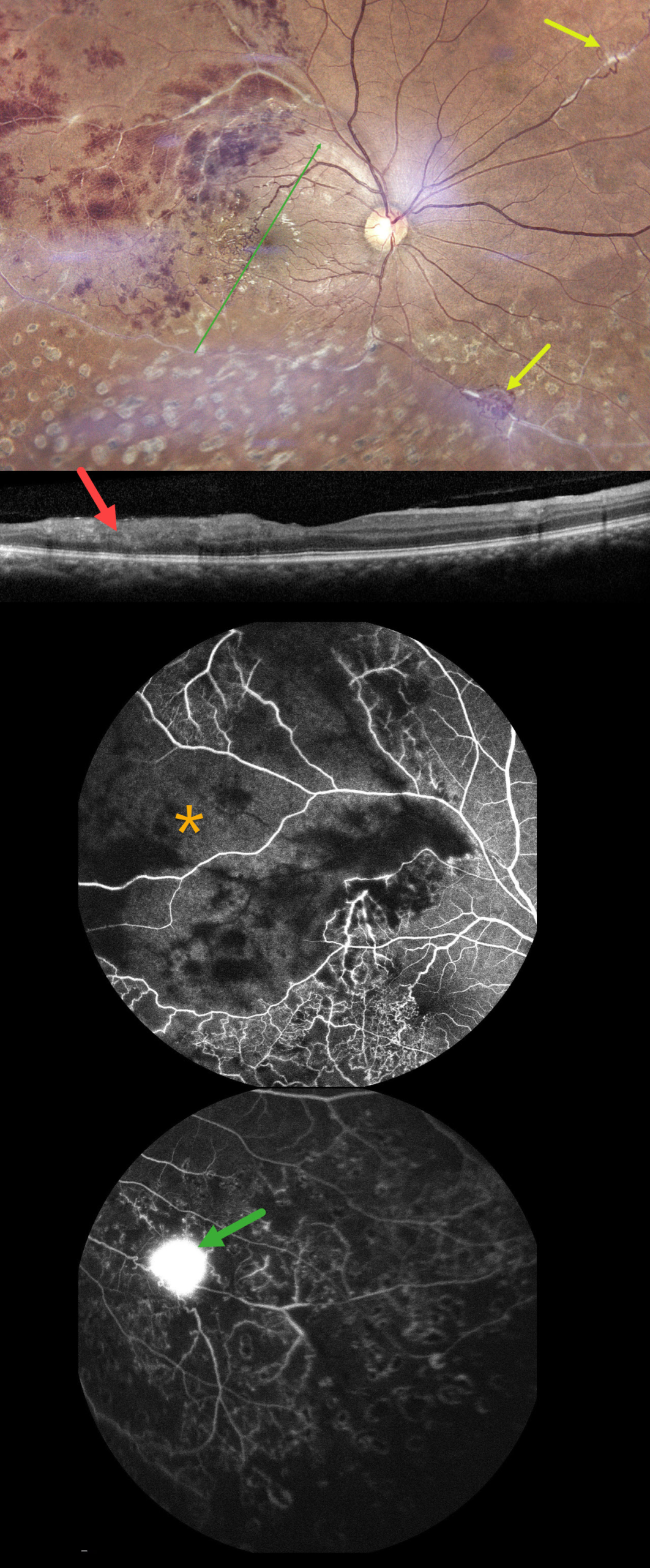
TUBERCULAR ISCHEMIC VASCULITIS
The European VitreoRetina Society (EVRS) and Abhishek Upadhyaya
Originally posted on @retina.rocks November 28, 2025
This 40YO male presented with 10 days of vision loss in his right eye. There was a history of prior laser treatment to this eye elsewhere. Vision was 20/40 OD and 20/20 in his normal OS.
Fundus photography shows superotemporal retinal hemorrhages consistent with a branch retinal vein occlusion (BRVO), with a possible additional BRVO inferotemporally. Collateral vessels are noted in the temporal fovea with scatter laser scarring in the inferior hemiretina. Skip vasculitis can be seen in the superotemporal quadrant along with venous sheathing in other areas as well. There are a few areas of fibrosed peripheral neovascularization (NV). OCT scanning shows variable temporal macular thinning with disorganization of retinal inner layers (DRIL). Fluorescein angiography shows profound ischemia within the superotemporal BRVO, along with vascular leakage and peripheral ischemia. Leakage from the Inferonasal NVE can be seen in the late stages.
Systemic workup was consistent with Ocular Tuberculosis (TB), including a positive Mantoux skin test (12mm), positive Quantiferon TB testing, and calcified lung nodules on contrast-enhanced computed tomography. He was started on oral steroids and antitubercular therapy, and scatter laser was planned for the areas of angiographic ischemia.
Learning Points:
The differential for occlusive peripheral retinal vasculitis (Huvard et al, Ophthalmology Retina 2022;6:43-48) includes TB, rheumatologic disorders/systemic vasculitides (granulomatosis with polyangiitis, Bechet’s disease, systemic lupus, etc.), idiopathic retinal vasculitis and neuroretinitis, and sarcoidosis. Historically, Eales disease has been used to describe a variety of heterogeneous disorders, including tuberculous vasculitis, and, due to its ambiguity, we prefer not to use this term.
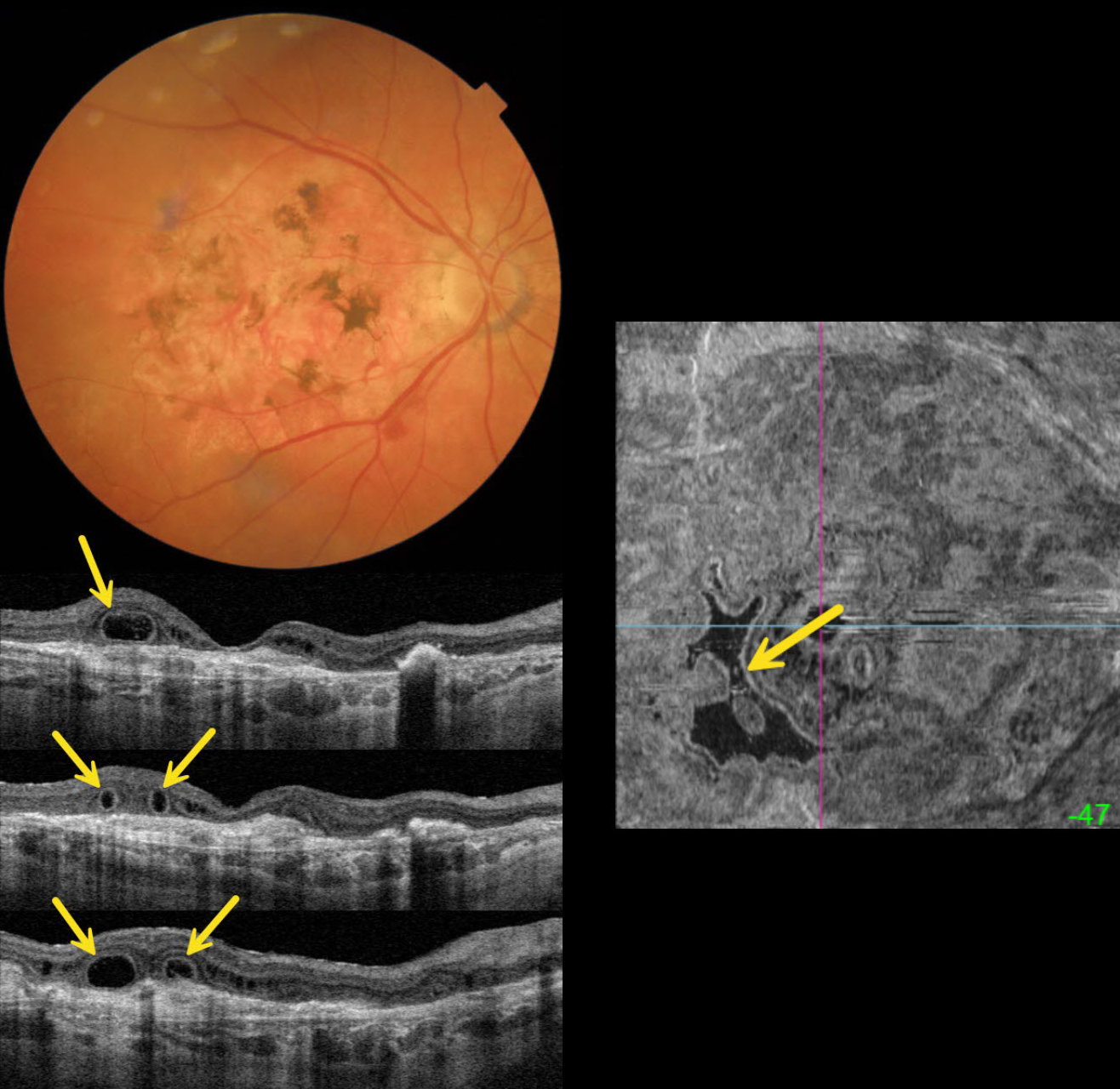
OUTER RETINAL TUBULATION IN NEOVASCULAR AMD
Originally posted on @retina.rocks November 18, 2025
This 89YO female is receiving ongoing intravitreal Eylea in her 20/40 left eye for neovascular AMD. She has a stable 20/400 atrophic disciform scar in her right eye.
Triton color imaging shows a variably pigmented flat disciform scar. Swept-source OCT shows variable outer retinal atrophy overlying a hyperreflective inactive macular neovascularization (MNV). A complex pattern of outer retinal tubulation (ORT) is seen on numerous B-scans. En face OCT shows another view of these meandering tubular structures.
Learning Points:
ORT is often noted overlying inactive MNV and should not be confused with exudative fluid or cysts, which lack a hyperreflective border. The outer hyperreflective band likely represents inner segment mitochondria undergoing fission and translocation toward the nucleus (Litts et al, Retina 2018;38:445-461). ORT, initially described by Zweifel et al (Arch Ophthalmol 2009;127:1596-1602), is a neurodegenerative condition of the photoreceptors and Muller cells associated with atrophy affecting the outer retina and retinal pigment epithelium, including advanced AMD and inherited retinal diseases.
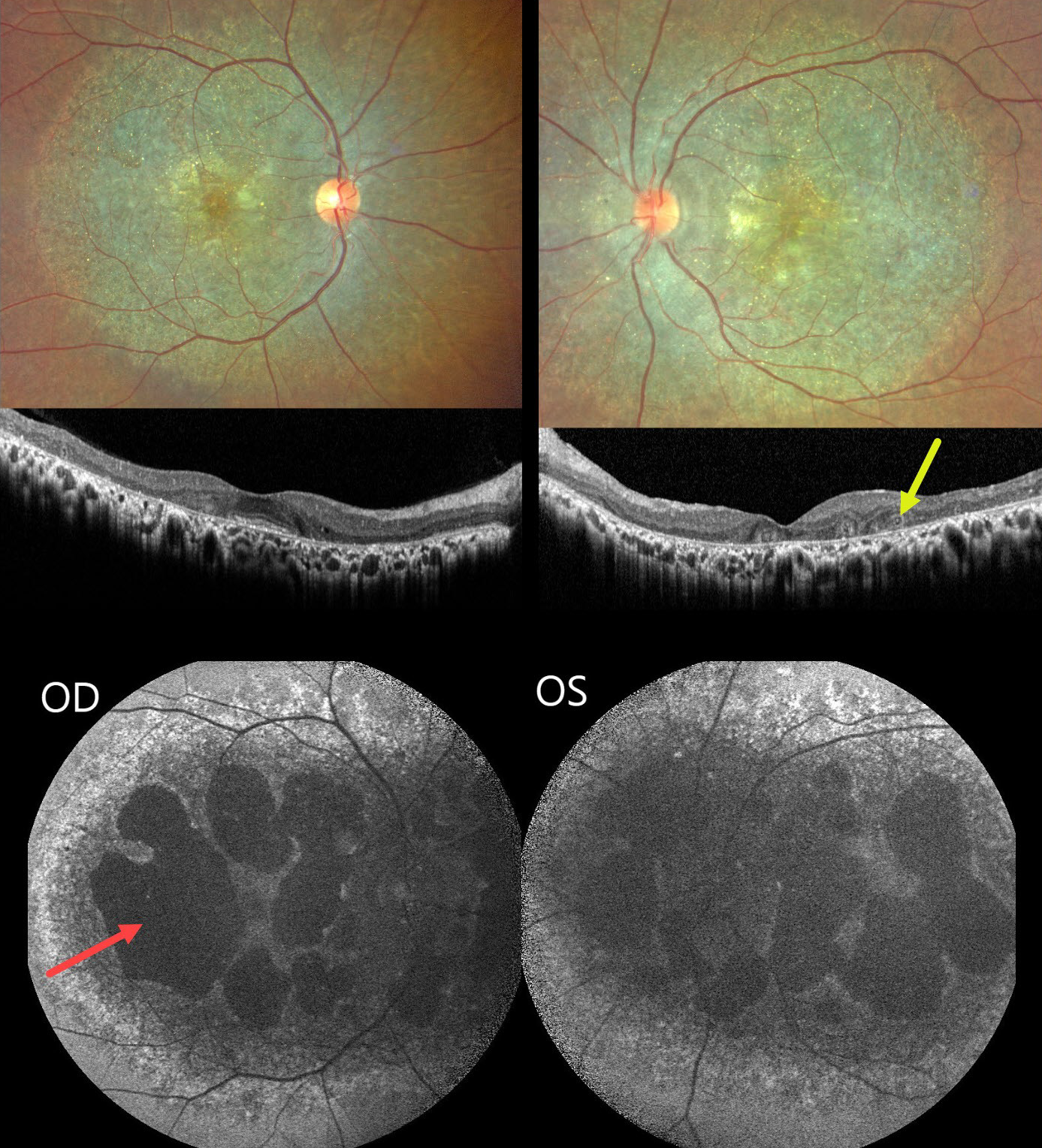
BIETTI CRYSTALLINE DYSTROPHY
The European VitreoRetina Society (EVRS) and Shishir Verghese
Originally posted on @retina.rocks October 24, 2025
This healthy 36YO female presented with a long history of bilateral vision loss with night blindness. Family history was negative. Vision was 20/80 bilaterally. Anterior segments were normal.
Fundus imaging shows extensive pigmentary degeneration with subretinal crystalline deposits involving both posterior poles. OCT scanning shows hyperreflective dots overlying the RPE-Bruch’s membrane complex with variable outer retinal and RPE loss. A few areas of outer retinal tubulation are noted temporally OS. Fundus autofluorescence (FAF) shows diffuse hypo-FAF throughout each posterior pole, dense hypo-FAF within areas of discrete atrophy, and dots of more peripheral hyper-FAF.
Learning Points:
Bietti crystalline dystrophy is an autosomal recessive ocular disorder that affects both the anterior and posterior segments. Clinical features include crystalline deposits in the retina, cornea, and rarely the crystalline lens, as well as retinal pigment epithelial clumping and atrophy. Mutations in CYP4V2 that cause dysregulated lipid metabolism have been implicated in its pathogenesis. While there is no definitive treatment, anti-VEGF therapy can be administered for secondary macular neovascularization. See Saatci et al for a recent review (Clinical Ophthalmology 2023;17:953-967).
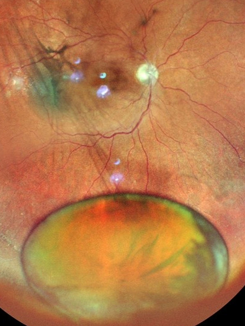
DISLOCATED LENS
The European VitreoRetina Society (EVRS), Akansha Sharma, Manish Nagpal, Malvika Singh and Tejaswita Verma
Originally posted on @retina.rocks October 10, 2025
This 67YO male presented with 3 weeks of decreased vision in his right eye. Vision was 20/120 OD with a +10 diopter sphere and 20/30 in his normal OS. Color photography shows a cataractous lens fully dislocated into the inferior vitreous cavity. A shallow macula-off rhegmatogenous retinal detachment is noted in the posterior pole without apparent breaks. Pars plana vitrectomy was performed the following day, which included lens extraction and retinal detachment repair.
Learning Points:
Posterior dislocation of the crystalline lens can be caused by trauma or by systemic disorders such as Marfan syndrome, homocystinuria, or Weill-Marchesani syndrome. Pars plana vitrectomy is needed for a completely dislocated lens, as with this patient, while lenses that are subluxed may be managed with phacoemulsification or other means.
Our patient’s lens spontaneously dislocated with no history of trauma. Pseudoexfoliation was noted on slit lamp examination of her left lens, which may have predisposed her to lens dislocation.
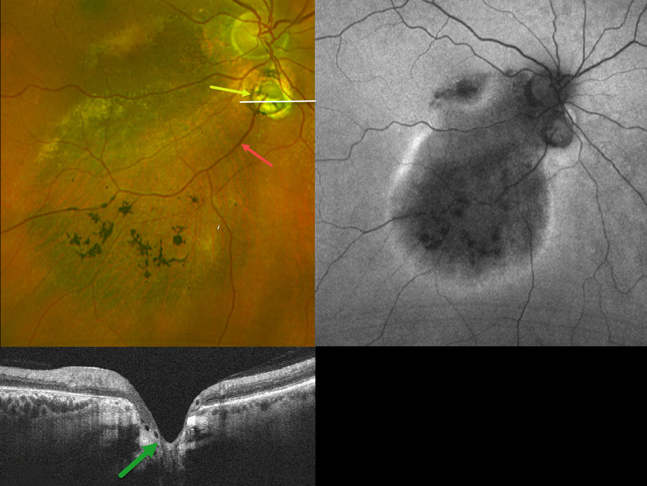
CHOROIDAL COLOBOMA WITH OPTIC NERVE PSEUDODUPLICATION
Originally posted on @retina.rocks October 8, 2025
This 60YO female was referred for asymptomatic unilateral fundus findings in her right eye. Vision was 20/30 OD.
Optos color RG imaging shows a choroidal coloboma just inferior to the disc, giving the appearance of a pseudoduplicated optic nerve. Coarse pigmentary changes with some pigment migration extend from the macula into the inferior midperiphery. An inferotemporal retinal vein drains directly into the choroid through the coloboma. Triton swept-source OCT shows that the coloboma overlies an area of ectatic sclera. Fundus autofluorescence (FAF) shows variable hypo-FAF within the area of clinical pigmentary changes, with a surrounding rim of hyper-FAF.
Learning Points:
Choroidal colobomas are most often due to failure of the optic vesicle and choroidal fissure to close during fetal development. Lesions like this may also be from other causes, including toxoplasmosis. Choroidal coloboma can present as solitary or multiple lesions. Visual prognosis depends on the location and involvement of the optic nerve and macula.
Pseudoduplication of the optic nerve occurs when a round choroidal coloboma or chorioretinal scar about the size of the optic nerve is associated with overlying radiating blood vessels (Bloom et al, Retinal Cases 2022;16:174-176). In our experience, it is most common for toxoplasmosis scars to be located near the optic nerve.
Under normal conditions, the retinal and choroidal circulations remain separate and distinct. Rarely, the two circulations connect directly through a chorioretinal anastomosis. These can occur in chorioretinal scars (typically from inactive toxoplasmosis lesions), in macular telangiectasia, and in disciform scars.
The prominent gutter of inferior pigmentary changes indicates a prior retinal detachment, most likely related to spontaneously resolved fluid associated with the coloboma (Tanaka et al, Ophthalmology Retina 2021;5:702-710). Similar pigmentary changes are seen in central serous retinopathy, although our patient’s macular OCTs showed no pachychoroid features (not shown). Finally, this may represent previously resolved pit-like macular fluid, which is rarely found in the absence of a pit or severe glaucomatous cupping (Fujimoto et al, Ophthalmology Retina 2023;7:811-818).
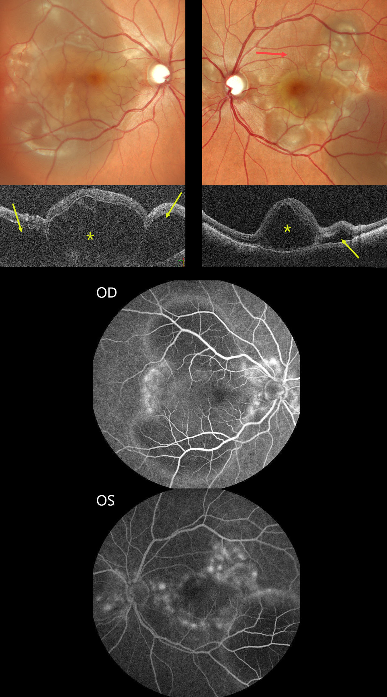
HARADA DISEASE
The European VitreoRetina Society (EVRS), Malvika Singh and Manish Nagpal
Originally posted on @retina.rocks October 3, 2025
This previously healthy 30YO female presented with sudden bilateral vision loss for 5 days. Vision was counting fingers OU.
Color SLO imaging shows bilateral variably confluent serous detachments involving the posterior poles. Retinal folds are also noted in the left superior macula. OCT scanning shows a complex pattern of bacillary layer detachments (asterisks) and subretinal fluid (arrows). Fluorescein angiography shows multifocal subretinal leaks with pooling in the pockets of subretinal and intraretinal spaces.
She was started on oral steroids and immunomodulators in conjunction with a rheumatologist. One month later, vision returned to 20/20 OU with normalization of the funduscopic findings (not shown).
Learning Points:
Harada disease is characterized by relatively acute bilateral panuveitis, optic nerve swelling, thickened choroid, chorioretinal folds, multifocal serous exudative retinal detachments, and bacillary layer detachments. Patients with only ocular findings (as in our patient) have Harada disease, whereas those with additional systemic findings (including vitiligo, poliosis, headache, vertigo, and hearing loss) have Vogt-Koyanagi-Harada disease.
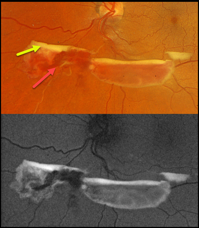
BRVO WITH RED AND YELLOW DEVITALIZED BLOOD
Originally posted on @retina.rocks September 29, 2025
This 48YO female was referred for asymptomatic retinal findings. Vision was 20/20 in the normal OD and 20/20 OS.
Optos color RGB imaging shows red and yellow preretinal blood just inferior to the nerve due to an ischemic extramacular inferonasal branch retinal vein occlusion (BRVO). On fundus autofluorescence (FAF), the red blood is hypo-FAF, and the devitalized yellow blood is markedly hyper-FAF. Intravitreal Avastin was given, and scatter laser was scheduled, although she was immediately lost to follow-up.
Learning Points:
Porphyrins are organic molecules that are part of the heme molecule in hemoglobin. When these compounds break down, the devitalized blood appears yellow and becomes intensely hyperautofluorescent (Bloom and Spaide, Retinal Cases & Brief Reports 2022;16:401-402).
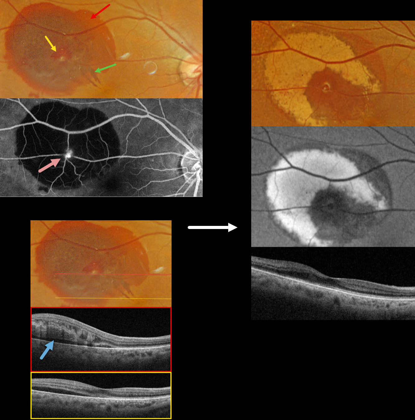
RETINAL ARTERIAL MACROANEURYSM
Originally posted on @retina.rocks September 22, 2025
This 83YO female presented with recent blurred vision in her right eye. Vision was 20/40.
Optos color RGB imaging shows subretinal blood in the superotemporal macula. A small amount of inner vs preretinal blood surrounds a small yellow-white retinal arterial macroaneurysm (RAM). In addition, some radiating outer plexiform blood radiates towards the macular center. Fluorescein angiography shows a hyperreflective, leaking RAM with blockage by surrounding subretinal blood. Swept source OCT shows variable outer retinal edema, hyperreflective blood, and subretinal fluid.
Observation was recommended. Six weeks later, the blood is absorbing and turning yellow superiorly and temporally. This devitalized blood is markedly hyper-autofluorescent. The foveal fluid resolved, and vision improved to 20/30.
Learning Points:
A RAM is thought to occur in a weakened arterial wall from arteriosclerosis and appears as a yellow-white dilated lesion along a retinal arterial within the first 3 bifurcations from the optic nerve. There are 2 distinct presentations, hemorrhagic or exudative. The hemorrhagic type often presents, as in this case, with pathognomonic multiple layers of blood. Acute macular exudation can be treated with observation, anti-VEGF injections, or thermal laser
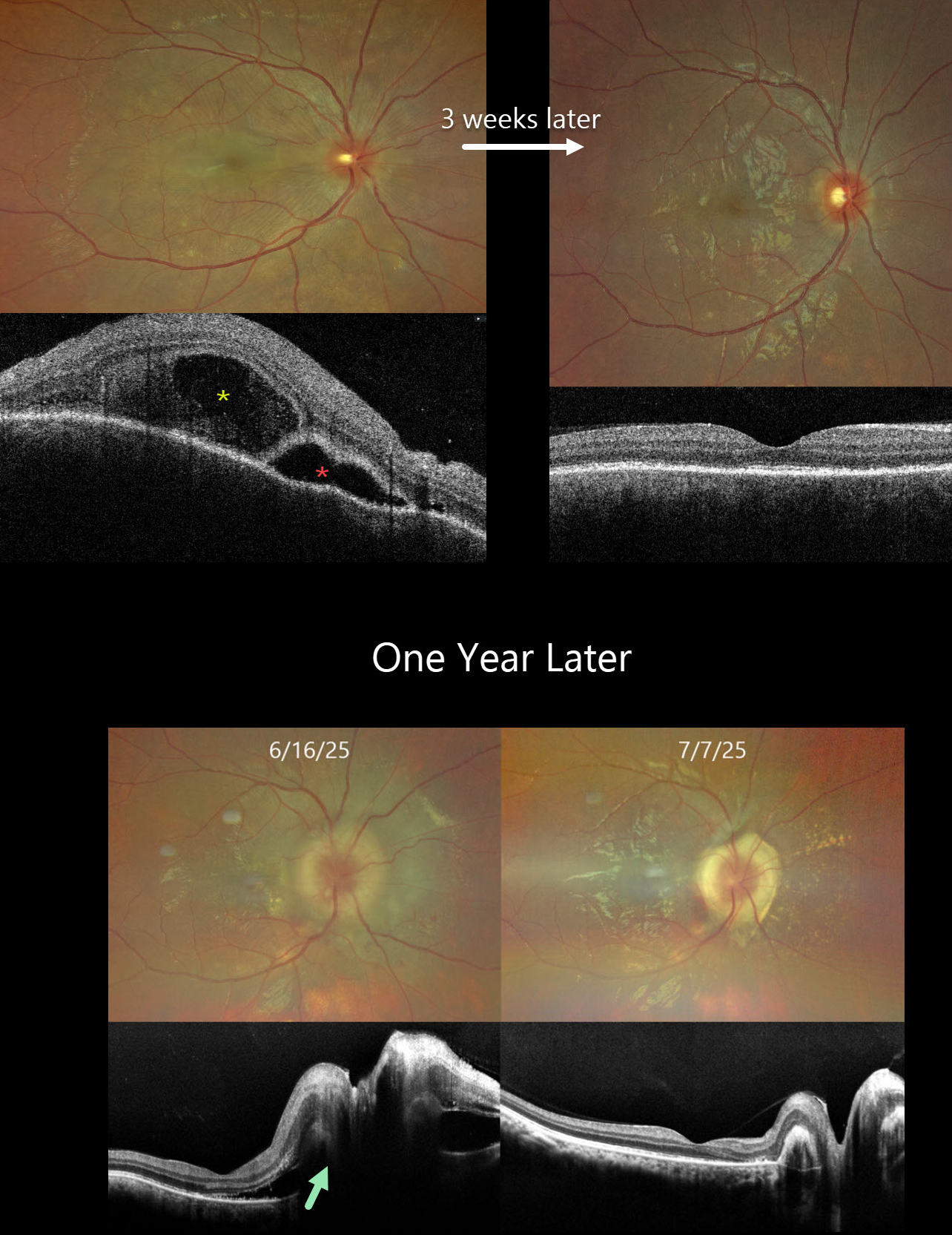
HARADA DISEASE
Nilesh Kumar
Originally posted on @retina.rocks August 20, 2025
This previously healthy 24YO male presented with acute vision loss in his right eye. Vision was 20/200 OD and 20/20 in his normal OS. There were no neurologic or systemic symptoms.
Color photography shows an exudative retinal detachment involving the posterior pole. OCT shows a bacillary layer detachment, subretinal fluid with thickened retina and choroid. He was diagnosed with unilateral Harada disease and started on oral prednisone. Three weeks later, vision improved to 20/25 OD with marked clinical improvement. The oral prednisolone was tapered weekly over the next 6 weeks and stopped.
He returned one year later with recurrent vision loss. Vision was 20/200 OD. Exudative fluid with distal lipid flecks and a few small retinal hemorrhages surrounded the right nerve. OCT confirmed marked disk and peripapillary thickening with subretinal fluid. Oral prednisone was restarted, and oral azathioprine therapy was added. Three weeks later, the vision improved to 20/30. The fluid completely resolved with a ring of subretinal fibrosis surrounding the disc. The oral prednisone was tapered weekly over the next 6 weeks, and the oral azathioprine is planned to continue for the next year with regular monitoring.
Learning Points:
Harada disease is characterized by relatively acute bilateral panuveitis, optic nerve swelling, thickened choroid, chorioretinal folds, multifocal serous exudative retinal detachments, and bacillary layer detachments. Patients with just ocular findings have Harada disease, whereas those with additional systemic findings (including vitiligo, poliosis, headache, vertigo, and hearing loss) have Vogt-Koyanagi-Harada (VKH) disease. Although classically bilateral, early stages may present unilaterally before involving the fellow eye.
The disease generally progresses through four clinical stages: Prodromal, resembling a viral illness with systemic symptoms (fever, headache, meningismus); Uveitic, characterized by blurred vision, photophobia, optic disc edema, posterior choroidal thickening, and multifocal serous retinal detachments; Chronic/Convalescent, where depigmentation of the choroid leads to a “sunset-glow fundus” along with cutaneous signs like vitiligo, poliosis, and alopecia; and Recurrent, marked by repeated bouts of anterior uveitis and complications such as cataract, glaucoma, or macular neovascularization.
Management centers on high-dose systemic corticosteroids, often followed by steroid-sparing immunosuppressive agents to control inflammation and prevent recurrences. Early, aggressive therapy improves visual outcomes.
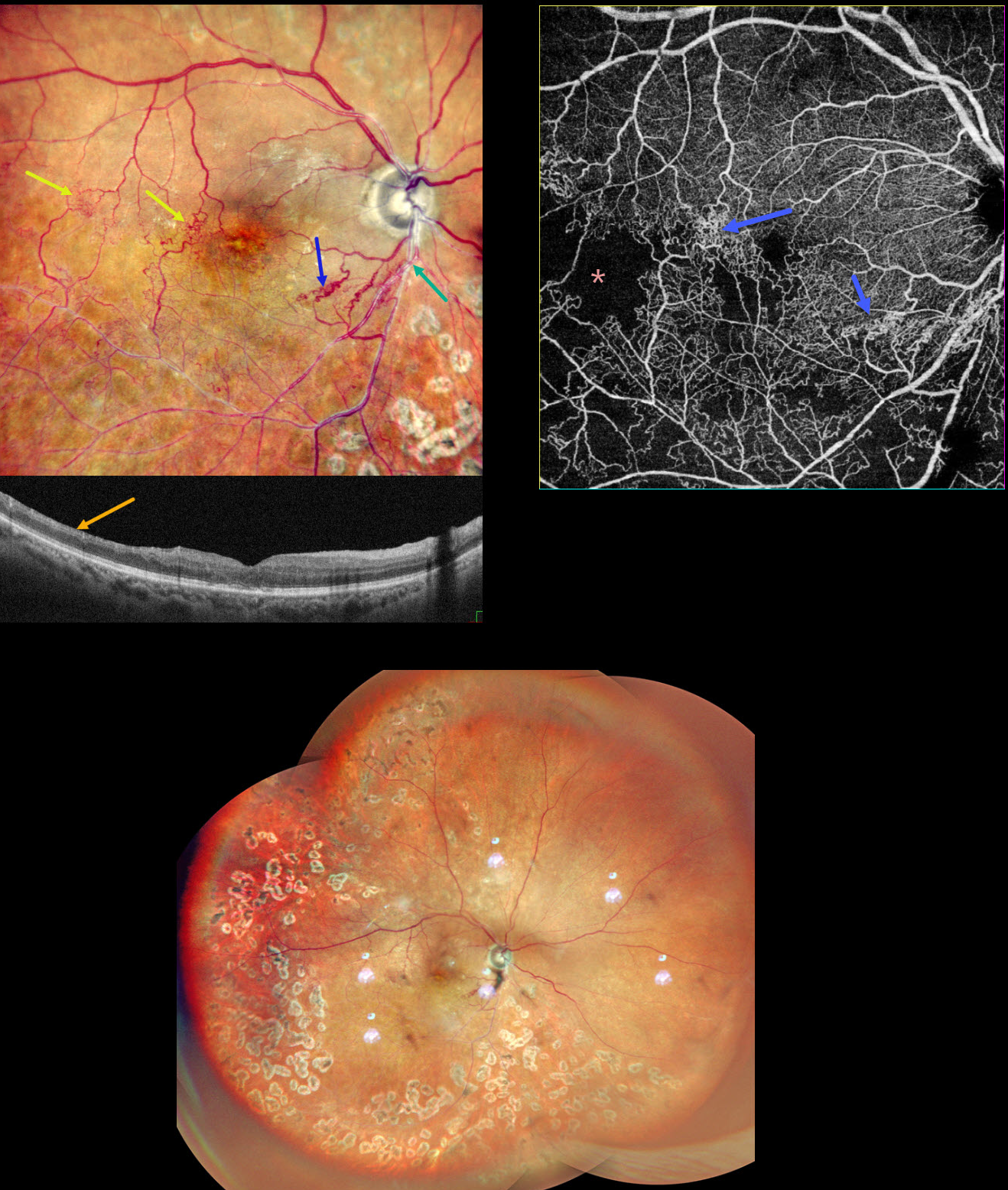
BRVO WITH COLLATERALS AND DRIL
Malvika Singh, Akansha Sharma and Manish Nagpal
Originally posted on @retina.rocks August 19, 2025
This 65YO male presented with a branch retinal vein occlusion (BRVO) in his right eye and 20/40 vision. He was treated elsewhere about a year ago with 3 intravitreal injections and scatter laser 7 years earlier.
Pseudocolor SLO shows an old major inferotemporal ischemic BRVO, which arises from an arteriovenous crossing just inferior to the disc. Extensive collateral vessels bypass the occlusion and cross the temporal horizontal raphe. OCT scanning shows thinned, featureless inner retina (disorganization of retinal inner layers, DRIL). Scatter laser is noted temporally and inferiorly. OCT angiography shows variable capillary loss throughout the BRVO distribution and highlights the collateral vessels.
Learning Points:
Collateral vessels are dilated, pre-existing vessels that bypass an occlusion. These are most commonly seen in the posterior pole following retinal venous occlusions and are exceedingly rare with arterial occlusions. The endothelial tight junctions are intact, so they don’t leak angiographically. This helps differentiate them from neovascularization, which does leak.
DRIL is seen in various retinal vascular disorders, including diabetic macular edema and retinal vein occlusions. It is generally associated with worse vision and increased anti-VEGF treatment burden (see Babiuch et al, JAMA Ophthalmol 2019;137:38-46).
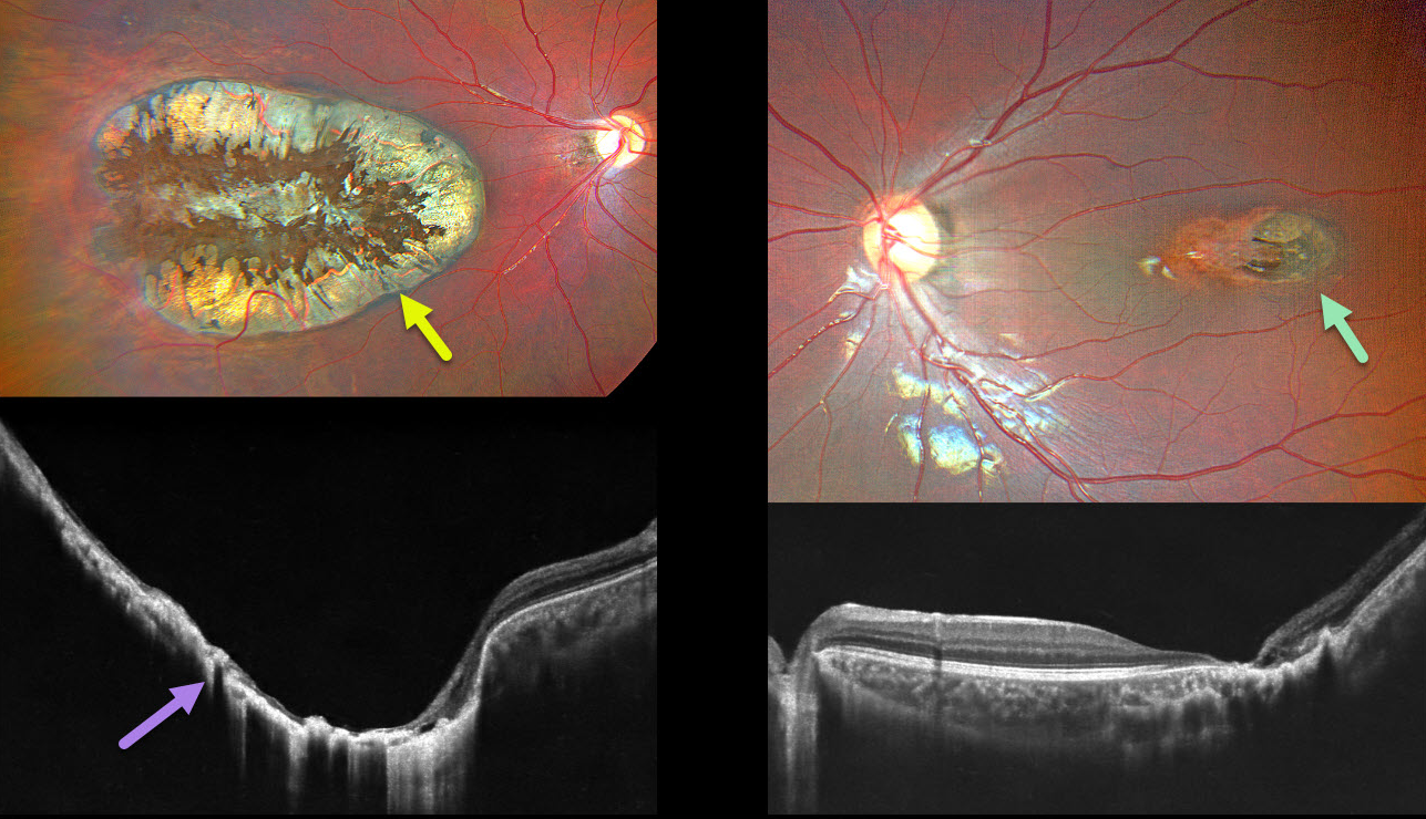
CHOROIDAL COLOBOMA
Ayushi Gupta and Vishal Agrawal
Originally posted on @retina.rocks August 4, 2025
This 7YO healthy boy has a history of esotropia in his amblyopic right eye. Vision was light perception OD and 20/40 OS.
Color photography of his right eye shows a large macular coloboma occupying nearly the entire macula. OCT scanning shows near complete atrophy and disorganization of the overlying RPE and neurosensory retina. A much smaller lesion is noted in the left temporal macula. Observation was recommended.
Learning Points:
Choroidal colobomas are most commonly caused by failure of the optic vesicle and choroidal fissure to close during embryonic development and are usually found inferonasally. They can present as solitary or multiple lesions. The differential diagnosis for congenital macular coloboma also includes post-inflammatory lesions (toxoplasmosis, cytomegalovirus, and Zika) and retinal dystrophies, including North Carolina macular dystrophy.
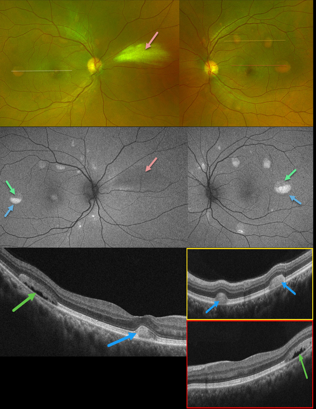
BEST DISEASE WITH MYELINATED NERVE FIBER LAYER
The European Society (EVRS) and Vaibhav Sethi
Originally posted on @retina.rocks July 25, 2025
This 39YO male presented with asymptomatic retinal lesions. Family history was negative. Vision was 20/20 OU.
Optos color RG imaging shows bilateral partially scrambled vitelliform lesions in each posterior pole. On OCT, the superior, scrambled portion of these lesions is hyporeflective, and the inferior, yellow portion is hyperreflective. On fundus autofluorescence (FAF), the superior portions are hypo-FAF, and the inferior vitelliform material is hyper-FAF. The incidental area of the nasal myelinated nerve fiber layer OD is hypo-FAF from blocking the underlying RPE.
Learning Points:
Vitelliform lesions are characterized by the accumulation of yellow subretinal material thought to be caused by impaired metabolism of photoreceptors or retinal pigment epithelium. They can be associated with a wide variety of retinal disorders, including Best vitelliform macular dystrophy (BVD, our patient), adult-onset foveomacular dystrophy, acute exudative polymorphous vitelliform maculopathy, as well as paraneoplastic, toxic, tractional, and degenerative etiologies (Iovino et al, Surv Ophthalmology 2023;68:361-379).
BVD is usually transmitted as an autosomal-dominant disorder due to a mutation in the BEST1 gene, which encodes bestrophin-1. Bestrophin-1, a calcium-activated chloride channel, is primarily found in the basolateral plasma membrane of the RPE. Unfortunately, our patient denied genetic testing due to cost.
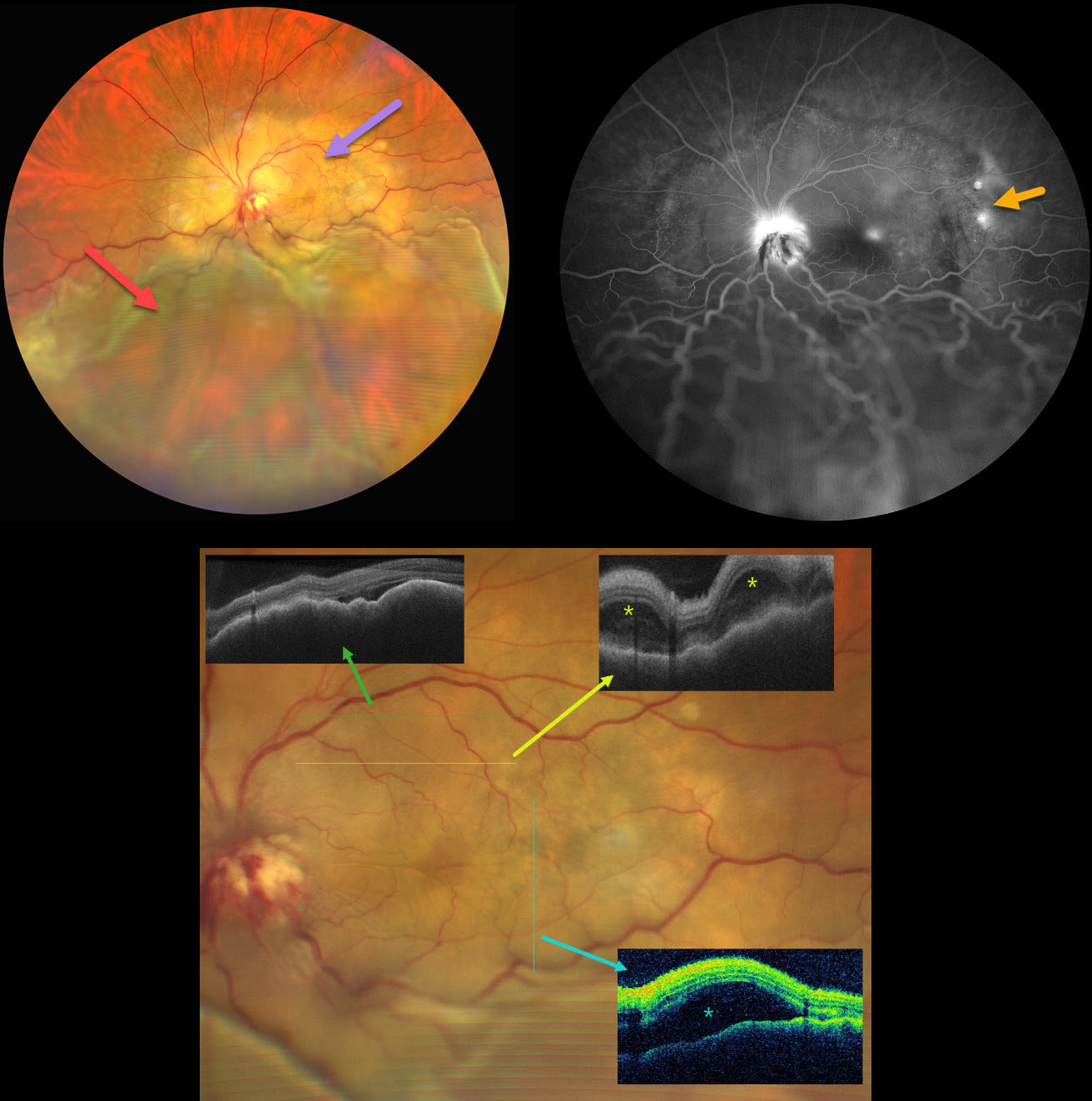
CHOROIDAL METASTASES WITH EXUDATIVE RETINAL DETACHMENT
Ayushi Gupta and Vishal Agrawal
Originally posted on @retina.rocks June 18, 2025
This 47YO female with a known history of metastatic breast carcinoma presented with several weeks of decreased vision in her left eye. She was on active chemotherapy and hormonal therapy. Vision was no light perception OD following trauma 30 years earlier, and hand motion OS.
Ultrawidefield color photography shows a large, amelanotic yellow choroidal lesion involving the entire macula and peripapillary choroid, which extends into the midperiphery. A large secondary inferior exudative retinal detachment involves the entire inferior hemisphere. This fluid shifted when laid supine. The optic disc is hyperemic with indistinct margins and associated blood.
OCT scanning shows a thickened, undulating choroid with variable overlying subretinal fluid and bacillary layer detachments. Fluorescein angiography shows multifocal subretinal leaks, choroidal lesion staining with possible overlying pooling, and intense nerve leakage.
Learning Points:
Although uveal melanoma is the most common primary intraocular malignancy, metastatic choroidal tumors are the most common intraocular malignancies. Lung and breast cancer are the most common sources.
Our patient had a known history of metastatic disease when she presented with this large lesion occupying her entire posterior pole. The extent of the metastasis and the overlying exudation are atypical in our experience. She was immediately referred to her oncologist for external-beam radiation, but was lost to follow-up.
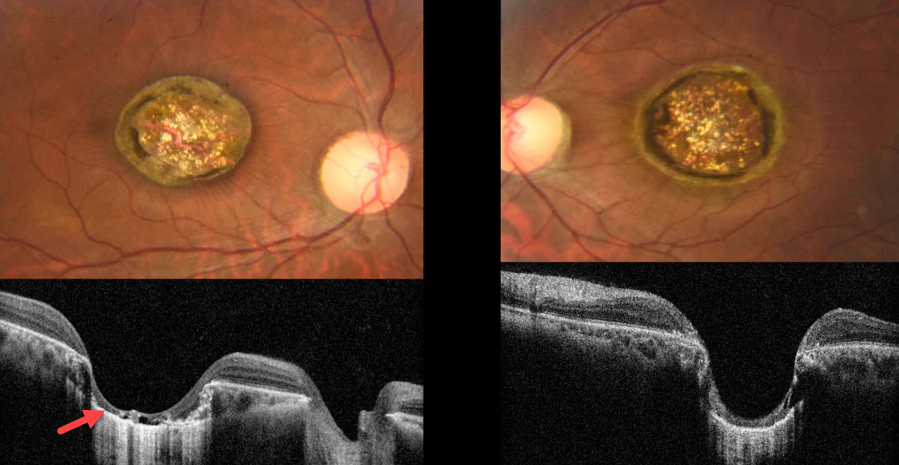
MACULAR COLOBOMA
Mauli Shah and Alay Banker
Originally posted on @retina.rocks May 6, 2025
This healthy 21YO male presented with a lifelong history of stable bilateral vision loss. There was no significant ocular family history, and ocular examination of family members revealed no abnormalities. Vision was 20/400 OU. There was bilateral horizontal nystagmus.
Color photography shows fairly symmetrical macular colobomas. Fine inner retinal striae radiate outwards from the coloboma OD. OCT scanning shows near-total choroidal loss within each coloboma, with a thinned, atrophic retina draped against bare sclera.
Learning Points:
Choroidal colobomas are most commonly due to failure of the optic vesicle and choroidal fissure to close during embryonic development and are usually found inferonasally. They can present as solitary or multiple lesions. Macular colobomas are most commonly either post-inflammatory (toxoplasmosis, cytomegalovirus, and Zika) or from an inherited retinal disorder (IRD), including North Carolina macular dystrophy (NCMD).
The symmetry of our patient’s lesions argues for an IRD. However, we doubt this is NCMD, given the lifelong history of poor vision and nystagmus. Due to financial constraints, the patient refused all blood work and genetic testing. It is
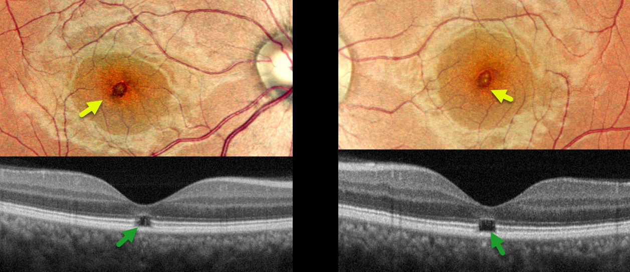
WELDING ARC MACULOPATHY
Rohan Jain and Manish Nagpal
Originally posted on @retina.rocks May 1, 2025
This 38YO male presented with 6-7 months of bilateral vision loss. He works as a welder and admits to rarely wearing his safety glasses. Vision was 20/25 OD and 20/20 OS.
Pseudocolor SLO imaging shows symmetrical, tiny yellow foveal scars. OCT shows outer foveal microdefects with hyporeflectivity due to loss of all bands deep to the external limiting membrane.
Learning Points:
The welding process emits optical radiation across various wavelengths and intensities, including infrared, visible, and ultraviolet (UV) light. Exposure to this UV light without proper eye protection can result in keratoconjunctivitis (the most common clinical presentation) and foveal lesions similar to those seen in solar retinopathy. Findings include a small foveal yellowish spot or scar with an outer foveal microdefect on OCT (Singh et al, Surv Ophthalmology 2023;68;655-668). Vision is usually good despite the persistent clinical and OCT findings.
Our patient was lucky to have good vision despite his poor work habits. He was advised to use protective eyewear during welding.
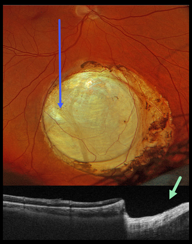
CHOROIDAL COLOBOMA
Originally posted on @retina.rocks April 8, 2025
This healthy 48YO male was referred for an asymptomatic lesion in his right eye. Vision was 20/20 OD and 20/20 in his healthy OS.
Optos color RGB imaging shows an oval choroidal coloboma in the inferior midperiphery. Variably pigmented chorioretinal atrophy surrounds the lesion. Triton swept-source OCT shows a fairly abrupt and near-complete loss of the neurosensory retina within the coloboma.
Learning Points:
Choroidal colobomas are most often due to failure of the optic vesicle and choroidal fissure to close during fetal development. Lesions like this may also be from other causes, including toxoplasmosis. Choroidal coloboma can present as solitary or multiple lesions. Visual prognosis depends on the location and involvement of the optic nerve and macula.
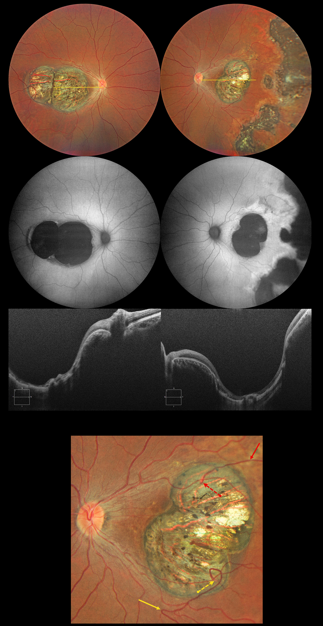
CHOROIDAL COLOBOMA
Evgenii Chernov
Originally posted on @retina.rocks March 18, 2025
This 32YO female has a history of congenital nystagmus and poor vision since birth. Vision was 20/130 OU.
Color photography shows bilateral macular colobomas with more peripheral multifocal variably pigmented colobomatous scarring in her left eye. These lesions are hypoautofluorescent. OCT scanning through the macular lesions shows thinned hyperreflective retina draped across the posteriorly bowed sclera.
The right macular coloboma also contains several retinal choroidal anastomoses. The major inferotemporal vein acutely bends superotemporally to enter the coloboma and drain into the choroidal circulation. A superotemporal vein also enters the coloboma to drain into a visible choroidal vortex.
Learning Points:
Choroidal colobomas are most commonly caused by failure of the optic vesicle and choroidal fissure to close during embryonic development and are usually found inferonasally. They can present as solitary or multiple lesions. The differential diagnosis for congenital macular coloboma also includes post inflammatory lesions (toxoplasmosis, cytomegalovirus, and zika) and retinal dystrophies including North Carolina macular dystrophy. The retina within the coloboma is variably thinned, which increases the risk for rhegmatogenous retinal detachment.
Under normal conditions the retinal and choroidal circulations remain separate and distinct. Rarely, the two circulations directly connect with each other through a chorioretinal anastomosis. These can occur in chorioretinal scars (typically from inactive toxoplasmosis lesions), in macular telangiectasia, and in disciform scars.
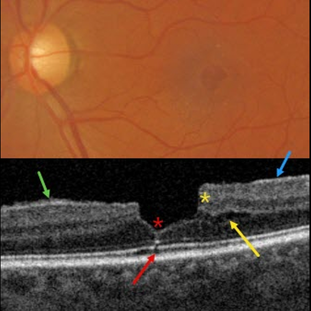
MACULAR PUCKER
Originally posted on @retina.rocks February 19, 2025
This patient presented with 20/30 vision due to a mildly symptomatic clinical macular pucker with a pseudohole; observation was recommended. Multimodal imaging reveals extensive pucker-related findings.
Triton color imaging shows an irregular macular pseudohole with a faint surrounding epiretinal membrane (ERM). True full-thickness macular holes are virtually always perfectly round, and these pucker-related pseudoholes are often irregularly round, as in this eye.
Swept-source OCT shows an adherent ERM with some shaggy-appearing adherent vitreous cortex, favoring epimacular proliferation. The fovea has a vertical contour, often present with ERM, due to the encircling membrane contracting towards the foveal center, dragging the inner retina centrally.
There are features of both a tractional lamellar macular hole (LMH) with splitting of the outer plexiform layer, along with a degenerative LMH with descending loss of tissue into the outer nuclear layer (Govetto et al, AJO 2016;164:99-109).
Finally, a tiny area of shadowing vs an outer foveal microdefect (Cohen et al, Ophthalmology Retina 2021;5:553-561) is seen.
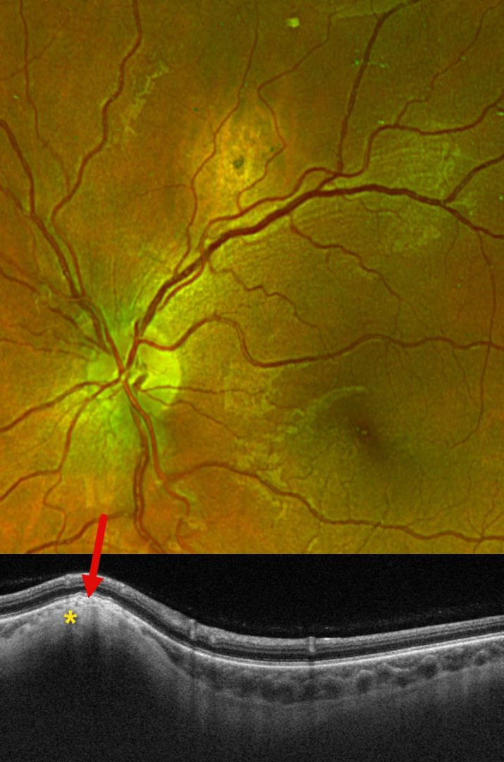
FOCAL SCLERAL NODULE
Originally posted on @retina.rocks February 13, 2025
This 29YO female presented with this asymptomatic amelanotic lesion above the right superotemporal arcade. Optos color RG imaging shows an apparent mostly depigmented chorioretinal scar. However, Triton swept-source OCT scanning shows hyperreflective scleral thickening, which compresses the overlying choroid and elevates the retina.
Learning Points:
Focal scleral nodule, previously known as solitary idiopathic choroiditis (SIC) and unifocal helioid choroiditis, presents as a single, yellow-white, elevated nodular, avascular subretinal lesion. On OCT, it arises from the inner sclera, causing marked choroidal compression. These lesions require no treatment, although they may rarely show slight growth (Fung and Li, Retinal Cases & Brief Reports 2024;18:526-528).
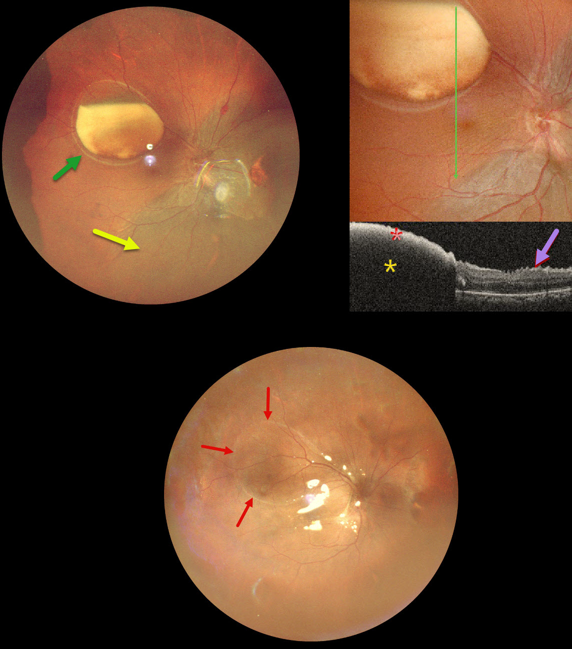
RHEGMATOGENOUS RETINAL DETACHMENT WITH SUB-ILM HEMORRHAGE
Originally posted on @retina.rocks February 12, 2025
This 39YO male underwent vitrectomy OD elsewhere for a vitreous hemorrhage one month earlier. The treating surgeon noticed a sub-internal limiting membrane (ILM) hemorrhage at a postoperative visit and referred him to us for further evaluation. Vision was 20/30 OD and 20/20 in his normal OS.
Ultrawidefield pseudocolor SLO imaging shows a large partially layered sub-ILM hemorrhage with devitalized yellow blood in the superotemporal midperiphery. A retinal detachment is noted inferiorly and nasally. Inner retinal folds are visible at higher magnification. The blood is highly reflective on OCT scanning with total posterior shadowing. The inner retinal folds are noted inferiorly.
We don’t have a good explanation for our patient’s sub-ILM hemorrhage following vitrectomy for a vitreous hemorrhage. The yellow devitalized blood must have been present preoperatively, even though the initial surgeon told us he just noticed it one month postoperatively. Possibly our patient developed a breakthrough vitreous hemorrhage from a Valsalva maneuver, but without further history, this is just speculative.
We repaired the detachment with vitrectomy and silicone oil, and drained the sub-ILM blood intraoperatively. The retina was completely attached 2 weeks later. A high-water mark defines the margins of the prior ILM detachment.
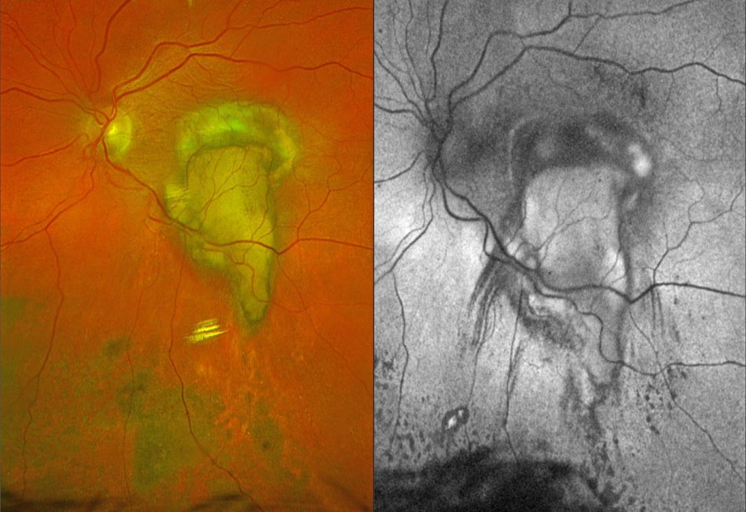
NEOVASCULAR AMD
Originally posted on @retina.rocks February 5, 2025
This 79YO female underwent vitrectomy for a breakthrough vitreous hemorrhage from neovascular AMD. Vision was 20/400.
Postoperative Optos color RGB imaging shows an oval fibrotic macular scar with a tongue-like projection of organized devitalized yellow blood extending inferiorly. Coarse subretinal pigment extends into the inferior midperiphery. On fundus autofluorescence (FAF), the fibrotic portion of the scar and inferior subretinal pigment are hypo-FAF, and the devitalized blood is hyper-FAF. Steaks of inferiorly radiating hypo-FAF also extend from the tongue-like yellow blood.
Learning Points:
This complex healing neovascular lesion has many signs of chronicity. There is no fresh, red blood. The remaining blood is yellow and devitalized, developing as heme products degrade. The coarse peripheral subretinal pigment is a marker of prior resolved subretinal blood and appears on OCT as subretinal hyperreflective material. Finally, the subretinal fibrosis where the breakthrough blood originated represents the usual endpoint of untreated neovascular disease, which is thankfully much less common since the advent of anti-VEGF therapy in the mid-2000s.
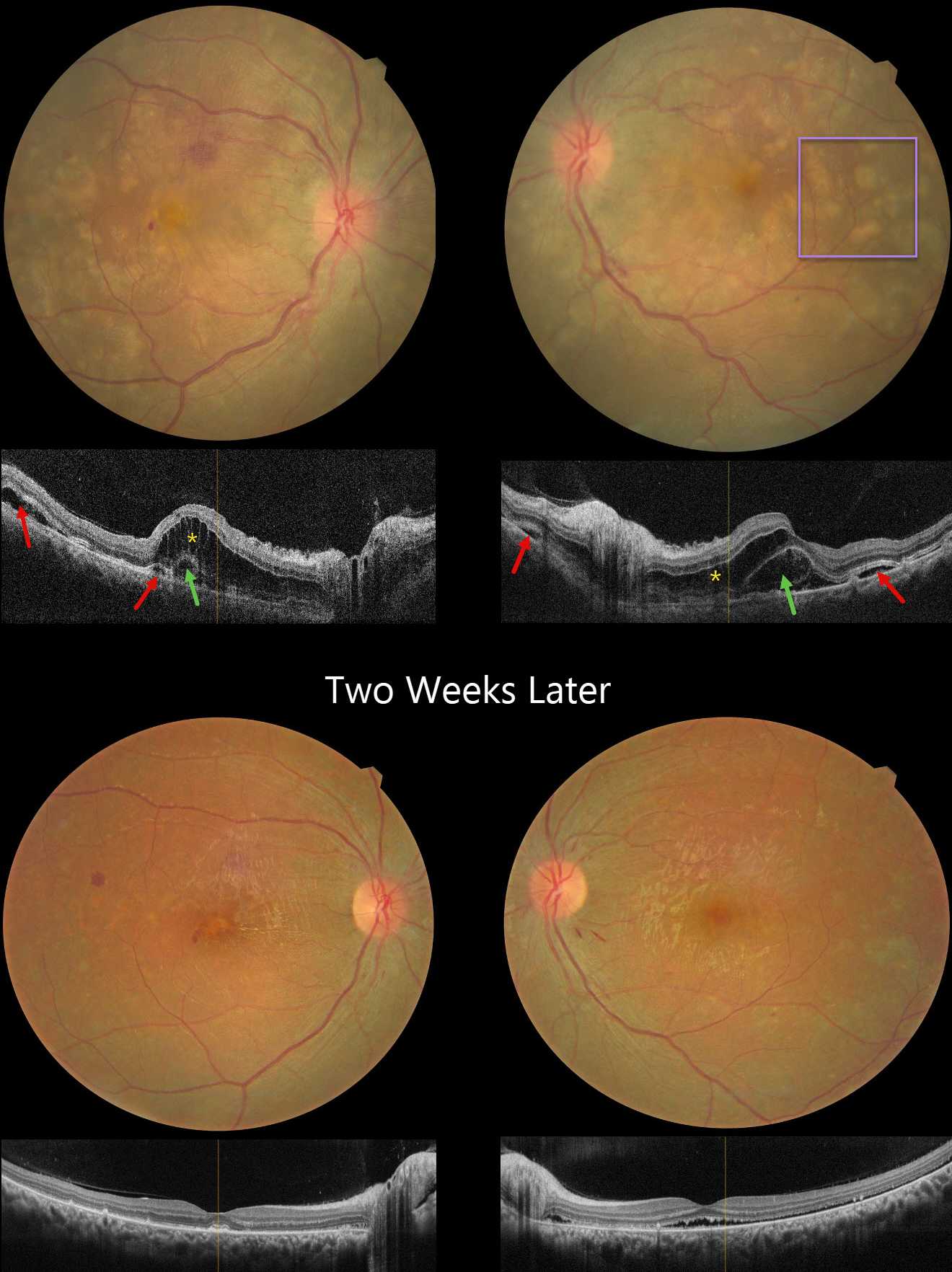
HARADA DISEASE
Navneet Mehrotra and Tamanna Patel
Originally posted on @retina.rocks January 23, 2025
This 24YO female presented with several days of bilateral vision loss. She has a history of anti-glomerular basement membrane disease (anti-GBM) and is on hemodialysis. Her renal disease was quiescent, and there were no acute systemic complaints. There was no anterior segment inflammation, although mild vitreous cells were present bilaterally. Vision was 20/200 OD and 20/80 OS.
Fundus photography shows bilateral multifocal yellow subretinal lesions, inner retinal folds, some retinal hemorrhages, and mild disc edema. OCT scanning shows pockets of subretinal fluid, outer nuclear layer edema, and bacillary layer detachments. The choroid was thickened in both eyes on ultrasonography (not shown).
Oral prednisone 60mg/day was started in a tapering dose. Two weeks later, vision improved to 20/40 OD and 20/80 OS. The acute fundus findings were mostly resolved bilaterally.
Learning Points:
Harada disease is characterized by relatively acute bilateral panuveitis, optic nerve swelling, thickened choroid, chorioretinal folds, multifocal serous exudative retinal detachments, and bacillary layer detachments. Patients with just ocular findings have Harada disease, whereas those with additional systemic findings (including vitiligo, poliosis, headache, vertigo, and hearing loss) have Vogt-Koyanagi-Harada disease.
Anti-GBM disease is a rare autoimmune disorder characterized by autoantibodies that primarily target the basement membranes of the capillaries in the kidneys and lungs. Retinal findings include drusen occurring at an early age (Mansour et al, AJO Case Reports 2017;7:83-90). Acute fundus findings are rarely reported and consist of possibly steroid-induced central serous-like changes with multifocal retinal pigment epithelial detachments and exudative retinal detachments.
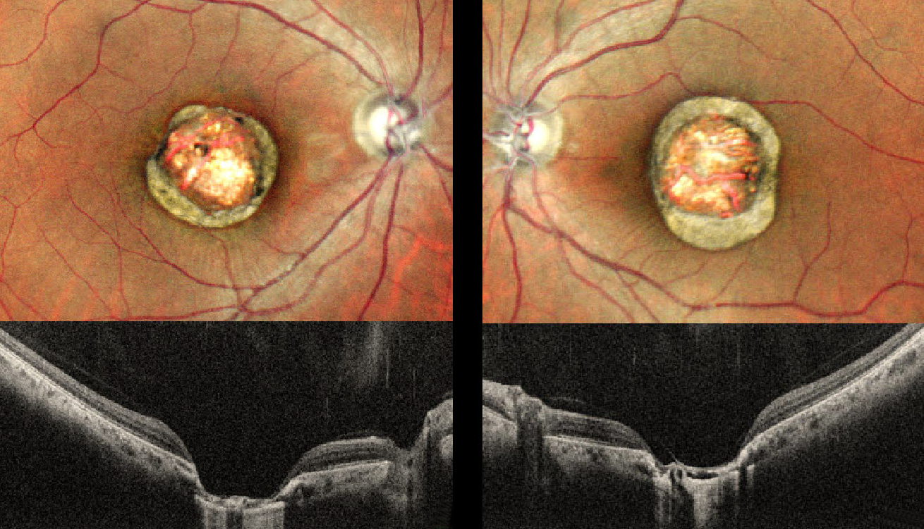
CHOROIDAL COLOBOMA
Anand Temkar and Manish Nagpal
Originally posted on @retina.rocks January 16, 2025
This healthy 24YO male presented with a lifelong history of stable bilateral vision loss. Vision was 20/80 OU.
Pseudocolor SLO imaging shows symmetrical macular colobomas. OCT scanning shows near-total choroidal loss within the coloboma, with a thinned, atrophic retina draped against bare sclera. Prior serologic testing elsewhere was negative for both toxoplasmosis and cytomegalovirus.
Learning Points:
Choroidal colobomas are most commonly due to failure of the optic vesicle and choroidal fissure to close during embryonic development and are usually found inferonasally. They can present as solitary or multiple lesions.
The differential diagnosis for congenital macular coloboma also includes post-inflammatory lesions (toxoplasmosis, cytomegalovirus, and Zika) and retinal dystrophies, including North Carolina macular dystrophy (NCMD). The retina within the coloboma is variably thinned, increasing the risk of rhegmatogenous retinal detachment (RRD).
The symmetry of the lesions argues for NCMD, but unfortunately, genetic testing was not performed due to cost.
VITAMIN A DEFICIENCY
Mariano Cotic, Ariel Schlaen, Saint Martin Madeleine, and Enzo Dilascio
Originally posted on @retina.rocks January 14, 2025
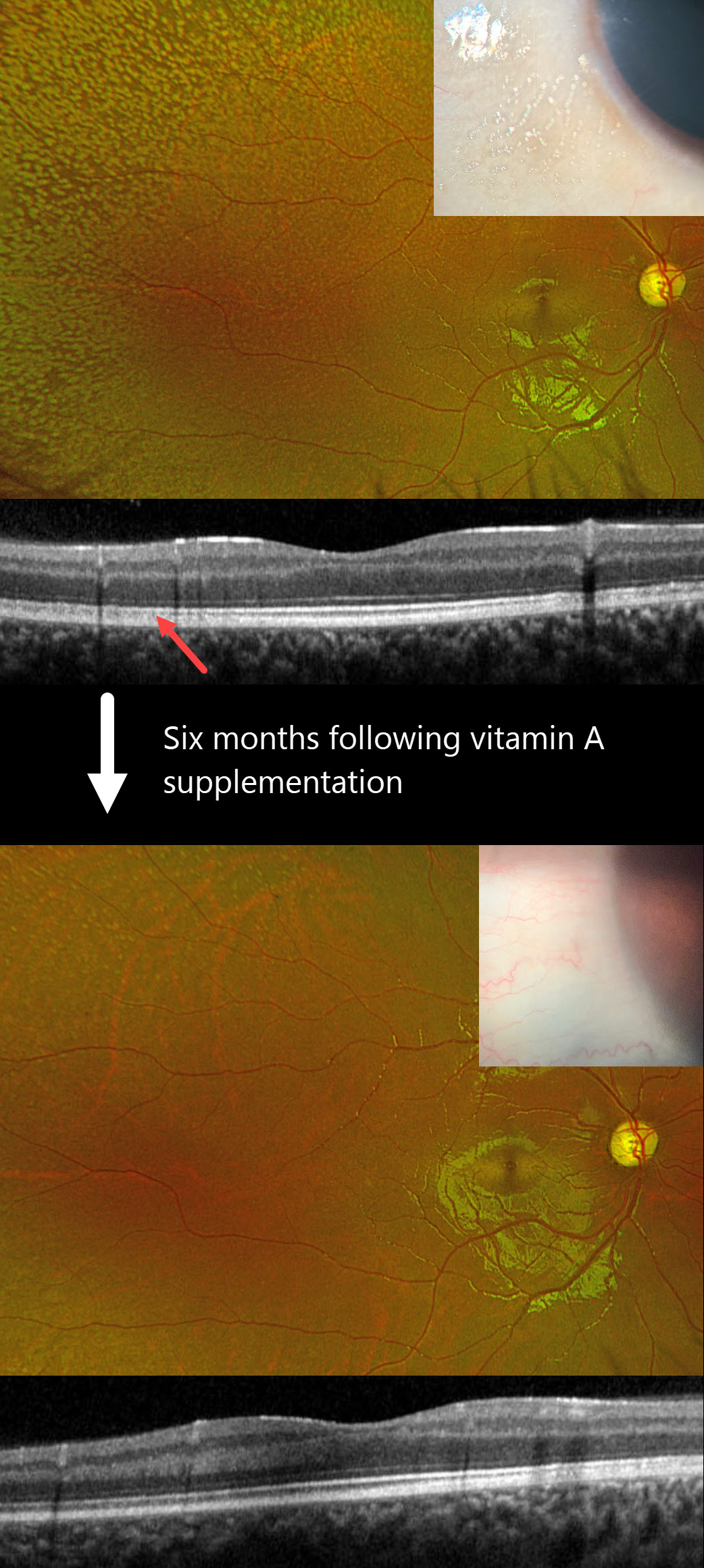
This 10YO boy presented with bilateral nyctalopia and vision loss. Vision was counting fingers OD and 20/100 OS. Identical findings were noted bilaterally, so only the images for the right eye are shown.
Bitot’s spots were observed on the temporal conjunctiva. Innumerable yellow-white subretinal deposits extend from the peripheral macula into the periphery. OCT scanning shows blurred detail of the outer segment bands due to diffuse deposition of this hyperreflective material.
On further questioning, our patient comes from a low socioeconomic environment with a diet consisting of french fries and pasta. Fasting serum vitamin A was severely low at <0.11 mg/L (normal 0.13-0.81), and supplemental oral vitamin A therapy was begun.
Six months later, vision improved to 20/70 OD and 20/30 OS with complete resolution of his nyctalopia. The conjunctival lesions and subretinal deposits had dramatically improved.
Learning Points:
Vitamin A deficiency is rare in developed countries and is sometimes related to malabsorption in chronic pancreatitis or inflammatory bowel disease. It leads to several well-documented ocular complications, including nyctalopia, Bitot spots, and xanthopsia. Outer nuclear layer thinning with disruption of the outer retinal bands can be visualized on OCT (Berkenstock et al, Int J Retin Vitr 2020;6:23). The outer retinal white dots colocalize to OCT outer retinal hyperreflective lesions (Aleman et al, Doc Ophthalmol 2013;127:239-243). Oral or intramuscular vitamin A replacement can reverse the retinal changes if initiated early enough.
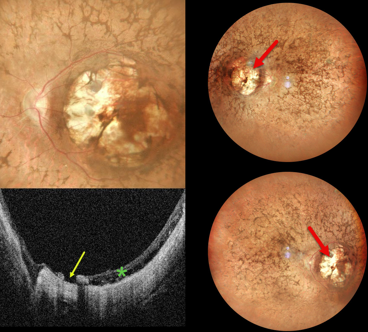
RETINITIS PIGMENTOSA WITH MACULAR COLOBOMA
Ankit Jain and Manish Nagpal
Originally posted on @retina.rocks January 7, 2025
This 32YO female was born with severe vision loss and nystagmus. There was no family history of eye disease. Vision was counting fingers bilaterally.
Pseudocolor SLO imaging shows bilateral symmetric macular colobomas with diffuse retinitis pigmentosa (RP) findings, including vascular narrowing, diffuse pigmentary changes, and intraretinal pigment migration (bone spicules). OCT shows thinned and variably disorganized retinal bands within the colobomas, and a small macular hole is noted nasally OS.
Learning Points:
Macular colobomas are rarely associated with RP (Parmeggiani et al, Eye 2004;18:421-428). North Carolina macular dystrophy, caused by a mutation in the PRDM13 gene (Small et al, Ophthalmology 2016;123:9-18), is characterized by macular coloboma-like lesions. However, these eyes do not have peripheral RP findings. Genetic testing in our patient was not performed, and observation was recommended.
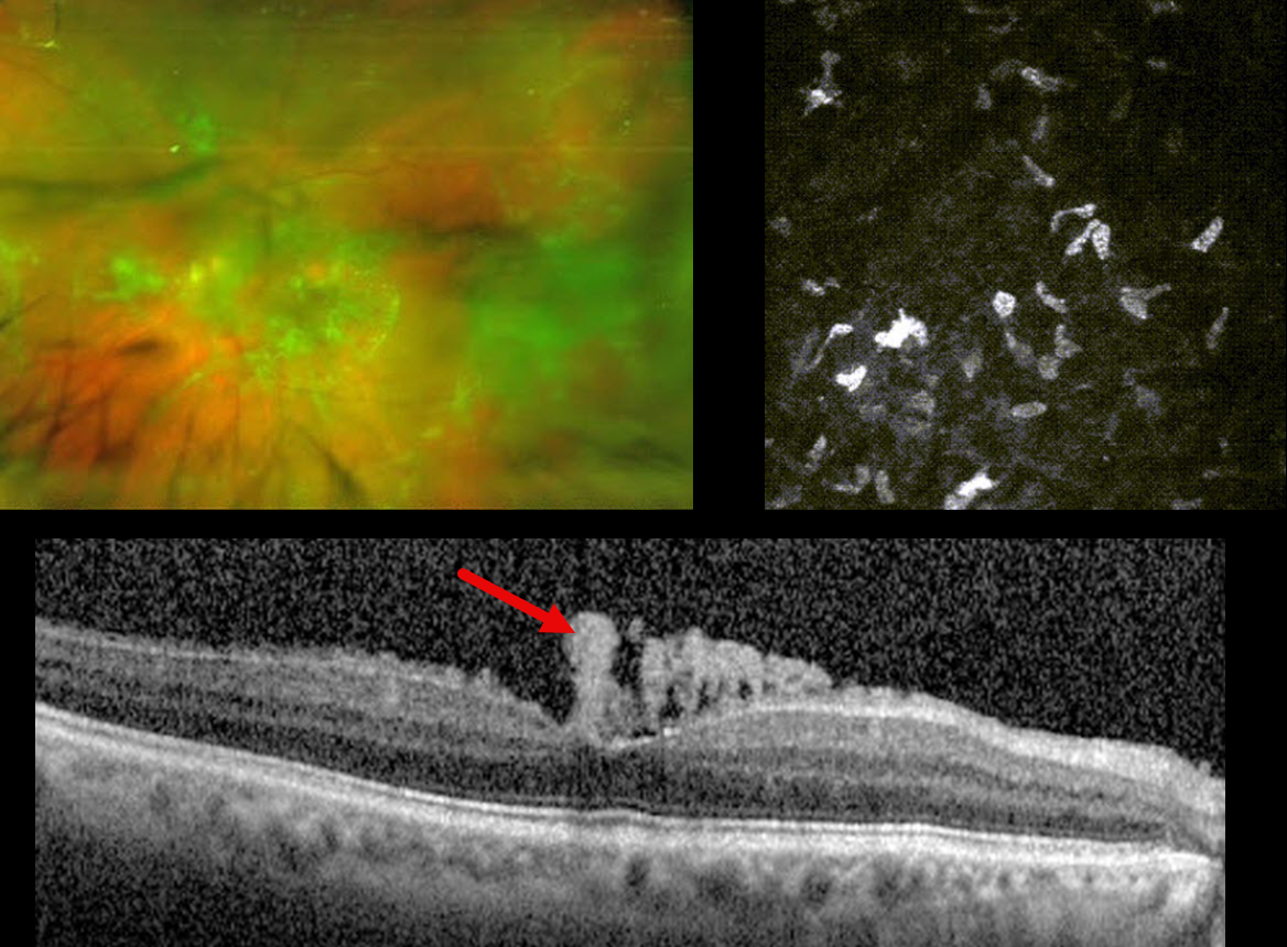
OCULAR AMYLOIDOSIS
Mariano Cotic, Natasha Gerschovsky, Madeleine Saint Martin, Enzo Dilascio, Marcelo Zas, and Pablo Chiaradia
Originally posted on @retina.rocks November 5, 2024
This 65YO male presented with chronic bilateral vision loss. Vision was 20/200 OD and 20/100 OS. There was no past medical history.
Optos color RG imaging of his right eye shows diffuse whitish, spider web-like fibrillar vitreous debris. In vivo corneal confocal microscopy reveals amyloid deposits.
Sequential therapeutic and diagnostic vitrectomy surgeries were performed on each eye. Vitreous and conjunctival biopsies were positive for amyloid deposits. Each fundus appeared normal postoperatively, and vision improved to 20/20 OU. Despite the normal fundus appearance, OCT scanning shows residual vertical amyloid deposits resting on the foveal internal limiting membrane.
Similar findings were present in his left eye (not shown).
Learning Points:
Vitreous amyloidosis is a rare disorder where bilateral vitreous opacities usually appear as fine, fibrillar, and often whitish or translucent deposits, which can reduce vision as the opaque amyloid material accumulates. OCT can reveal pathognomonic fine vertical needle-like hyperreflective deposits extending into the vitreous from the internal limiting membrane (Tasiopoulou et al, Ophthalmology Retina 2021;5:99-101). It is almost always related to mutant transthyretin (TTR), a plasma protein that transports thyroxine and vitamin A, often associated with familial amyloid polyneuropathy (Venkatesh et al, Ophthalmology 2017;124:1014-1022). Systemic amyloidosis can also cause ocular manifestations, including conjunctival, temporal artery, extraocular muscle, trabecular meshwork, and cranial nerve deposition (Reynolds et al, Retina 2018;38:1371-1376).
Our patient’s systemic workup was negative. Genetic testing for TTR was ordered and is pending.
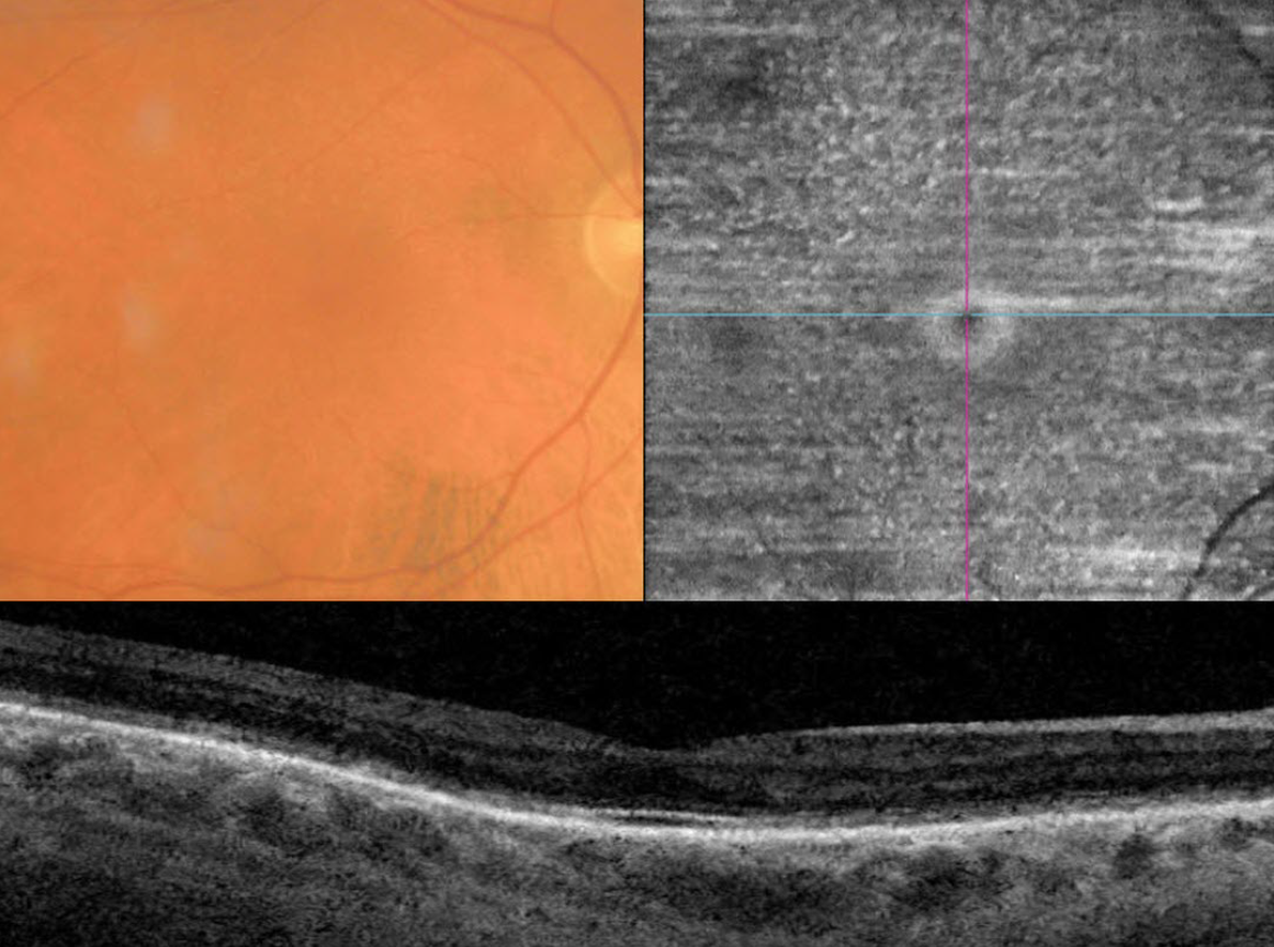
VITAMIN A DEFICIENCY
Mattie Adams
Originally posted on @retina.rocks November 4, 2024
This 70YO male presented with six months of vision loss. He has a history of severe Crohn’s disease. He has undergone 9 bowel surgeries resulting in short bowel syndrome being treated with total parenteral nutrition. Vision was 20/50 OD and 20/40 OS.
Triton color imaging shows numerous smaller subretinal yellow-white dots. These dots are well-visualized on en face swept-source OCT through the outer retina. OCT B scan shows outer retinal thinning and loss of the hyperreflective bands detail.
Fasting serum vitamin A was severely low at 2.5 (normal 20-60), and he was started on parenteral vitamin A therapy.
Learning Points:
Vitamin A deficiency is rare in the United States and is sometimes related to malabsorption in chronic pancreatitis or inflammatory bowel disease. It leads to several well-documented ocular complications including nyctalopia, Bitot spots and xanthopsia. Outer nuclear layer thinning with disruption of the outer retinal bands can be visualized on OCT (Berkenstock et al, Int J Retin Vitr 2020;6:23). The outer retinal white dots colocalize to OCT outer retinal hyperreflective lesions (Aleman et al, Doc Ophthalmol 2013;127:239-243). Oral or intramuscular vitamin A replacement has been shown to reverse the retinal changes if initiated early enough. We are hoping that our patient’s vision and retinal findings will improve with continued treatment.
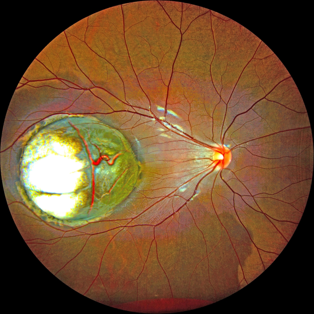
CHOROIDAL COLOBOMA
Kanwaljeet Harjot Madan
Originally posted on @retina.rocks October 30, 2024
This healthy 5YO boy’s parents noticed that his right eye was wandering for a few weeks. Vision was 20/200 OD and 20/20 in the normal OS.
Color fundus photography shows a large choroidal coloboma occupying the central and temporal macula. The underlying outer choroidal vessels are visible. His systemic examination was normal. Serologic testing for toxoplasmosis and cytomegalovirus was negative.
Learning Points:
Choroidal colobomas are most commonly due to failure of the optic vesicle and choroidal fissure to close during embryonic development and are usually found inferonasally. They can present as solitary or multiple lesions. The differential diagnosis for congenital macular coloboma also includes post-inflammatory lesions (toxoplasmosis, cytomegalovirus, and Zika) and retinal dystrophies, including North Carolina macular dystrophy. The retina within the coloboma is variably thinned, increasing the risk of rhegmatogenous retinal detachment (RRD).
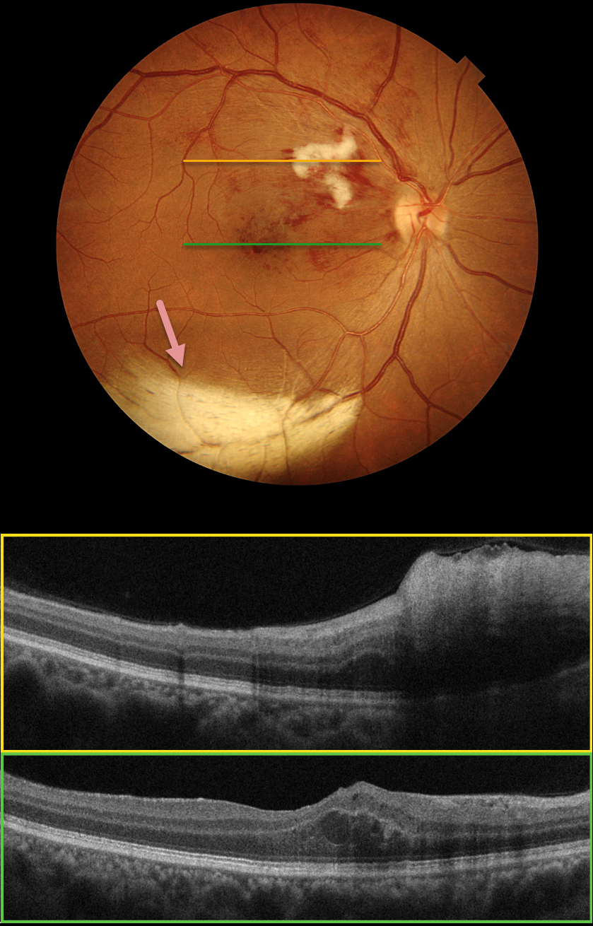
BRVO + MYELINATED NERVE FIBER LAYER
Evgenii Chernov
Originally posted on @retina.rocks September 30, 2024
Color photography of this 48YO female’s right eye shows a major superotemporal branch retinal vein occlusion (BRVO) with foveal blood. A prominent nerve fiber layer infarct (cotton-wool spot, CWS) is present in the superonasal macula. OCT scanning shows diffuse hyperreflective inner retinal thickening through the CWS and some central foveal fluid in the outer nuclear layer. An unrelated patch of myelinated nerve fiber layer (NFL) is noted along the inferotemporal arcade. The macular edema resolved following 3 monthly intravitreal Lucentis injections (not shown).
Our patient’s eye shows two different pathologies that each involve the NFL. Myelinated NFL occurs when retinal nerve fibers develop a myelin sheath, which usually stops posterior to the lamina cribrosa. The white myelin beautifully displays the normally transparent NFL. Myelination may represent an oligodendrocytic choristoma (see Rao et al Retina 2019;39:1125-1132). Nerve fiber layer infarcts cause a more fluffy, white ischemic opacification of the NFL and are a relatively nonspecific finding in numerous disorders, including diabetes, retinal venous occlusion, hypertension, and HIV/AIDS.
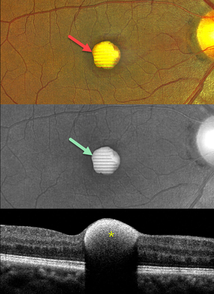
VALSALVA RETINOPATHY
Nilesh Kumar
Originally posted on @retina.rocks September 26, 2024
This 43YO male presented with a 1-week history of sudden decreased vision in his right eye. He denied any Valsalva maneuver. Vision was 20/70 in his right eye and 20/30 in his left eye.
Color imaging shows a foveal yellow sub-internal limiting membrane (ILM) hemorrhage. Fundus autofluorescence (FAF) shows intense hyper-FAF. Spectral domain OCT shows a hyperreflective sub-ILM lesion that shadows posteriorly.
Our patient was treated with observation, and the blood levels were greatly decreased 3 weeks later (not shown).
Learning Points:
Valsalva retinopathy was first described by Thomas Duane (Trans Am Ophthalmol Soc 1972; 70:298-313). It is characterized by usually unilateral retinal and preretinal blood caused by raised intrathoracic or intra-abdominal pressure. Various causes include coughing, heavy lifting, and vomiting. However, as in our case, sometimes patients present with these findings, denying any Valsalva maneuver (Forshaw et al, Acta Ophthalmologica 2024;102:122-123)
These hemorrhages virtually always resolve without sequelae, although the preretinal blood in severe cases can be drained into the inferior vitreous by creating a small ILM opening using either a thermal or Nd:YAG laser (Durukan et al, Eye 2008;22:214-218).
Porphyrins are organic molecules that are part of the heme molecule in hemoglobin. When these compounds break down, the devitalized blood appears yellow and becomes intensely hyperautofluorescent (Bloom and Spaide, Retinal Cases & Brief Reports 2022;16:401-402).
PROLIFERATIVE DIABETIC RETINOPATHY (PDR)
Originally posted on @retina.rocks September 25, 2024
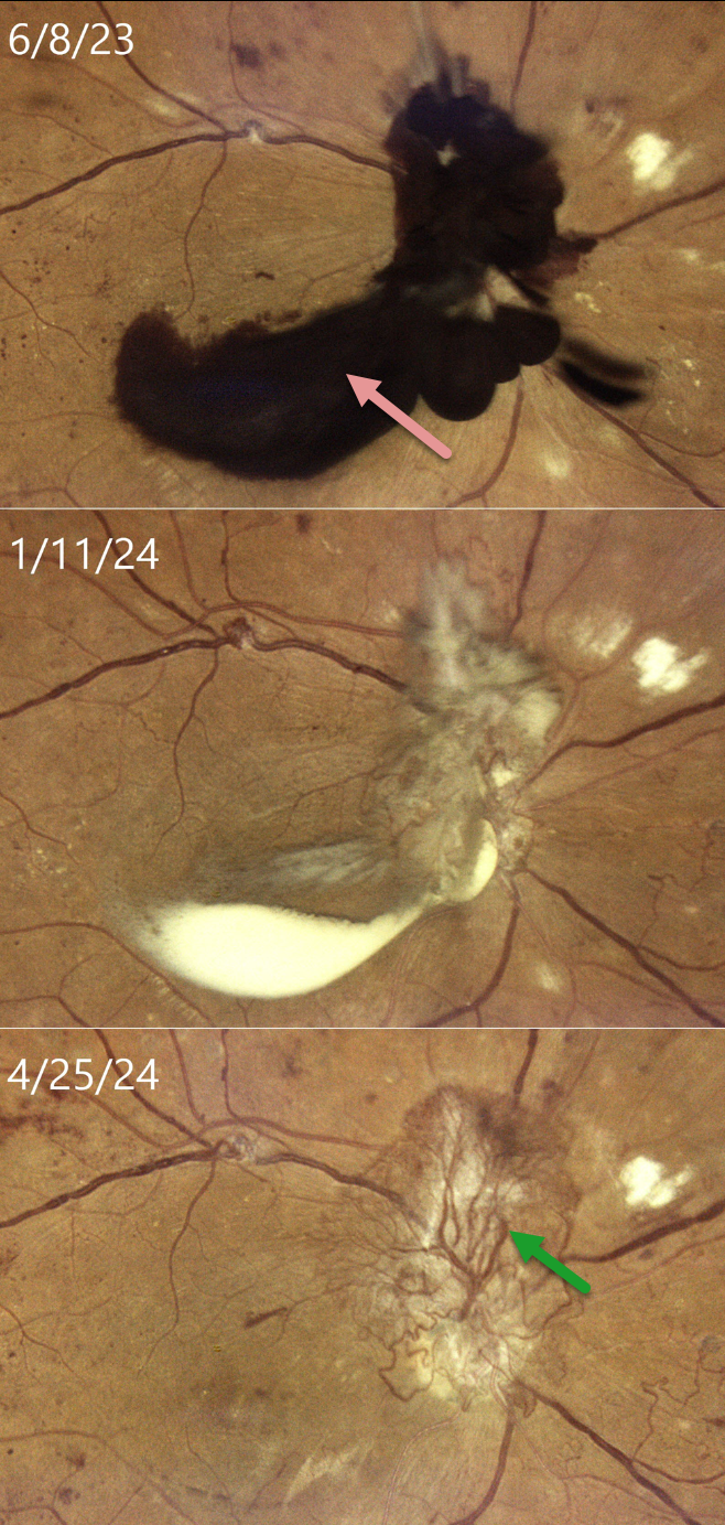
This 33YO female with type 2 diabetes was in the process of being treated with intravitreal Eylea, macular laser, and panretinal photocoagulation (PRP) for bilateral center-involved diabetic macular edema and proliferative diabetic retinopathy (PDR). She presented on 6/8/23 with recent vision loss OD and was overdue for this visit because of numerous medical issues. Vision had decreased to 20/400 OD.
Color imaging shows dark red preretinal blood obscuring the optic nerve and central macula. After several monthly Eylea injections and completion of the PRP, the preretinal blood became yellow and devitalized on 1/11/24, before finally completely absorbing, as noted at last examination on 4/25/24, with stable 20/400 vision. Despite full PRP, diffuse variably fibrosed neovascularization persists, along with venous beading. OCT scanning revealed no edema with central atrophy (not shown).
Our patient was initially in no rush for vitrectomy surgery, as vision in her fellow eye was 20/50. Conservative management with intravitreal injections and PRP was thankfully successful. This approach was also found to be safe and effective by Brar et al in a retrospective series of 22 eyes with foveal preretinal blood from PDR (Ind J Ophthalmol 2024;72:687-691).
PERSISTENT FETAL VASCULATURE
Gokcen Deniz Gulpinar Ikiz and Sengül Özdek
Originally posted on @retina.rocks September 16, 2024
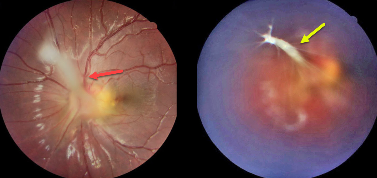
This 7YO girl presented with blurred vision in her esotropic left eye, which had previously been treated with patching of her normal right eye. Vision was 20/20 OD and 20/50 OS. Refraction was +0.25 +0.25 x180 OD and +1.00-1.50 x60 OS.
Color imaging of her left eye shows a fibrotic stalk that extends from the optic nerve head superonasally, terminating in the mid-vitreous in a spider web configuration. The macula is dragged towards the nerve, causing a shallow nasal foveal traction detachment. Lens-sparing vitrectomy will be performed in the near future.
Learning Points:
Persistent fetal vasculature (PFV), previously recognized as persistent hyperplastic primary vitreous (PHPV), represents a failure of regression of fetal vessels. It is a common cause of leukocoria and subsequent amblyopia. There is no identifiable genetic association.
Characteristically, the affected eye is microphthalmic with a shallow chamber predisposed to secondary angle closure glaucoma, a varying degree of cataract, and retrolental and/or posterior fibrovascular membranes that can cause retinal traction. If diagnosed early in life, PFV can be surgically treated with a combined lensectomy and vitrectomy. Anterior-type PFV is associated with better vision than combined or posterior types, and younger age at presentation correlates with improved vision after treatment.
DISLOCATED CATARACT
Anjana Mirajkar and Manish Nagpal
Originally posted on @retina.rocks September 3, 2024
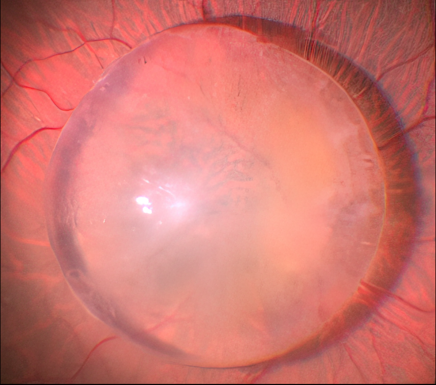
Just a cool intraoperative photo of a posteriorly dislocated cataract.
Unfortunately, we don’t have any further clinical information for this patient.
LEUKEMIA
Kanwaljeet Harjot Madan
Originally posted on @retina.rocks August 28, 2024
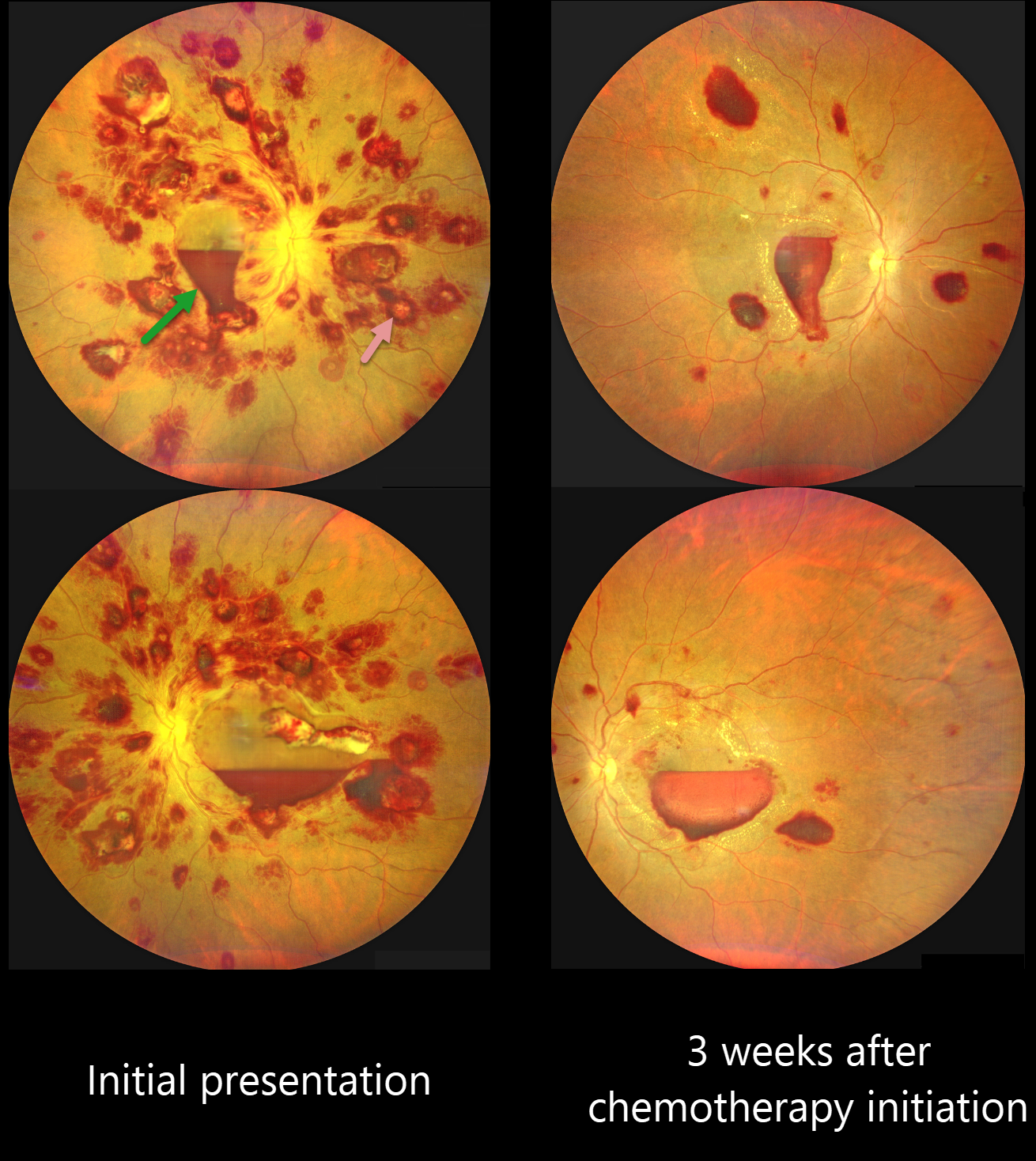
This 42YO previously healthy male presented with one week of bilateral decreased vision. Vision was 20/100 OD and 20/200 OS.
Color photography shows an extensive bilateral nerve fiber layer and deep white-centered retinal hemorrhages. Layered macular sub-internal limiting membrane (ILM) hemorrhages are noted as well, with some yellow devitalized blood in the temporal left macula. Hematologic workup revealed elevated blast cells, severe anemia (HgB 5.4) and thrombocytopenia.
He was diagnosed with acute myeloid leukemia and referred to oncology. He started chemotherapy, and 3 weeks later, his vision improved to 20/70 OU. The HgB was 8.2 with decreased blast cells. The bilateral retinal hemorrhages were markedly decreased.
This case was submitted by Kanwaljeet Harjot Madan.
Learning Points:
The term “Roth spot” was named after Dr. Moritz Roth, who, in 1872, noted white-centered hemorrhages in patients with bacterial endocarditis. He felt that these white spots were septic emboli that originated from an infected cardiac valve. However, similar lesions can be seen in many other disorders, including leukemia, hyperviscosity, anemia, diabetes, and hypertension (Duane et al Ophthalmology 1980;87:66-69). The white material may represent a variety of causes, including fibrin. So, these lesions are best called white-centered hemorrhages, unless one is specifically referring to those found associated with bacterial endocarditis.
Ocular leukemic manifestations are primarily caused by accompanying hematologic abnormalities, including hyperviscosity, anemia, and thrombocytopenia (Soman et al, Ophthalmology Retina 2018;2:17-23). Posterior segment findings include retinal hemorrhages (sub-ILM, nerve fiber layer or deeper, white-centered), cotton-wool spots, dilated retinal veins, and serous retinal detachment. Leukemic infiltration of the optic nerve, retina, and choroid can also occur. The sub-ILM hemorrhages are usually self-limiting and resolve within several months of treating the underlying disease (Ophthalmology Retina 2018;2:494-501).
COUCHED CATARACT
Ogugua Okonkwo, Adekunle Olubola Hassan, and Zainab Ogunsakin
Originally posted on @retina.rocks August 8, 2024
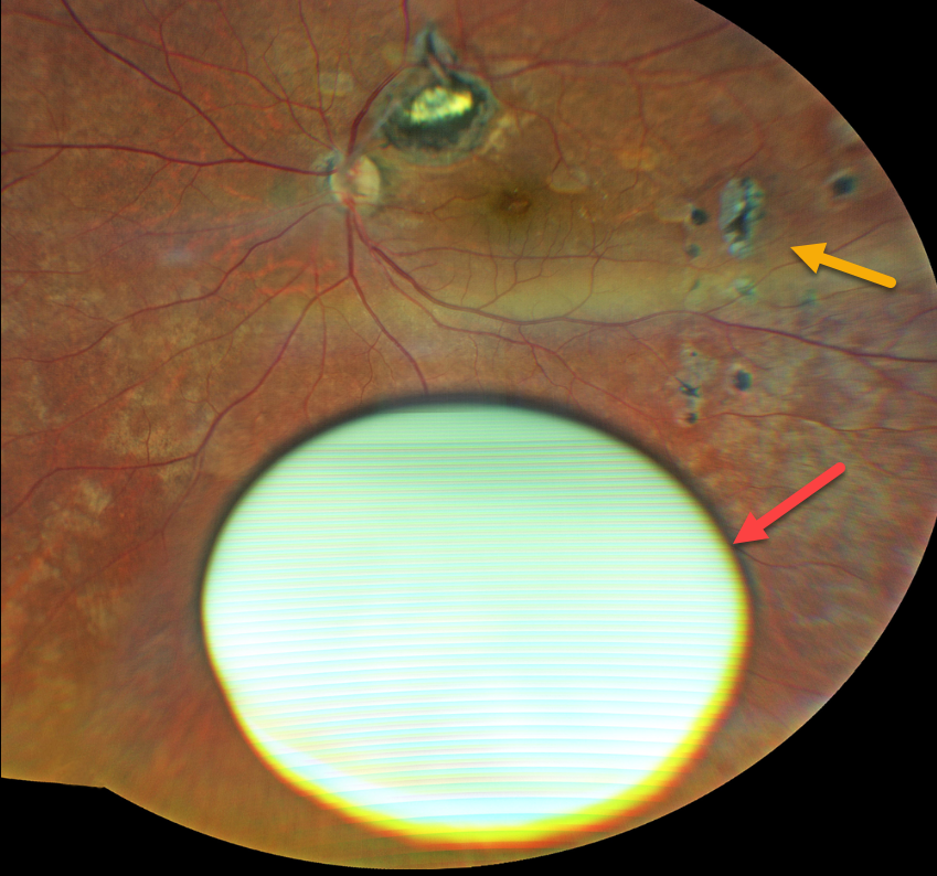
This 67YO female underwent a couching procedure for a left cataract in 2018. Cataract surgery with a posterior chamber IOL OD was performed in 2020, and 2 months later, the cataract surgeon referred her to us for evaluation of the couched lens OS.
Vision was 20/40 OS with a +10.00 diopter lens. She was lost to follow-up until she returned in March 2024 with counting-fingers vision OS. Her intraocular pressure was slightly elevated, and she had new focal lesions in the peripheral retina, which were felt to represent inflammation from leaking lens protein.
Fundus photography shows a dislocated intumescent hypermature cataract in the inferior vitreous. Multifocal chorioretinal scars are scattered throughout the fundus. Pars plana vitrectomy, lensectomy, and anterior chamber lens implantation were performed. Vision without correction was 20/200 OS one month postoperatively.
Learning Points:
Couching is the earliest documented form of cataract surgery and was widely practiced worldwide (Leffler et al, Ann Transl Med 2020;8(22):1551). It was described in 600-800 BC by an ancient Indian surgeon. This practice, though now largely abandoned with the advent of modern cataract surgery, is still rarely practiced in rural areas of some African countries, including Nigeria.
Couching is performed in the ‘comfort’ of the patient’s home by the ‘coucher’ who moves around in the rural community where their service is needed. The procedure involves using a sharp instrument to push the cataractous lens backward into the vitreous. If all goes well. A high hyperopic spectacle correction is prescribed. It can, however, be complicated by glaucoma, lens dislocation into the anterior chamber, cornea decompensation, uveitis, retinal detachment, vitreous hemorrhage, and intraocular infection.
CHOROIDAL COLOBOMA + MYELINATED NERVE FIBER LAYER
Originally posted on @retina.rocks July 31, 2024
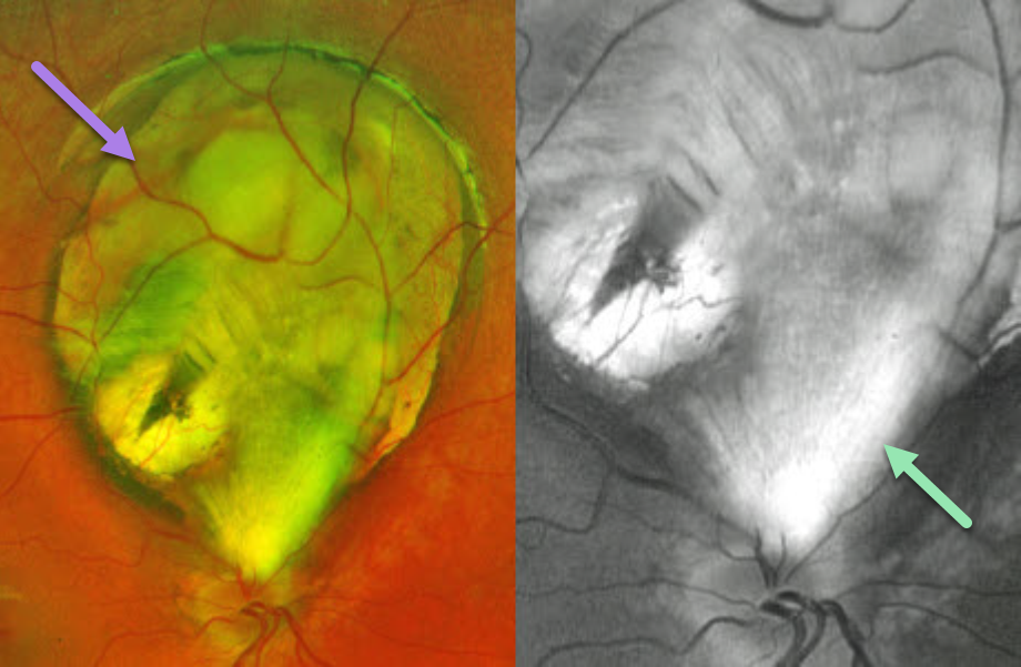
This healthy 8YO girl was referred for an asymptomatic retinal finding in her right eye. Vision was 20/30 in her right eye and 20/30 in her left eye. Anterior segments were normal.
Optos color RG imaging shows a superior choroidal coloboma that almost extends to the disc margin. An incidental patch of myelinated nerve fiber layer (NFL), best imaged in the green channel, extends from the superior nerve into the inferior aspect of the coloboma.
Learning Points:
Choroidal colobomas are caused by failure of the optic vesicle and choroidal fissure to close during fetal development. Choroidal coloboma can present as solitary or multiple lesions. Visual prognosis depends on the location and involvement of the optic nerve and macula. We will follow our patient yearly due to her increased risk of retinal detachment.
Myelinated NFL is a relatively uncommon and usually isolated finding present at birth. Most patients are asymptomatic, although some may have a corresponding relative scotoma. Myelination can progress in about 10% of cases and can also resolve following retinal ischemic events. Myelinated NFL occurs when retinal nerve fibers develop a myelin sheath, which usually stops posterior to the lamina cribrosa. The myelination may represent an oligodendrocytic choristoma (see Rao et al Retina 2019;39:1125-1132).
DISLOCATED IOL
Anjana Mirajkar and Manish Nagpal
Originally posted on @retina.rocks July 30, 2024
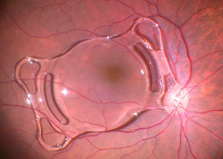
This patient presented with a spontaneously posteriorly dislocated intraocular lens (IOL). The lens was explanted, and a secondary scleral-fixated lens was placed. Unfortunately, we don’t have any further clinical information.
LASER MACULOPATHY
Mattie Adams
Originally posted on @retina.rocks April 22, 2024
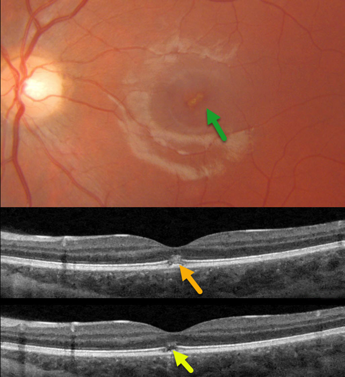
This 13YO girl presented with immediate vision loss in her left eye after looking at a laser pointer one week earlier. Vision was 20/20 in her normal right eye and 20/70 in her left eye.
Triton color photography shows an oblique, slightly linear area of foveal pigment loss. Swept-source OCT scans through this lesion show focal hyperreflectivity with variable loss of the ellipsoid zone and outer segments.
Learning Points:
Sun gazing is thought to cause acute damage to the RPE and photoreceptors via photochemical mechanisms involving reactive oxygen species. Similar photochemical damage can follow laser pointer exposure, although the clinical findings can be different (Bloom and Singal, Retinal Cases 2022;16:
89-91).
Sun gazing acutely produces bilateral, symmetric, pinpoint subfoveal yellow-white lesions in the outer retina, followed by variable clinical depigmented foveal RPE and focal photoreceptor OCT defects extending from the external limiting membrane to the RPE.
Laser pointer maculopathy can cause a similar lesion, but presumed more prolonged viewing, saccades, and laser pointer movement can also lead to larger areas of macular damage.
Outer foveal microdefects (Cohen et al, Ophthalmology Retina 2021;5:553-561) have been described in numerous conditions, including macular telangiectasia, tamoxifen use, ABCA4 disorders (cone-rod dystrophies, Stargardt disease and fundus flavimaculatus), phototoxicity, trauma, and vitreomacular traction disorders.
INTRACHOROIDAL CAVITATION
Originally posted on @retina.rocks April 8, 2024
This 84YO male was referred for an asymptomatic macular lesion in his right eye. He was a +6 diopter hyperope. Vision was 20/30 in his right eye and 20/30 in his left eye.
Optos color RG imaging shows a subretinal, mostly depigmented lesion in the superior macula. Triton swept-source OCT shows a hyporeflective outer choroidal lesion that bows posteriorly. The choroid surrounding this lesion is relatively hyperreflective and disorganized. Observation was recommended.
Learning Points:
Initially described by Freund et al (Arch Ophthalmol 2003;121:197-204), intrachoroidal cavitations are most commonly found in high myopes as a peripapillary yellow-orange subretinal lesion. Our case is quite unique, given the location and occurrence in a hyperope.
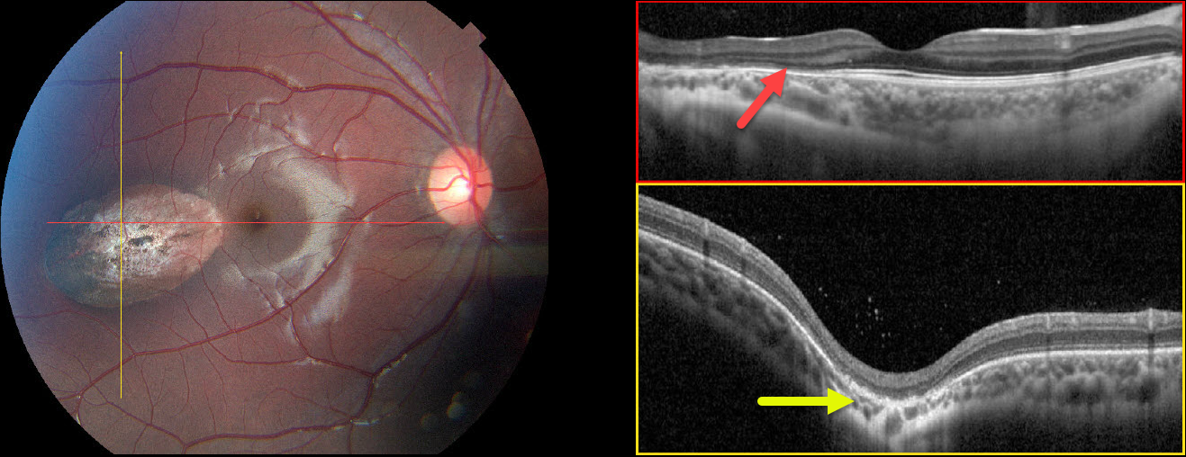
CHOROIDAL COLOBOMA
Neeket Patel
Originally posted on @retina.rocks March 18, 2024
This 12YO female complained of several years of daily frontal headaches associated with bilateral blurriness. Vision was 20/25 OU.
Color photography of her right eye shows an oval depigmented lesion in the temporal macula. Horizontal B-scan OCT shows outer retinal atrophy overlying the lesion. In the vertical B-scan, the lesion is colobomatous with outer retinal thinning. Some hyperreflective vitreous cells are noted. The left fundus was normal.
Learning Points:
Torpedo maculopathy was originally described by Gass as a solitary hypopigmented RPE nevus (Arch Ophthalmology 1992;110:1358-1359). These benign, unilateral lesions are tear-drop shaped and almost always located in the horizontal meridian in the temporal macula.
OCT scanning shows attenuation of the RPE and outer retinal layers. Vision is almost always normal, although there is a small risk for macular neovascularization. There are no known systemic associations.
Choroidal colobomas are most commonly due to failure of the optic vesicle and choroidal fissure to close during fetal development. Lesions like this may also be from other causes, including toxoplasmosis.
Although our patient’s lesion strongly resembles torpedo maculopathy, the coloboma would be highly atypical. Our best guess, given the vitreous cells, is that this lesion is due to toxoplasmosis.
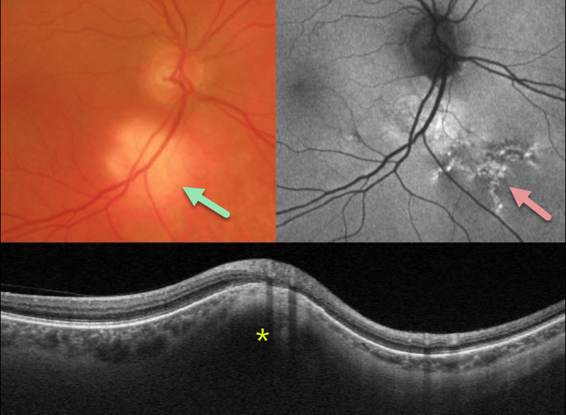
FOCAL SCLERAL NODULE
Janelle Adeniran
Originally posted on @retina.rocks March 6, 2024
This 73YO male presented with this asymptomatic amelanotic lesion inferior to the right optic nerve. Optos fundus autofluorescence (FAF) shows overlying hyper-FAF with some dendritic radiating lines of hyper- and hypo-FAF extending inferonasally.
Triton swept-source OCT scanning through this region shows hyperreflective scleral thickening, which compresses the overlying choroid and elevates the retina.
Learning Points:
Focal scleral nodule, previously known as solitary idiopathic choroiditis (SIC) and unifocal helioid choroiditis, presents as a single, yellow-white, elevated nodular, avascular subretinal lesion. On OCT, it arises from the inner sclera, causing marked compression of the choroid.
For the largest series to date and the rationale behind its renaming, see Fung et al, Ophthalmology 2020;127:1567-1577.
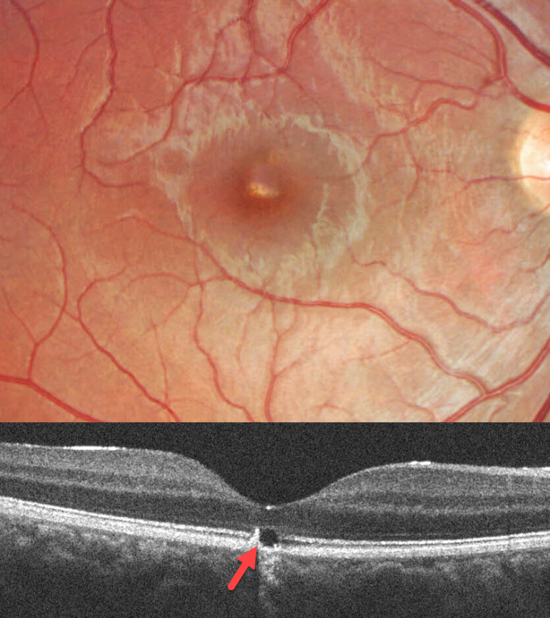
WHIPLASH MACULOPATHY
Originally posted on @retina.rocks March 5, 2024
This 17YO male was in a car accident several weeks earlier with a whiplash injury. He noticed mild vision loss in his right eye since. Vision was 20/30.
Color imaging shows a somewhat linear area of foveal pigment loss. OCT scanning of this region shows an outer foveal microdefect with adjacent hyperreflectivity along its temporal edge.
Learning Points:
Initially described by Kelley et al (Arch Ophthalmol 1978;96:834-835), whiplash maculopathy is thought to occur from a vitreous coup-contrecoup injury following a motor vehicle accident. This can cause contusive RPE injury with transient macular edema (AJO 2007;143:348-350) or, more typically, a solar retinopathy-like defect in the outer retina.
Outer foveal microdefects (Cohen et al, Ophthalmology Retina 2021;5:553-561) have been described in numerous conditions, including macular telangiectasia, tamoxifen use, ABCA4 disorders (cone-rod dystrophies, Stargardt disease, and fundus flavimaculatus), phototoxicity, trauma, and vitreomacular traction disorders.
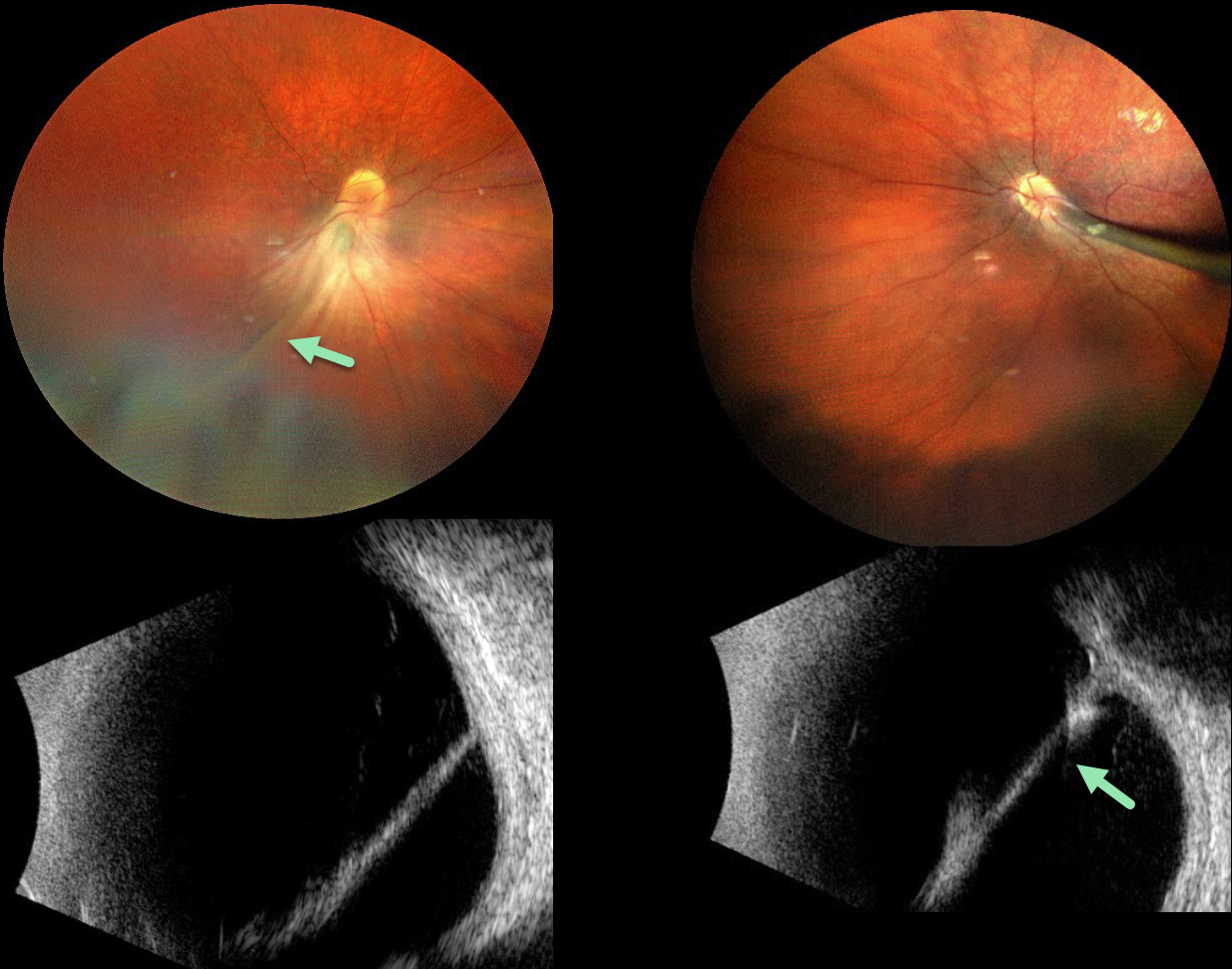
PERSISTENT FETAL VASCULATURE
Emma Oreškovič and Nataša Drača
Originally posted on @retina.rocks January 16, 2024
The 9YO girl presented with a known history of persistent fetal vasculature (PFV) since age 4. She was the product of a normal, full-term pregnancy. Vision was 20/400 OU. She also has congenital nystagmus.
Color photography shows bilateral fibrous stalks extending from the optic disc to the posterior lens capsule. The stalks are hyperreflective on B-scan ultrasonography. Falciform folds are also observed. Each retina was otherwise completely attached. She was referred to a pediatric retinal specialist for further management.
Learning Points:
Persistent fetal vasculature (PFV), previously recognized as persistent hyperplastic primary vitreous (PHPV), represents a failure of regression of fetal vessels. It is a common cause of leukocoria and subsequent amblyopia. There is no identifiable genetic association.
Characteristically, the affected eye is microphthalmic with a shallow chamber predisposed to secondary angle closure glaucoma, a varying degree of cataract, and retrolental and/or posterior fibrovascular membranes that can cause retinal traction.
If diagnosed early in life, PFV can be surgically treated with a combined lensectomy and vitrectomy. Anterior-type PFV is associated with better vision than combined or posterior types, and younger age at presentation correlates with improved vision after treatment.
For a review of PFV signs and symptoms, see Goldberg, AJO 1997;124:587-626.
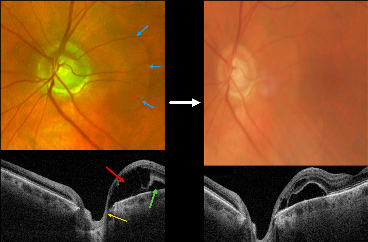
GLAUCOMATOUS PERIPAPILLARY RETINOSCHISIS
Devin Cohen and Fraser McKay
Originally posted on @retina.rocks December 28, 2023
This 70YO male was referred for possible macular edema in his left eye. He underwent a successful vitrectomy for a retinal detachment in his right eye 8 months earlier. He had no visual complaints, and vision was 20/20 OD and 20/25 OS. Intraocular pressure was 14 mmHg OD and 22 mmHg OS.
Optos color RG imaging shows increased optic nerve cupping with a blister of retinal fluid extending temporally. Triton swept-source OCT shows both outer retinal elevation and schisis, and a small area of more temporal subretinal fluid. The outer retinal schisis directly communicates with the nerve.
At least 0.8 optic nerve cupping was noted OU, along with dense superior arcuate scotoma on 30-2 visual field testing (not shown).
Generic Cosopt OU BID was prescribed. Six weeks later, vision was 20/30 OD and 20/25 OS. IOP was 9 mmHg OD and 11 mmHg OS. The outer retinal schisis and fluid were decreasing.
Learning Points:
While peripapillary retinoschisis (PPRS) secondary to congenital optic disc cavitary abnormalities is well known, similar findings in patients with glaucomatous cupping but without nerve head cavitation are a more recently described and less common finding (Fujimoto et al., Ophthalmology Retina 2023;7:811-818).
The retinal nerve fiber layer is most commonly involved in glaucoma, whereas in optic pit maculopathy, outer and inner nuclear layer schisis, foveal involvement, and subretinal fluid are more frequently noted.
Most cases of glaucoma-related PPRS are asymptomatic and spontaneously resolve. However, in fovea-involving or symptomatic cases, surgical intervention may improve vision and decrease time to resolution.
There is no consensus on the optimal treatment approach, including pars plana vitrectomy alone or with juxtapapillary laser treatment, ILM peeling, inner retinal fenestration, and/or autologous platelet concentrate.
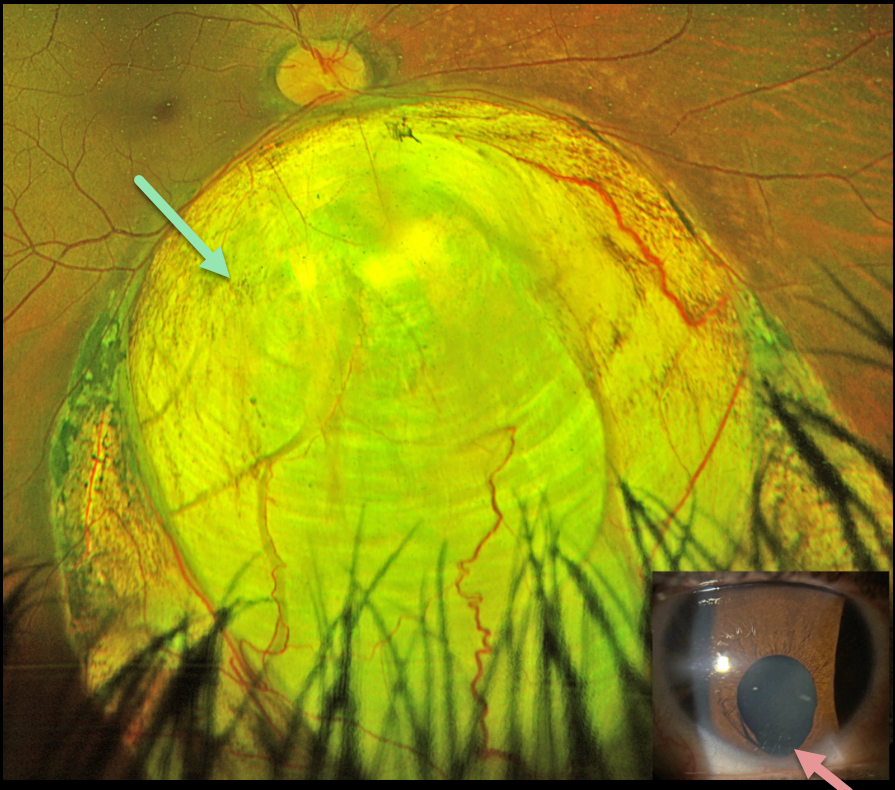
CHOROIDAL COLOBOMA
Austin Eckel
Originally posted on @retina.rocks December 14, 2023
This healthy 25YO female’s primary care doctor noticed an unusual finding in her right eye and referred her to us for an examination. She said that her right pupil had looked different.
Optos color RG imaging shows a large inferior choroidal coloboma extending from the inferior disc margin into the retinal periphery. An inferior iris coloboma is noted.
Learning Points:
Choroidal colobomas are caused by failure of the optic vesicle and choroidal fissure to close during fetal development. Choroidal coloboma can present as solitary or multiple lesions, as in this case. Visual prognosis depends on the location and involvement of the optic nerve and macula.
We will follow our patient yearly due to her increased risk of retinal detachment and expect her to do well, given the normal central macular appearance.
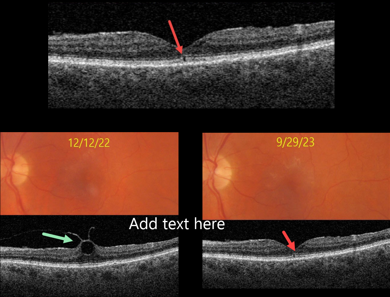
OUTER FOVEAL MICRODEFECT
Originally posted on @retina.rocks December 13, 2023
This 76YO female presented with no visual symptoms, 20/40 vision, and an abnormal OCT in her left eye. What caused this subtle OCT finding?
She was previously being followed for asymptomatic focal vitreomacular traction (VMT) in this eye. On 12/12/22, Triton color imaging showed a subtle foveal cyst with focal VMT on swept-source OCT.
We recommended observation due to the absence of symptoms, and when she returned on 9/29/23, these findings spontaneously resolved as the vitreous separated, leaving a tiny outer foveal microdefect.
Learning Points:
Outer foveal microdefects (Cohen et al, Ophthalmology Retina 2021;5:553-561) have been described in numerous conditions, including macular telangiectasia, tamoxifen use, ABCA4 disorders (cone-rod dystrophies, Stargardt disease, and fundus flavimaculatus), phototoxicity, trauma, and vitreomacular traction disorders.
The prevalence of VMT increases with age, and is present in about 1% of people over 40YO. The natural history is still being defined, but most patients have stable traction and vision. VMT can also resolve spontaneously in about 20% (Errera et al, Ophthalmology 2018;125:701-707).
Many patients with VMT remain completely asymptomatic despite impressive OCT findings. We therefore almost always observe VMT, as in this case, unless the patient has significant symptoms.
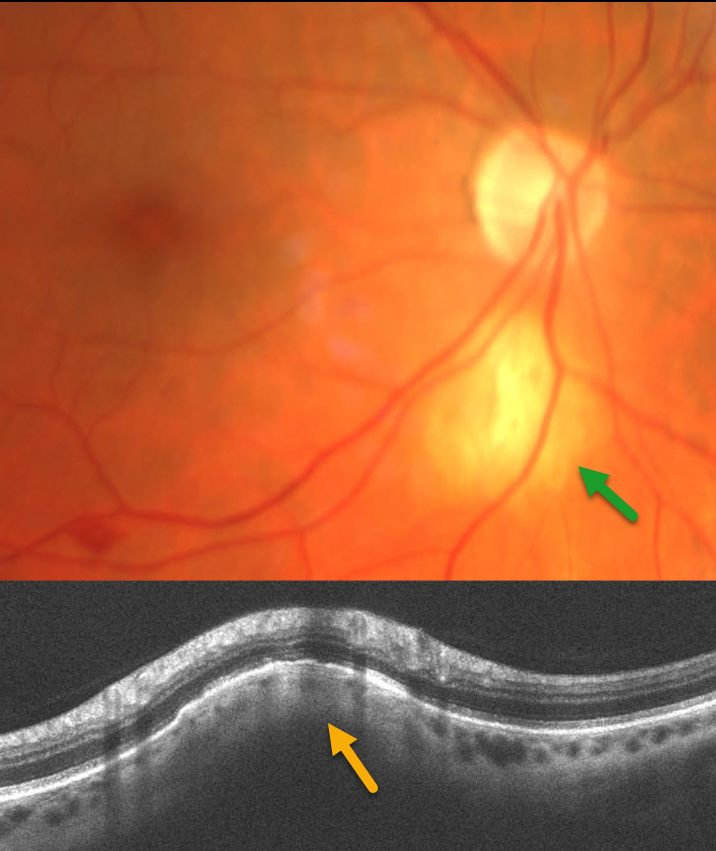
FOCAL SCLERAL NODULE
Originally posted on @retina.rocks November 6, 2023
This patient presented with an asymptomatic amelanotic lesion inferior to the right optic nerve. OCT scanning through this region shows hyperreflective scleral thickening, which compresses the overlying choroid and elevates the retina.
Learning Points:
Focal scleral nodule, previously known as solitary idiopathic choroiditis (SIC) and unifocal helioid choroiditis, presents as a single, yellow-white, elevated nodular, avascular subretinal lesion. On OCT, it arises from the inner sclera, causing marked compression of the choroid.
For the largest series to date and the rationale behind its renaming, see Fung et al, Ophthalmology 2020;127:1567-1577.
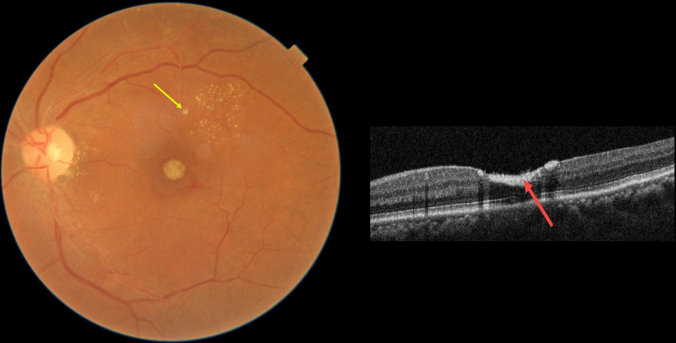
RETINAL ARTERIAL MACROANEURYSM (RAM)
Asma Samsudeen and Ashish Sharma
Originally posted on @retina.rocks October 26, 2023
This 54YO female presented with 3 weeks of vision loss in her left eye. Vision was 20/20 in her normal OD and 20/200 OS.
Fundus photography shows devitalized yellow prefoveal blood, some lipid flecks centered in the superotemporal macula, and a tiny fibrosed superior retinal arterial macroaneurysm (RAM).
OCT scanning through the fovea confirms the sub-internal limiting membrane (ILM) location of the hyperreflective blood.
This case was submitted by Asma Samsudeen and Ashish Sharma.
Learning Points:
A RAM is thought to occur in a weakened arterial wall from arteriosclerosis, and appears as a yellow-white dilated lesion along a retinal artery within the first 3 bifurcations from the optic nerve.
There are 2 distinct presentations, hemorrhagic or exudative, and our patient has features of both. Although she reported only a several-week history of vision loss, the retinal findings suggest a much longer course.
The devitalized blood usually takes months to turn yellow, and was likely much larger in extent originally. The blood elsewhere has largely resolved. There was also likely macular thickening and possibly subretinal fluid, which spontaneously resolved, leaving behind residual peripheral macular lipid.
Observation was recommended, and we expect her vision to significantly improve, as the retina appears structurally fairly normal on OCT deep to the blood. Subretinal blood, especially devitalized, is toxic to the outer retina, which is why removal and/or displacement of thick subretinal blood is often recommended. Blood in the sub-ILM or preretinal space does not seem to damage the retina, so it can more safely be observed (see Bloom and Spaide, Retinal Cases 2022;16;401-402).
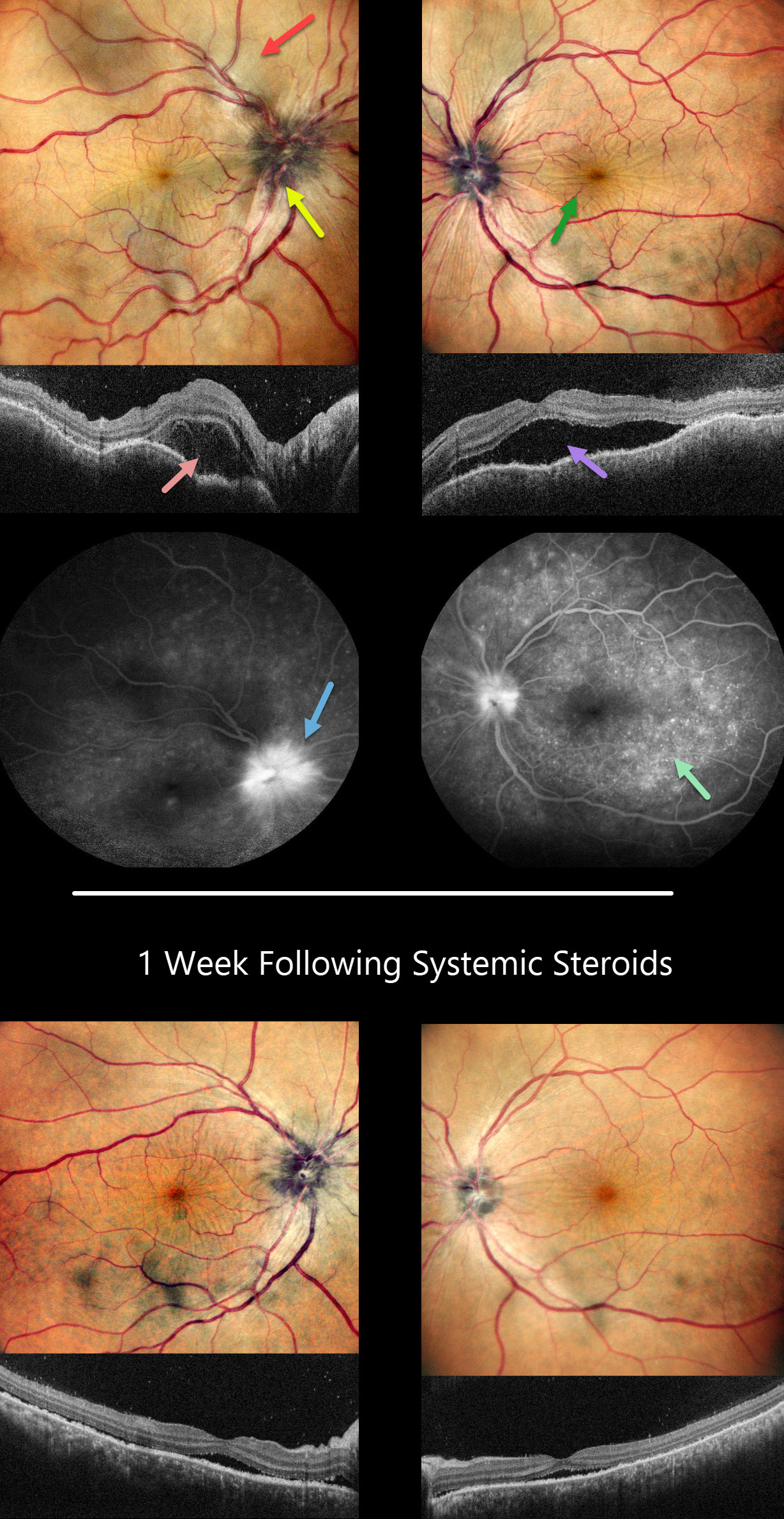
HARADA DISEASE
Anjana Mirajkar, Manish Nagpal and Navneet Mehrotra
Originally posted on @retina.rocks October 18, 2023
This 41YO female presented with headaches, redness, and pain OD for 1.5 months and OS for 2 weeks. She was diagnosed elsewhere with angle closure glaucoma, received a YAG peripheral iridotomy, and started on glaucoma drops. She then saw multiple other doctors who diagnosed her with papilledema.
Her CSF opening pressure was 21, and she was started on oral acetazolamide for a presumed diagnosis of idiopathic intracranial hypertension (IIH). In our office, vision was 20/100 OD and 20/60 OS. Intraocular pressure (IOP) was 30mmHG.
Anterior segments showed shallow anterior chambers with retrolental vitreous cells. Multicolor imaging shows bilateral irregular chorioretinal folds, multifocal serous detachments, and disc hyperemia.
OCT scanning shows a bacillary layer detachment OD, subretinal fluid OS, and a bilateral undulating thickened choroid. Fluorescein angiography shows bilateral optic nerve involvement and pinpoint subretinal leakage.
She was diagnosed with Harada’s disease and started on intravenous methylprednisolone for 3 days, followed by 60mg prednisone PO daily. She was also referred to rheumatology to start immunosuppressants. The acetazolamide was tapered and discontinued for the misdiagnosed IIH.
One week later, vision improved to 20/40 OU. IOP was 10mmHG OU with marked bilateral improvement in the chorioretinal folds, macular fluid, and choroidal thickening. A slow steroid taper was initiated pending initiation of immunosuppressants.
Learning Points:
This case has many classic findings for Harada’s disease, including bilateral panuveitis, optic nerve swelling, thickened choroid, chorioretinal folds, and multifocal serous exudative retinal detachments.
Patients with just ocular findings have Harada’s disease, whereas those with additional systemic findings (including vitiligo, poliosis, headache, vertigo, and hearing loss) have Vogt-Koyanagi-Harada (VKH) disease.
This case also reminds us that it is easy to misdiagnose these complex uveitis cases, especially when not all ocular findings are taken into full consideration in an attempt to give the patient a single, unifying diagnosis.
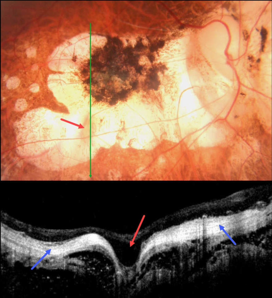
HIGH MYOPIA
Asma Samsudeen and Ashish Sharma
Originally posted on @retina.rocks September 14, 2023
This 58YO male with a history of degenerative myopia presented with 6 months of vision loss in his right eye. He was blind in his left eye following vitrectomy surgery 7 years earlier. Vision was counting fingers OD.
Color photography shows severe variably pigmented atrophic myopic degeneration. An area of orange coloration is seen within the larger area of atrophy inferiorly.
On OCT, this area appears as a focal staphyloma. The overlying retina is draped across the staphyloma. The choroid is so thin as to be invisible on OCT, with a hyperreflective underlying sclera. The orbital tissue is well seen posterior to the sclera.
Learning Points:
Degenerative myopia is a major cause of worldwide visual impairment. Due to the stretching of the posterior retinal structures, there is an increased risk for numerous findings, including atrophic retinal and choroidal thinning, posterior staphyloma, macular schisis/traction, and macular neovascularization (see Ohno-Matsui et al for a recent review, Invest Ophthalmol Vis Sci 2021;62(5);5)..
Initially described by Freund et al (Arch Ophthalmol 2003;121:197-204), intrachoroidal cavitations are most commonly found in high myopes as a peripapillary yellow-orange subretinal lesion. It is difficult to determine whether our patient has a cavitation above the staphyloma.
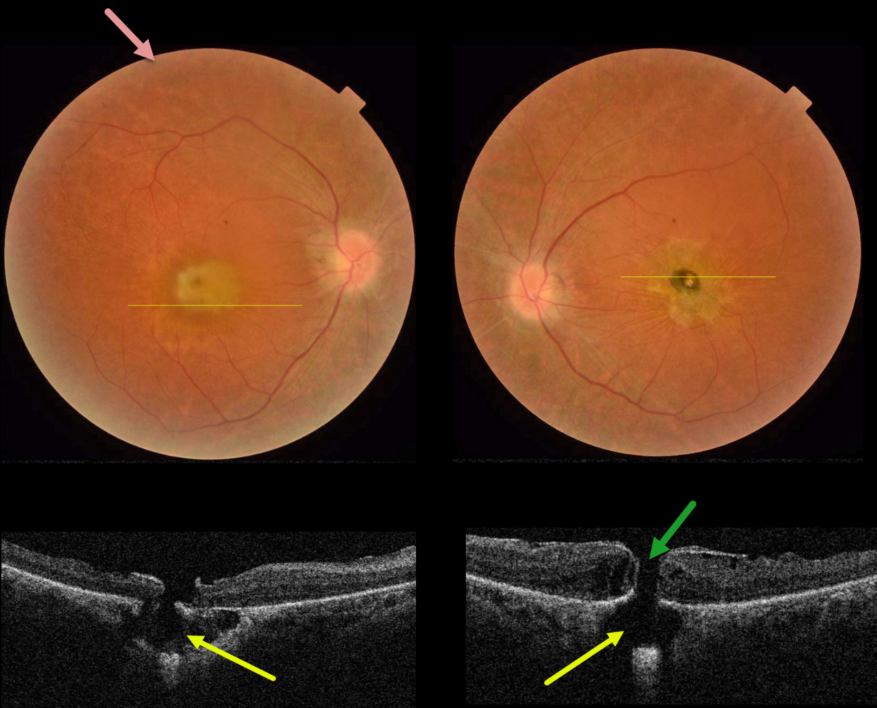
RETINITIS PIGMENTOSA WITH CHOROIDAL CAVITATIONS
Omar Mulki and Faisal Fayyad
Originally posted on @retina.rocks August 16, 2023
This 32YO female presented with a known history of severe vision loss from retinitis pigmentosa (RP). Her parents are first-degree cousins, and there was no family history of RP. Vision was hand motion OD and counting fingers OS.
Fundus photography shows bilateral irregular yellow central macular pigmentary changes, with a central hyperpigmented spot OS.
OCT scanning through each macula shows a full-thickness macular hole with an underlying defect in the RPE. Each macular hole is suspended over a choroidal cavitation.
Surgical repair was offered with appropriate counseling regarding the poor visual prognosis, and the patient elected to be observed.
Learning Points:
Initially described by Freund et al (Arch Ophthalmol 2003;121:197-204), intrachoroidal cavitations are most commonly found in high myopes as a peripapillary yellow-orange subretinal lesion.
Ornek and Ornek reported a patient with pathologic myopia and a unilateral macular hole communicating with a choroidal cavitation (Retinal Cases 2020;14:328-330).
Although macular holes can rarely be associated with RP (Jin et al, Retina 2008;28:610-614), to the best of our knowledge, ours is the first occurrence of RP with bilateral macular holes and underlying choroidal cavitations.
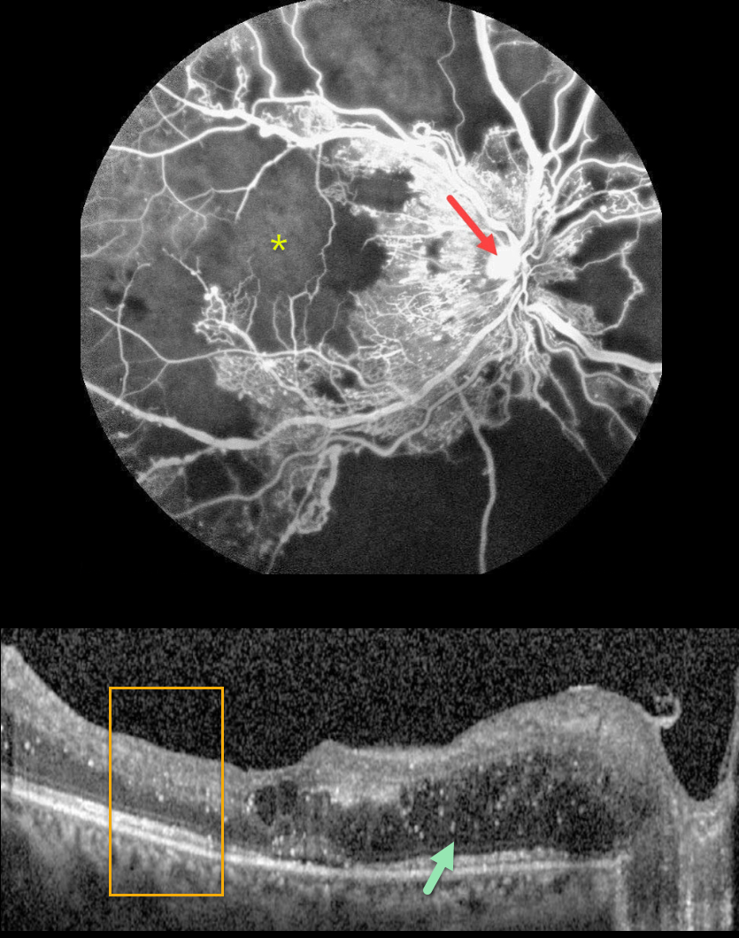
PROLIFERATIVE DIABETIC RETINOPATHY (PDR)
Sehrish Momin and Haroon Tayyab
Originally posted on @retina.rocks July 31, 2023
This 27YO female with a history of uncontrolled type 1 diabetes presented with bilateral vision loss. Vision was 20/40 OD and 20/200 OS.
Fluorescein angiography shows severe capillary loss that extends through the central macula. The remaining vessels are telangiectatic, especially bordering the areas of frank loss, and an area of leaking disc neovascularization is noted.
OCT scanning shows mostly nasal edema of the outer nuclear layer with numerous suspended hyperreflective particles (hyperreflective foci, HRF), along with more central cysts and trace subretinal fluid. The inner retinal layers have lost some of their normal hyper- and hypo-reflective structures (disorganization of the retinal inner layers, DRIL). Similar findings were noted in her left eye (images not shown).
Monthly anti-VEGF therapy was recommended although the patient was subsequently lost to follow-up.
Learning Points:
Anti-VEGF therapy has become the standard treatment for central-involved diabetic macular edema (CI-DME), with the vast majority of eyes showing improvements in macular thickness and vision.
Although we expect our patient’s edema to improve if she returns for treatment, we are more guarded regarding her final acuity since the capillary loss is permanent. Eyes with CI-DME and baseline central DRIL have a worse visual prognosis (Sun et al, JAMA Ophthalmol 2014;123:1309-1316).
HRF are small dot-like retinal opacities found in some eyes with diabetic macular edema. Although their pathogenesis is debated, they likely represent extravasated lipoproteins that can later become clinically evident lipid exudates (Ganne et al, Indian J Ophthalmol 2021;69:3208-3217).
Although HRF numbers decrease with treatment, it is unclear whether they serve as a biomarker for predicting visual outcome (Huang et al, Ophthalmology Retina 2022;6:814-827).
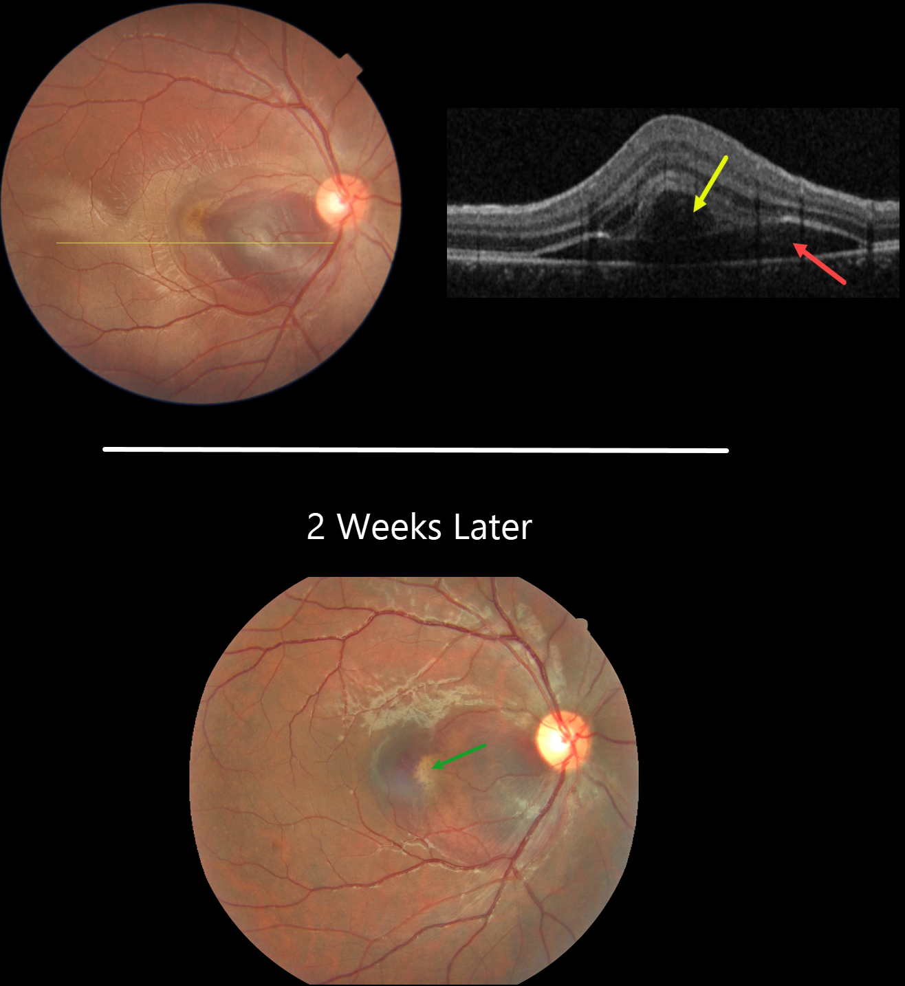
CHOROIDAL RUPTURE
Asma Samsudeen and Ashish Sharma
Originally posted on @retina.rocks July 6, 2023
This 21YO male presented with immediate vision loss after being hit with a cricket ball in his right eye one day earlier. Vision was 20/60.
Fundus photography shows a subretinal hemorrhage in the nasal macula, a small rim of surrounding subretinal fluid, and a more temporal ill-defined commotio retina.
OCT scanning shows hyporeflective retinal elevation from the subretinal blood and fluid, along with an overlying bacillary layer detachment.
Our patient shows several findings classic for acute blunt trauma, including commotio retinae and subretinal blood. This subretinal blood almost always indicates an underlying choroidal rupture, which did, in fact, reveal itself on follow-up 2 weeks later, when vision returned to 20/20. The visual prognosis for these choroidal ruptures is good as long as the rupture spares the foveal center, although patients need lifelong surveillance due to the risk of secondary macular neovascularization.
Learning Points:
A bacillary layer (cone and rod inner and outer segments) detachment (BLD) appears as a unique dome-shaped collection of intraretinal fluid from photoreceptor splitting. These detachments can be observed in an increasing number of conditions, most classically Vogt-Koyanagi-Harada disease (Cicinelli et al, Ophthalmology Retina 2020;4:454-456).
About 8% of eyes with acute non-penetrating ocular trauma have a BLD (Venkatesh et al, Can J Ophthalmol 2022;57:328-336). These resolve within 2-10 days without visual or anatomic sequelae.
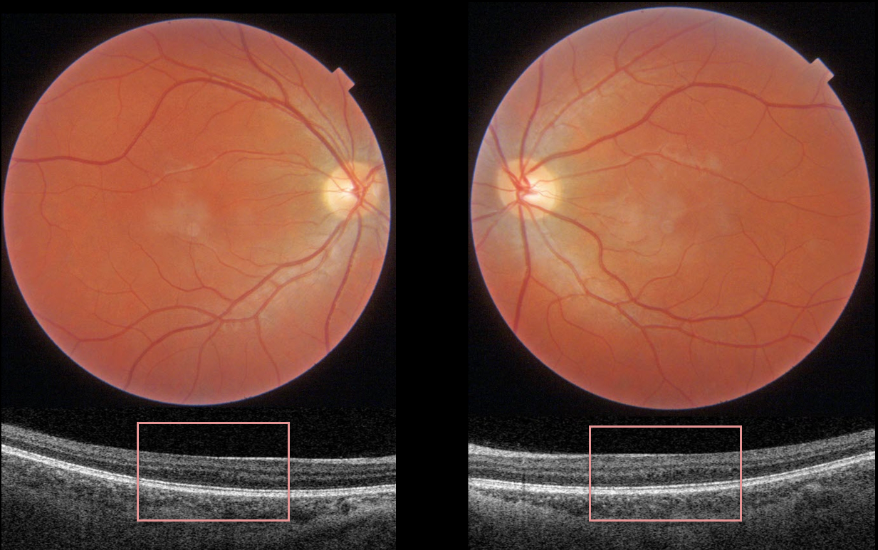
FOVEA PLANA
Originally posted on @retina.rocks June 22, 2023
This healthy 24YO female was referred for asymptomatic OCT findings.
Color macular photography is normal, although swept-source OCT through each macular center reveals a flat foveal contour.
Learning Points:
Fovea plana is characterized by the absence of the foveal pit, which is formed embryologically by the inner retinal tissues and vasculature being displaced centrifugally.
It is most commonly associated with albinism, prematurity, or as an isolated finding. Despite the abnormal foveal architecture, vision is usually normal.
On further questioning, our patient was born 5 weeks prematurely and weighed about 5 pounds. We’re not sure if this caused her OCT findings.
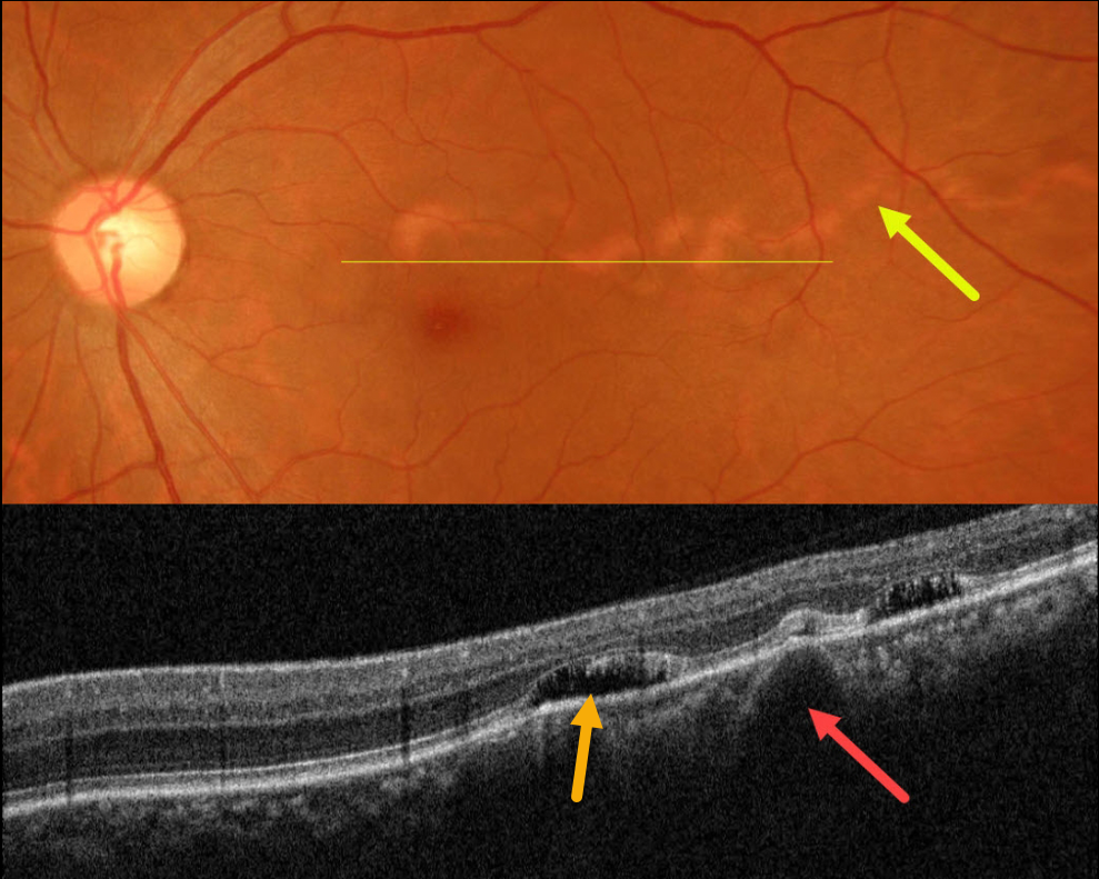
CHOROIDAL MACROVESSEL
Asma Samsudeen and Ashish Sharma
Originally posted on @retina.rocks June 20, 2023
This healthy 41YO female presented with an asymptomatic lesion in her left eye. Vision was 20/20 bilaterally. She is a -7.00 myope.
Color photography shows a serpiginoid-like choroidal atrophic tract extending from the superior macula into the temporal midperiphery.
OCT shows a large dilated choroidal vessel corresponding to this lesion with some pockets of overlying subretinal fluid.
Fortunately, her fovea is uninvolved, and we are following her without treatment.
Learning Points:
Originally reported by Lima et al (BJO 2011;95:1333-1334), choroidal macrovessels are benign dilated choroidal vessels that radiate from the temporal macula towards the midperiphery, somewhat resembling a parasitic tract. They are usually unilateral and found as an incidental finding in an asymptomatic eye.
They appear on OCT as enlarged hyporeflective choroidal structures that compress the surrounding choroid, and changes in the outer retinal bands and RPE are commonly found (Gallo et al, BJO 2020;106:568-575). Localized subretinal fluid overlying the macrovessel is seen in about one-quarter of eyes, which may be caused by overlying choriocapillaris compression with RPE dysfunction.
See Bowen et al (Survey Ophthalmology 2022;67:570-578) for a systematic review, suggesting that choroidal macrovessels are dilated posterior ciliary arteries.
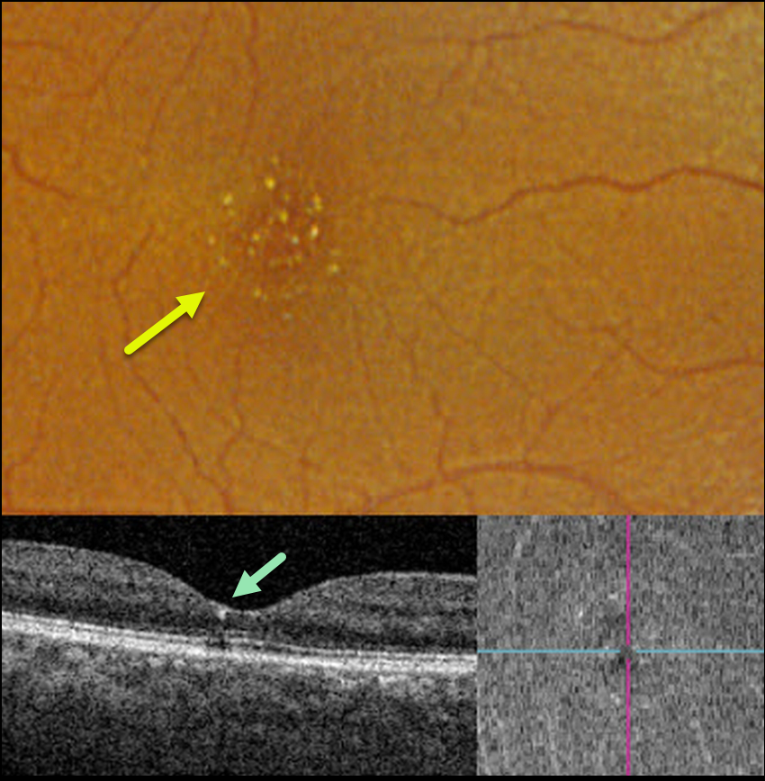
TAMOXIFEN RETINOPATHY
Aaron McNulty
Originally posted on @retina.rocks June 19, 2023
This 56YO female was referred for asymptomatic macular findings. The referring doctor mentioned that she may have retinal changes from the Tamoxifen that she was taking for over 3 years for breast cancer. Vision was 20/50 OD and 20/40 OS.
Optos color RGB imaging shows numerous tiny inner retinal crystalline deposits scattered throughout each fovea. These deposits appeared as tiny hyperreflective dots beneath the ILM on OCT B-scan and en face.
Our patient had already spoken with her oncologist before seeing us and had already stopped the Tamoxifen. The crystalline changes will likely remain and, hopefully, not progress going forward.
Learning Points:
Tamoxifen retinopathy shares phenotypes very similar to those of macular telangiectasia type 2 (MacTel 2, Lee et al., Ophthalmology Retina 2020;3:681-689), especially in the early stages, including retinal cavitations, right-angle venules, and inner retinal crystals.
The changes in Tamoxifen are confined to the central macula, whereas in MacTel2 they are present in a slightly larger area with an epicenter temporal to the foveal center (Hess et al, Ophthalmology Retina 2023;7:101-110). The retinal changes for both disorders likely share Muller cell dysfunction as the common cause.
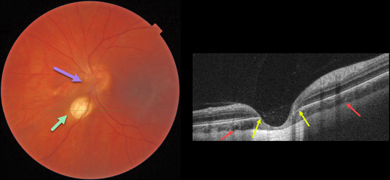
CHOROIDAL COLOBOMA + PERSISTENT FETAL VASCULATURE
Lesia Khrystych
Originally posted on @retina.rocks May 22, 2023
This 35YO female has a history of decreased vision in her left eye since childhood. Vision was 20/20 in her right eye and stable at 20/200 in her left eye. Anterior segments were normal except for a posterior polar cataract OS.
Color imaging of the left posterior pole shows a fairly round, one-disc-diameter depigmented lesion just inferonasal to the disc. An area of non-specific pigmentary changes is noted just nasal to the nerve, along with persistent fetal vasculature (hyaloid artery).
OCT through the depigmented lesion shows a choroidal coloboma. The underlying sclera is intact and slightly bowed posteriorly. Within the coloboma, there is loss of the outer retinal layers with draping of the inner retina against the bare sclera. The RPE is also absent within the coloboma.
Learning Points:
Choroidal colobomas result from a failure of closure of the optic vesicle and choroidal fissure during normal fetal development, but can also be caused by congenital toxoplasmosis and Zika virus. They can be solitary or multiple, and the visual potential depends on the extent of optic nerve and macular involvement. Patients with choroidal colobomas are at an increased risk for retinal detachment and macular neovascularization.
Embryologically, the hyaloid artery is vital to the proper development of the anterior segment, and usually regresses at about 10 weeks of gestation; however, it may persist in some cases. In severe cases, the eye may be microphthalmic with leukocoria and retinal traction. These eyes are typically amblyopic. A genetic association has not been identified.
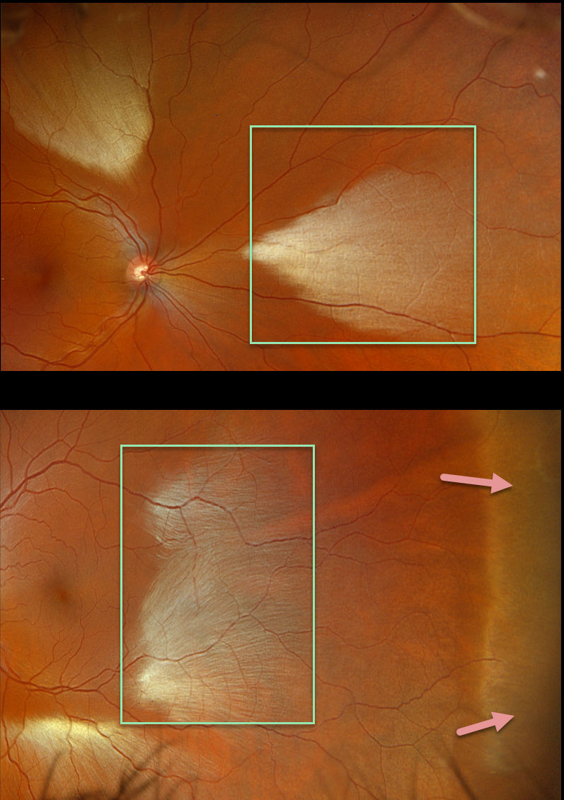
MYELINATED NERVE FIBER LAYER + WWP
Originally posted on @retina.rocks May 18, 2023
This healthy 35YO male was referred for asymptomatic fundoscopic changes. Vision was 20/20 OU.
Opto imaging shows large multifocal patches of myelinated nerve fiber layer (NFL) in both eyes. Temporal white without pressure (WWP) is also noted in his left eye.
Learning Points:
Myelinated NFL is a relatively uncommon, unilateral or bilateral, usually isolated finding present at birth. The myelinated NFL is often connected to the optic nerve, but not necessarily as in this case. Most patients are asymptomatic, although some may have a corresponding relative scotoma. The myelination can progress in about 10% of cases and can also resolve following retinal ischemic events.
Myelinated NFL occurs when retinal nerve fibers develop a myelin sheath, which usually stops posterior to the lamina cribrosa. The myelination may represent an oligodendrocytic choristoma (see Rao et al Retina 2019;39:1125-1132).
WWP is a relatively common benign peripheral finding, most often seen in darkly pigmented individuals. Typically, there are bilateral peripheral geographic areas of whitish retinal discoloration. WWP gets its name because the area appears white without indentation, unlike scleral depression (white with pressure). It can sometimes be confused with retinal breaks or detachment.
The exact cause of this phenomenon is unknown. Historically, it was felt to be due to a benign vitreoretinal interface change, but more recent OCT findings show increased reflectivity in the outer retina (see Diaz et al, Retina 2014;34:1020-1021).
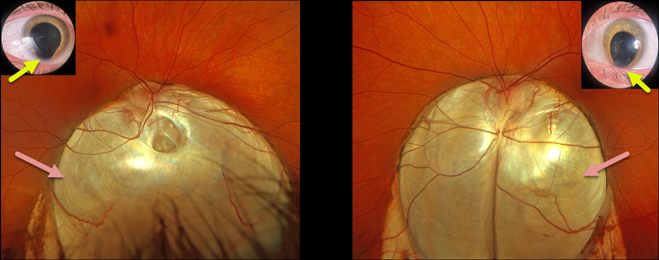
CHOROIDAL COLOBOMA
Originally posted on @retina.rocks May 15, 2023
This 30YO female presented with a 6-month history of vision loss in her right eye. Despite being born with bilateral iris and choroidal colobomas, her vision in each eye was excellent until recently. Vision was 20/200 OD and 20/40 OS.
Optos RGB imaging shows bilateral inferior choroidal colobomas extending from just superior to the optic nerves towards the inferior peripheries. A shallow macula-off retinal detachment extends from the temporal edge of the right coloboma into the 7 to 10 o’clock periphery. In the office, we could not identify any retinal breaks. At vitrectomy, a small anterior retinal tear was found at the superior edge of the detachment.
Learning Points:
Choroidal colobomas are caused by failure of the optic vesicle and choroidal fissure to close during fetal development. Choroidal coloboma can present as solitary or multiple lesions, as in this case. Visual prognosis depends on the location and involvement of the optic nerve and macula.
The retina within the coloboma is variably thinned, increasing the risk of rhegmatogenous retinal detachment (RRD). More centrally within the coloboma, the retina consists of a monolayer of atrophic glial structures and neurons, known as the intercalary membrane (ICM). Between the edge of the coloboma and the ICM is a region of thinned retina lacking RPE and choroid, the marginal ICM (MICM). Breaks within the MICM, which extend about 2 disc diameters from the coloboma margin, are usually the cause of RRD (Tanaka et al, Ophthalmology Retina 2021;5;702-710).
Fortunately, our patient had a more typical RRD that was independent of her coloboma. Laser treatment along the edge of the coloboma was therefore not performed, thereby avoiding the risk of iatrogenic paracentral vision loss. The retina remained attached 5 weeks postoperatively, and vision improved to 20/100.
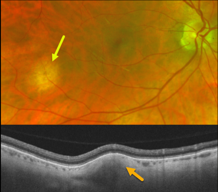
FOCAL SCLERAL NODULE
Ryan Kern and Alex Hynes
Originally posted on @retina.rocks May 11, 2023
Optos color imaging shows an amelanotic subretinal lesion in the right inferotemporal midperiphery. Swept-source OCT through this lesion shows hyperreflective scleral thickening, which compresses the overlying choroid.
Learning Points:
Focal scleral nodule (previously known as solitary idiopathic choroiditis [SIC] and unifocal helioid choroiditis), presents as a single, yellow-white, elevated nodular, avascular subretinal lesion. On OCT it arises from the inner sclera, causing marked choroidal compression.
For the largest series to date and the rationale behind the renaming of this entity, see Fung et al, Ophthalmology 2020;127:1567-1577.
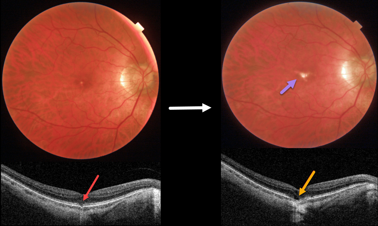
OUTER FOVEAL MICRODEFECT
Originally posted on @retina.rocks April 13, 2023
This 58YO male was referred for an asymptomatic foveal lesion in his right eye. There was a history of high myopia prior to LASIK surgery years earlier. Vision was 20/40 OD.
Color photography shows a small atrophic foveal scar. OCT shows a focal EZ defect with a possible tiny choroidal excavation. He returned about 2 years later for a routine follow-up visit.
Although he was binocularly without a new complaint, his vision had decreased to 20/200. There was now a definite, much enlarged foveal lacquer crack. OCT scanning shows a new outer foveal microdefect.
Learning Points:
Outer foveal microdefects (Cohen et al, Ophthalmology Retina 2021;5:553-561) have been described in numerous conditions, including macular telangiectasia, tamoxifen use, ABCA4 disorders (cone-rod dystrophies, Stargardt disease, and fundus flavimaculatus), phototoxicity, trauma, and vitreomacular traction disorders.
We are not aware of a prior report showing these lesions in association with myopic lacquer cracks. These defects, caused by focal loss of the outer retinal layers, leave an optically empty space that can be mistaken for subretinal fluid but is more of an optical gap within the outer retina.
Focal choroidal excavation (FCE), originally described by Jampol et al in 2006, is likely part of the pachychoroid spectrum since it is usually associated with a thickened choroid and pachyvessels. The FCE can be observed, although symptomatic manifestations, including active central serous retinopathy or macular neovascularization, often require treatment. An excellent review is by Verma et al, Br J Ophthalmol 2021;105:1043-1048.
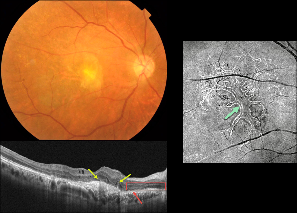
WET AMD WITH OUTER RETINAL TUBULATION
Originally posted on @retina.rocks April 12, 2023
This 63YO male received several intravitreal anti-VEGF injections for neovascular AMD in his right eye, the most recent being 6 months earlier. Vision is counting fingers.
Color photography shows an inactive atrophic foveal scar. OCT scanning shows a hyperreflective macular neovascularization (MNV). The outer retina overlying the MNV is disorganized with several overlying hyporeflective lesions with surrounding hyperreflective rims. More nasally, there is complete RPE loss with bare Bruch’s membrane. The overlying retina is anomalous with additional hypo- and hyperreflective bands.
En face OCT scanning through the outer retina provides a more dramatic view of the extensive tubulation-branching network.
Learning Points:
Outer retinal tubulation (ORT) is often noted overlying inactive MNV with ongoing anti-VEGF therapy and should not be confused with exudative fluid or cysts, which lack a hyperreflective border. The outer hyperreflective band likely represents inner segment mitochondria undergoing fission and translocation toward the nucleus (Litts et al, Retina 2018;38:445-461).
ORT, initially described by Zweifel et al (Arch Ophthalmol 2009;127:1596-1602), is a neurodegenerative condition of the photoreceptors and Muller cells associated with atrophy affecting the outer retina and retinal pigment epithelium, including advanced AMD and inherited retinal diseases.
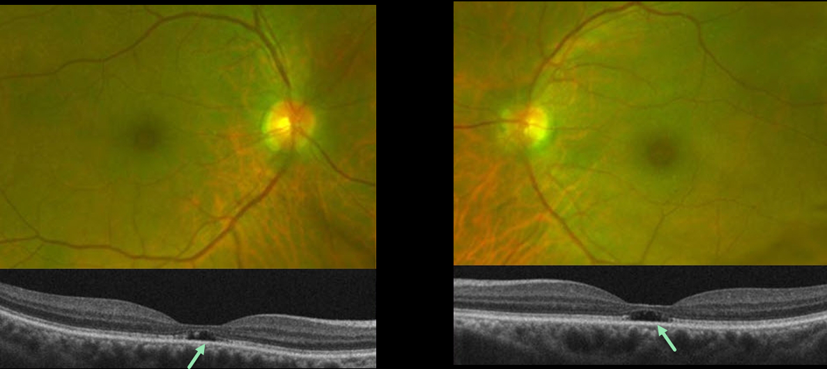
OUTER FOVEAL MICRODEFECTS FROM POSSIBLE IRD
Emily Shepherd
Originally posted on @retina.rocks March 10, 2023
This 65YO presented complaining of difficulty seeing at a distance for some time. She gave a history of radial keratotomy in both eyes about 20 years prior. There was no significant past medical history or family history of eye disease. Vision was 20/60 OD and 20/70 OS.
Optos color imaging shows mild symmetrical pericentral pigmentary change. Fundus autofluorescence and fluorescein angiography were unremarkable (not shown). However, OCT B-scans show bilateral subfoveal loss of the outer retinal tissue external to the external limiting membrane.
An ERG showed rod receptor dysfunction and abnormal dark adaptation with delayed adaptation, while a multifocal ERG was consistent with normal cone function.
Our patient underwent genetic testing and was found to have a heterozygous mutation for FLVCR1. This gene is associated with autosomal recessive posterior column ataxia with retinitis pigmentosa.
A recent report by Kuehlewein et al (Graefe’s Arch Clin Exp Ophthalmol 2019;257:629-638) showed a variety of phenotypes, none of which matched our patient. It is therefore difficult to say whether our patient’s findings are isolated (given the lack of funduscopic, autofluorescence, and angiographic findings beyond the OCT outer defects) or represent an atypical manifestation (given the abnormal electrophysiology) of FLVCR1 disease.
She is scheduled to return for follow-up imaging and visual field testing.
Learning Points:
Outer foveal microdefects (Cohen et al, Ophthalmology Retina 2021;5:553-561) have been described in numerous conditions, including macular telangiectasia, tamoxifen use, ABCA4 disorders (cone-rod dystrophies, Stargardt disease, and fundus flavimaculatus), phototoxicity, trauma, and vitreomacular traction disorders.
In our experience, in patients with inherited retinal diseases (IRD), these findings are most common with a pathogenic ABCA4 mutation (Leng et al, Retina 2012;32:1411-1419 and Wang et al Invest Ophthalmol Vis Sci 2022;53(5):28). These defects, caused by focal loss of the outer retinal layers, leave an optically empty space which can be confused with subretinal fluid but are more of an optical gap within the outer retina.
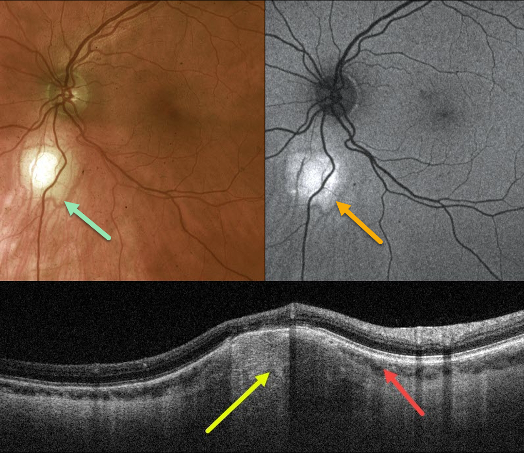
FOCAL SCLERAL NODULE
Originally posted on @retina.rocks March 7, 2023
This 45YO healthy male was referred for an asymptomatic lesion in his left eye. Vision was 20/20 OU.
Optos color imaging shows an amelanotic subretinal lesion just inferior to the left optic nerve. Fundus autofluorescence (FAF) shows this lesion to be hyper-FAF.
Swept-source OCT through this lesion shows a hyperreflective scleral thickening that compresses the overlying choroid.
Learning Points:
Focal scleral nodule (previously known as solitary idiopathic choroiditis [SIC] and unifocal helioid choroiditis), presents as a single, yellow-white, elevated nodular, avascular subretinal lesion. On OCT it arises from the inner sclera, causing marked choroidal compression.
For the largest series to date and the rationale behind the renaming of this entity, see Fung et al, Ophthalmology 2020;127:1567-1577.
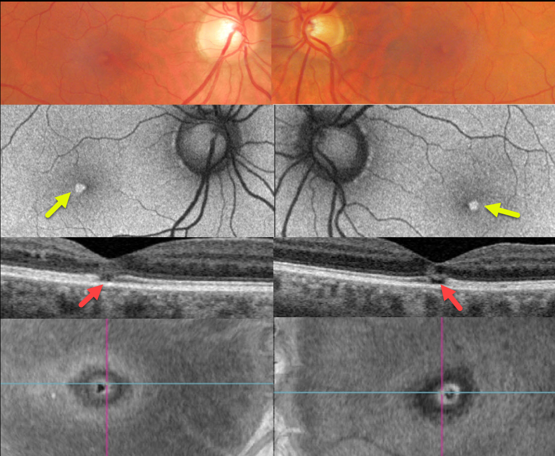
SOLAR RETINOPATHY
Originally posted on @retina.rocks February 27, 2023
This healthy 46YO male complained of mild bilateral central vision loss for about 5 years. Vision was 20/25 OD and 20/30 OS.
Optos color imaging shows subtle central foveal pigment loss. Fundus autofluorescence (FAF) shows these lesions to be hyper-FAF.
Swept-source OCT shows small central foveal outer retinal defects, and en face OCT of the outer retina shows a unique perspective of these lesions.
On further questioning, he noted that his visual symptoms immediately followed his viewing the Great Eclipse of 2017 without proper eye protection.
Learning Points:
The outer retinal damage in solar retinopathy is thought to be caused by photochemical outer retinal and RPE damage instead of a thermal burn. Vision is usually fairly good despite the outer retinal and RPE findings.
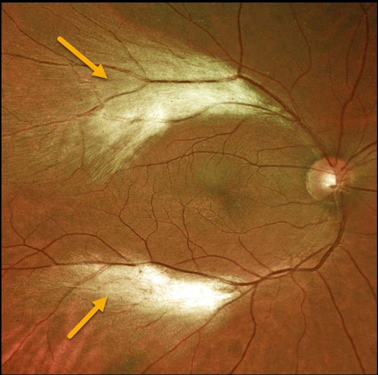
MYELINATED NERVE FIBER LAYER
Originally posted on @retina.rocks February 8, 2023
This 47YO female presented with two distinct patches of myelinated nerve fiber layer (NFL) along the distal temporal arcades in the right eye. Her ocular examinations were otherwise normal.
Learning Points:
Myelinated NFL is a relatively uncommon and usually isolated finding present at birth. Most patients are asymptomatic, although some may have a corresponding relative scotoma. Myelination can progress in about 10% of cases and can also resolve following retinal ischemic events.
Myelinated NFL occurs when retinal nerve fibers develop a myelin sheath, which usually stops posterior to the lamina cribrosa. The myelination may represent an oligodendrocytic choristoma (see Rao et al Retina 2019;39:1125-1132).
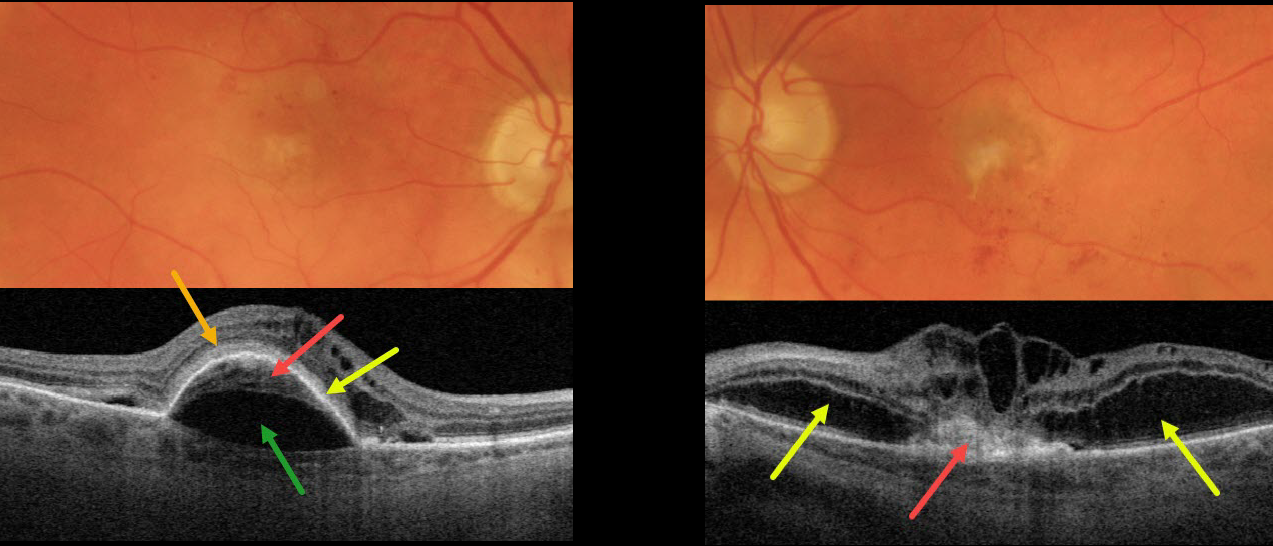
NEOVASCULAR AMD PRECHOROIDAL CLEFT + BACILLARY DETACHMENT
Originally posted on @retina.rocks November 29, 2022
This 87YO patient presented with vision of 20/200 OD and counting finger vision OS from bilateral active neovascular AMD (nAMD).
The right macula has a few dots of central and superior macular blood. OCT scanning shows a type 1 macular neovascularization (MNV, located below the RPE) adherent to an overlying RPE detachment. A prechoroidal cleft separates the MNV from the underlying choroid. Subretinal hyperreflective material (SRHM) lies above the RPE detachment (orange arrow). Intraretinal fluid is noted nasally with trace subretinal fluid on either side of the RPE detachment.
The left macula has faint subfoveal fibrosis with some scattered more inferior subretinal blood. OCT scanning shows a type 2 MNV (located above the RPE), which is tethered to the overlying edematous retina. Bacillary layer detachments are noted nasal and temporal to the MNV.
Learning Points:
A prechoroidal cleft is a hyporeflective space between the RPE and Bruch’s membrane. These are seen in up to about 20% of treated eyes with nAMD, particularly with type 3 neovascularization (intraretinal MNV) and polypoidal choroidal vasculopathy.
They are usually associated with worse visual acuity due to potential complications, including RPE tear and subretinal hemorrhages (Kim et al, Retina 2017;37:2047-2055).
SHRM is an OCT finding that is invisible clinically. This material, located between the neurosensory retina and retinal pigment epithelium (RPE), is thought to consist of fluid, fibrin, blood, and other fibrovascular tissues.
SHRM is a biomarker of active disease that decreases with successful anti-VEGF therapy (Willoughby et al., Ophthalmology 2015;122:1846-1853). Persistent SHRM is associated with an increased incidence of scar formation and worse VA.
Bacillary layer (cone and rod inner and outer segments) detachment appears as a unique dome-shaped collection of intraretinal fluid from photoreceptor splitting. These detachments can be observed in an increasing number of conditions, most classically Vogt-Koyanagi-Harada disease (Cicinelli et al, Ophthalmology Retina 2020;4:454-456).
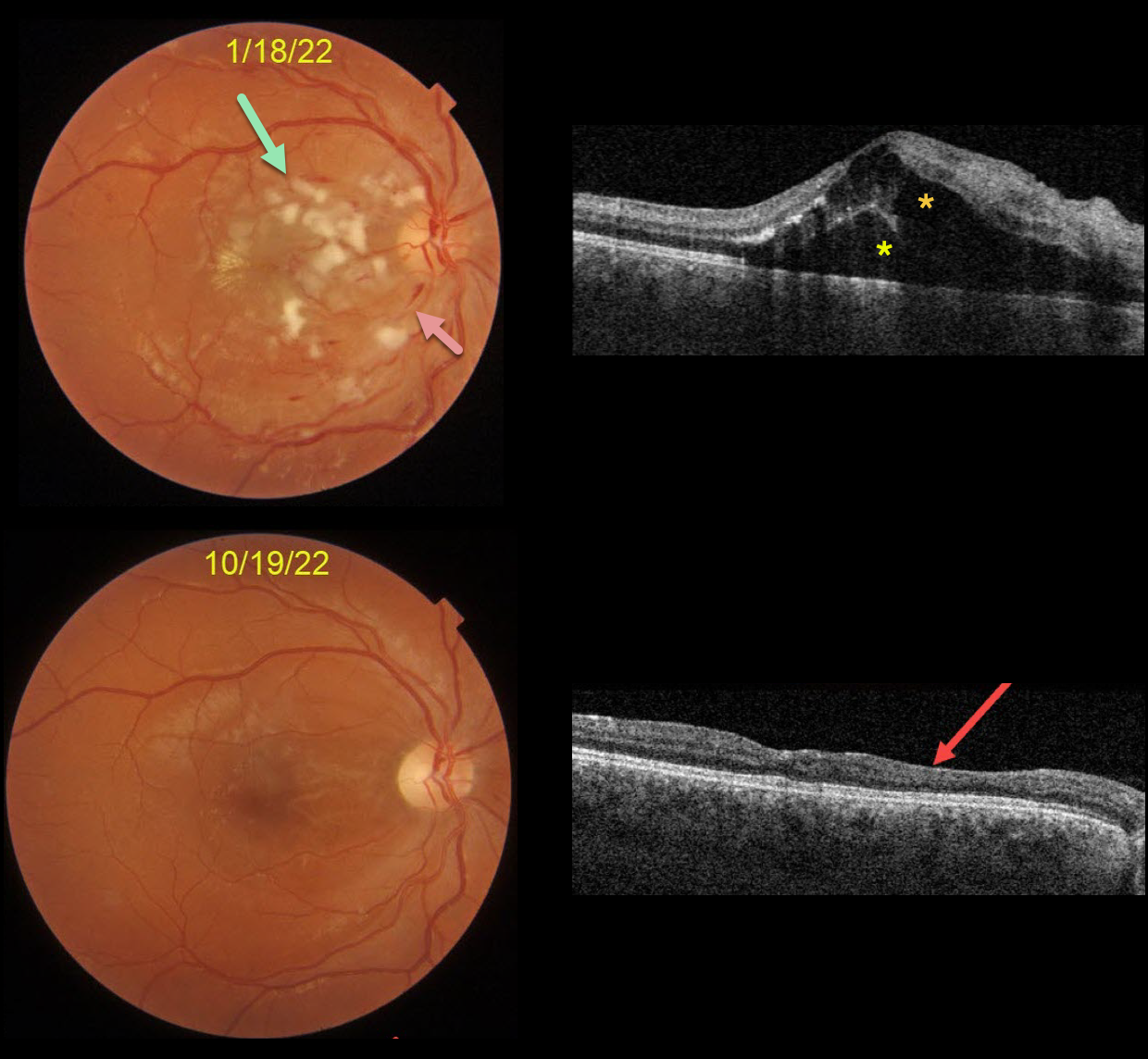
HYPERTENSIVE RETINOPATHY
Originally posted on @retina.rocks November 24, 2022
This is a follow-up from our 2/4/22 post of a patient with malignant hypertension.
This 22YO male presented on 1/18/22 with subacute vision loss of 20/200 OD and 20/50 OS. Blood pressure was 169/135. Color imaging shows nerve fiber layer infarcts (cotton-wool spots) and mild inner retinal hemorrhages.
OCT scanning shows mostly outer macular edema emanating from the nerve, with foveal subretinal fluid.
Nine months later, the macular and OCT findings have mostly normalized. OCT scanning shows thinning with disorganization of retinal inner layers (DRIL) nasally OD.
Learning Points:
Malignant hypertension, defined as blood pressure above 180 systolic and/or 120 diastolic, is a life-threatening medical emergency. Eye doctors are in a unique position to often diagnose this condition.
Patients will often present with bilateral optic nerve swelling, flame-shaped retinal hemorrhages, and, with more chronic disease, lipid precipitates in the nasal macular outer plexiform layer.
Since the macular fluid emanates from the optic nerve, the macular thickening is always worse nasally (as in our case), and is an important clinical clue for this diagnosis.
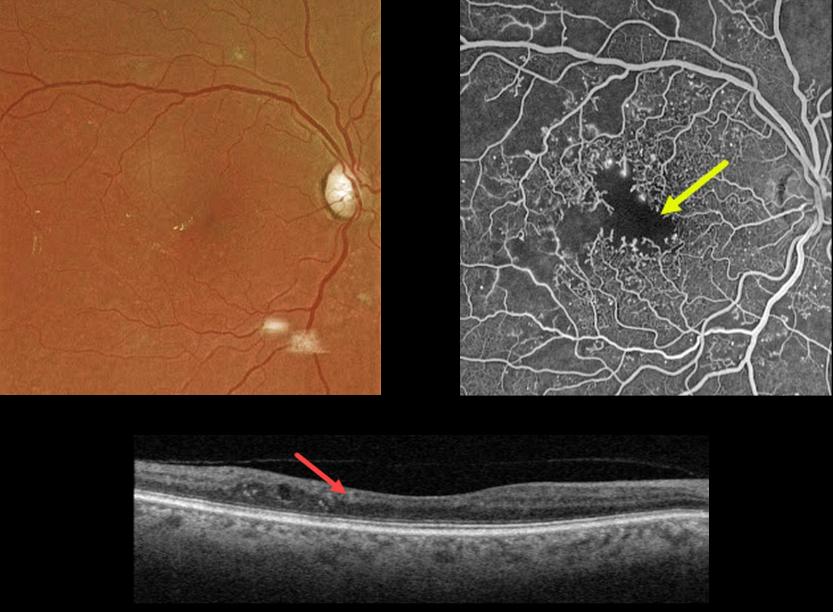
DIABETIC MACULAR ISCHEMIA
Originally posted on @retina.rocks November 23, 2022
This 67YO male has a history of type 2 diabetes. Vision is 20/40.
Optos color imaging is fairly unremarkable, with some mild temporal foveal lipid. However, fundus fluorescein angiography shows foveal ischemia with an enlarged, irregular foveal avascular zone along with more peripheral superotemporal retinal ischemia.
Swept-source OCT shows a thinned, relatively featureless inner retina, also known as disorganization of inner retinal layers (DRIL).
Observation was recommended. The ischemic retina and DRIL will not improve with anti-VEGF therapy.
Learning Points:
DRIL is seen in various retinal vascular disorders, including diabetic macular edema and retinal vein occlusions. It is generally associated with worse vision and increased anti-VEGF treatment burden (see Babiuch et al, JAMA Ophthalmol 2019;137:38-46).
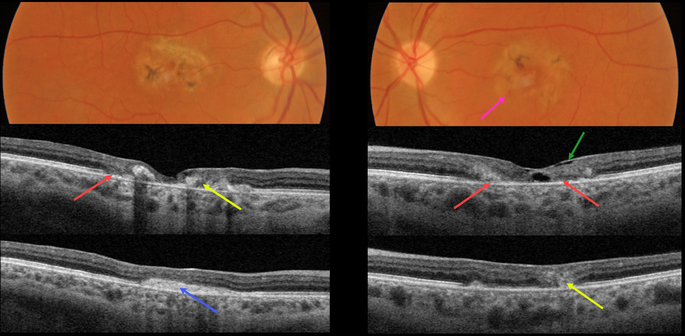
MACULAR TELANGIECTASIA
Originally posted on @retina.rocks November 8, 2022
This 49YO female presented with several weeks of vision loss in her left eye. A macular neovascularization (MNV) in her right eye was treated 4 years earlier with intravitreal Avastin. Vision was 20/200 OD and 20/70 OS.
Both maculas had central pigment loss with intraretinal pigment migration. A small dot of blood was noted inferonasally in the left macula.
OCT scanning shows outer retinal atrophy, pericentral retinal disorganization, a fibrosed, inactive MNV in the right macula, and hyporeflective loss of retinal tissue (cavitation) in the left outer macula. Faint loss of the temporal inner retinal tissue is highlighted by intact internal limiting membrane (ILM drape sign).
Anti-VEGF therapy was started for a presumed MNV in her left eye.
Learning Points:
Originally described by Gass (Arch Ophthalmology 1982;100:769-780), MacTel2 is a neurodegenerative disorder, most likely originating from Muller cell dysfunction.
The term, telangiectasia, is misleading in that the funduscopic findings are mostly non-vascular. The refractile inner retinal ‘crystals’ are thought to be Muller cell footplates. The foveal retina shows a whitish discoloration, most likely due to retinal opacification caused by dysfunctional Muller cells.
Photoreceptor loss allows RPE cells to migrate along intraretinal capillaries, causing the coarse clinical pigment clumping.
All of these changes are usually in the temporal fovea, so our patient is atypical in that they involve the entire central macular regions.
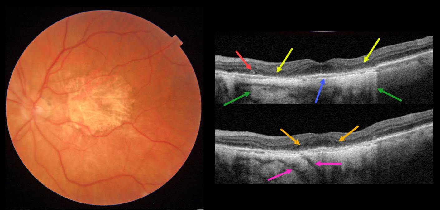
AMD MACULAR ATROPHY
Originally posted on @retina.rocks November 2, 2022
This 84YO female has stable advanced non-exudative AMD with bilateral macular atrophy (MA). There are numerous classic findings on multimodal imaging of her left eye.
Color imaging shows foveal MA. Swept-source OCT shows pericentral outer retinal and RPE atrophy. This atrophy allows for more light to illuminate the underlying choroid and sclera, as well as visualizing two short posterior ciliary arteries posterior to the sclera. The variably atrophic RPE allows for visualization of Bruch membrane, which is normally fused in the RPE-Bruch layer seen in normal eyes. There is a tiny area of outer retinal tubulation (ORT) nasally. Finally, hyporeflective outer plexiform layer wedge defects are noted along the descending outer retinal atrophy.
Wedge defects develop at the boundaries of the degenerating outer plexiform layer in about 75% of MA eyes (Mones et al, Ophthalmology 2012;119:1412-1419).
ORTs represent photoreceptor and Muller cell degeneration (Dolz-Marco et al, Ophthalmology 2017;124:1353-1367). These ORTs have a hyper-reflective border with a central hyporeflective core.
Learning Points:
In 1970, Dr. Gass originally described macular atrophy from AMD as “geographic areas of atrophy” in the setting of “senile macular choroidal degeneration” (Schmitz-Valkenberg, Retina 2016;36:2250-2264).
Today, the terminology is much more specific due to advances in technology and improved visualization of retinal and choroidal structures.
From Sadda et al, Ophthalmology 2018:537-548, the new classification is as follows: Complete RPE and Outer Retinal Atrophy (cRORA), Incomplete RPE and Outer Retinal Atrophy (iRORA), Complete Outer Retinal Atrophy (cORA), and Incomplete Outer Retinal Atrophy (iORA).
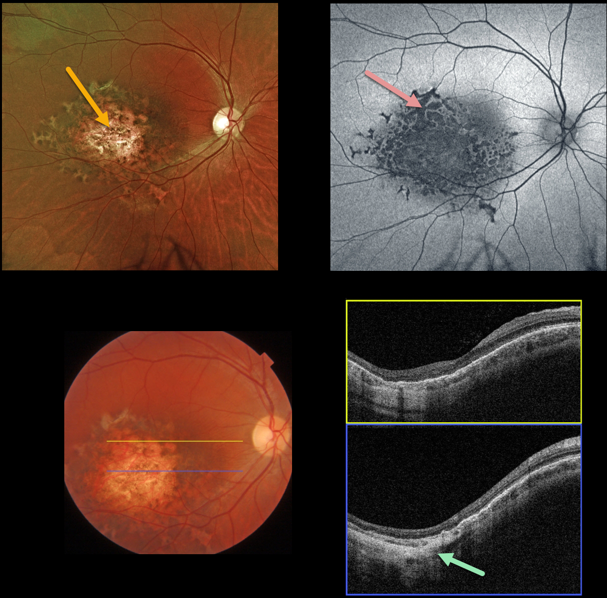
RPE DYSGENESIS WITH CHOROIDAL COLOBOMA
Originally posted on @retina.rocks October 31, 2022
This healthy 32YOF was referred for an asymptomatic lesion in her right macula. Vision was 20/30 OU, and her left fundus was normal.
Optos color imaging shows an atrophic, variably pigmented macular colobomatous lesion with a surrounding rim of scalloped pigmentation.
Fundus autofluorescence (FAF) shows variable, mostly hypo-FAF, indicating RPE dysfunction/loss.
Swept source OCT scanning shows outer retinal/RPE thinning with a colobomatous/staphylomatous outpouching.
Learning Points:
Retinal pigment epithelium dysgenesis (RPED) was first described by Cohen et al (Arch Ophthalmol 2002;120:512-516) as a unilateral, idiopathic lesion affecting the RPE and outer retina. These rare lesions have a pathognomonic scalloped margin with associated RPE atrophy and fibrosis.
Although initially described as a unilateral condition, it can also appear bilaterally (Renz et al, Arch Ophthalmol 2012;130:1341-1340). The lesions can enlarge with time and also carry a small risk for secondary macular neovascularization (Shimoyama et al, Case Rep Ophthalmol 2014;5:34-37) and presumed RPE tumor (Gal-Or et al, Retinal Cases & Brief Reports 2019;13:121-126).
To the best of our knowledge, our patient’s lesion is unique by the colobomatous appearance which has not been previously described.
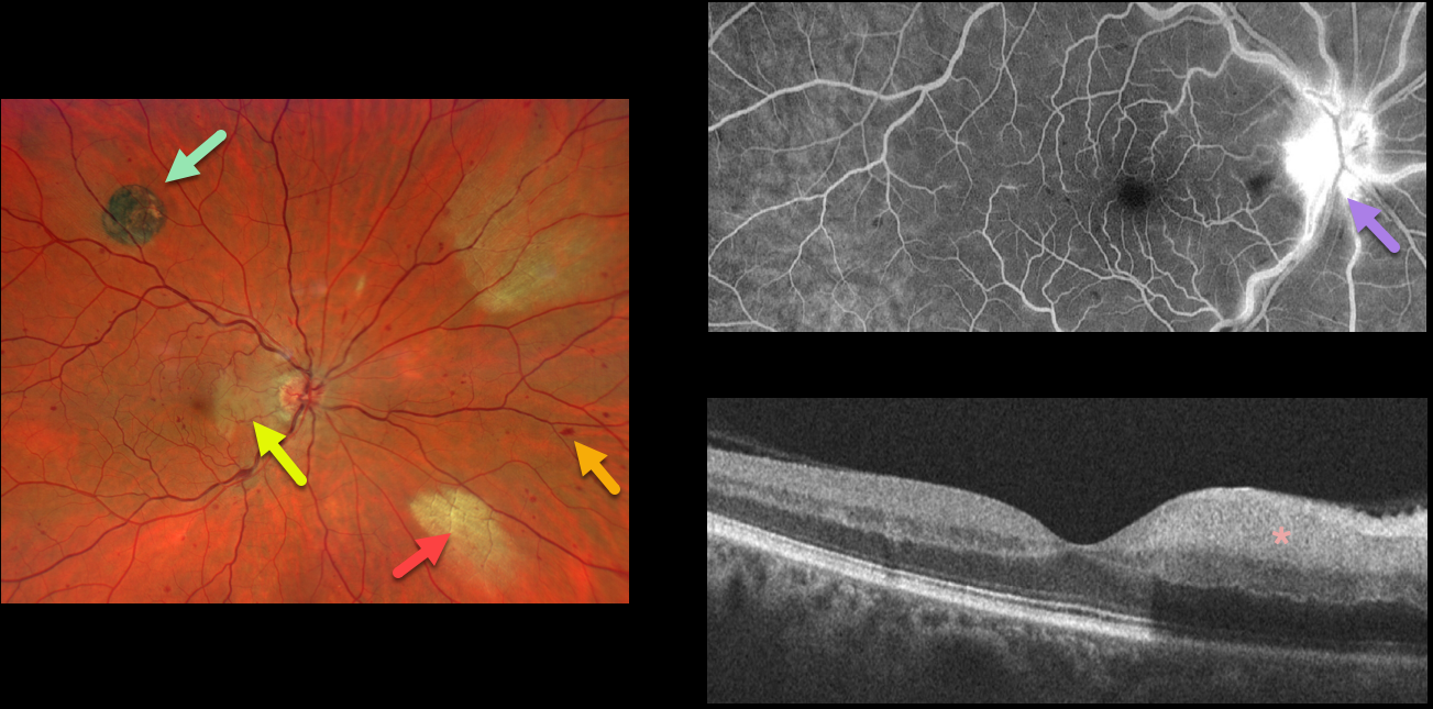
CRVO + CHRPE + MYELINATED NERVE FIBER LAYER
Will Gibson
Originally posted on @retina.rocks October 27, 2022
This 52YO male presented with acute visual symptoms in his right eye. Vision was 20/20.
Optos imaging shows a non-ischemic central retinal vein occlusion (CRVO) with mild scattered retinal hemorrhages in all quadrants. An acute nasal macular branch retinal artery occlusion (BRAO) is noted, along with a small round patch of congenital hypertrophy of the RPE (CHRPE) superotemporally, and two patches of myelinated nerve fiber layer (NFL) nasally.
Fluorescein angiography shows a well-perfused central retinal vein occlusion (CRVO) with late optic nerve leakage. OCT scanning shows hyperreflectivity of the inner nasal retina.
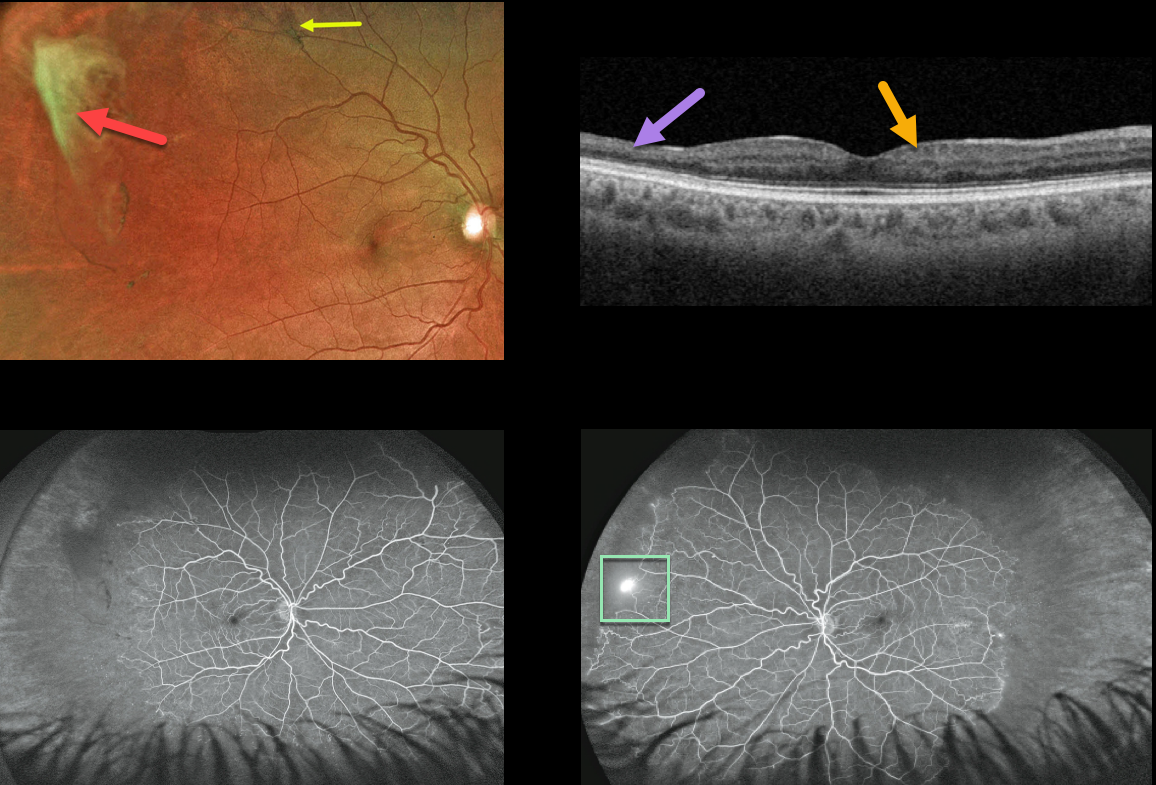
SICKLE CELL RETINOPATHY
Originally posted on @retina.rocks October 21, 2022
This 39YO African American male presented with asymptomatic proliferative sickle cell retinopathy with spontaneously avulsed bilateral superotemporal peripheral fibrosed retinal neovascularization (sea fans). A small midperipheral black sunburst lesion is seen superotemporally in his right eye.
Swept-source OCT shows variable inner retinal thinning and disorganization of the retinal inner layers (DRIL).
Fluorescein angiography shows a patch of retinal neovascularization OS at the junction of perfused and non-perfused retina.
Since these lesions often auto-infarct, as they did in our patient, we elected to observe the small area of neovascularization in his right eye. He is also at low risk for a vitreous hemorrhage or traction detachment since the vitreous is already detached in this area.
Learning Points:
Patients with sickle cell disease develop symptoms throughout their bodies as stiff, sickled red blood cells block capillary beds. In the eye, this can cause proliferative sickle cell retinopathy (PSR) with sea-fan-shaped areas of peripheral retinal neovascularization developing at the junction of perfused and ischemic retina.
The variable macular thinning noted on OCT is caused by microinfarcts to the retinal microcirculation. Enlargement of the foveal avascular zone is a common OCT angiographic finding (Fares, AJO 2021;224:7-17).
The black sunburst lesions are thought to occur from intraretinal RPE migration in response to photoreceptor damage from intraretinal and subretinal hemorrhage (see Ausdourian et al, BJO 1975;59:710-716).
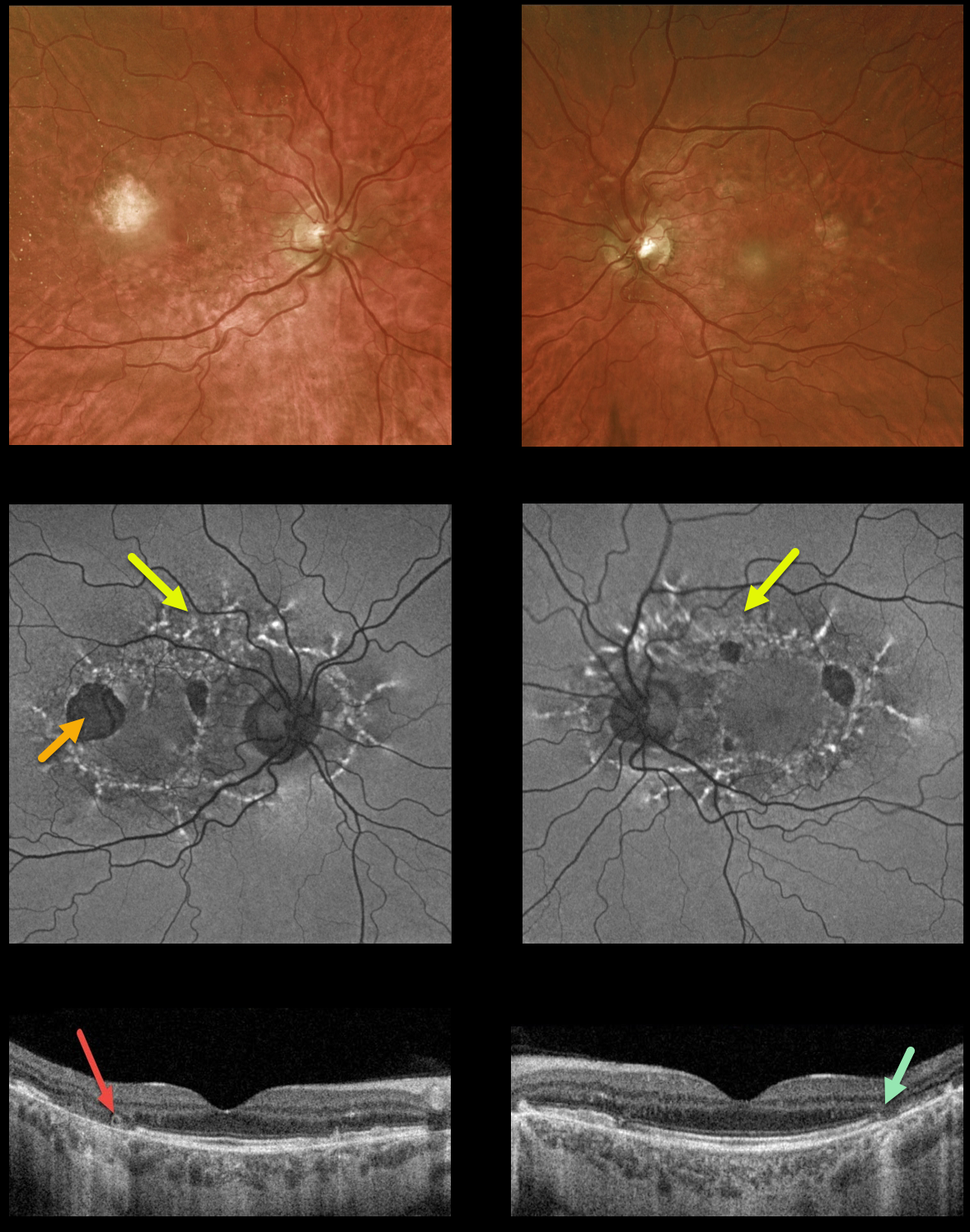
MATERNALLY INHERITED DIABETES AND DEAFNESS (MIDD) + OUTER RETINAL TUBULATION (ORT)
Originally posted on @retina.rocks October 3, 2022
This 59YO male presented with vision of 20/400 OD and 20/30 OS. He has severe lifelong hearing loss, and both he and his mother have type 2 diabetes.
Optos ultrawidefield imaging shows temporal macular atrophy. Fundus autofluorescence shows the true extent of the pathology, with linear interconnected subretinal streaks of hyper-FAF associated with areas of hypo-FAF macular atrophy.
Swept-source OCT shows variable, mostly temporal outer retinal and RPE atrophy, along with a small area of temporal outer retinal tubulation (ORT) OD.
Learning Points: Maternally inherited diabetes and deafness (MIDD) is responsible for up to 3% of all cases of diabetes, and results from a mutation of mitochondrial DNA A3243G. MIDD often masquerades as a pattern macular dystrophy.
Fundus autofluorescence (FAF) in our experience is the best way to visualize these changes. The FAF appearance somewhat resembles that seen with Elmiron toxicity.
Peripapillary hypoautofluorescence, more densely-packed macular autofluorescent changes, and earlier central macular involvement suggest Elmiron toxicity over other causes (see Barnes et al Ophthalmology Retina 2020;4:1196-1201). Note that our case spares the central macula and peripapillary retina, which is more consistent with MIDD.
ORT is often noted overlying inactive MNV with ongoing anti-VEGF therapy and should not be confused with exudative fluid or cysts which lack a hyperreflective border. The outer hyperreflective band likely represents inner segment mitochondria undergoing fission and translocation toward the nucleus (Litts et al, Retina 2018;38:445-461).
ORT, initially described by Zweifel et al (Arch Ophthalmol 2009;127:1596-1602), is a neurodegenerative condition of the photoreceptors and Muller cells associated with atrophy affecting the outer retina and retinal pigment epithelium, including advanced AMD and inherited retinal diseases.
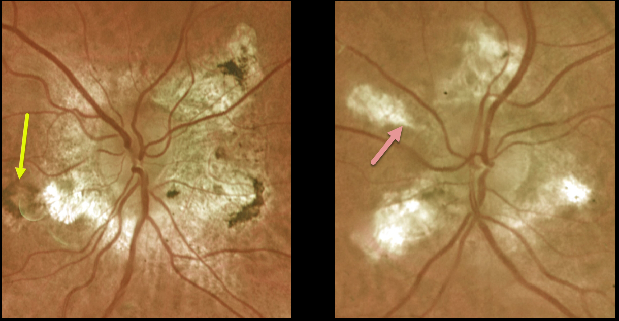
HELICOID PERIPAPILLARY CHORIORETINAL DEGENERATION
Originally posted on @retina.rocks September 30, 2022
This 65YO female is receiving ongoing anti-VEGF therapy in her right eye for macular neovascularization (MNV).
Optos imaging shows an inactive pigmented MNV OD and bilateral peripapillary degenerative propeller-shaped lesions radiating from the optic nerves.
Vision is 20/40 OD and 20/30 OS.
Learning Points:
Helicoid peripapillary chorioretinal degeneration (HPCD) is a benign autosomal dominant disorder caused by a mutation in the TEAD1 gene.
It is characterized by a unique pattern of bilateral peripapillary chorioretinal atrophy. These degenerative wing or propeller-shaped lesions radiate centrifugally from the optic nerve head.
Vision is usually normal, although central vision can rarely be affected if atrophy progresses through the fovea. MNV can rarely develop as in our patient (see Triantafylla et al, Eur J Ophthalmol 2016;26:e30-e31).
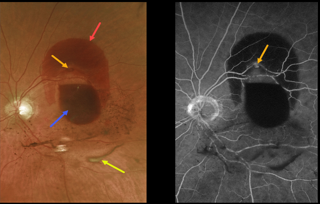
RETINAL ARTERIAL MACROANEURYSM (RAM)
Originally posted on @retina.rocks September 1, 2022
This 85 YO female presented with a retinal arterial macroaneurysm (RAM) with counting fingers vision.
The macroaneurysm has ruptured, causing multiple levels of blood shown on fundus photography and fluorescein angiography. The blood is subretinal superiorly, subhyaloid more centrally, and in the vitreous (both red and devitalized). We are observing this patient.
Learning Points:
A RAM is thought to occur in a weakened arterial wall from arteriosclerosis, and appears as a yellow-white dilated lesion along a retinal arterial within the first 3 bifurcations from the optic nerve.
There are 2 distinct presentations, hemorrhagic or exudative. The hemorrhagic type often presents, as in this case, with pathognomonic multiple layers of blood.
Acute macular hemorrhage can be treated with observation, anti-VEGF injections, or thermal laser.
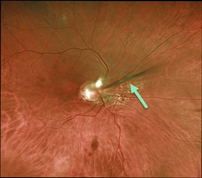
PERSISTENT FETAL VASCULATURE
Originally posted on @retina.rocks August 23, 2022
This 48YO male gave a history of blindness in his right eye since birth. Vision was no light perception. The anterior chamber was shallow with a stalk of tissue radiating from the nerve to the back of his crystalline lens. The macula was hypoplastic. The left eye was completely normal.
Learning Points:
Persistent fetal vasculature (PFV) is a failure of regression of fetal vessels and a common cause of leukocoria and subsequent amblyopia. There is no identifiable genetic association.
Characteristically, the affected eye is microphthalmic with a shallow chamber, a varying degree of cataract, and retrolental and/or posterior fibrovascular membranes that can cause retinal traction.
If diagnosed earlier in life, PFV can be surgically treated with a combined lensectomy and vitrectomy.
We are observing our patient, and it is unclear why he has no light perception vision, given the relatively mild structural findings.
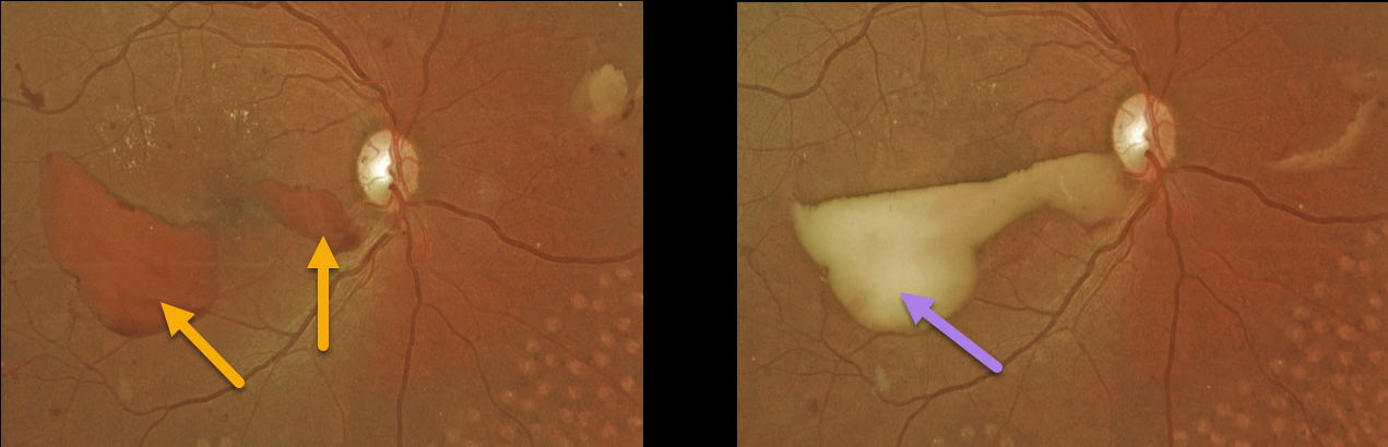
PROLIFERATIVE DIABETIC RETINOPATHY (PDR)
Originally posted on @retina.rocks August 17, 2022
This 37YO female presented with recent vision loss in her right eye. She had a history of prior laser treatment elsewhere several years ago. Vision was 20/30 OD and 20/25 OS.
Red preretinal blood was seen in the inferotemporal right macula with partial PRP scarring. One month following a single Avastin injection, the devitalized preretinal blood was yellowing. Bilateral PRP is planned.
Learning Points:
PRP has been around since the 1960s and still remains the mainstay for treating proliferative disease. Often considered a “one and done”, patients can still develop break-through hemorrhage or progressive neovascular growth and traction.
Intravitreal anti-VEGF injections are often helpful, but some will require vitrectomy.
In our patient’s case, the new preretinal blood was most likely due to her having been incompletely treated rather than a PRP failure.
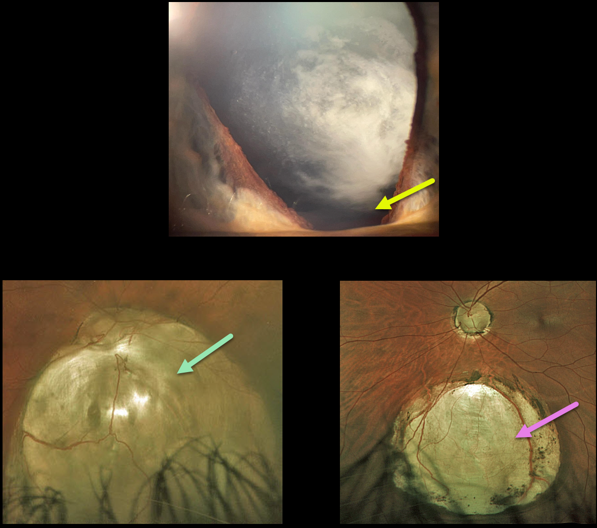
CHOROIDAL + IRIS COLOBOMA
Originally posted on @retina.rocks July 5, 2022
This 42YO state trooper presented with a few months of vision loss in his right eye, including significant glare with headlights. He reported a history of excellent vision throughout his life. Vision was 20/200 OD and 20/20 OS.
He had 3+ nuclear/cortical spoke cataracts consistent with this vision loss. Slit lamp examination shows an inferior iris coloboma OD, and it was normal OS.
Optos imaging of his right eye shows an inferior choroidal coloboma extending from the nerve inferiorly. The macula is normal, and OCT through the macular center is also normal (not shown). A smaller inferior midperipheral choroidal coloboma is noted in his left eye.
Cataract surgery is scheduled, and we expect his vision to return to normal, given his prior history and normal macular anatomy.
Learning Points:
Choroidal colobomas are most commonly due to failure of the optic vesicle and choroidal fissure to close during fetal development. Lesions like this may also be from other causes, including toxoplasmosis. Choroidal coloboma can present as solitary or multiple lesions. Visual prognosis depends on the location and involvement of the optic nerve and macula.
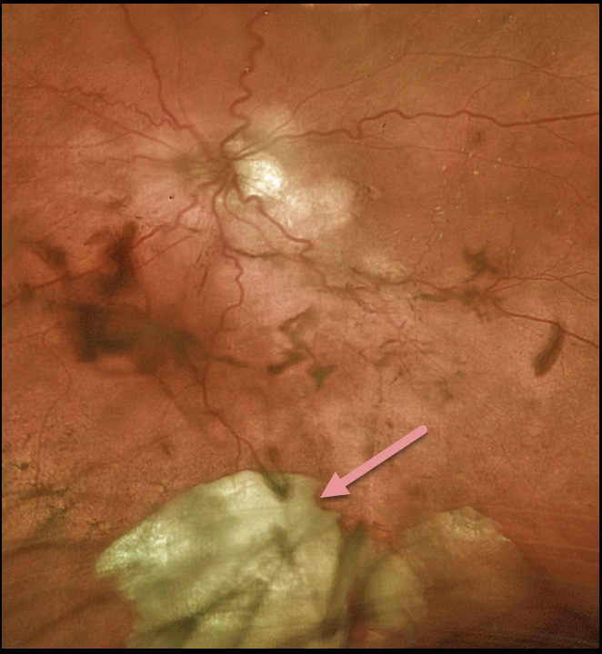
RETAINED LENS FRAGMENT
Originally posted on @retina.rocks June 24, 2022
This 83YO female underwent cataract extraction for a hypermature lens. The posterior capsule tore, and a large lens fragment fell posteriorly. A 3 piece intraocular lens was successfully placed in the sulcus at the time of surgery.
She had previously received multiple intravitreal injections from multiple outside providers for exudative macular degeneration. We have no documentation if the capsule had ever been compromised.
Learning Points:
Posteriorly dislocated lens fragments are a known complication of cataract surgery. The crystalline lens is an immune privileged tissue. When lens fragments get exposed following cataract surgery, severe inflammation and increased eye pressure usually follow.
Rarely, permanent vision loss occurs from glaucoma or cystoid macular edema. Small lens fragments can be managed conservatively, but larger fragments like this one need to be surgically removed.
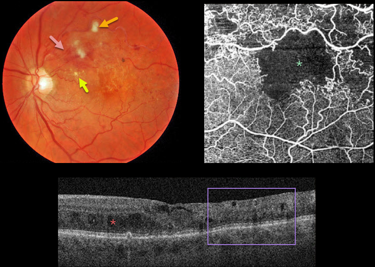
BRVO WITH MACULAR ISCHEMIA
Originally posted on @retina.rocks June 10, 2022
This 79YO male presented with a major superotemporal branch retinal vein occlusion (BRVO) and counting fingers vision.
Fundus photography shows superior macular hemorrhages with inner retinal ischemia (cotton-wool spots) and some nasal lipid. The vessels, especially around the superotemporal fovea, are nonperfused.
The clinically suspected foveal ischemia is confirmed on OCT angiography. Spectral-domain OCT shows disorganization of the inner retinal layers (DRIL), particularly within the clinically ischemic temporal macula, along with scattered inner and outer retinal cysts.
Learning Points:
DRIL is seen in various retinal vascular disorders, including diabetic macular edema and retinal vein occlusions. It is generally associated with worse vision and increased anti-VEGF treatment burden (see Babiuch et al, JAMA Ophthalmol 2019;137:38-46).
Since the patient has no central foveal thickening or retinal neovascularization, we are observing for now. Anti-VEGF therapy will not restore the foveal circulation.
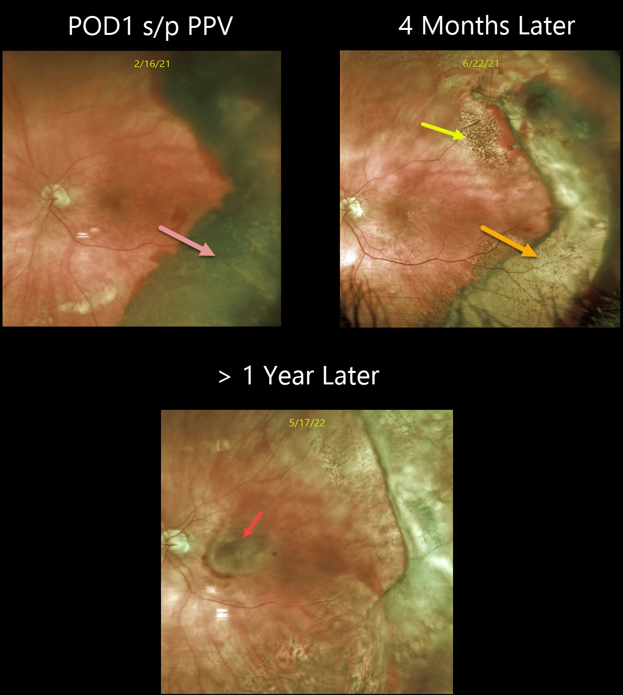
PERIPHERAL EXUDATIVE HEMORRHAGIC CHORIORETINOPATHY
Originally posted on @retina.rocks May 26, 2022
This 80YO female with a known history of dry AMD presented with a 3-week history of poor vision in her left eye. Vision was hand motion from a dense vitreous hemorrhage. B-scan ultrasonography showed a thickened temporal subretinal lesion without retinal detachment (not shown).
One day following vitrectomy, vision improved to 20/100. The temporal subretinal and RPE blood somewhat resembled a uveal malignant melanoma. However, the well-demarcated location and shape of the lesion, along with the posterior rim of subretinal blood, helped establish the diagnosis of a breakthrough vitreous hemorrhage from ectopic choroidal neovascularization (CNV), also known as peripheral exudative hemorrhagic chorioretinopathy (PEHCR).
Four months later, the resolved devitalized blood was turning yellow. The coarse subretinal pigmentation, most prominent superotemporally, is pathognomonic for prior subretinal blood.
She was subsequently lost to follow-up until she presented with a 6-month history of decreased vision. Although the PEHCR ridge continued to contract, vision was counting fingers due to a new macular neovascularization. Anti-VEGF therapy was started.
Learning Points:
PEHCR is an exudative process similar to wet AMD but located in the retinal periphery. Ectopic CNV can often masquerade as a choroidal mass or uveal melanoma.
It can also cause extensive choroidal/suprachoroidal, subretinal, and vitreous hemorrhage, more commonly in patients who are on blood thinners.
The subretinal blood often spontaneously regresses without treatment, although anti-VEGF injections can be used for symptomatic or increasing exudation.
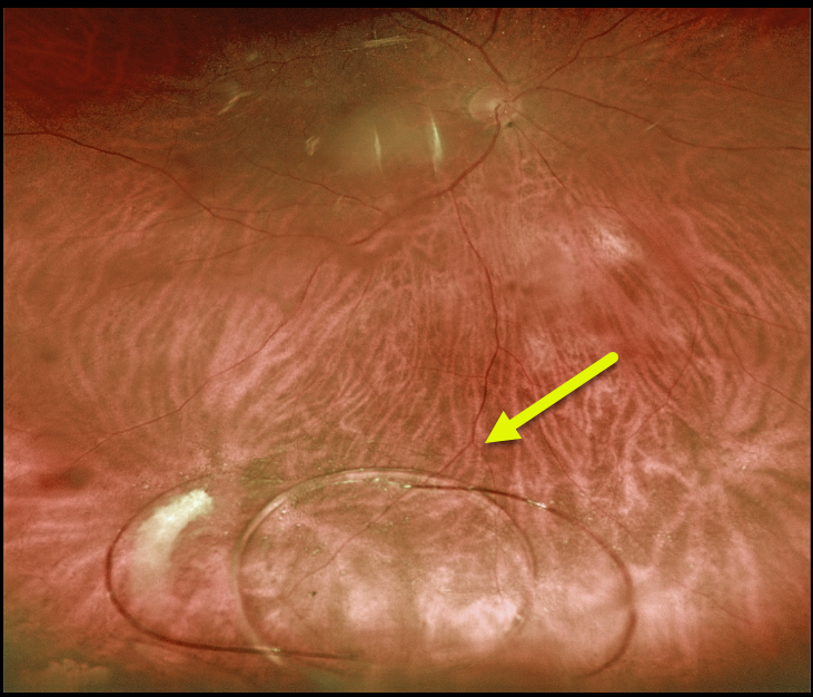
DISLOCATED IOL
Originally posted on @retina.rocks May 24, 2022
This 78YO male presented with a several-month history of decreased vision in his right eye. He had prior bilateral cataract surgery years earlier at an outside practice. Vision was 20/25 OU. His right posterior chamber implant was decentered inferiorly, but the optic remained in the visual axis.
The lens subsequently completely dislocated into the inferior vitreous before the anterior segment surgeon could perform scleral fixation.
He subsequently underwent successful vitrectomy, lens explantation with placement of an anterior chamber implant.
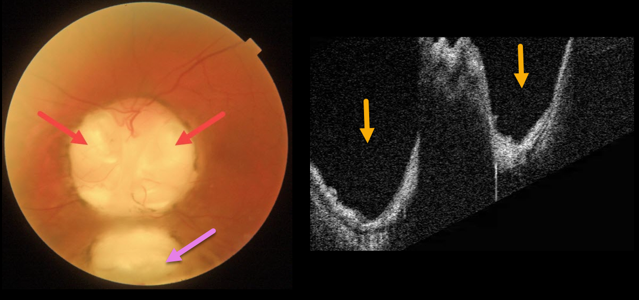
OPTIC NERVE COLOBOMA
Originally posted on @retina.rocks May 9, 2022
This 66YO male has mild lifelong decreased vision in his left eye. Vision was 20/70.
Triton imaging of the optic nerve coloboma shows optic nerve pits temporally and nasally, as well as a more inferior choroidal coloboma. Swept-source OCT dramatically reveals the pits.
We are following him yearly.
Learning Points:
Both optic nerve and choroidal colobomas can be unilateral or bilateral and are caused by incomplete closure of the embryonic fissure.
Patients with choroidal colobomas are at increased lifetime risk of rhegmatogenous retinal detachment due to breaks in the overlying thinned retina.
Optic nerve pits are also associated with serous macular detachment and macular neovascularization.
For a great review of cavitary optic nerve abnormalities, see Jain and Johnson, AJO 2014;158:423-435.
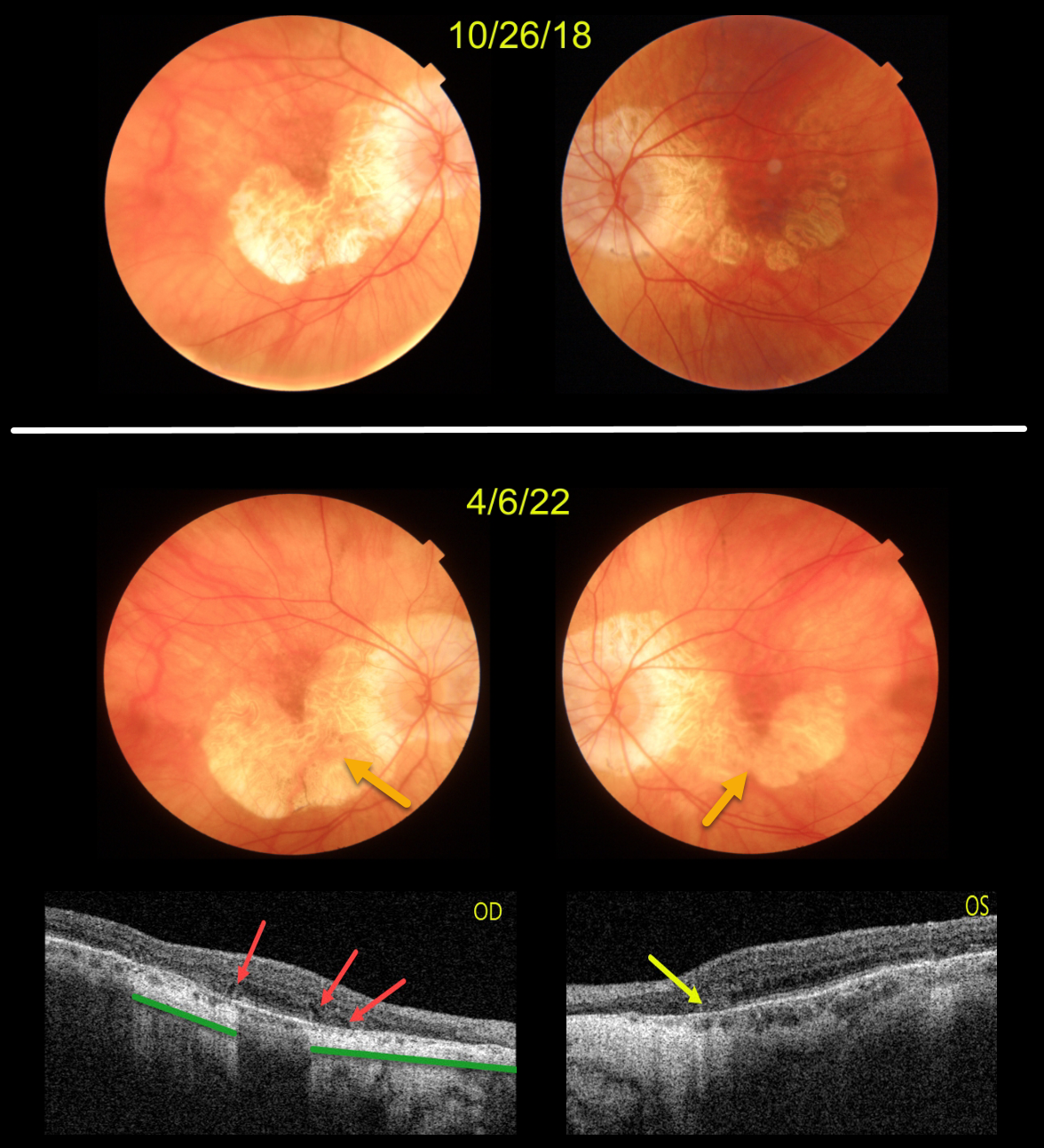
AMD MACULAR ATROPHY
Originally posted on @retina.rocks April 28, 2022
This patient originally presented in 2018 with vision of 20/40 OD and 20/50 OS. Areas of AMD-related macular atrophy (MA) skirted each macular center.
Four years later, vision had decreased to 20/100 OU due to progressive atrophy. Comparing the two images from 2018 to 2022, there is a bilateral linear enlargement for all margins of the atrophy, particularly in the left eye.
Triton swept-source OCT B-scans show classic findings for MA. The right eye has multiple wedge defects, hyporeflective wedges which develop at the boundaries of the degenerating outer plexiform layer in about 75% of MA eyes (Mones et al, Ophthalmology 2012;119:1412-1419). In addition, loss of the outer retina and RPE allows more OCT light to be transmitted into the choroid, resulting in distinct areas of choroidal and scleral hyperreflectivity.
There is a tiny area of outer retinal tubulation (ORT) nasally in the left eye, which represents photoreceptor and Muller cell degeneration (Dolz-Marco et al, Ophthalmology 2017;124:1353-1367). These ORTs have a hyper-reflective border with a central hyporeflective core.
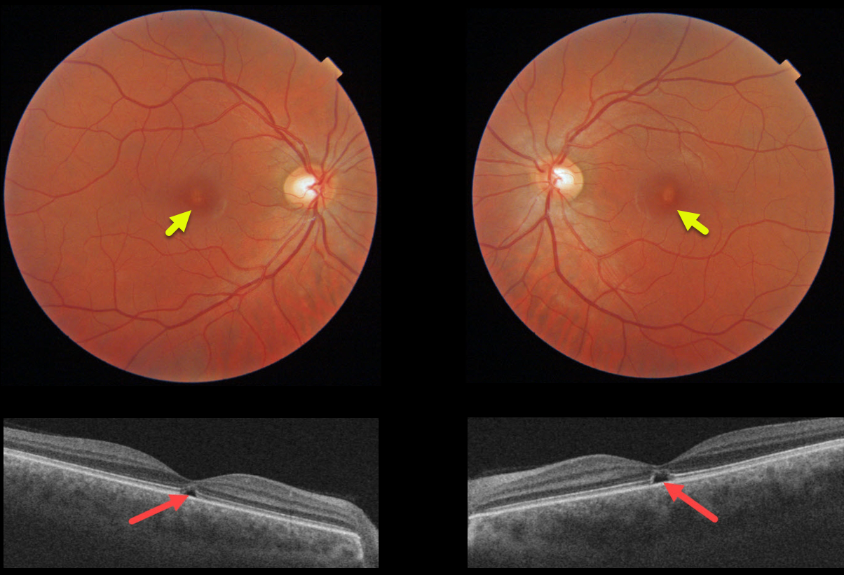
SOLAR RETINOPATHY
Originally posted on @retina.rocks April 27, 2022
This 31YO male was referred for relatively asymptomatic central macular pigmentary changes. He gave a long history of twice-daily sun gazing, which always caused a very pleasurable sneezing episode. Vision was 20/30 OD and 20/40 OS.
Triton color imaging shows tiny foveal pigment loss. Swept-source OCT shows bilateral ellipsoid zone and outer segment defects.
We diagnosed him with the ACHOO reflex (Autosomal Dominant Compelling Helioophthalmic Outburst). And no, this isn’t an April Fools post, and we’re not talented enough to even make this stuff up if we wanted to! Patients with the ACHOO reflex sneeze when exposed to bright lights, typically sunlight.
We advised him to stop his sun gazing ritual, although we’re not sure this will be possible for him.
Learning Points:
The outer retinal damage in solar retinopathy is thought to be caused by photochemical outer retinal and RPE damage instead of a thermal burn. Vision is usually fairly good despite the outer retinal and RPE findings.
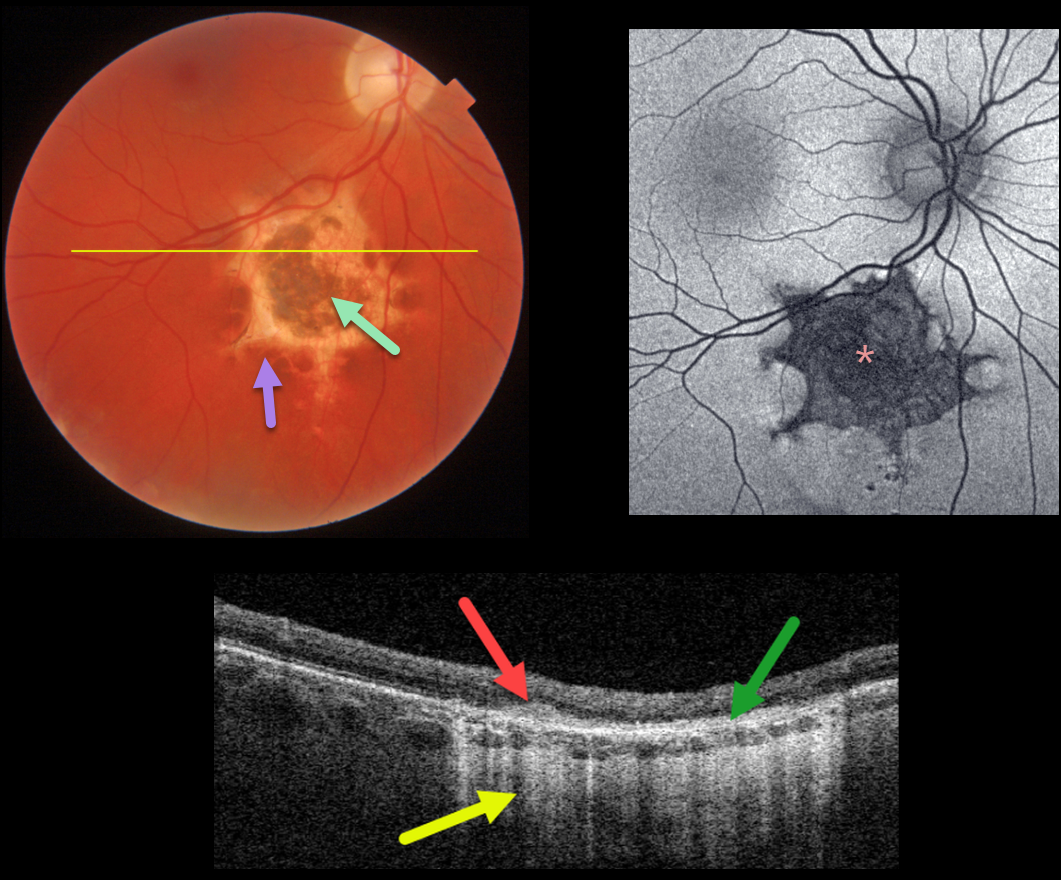
RPE DYSGENESIS
Originally posted on @retina.rocks April 25, 2022
This asymptomatic 34YO male was referred for an asymptomatic lesion just inferior to the right macula. The 2-disc-diameter lesion is hyperpigmented centrally with a scalloped hypopigmented border.
Optos fundus autofluorescence (FAF) shows diffuse hypo-FAF of the lesion due to a lack of RPE lipofuscin from the RPE and outer retinal atrophy.
Triton swept-source OCT through this lesion shows outer retinal hyperreflectivity within the area of hyperpigmentation, disorganized atrophic outer retinal layers and RPE, and hyperreflectivity of the choroid and sclera due to increased light transmittance through the outer retinal and RPE atrophy.
The patient will be monitored yearly for any changes.
Learning Points:
Retinal pigment epithelium dysgenesis (RPED) was first described by Cohen et al (Arch Ophthalmol 2002;120:512-516) as a unilateral, idiopathic lesion affecting the RPE and outer retina. These rare lesions have a pathognomonic scalloped margin with associated RPE atrophy and fibrosis.
Although initially described as a unilateral condition, it can also appear bilaterally (Renz et al, Arch Ophthalmol 2012;130:1341-1340). The lesions can enlarge over time and also carry a small risk of secondary macular neovascularization (Shimoyama et al., Case Rep Ophthalmol 2014;5:34-37) and a presumed RPE tumor (Gal-Or et al., Retinal Cases & Brief Reports 2019;13:121-126).
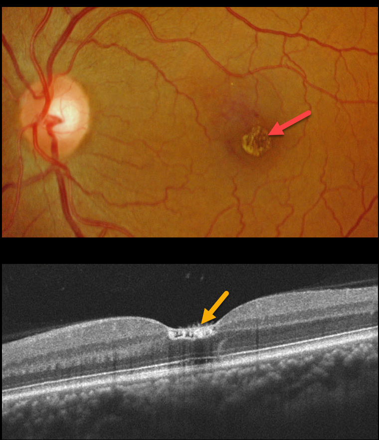
WHITE DOT FOVEA
Originally posted on @retina.rocks April 19, 2022
This 47YO male has 20/30 vision acuity and discrete unilateral inner foveal white dots. He was recently hospitalized with double pneumonia and was significantly immunocompromised.
OCT shows foveal hyperreflective deposits extending from the internal limiting membrane (ILM) to the outer plexiform layer. Some hyperreflective material is also noted within the ILM. The right macula was normal. We felt this most likely represented the white dot fovea.
Learning Points:
Initially described by Yokotsuka et al (AJO 1997;123:76-83), white dot fovea appears as usually bilateral tiny foveal white dots distributed diffusely on the inner fovea or along its margin. Histologically, it remains unclear what the white dots represent, but the configuration may suggest Müller cell end feet.
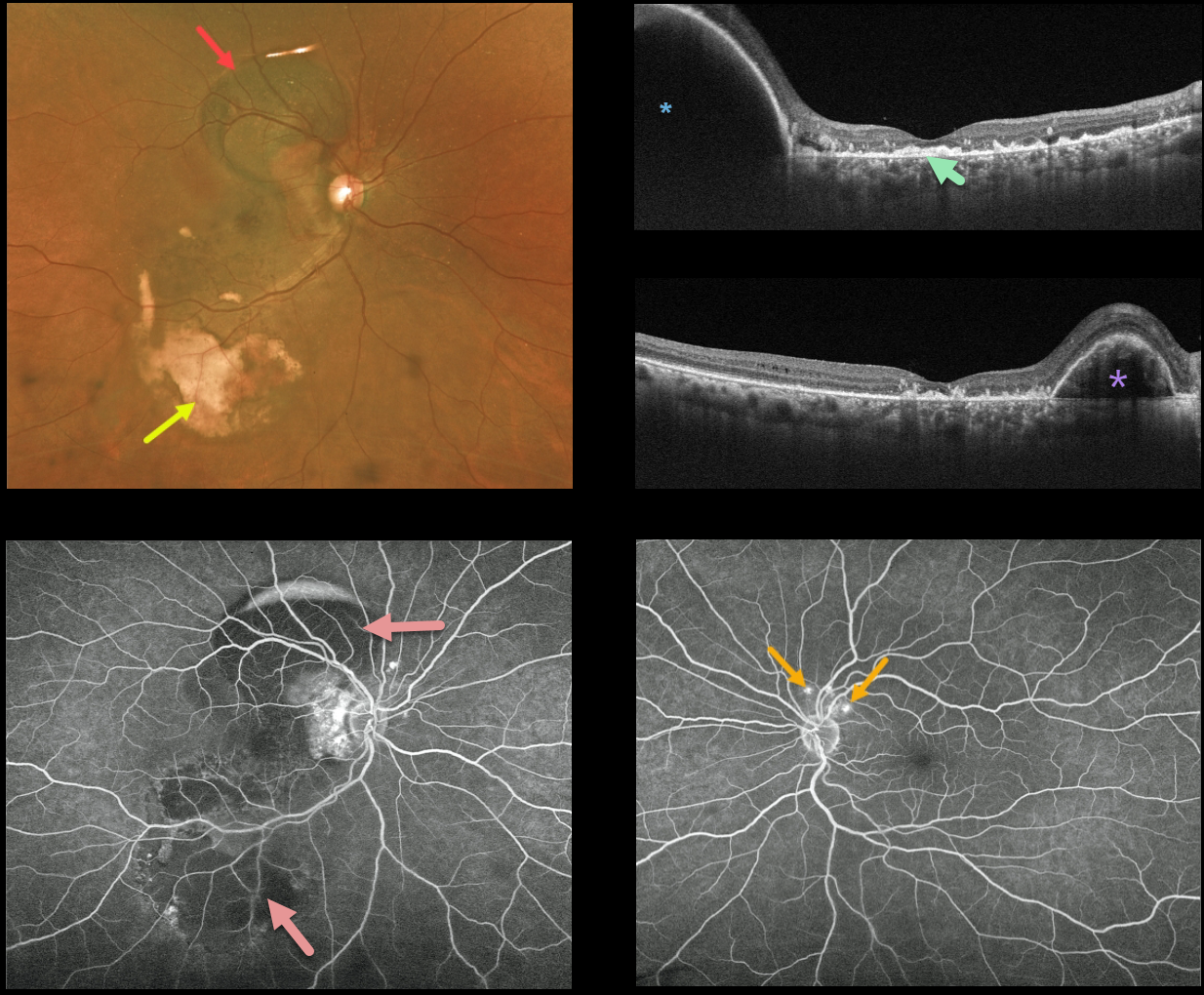
POLYPOIDAL CHOROIDAL VASCULOPATHY
Originally posted on @retina.rocks April 6, 2022
This 59YO female patient presented with variable mostly peripheral submacular blood in her right eye and counting fingers vision. Optos imaging shows the extent of the subretinal blood, which is dark superiorly and yellow inferiorly.
Triton swept-source OCT shows variable outer retinal and subretinal hyperreflective material from resolved submacular blood, and this is seen clinically as the coarse subretinal pigment most evident in the inferotemporal macula. A large RPE detachment (PED) is seen superiorly in the vertical scan, with a smaller PED nasally in the horizontal scan.
Fluorescein angiography shows mostly blockage from the subretinal blood. Several small asymptomatic polyps are noted superior to the left nerve.
Learning Points:
Polypoidal choroidal vasculopathy (PCV) is a variant of type 1 macular neovascularization. PCV is likely distinct from age-related macular degeneration, occurring in a different population (more common in African Americans and Asians), with a relative lack of drusen and a thicker choroid.
The sub-RPE vessels were classically characterized using indocyanine green angiography, although recent literature suggests that OCT is just as effective at diagnosing these vascular polyps.
Historically, these lesions were believed to be somewhat resistant to anti-VEGF monotherapy, often requiring combined treatment with photodynamic therapy.
More recent data from the PLANET study suggest that Eylea monotherapy is also effective (AJO 2019;204:80-89).
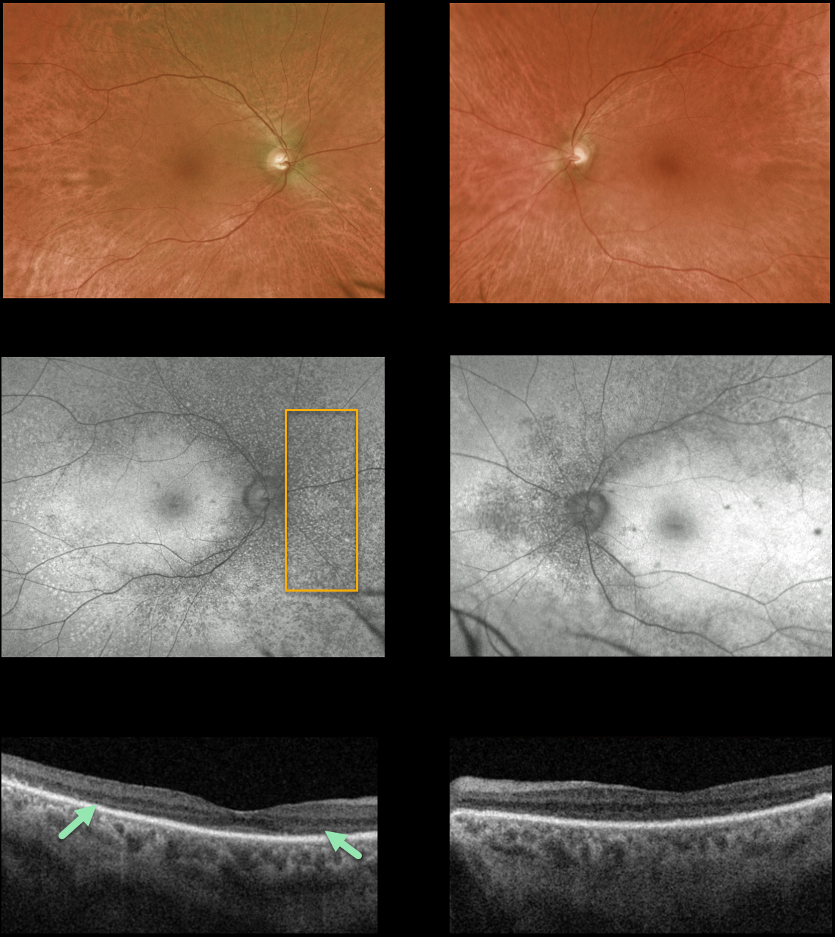
VITAMIN A DEFICIENCY
Originally posted on @retina.rocks March 30, 2022
This 64YO man presented with 1 year of gradual severe bilateral vision loss. Vision was counting fingers at 5 feet OU.
Retinal examinations showed fine subretinal pigmentary changes with tiny whitish dots throughout the extramacular retina, best visualized as hypoautofluorescent spots on Optos ultra-widefield autofluorescence.
Triton swept-source OCT shows profound loss of the outer segment layers.
The patient denied any systemic diseases or current medications, though he admitted he hadn’t been to a physician in years. He appeared in poor health and malnourished, so we ordered nutritional testing, including folate, B12, vitamin A, antiretinal antibodies, and Invitae genetic testing.
All tests were negative except for a decreased Vitamin A level. He subsequently started daily vitamin A supplementation at 20,000 IU, and after a few weeks, his vision improved to 20/400 OD and 20/200 OS without funduscopic or OCT changes.
Learning Points:
Vitamin A deficiency is rare in the United States and is sometimes related to malabsorption in chronic pancreatitis or inflammatory bowel disease. It leads to several well-documented ocular complications, including nyctalopia, Bitot spots, and xanthopsia.
Outer nuclear layer thinning with disruption of the outer retinal bands can be visualized on OCT (Berkenstock et al, Int J Retin Vitr 2020;6:23).
Oral or intramuscular vitamin A replacement has been shown to reverse the retinal changes if initiated early enough. We hope our patient’s vision and retinal findings will improve with continued treatment.
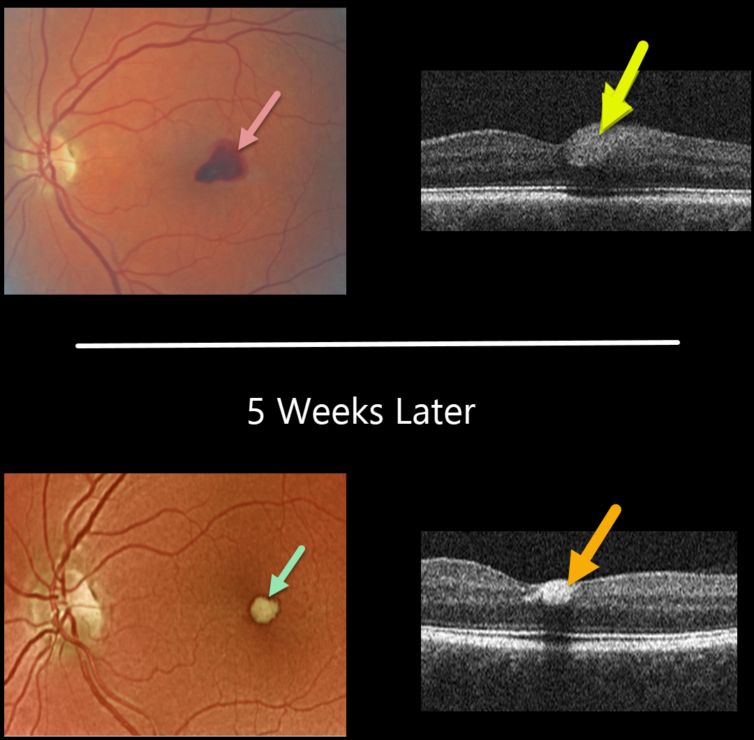
VALSALVA RETINOPATHY
Originally posted on @retina.rocks March 24, 2022
This 56YO female presented with acute vision loss in her left eye from Valsalva retinopathy. Vision was 20/100. A temporal foveal retinal hemorrhage is seen, located within the inner retina on OCT B-scan.
Five weeks later, the devitalized blood is contracting and turning yellow. The blood also became more hyperreflective on the OCT B-scan. All blood subsequently resolved with the return of normal vision.
Learning Points:
Valsalva hemorrhages are usually in the sub-internal limiting membrane (ILM) space, although this case was in the inner retina. These hemorrhages virtually always resolve without sequelae, although the sub-ILM blood can be drained into the inferior vitreous by creating a small opening in the ILM using either a thermal or Nd:YAG laser.
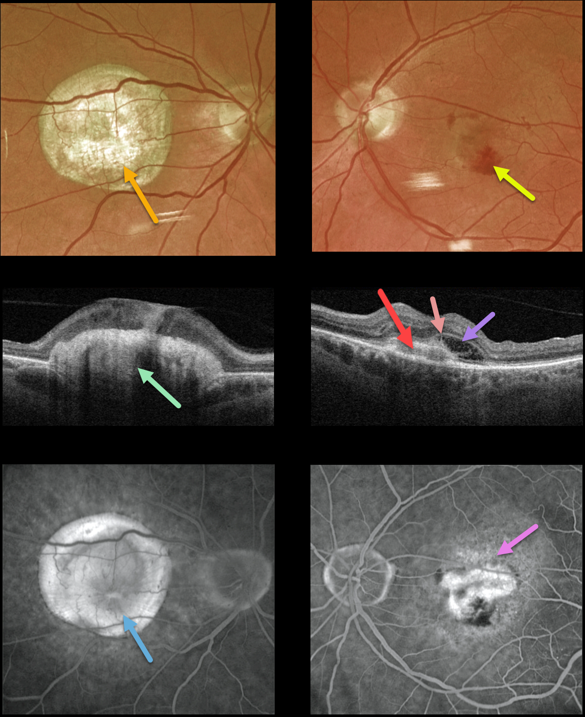
NEOVASCULAR AMD
Originally posted on @retina.rocks March 22, 2022
This 77YO male presented with counting fingers vision OD from a large fibrotic disciform scar and 20/200 vision OS from a new macular neovascularization.
The disciform scar is contracted and fibrotic, with marked hyperreflectivity on swept-source OCT. The MNV in his left eye is seen as opaque fluid with some subretinal blood. The type 2 MNV (located above the RPE) is noted on the OCT B-scan. There is also a bacillary detachment and a hyperreflective line indicating a possible pitchfork sign.
Optos fluorescein angiography shows diffuse staining of the subretinal scarring OD and leakage from the MNV OS.
Learning Points:
Disciform scars were the norm for untreated wet AMD before the anti-VEGF era that began in the mid-2000s. New-onset wet AMD diagnosed and treated with appropriate anti-VEGF dosing virtually never goes on to develop these lesions. There may be progressive macular atrophy following years of therapy, but not this type of fibrotic scarring.
An excellent review of wet AMD fibrotic lesions is in Souied et al’s AOS thesis (AJO 2020;214:151-171).
The pitchfork sign was originally described by Hoang et al (Retina 2013;33:1049-1055) as a unique OCT finding in inflammatory MNV. Falavarajani et al more recently noted that this finding is also associated with non-inflammatory MNV (Ophthalmic Surg Lasers Imaging Retina 2019;50:719-725).
The pathogenesis of the avascular pitchfork spikes is unknown, but may be related to outer retinal traction or Müller cell activation.
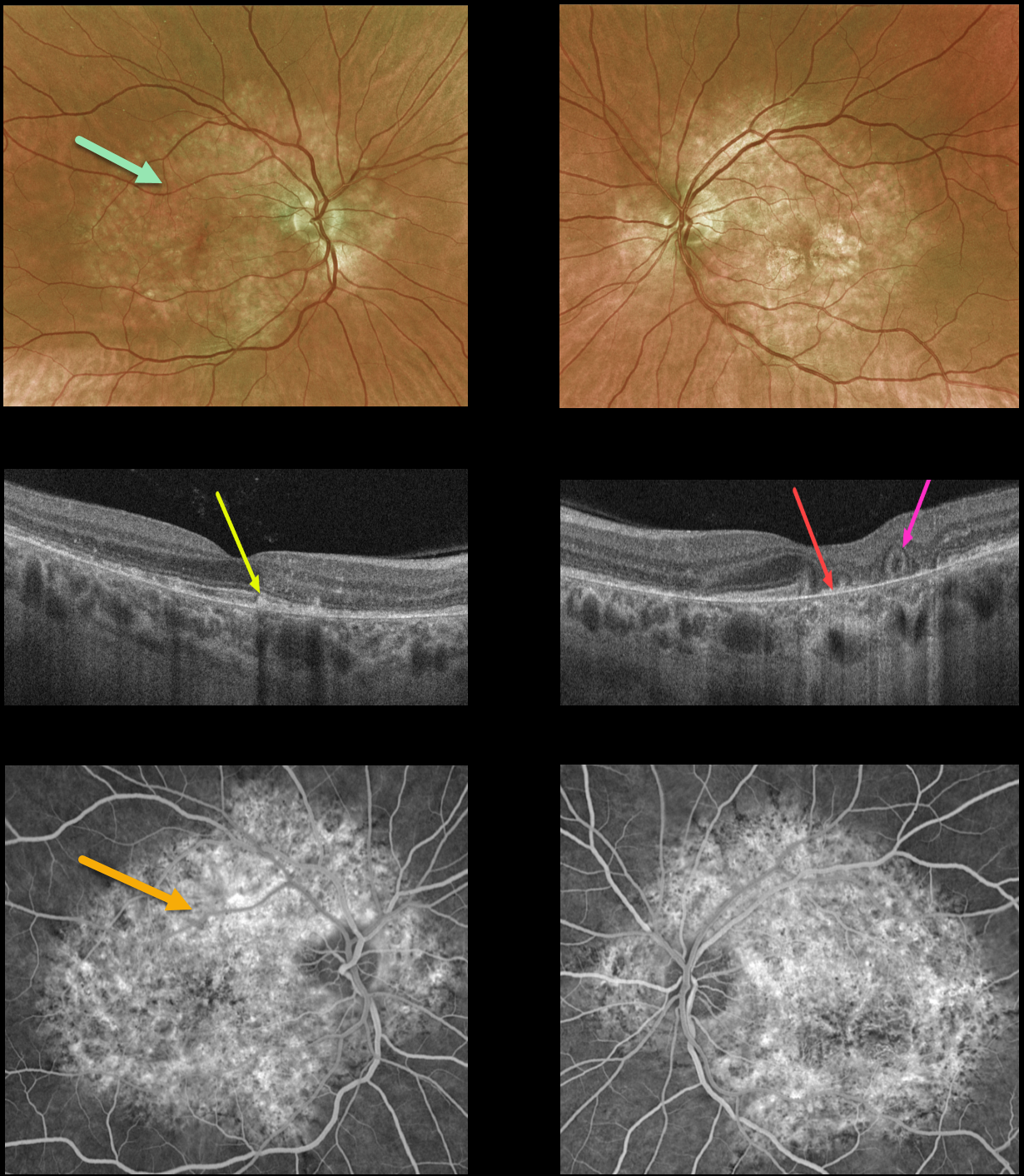
PENTOSAN POLYSULFATE SODIUM MACULOPATHY + OUTER RETINAL TUBULATION
Originally posted on @retina.rocks March 1, 2022
This 54YO female presented with a 9-month history of blurred vision in her left eye. She was on Elmiron (pentosan polysulfate sodium, PPS) for about 20 years due to interstitial cystitis, but stopped the medication before seeing us due to the widespread publicity about its potential retinal toxicity.
Optos color imaging shows variable yellow-orange atrophic pigmentary changes throughout the macular and peripapillary posterior poles.
OCT of the right macula shows scattered hyperreflective lesions mostly within the outer segment layers. The left macular OCT shows variable outer retinal and RPE loss, especially centrally, where bare Bruch’s membrane is visible. There is also an area of outer retinal tubulation (ORT) more temporally.
Fluorescein angiography shows window defects within these regions.
Learning Points:
Elmiron was approved by the FDA in 1996 for treating interstitial cystitis. Recently, a unique PPS retinopathy has been described, and our patient shows classic findings.
Toxicity seems to develop over many years and can mimic more common disorders, including age-related macular degeneration and macular dystrophies.
Peripapillary hypoautofluorescence, more densely packed macular autofluorescent changes, and earlier central macular involvement suggest PPS toxicity over other causes (see Barnes et al Ophthalmology Retina 2020;4:1196-1201), including maternally inherited diabetes and deafness (MIDD).
ORT is often noted overlying inactive macular neovascularization with ongoing anti-VEGF therapy and should not be confused with exudative fluid or cysts, which lack a hyperreflective border. The outer hyperreflective band likely represents inner segment mitochondria undergoing fission and translocation towards the nucleus (Litts et al, Retina 2018;38:445-461).
ORT, initially described by Zweifel et al (Arch Ophthalmol 2009;127:1596-1602), is a neurodegenerative condition of the photoreceptors and Muller cells associated with atrophy affecting the outer retina and retinal pigment epithelium, including advanced AMD and inherited retinal diseases.
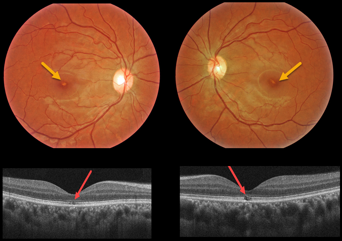
SOLAR RETINOPATHY
Originally posted on @retina.rocks February 24, 2022
This 32YO female with schizophrenia reported regular sun gazing every morning for many years. Vision was 20/40 OU.
Triton color imaging shows tiny foveal pigment loss. Swept-source OCT shows bilateral ellipsoid zone and outer segment defects.
Learning Points:
The outer retinal damage in solar retinopathy is thought to be caused by photochemical outer retinal and RPE damage instead of a thermal burn.
Vision is usually fairly good despite the outer retinal and RPE findings.
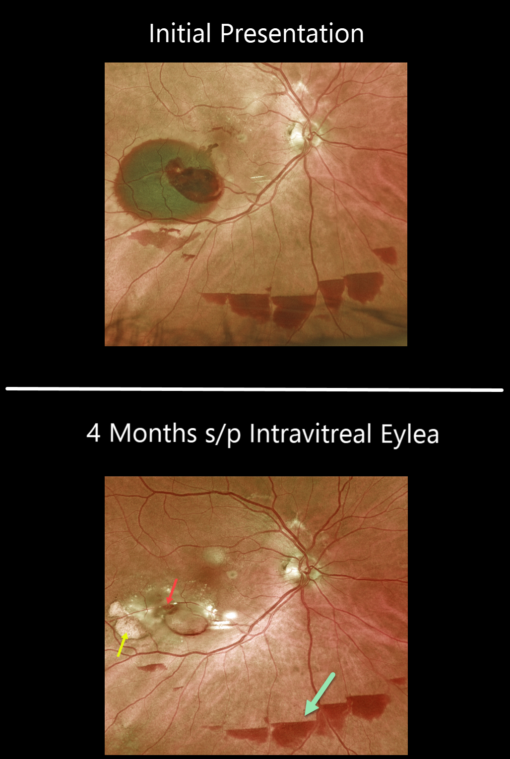
RETINAL ARTERIAL MACROANEURYSM (RAM)
Originally posted on @retina.rocks February 14, 2022
This 85 YO female with a retinal arterial macroaneurysm (RAM) continues to improve with 20/30 vision following a single Eylea injection four months earlier.
The macroaneurysm still appears patent. The old subretinal devitalized blood is devitalized and yellow. There are also multiple patches of resolving inferior subhyaloid blood.
Learning Points:
A RAM is thought to occur in a weakened arterial wall from arteriosclerosis and appears as a yellow-white dilated lesion along a retinal arteriole within the first 3 bifurcations from the optic nerve.
There are 2 distinct presentations: hemorrhagic or exudative. The hemorrhagic type often presents, as in this case, with pathognomonic preretinal, intraretinal, and subretinal blood. Acute macular hemorrhage can be treated with observation, anti-VEGF injections, or thermal laser.
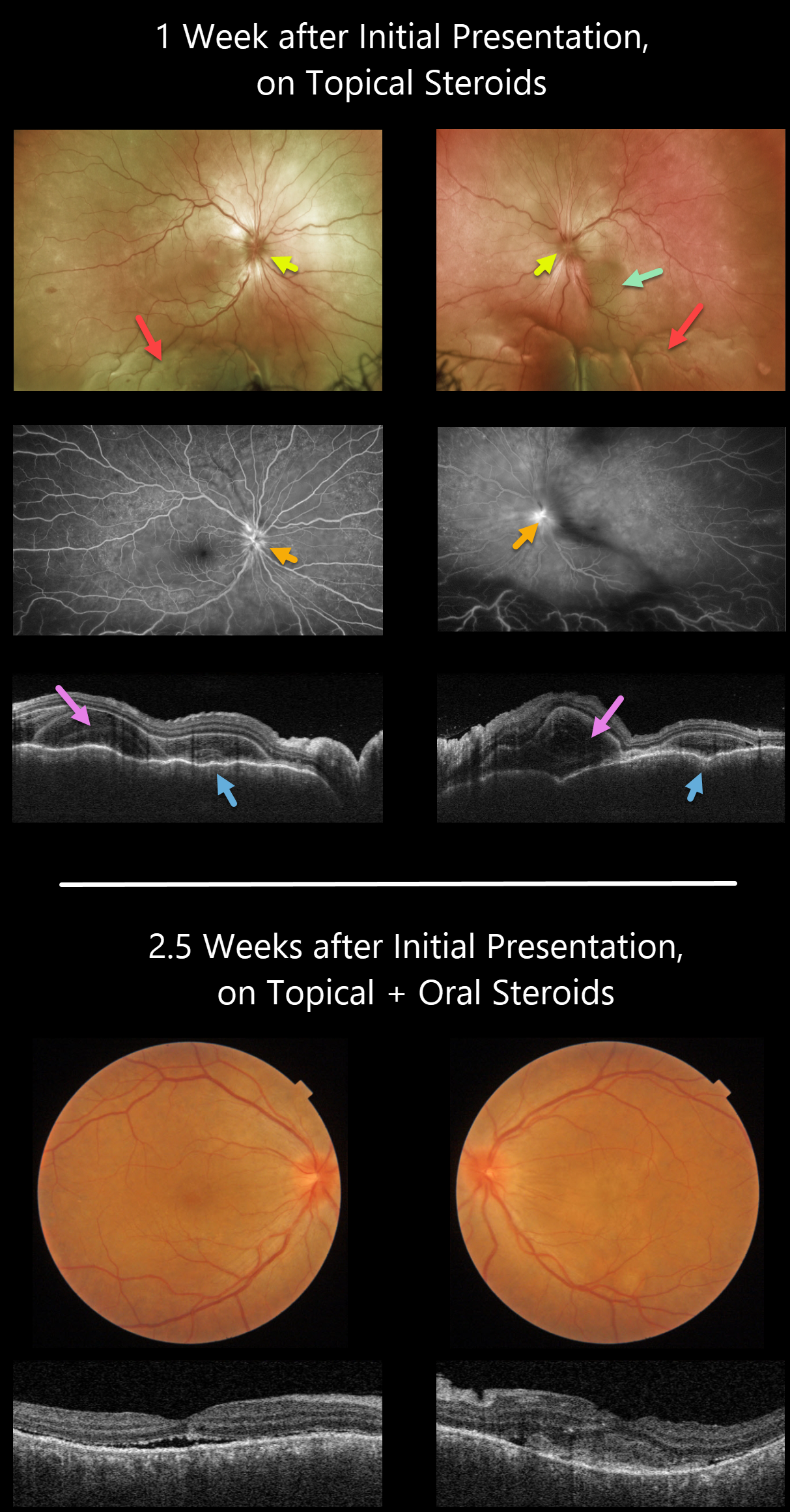
HARADA DISEASE
Originally posted on @retina.rocks February 7, 2022
This 35YO female presented with a 2-week history of severe bilateral vision loss, photophobia, and red eyes. There was no significant past medical history.
Vision was counting fingers OU. There was moderate bilateral conjunctival injection, severe cells with moderate flare, moderate fibrin, and 360-degree posterior synechiae. Topical cycloplegia and steroids were prescribed.
One week later, she was referred to our retinal service. There was marked improvement of her anterior uveitis and the posterior synechiae were mostly broken throughout. There were moderate bilateral vitreous cells, markedly swollen optic nerves, irregular diffuse macular exudative fluid with inferior exudative retinal detachments.
Fluorescein angiography showed bilateral optic nerve leakage. Triton swept-source OCT showed bilateral markedly thickened and rippled choroid with multiple bacillary pockets of outer retinal thickening and fluid. She started oral prednisone 80mg daily.
Around 10 days later, vision improved to 20/200 OU, with improvement in optic nerve edema, macular fluid, bacillary detachments, and choroidal thickening. The inferior exudative detachments were also resolving (not imaged).
Learning Points:
This case has many classic findings for Harada disease, including bilateral panuveitis, optic nerve swelling, thickened choroid, chorioretinal folds, and multifocal serous exudative retinal detachments.
Given the severity of the panuveitis and exudative subretinal fluid, we were surprised that angiography did not show the more classic multifocal subretinal leaks with pooling in the overlying serous detachments and bacillary cavities.
Patients with just ocular findings have Harada disease, whereas those with additional systemic findings (including vitiligo, poliosis, headache, vertigo, and hearing loss) have Vogt-Koyanagi-Harada disease.
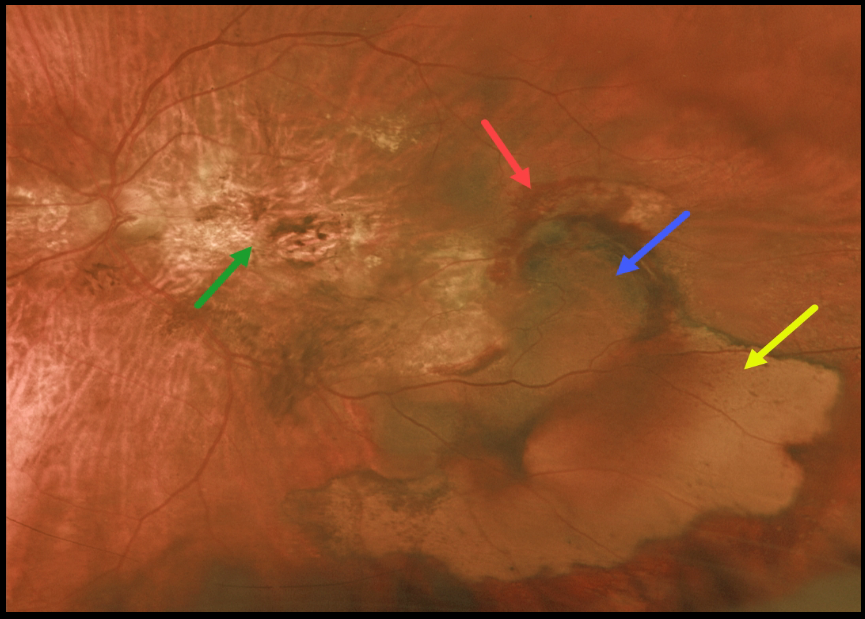
PERIPHERAL EXUDATIVE HEMORRHAGIC CHORIORETINOPATHY
Originally posted on @retina.rocks January 28, 2022
This 77YO female has been followed for years with vision of 20/200 OD and counting fingers OS from inactive age-related disciform scars. She then came in with sudden symptomatic vision loss and floaters in her left eye.
Vision was hand motion OS from a breakthrough vitreous hemorrhage from an inferotemporal ectopic choroidal neovascularization (CNV) that grew from the edge of the original macular scar. She was not taking anticoagulants or aspirin.
Following two monthly intravitreal Avastin injections, vision remained at hand motion. The vitreous hemorrhage had cleared, and the subretinal blood was resolving.
Optos imaging shows the original macular scarring, along with varying stages of resolving rub-RPE blood, subretinal blood, and devitalized, yellow subretinal blood.
Learning Points:
Ectopic CNV, also known as peripheral exudative hemorrhagic chorioretinopathy, is an exudative process similar to wet AMD but located in the retinal periphery.
Ectopic CNV can often masquerade as a choroidal mass or uveal melanoma. It can also cause extensive choroidal/suprachoroidal, subretinal, and vitreous hemorrhage, more commonly in patients who are on blood thinners.
The subretinal blood often spontaneously regresses without treatment, although anti-VEGF injections can be used for symptomatic or increasing exudation.
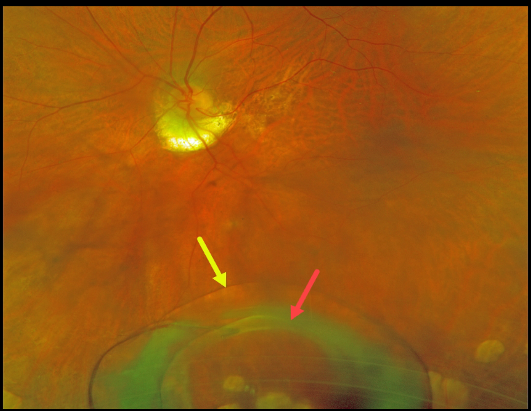
DISLOCATED LENS
Originally posted on @retina.rocks January 24, 2022
This 80YO female presented with subacute, painless vision loss in her left eye. Previously, she had been treated for macular neovascularization from ocular histoplasmosis but had been lost to follow-up.
Optos imaging shows a 3-piece intraocular lens with an intact capsule settled in the inferior midperiphery.
She denied any trauma, and previous exams showed no zonular dehiscence. The cataract surgery had been performed nearly 25 years earlier.
The lens will be explanted during vitrectomy with placement of a scleral-fixated lens.
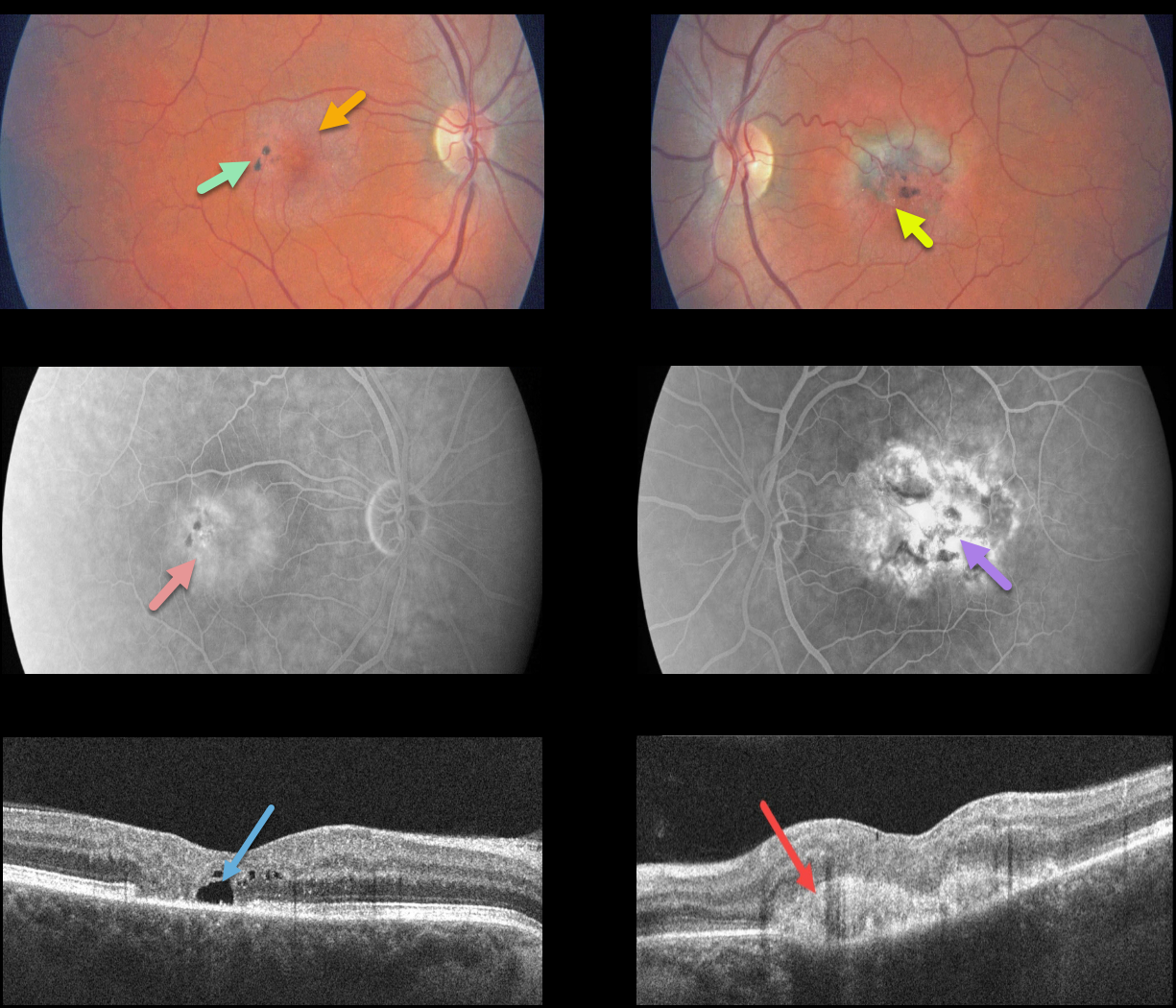
MACULAR TELANGIECTASIA
Originally posted on @retina.rocks December 27, 2021
This patient presented with classic findings of macular telangiectasia type 2 (MacTel2), including an opaque perifoveal retina, inner retinal crystal, and black clumps of intraretinal pigment migration.
Deep retinal angiographic leakage is noted in the right eye with more diffuse staining in the left eye. OCT scanning shows an outer retinal cavitation OD and type 2 macular neovascularization (MNV) OS.
Learning Points:
Originally described by Gass (Arch Ophthalmology 1982;100:769-780), MacTel2 is a neurodegenerative disorder, most likely originating from Müller cell dysfunction.
The term telangiectasia is misleading because the funduscopic findings are mostly nonvascular.
The refractile inner retinal ‘crystals’ are thought to be Müller cell footplates. The foveal retina shows a whitish discoloration, most likely due to retinal opacification resulting from Müller cell dysfunction.
Photoreceptor loss allows RPE cells to migrate along intraretinal capillaries, causing the coarse clinical pigment clumping.
The majority of patients will also have subclinical chorioretinal anastomosis associated with right-angle venules (Spaide et al., Retina 2018;38:1920-1929).
Secondary MNV can be successfully treated per age-related macular degeneration protocols.
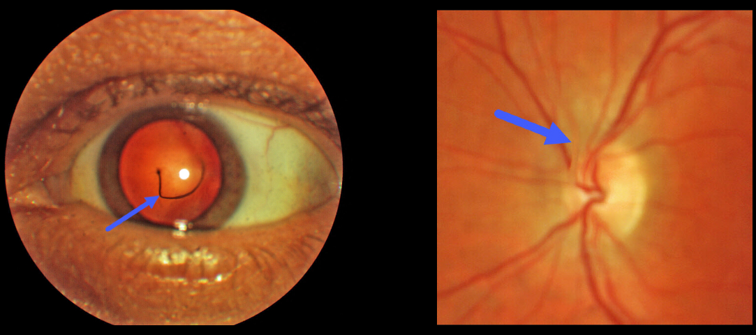
PERSISTENT HYALOID ARTERY
Originally posted on @retina.rocks November 29, 2021
This 44YO male was referred for an asymptomatic vitreous floater. Upon examination, a large translucent strand was noted posterior to the lens, connecting the posterior capsule of the lens to the optic nerve.
Learning Points:
The hyaloid vascular system, embryologically located within the hyaloid canal, provides the developing anterior segment with nutrients. The hyaloid artery usually regresses around 10 weeks of gestation, but in some cases, it may not fully regress.
An anterior remnant may remain on the posterior capsule, which is known as a Mittendorf Dot. Posterior remnants, attached to the optic disc, are otherwise known as Bergmeister’s papilla.
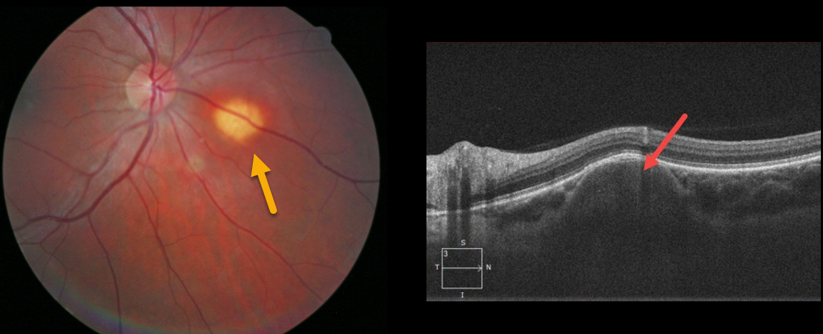
FOCAL SCLERAL NODULE
Originally posted on @retina.rocks October 28, 2021
This patient presented with an asymptomatic yellowish, nodular subretinal lesion inferonasal to the right optic nerve.
Spectral domain optical coherence tomography shows that the lesion originates from the sclera.
Learning Points:
Focal scleral nodule (previously known as solitary idiopathic choroiditis [SIC] and unifocal helioid choroiditis) presents as a single, yellow-white, elevated nodular, avascular subretinal lesion. On OCT, it arises from the inner sclera, causing marked choroidal compression.
For the largest series to date and the rationale behind the renaming of this entity, see Fung et al, Ophthalmology 2020;127:1567-1577.
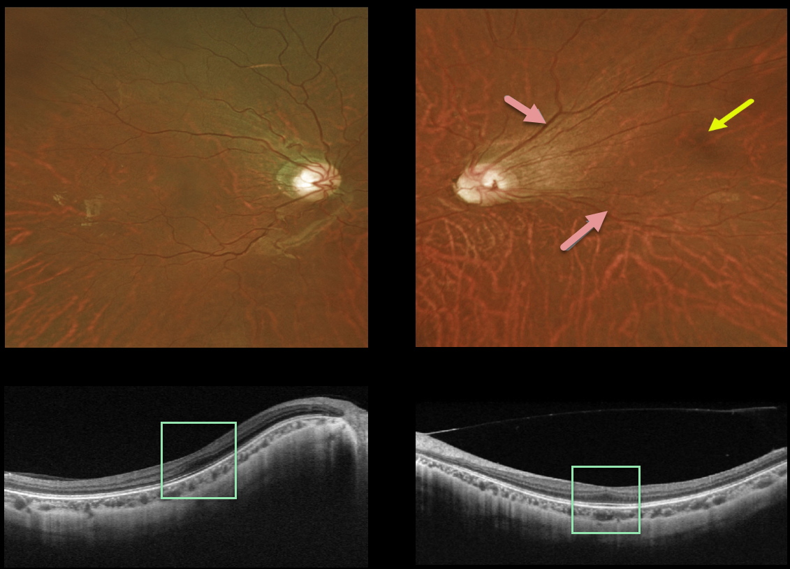
FOVEA PLANA
Originally posted on @retina.rocks October 12, 2021
This 20YO female presented with these asymptomatic macular findings. She was born prematurely at 24 weeks of gestation. Vision was 20/25 OU.
The right posterior pole was fairly normal clinically, although the left major temporal arcades were straightened and dragged temporally. The left fovea was also temporally displaced.
Triton swept-source OCT showed a flattening of the normal foveal depression bilaterally.
Learning Points:
Temporal dragging and straightening of the vascular arcades are common findings in regressed retinopathy of prematurity (ROP). This is caused by involuted peripheral temporal neovascularization displacing the posterior retina.
The macular center is usually about 2 disc diameters temporal to the edge of the optic nerve, and our patient’s is about 3 disc diameters.
Fovea plana is characterized by the absence of the foveal pit, which is formed embryologically by the inner retinal tissues and vasculature being displaced centrifugally.
It is most commonly associated with albinism, prematurity (as in our case), or as an isolated finding. Despite the abnormal foveal architecture, vision is usually normal.
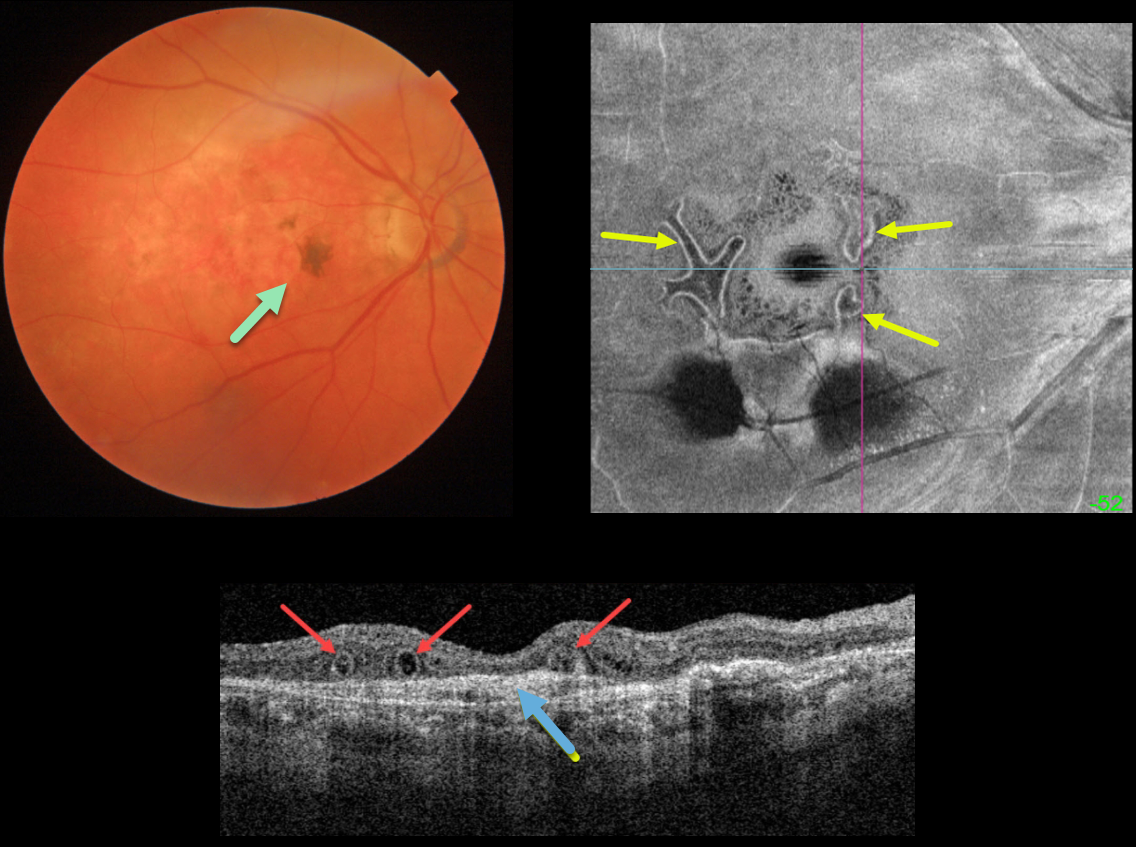
OUTER RETINAL TUBULATION FOLLOWING ONGOING ANTI-VEGF FOR NEOVASCULAR AMD
Originally posted on @retina.rocks September 23, 2021
This is an 86YO female who has stable dry macular scarring from wet age-related macular degeneration with ongoing anti-VEGF injections.
Triton swept-source OCT shows numerous outer retinal tubulations (ORT) overlying an inactive type 1 macular neovascularization (MNV). These tubules are more dramatically seen with en face imaging of the outer retina.
Learning Points:
Outer retinal tubulation is often noted overlying inactive MNV with ongoing anti-VEGF therapy and should not be confused with exudative fluid or cysts, which lack a hyperreflective border.
ORT, initially described by Zweifel et al (Arch Ophthalmol 2009;127:1596-1602), is a neurodegenerative condition of the photoreceptors and Muller cells associated with atrophy affecting the outer retina and retinal pigment epithelium, including advanced AMD and inherited retinal diseases.
The outer hyperreflective band likely represents inner segment mitochondria undergoing fission and translocation toward the nucleus (Litts et al, Retina 2018;38:445-461).
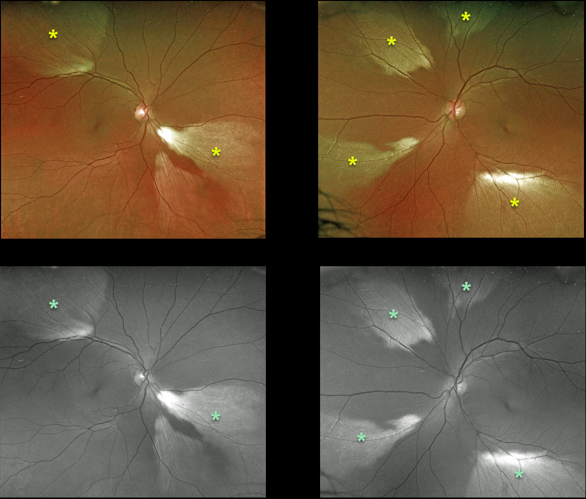
MYELINATED NERVE FIBER LAYER
Originally posted on @retina.rocks September 15, 2021
This 50YO female presented with multiple bilateral patches of myelinated nerve fiber layer (NFL). The findings are beautifully imaged using both the Optos color RG and green channels.
Myelinated NFL is a relatively uncommon, unilateral or bilateral, usually isolated finding present at birth. The myelinated NFL is often connected to the optic nerve, but not necessarily as in this case. Most patients are asymptomatic, although some may have a corresponding relative scotoma. Myelination can progress in about 10% of cases and can also resolve following retinal ischemic events. Myelinated NFL occurs when retinal nerve fibers develop a myelin sheath, which usually stops posterior to the lamina cribrosa. The myelination may represent an oligodendrocytic choristoma (see Rao et al, Retina 2019;39:1125-1132).
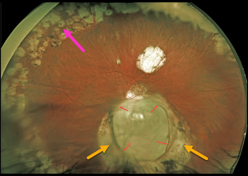
CHOROIDAL COLOBOMA
Originally posted on @retina.rocks September 10, 2021
This 57YO male underwent successful vitrectomy surgery 8 years earlier for rhegmatogenous retinal detachment (RRD). Vision is now 20/40.
The detachment was caused by a retinal break associated with an inferior choroidal coloboma. The margins of the coloboma are surrounded by confluent depigmented scarring from prior laser photocoagulation. Prophylactic peripheral laser was also applied intraoperatively.
Learning Points:
Choroidal colobomas arise from incomplete closure of the embryonic fissure during the second month of embryonic development.
Patients are at lifelong risk of rhegmatogenous retinal detachment due to breaks in the overlying thinned retina.
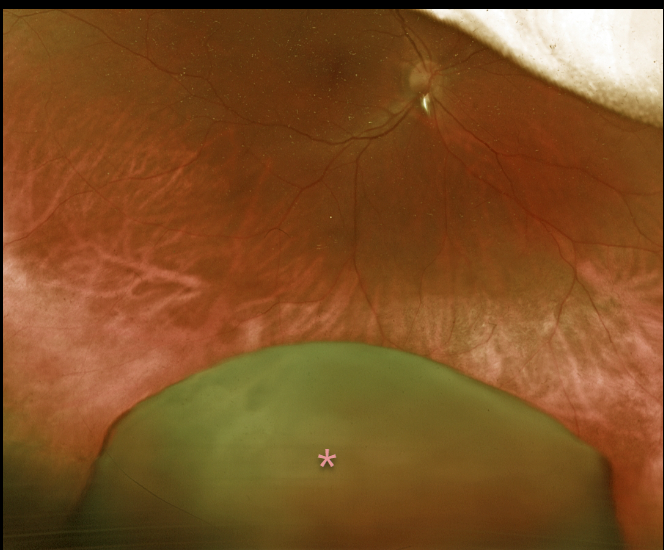
DISLOCATED LENS
Originally posted on @retina.rocks August 26, 2021
This 51YO female presented with a 6-month history of vision loss in her right eye. She remembered falling 7 months earlier, then woke a few weeks later with severe unilateral vision loss. Vision was counting fingers from a total dislocation of her crystalline lens into the inferior vitreous.
Vitrectomy surgery to remove the dislocated cataract, along with placement of an anterior chamber intraocular lens, was performed, resulting in a return to 20/30 vision.
Learning Points:
Posterior dislocation of the crystalline lens can be caused by trauma or by systemic disorders such as Marfan syndrome, homocystinuria, or Weill-Marchesani syndrome. Pars plana vitrectomy is needed for a completely dislocated lens, as in this patient, whereas subluxed lenses may be managed with phacoemulsification or other means.
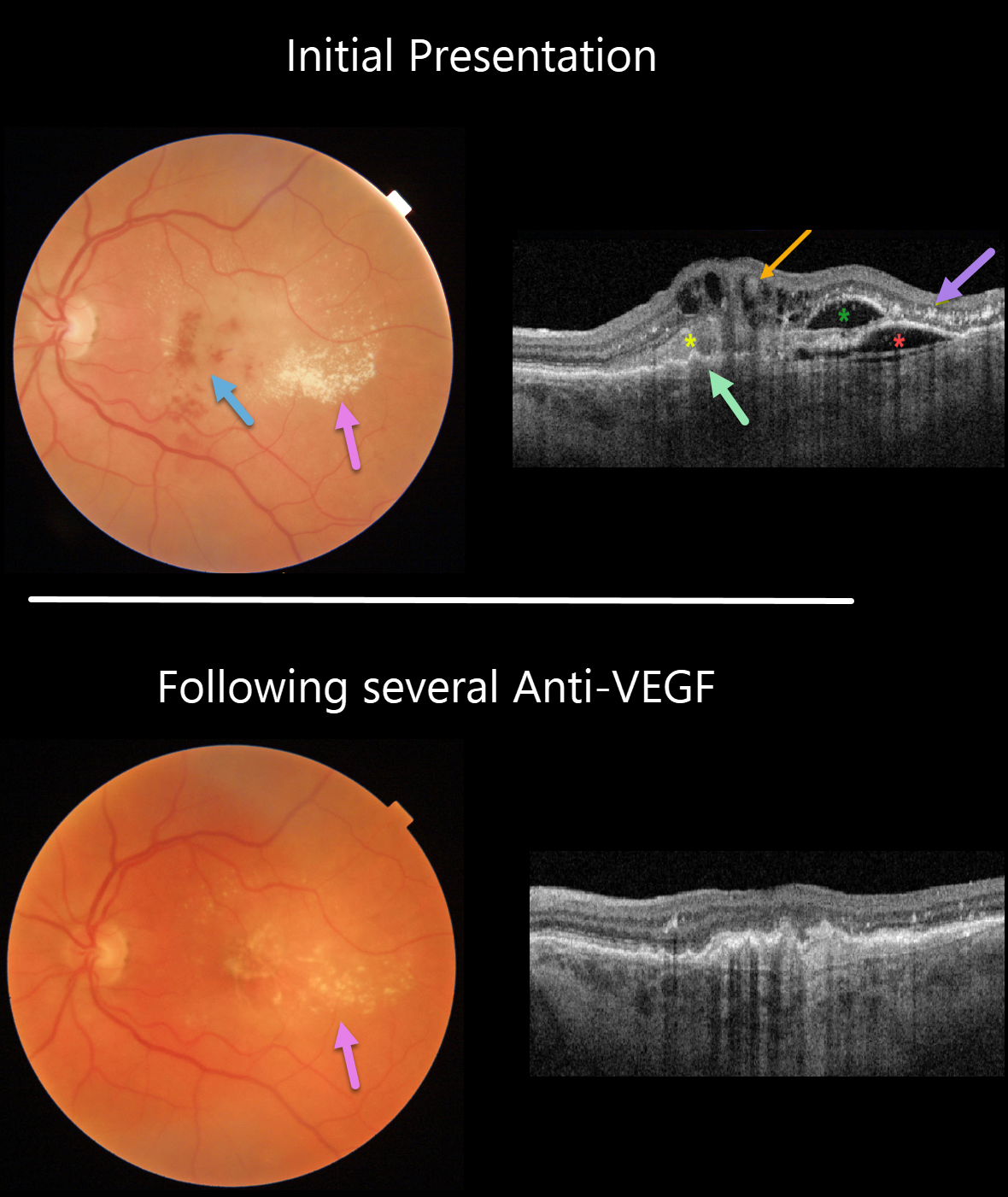
RETINAL ANGIOMATOUS PROLIFERATION (RAP)
Originally posted on @retina.rocks August 18, 2021
This 74YO man presented with counting fingers vision from previously untreated wet age-related macular degeneration (AMD).
Triton color imaging and swept-source OCT beautifully highlight this complex macular neovascularization (MNV). There are scattered inner and deep retinal hemorrhages, opaque fluid, and temporal lipid.
OCT shows an irregular shallow elevation of the RPE more nasally, possibly indicating a sub-RPE (type 1 MNV), with overlying subretinal hyperreflective material. More temporally, there is a more prominent area of RPE elevation that is more hyporeflective with an overlying pocket of intraretinal fluid vs a bacillary layer detachment. Finally, central inner retinal thickening and cysts are seen, indicating intraretinal neovascularization (type 3 MNV), also known as a retinal angiomatous proliferation (RAP) lesion. The lipid appears as hyperreflective clumps in the outer plexiform and outer nuclear layers.
The fluid and blood regressed, with significantly decreased lipid, after several monthly Avastin injections (image 2). Vision improved to 20/400.
Learning Points:
Unlike more typical age-related MNV that start either below (type 1 MNV) or occasionally above (type 2 MNV) the RPE, RAP lesions originate in the retina.
These incompetent vessels leak fluid and blood into the surrounding tissue. Their intraretinal location is a biomarker for RAP lesions.
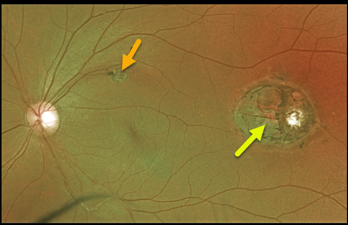
TOXOPLASMOSIS
Originally posted on @retina.rocks August 12, 2021
This 44YO male presented with 20/25 vision and a presumed inactive toxoplasmosis scar in the distal left macula. Although we do not have an OCT through this scar, it likely represents a colobomatous lesion.
This lesion somewhat resembles torpedo maculopathy, but the additional scar in the superior macula more suggests a prior inflammatory event.
Learning Points:
For a great discussion on how to differentiate these infectious macular colobomatous lesions (congenital toxoplasmosis, Zika, and cytomegalovirus) from inherited dystrophies like North Carolina Macular Dystrophy (NCMD), see Kumar and Mahalingam AJO 2019;200;47-56.
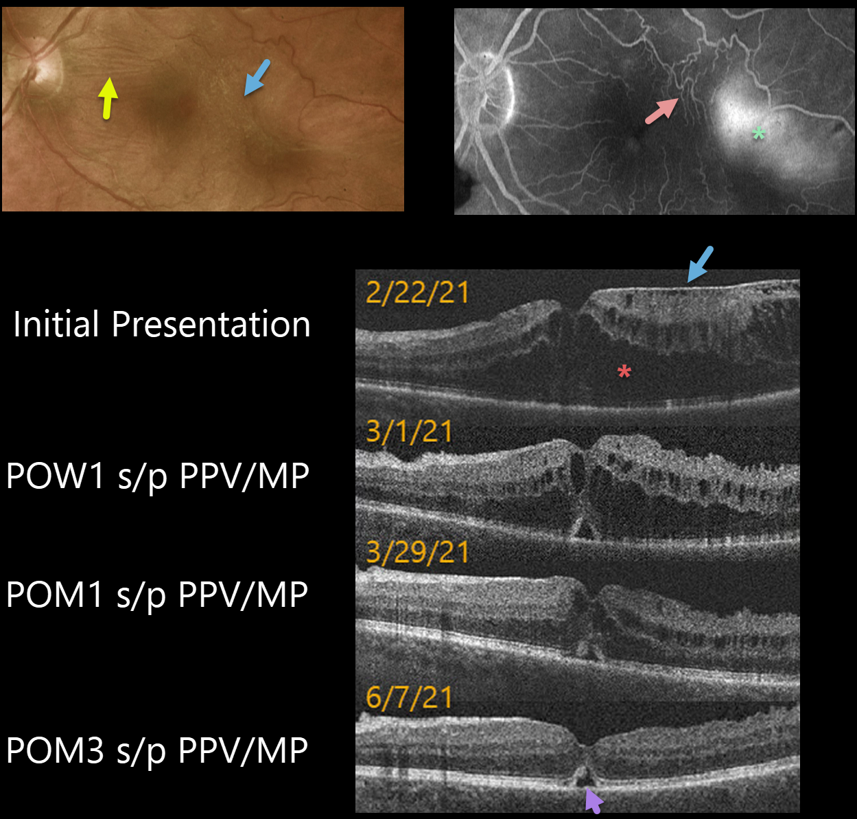
MACULAR PUCKER
Originally posted on @retina.rocks August 5, 2021
This 62YO male presented with 20/200 vision in his left eye. An epiretinal membrane (ERM) was noted in the temporal macula, associated with more nasal horizontal retinal striae.
Optos fluorescein angiography shows distortion of the temporal macular vessels with late leakage.
Vitrectomy with membrane peeling was performed the following day, and one week later, on 3/1/21, vision improved to 20/70 with a significant decrease in macular thickening.
When last examined on 6/7/21, about 3 months post-op, vision was still 20/70, but the macular thickening and contour were almost normal except for a small blister of foveal subretinal fluid.
Learning Points:
OCT scanning confirmed the temporal ERM, but the severe cystic macular thickening with possible tractional schisis that extended into the nasal macula seemed disproportionate to the imaged temporal ERM. Fortunately, both the vision and macular thickening significantly improved following vitrectomy with membrane peeling.
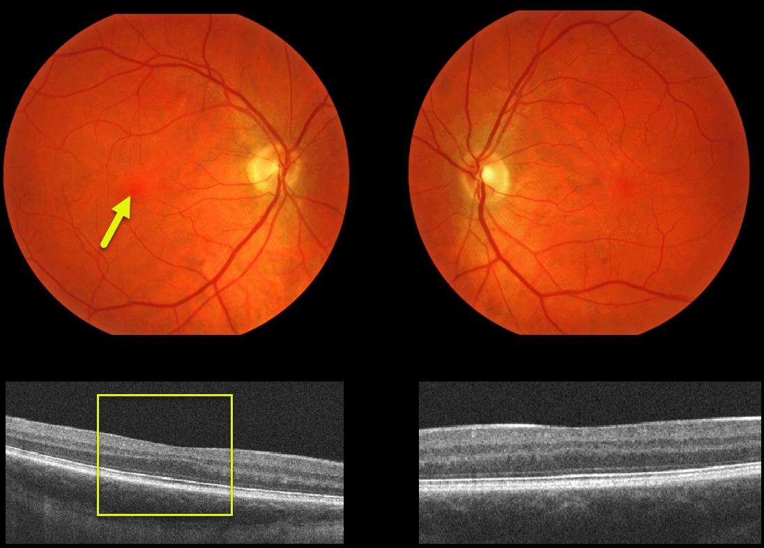
FOVEA PLANA
Originally posted on @retina.rocks June 21, 2021
This patient has normal vision and no history of albinism or prematurity. Fundus photos and OCT images demonstrate the incidental absence of the foveal pit.
Learning Points:
Fovea plana is characterized by the absence of the foveal pit, which is formed embryologically by the inner retinal tissues and vasculature being displaced centrifugally. It is most commonly associated with albinism or prematurity, or found as an isolated finding, as in this case. Despite the abnormal foveal architecture, vision is usually normal.
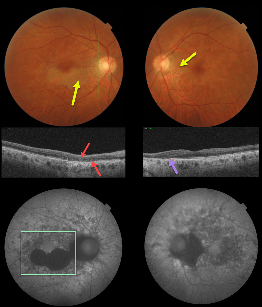
ACUTE POSTERIOR MULTIFOCAL PLACOID PIGMENT EPITHELIOPATHY (APMPPE) WITH OUTER RETINAL TUBULATION
Originally posted on @retina.rocks June 4, 2021
This patient has bilateral fundus findings consistent with resolved acute posterior multifocal placoid pigment epitheliopathy (APMPPE).
Triton fundus photos show placoid areas of atrophic pigmentary changes, which are variably hypo-autofluorescent.
Triton swept-source OCT in the right eye shows outer retinal atrophy with outer retinal tubulation (ORT) and nasal outer retinal atrophy in the left eye.
Learning Points:
ORT, initially described by Zweifel et al (Arch Ophthalmol 2009;127;1596-1602), is a neurodegenerative condition of the photoreceptors and Muller cells associated with atrophy affecting the outer retina and retinal pigment epithelium, including advanced AMD and inherited retinal diseases.
ORT is often seen overlying inactive macular neovascularization in the setting of ongoing anti-VEGF therapy and should not be confused with exudative fluid or cysts, which lack a hyperreflective border. The outer hyperreflective band likely represents inner segment mitochondria undergoing fission and translocation toward the nucleus (Litts et al, Retina 2018;38;445-461).
Acute posterior multifocal placoid pigment epitheliopathy (APMPPE) is a usually bilateral inflammatory vs ischemic disorder of the inner choroid, RPE, and outer retina.
Blurry vision and paracentral/central scotomas often follow a viral illness. Acutely multifocal creamy yellow or grayish-white subretinal placoid lesions are scattered throughout the posterior poles. The lesions and visual symptoms usually resolve within 4 to 8 weeks with secondary chorioretinal changes. Recurrent lesions are rare, and vision is usually fairly good.
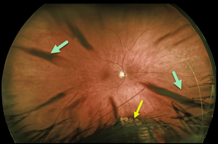
CORTICAL CATARACT
Originally posted on @retina.rocks June 1, 2021
This patient’s Optos image shows beautiful cortical cataract spoke artifacts along with congenital hypertrophy of the RPE (CHRPE) inferiorly, which is mostly obstructed by the patient’s hair and eyelashes.
Learning Points:
Cataracts or other anterior segment findings can create artifacts in fundus photography or optical coherence tomography.
Superior eyelashes often obscure the inferior retinal periphery on Optos imaging, so the technician needs to remind the patient to open their eyes wide and sometimes manually elevate the lid.
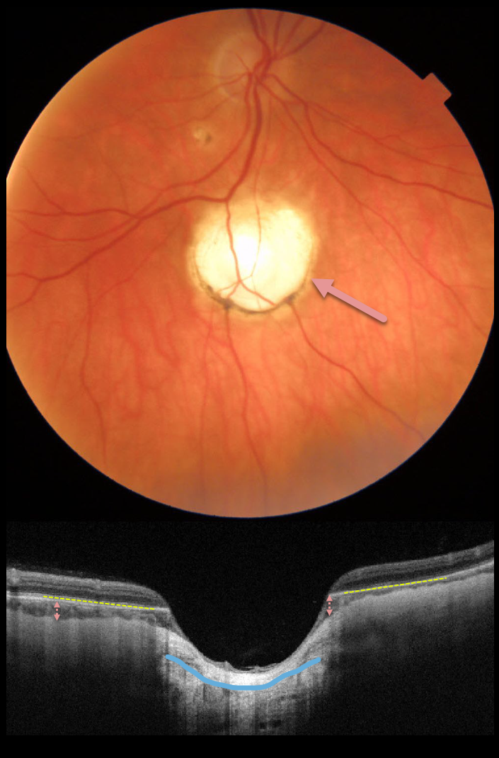
CHOROIDAL COLOBOMA
Originally posted on @retina.rocks May 24, 2021
This 57yo female presented with an asymptomatic, unilateral choroidal coloboma inferior to the right optic nerve.
Triton swept-source OCT shows a total loss of the RPE and a near-complete absence of the choroid within the coloboma, with marked thinning of the overlying retina. There is also an outpouching of the underlying sclera.
Learning Points:
Choroidal colobomas are most commonly due to failure of the optic vesicle and choroidal fissure to close during fetal development. Lesions like this may also be from other causes, including toxoplasmosis. Choroidal coloboma can present as solitary or multiple lesions. Visual prognosis depends on the location and involvement of the optic nerve and macula.
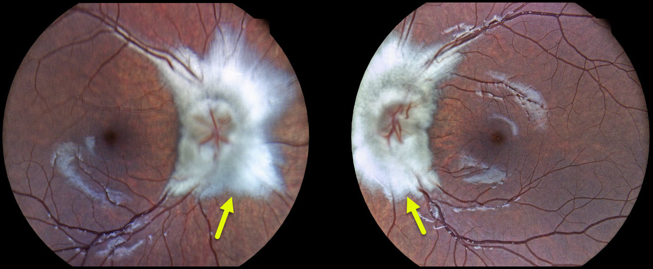
MYELINATED NERVE FIBER LAYER
Kaitlin Spikes
Originally posted on @retina.rocks May 7, 2021
This 9YO girl presented with bilateral optic nerve myelination. The patient had no visual complaints and had 20/20 vision in both eyes.
Learning Points:
Myelinated NFL is a relatively uncommon, unilateral or bilateral, usually isolated finding present at birth. Most patients are asymptomatic, although some may have a corresponding relative scotoma.
Myelination can progress in about 10% of cases and can also resolve following retinal ischemic events.
Myelinated NFL occurs when retinal nerve fibers develop a myelin sheath, which usually stops posterior to the lamina cribrosa. The myelination may represent an oligodendrocytic choristoma.
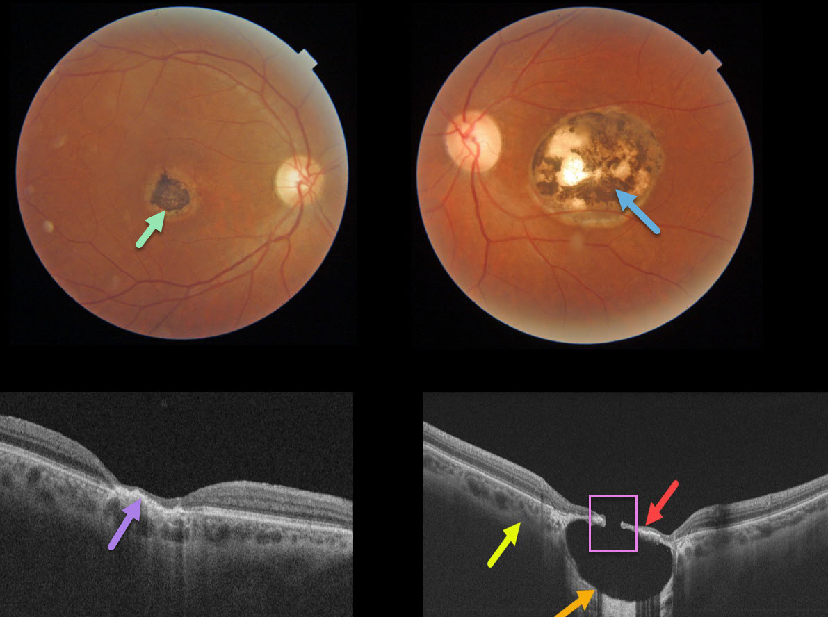
TOXOPLASMOSIS
Originally posted on @retina.rocks March 26, 2021
This 57YO male gave a history of lifelong poor vision in his left eye. Vision was 20/40 OD and 20/200 OS.
A small hyperpigmented foveal scar is noted in his right eye with neurosensory retinal atrophy and irregular elevation of the RPE.
A larger, round, variably pigmented, excavated macular scar is noted in his left eye. Triton swept-source OCT reveals a thinned atrophic retina and intact RPE suspended over an optically empty hyporeflective excavated colobomatous type lesion. The plane of the intact surrounding sclera is indicated by the yellow arrow. A small full-thickness retinal and RPE defect is noted centrally.
Learning Points:
For a great discussion on how to differentiate these infectious macular colobomatous lesions (congenital toxoplasmosis, Zika, and cytomegalovirus) from inherited dystrophies like North Carolina Macular Dystrophy (NCMD), see Kumar and Mahalingam AJO 2019;200;47-56.
In case you’re thinking our patient has NCMD, also see the first OCT description of congenital toxoplasmosis macular lesions (Garg et al, Retina 2009;29;631-637). Their Figures 4 and 6 look eerily similar to our patient’s right and left eyes!
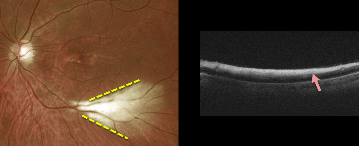
MYELINATED NERVE FIBER LAYER
Originally posted on @retina.rocks February 19, 2021
This patient had a large area of myelinated nerve fiber layer (NFL) in the distal inferotemporal left macula. OCT scanning through this area showed marked inner retinal hyperreflectivity.
Learning Points:
Myelinated NFL is a relatively uncommon and usually isolated finding present at birth. Most patients are asymptomatic, although some may have a corresponding relative scotoma.
Myelination can progress in about 10% of cases and can also resolve following retinal ischemic events.
Myelinated NFL occurs when retinal nerve fibers develop a myelin sheath, which usually stops posterior to the lamina cribrosa. The myelination may represent an oligodendrocytic choristoma (Rao et al, Retina 2019;39:1125-1132).
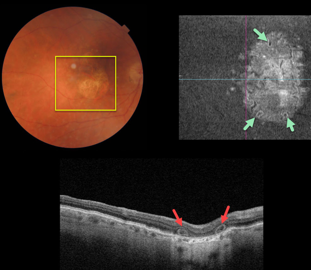
OUTER RETINAL TUBULATION
Originally posted on @retina.rocks February 16, 2021
This 80YO female was 20/200 in her right eye due to central macular atrophy from atrophic age-related macular degeneration (AMD).
Triton swept-source optical coherence tomography showed outer retinal tubulation (ORT) in the outer retinal layers. The extent of these tubular structures was best visualized on en face imaging.
Learning Points:
Outer retinal tubulation, initially described by Zweifel et al (Arch Ophthalmol 2009;127;1596-1602), is a neurodegenerative condition of the photoreceptors and Muller cells associated with atrophy affecting the outer retina and retinal pigment epithelium, including advanced AMD and inherited retinal diseases.
ORT is often seen overlying inactive macular neovascularization in the setting of ongoing anti-VEGF therapy and should not be confused with exudative fluid or cysts, which lack a hyperreflective border.
The outer hyperreflective band likely represents inner segment mitochondria undergoing fission and translocation toward the nucleus (Litts et al Retina 2018;38;445-461).
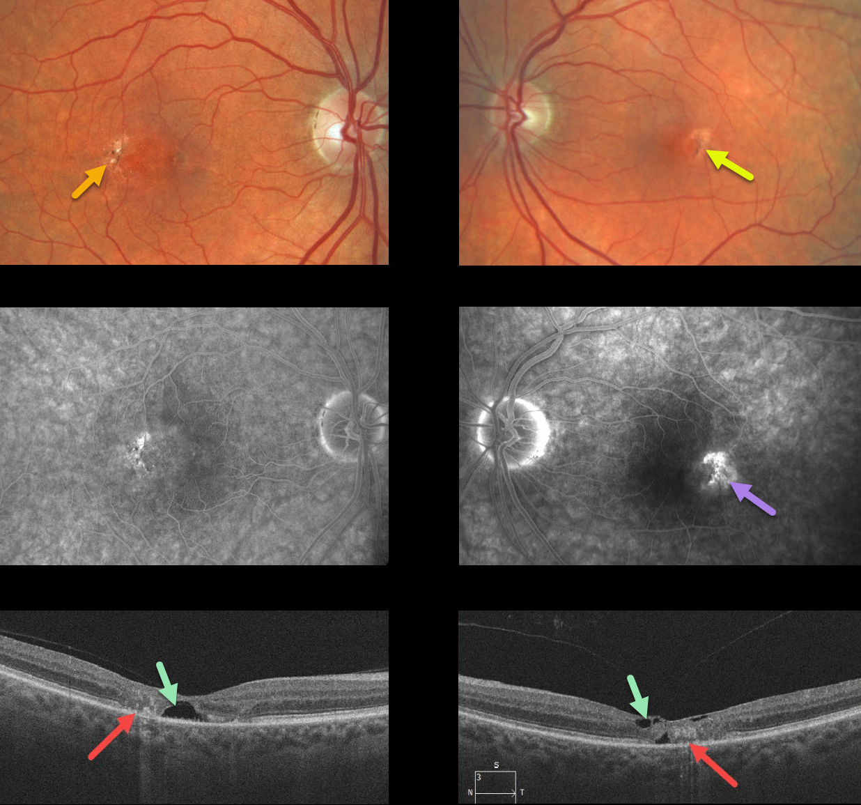
MACULAR TELANGIECTASIA
Originally posted on @retina.rocks January 22, 2021
Our patient shows classic bilateral findings of macular telangiectasia type 2 (MacTel2) with temporal foveal intraretinal pigment migration and inner retinal crystals, as well as angiographic staining and leakage.
Our patient’s optical coherence tomography scans also show classic findings of mostly temporal foveal retinal atrophy, disorganization, and hyporeflective tissue loss (cavitations).
Learning Points:
MacTel type 2 is a neurodegenerative disorder, most likely originating from Müller cell dysfunction. The term telangiectasia is misleading because the funduscopic findings are mostly nonvascular.
It is thought that photoreceptor loss allows RPE cells to migrate along intraretinal capillaries, creating the pigment clumping. This is a similar mechanism to that found in other causes of intraretinal pigment migration, including photoreceptor loss or damage, such as retinitis pigmentosa, chronic retinal detachment, and blunt trauma.
The refractile inner retinal ‘crystals’ are thought to be Muller cell footplates, similar to the refractile dots found in retinoschisis.
Although much remains to be learned about its pathophysiology and there is no treatment for the underlying disease, secondary macular neovascularization can be successfully treated according to age-related macular degeneration protocols.
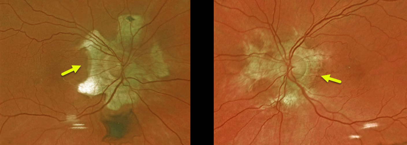
HELICOID PERIPAPILLARY CHORIORETINAL DEGENERATION
Originally posted on @retina.rocks January 19, 2021
This asymptomatic patient was found to have a unique bilateral peripapillary chorioretinal atrophy called helicoid peripapillary chorioretinal degeneration.
Vision is usually normal, although central vision can rarely be affected if atrophy progresses through the fovea.
Learning Points:
Helicoid peripapillary chorioretinal degeneration is a benign autosomal dominant condition with degenerative wing or propeller-shaped lesions that radiate centrifugally from the optic nerve head.
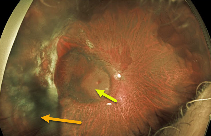
PERIPHERAL EXUDATIVE HEMORRHAGIC CHORIORETINOPATHY
Originally posted on @retina.rocks January 15, 2021
This 71YO male had a vitrectomy in 2018 for a breakthrough vitreous hemorrhage from ectopic choroidal neovascularization (CNV). He did well with 20/40 vision until a recurrent vitreous hemorrhage developed two years later.
The vitreous cleared with several monthly intravitreal Avastin injections, but vision remains at hand motion due to submacular blood, some of which is yellow and devitalized. We discussed repeat vitrectomy with subretinal TPA, but he elected for in-office anti-VEGF injections. He is not on any systemic blood thinners.
Learning Points:
Ectopic CNV, also known as peripheral exudative hemorrhagic chorioretinopathy, is a degenerative and exudative process similar to wet AMD but located in the retinal periphery.
Ectopic CNV can often masquerade as a choroidal mass or uveal melanoma. It can also cause extensive subretinal and vitreous hemorrhage, more commonly in patients who are on blood thinners.
The subretinal blood often spontaneously regresses without treatment, although anti-VEGF injections can be used for symptomatic or increasing exudation.
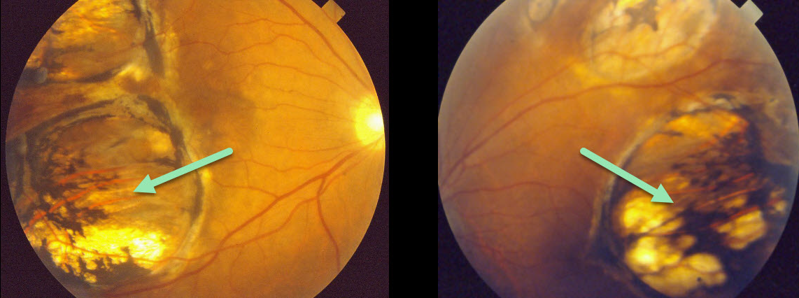
TOXOPLASMOSIS
Originally posted on @retina.rocks January 12, 2021
This patient presented in 1990 with what we assumed was bilateral inactive colobomatous congenital toxoplasmosis scarring.
Learning Points:
Choroidal colobomas are most commonly due to failure of the optic vesicle and choroidal fissure to close during fetal development. Multifocal lesions, as seen in our patient, can be caused by congenital toxoplasmosis and Zika.
For a comprehensive review of ocular coloboma, see Onwochei et al Surv Ophthalmol 2000;45:175-194.
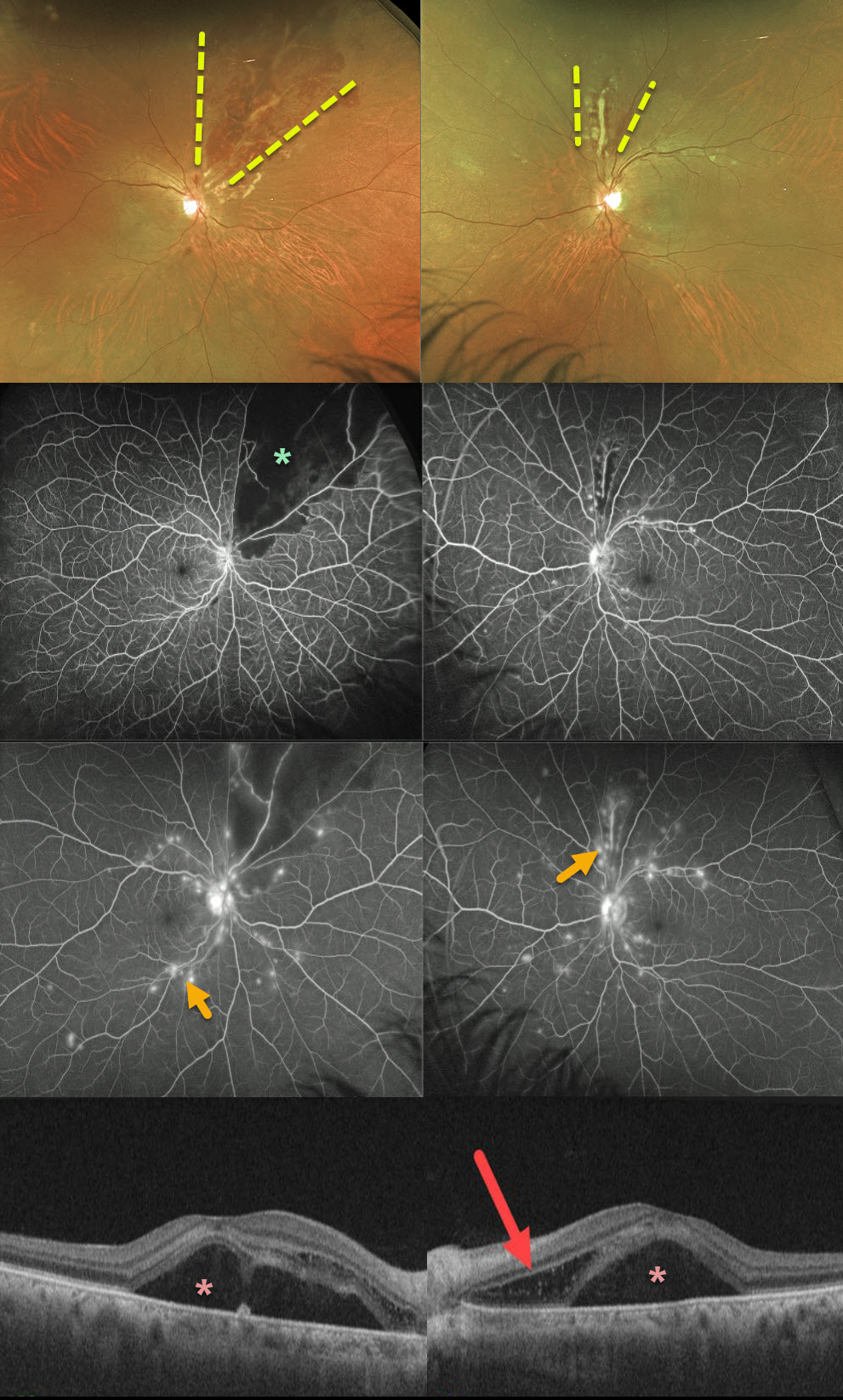
SYSTEMIC LUPUS ERYTHEMATOSUS (SLE)
Originally posted on @retina.rocks December 29, 2020
This 52YO female presented with severe bilateral vision loss (20/400 OD, counting fingers OS) following a several-week gastrointestinal illness with 60-pound weight loss.
She has bilateral areas of hemorrhagic retinal vasculitis, with areas of ischemia and leakage seen on fluorescein angiography. OCT shows macular subretinal fluid and a bacillary detachment in the left macula.
She had been hospitalized recently for hypertension and kidney failure and was diagnosed with a previously asymptomatic stroke. Extensive blood work was negative, and she denied symptoms of Behcet’s.
We suspected an underlying rheumatologic cause, and she was subsequently diagnosed with severe systemic lupus erythematosus (SLE) vasculitis.
She was started on high-dose oral prednisone followed by Cytoxan and hydroxychloroquine. Her ocular findings rapidly improved, and at her last examination 4 months later, vision had improved to 20/40 OD and 20/100 OS.
Learning Points:
SLE-associated retinopathy more typically appears as hypertensive retinopathy (bilateral nerve fiber layer infarcts and retinal hemorrhages), often in patients with cerebral vasculitis. Purtscher-like retinopathy can also be seen.
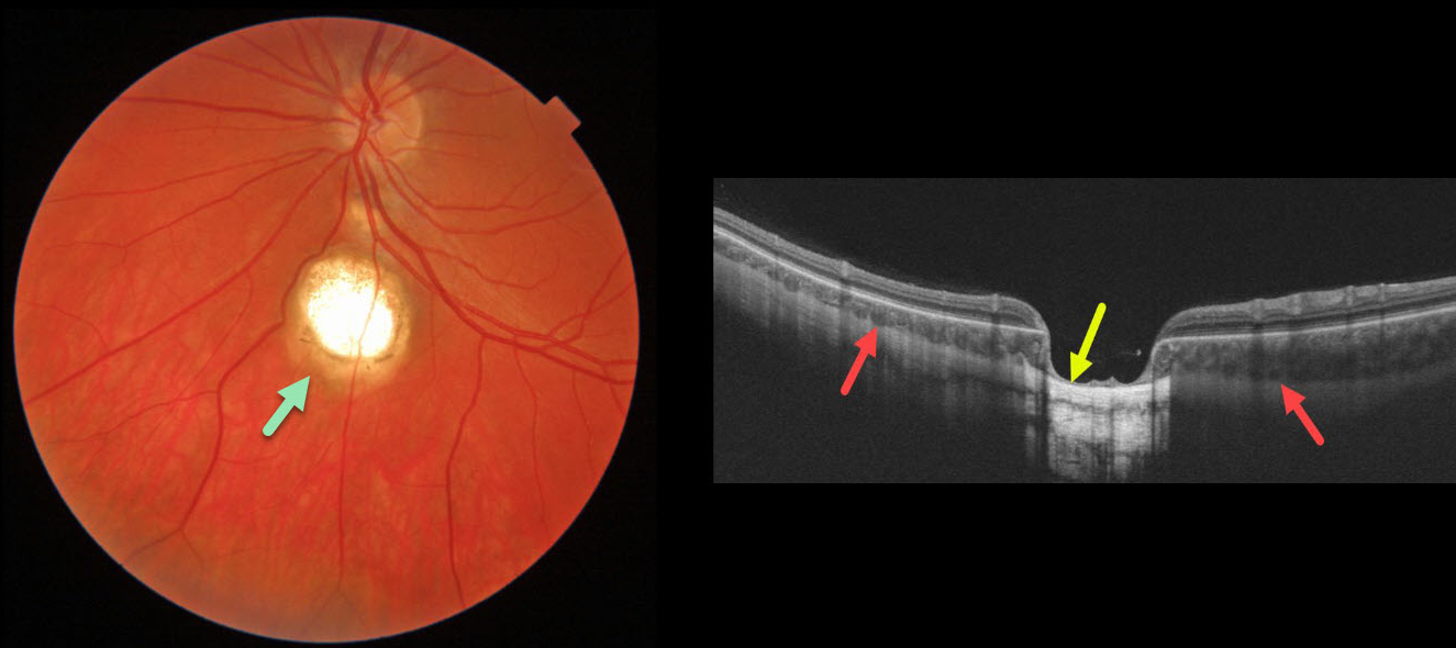
CHOROIDAL COLOBOMA
Originally posted on @retina.rocks December 17, 2020
This 25yo was referred for this asymptomatic choroidal coloboma.
A B-scan OCT through our patient’s coloboma shows that the sclera is continuous and non-displaced within the coloboma.
A staphyloma, by definition, is an outpouching of the entire eye wall. Choroidal coloboma always involves a choroidal defect, but may or may not also involve the sclera.
Learning Points:
Choroidal colobomas are most commonly due to failure of the optic vesicle and choroidal fissure to close during fetal development. Lesions like this may also be from other causes, including toxoplasmosis. Choroidal coloboma can present as solitary or multiple lesions. Visual prognosis depends on the location and involvement of the optic nerve and macula.
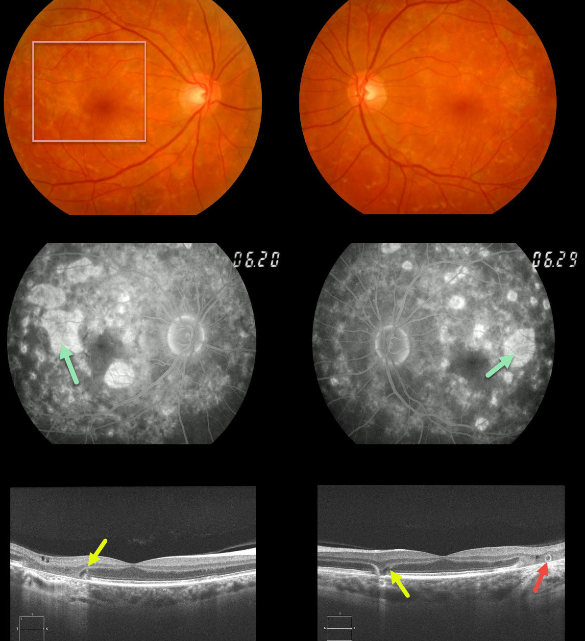
MATERNALLY INHERITED DIABETES AND DEAFNESS (MIDD)
Originally posted on @retina.rocks December 4, 2020
This 62yo female with a history of type 2 diabetes and mild hearing loss initially presented in 2012 with asymptomatic 20/30 vision OU and bilateral areas of focal macular atrophy and subretinal yellow fleck-like lesions.
The retinal pigment epithelial changes were much more pronounced on fluorescein angiography. OCT scanning shows classic wedge defects along the border of the macular atrophy as well as an area of outer retinal tubulation in the left eye.
Learning Points:
Maternally inherited diabetes and deafness (MIDD) accounts for up to 3% of all cases of diabetes and results from a mutation in mitochondrial DNA at position A3243G. MIDD often masquerades as a pattern macular dystrophy.
Fundus autofluorescence (FAF), in our experience, is the best way to visualize these changes, but it was not available at the office when we initially saw her. MIDD patients need to be screened for other potential co-existing medical conditions, including cardiac arrhythmias, myopathy, and renal disease.
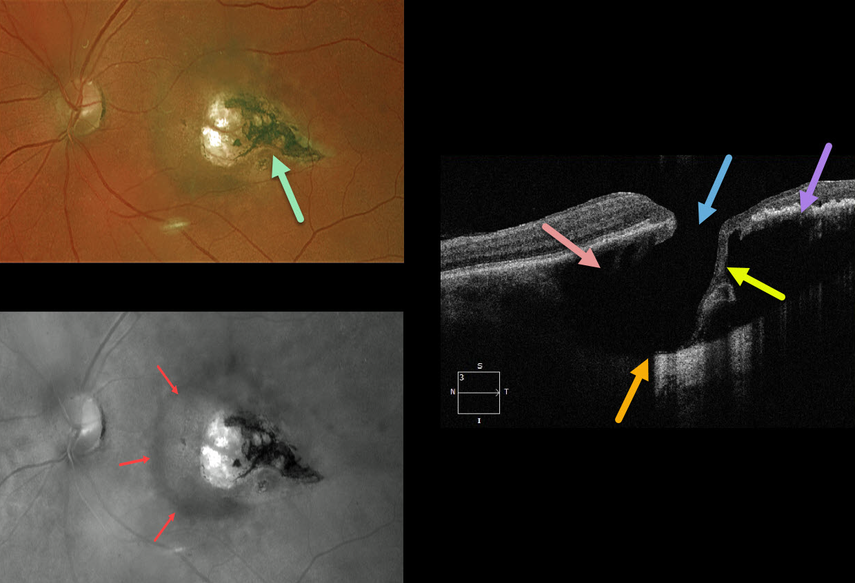
TOXOPLASMOSIS
Originally posted on @retina.rocks November 16, 2020
This 67YO female was referred with 20/30 vision and asymptomatic retinal findings. There was a tear-drop-shaped, variably pigmented chorioretinal scar in the temporal macula. The associated increased choroidal pigmentation is best seen in the red-channel image.
The OCT shows some unexpected findings. There is either a coloboma or posterior staphyloma causing marked posterior displacement of the sclero-choroidal junction. Temporally thinned retina dives posteriorly with an adjacent full-thickness retinal defect.
Temporally, the increased pigmentation of the scarring likely makes it difficult to see more posterior choroidal detail due to shadowing. Nasally, the faint, increased choroidal pigmentation also causes shadowing.
What do you think this is? Our best guess is a choroidal cavitation or an atypical colobomatous toxoplasmosis scar.
Learning Points:
For a great discussion on how to differentiate these infectious macular colobomatous lesions (congenital toxoplasmosis, Zika, and cytomegalovirus) from inherited dystrophies like North Carolina Macular Dystrophy (NCMD), see Kumar and Mahalingam AJO 2019;200:47-56. Also see the first OCT description of congenital toxoplasmosis macular lesions (Garg et al, Retina 2009;29;631-637).
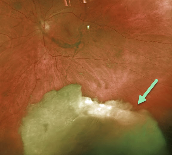
RETAINED LENS FRAGMENT
Originally posted on @retina.rocks November 2, 2020
This patient came to see us for a second opinion following cataract surgery elsewhere six weeks earlier. He has been on topical steroid drops since his surgery. His pressure was 42 mmHg with significant anterior chamber cell and flare.
Virtually the entire cataract was floating in the inferior vitreous. He was scheduled for immediate pars plana vitrectomy and lensectomy.
Learning Points:
Posteriorly dislocated lens fragments are a known complication of cataract surgery. The crystalline lens is an immunoprecipitated tissue.
When lens fragments are exposed after cataract surgery, severe inflammation and increased intraocular pressure usually follow. Rarely, permanent vision loss occurs from glaucoma or cystoid macular edema.
Small lens fragments can be managed conservatively, but larger fragments require surgical removal.
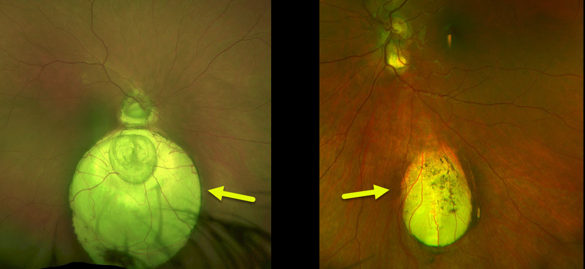
CHOROIDAL COLOBOMA
Originally posted on @retina.rocks October 26, 2020
Our asymptomatic patient is being followed yearly for bilateral choroidal colobomas.
These patients are at risk for retinal detachment and choroidal neovascularization. Choroidal neovascularization has an increased tendency to develop at the superotemporal edge of the coloboma, which is thought to be caused by the abnormal architecture of the RPE and Bruch’s membrane.
Learning Points:
Choroidal colobomas are caused by failure of the optic vesicle and choroidal fissure to close during fetal development. Choroidal coloboma can present as solitary or multiple lesions as in this case. Visual prognosis depends on the location and involvement of the optic nerve and macula.
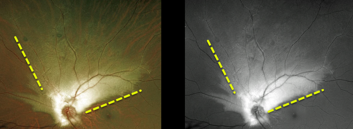
MYELINATED NERVE FIBER LAYER
Originally posted on @retina.rocks October 6, 2020
This 20 YO male has a large area of myelinated nerve fiber layer (NFL) extending superiorly from the left optic nerve.
This relatively uncommon and usually isolated finding is present at birth. Most patients are asymptomatic, although some may have a corresponding relative scotoma.
Myelination can progress in about 10% of cases and can also resolve following retinal ischemic events.
Learning Points:
Myelinated NFL occurs when retinal nerve fibers develop a myelin sheath, which usually stops posterior to the lamina cribrosa. The myelination may represent an oligodendrocytic choristoma.
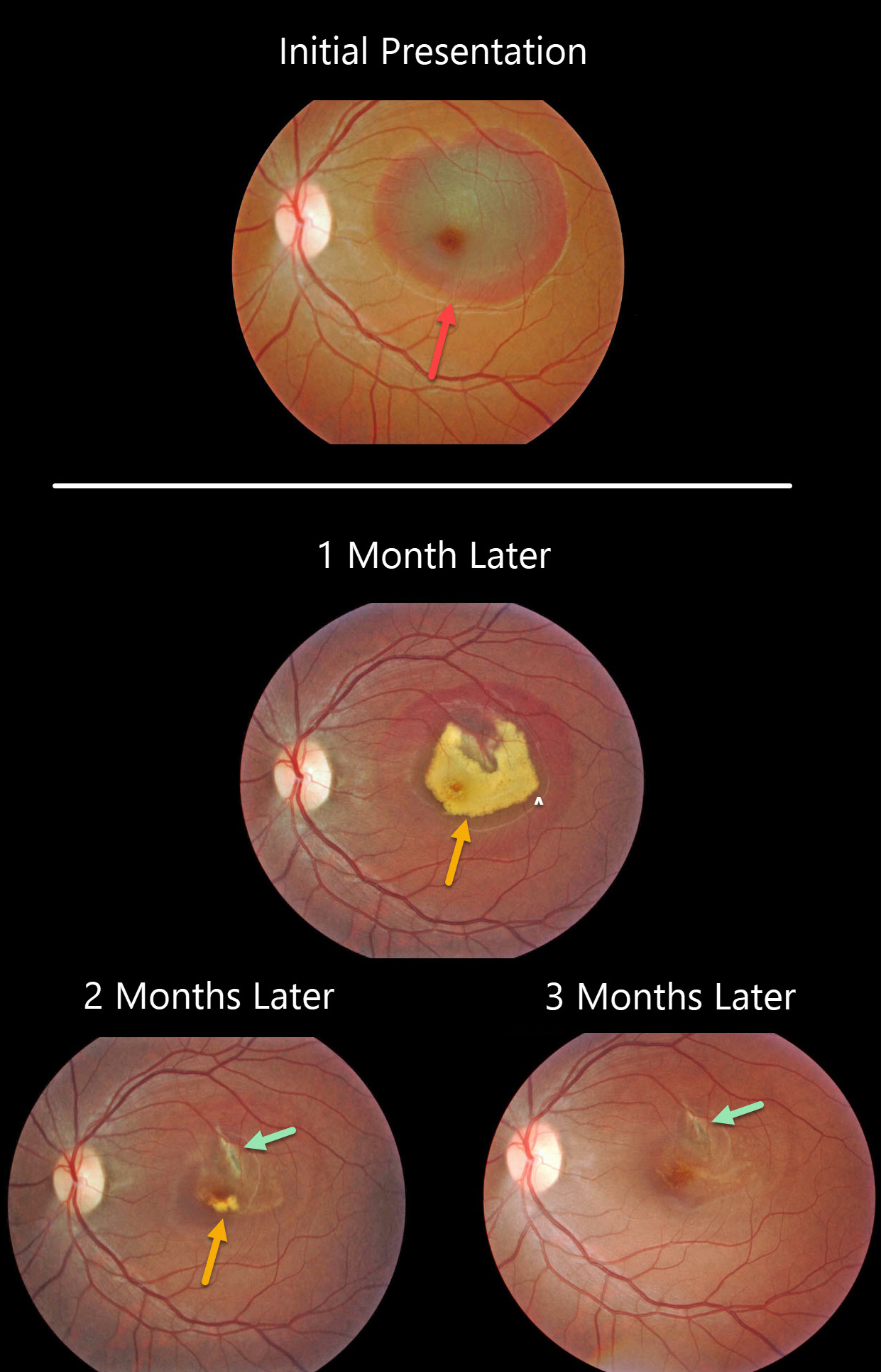
CHOROIDAL RUPTURE
Originally posted on @retina.rocks October 1, 2020
This patient presented with an acute submacular hemorrhage following a finger-to-eye injury.
The subretinal blood slowly resolved over the following three months. An underlying choroidal rupture is almost always the source of this traumatic blood.
Final vision was 20/400, either due to contusive foveal damage or possible toxicity from the subretinal blood.
Learning Point:
Choroidal ruptures are caused by blunt ocular trauma and are always circumferential with respect to the optic nerve.
These patients need to be followed since macular neovascularization can develop later in life.
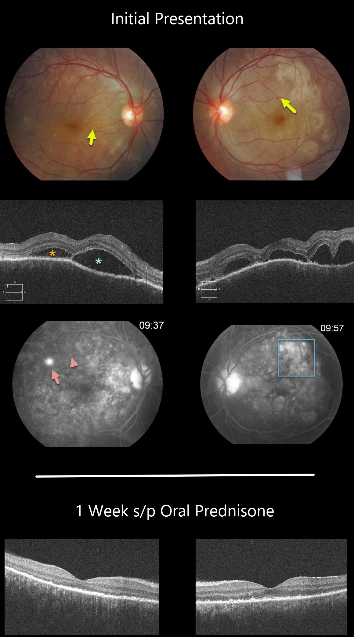
HARADA DISEASE
Originally posted on @retina.rocks September 23, 2020
This patient has classic posterior segment findings of Harada disease, including multifocal exudative serous retinal detachments.
Choroidal inflammation produces a markedly thickened choroid with overlying chorioretinal folds (seen in both eyes as variably radiating outer retinal folds).
The OCT scans show multifocal serous detachments with bacillary detachments. Fluorescein angiography shows multiple subretinal leaks with pooling of fluorescein within the various subretinal and bacillary spaces.
The patient started 60mg of oral prednisone daily. One week later, there was a dramatic improvement, with complete resolution of all intra- and subretinal fluid and normalization of choroidal thickness.
Learning Points:
Harada disease is a bilateral granulomatous panuveitis. These patients often need extended treatment with oral prednisone. Most can be successfully weaned off of steroids, although chronic treatment is sometimes necessary.
Choroidal thickening is a good biomarker for disease activity and thins with controlled disease activity.
Remember that in Harada disease, there are only ocular findings. Vogt-Koyanagi-Harada disease occurs when there are associated auditory, integumentary or neurologic symptoms.
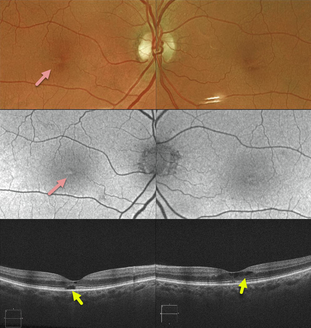
TAMOXIFEN RETINOPATHY
Originally posted on @retina.rocks September 9, 2020
This 72YO female recently completed a 5-year course of Tamoxifen as adjunctive treatment for breast cancer. She had no visual symptoms with 20/30 vision bilaterally.
Clinically, there were subtle bilateral foveal pigmentary changes. B-scan and en-face OCT showed bilateral cavitary changes.
Learning Points:
Tamoxifen retinopathy shares findings very similar to those of macular telangiectasia type 2 (MacTel2), including retinal cavitations, right-angle venules, and inner retinal crystals. The changes in MacTel2 are usually confined to the temporal fovea, whereas with Tamoxifen, they seem more diffusely distributed throughout the entire central macula. The retinal changes for both disorders likely share Muller cell injury as the underlying cause.
The prevalence of Tamoxifen retinopathy may be as high as 12%. Although most oncologists do not require this, periodic ophthalmic screening, OCT, and examinations may be indicated since patients can lose central vision if toxicity develops.
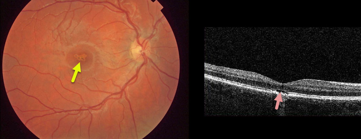
SOLAR RETINOPATHY
Originally posted on @retina.rocks July 31, 2020
This asymptomatic 10YO boy presented with 20/40 vision OD and 20/20 vision OS.
There were multifocal small subretinal pigment clumps with surrounding pigment loss in the right central macula.
OCT shows foveal outer segment loss with ragged RPE loss. The left macula was normal.
He denied looking at a laser pointer but did admit to a history of sun-gazing. He likely closed his left eye each time he looked at the sun, which explains the unusual finding of unilateral solar retinopathy.
Learning Points:
The outer retinal damage in solar retinopathy is thought to be caused by photochemical outer retinal and RPE damage instead of a thermal burn. Vision is usually fairly good.
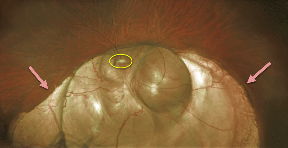
CHOROIDAL COLOBOMA
Originally posted on @retina.rocks July 30, 2020
This patient has a large choroidal coloboma extending through the left optic nerve and macula. It’s so extensive that it hides the optic nerve.
These arise from incomplete closure of the embryonic fissure during the second month of embryonic development. Patients are at lifelong risk for rhegmatogenous retinal detachment due to breaks that occur in the overlying thinned retina.
Learning Points:
Defects in closure have a wide range of results such as microphthalmia, anophthalmia, iris coloboma, ciliary body coloboma, choroid coloboma, etc.
Colobomas of the iris or ciliary body result from failure of anterior closure while choroid, retina and optic nerve result from failure of posterior closure.
A staphyloma by definition is an outpouching of the entire eye wall. Choroidal coloboma, always involves a choroidal defect, but may or may not also involve the sclera (as in this case).
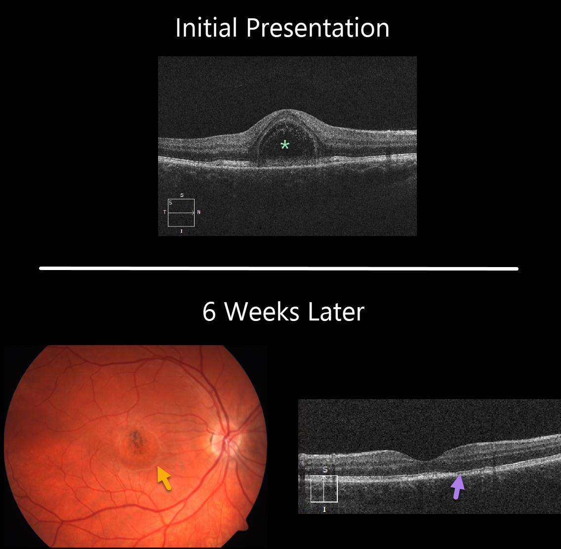
ACUTE IDIOPATHIC MACULOPATHY (AIM)
Originally posted on @retina.rocks July 23, 2020
This 22YO female presented with 1 week of unilateral vision loss and 20/200 vision in her right eye.
The macular OCT line scan demonstrates a bacillary layer (cone and rod inner and outer segments) detachment. This appears as a unique dome shaped collection of intraretinal fluid from photoreceptor myoid splitting.
Six weeks later vision improved to 20/40. The macular fluid resolved, with the development of a dry area of macular hyperpigmentation surrounded by a rim of pigment loss. OCT shows some subtle outer retinal foveal thinning with ellipsoid zone irregularities.
Learning Points:
Acute idiopathic maculopathy (AIM) is a usually unilateral idiopathic condition usually found in young people, often following a viral illness. It is usually self-limited, resolving within several weeks with significant visual improvement. The appearance of a bulls-eye lesion, as occurred in our patient, is classic for this disease.
Bacillary detachments can be observed in multiple conditions including Vogt-Koyanagi-Harada (VKH) disease, AIM, blunt trauma, or APMPPE.
See Fernandez-Avellaneda et al Retinal Cases & Brief Reports 2019 (published ahead of print) for a discussion of bacillary layer detachment in AIM.
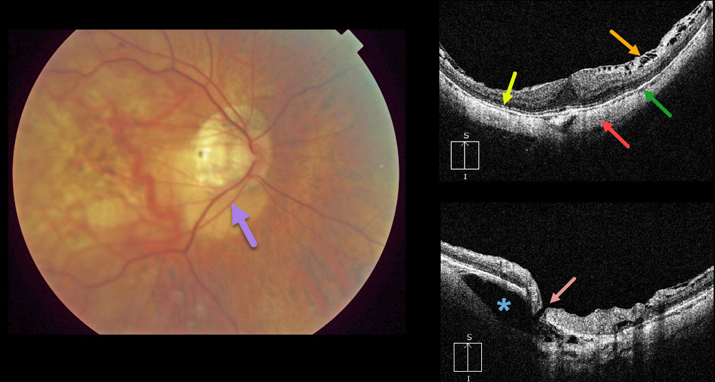
HIGH MYOPIA & INTRACHOROIDAL CAVITATION
Originally posted on @retina.rocks July 9, 2020
Peripapillary intrachoroidal cavitation is a relatively rare abnormality most commonly found in high myopia. Clinically, these subretinal lesions have an orange coloration and are usually located inferior to the optic nerve.
OCT scanning of the central macula shows a variety of typical myopic findings, including posterior bowing of the sclera, variable attenuation of the ellipsoid zone, markedly thinned choroid, and a variable epiretinal membrane with inner retinal schisis most prominent superiorly.
OCT scanning centered over the optic nerve reveals the intrachoroidal cavitation. There also seems to be a tiny full-thickness retinal defect that communicates directly with the cavitation.
Learning Points:
To learn more about intrachoroidal cavitations and high myopia, see Spaide et al Retina 2012;32;1037-1044.
See Ikuno Retina 2017;37;2347-2351 for a nice overview of macular findings in high myopia.
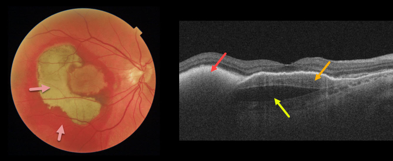
MACULAR NEOVASCULARIZATION (MNV)
Originally posted on @retina.rocks July 8, 2020
This patient’s vision remained 20/100 one month following vitrectomy with subretinal tissue plasminogen activator.
Surgery accomplished its goal of displacing most of the subfoveal blood, which is variably red and devitalized (yellow).
OCT shows the remaining subretinal hyperreflective blood, the causative sub-RPE type 1 macular neovascularization, and a subfoveal prechoroidal cleft.
Learning Points:
A prechoroidal cleft is a hyporeflective space between the RPE and Bruch’s membrane.
These are seen in up to about 20% of treated eyes with wet AMD, particularly with type 3 neovascularization and polypoidal choroidal vasculopathy.
They are usually associated with worse visual acuity due to potential complications, including RPE tear and subretinal hemorrhages.
For a more detailed discussion of prechoroidal clefts, see Kim et al Retina 2017;37;2047-2055.
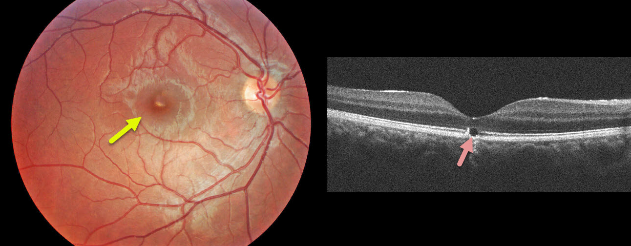
WHIPLASH MACULOPATHY
Originally posted on @retina.rocks July 1, 2020
This 17YO male presented with sudden unilateral vision loss immediately following a whiplash from a motor vehicle accident.
Clinically, he has a yellowish linear subretinal foveal discoloration. OCT shows a focal loss of the outer subfoveal retina.
Learning Points:
These findings are classic for whiplash maculopathy, in which sudden acceleration/deceleration causes the vitreous to tug on or impact the retina, resulting in outer retinal and RPE damage.
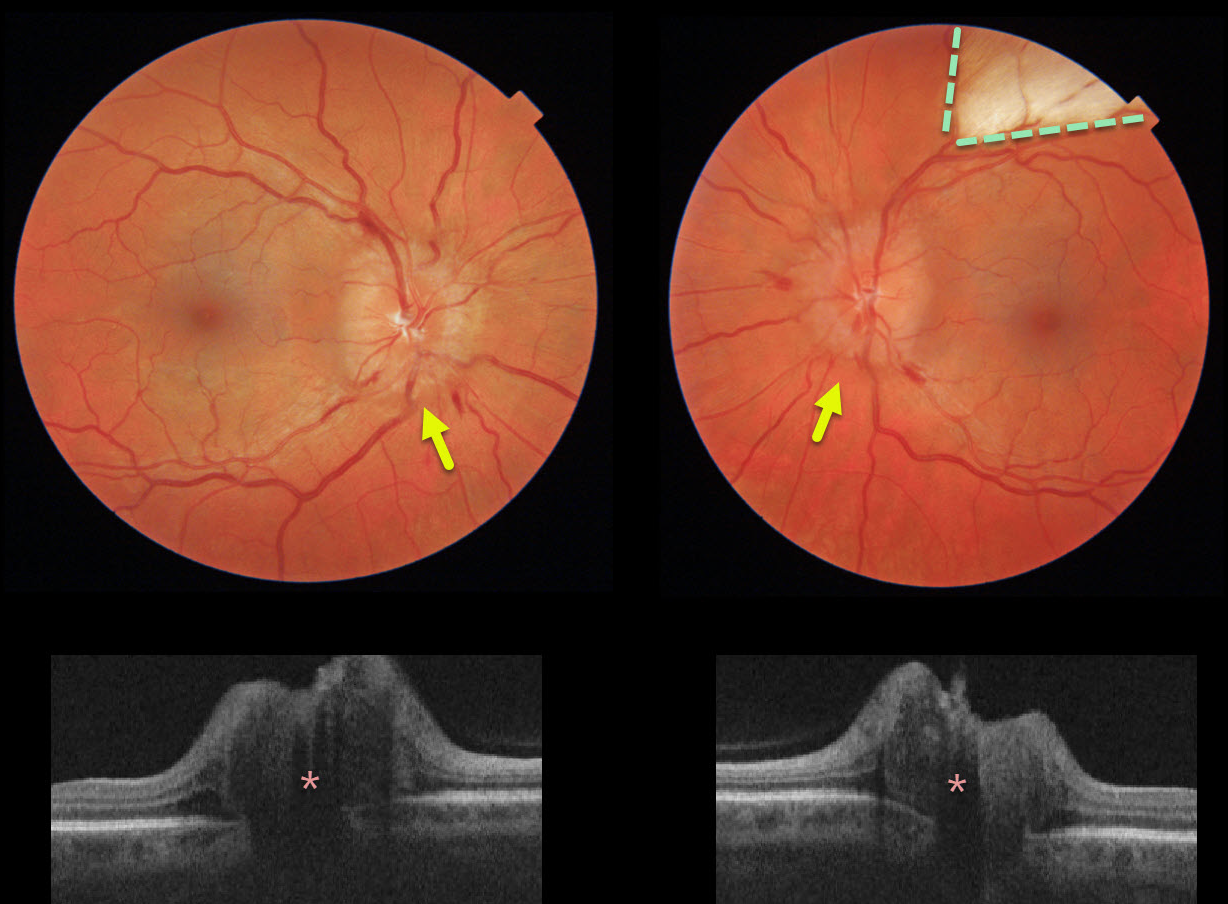
PAPILLEDEMA
Originally posted on @retina.rocks June 26, 2020
This healthy 40yo female presented with chronic headaches. She was on no systemic medications, and her blood pressure was normal. Her nerves were swollen bilaterally, and a myelinated nerve fiber layer was also noted in her left eye.
The retinal nerve fiber layer on OCT was severely thickened bilaterally.
Learning Points:
The diagnosis of idiopathic intracranial hypertension (pseudotumor cerebri) was made after an MRI scan was normal and a lumbar puncture revealed a markedly elevated opening pressure.
She is currently being successfully managed with oral acetazolamide.
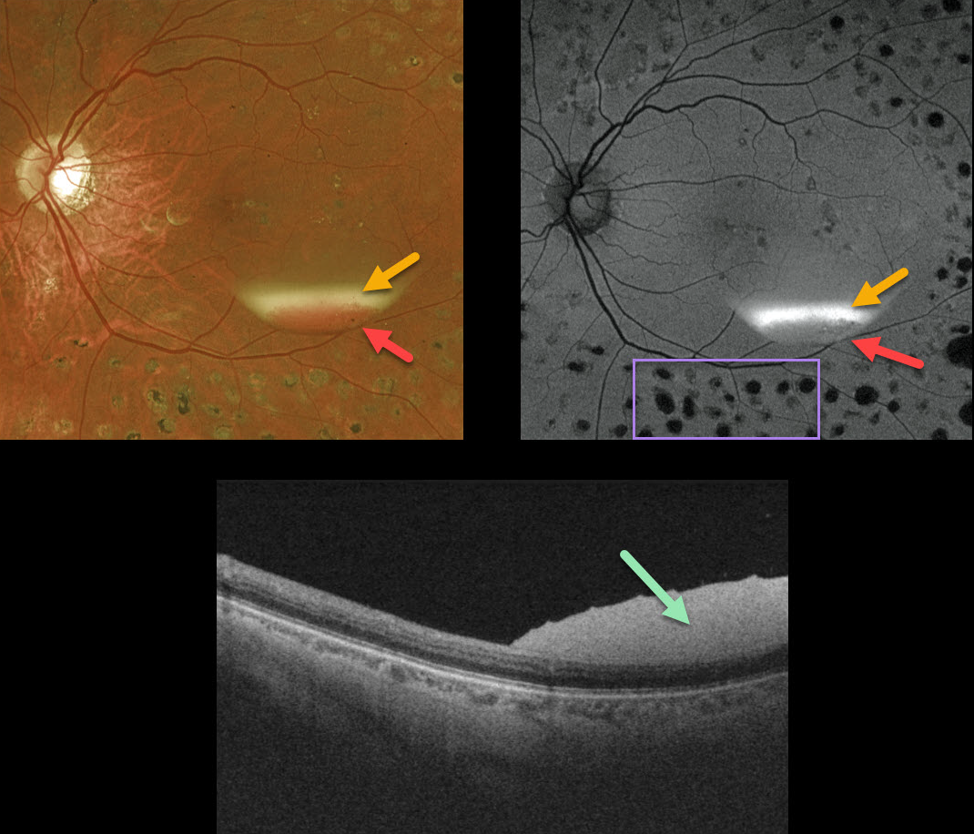
PROLIFERATIVE DIABETIC RETINOPATHY (PDR)
Originally posted on @retina.rocks June 16, 2020
This patient, previously treated for proliferative diabetic retinopathy with panretinal photocoagulation (PRP), presented with an asymptomatic resolving two-toned subhyaloid hemorrhage. The devitalized yellow blood indicates chronicity, although some red blood remains.
The OCT confirms the subhyaloid location of the hyperreflective blood.
Red blood is dark on fundus autofluorescence (FAF), although yellow blood is extremely hyperautofluorescent. Lipofuscin within the RPE is the main fluorophore in FAF. The laser PRP scars are hypoautofluorescent due to loss of the RPE.
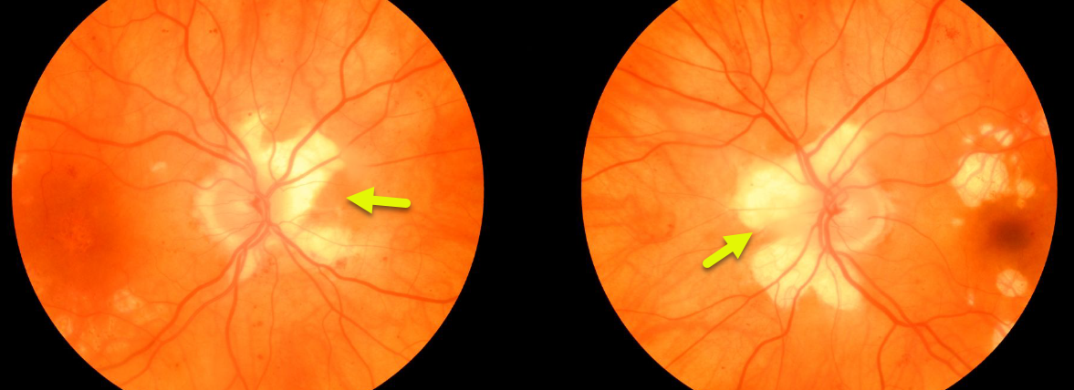
HELICOID PERIPAPILLARY CHORIORETINAL DEGENERATION
Originally posted on @retina.rocks March 27, 2020
This asymptomatic patient was found to have a unique bilateral peripapillary chorioretinal atrophy called helicoid peripapillary chorioretinal degeneration.
Vision is usually normal although central vision can rarely be affected if atrophy progresses through the fovea.
Learning Points:
Helicoid peripapillary chorioretinal degeneration is a benign autosomal dominant condition with degenerative wing or propeller-shaped lesions that radiate centrifugally from the optic nerve head.
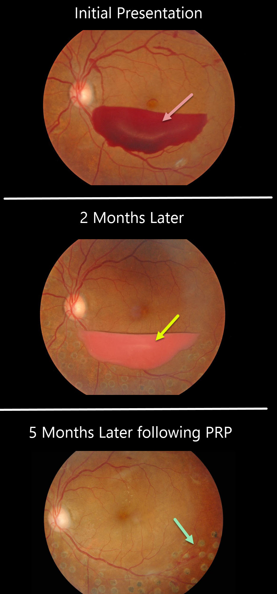
PROLIFERATIVE DIABETIC RETINOPATHY
Originally posted on @retina.rocks December 24, 2019
This patient presented with a sub-hyaloid hemorrhage due to proliferative diabetic retinopathy.
A subsequent photo two months later shows devitalized (yellowing) sub-hyaloid hemorrhage. He received panretinal photocoagulation (PRP) in the left eye and the sub-hyaloid hemorrhage completely resolved three months later. Anti-VEGF therapy was not used.
Learning Points:
PRP has been around since the 1960s and still remains the mainstay for treating proliferative disease. Often considered a “one and done,” patients can still develop break-through hemorrhages. Intravitreal anti-VEGF injections are often helpful, but some will require vitrectomy.


