

Achromatopsia
<< Back to Cases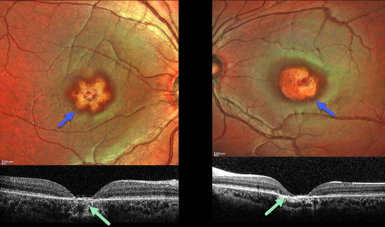
ACHROMATOPSIA
The European VitreoRetinal Society (EVRS), Vinod Kumar, Smile Singla and Bhupinder Singh
Originally posted on @retina.rocks April 24, 2026
This healthy 5YO boy presented with decreased vision and repetitive eye movements. Family history was negative. Vision was 20/200 OU. Low-amplitude horizontal nystagmus was noted bilaterally.
MultiColor imaging shows star-shaped macular atrophy OD with a more rounded area of atrophy OS. OCT shows complete central RPE and outer retinal atrophy. A homozygous pathogenic missense mutation (c.1279C>T) in exon 8 of the CNGA3 gene led to the diagnosis of achromatopsia.
Learning Points:
CNGA3 mutations, which encode for components of the cone phototransduction cascade, cause achromatopsia, a rare autosomal recessive cone photoreceptor disorder. Clinical findings range from a normal-appearing fundus to progressive macular degeneration. OCT findings may be normal or show variable outer retinal changes, including foveal ellipsoid zone (EZ) absence or disruption, a hyporeflective zone, and outer retinal atrophy (Zobor et al, IOVS 2017;58:821-832). Most eyes have foveal hypoplasia. The retinal phenotype shows considerable heterogeneity even among patients with the same genotype.


