

Acute Exudative Polymorphous Vitelliform Maculopathy
<< Back to Cases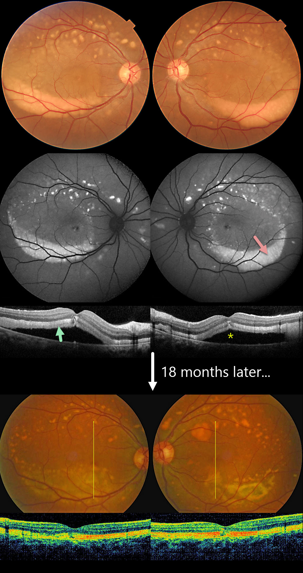
ACUTE EXUDATIVE POLYMORPHOUS VITELLIFORM MACULOPATHY
Krishnendu Nandi
Originally posted on @retina.rocks June 24, 2025
This 38YO male presented with 6 months of bilateral vision loss. Vision was 20/80 OU.
Color imaging shows multiple symmetrical yellowish subretinal variably round lesions centered in each superior macula and extending superiorly. Confluent subretinal vitelliform material is settled in the inferior maculas. This material is markedly hyper-autofluorescent. On OCT, the outer retina is diffusely infiltrated by this hyperreflective material, with associated serous macular detachments.
Oncologic workup was completely negative. When last examined 18 months later, vision was 20/30 OD and 20/30 OS. Color imaging shows remodeling of both the superior macular lesions and the inferiorly layered vitelliform material. The subretinal fluid is resolved with variable outer retinal band defects.
Learning Points:
Acute exudative polymorphous vitelliform maculopathy (AEPVM) is a rare condition characterized by acute vision loss from multifocal serous retinal detachments that later progress to subretinal vitelliform lesions. Initially, serous detachments can mimic central serous retinopathy, including choroidal thickening. The condition is often self-limiting, and vision usually recovers within several months. The yellow vitelliform lesions may take 1-2 years to completely resolve. These patients need a systemic workup for metastatic disease, since paraneoplastic retinopathy, including metastatic melanoma, can mimic idiopathic AEPVM.
ACUTE EXUDATIVE POLYMORPHOUS VITELLIFORM MACULOPATHY
Originally posted on @retina.rocks May 18, 2022
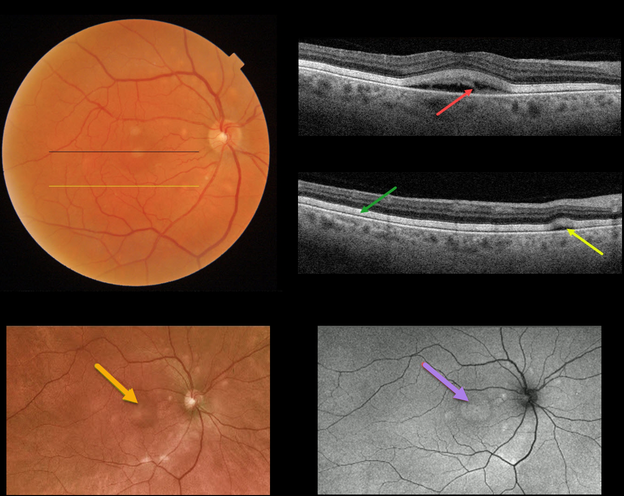
This 72YO male presented with recent difficulties with dark adaptation and 20/60 vision in his right eye. His left eye was enucleated 2 years earlier for a uveal malignant melanoma.
Triton color imaging shows multiple yellowish subretinal round lesions scattered around the optic nerve and macula.
Triton Swept Source OCT through the fovea shows a partially scrambled vitelliform lesion. An OCT B-scan through the inferior macula shows the outer retina diffusely infiltrated by hyperreflective vitelliform material, along with a small serous detachment.
Optos imaging shows an ultrawide view of the smaller and larger central vitelliform lesions, and fundus autofluorescence (FAF) shows hyper-FAF from the corresponding vitelliform lesions. ICG and FA were both normal.
Learning Points:
Acute exudative polymorphous vitelliform maculopathy (AEPVM) is a rare condition characterized by acute vision loss from multifocal serous retinal detachments that later progress to subretinal vitelliform lesions.
Initially, the serous detachments can mimic central serous retinopathy, including choroidal thickening. The condition is often self-limiting, and vision usually recovers within several months. The yellow vitelliform lesions may take 1-2 years to resolve completely.
These patients need a systemic workup for metastatic disease, since paraneoplastic retinopathy, including metastatic melanoma, can mimic idiopathic AEPVM.
Sadly, preliminary metastatic workup for this patient showed two possible liver metastases.
ACUTE EXUDATIVE POLYMORPHOUS VITELLIFORM MACULOPATHY
Originally posted on @retina.rocks March 2, 2020
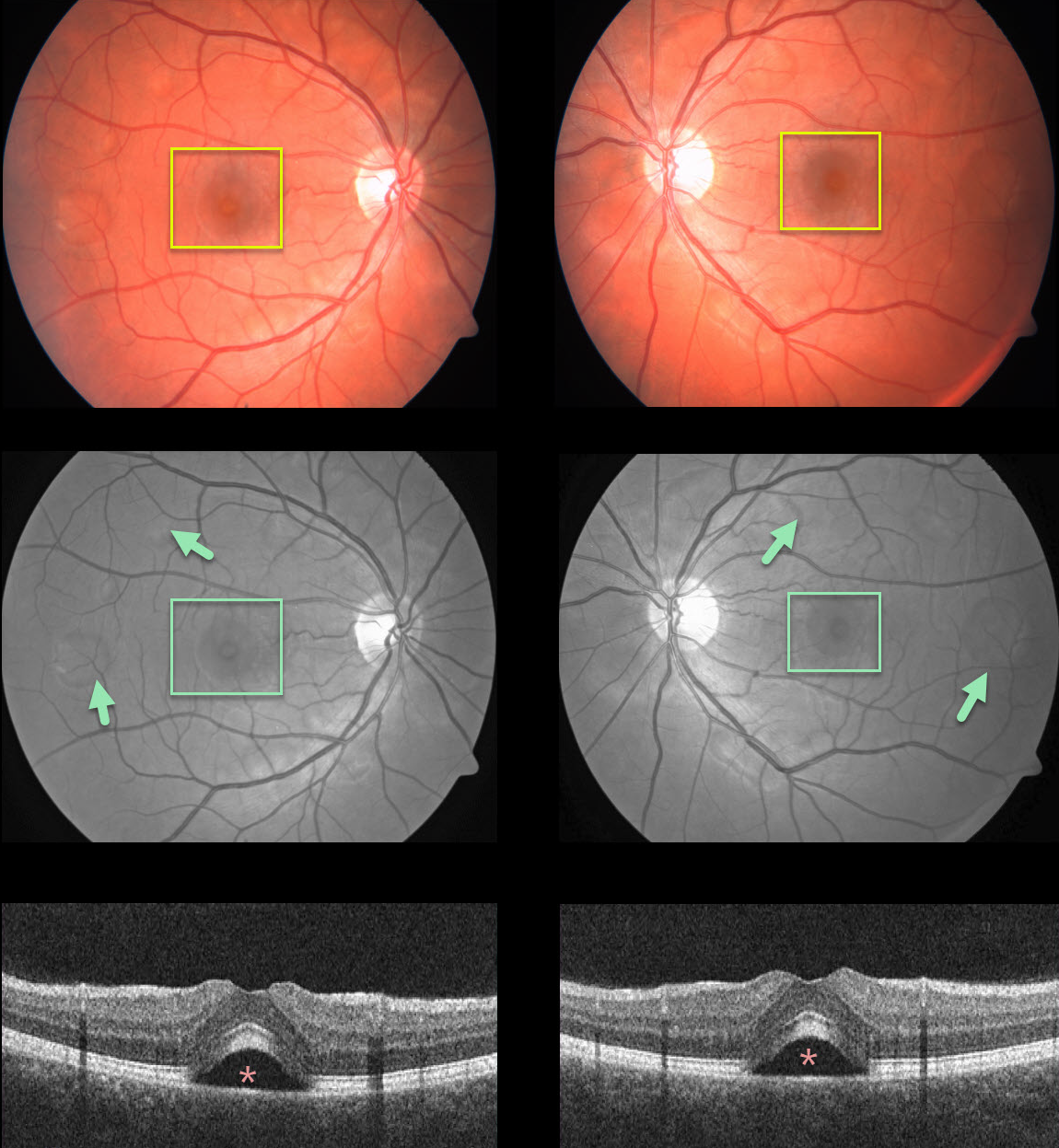
Our patient presented with bilateral multifocal serous retinal detachments, which are better seen on red-free fundus imaging and OCT. Unfortunately, we don’t have any further clinical information.
Learning Points:
Acute exudative polymorphous vitelliform maculopathy (AEPVM) is a rare condition characterized by acute vision loss from multifocal serous retinal detachment that later progress to subretinal vitelliform lesions.
Initially the serous detachments can mimic central serous retinopathy, including thickening of the choroid. The yellow vitelliform lesions may take 1-2 years to completely resolve.
The condition is often self-limiting, and vision usually recovers within several months.
These patients need a systemic workup for metastatic disease, since paraneoplastic retinopathy, including metastatic melanoma, can mimic AEPVM.


