

Acute Idiopathic Maculopathy (AIM)
<< Back to Cases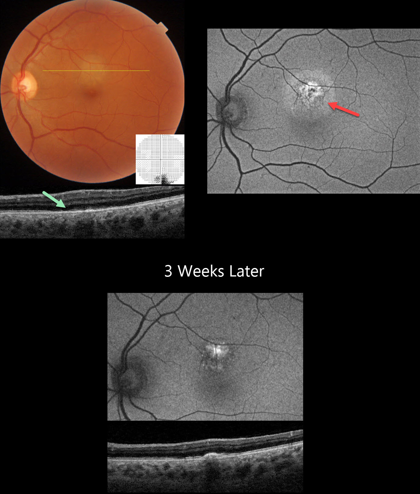
ACUTE IDIOPATHIC MACULOPATHY (AIM)
Mattie Adams
Originally posted on @retina.rocks July 17, 2025
This healthy 46YO male presented with 5 days of an inferior paracentral scotoma in his left eye. Vision was 20/15 in his normal OD and 20/20 OS.
Triton color imaging shows a round, deep retinal amelanotic lesion just superior to the macular center. Swept-source OCT shows loss of the outer retinal band detail with ill-defined hyperreflective material overlying the RPE. Central 10-2 visual field shows inferior loss corresponding to the subretinal lesion. Optos fundus autofluorescence (FAF) of this lesion shows central hyper-FAF with a surrounding rim of less-intense hyper-FAF.
Coxsackie B virus antibody titers were markedly positive at >= 1:640, and observation was recommended. He returned 3 weeks later with stable symptoms and vision, although the OCT and FAF had improved.
Learning Points:
Acute idiopathic maculopathy (AIM) is an idiopathic inflammation of the outer retina, RPE, and inner choroid. There is often a preceding viral prodrome (lacking in our patient) caused by the Coxsackie virus. Healthy young patients typically present with acute unilateral moderately severe central vision loss.
The initial funduscopic changes include macular fluid and, sometimes, deep retinal blood that can mimic macular neovascularization. The OCT shows heterogeneous hyperreflective thickening of the outer retinal/RPE and fluid. Fluorescein angiography usually shows deep retinal leakage. The lesion spontaneously resolves with significant visual improvement after several weeks, usually leaving a pathognomonic bull’s-eye lesion as seen in our patient.
ACUTE IDIOPATHIC MACULOPATHY (AIM)
Emily Shepherd
Originally posted on @retina.rocks March 3, 2023
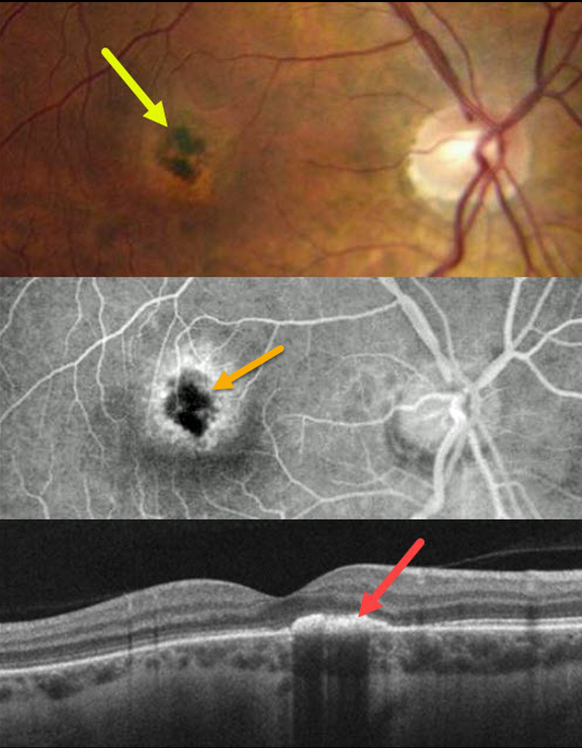
This 33YO healthy female complained of 1 to 2 weeks of a blind spot and ring of light in her right eye. She had a recent non-COVID viral illness when her symptoms started. Vision was 20/60 OD and 20/20 OS.
Color imaging shows a subfoveal area of central hyperpigmentation surrounded by a ring of pigment loss. Angiographically, the pigmentation blocks with a surrounding rim of staining.
OCT B-scan shows a hyperreflective plaque in the outer retina.
Learning Points:
Acute idiopathic maculopathy (AIM) is an idiopathic inflammation of the outer retina, RPE, and inner choroid. There is often a preceding viral prodrome caused by the Coxsackie virus. Healthy young patients typically present with acute unilateral moderately severe central vision loss.
The initial funduscopic changes show macular fluid and, sometimes, deep retinal blood, which can simulate macular neovascularization. The OCT shows heterogeneous hyperreflective thickening of the outer retina/RPE and fluid.
Fluorescein angiography usually shows deep retinal leakage. The lesion spontaneously resolves with significant visual improvement after several weeks, usually leaving a pathognomonic bull’s-eye lesion as seen in our patient.
ACUTE IDIOPATHIC MACULOPATHY (AIM)
Originally posted on @retina.rocks August 3, 2021
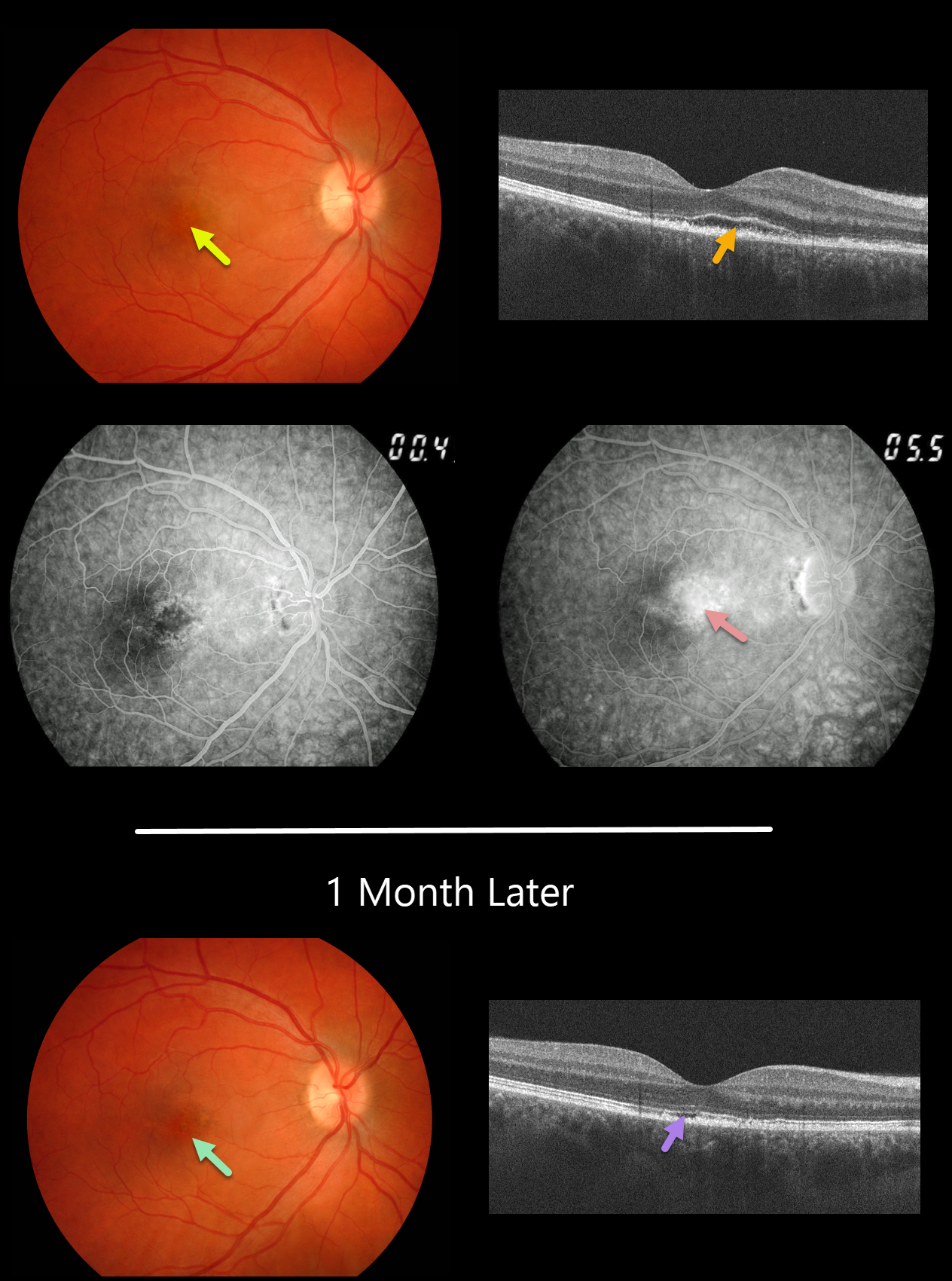
This 29YO male presented with a 1-week history of acute blurred vision in his right eye. Vision was 20/40 OD.
Fundus photography and fluorescein angiography reveal pigment mottling and late-stage leakage. OCT scanning shows an irregularly elevated ellipsoid zone with underlying outer segment disruption vs subretinal fluid and a thickened RPE.
We felt the patient had acute idiopathic maculopathy (AIM) and observed him without treatment.
One month later, the outer fluid resolved with residual irregularities in the outer retinal and RPE bands on OCT and an area of macular hyperpigmentation surrounded by a rim of pigment loss.
Learning Points:
Originally described by Yannuzzi et al. in 1991 (Arch Ophthalmology 1991;109:1411-1416), AIM is an idiopathic inflammation of the outer retina, RPE, and inner choroid, usually found in young people and often following a viral illness. It is usually self-limited, resolving within several weeks with significant visual improvement. The appearance of a bull’s-eye lesion, as occurred in our patient, is classic for this disease.
ACUTE IDIOPATHIC MACULOPATHY (AIM)
Originally posted on @retina.rocks July 23, 2020
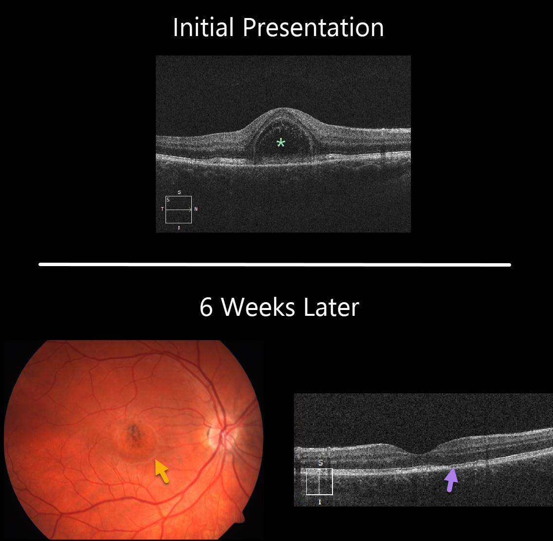
This 22YO female presented with 1 week of unilateral vision loss and 20/200 vision in her right eye.
The macular OCT line scan demonstrates a bacillary layer (cone and rod inner and outer segments) detachment. This appears as a unique dome shaped collection of intraretinal fluid from photoreceptor myoid splitting.
Six weeks later vision improved to 20/40. The macular fluid resolved, with the development of a dry area of macular hyperpigmentation surrounded by a rim of pigment loss. OCT shows some subtle outer retinal foveal thinning with ellipsoid zone irregularities.
Learning Points:
Acute idiopathic maculopathy (AIM) is a usually unilateral idiopathic condition usually found in young people, often following a viral illness. It is usually self-limited, resolving within several weeks with significant visual improvement. The appearance of a bulls-eye lesion, as occurred in our patient, is classic for this disease.
Bacillary detachments can be observed in multiple conditions including Vogt-Koyanagi-Harada (VKH) disease, AIM, blunt trauma, or APMPPE.
See Fernandez-Avellaneda et al Retinal Cases & Brief Reports 2019 (published ahead of print) for a discussion of bacillary layer detachment in AIM.


