

Acute Posterior Multifocal Placoid Pigment Epitheliopathy (APMPPE)
<< Back to Cases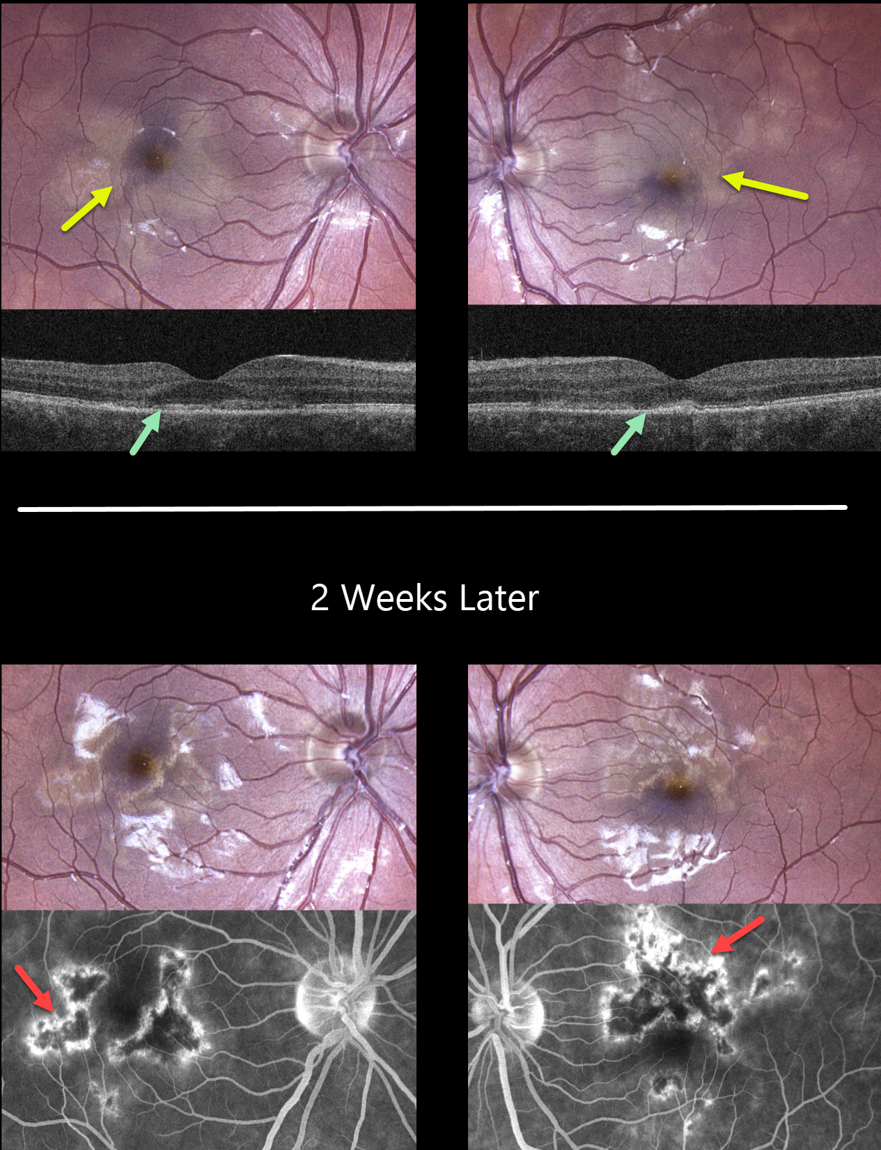
ACUTE POSTERIOR MULTIFOCAL PLACOID PIGMENT EPITHELIOPATHY (APMPPE)
Mattie Adams
Originally posted on @retina.rocks September 12, 2023
This healthy 25YO female presented with 3 weeks of painless vision loss, especially in her left eye. Vision was 20/20 OD and 20/100 OS. There was a moderate bilateral granulomatous anterior uveitis. The vitreous was clinically free of cells.
Color photography shows bilateral creamy subretinal macular placoid lesions. OCT scanning shows variable loss of the ellipsoid and interdigitation zone layers. Some vitreous cells are noted bilaterally.
Laboratory testing was all negative, including CBC, ESR, C-reactive protein, ACE, syphilis, and QuantiFERON-TB Gold Plus. The patient was started on topical prednisolone acetate QID with a weekly taper.
When examined 2 weeks later, her symptoms were much improved with vision of 20/20 OD and 20/30 OS. The placoid lesions were resolving with angiographic staining.
Learning Points:
Acute posterior multifocal placoid pigment epitheliopathy (APMPPE) is a usually bilateral inflammatory vs ischemic disorder of the inner choroid, RPE, and outer retina. Blurry vision and paracentral/central scotomas often follow a viral illness.
Acutely multifocal creamy yellow or grayish-white subretinal placoid lesions are scattered throughout the posterior poles. The lesions and visual symptoms usually resolve within 4 to 8 weeks with secondary chorioretinal changes. Recurrent lesions are rare, and vision is usually fairly good.
The debate over whether APMPPE is primarily an inflammatory disorder of the outer retina and RPE or an ischemic disease of the inner choroid has raged since Dr. Gass first described this entity in 1968.
Our case supports the current theory that APMPPE is an inflammation of the inner choroid with secondary outer retinal damage.
Anterior uveitis, as seen in our patient, is rarely seen in APMPPE. This was one reason we performed blood work, which fortunately came back negative. Her spontaneous and rapidly resolving posterior segment findings also support our diagnosis of APMPPE.
ACUTE POSTERIOR MULTIFOCAL PLACOID PIGMENT EPITHELIOPATHY (APMPPE) WITH OUTER RETINAL TUBULATION
Originally posted on @retina.rocks June 4, 2021
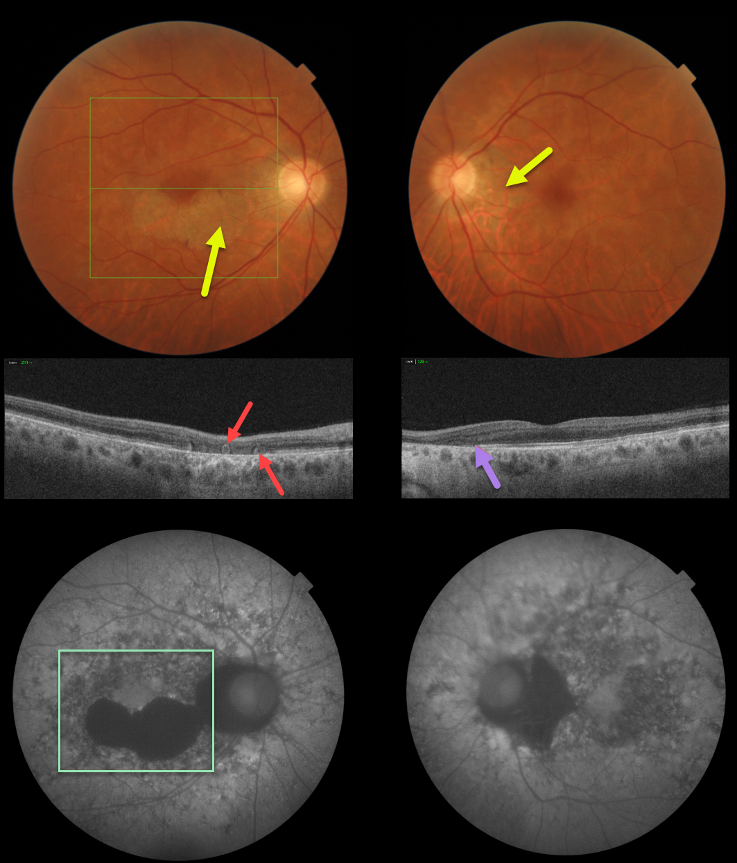
This patient has bilateral fundus findings consistent with resolved acute posterior multifocal placoid pigment epitheliopathy (APMPPE).
Triton fundus photos show placoid areas of atrophic pigmentary changes, which are variably hypo-autofluorescent.
Triton swept-source OCT in the right eye shows outer retinal atrophy with outer retinal tubulation (ORT) and nasal outer retinal atrophy in the left eye.
Learning Points:
ORT, initially described by Zweifel et al (Arch Ophthalmol 2009;127;1596-1602), is a neurodegenerative condition of the photoreceptors and Muller cells associated with atrophy affecting the outer retina and retinal pigment epithelium, including advanced AMD and inherited retinal diseases.
ORT is often seen overlying inactive macular neovascularization in the setting of ongoing anti-VEGF therapy and should not be confused with exudative fluid or cysts, which lack a hyperreflective border. The outer hyperreflective band likely represents inner segment mitochondria undergoing fission and translocation toward the nucleus (Litts et al, Retina 2018;38;445-461).
Acute posterior multifocal placoid pigment epitheliopathy (APMPPE) is a usually bilateral inflammatory vs ischemic disorder of the inner choroid, RPE, and outer retina.
Blurry vision and paracentral/central scotomas often follow a viral illness. Acutely multifocal creamy yellow or grayish-white subretinal placoid lesions are scattered throughout the posterior poles. The lesions and visual symptoms usually resolve within 4 to 8 weeks with secondary chorioretinal changes. Recurrent lesions are rare, and vision is usually fairly good.


