

Acute Retinal Necrosis Syndrome (ARNS)
<< Back to Cases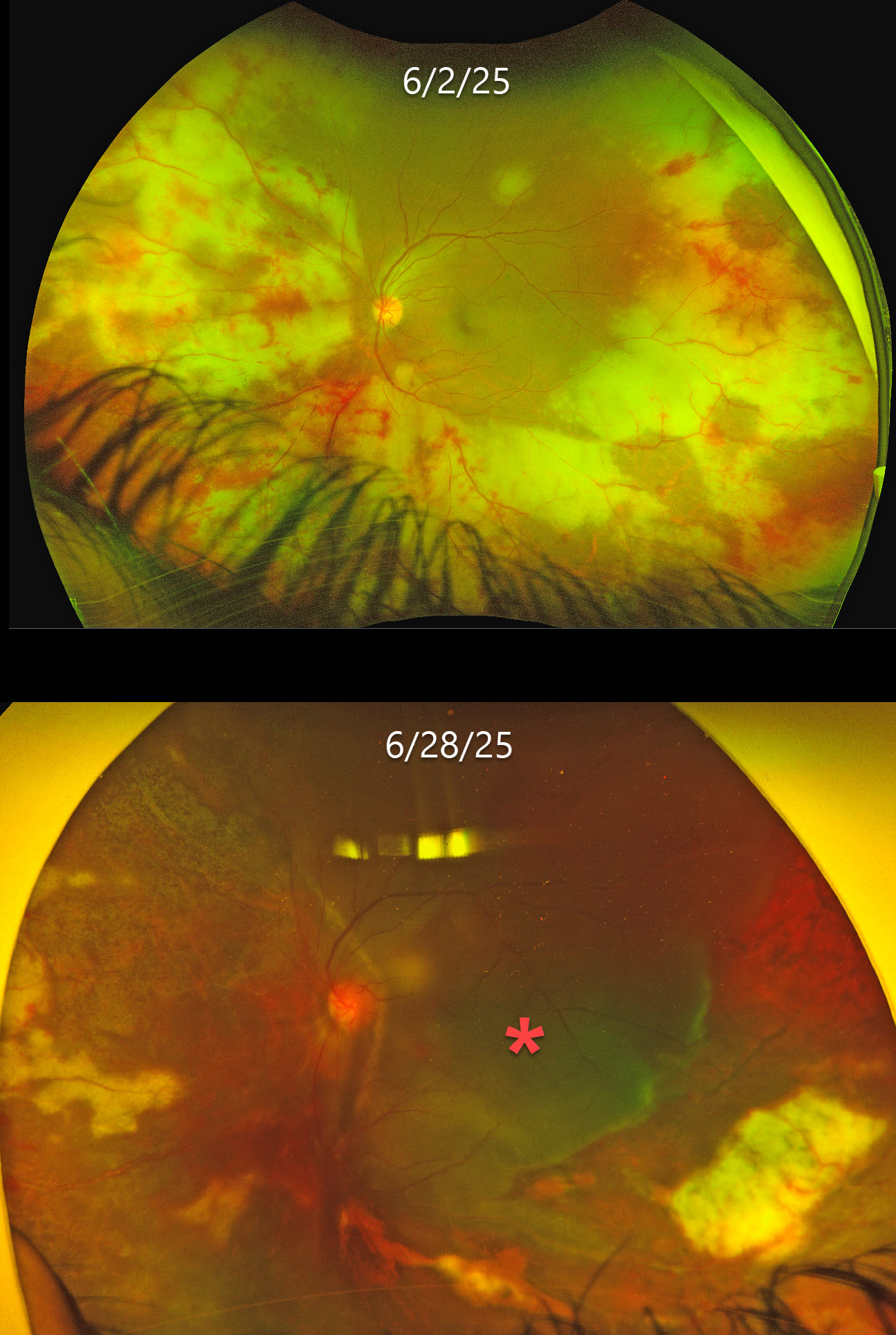
ACUTE RETINAL NECROSIS SYNDROME
César Adrián Gómez Valdivia
Originally posted on @retina.rocks November 26, 2025
This previously healthy 35YO male presented with 1 week of floaters in his left eye. Vision was 20/20 in his normal OD and 20/20 OS. Anterior segments were normal.
Optos color RG imaging shows variably confluent outer retinal opacification with retinal hemorrhages extending from the periphery into the posterior pole. The retinal vessels are variably narrowed. He was initially diagnosed with possible cytomegalovirus (CMV) retinitis, started on valganciclovir, and referred to infectious diseases (ID).
He returned 3 weeks later with counting fingers vision despite improved retinitis and stopping systemic treatment after 10 days due to financial limitations. He returned 5 days later with sudden loss of vision. Vision was light perception with improved retinitis but with a new macula-off retinal detachment, which was repaired with lensectomy, vitrectomy, scleral buckle, retinectomy, and silicone oil. Two months later, vision was 20/150, and the retina remained completely attached.
Learning Points:
Acute retinal necrosis syndrome (ARNS) is usually caused by the herpes zoster virus. It generally occurs in immunocompetent individuals and presents as rapidly progressive panuveitis with hemorrhagic and ischemic unilateral (ARNS) or bilateral (BARNS) retinal necrosis, typically beginning in the retinal periphery.
Our patient was eventually evaluated by ID, who diagnosed him with HIV and a CD4 count of 45 with initiation of HARRT therapy. It is unclear if our patient’s underlying previously undiagnosed HIV infection was contributory to his retinitis. The lack of the usual anterior chamber and vitreous inflammation is likely due to his immunosuppression.
Both CMV retinitis and ARNS cause retinal necrosis and hemorrhages. However, CMV usually starts in the posterior pole, often beginning in a paravascular location, which then spreads outwards. The retinitis involves the full-thickness retina, so the hemorrhages and opaque retina also involve the inner retina and obscure the underlying larger retinal vessels. Our patient’s findings are more pathognomonic for ARNS, given the outer retinal findings that likely began in the retinal periphery and extended posteriorly, with no paravascular location.
ARNS is often a devastating and blinding infection due to the high risk for retinal detachment as well as macular and optic nerve involvement. The latest meta-analysis argues for systemic antiviral therapy and prophylactic vitrectomy, with uncertain benefits for prophylactic laser photocoagulation or adjunctive intravitreal antivirals (Zhao et al, Retina 2021;41:965-978).
Treatment should be initiated as soon as the diagnosis is suspected, without waiting for laboratory results. Oral valacyclovir is clinically equivalent to intravenous acyclovir (Baltinas et al, AJO 2018;188:173-180), and aspirin is often recommended to minimize vascular thrombosis. Although valganciclovir is not the treatment of choice, our patient’s retinitis still rapidly improved despite only 10 days of therapy. Treatment is continued until all retinitis becomes inactive, and usually for at least 3-6 months thereafter to reduce the risk of fellow-eye involvement.
ACUTE RETINAL NECROSIS SYNDROME
Ayushi Gupta and Vishal Agrawal
Originally posted on @retina.rocks June 26, 2025
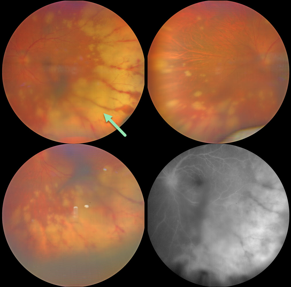
This 56YO female presented with 2 weeks of decreased vision in her left eye. She was treated for herpes zoster one month earlier, and there were a few crusted lesions on the right side of the forehead. Vision was 20/20 in her normal OD and 20/200 OS. There were moderate anterior chamber and vitreous cells.
Color photography shows scattered, midperipheral, variably confluent patches of creamy white deep retinitis. Fluorescein angiography shows optic nerve and deep retinal leakage.
An anterior chamber tap was performed, and PCR analysis confirmed varicella zoster. Intravenous acyclovir was given for 1 week, followed by oral valacyclovir 1 gram daily. When examined 3 weeks later, vision improved to 20/40 with resolving retinitis (not shown).
Learning Points:
The acute retinal necrosis syndrome is usually caused by the herpes zoster virus. It generally occurs in immunocompetent individuals and presents as a rapidly progressive panuveitis with hemorrhagic and ischemic unilateral (ARNS) or bilateral (BARNS) retinal necrosis, typically beginning in the retinal periphery.
It is often a devastating and blinding infection due to the high risk for retinal detachment as well as macular and optic nerve involvement. The latest meta-analysis argues for systemic antiviral therapy and prophylactic vitrectomy, with uncertain benefits for prophylactic laser photocoagulation or adjunctive intravitreal antivirals (Zhao et al, Retina 2021;41:965-978).
Treatment should be initiated as soon as the diagnosis is suspected, without waiting for laboratory results. Oral valacyclovir is clinically equivalent to intravenous acyclovir (Baltinas et al, AJO 2018;188:173-180), and aspirin is often recommended to minimize vascular thrombosis. Treatment is continued until all retinitis becomes inactive, and usually for at least 3-6 months thereafter to reduce the risk of fellow-eye involvement.
ACUTE RETINAL NECROSIS SYNDROME
Originally posted on @retina.rocks November 29, 2023
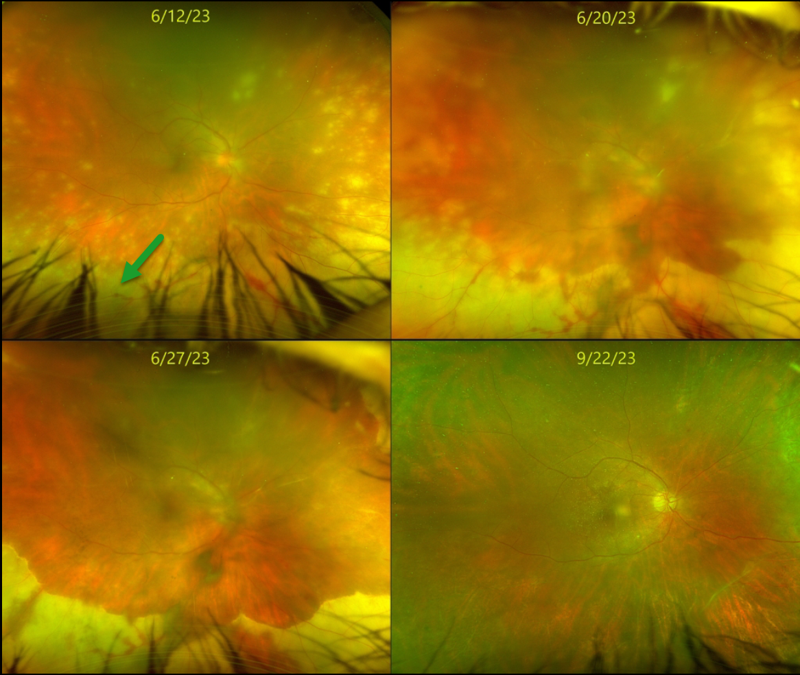
This healthy 55YO female presented with about 2 weeks of vision loss in her right eye. Vision was counting fingers OD and 20/30 in her normal left eye.
Slit lamp showed moderate anterior granulomatous inflammation. Moderate vitreous debris was noted. Optos color RG imaging shows multifocal white patches of variably confluent outer retinal inflammation extending anteriorly from the midperiphery. She was started on topical steroids and atropine, valacyclovir 1 gram PO BID, and a baby aspirin daily for acute retinal necrosis syndrome (ARNS).
After 1 week, vision remained at counting fingers but with subjective improvement. The retinitis was consolidating. The retinitis gradually improved over the following 3 months. When last examined on 9/22/23, vision was 20/70, with complete resolution of disease on valacyclovir 1 gram PO daily. We continue to follow her closely.
Learning Points:
ARNS is most commonly caused by the Herpes zoster virus. It usually occurs in immunocompetent individuals, and is a rapidly progressive panuveitis with hemorrhagic and ischemic unilateral (ARNS) or bilateral (BARNS) retinal necrosis that typically begins in the retinal periphery.
ARNS is often a devastating and blinding infection due to the high risk for retinal detachment as well as macular and optic nerve involvement. The latest meta-analysis argues for systemic antiviral therapy and prophylactic vitrectomy, with uncertain benefits for prophylactic laser photocoagulation or adjunctive intravitreal antivirals (see Zhao et al, Retina 2021;41:965-978).
ACUTE RETINAL NECROSIS SYNDROME
Originally posted on @retina.rocks July 15, 2022
This 58YO male has an extremely complicated past ocular history beginning with pseudoexfoliative glaucoma with a steroid response. Following cataract surgery in 2012, his intraocular lens dislocated in February 2016, requiring vitrectomy with a secondary anterior chamber implant. Pseudophakic bullous keratopathy developed, requiring DMEK surgery in July 2018.
His intraocular pressure subsequently increased, eventually requiring multiple glaucoma procedures, including Baerveldt implantation. Additional DMEK procedures were performed in September 2020 and December 2021. A minor additional corneal procedure with injection of gas into the anterior chamber was performed on 5/5/22 for some host Descemet’s that extended under the donor graft, causing recurrent corneal edema.
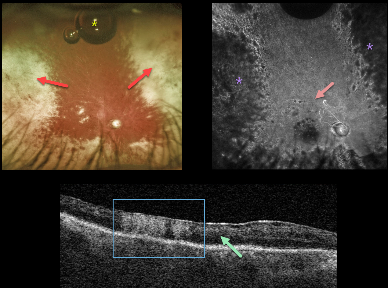
When examined on 6/24/22, the cornea was finally clear enough to allow for visualization of the posterior segment, which hadn’t been possible during his multiple anterior segment procedures. Vision was light perception.
Optos imaging shows white chorioretinal scarring in the nasal and temporal peripheries with a small amount of residual intraocular gas that had migrated into the vitreous cavity. Triton swept source OCT shows diffuse disorganization of all retinal layers, with some areas of full-thickness hyperreflectivity. Fluorescein angiography shows near total loss of all retinal perfusion, with peripheral hypofluorescence from the white peripheral chorioretinal scarring.
We felt his findings were most consistent with acute retinal necrosis, and oral valaciclovir was started.
Learning Points:
ARNS is most commonly caused by the Herpes zoster virus. It usually occurs in immunocompetent individuals, and is a rapidly progressive panuveitis with hemorrhagic and ischemic unilateral (ARNS) or bilateral (BARNS) retinal necrosis that typically begins in the retinal periphery.
ARNS is often a devastating and blinding infection due to the high risk for retinal detachment as well as macular and optic nerve involvement. The latest meta-analysis argues for systemic antiviral therapy and prophylactic vitrectomy, with uncertain benefits for prophylactic laser photocoagulation or adjunctive intravitreal antivirals (see Zhao et al, Retina 2021;41:965-978).
ACUTE RETINAL NECROSIS SYNDROME
Originally posted on @retina.rocks March 10, 2022
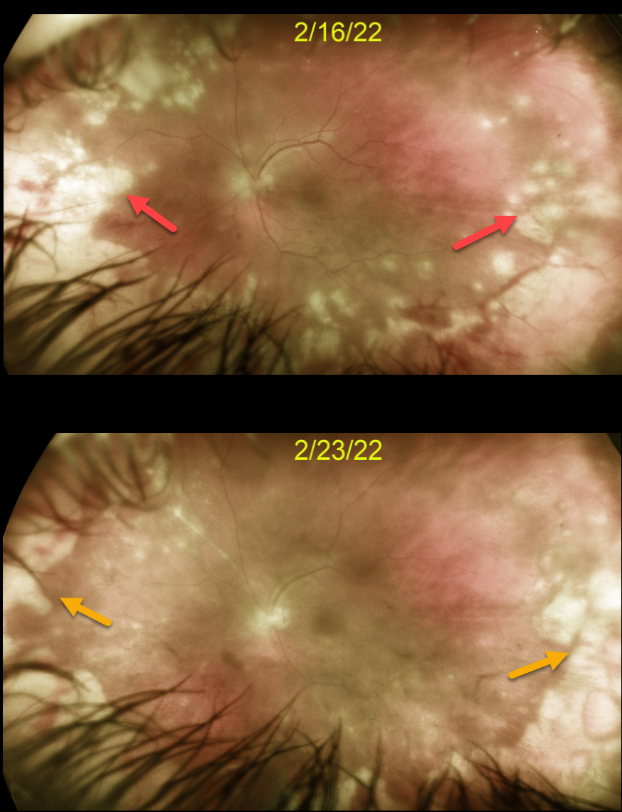
This 44YO otherwise healthy male presented with 1 week of floaters, blurred vision and pain in his left eye. Vision was 20/20 in his normal right eye and 20/100 in his left eye. There was panuveitis in his left eye, including a moderate granulomatous anterior uveitis, vitritis, and peripheral multifocal mostly outer white retinitis.
We diagnosed him with acute retinal necrosis syndrome (ARNS), and started him on oral valganciclovir 900 mg BID and a baby aspirin daily. Several days later, oral prednisone 60mg/day was begun.
After 1 week, vision remained at 20/100 but with subjective improvement. The retinitis was significantly improving. We continue to follow him very closely.
Learning Points:
The Herpes zoster virus most commonly causes ARNS. It usually occurs in immunocompetent individuals and is a rapidly progressive panuveitis with hemorrhagic and ischemic unilateral (ARNS) or bilateral (BARNS) retinal necrosis, typically beginning in the retinal periphery.
ARNS is often a devastating and blinding infection due to the high risk for retinal detachment as well as macular and optic nerve involvement.
The latest meta-analysis argues for systemic antiviral therapy and prophylactic vitrectomy, with uncertain benefits for prophylactic laser photocoagulation or adjunctive intravitreal antivirals (see Zhao et al, Retina 2021;41:965-978).
ACUTE RETINAL NECROSIS SYNDROME
Originally posted on @retina.rocks May 19, 2021
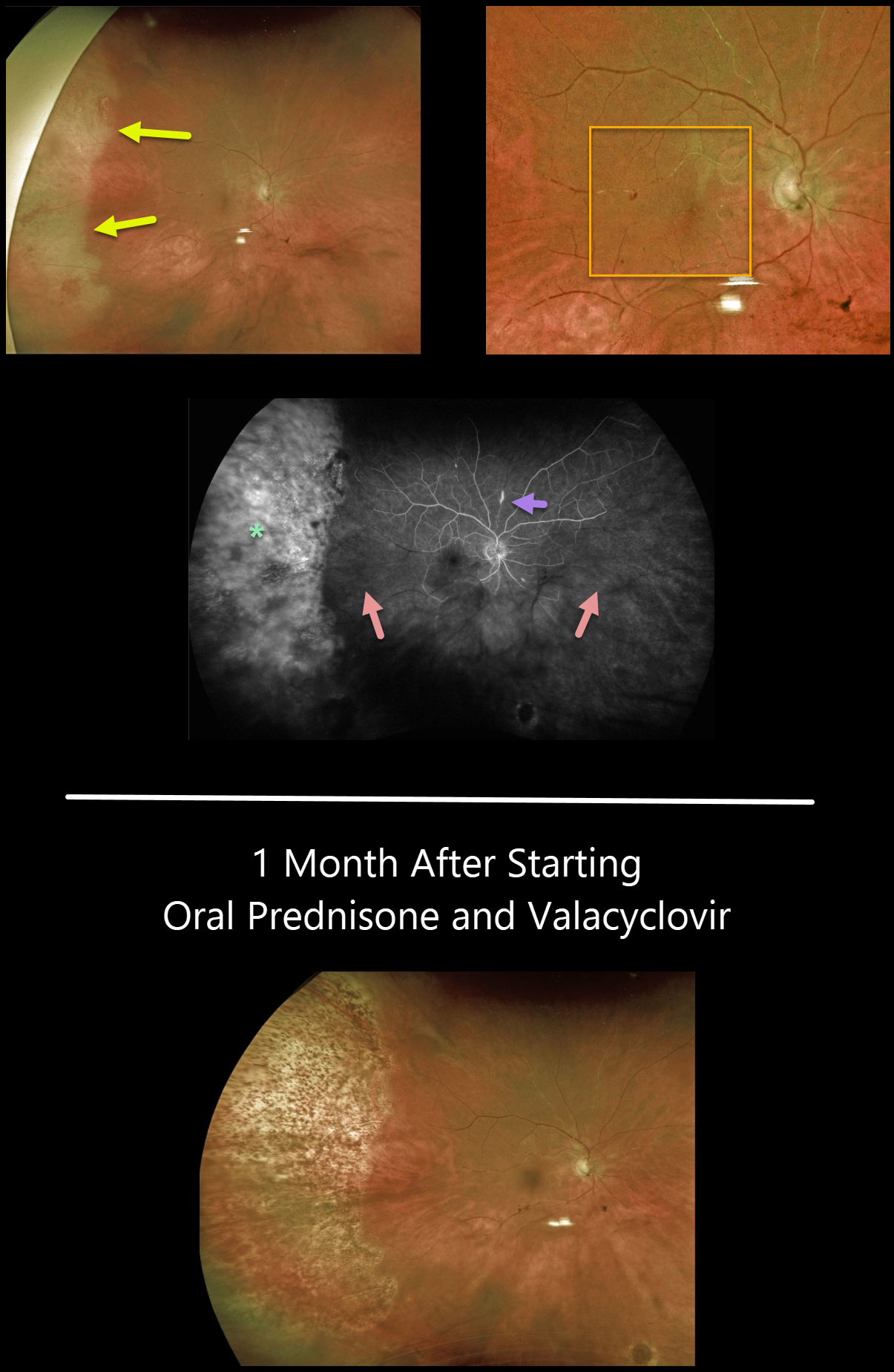
This 80YO female presented with a sudden onset of floaters while battling 2 months of persistent iridocyclitis from Herpes zoster ophthalmicus.
There was increased anterior uveitis, along with new peripheral temporal retinitis. Despite significant macular ischemia on clinical examination, vision was 20/60. Our patient’s posterior involvement was likely seeded from her anterior segment.
Optos fluorescein angiography shows diffuse staining of the peripheral retina, extensive nonperfusion throughout the posterior pole, and a few scattered areas of focal staining vasculitis.
She was started on high-dose oral prednisone and valacyclovir. One month later, vision improved to 20/40, and the retinitis resolved with secondary chorioretinal scarring.
We are slowly tapering her steroids and plan to leave her on low-dose valacyclovir as prophylaxis against recurrent infection in her right eye, as well as infection in her normal left eye.
Learning Points:
Acute retinal necrosis syndrome (ARNS) is most commonly caused by the Herpes zoster virus. ARNS, which usually occurs in immunocompetent individuals, is a rapidly progressive panuveitis with hemorrhagic and ischemic unilateral (ARNS) or bilateral (BARNS) retinal necrosis that typically begins in the retinal periphery.
ARNS is often a devastating and blinding infection due to the high risk for retinal detachment as well as macular and optic nerve involvement.
The latest meta-analysis argues for systemic antiviral therapy and prophylactic vitrectomy, with uncertain benefits for prophylactic laser photocoagulation or adjunctive intravitreal antivirals (see Zhao et al, Retina 2021;41:965-978).


