

Albinism
<< Back to Cases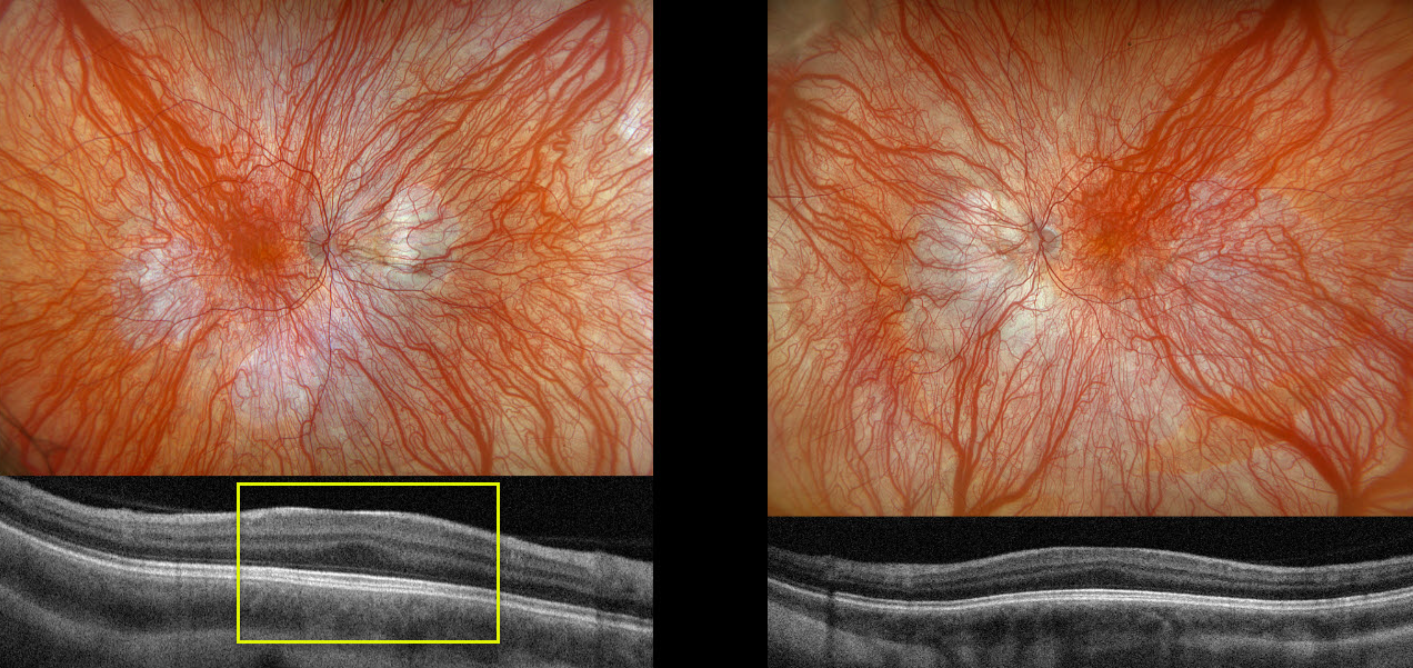
OCULOCUTANEOUS ALBINISM
The European VitreoRetinal Society (EVRS) and João Pedro Marques
Originally posted on @retina.rocks December 26, 2025
This 9YO girl was referred by her local ophthalmologist for esotropia, nystagmus, light hair, and skin, first noticed when she was 3 years old. She has a known history of Hermansky-Pudlak syndrome (HPS), diagnosed with genetic testing 3 years earlier. There is no history of parental consanguinity or of familial inherited retinal diseases. Vision was 20/63 OD and 20/80 OS. There were bilateral iris transillumination defects.
Optos color RGB imaging shows bilateral marked choroidal hypopigmentation with foveal hypoplasia. OCT scanning shows a flattened foveal contour bilaterally (fovea plana).
Learning Points:
Albinism is a group of genetic disorders characterized by abnormal melanin production due to faulty amino acid production. Patients have either ocular (eye) albinism or oculocutaneous (eye and skin) involvement. Inheritance patterns include autosomal dominant, autosomal recessive, and X-linked. Ocular findings include strabismus, nystagmus, iris transillumination defects, blonde fundus, and foveal hypoplasia (seen on OCT as fovea plana). There are often a higher number of crossed nerve fibers at the optic chiasm.
HPS is a rare autosomal recessive multisystem disorder characterized by oculocutaneous albinism, bleeding diathesis due to platelet storage pool deficiency, progressive pulmonary fibrosis, and granulomatous colitis. The underlying pathogenesis involves mutations in at least 10 genes encoding components of protein complexes (BLOC-1, BLOC-2, BLOC-3, and AP-3) essential for the biogenesis and trafficking of lysosome-related organelles in melanocytes, platelets, and other cell types. Our patient had a pathogenic c.223C>T p.(Gln75) homozygous variant in the HPS6 gene, which encodes for a key protein in the BLOC-2 pathway.
OCULOCUTANEOUS ALBINISM
Nilesh Kumar
Originally posted on @retina.rocks September 23, 2025
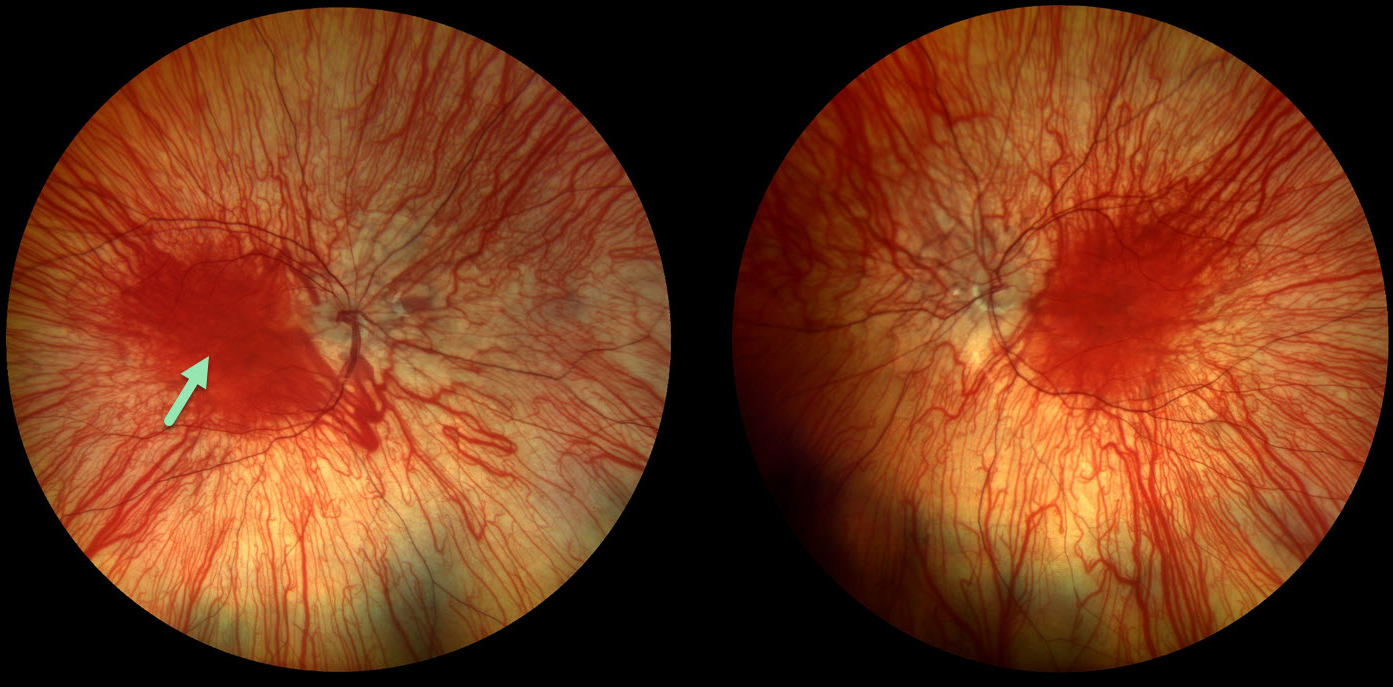
This 14YO female has a history of nystagmus and vision loss from oculocutaneous albinism. There was no significant family history. Vision was 20/120 OU.
Color photography shows bilateral marked choroidal hypopigmentation with foveal hypoplasia. No genetic testing or pedigree analysis was performed.
Learning Points:
Albinism is a group of genetic disorders characterized by abnormal melanin production due to faulty amino acid production. Patients have either only eye involvement (ocular albinism) or eye and skin (oculocutaneous albinism). Inheritance patterns include autosomal dominant, autosomal recessive, and X-linked. Ocular findings include strabismus, nystagmus, iris transillumination defects, blonde fundus, and foveal hypoplasia. There are often a higher number of crossed nerve fibers at the optic chiasm.
OCULOCUTANEOUS ALBINISM
Ayushi Gupta and Vishal Agrawal
Originally posted on @retina.rocks July 21, 2025
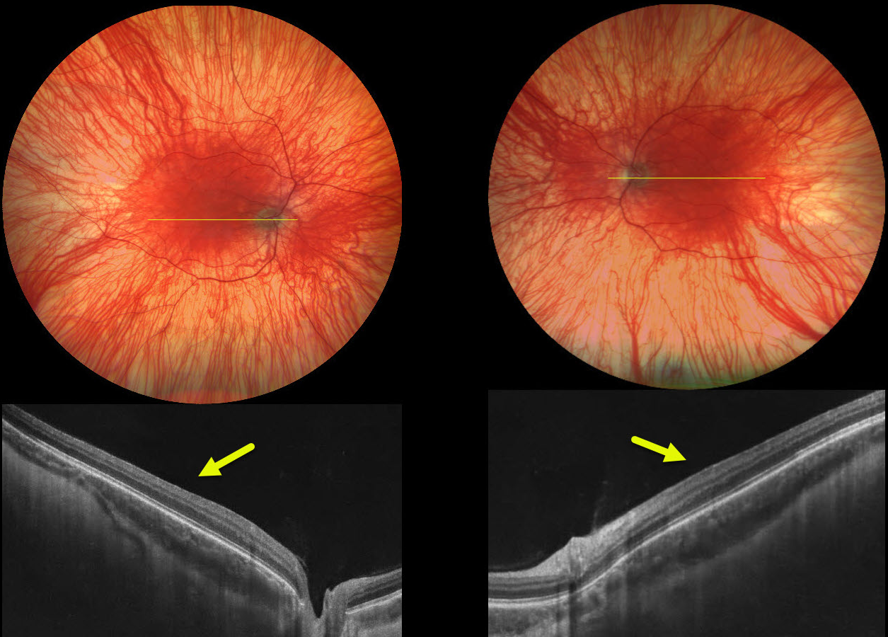
This 25YO male has a history of nystagmus and vision loss from oculocutaneous albinism. Her grandfather also had albinism. Vision was 20/70 bilaterally.
Color photography shows bilateral marked choroidal hypopigmentation with foveal hypoplasia. OCT scanning shows the absence of the normal foveal depression.
Learning Points:
Albinism is a group of genetic disorders characterized by abnormal melanin production due to faulty amino acid production. Patients have either eye-only involvement (ocular albinism) or eye and skin involvement (oculocutaneous albinism). Inheritance patterns include autosomal dominant, autosomal recessive, and X-linked. Ocular findings include strabismus, nystagmus, iris transillumination defects, blonde fundus, and foveal hypoplasia. There are often a higher number of crossed nerve fibers at the optic chiasm.
Fovea plana is characterized by the absence of the foveal pit, which is formed embryologically by the inner retinal tissues and vasculature being displaced centrifugally. It is most commonly associated with albinism, prematurity, or as an isolated finding. Despite the abnormal foveal architecture, vision is usually normal, unless due to albinism.
OCULAR ALBINISM
Borivoje Ivezić, Emma Oreškovič, and Nataša Drača
Originally posted on @retina.rocks December 21, 2023
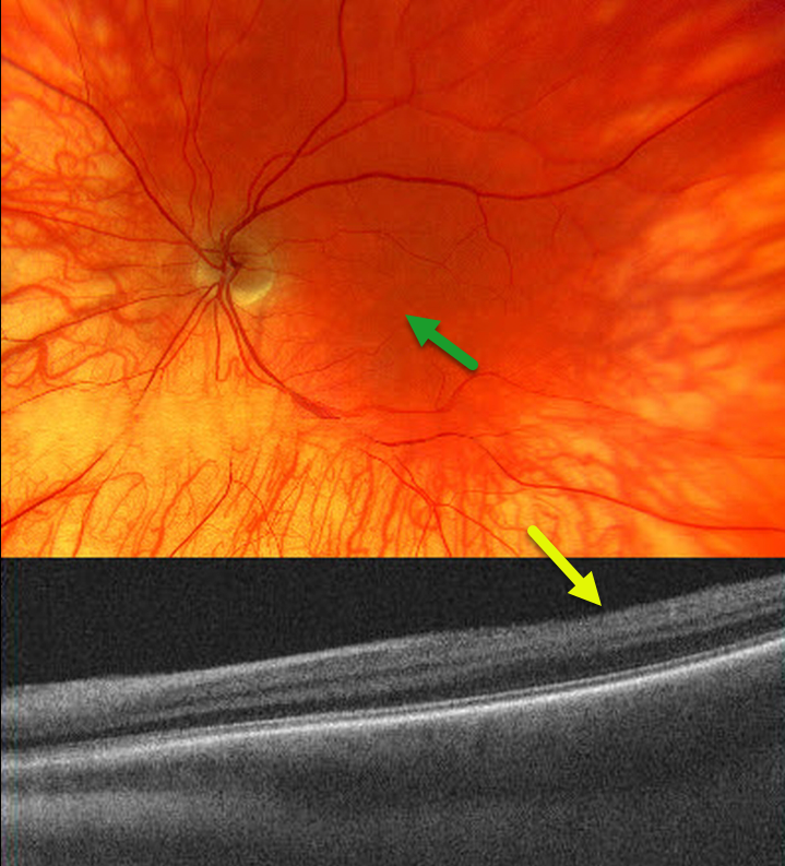
The 53YO male has a known history of ocular albinism, reporting poor visual acuity and horizontal nystagmus since childhood. Vision was 20/200 bilaterally.
Color imaging of the left eye shows a diffusely hypo-pigmented fundus with foveal hypoplasia.
OCT imaging shows a flat foveal contour (fovea plana). Similar findings were noted in the right eye (not shown).
Learning Points:
Ocular albinism is an X-linked recessive disorder that affects only the eyes and is caused by a mutation in the OA1 gene.
Findings seen in ocular albinism include strabismus, nystagmus, iris transillumination defects, blonde fundus, and foveal hypoplasia. There are often a higher number of crossed nerve fibers at the optic chiasm.
Fovea plana is characterized by the absence of the foveal pit, which is formed embryologically by the inner retinal tissues and vasculature being displaced centrifugally. It is most commonly associated with albinism, prematurity, or as an isolated finding. Despite the abnormal foveal architecture, vision is usually normal, unless due to albinism.
OCULAR ALBINISM
Erdem Dinc
Originally posted on @retina.rocks February 3, 2022
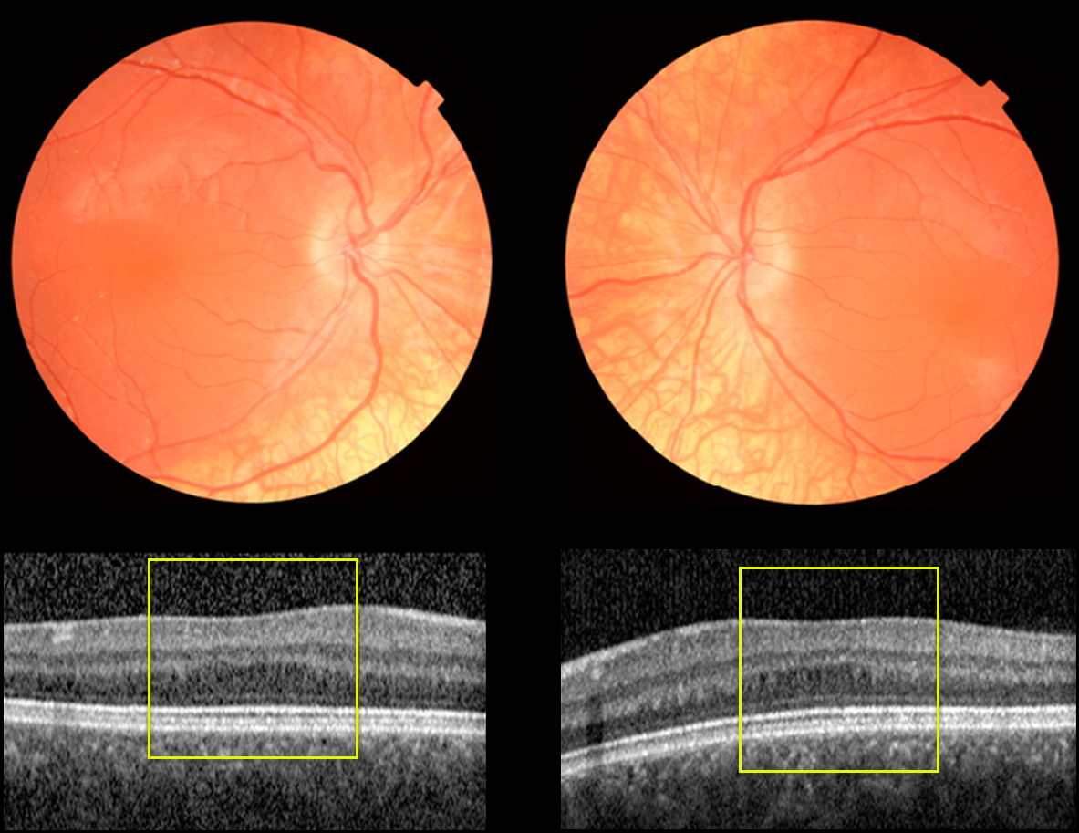
This 11YO female presented with 20/50 vision OU. She was healthy with normal skin and hair pigmentation and was the product of a consanguineous marriage. Motility was normal without nystagmus. Bilateral iris transillumination defects were noted.
Fundus photography shows marked decreased choroidal pigmentation and absent foveal reflex. OCT scanning shows bilateral absence of the normal foveal depression (fovea plana).
Learning Points:
Ocular albinism is an X-linked recessive disorder that affects only the eyes and is caused by a mutation in the OA1 gene. Findings seen in ocular albinism include strabismus, nystagmus, iris transillumination defects, blonde fundus, and absent or blunted foveal reflex (fovea plana). There are often a higher number of crossed nerve fibers at the optic chiasm.
OCULAR ALBINISM
Originally posted on @retina.rocks March 9, 2020
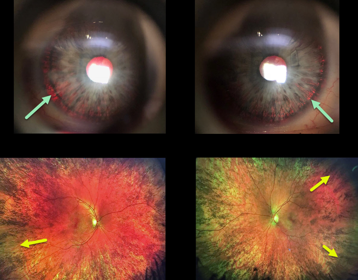
This woman has a history for both a congenital rubella infection and a positive family history for albinism in both an uncle and cousin.
We think that she is likely a carrier for OA1 (ocular albinism type 1) given the presence of iris transillumination defects and pigmentary changes of the peripheral retina, although the fundus changes may also be due to congenital rubella.
Learning Points:
Ocular albinism is a X-linked recessive disorder that affects only the eyes and is caused by a mutation in the OA1 gene. Findings seen in ocular albinism include strabismus, nystagmus iris transillumination defects, blonde fundus, and absent or blunted foveal reflex (fovea plana). There is often a higher number of crossed nerve fibers at the optic chiasm.
OA1 carriers will often have partial iris transillumination defects and up to 90% of OAT1 female carriers exhibit a salt and pepper (or mud-splattered) fundus due to the patches of amelanotic RPE.
This salt and pepper appearance can also be seen in congenital rubella, Leber’s congenital amaurosis, congenital syphilis, and phenothiazine toxicity.
Oculocutanoeus albinism involves both eye and skin findings and is caused by mutations in the tyrosine gene. There is a complete tyrosinase-negative form in which no melanin is produced and a tyrosinase-positive form in which only partial amounts of melanin are produced,


