

Alport Syndrome
<< Back to Cases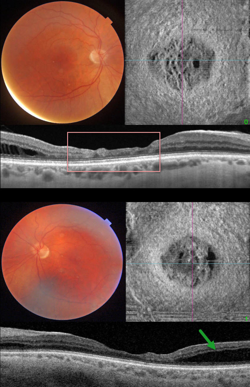
ALPORT SYNDROME
Fraser McKay
Originally posted on @retina.rocks April 9, 2025
This 46YO male presented with increasing difficulties with watching TV and working on the computer. He had a known history of Alport syndrome, including renal failure with prior renal transplantation and hearing loss. Vision was 20/40 OD and 20/50 OS. Slit lamp examinations showed bilateral cataracts without anterior lenticonus. Cataract surgery was performed, and vision remarkably improved to 20/20 OU.
Triton color imaging shows a blunted foveal reflex. Swept-source OCT shows central inner macular thinning and disorganization. Outer nuclear layer schisis is noted temporally. En face OCT reveals another view of these central macular findings. Similar changes are noted in his left eye.
Learning Points:
Alport syndrome is a rare genetic disorder, most commonly inherited in an X-linked recessive pattern with mutations in the COL4A5 gene, which encodes for collagen type IV alpha. Basement membrane thinning and lamellation cause hemorrhagic nephritis and renal failure, sensorineural hearing loss, and ocular findings, including corneal abnormalities and anterior lenticonus. Retinal findings include macular and peripheral retinoschisis and macular yellow-white perimacular dots and flecks in the inner retina.
In more severe forms, a dull macular reflex with inner macular thinning (lozenge sign), best seen with red-free imaging, can occur (Colville et al, Br J Ophthalmol 2009;93:383-386). Multimodal imaging, including wide-field OCT, shows a spectrum of internal limiting membrane (ILM) findings, including ILM granularity, progressive ILM lamellation, retinal nerve fiber layer (RNFL) dehiscence, vascular lacunae, and course arrangement of RNFL toward the disc (Cicinelli et al, Retina 2022;42:274-282).


