

AMD Disciform Scar + Subretinal Fibrosis
<< Back to Cases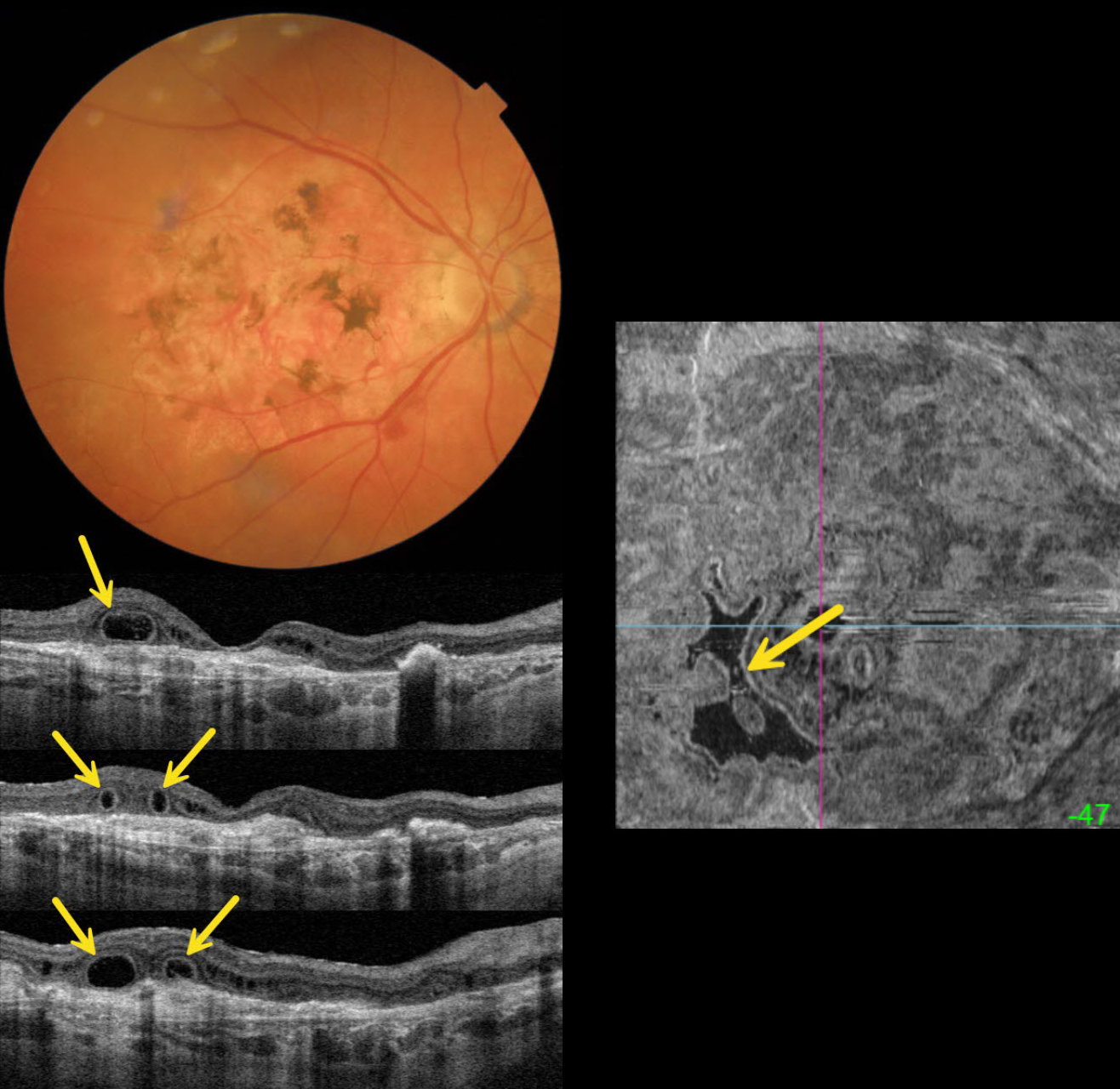
OUTER RETINAL TUBULATION IN NEOVASCULAR AMD
Originally posted on @retina.rocks November 18, 2025
This 89YO female is receiving ongoing intravitreal Eylea in her 20/40 left eye for neovascular AMD. She has a stable 20/400 atrophic disciform scar in her right eye.
Triton color imaging shows a variably pigmented flat disciform scar. Swept-source OCT shows variable outer retinal atrophy overlying a hyperreflective inactive macular neovascularization (MNV). A complex pattern of outer retinal tubulation (ORT) is seen on numerous B-scans. En face OCT shows another view of these meandering tubular structures.
Learning Points:
ORT is often noted overlying inactive MNV and should not be confused with exudative fluid or cysts, which lack a hyperreflective border. The outer hyperreflective band likely represents inner segment mitochondria undergoing fission and translocation toward the nucleus (Litts et al, Retina 2018;38:445-461). ORT, initially described by Zweifel et al (Arch Ophthalmol 2009;127:1596-1602), is a neurodegenerative condition of the photoreceptors and Muller cells associated with atrophy affecting the outer retina and retinal pigment epithelium, including advanced AMD and inherited retinal diseases.
PSEUDO-UVEAL MELANOMA FROM NEOVASCULAR AMD SUBRETINAL FIBROSIS
Fraser McKay
Originally posted on @retina.rocks June 20, 2024
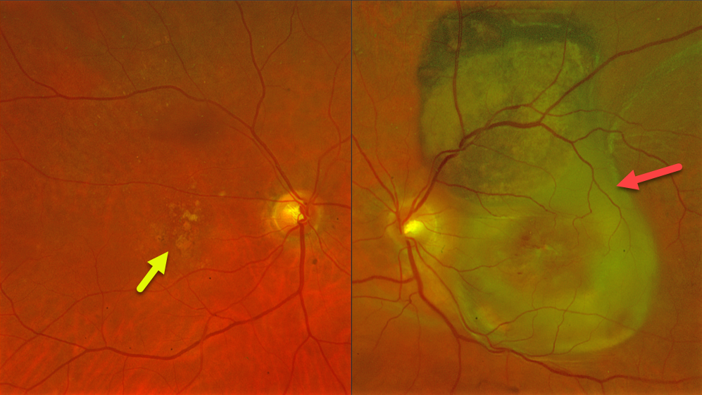
This 78YO female presented with at least several months of vision loss in her left eye. Vision was 20/40 OD and light perception OS. She was examined 2 years earlier with 20/400 vision OS from macular atrophy.
Optos color RG imaging OS shows a variably pigmented and slightly thickened subretinal lesion extending from the superior macula into the superior midperiphery. Subretinal fibrosis extends through the macula, along with retinal blood dots. Intermediate AMD with mixed drusen was noted OD.
Learning Points:
Suprachoroidal, subretinal, or sub-RPE blood can rarely simulate a uveal malignant melanoma. We suggested a trial of monthly anti-VEGF therapy for this treatment naïve neovascular AMD lesion, although the visual prognosis remains quite guarded given the extent of submacular fibrosis.
SUBRETINAL FIBROSIS
Originally posted on @retina.rocks April 30, 2024
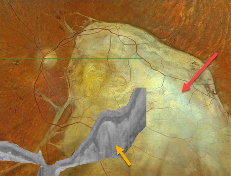
This 69YO male has stable vision loss in his left eye from an age-related disciform scar.
Optos color RGB imaging shows massive subretinal fibrosis occupying virtually the entire macula, which extends into the inferior midperiphery. Optos Silverstone OCT shows markedly thickened hyperreflective subretinal fibrosis. Vision was remarkably 20/400. Continued observation was recommended.
Learning Points:
Disciform scars were the norm for untreated neovascular AMD (nAMD) prior to the anti-VEGF era that began in the mid-2000s. New-onset nAMD diagnosed and treated with timely anti-VEGF dosing virtually never goes on to develop these lesions. There may be progressive macular atrophy following years of therapy, but not this type of fibrotic scarring.
For an extensive discussion on the SD-OCT findings of these age-related fibrotic lesions, see the AOS thesis by Souied et al (AJO 2020;214:151-171).
DISCIFORM SCAR
Originally posted on @retina.rocks October 24, 2023
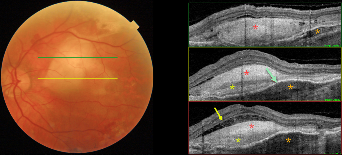
This 81YO male has a history of counting-finger vision in his left eye due to a stable age-related fibrosed disciform scar.
Color imaging shows a fibrosed disciform scar in the central and superior macula. Triton swept-source OCT through different regions of the scar shows a constellation of interesting findings. The scar is variably hyperreflective, with areas of increased and relatively decreased reflectivity. Most inferiorly, the outer plexiform layer is clearly visible overlying an area of fluid and disorganized outer retina. Superiorly, the fibrosis indents and compresses the underlying RPE and choroid. Finally, the fibrosed neovascular lesion is both above (type 2) and below (type 1) the RPE, with the RPE variably elevated.
Learning Points:
Disciform scars were the norm for untreated wet AMD prior to the anti-VEGF era that began in the mid-2000s. This type of scarring is virtually no longer seen in eyes treated early in the course of their neovascular disease, although some subretinal fibrosis remains in 40% of eyes after 10 years of anti-VEGF treatment (Chong et al, Retina 2020;40:2285-2295).

AMD DISCIFORM SCAR
Originally posted on @retina.rocks November 28, 2022
This 70YO male presented with counting fingers vision in his right eye due to a large disciform scar with radiating inner retinal folds. The radiating folds appear as corrugations on OCT B-scan and as radiating lines on OCT en face.
Learning Points:
Disciform scars were the norm for untreated wet AMD prior to the anti-VEGF era that began in the mid-2000s. New-onset wet AMD diagnosed and treated with appropriate anti-VEGF dosing virtually never goes on to develop these lesions.
There may be progressive macular atrophy following years of therapy, but not this type of fibrotic scarring.
The radiating folds in our patient are caused by contraction of the fibrosed macular neovascularization which is centripetally dragging the overlying neurosensory retina.
AMD SCAR
Originally posted on @retina.rocks October 4, 2022
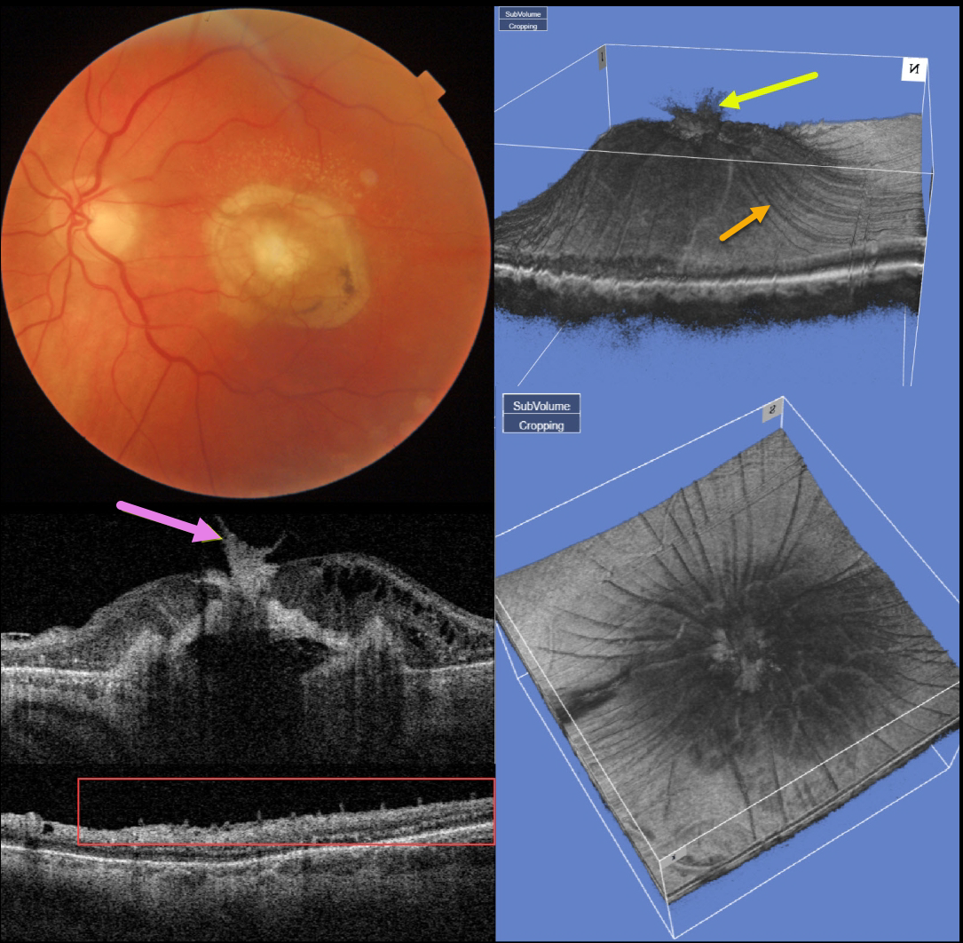
This 77YO man presented counting fingers vision OS with what seems at first to be an uneventful fibrotic disciform scar from neovascular AMD. But multimodal imaging shows several interesting findings.
Swept-source OCT through the macular center shows that the central scar extends through the full-thickness retina and becomes incorporated with the attached vitreous.
A 3D macular reconstruction viewed from above also shows how scar contraction is creating radiating retinal folds.
These folds on a B-scan taken through the peripheral macula give the appearance of numerous needle-like projections emanating from the inner retina.
The needle-like projections, created by imaging the radiating inner retinal folds perpendicular to their orientation, mimic findings felt to be pathognomonic for ocular amyloidosis (Kakuhara et al, Ophthalmology Retina 2021;5:1005-1008).
NEOVASCULAR AMD
Originally posted on @retina.rocks July 20, 2022
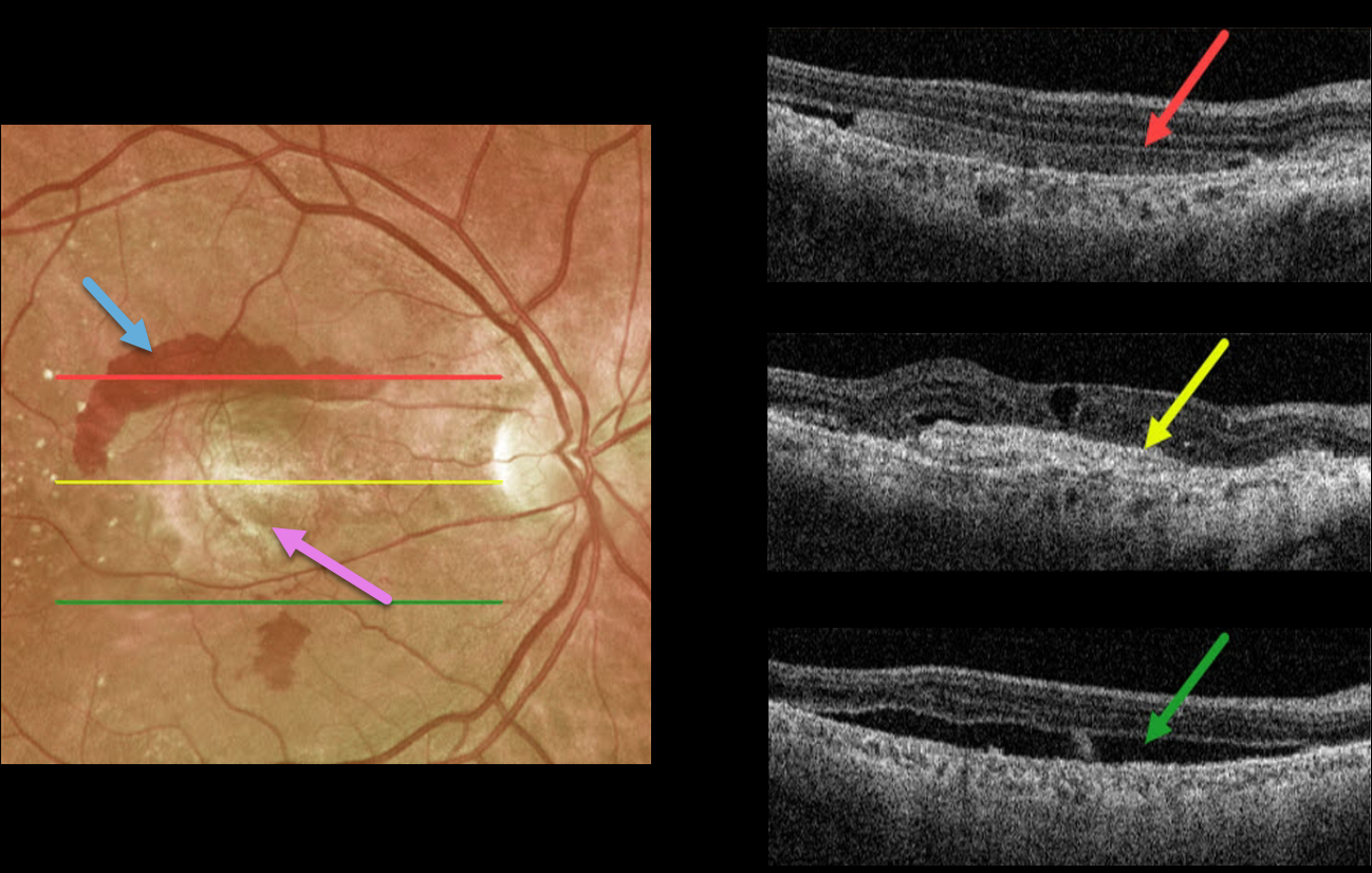
This 72YO male presented with longstanding decreased vision in his right eye. Vision was counting fingers due to a fibrosed scar from neovascular age-related macular degeneration (nAMD).
Optos imaging shows a central fibrosed macular scar with superior and inferior subretinal blood. Triton swept-source imaging shows multiple cool findings from this single neovascular lesion. Superiorly, the subretinal blood appears as hyperreflective material in the subretinal space. More centrally, the fibrotic scar/type 2 macular neovascularization is hyperreflective. There are also some overlying chronic cystic retinal changes. More inferiorly is shallow, subclinical subretinal fluid.
A therapeutic trial of monthly anti-VEGF injections was initiated, although we’re not sure this will have a visual effect given the subfoveal fibrosis.
NEOVASCULAR AMD
Originally posted on @retina.rocks March 22, 2022
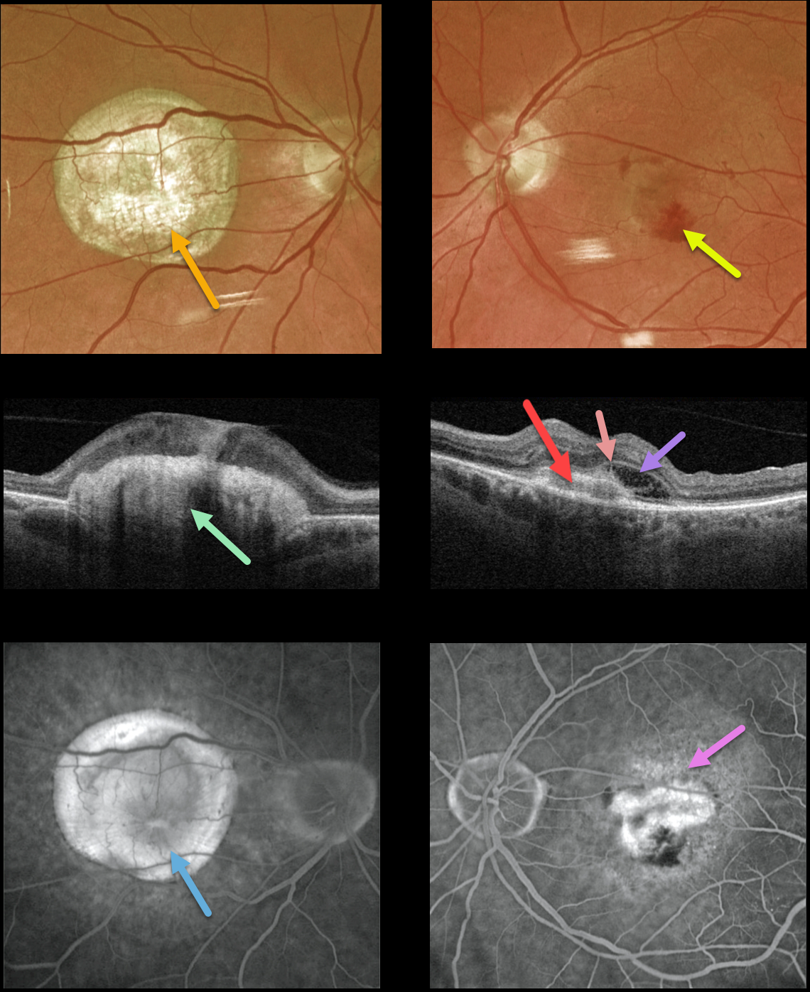
This 77YO male presented with counting fingers vision OD from a large fibrotic disciform scar and 20/200 vision OS from a new macular neovascularization.
The disciform scar is contracted and fibrotic, with marked hyperreflectivity on swept-source OCT. The MNV in his left eye is seen as opaque fluid with some subretinal blood. The type 2 MNV (located above the RPE) is noted on the OCT B-scan. There is also a bacillary detachment and a hyperreflective line indicating a possible pitchfork sign.
Optos fluorescein angiography shows diffuse staining of the subretinal scarring OD and leakage from the MNV OS.
Learning Points:
Disciform scars were the norm for untreated wet AMD before the anti-VEGF era that began in the mid-2000s. New-onset wet AMD diagnosed and treated with appropriate anti-VEGF dosing virtually never goes on to develop these lesions. There may be progressive macular atrophy following years of therapy, but not this type of fibrotic scarring.
An excellent review of wet AMD fibrotic lesions is in Souied et al’s AOS thesis (AJO 2020;214:151-171).
The pitchfork sign was originally described by Hoang et al (Retina 2013;33:1049-1055) as a unique OCT finding in inflammatory MNV. Falavarajani et al more recently noted that this finding is also associated with non-inflammatory MNV (Ophthalmic Surg Lasers Imaging Retina 2019;50:719-725).
The pathogenesis of the avascular pitchfork spikes is unknown, but may be related to outer retinal traction or Müller cell activation.
NEOVASCULAR AMD + RETINAL CHOROIDAL ANASTAMOSIS
Originally posted on @retina.rocks November 25, 2021
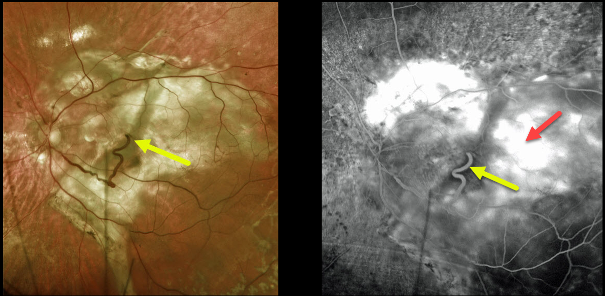
This 74YO female has a large complex disciform scar from end-stage age-related macular degeneration (AMD).
A very prominent retinal vein drains directly into the substance of the scar inferiorly, indicating a chorioretinal anastomosis. Fluorescein angiography shows diffuse staining of the subretinal scarring.
Learning Points:
Under normal conditions, the retinal and choroidal circulations remain separate and distinct. Rarely, the two circulations connect directly through a chorioretinal anastomosis.
These can occur in chorioretinal scars (typically from inactive toxoplasmosis lesions), in macular telangiectasia, and in disciform scars.
SUBRETINAL FIBROSIS
Originally posted on @retina.rocks March 11, 2021
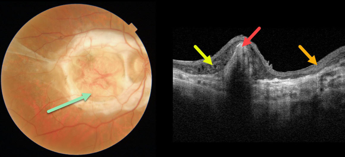
This 69YO male has counting-finger vision in his right eye from a fibrotic disciform scar due to wet age-related macular degeneration.
Triton swept-source OCT scanning shows hyperreflective subretinal scarring, variable disorganized thinned overlying retinal layers, and outer retinal thickening.
Learning Points:
This type of end-stage wet AMD was the norm prior to the anti-VEGF era, which began in 2005. Fortunately, with early and timely intravitreal injections, disciform scars are now virtually only seen in untreated eyes.
DISCIFORM SCAR
Originally posted on @retina.rocks December 15, 2020
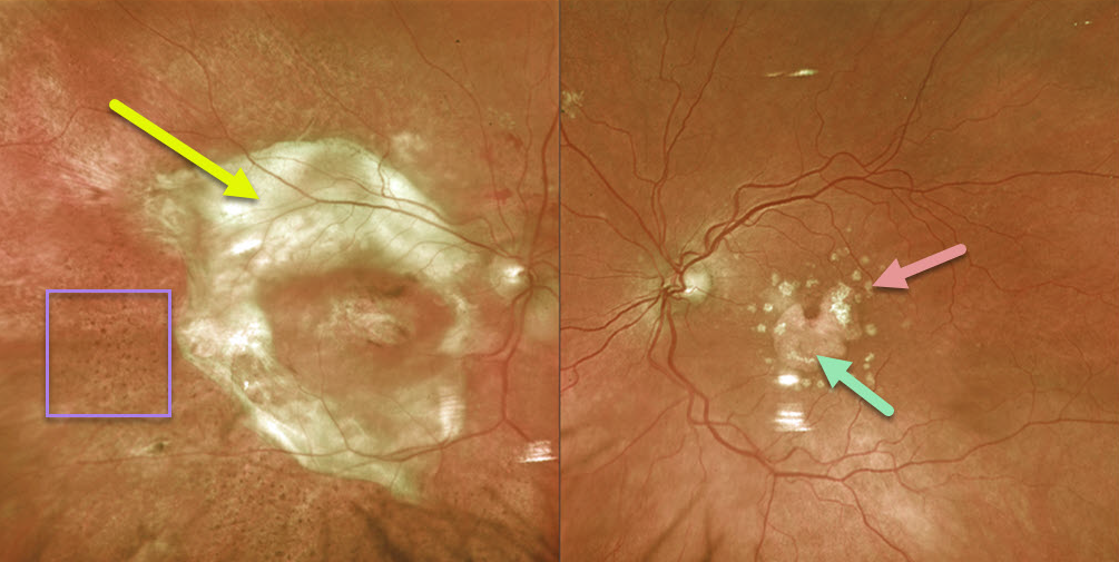
This patient with age-related macular degeneration (AMD) has a large fibrotic disciform scar in the right eye and dry AMD in the left eye with macular atrophy skirting the foveal center and more distal calcified drusen.
The coarse pigmentary changes noted inferiorly and temporally in the right eye are a classic tell-tale finding for prior resolved subretinal blood (see Hussnain et al Retina 2019;39:1925-1935).
Learning Points:
Disciform scars were the norm for untreated wet AMD prior to the anti-VEGF era that began in the mid-2000s.
New-onset wet AMD diagnosed and treated with appropriate anti-VEGF dosing virtually never goes on to develop these lesions.
There may be progressive macular atrophy following years of therapy, but not this type of fibrotic scarring.
DISCIFORM SCAR
Originally posted on @retina.rocks June 9, 2020
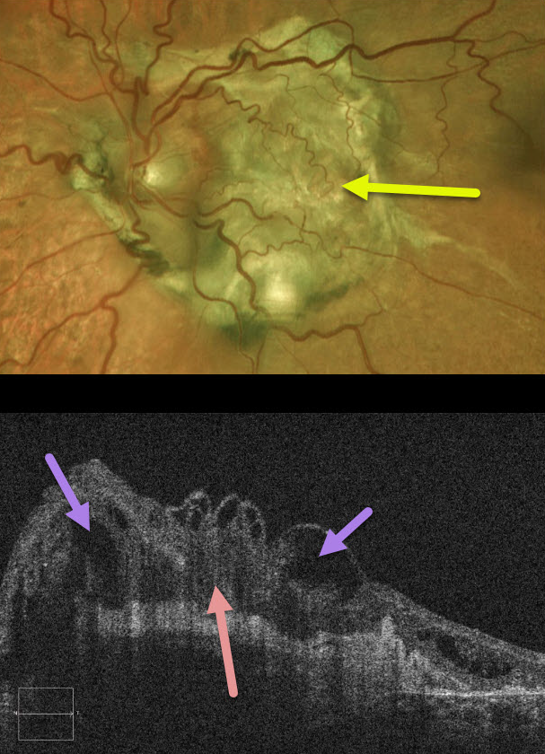
This patient has a large subretinal fibrotic disciform scar from end-stage wet age-related macular degeneration.
OCT shows marked overlying retinal disorganization and cystic changes.
Learning Points:
These fibrotic scars are usually the result of untreated macular neovascularization. These were more the rule than the exception prior to the introduction of anti-VEGF therapy in the mid-2000’s.
With early and aggressive treatment, this type of outcome is now rarely, if ever, seen.


