

AMD Drusen and Pigment Clumping
<< Back to Cases
FLATTENED DRUSENOID PED CAUSING SECONDARY MACULAR ATROPHY
Originally posted on @retina.rocks June 23, 2026
This 72YO female has been followed for years with intermediate AMD. On 3/26/25, she remained asymptomatic with 20/40 vision.
Color photography shows confluent central drusen creating a drusenoid PED with a radiating pigment figure, surrounded by large soft drusen. The drusenoid RPE detachment (PED) is confirmed on swept-source OCT. The RPE and outer retinal bands are obscured by subretinal hyperreflective material. She returned 6 months later with stable vision and no new symptoms. The PED is mostly flattened with some resolution of the hyperreflective material.
Learning Points:
Drusen commonly remodel over time and their collapse, as in our patient, can lead to drusenoid atrophy of the RPE (Filho et al, Retina 2013;33;1558-1566). Vision may or may not decrease following drusen regression. In our experience, drusenoid atrophy remains stable, unlike primary age-related macular atrophy, whose radius enlarges linearly over time (Shen et al, Ophthalmology Retina 2020;4;899-910). We therefore do not consider intravitreal complement inhibitors for these patients.
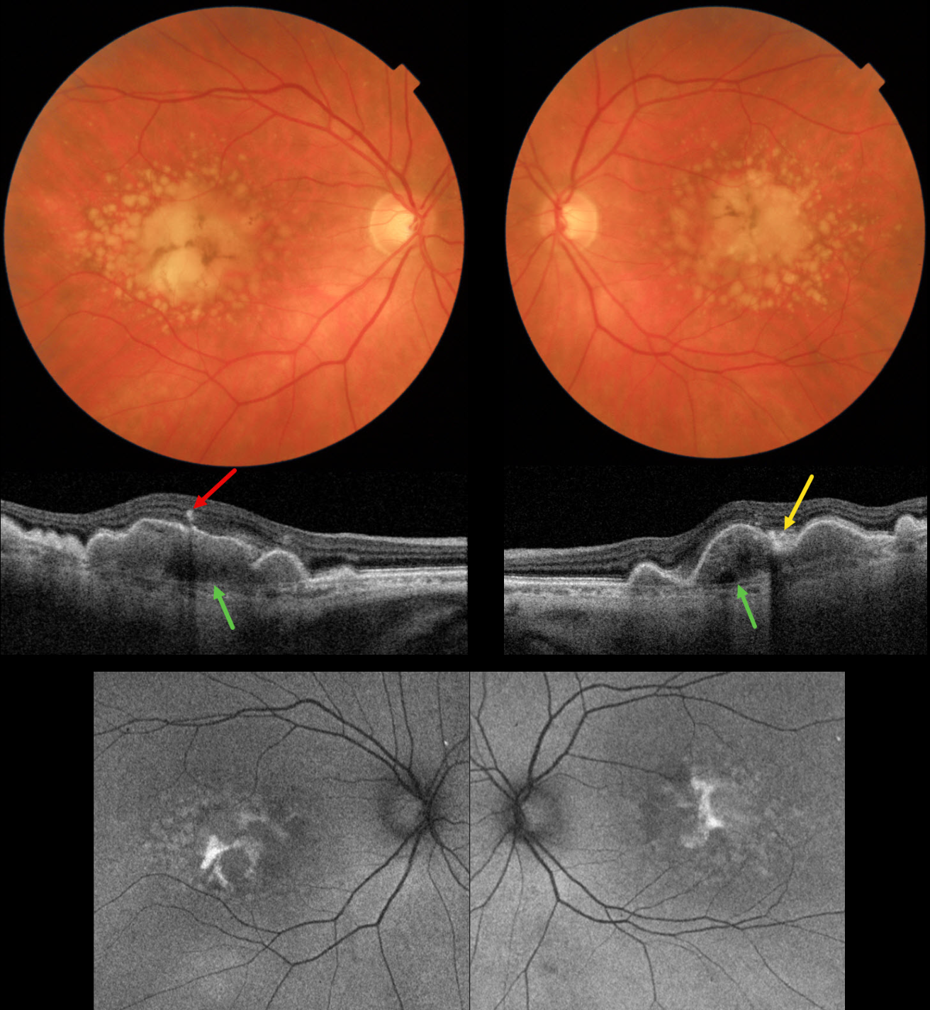
INTERMEDIATE AMD WITH DRUSENOID PEDS
Originally posted on @retina.rocks January 8, 2026
This 72YO female presented for her 6-month AMD examination. She was visually asymptomatic, and vision was 20/40 OU.
Triton color imaging shows bilateral chronic foveal RPE detachments (PEDs) with surrounding soft drusen. A radiating pigment figure overlies each PED. Swept-source OCT shows hyperreflective material beneath each PED, with some overlying hyperreflective foci and hyperreflective material likely representing lipofuscin. Bruch membrane is visible beneath the PED and surrounding soft drusen. Fundus autofluorescence (FAF) shows variable hyper-FAF from the pigment figures and lipofuscin.
Learning Points:
RPE detachments are most commonly associated with AMD and the central serous-pachychoroid spectrum. In cases of AMD, lesions can be avascular (as in our patient) or vascularized as part of a type 1 neovascular complex (below the RPE). AMD-related PEDs can form primarily or as a confluence of soft drusen. When they collapse, they may lead to secondary macular atrophy (Yu et al, Ophthalmology 2019;126:261-273).
The radiating pigment figure, described originally by Dr. Gass, is pathognomonic for a chronic PED and persists even after a PED flattens. Avascular PEDs are typically not treated. In contrast, anti-VEGF therapy is both safe and effective for vascularized PEDs, despite the risk of a RPE tear (Khanani et al, Retina 2018;38;2103-2117).
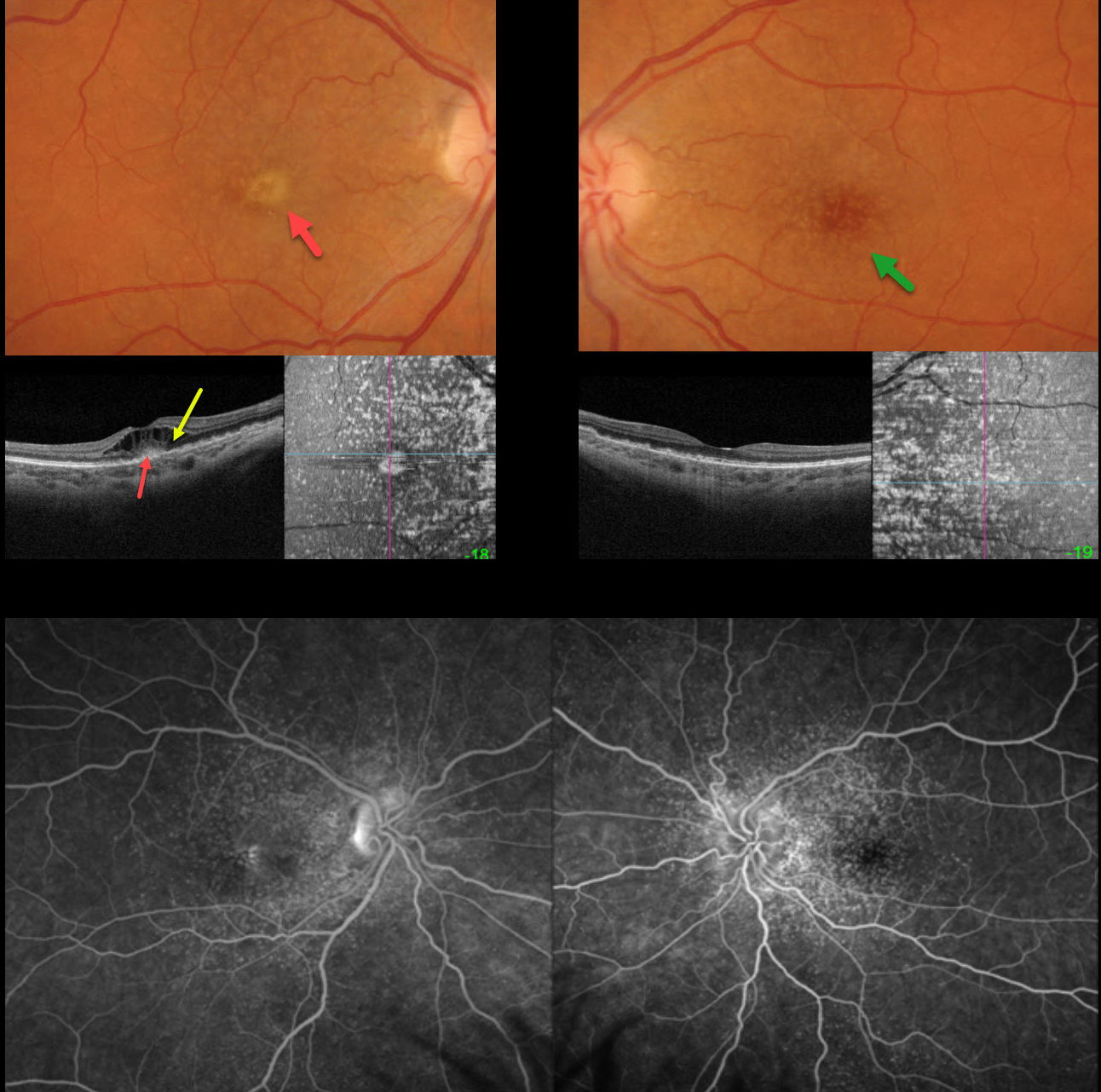
CUTICULAR DRUSEN WITH MACULAR NEOVASCULARIZATION
Originally posted on @retina.rocks August 5, 2025
This 47YO female presented with one week of vision loss in her right eye. Vision was counting fingers OD and 20/25 OS.
Optos color RGB imaging shows innumerable cuticular macular drusen, which appear on swept-source OCT B-scanning as arrowhead-like RPE elevations and on en face imaging as numerous hyperreflective spots. A yellow subfoveal lesion OD appears on OCT as hyperreflectivity in the outer retina. Marked overlying cystoid edema is seen. Fluorescein angiography shows late pooling in the foveal cysts OD, with hyperfluorescent window defects from the drusen.
One month following an initial intravitreal Avastin injection, vision improved to 20/80, with complete resolution of the vitelliform lesion and cystoid edema. Treat-and-extend treatment was started.
Learning Points:
Originally described by Gass as basal laminar drusen (AJO 1985;99:445-459), cuticular drusen were initially thought to represent nodular thickening of the RPE basement membrane. They appear clinically as numerous, round, yellow-white ‘hard drusen’, and on OCT as small, arrowhead-shaped drusen located above Bruch’s membrane and below the RPE (Spaide and Curcio, Retina 2010;30:1441-1454).
Our patient’s angiographic appearance echoes Dr. Gass’ original description of “a beautiful stars-in-the-sky or Milky-Way pattern” of hyperfluorescence. Cuticular drusen are classically associated with avascular vitelliform lesions (Finger et al, Retina 2010;30:1455-1464).
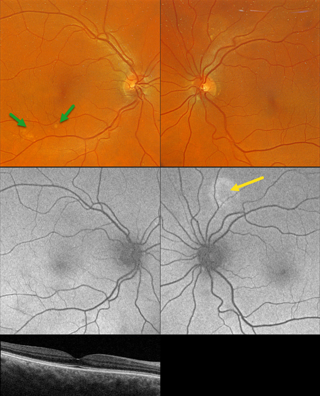
PACHYCHOROID SPECTRUM PREVIOUSLY MISDIAGNOSED AS AMD
Originally posted on @retina.rocks July 30, 2025
This healthy 65YO founder of Retina Rocks presented for his once-per-decade ‘eye examination’ which included an undilated slit lamp examination by his partner, Triton swept-source OCT, and Optos color RGB imaging. His mother had a history of neovascular AMD, and he has a known history of drusen. Given his family history, he has been taking AREDS2 supplementation for many years. There were no ocular symptoms, and vision was 20/20 bilaterally.
Triton color imaging shows some pachydrusen in the inferotemporal right macula. Swept-source OCT is normal except for a thickened choroid. Optos fundus autofluorescence (FAF) shows a round area of hyper-FAF superior to the left nerve.
Learning Points:
Pachydrusen were described by Spaide as a distinct pachychoroid phenotype (Retina 2018;38:708-716). Unlike typical AMD, where cone-based soft drusen are symmetrically localized to the foveal region, pachydrusen are eccentrically located, far less numerous, and have an irregular contour. Since choroidal thickness is influenced by numerous factors, including age and refractive status (Xie et al, IOVS 2022;63;2;34), these must always be considered when reviewing an individual patient’s scans. Eyes with AMD typically have age-related choroidal thinning, but eyes with pachydrusen, as in this case, are associated with a relatively thickened choroid.
Our patient became aware of his macular drusen well before pachychoroid was described by Freund in 2013 (Warrow et al, Retina 2013;33:1659-1672), and he attributed his early macular drusen to AMD. In 2025, he understands that his findings are consistent with the pachychoroid spectrum, as evidenced by the bilaterally thickened choroid, pachydrusen OD, and FAF findings suggestive of prior extramacular central serous OS. He does not have intermediate AMD, is at minimal risk for developing AMD, and thus doesn’t need AREDS2 supplementation. However, like the rest of us, he is only human and finds emotional comfort in continuing to take these supplements.
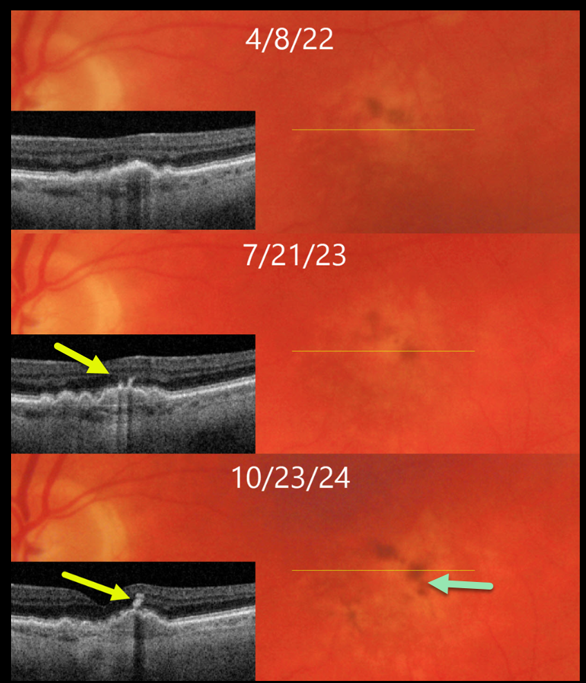
DRY AMD WITH PIGMENT CLUMPING REMODELING
Originally posted on @retina.rocks April 10, 2025
This 71YO male has been followed for several years with intermediate AMD.
On 4/8/22, vision was 20/40 in his asymptomatic left eye. There is a shallow RPE detachment (PED) with faint surrounding drusen and pigment loss on both Triton color imaging and swept-source OCT. Clumping of subretinal pigment is noted along the superior edge of the PED on color imaging.
He returned for his 1-year examination on 7/21/23 with stable 20/40 vision. A new pigment clump is noted along the temporal edge of the PED, and this is seen as a hyperreflective plume on OCT.
On 10/23/24, vision decreased to 20/100, with extension of the plume into the inner retina. There is continued remodeling of the pigment clumps on color imaging.
Learning Points:
The widely used Beckman AMD clinical classification defines intermediate AMD as including large drusen > 125 microns and/or any AMD pigmentary abnormalities (Ferris et al, Ophthalmology 2013;120:844-851). This classification was modified in 2024 to include a third risk factor, subretinal drusenoid deposits, which significantly increases the risk for progression to late AMD (Agron et al, Ophthalmology 2024;131:1164-1174).
These pigment clumps, which are visualized on OCT as hyperreflective foci (HRF), are found in over 50% of eyes with AMD (Ho et al, Ophthalmology 2011;118:687-693), and are thought to represent thickened RPE or intraretinal migrating RPE cells (Laiginhas et al, Ophthalmology Science 2022;2:100116). The migrating RPE cells often track along Muller cells creating a hyperreflective plume (Cao et al, IOVS 1021;62(10):34). These RPE cells transdifferentiate into macrophage-like cells. Macular atrophy will often develop underlying the HRF.
CUTICULAR DRUSEN WITH VITELLIFORM LESION
Originally posted on @retina.rocks May 28, 2024
This 57YO female was referred for asymptomatic macular changes. Vision was 20/20 bilaterally.
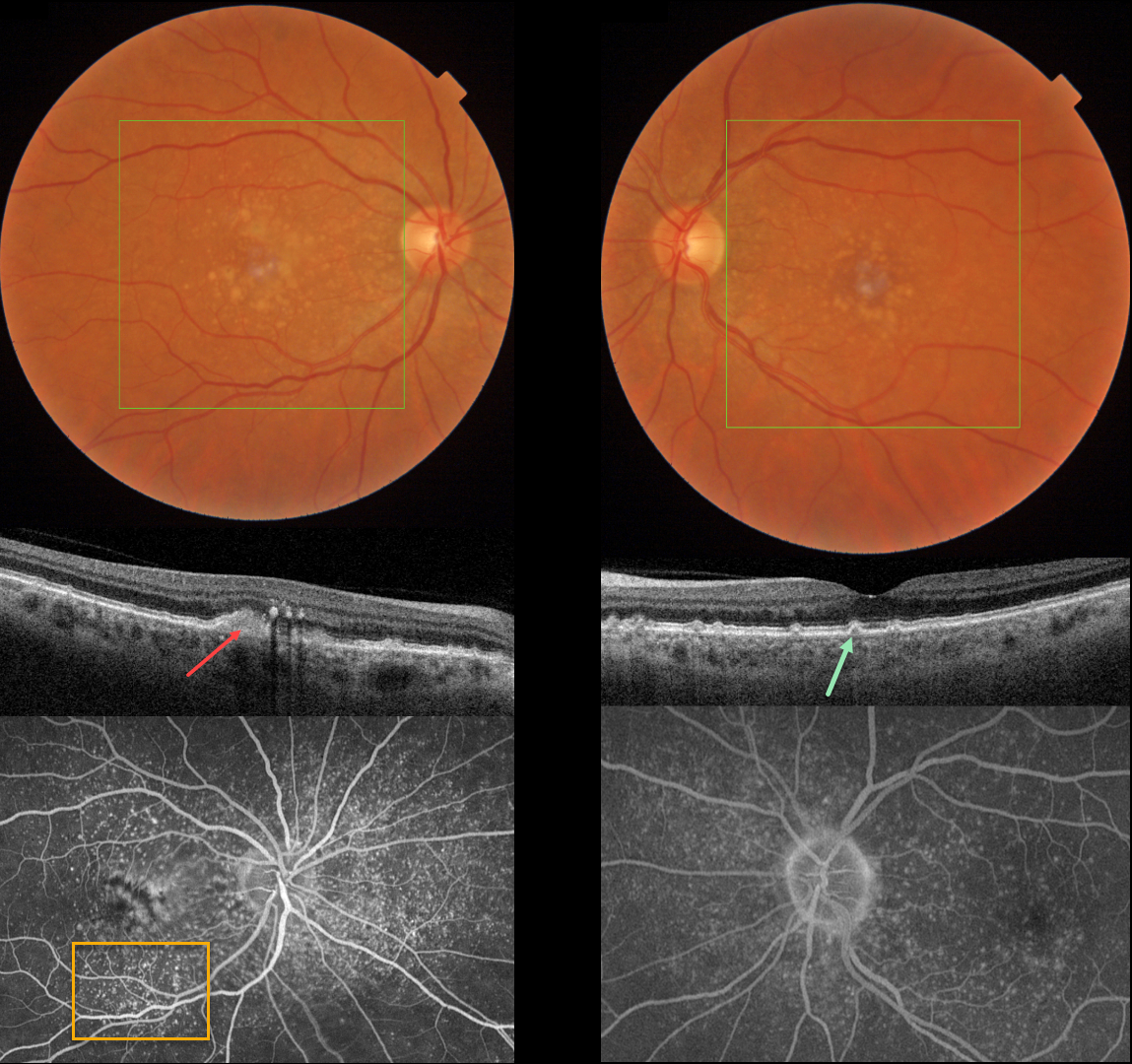
Triton color imaging shows bilateral confluent central soft drusen and smaller, more peripheral macular drusen. Swept-source OCT shows bilateral, small arrowhead-shaped cuticular drusen. A hyperreflective vitelliform lesion is noted OD, along with some hyperreflective deposits in the outer plexiform layer. Optos fluorescein angiography shows extensive bilateral hyperfluorescent cuticular drusen along with a staining vitelliform lesion OD.
Learning Points:
Originally described by Gass as basal laminar drusen (AJO 1985;99:445-459), cuticular drusen were initially thought to represent nodular thickening of the RPE basement membrane. They appear clinically as numerous, round, yellow-white ‘hard drusen’, and on OCT as small, arrowhead-shaped drusen located above Bruch’s membrane and below the RPE (Spaide and Curcio, Retina 2010;30:1441-1454).
Our patient’s angiographic appearance echoes Dr. Gass’ original description of “a beautiful stars-in-the-sky or Milky-Way pattern” of hyperfluorescence. Cuticular drusen are classically associated with avascular vitelliform lesions (Finger et al, Retina 2010;30:1455-1464), as in this case, but can also develop macular neovascularization.
FLATTENED DRUSEN CAUSING SECONDARY MACULAR ATROPHY
Originally posted on @retina.rocks January 8, 2024
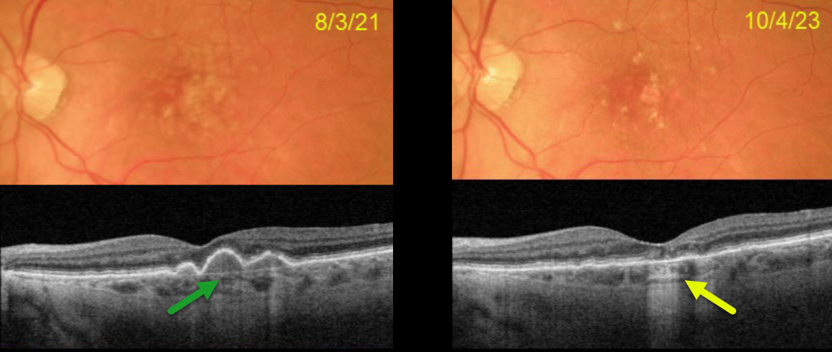
This 77YO female presented on 8/3/21 with 20/30 vision in her left eye. Optos color RG imaging shows confluent central soft drusen, which are confirmed on Triton swept-source OCT.
On 10/4/23, vision was 20/25. The central drusen flattened with secondary drusenoid atrophy. Continued observation was recommended.
Learning Points:
Drusen commonly remodel over time and their collapse, as in our patient, can lead to drusenoid atrophy of the RPE (Filho et al, Retina 2013;33;1558-1566). Vision may or may not decrease following regression of drusen.
In our experience, drusenoid atrophy remains stable, unlike primary age-related macular atrophy, whose radius enlarges linearly over time (Shen et al, Ophthalmology Retina 2020;4;899-910). We therefore do not consider intravitreal complement inhibitors for these patients.
CUTICULAR DRUSEN
Originally posted on @retina.rocks October 26, 2022
This 55YO female is being followed for asymptomatic dry AMD. Vision is 20/30 OD and 20/25 OS.
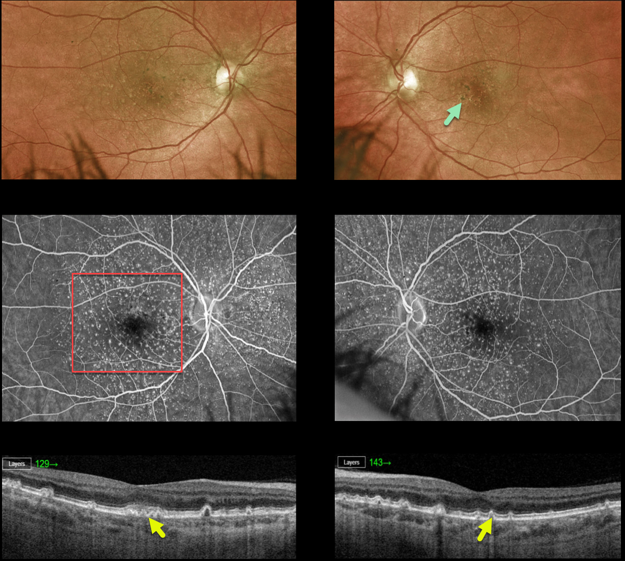
Optos color imaging shows extensive small drusen with foveal pigment clumping. Fluorescein angiography shows the classic ‘starry sky’ pattern of hyperfluorescent cuticular drusen. Swept-source OCT shows mostly cuticular drusen with arrowhead-like small elevations of the RPE.
Learning Points:
Originally described by Gass as basal laminar drusen (AJO 1985;99:445-459), cuticular drusen were initially thought to represent nodular thickening of the RPE basement membrane.
They appear clinically as numerous, round, yellow-white ‘hard drusen’, and on OCT as small, arrowhead-shaped drusen located above Bruch’s membrane and below the RPE (Spaide and Curcio, Retina 2010;30:1441-1454).
Our patient’s angiographic appearance echoes Dr. Gass’ original description of “a beautiful stars-in-the-sky or Milky-Way pattern” of hyperfluorescence. Cuticular drusen are classically associated with avascular vitelliform lesions, but can also develop macular neovascularization.
DRY AMD
Originally posted on @retina.rocks September 7, 2022
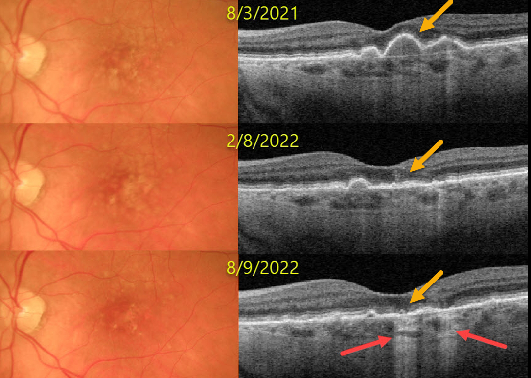
This 77YO female presented on 8/3/21 with asymptomatic bilateral confluent soft drusen. Vision was 20/30 OU.
Over the next year, the drusen flattened with secondary drusenoid atrophy. Transmission defects from incomplete outer retinal and RPE atrophy are noted at the most recent visit. Vision was 20/20 OU on the 8/9/22 visit.
Learning Points:
Drusen commonly remodel over time, and their collapse can lead to drusenoid atrophy of the RPE, as in our patient. Vision may or may not decrease following drusen regression.
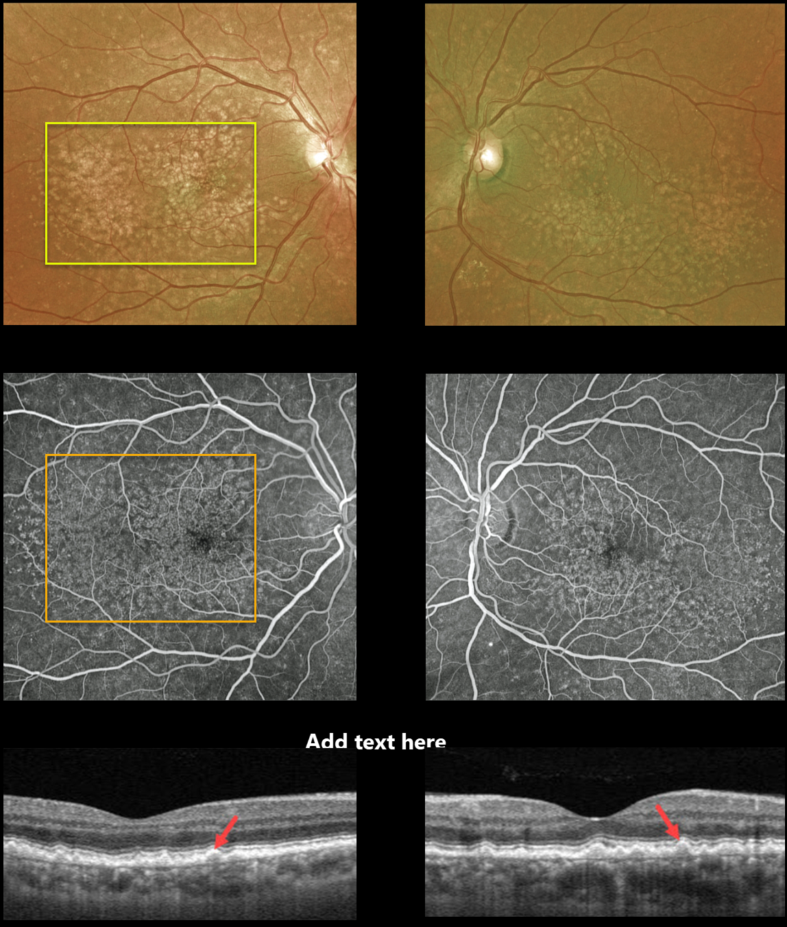
CUTICULAR DRUSEN
Originally posted on @retina.rocks March 17, 2022
This 62YO male presented with 20/20 vision OU and asymptomatic macular drusen. Optos photos of both eyes show numerous scattered cuticular drusen throughout each macula.
Fluorescein angiography shows a dramatic bilateral starry sky pattern of innumerable hyperfluorescent drusen.
Triton swept-source OCT reveals multiple tooth-like cuticular drusen.
Learning Points:
Cuticular drusen, formerly known as basal laminar drusen, appear as numerous, round, yellow-white punctate accumulations located above Bruch’s membrane and below the RPE.
Originally described by Gass as a unique entity (AJO 1985;99;445-459), they are now thought to fall at least in part within the AMD spectrum. They are classically associated with avascular vitelliform lesions, but can also cause macular neovascularization.
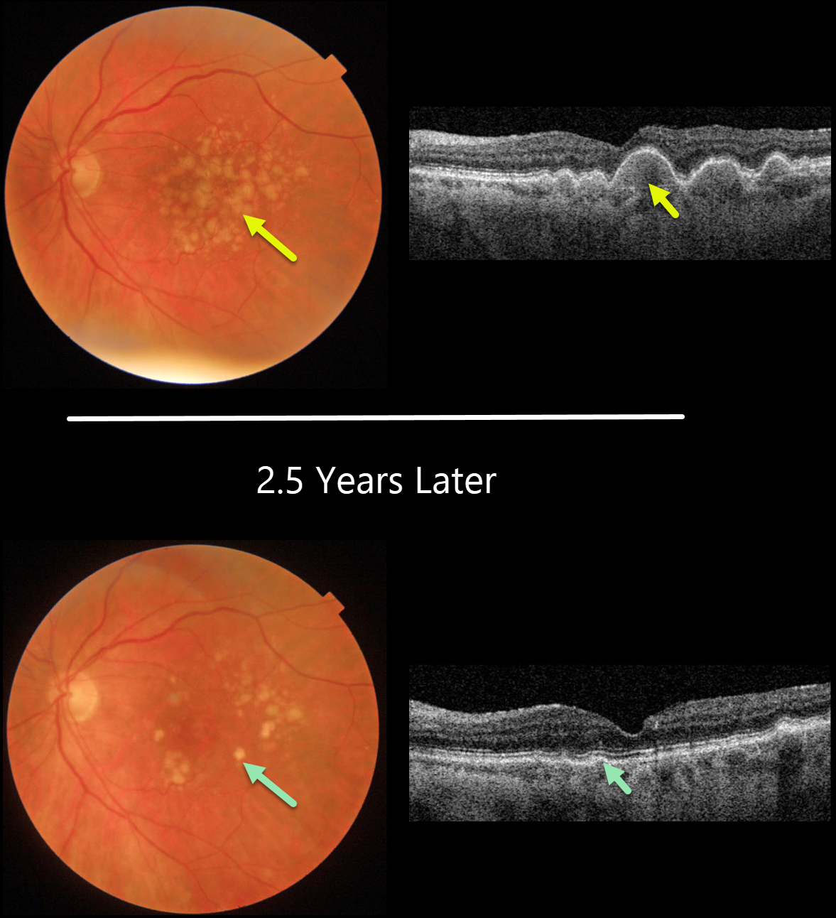
DRUSEN RESOLUTION
Originally posted on @retina.rocks March 16, 2022
This 77YO female presented with diffuse, large, confluent soft drusen in both eyes (only left eye pictured). Vision was 20/50.
Two and a half years later, her macular examinations appeared quite different with marked drusen resolution. OCT shows near-complete flattening of all drusen, with irregularity of the outer retinal bands and RPE. Vision actually improved to 20/25.
Learning Points:
Drusen commonly remodel over time, and their collapse can lead to drusenoid atrophy of the RPE.
Although our patient’s outer retina is certainly abnormal on OCT, her clinical macular findings are remarkably benign given the extent of drusen several years ago.
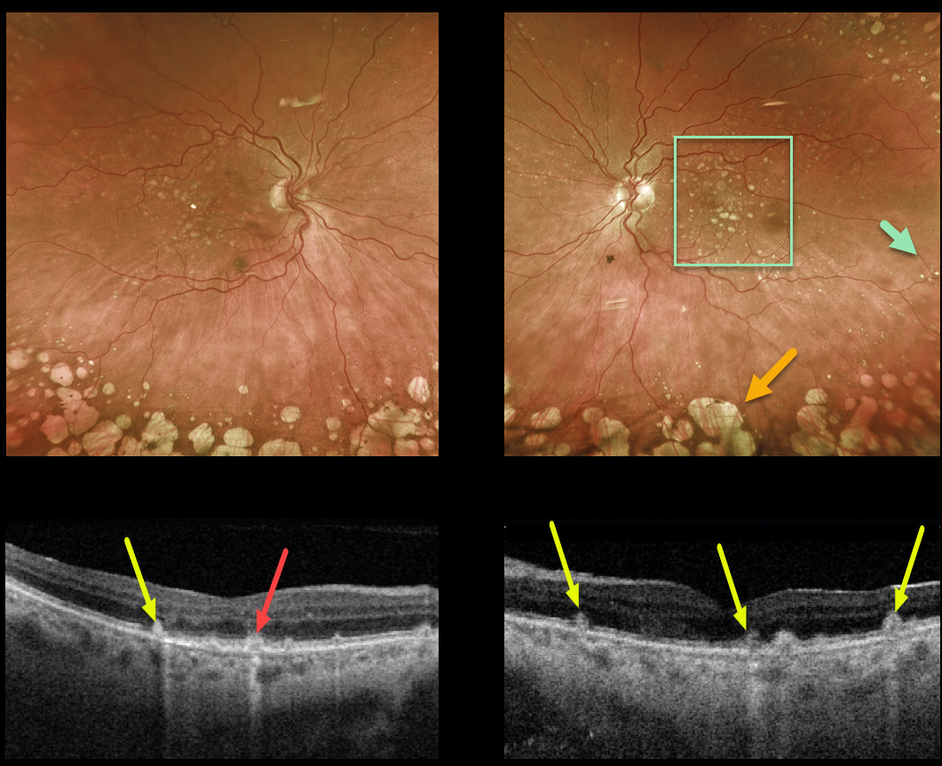
DRY AMD
Originally posted on @retina.rocks June 11, 2021
This 69YO female presented with 20/50 vision OU and bilateral mixed macular and peripheral drusen, along with extensive peripheral cobblestone degeneration.
Triton swept-source OCT shows a mixture of drusen and subretinal drusenoid deposits.
Learning Points:
Macular drusen and subretinal drusenoid deposits are some of the hallmark features of non-exudative age-related macular degeneration.
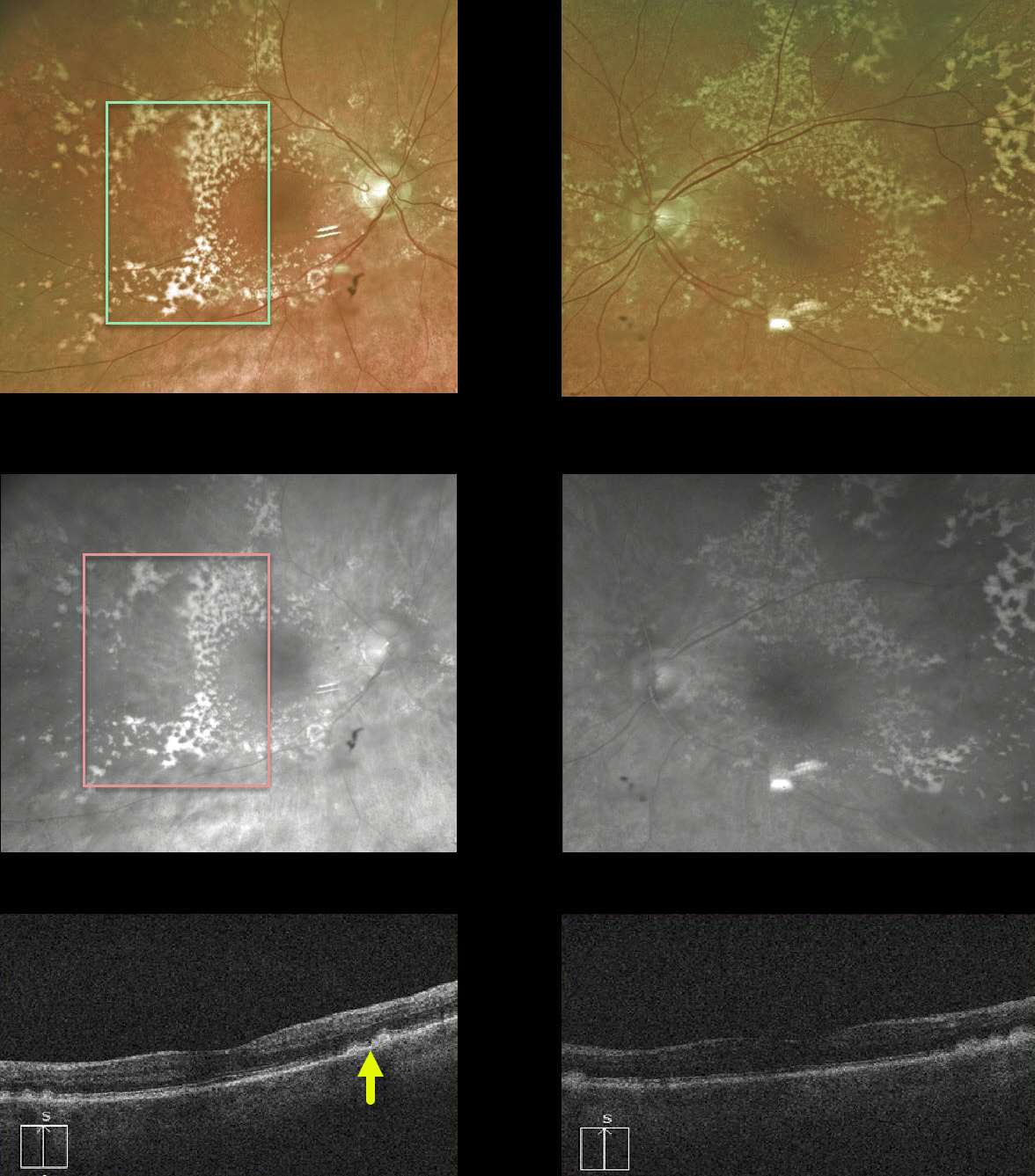
DRUSEN
Originally posted on @retina.rocks July 7, 2020
This 83YO female has extensive, confluent drusen outside the perifoveal area.
The drusen, located beneath the RPE, are well visualized with the Optos red channel. Subretinal drusenoid deposits (not present), which form above the RPE, would disappear with the red channel.
OCT also demonstrates drusen.
Learning Points:
Sub-RPE drusen are a hallmark feature of non-exudative age-related macular degeneration.


