

AMD Macular (Geographic) Atrophy
<< Back to Cases
FLATTENED DRUSENOID PED CAUSING SECONDARY MACULAR ATROPHY
Originally posted on @retina.rocks June 23, 2026
This 72YO female has been followed for years with intermediate AMD. On 3/26/25, she remained asymptomatic with 20/40 vision.
Color photography shows confluent central drusen creating a drusenoid PED with a radiating pigment figure, surrounded by large soft drusen. The drusenoid RPE detachment (PED) is confirmed on swept-source OCT. The RPE and outer retinal bands are obscured by subretinal hyperreflective material. She returned 6 months later with stable vision and no new symptoms. The PED is mostly flattened with some resolution of the hyperreflective material.
Learning Points:
Drusen commonly remodel over time and their collapse, as in our patient, can lead to drusenoid atrophy of the RPE (Filho et al, Retina 2013;33;1558-1566). Vision may or may not decrease following drusen regression. In our experience, drusenoid atrophy remains stable, unlike primary age-related macular atrophy, whose radius enlarges linearly over time (Shen et al, Ophthalmology Retina 2020;4;899-910). We therefore do not consider intravitreal complement inhibitors for these patients.
FLATTENED DRUSEN CAUSING SECONDARY MACULAR ATROPHY
Originally posted on @retina.rocks January 8, 2024
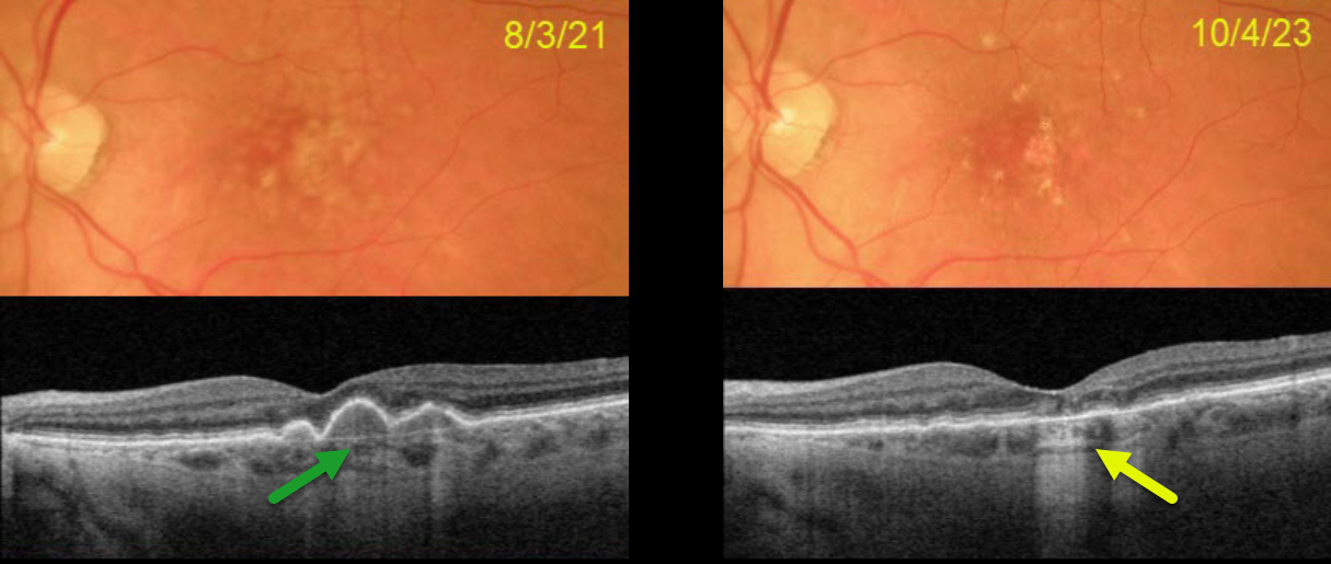
This 77YO female presented on 8/3/21 with 20/30 vision in her left eye. Optos color RG imaging shows confluent central soft drusen, which are confirmed on Triton swept-source OCT.
On 10/4/23, vision was 20/25. The central drusen flattened with secondary drusenoid atrophy. Continued observation was recommended.
Learning Points:
Drusen commonly remodel over time and their collapse, as in our patient, can lead to drusenoid atrophy of the RPE (Filho et al, Retina 2013;33;1558-1566). Vision may or may not decrease following regression of drusen.
In our experience, drusenoid atrophy remains stable, unlike primary age-related macular atrophy, whose radius enlarges linearly over time (Shen et al, Ophthalmology Retina 2020;4;899-910). We therefore do not consider intravitreal complement inhibitors for these patients.
HIGH MYOPIA
Originally posted on @retina.rocks September 11, 2023
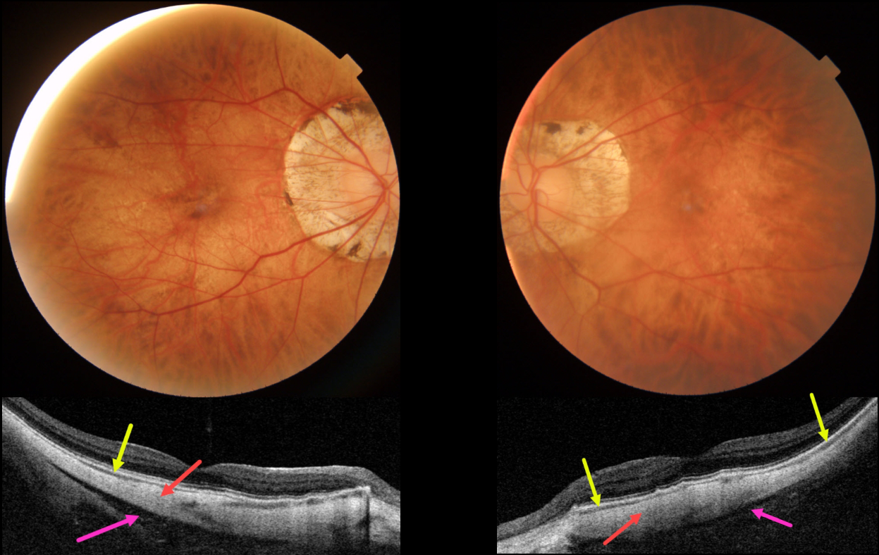
This 63YO female with severe high myopia underwent bilateral cataract surgery many years ago, along with successful vitrectomy for retinal detachment in her right eye. Vision was 20/50 OD and 20/30 OS.
Color imaging shows bilateral myopic peripapillary atrophy and macular pigmentary loss. Swept-source OCT shows a dramatically thinned or absent choroid and full-thickness, thinned sclera measuring about 300 microns bilaterally (normal posterior scleral thickness should be about 1mm). A hyporeflective region behind the sclera likely represents Tenon’s capsule, with the orbit visible more posteriorly.
Learning Points:
The era of ocular OCT began in 1991 with the initial report from Dr. Fujimoto’s lab at MIT (Huang et al, Science 1991;254:1178-1181). The technology was acquired by Zeiss in 1994, and after several failed commercial iterations, the Zeiss Stratus time-domain OCT in 2002 made it a must-have instrument for eye care.
Spectral domain units became available in 2008, followed by swept source units in 2015. Each subsequent generation can capture more information at greater retinal/choroidal depth. The swept source technology, in particular, is best able to visualize choroidal detail.
Our patient’s axial myopia with thinned choroid and sclera, along with the swept-source technology, provides a unique ability to visualize Tenon’s capsule and the anterior orbit.
For a wonderful historical review of the development, commercialization, and impact of OCT, see Fujimoto and Swanson, Invest Ophthalmol Vis Sci 2016;57;OCT1-OCT13.
PENTOSAN POLYSULFATE SODIUM TOXICITY VS AMD MACULAR ATROPHY
Will Gibson
Originally posted on @retina.rocks September 4, 2023
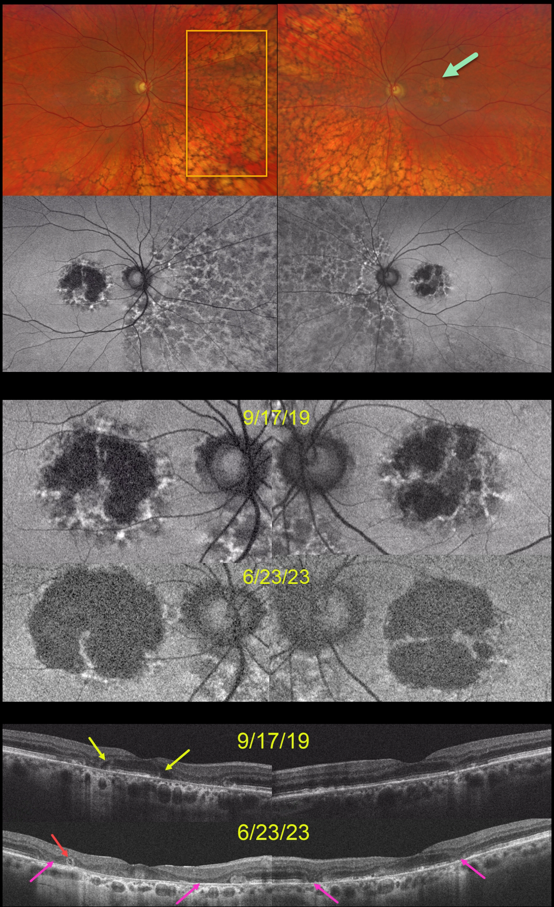
This 59YO was examined on 9/17/19. She was taking Elmiron (pentosan polysulfate sodium, PPS) for 17 years for interstitial cystitis. Vision was 20/40 OD and 20/30 OS.
Optos color RG imaging shows bilateral central-sparing macular atrophy (MA) and extensive, posterior peripheral reticular degeneration of the RPE (PRDRPE).
Fundus autofluorescence shows hypo-FAF from the MA with surrounding punctate and linear areas of hyper-FAF. Based on these findings, she decided to stop taking Elmiron.
When last examined on 6/23/23, vision dropped to 20/800 OD and was relatively stable at 20/40 OS. The MA progressed bilaterally clinically and on FAF and OCT. Hyporeflective OPL degeneration (wedge defects) is noted on the initial 9/17/19 OCT. On the 6/23/23 OCTs, an area of outer retinal tubulation (ORT) is seen temporally OD, and progressive outer retinal and RPE atrophy are noted bilaterally.
Learning Points:
Elmiron was approved by the FDA in 1996 for the treatment of interstitial cystitis. Recently, a unique PPS retinopathy has been described. Toxicity seems to develop over many years and can mimic more common disorders, including age-related macular degeneration and macular dystrophies.
Peripapillary hypoautofluorescence, more densely packed macular autofluorescent changes, and earlier central macular involvement suggest PPS toxicity over other causes (see Barnes et al Ophthalmology Retina 2020;4:1196-1201), including maternally inherited diabetes and deafness (MIDD).
ORT is often noted overlying inactive macular neovascularization with ongoing anti-VEGF therapy and should not be confused with exudative fluid or cysts, which lack a hyperreflective border. The outer hyperreflective band likely represents inner segment mitochondria undergoing fission and translocation towards the nucleus (Litts et al, Retina 2018;38:445-461).
ORT, initially described by Zweifel et al (Arch Ophthalmol 2009;127:1596-1602), is a neurodegenerative condition of the photoreceptors and Muller cells associated with outer retinal and retinal pigment epithelium atrophy, including advanced AMD and inherited retinal diseases.
The hyper-FAF surrounding the initial MA is more typical for PPS toxicity, and progressive atrophy is not uncommon even with stopping therapy (Jung et al, JAMA Ophthalmol 2023;141:260-266). However, in our experience, PPS usually produces more widespread macular findings. Given the PRDRPE, the MA could also be age-related. Or maybe our patient had baseline age-related MA superimposed on Elmiron toxicity?
AMD MACULAR ATROPHY
Originally posted on @retina.rocks November 2, 2022
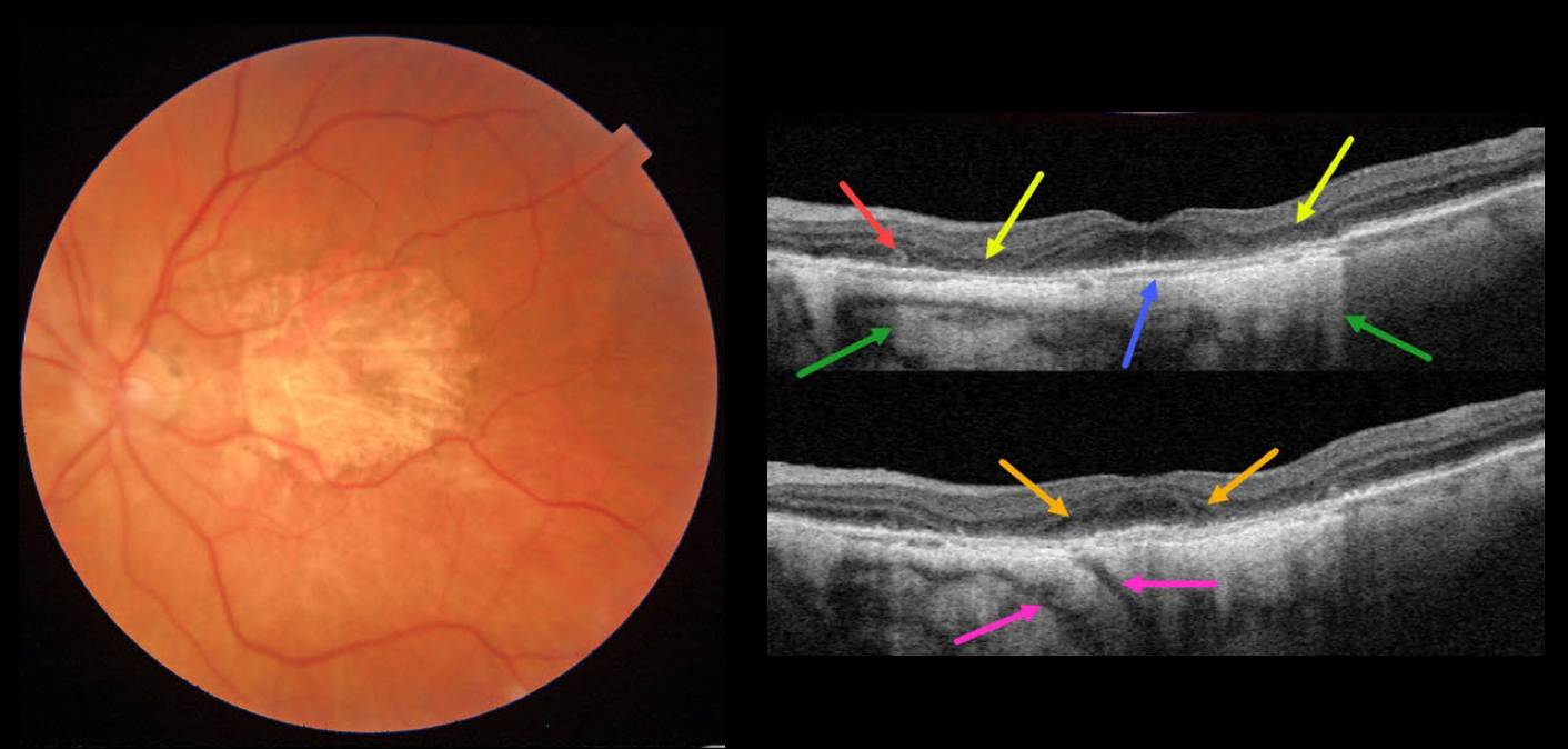
This 84YO female has stable advanced non-exudative AMD with bilateral macular atrophy (MA). There are numerous classic findings on multimodal imaging of her left eye.
Color imaging shows foveal MA. Swept-source OCT shows pericentral outer retinal and RPE atrophy. This atrophy allows for more light to illuminate the underlying choroid and sclera, as well as visualizing two short posterior ciliary arteries posterior to the sclera. The variably atrophic RPE allows for visualization of Bruch membrane, which is normally fused in the RPE-Bruch layer seen in normal eyes. There is a tiny area of outer retinal tubulation (ORT) nasally. Finally, hyporeflective outer plexiform layer wedge defects are noted along the descending outer retinal atrophy.
Wedge defects develop at the boundaries of the degenerating outer plexiform layer in about 75% of MA eyes (Mones et al, Ophthalmology 2012;119:1412-1419).
ORTs represent photoreceptor and Muller cell degeneration (Dolz-Marco et al, Ophthalmology 2017;124:1353-1367). These ORTs have a hyper-reflective border with a central hyporeflective core.
Learning Points:
In 1970, Dr. Gass originally described macular atrophy from AMD as “geographic areas of atrophy” in the setting of “senile macular choroidal degeneration” (Schmitz-Valkenberg, Retina 2016;36:2250-2264).
Today, the terminology is much more specific due to advances in technology and improved visualization of retinal and choroidal structures.
From Sadda et al, Ophthalmology 2018:537-548, the new classification is as follows: Complete RPE and Outer Retinal Atrophy (cRORA), Incomplete RPE and Outer Retinal Atrophy (iRORA), Complete Outer Retinal Atrophy (cORA), and Incomplete Outer Retinal Atrophy (iORA).
DRY AMD
Originally posted on @retina.rocks September 7, 2022
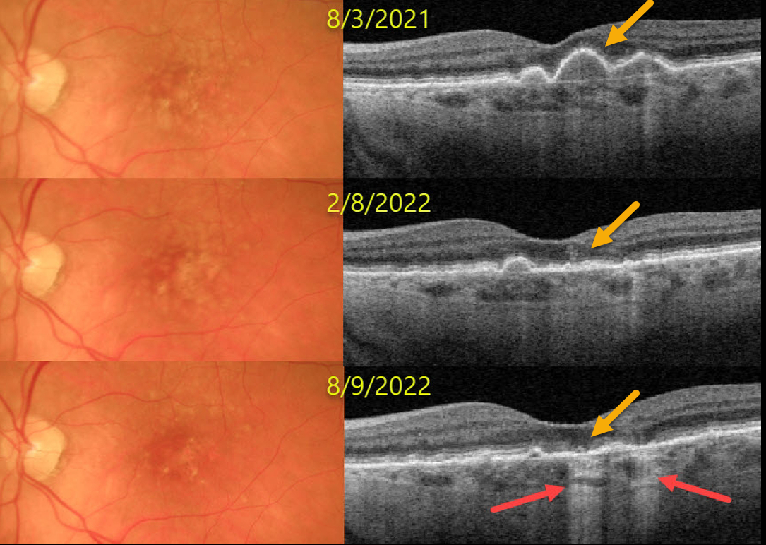
This 77YO female presented on 8/3/21 with asymptomatic bilateral confluent soft drusen. Vision was 20/30 OU.
Over the next year, the drusen flattened with secondary drusenoid atrophy. Transmission defects from incomplete outer retinal and RPE atrophy are noted at the most recent visit. Vision was 20/20 OU on the 8/9/22 visit.
Learning Points:
Drusen commonly remodel over time, and their collapse can lead to drusenoid atrophy of the RPE, as in our patient. Vision may or may not decrease following drusen regression.
AMD MACULAR ATROPHY
Originally posted on @retina.rocks April 28, 2022
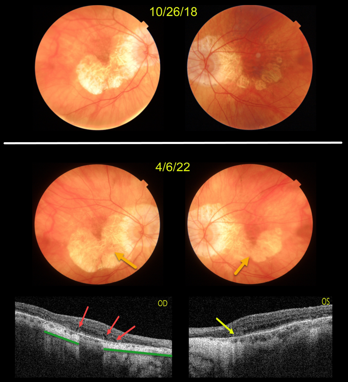
This patient originally presented in 2018 with vision of 20/40 OD and 20/50 OS. Areas of AMD-related macular atrophy (MA) skirted each macular center.
Four years later, vision had decreased to 20/100 OU due to progressive atrophy. Comparing the two images from 2018 to 2022, there is a bilateral linear enlargement for all margins of the atrophy, particularly in the left eye.
Triton swept-source OCT B-scans show classic findings for MA. The right eye has multiple wedge defects, hyporeflective wedges which develop at the boundaries of the degenerating outer plexiform layer in about 75% of MA eyes (Mones et al, Ophthalmology 2012;119:1412-1419). In addition, loss of the outer retina and RPE allows more OCT light to be transmitted into the choroid, resulting in distinct areas of choroidal and scleral hyperreflectivity.
There is a tiny area of outer retinal tubulation (ORT) nasally in the left eye, which represents photoreceptor and Muller cell degeneration (Dolz-Marco et al, Ophthalmology 2017;124:1353-1367). These ORTs have a hyper-reflective border with a central hyporeflective core.
OUTER RETINAL TUBULATION
Originally posted on @retina.rocks February 16, 2021
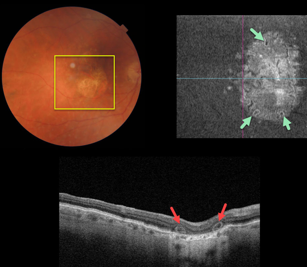
This 80YO female was 20/200 in her right eye due to central macular atrophy from atrophic age-related macular degeneration (AMD).
Triton swept-source optical coherence tomography showed outer retinal tubulation (ORT) in the outer retinal layers. The extent of these tubular structures was best visualized on en face imaging.
Learning Points:
Outer retinal tubulation, initially described by Zweifel et al (Arch Ophthalmol 2009;127;1596-1602), is a neurodegenerative condition of the photoreceptors and Muller cells associated with atrophy affecting the outer retina and retinal pigment epithelium, including advanced AMD and inherited retinal diseases.
ORT is often seen overlying inactive macular neovascularization in the setting of ongoing anti-VEGF therapy and should not be confused with exudative fluid or cysts, which lack a hyperreflective border.
The outer hyperreflective band likely represents inner segment mitochondria undergoing fission and translocation toward the nucleus (Litts et al Retina 2018;38;445-461).


