

AMD Neovascular
<< Back to Cases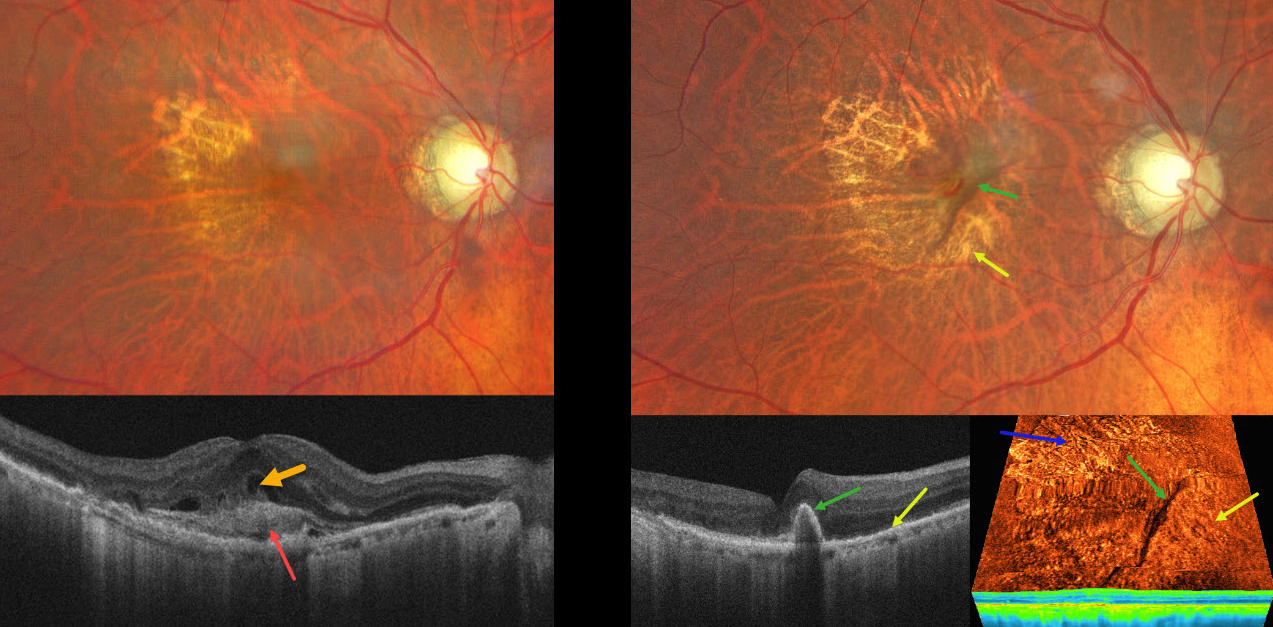
NEOVASCULAR AMD WITH RAPIDLY RESOLVED SHRM FOLLOWING RPE TEAR
Ayushi Gupta and Vishal Agrawal
Originally posted on @retina.rocks August 18, 2025
This 82YO male presented with recent vision loss in his right eye. Vision was 20/200.
Color photography shows a poor foveal reflex with superotemporal geographic atrophy. OCT scanning shows subretinal hyperreflective material (SHRM) with overlying retinal fluid. A possible bacillary layer detachment is also noted. About 6 weeks following a single intravitreal anti-VEGF injection, vision improved to 20/50. A new RPE tear is noted, with the bed of the tear inferonasal to the retracted hyperpigmented RPE. Despite persistent flow signals on OCT angiography, there is no fluid on structural OCT. The SHRM has completely resolved. A treat-and-extend anti-VEGF regimen was begun.
Learning Points:
A tear in the RPE is a fairly common finding in RPE detachment (PED) associated with type 1 macular neovascularization (MNV) located below the RPE. The neovascularization can spontaneously contract or involute following treatment. It then puts traction on the overlying RPE, which can then rip at the opposite edge of the PED. Like a cheap window blind, the torn RPE retracts and scrolls towards the MNV. This leaves a depigmented region where the RPE was and is now absent, along with a pigmented, relatively straight subretinal lesion from the scrolled RPE edge.
SHRM is an OCT finding that is invisible clinically. This material, located between the neurosensory retina and RPE, is thought to consist of fluid, fibrin, blood, and other fibrovascular tissues. SHRM is a biomarker of active disease that decreases with successful anti-VEGF therapy (Willoughby et al, Ophthalmology 2015;122:1846-1853). Persistent SHRM is associated with an increased incidence of scar formation and worse vision.
It is impossible to determine whether the anti-VEGF injection alone or in combination with the RPE tear caused such a rapid resolution of the SHRM. Fortunately, our patient’s vision remained quite good since the RPE tear just spared the foveal center.
CUTICULAR DRUSEN WITH MACULAR NEOVASCULARIZATION
Originally posted on @retina.rocks August 5, 2025
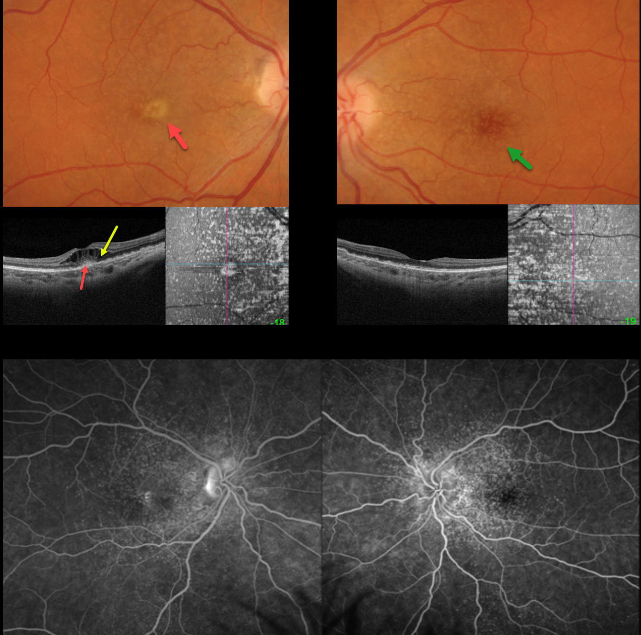
This 47YO female presented with one week of vision loss in her right eye. Vision was counting fingers OD and 20/25 OS.
Optos color RGB imaging shows innumerable cuticular macular drusen, which appear on swept-source OCT B-scanning as arrowhead-like RPE elevations and on en face imaging as numerous hyperreflective spots. A yellow subfoveal lesion OD appears on OCT as hyperreflectivity in the outer retina. Marked overlying cystoid edema is seen. Fluorescein angiography shows late pooling in the foveal cysts OD, with hyperfluorescent window defects from the drusen.
One month following an initial intravitreal Avastin injection, vision improved to 20/80, with complete resolution of the vitelliform lesion and cystoid edema. Treat-and-extend treatment was started.
Learning Points:
Originally described by Gass as basal laminar drusen (AJO 1985;99:445-459), cuticular drusen were initially thought to represent nodular thickening of the RPE basement membrane. They appear clinically as numerous, round, yellow-white ‘hard drusen’, and on OCT as small, arrowhead-shaped drusen located above Bruch’s membrane and below the RPE (Spaide and Curcio, Retina 2010;30:1441-1454).
Our patient’s angiographic appearance echoes Dr. Gass’ original description of “a beautiful stars-in-the-sky or Milky-Way pattern” of hyperfluorescence. Cuticular drusen are classically associated with avascular vitelliform lesions (Finger et al, Retina 2010;30:1455-1464).
NEOVASCULAR AMD
Originally posted on @retina.rocks February 5, 2025
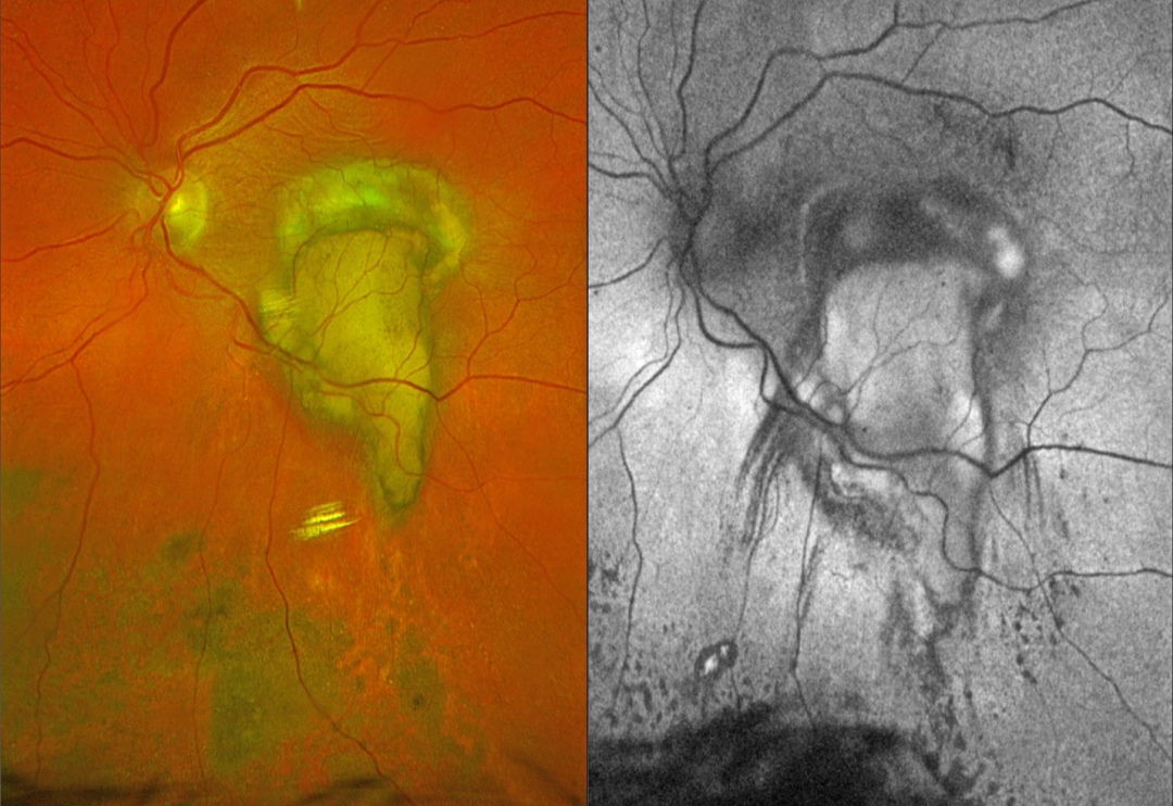
This 79YO female underwent vitrectomy for a breakthrough vitreous hemorrhage from neovascular AMD. Vision was 20/400.
Postoperative Optos color RGB imaging shows an oval fibrotic macular scar with a tongue-like projection of organized devitalized yellow blood extending inferiorly. Coarse subretinal pigment extends into the inferior midperiphery. On fundus autofluorescence (FAF), the fibrotic portion of the scar and inferior subretinal pigment are hypo-FAF, and the devitalized blood is hyper-FAF. Steaks of inferiorly radiating hypo-FAF also extend from the tongue-like yellow blood.
Learning Points:
This complex healing neovascular lesion has many signs of chronicity. There is no fresh, red blood. The remaining blood is yellow and devitalized, developing as heme products degrade. The coarse peripheral subretinal pigment is a marker of prior resolved subretinal blood and appears on OCT as subretinal hyperreflective material. Finally, the subretinal fibrosis where the breakthrough blood originated represents the usual endpoint of untreated neovascular disease, which is thankfully much less common since the advent of anti-VEGF therapy in the mid-2000s.
NEOVASCULAR AMD WITH HENLE LAYER HEMORRHAGE
Originally posted on @retina.rocks July 29, 2024
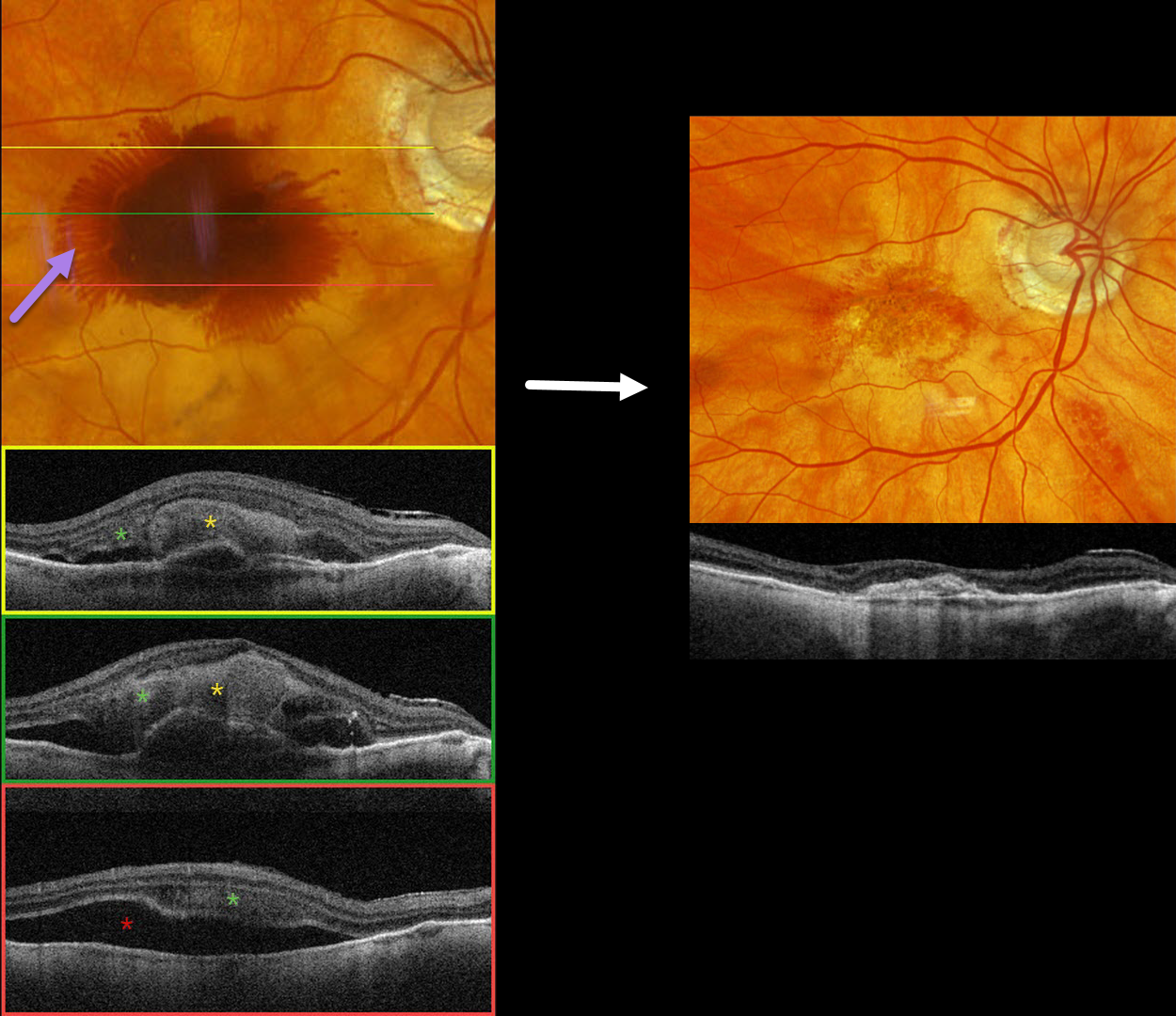
This 97YO male has a history of counting-finger vision in his left eye due to foveal macular atrophy. On 2/29/24, vision was 20/40 in his non-exudative right eye. He presented on 3/12/24 with sudden vision loss OD and new hand motions vision.
Optos color RGB imaging shows fairly dense central subretinal blood, with surrounding radiating blood in the deeper Henle’s layer (outer plexiform layer). Triton swept-source OCT shows variable thick subretinal blood and more inferior subretinal fluid. Henle’s blood is seen as a diffuse hyperreflective obscuration of the outer retinal layers. Vitrectomy with subretinal TPA was performed two days later. Two months later, vision was counting fingers, and the macula was completely free of fluid and blood.
Learning Points:
Radiating deep macular hemorrhages are seen in multiple conditions, including Valsalva and trauma. We have not previously seen these hemorrhages in neovascular AMD. Their unique radiating appearance results from blood accumulating in Henle’s layer. Kon Graversen et al described hemorrhagic unilateral retinopathy, an idiopathic disorder with a similar appearance (Retina 2014;34:483-489).
OCCULT MNV
Asma Samsudeen and Ashish Sharma
Originally posted on @retina.rocks November 23, 2023
This 62YO female presented with 3 weeks of vision loss in her left eye. Vision was 20/30.
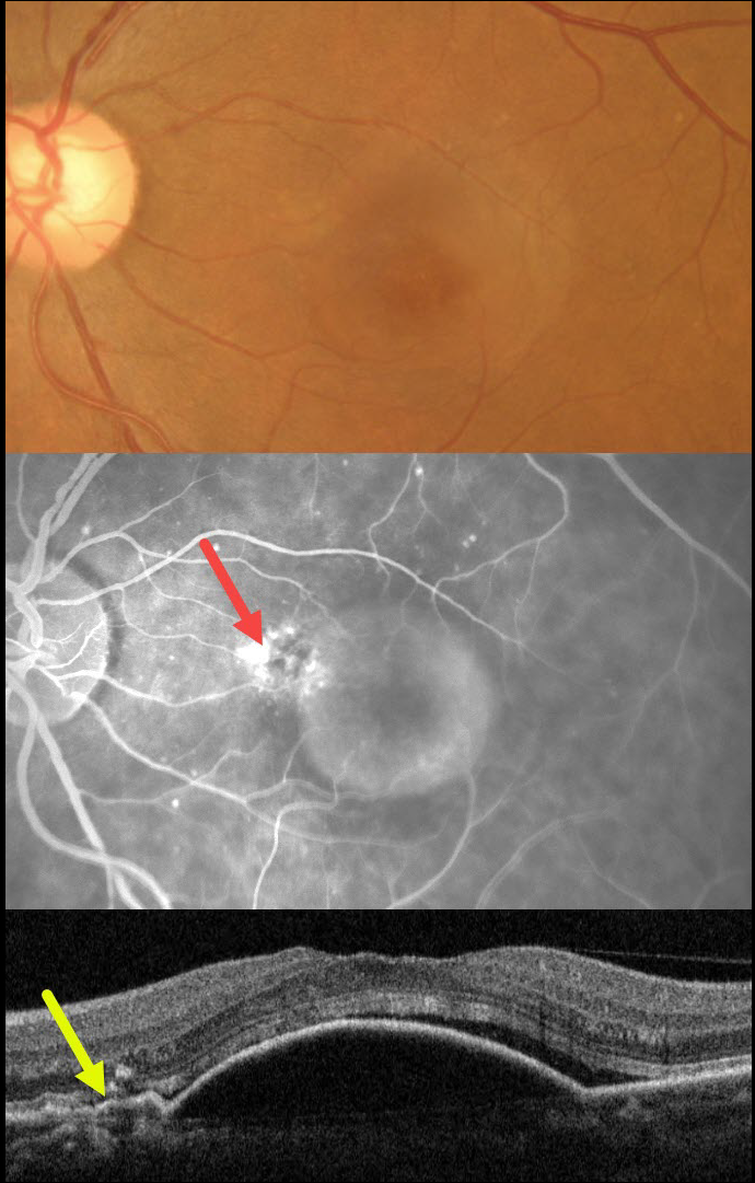
Color imaging shows a large central macular retinal pigment epithelial detachment (PED). Fluorescein angiography shows a somewhat unevenly filling PED, with staining vs leakage along its superonasal edge. OCT scanning shows a large PED, shallow overlying temporal subretinal fluid, and shallow irregular elevation of the RPE nasally indicative of a type 1 macular neovascularization (MNV).
The uneven angiographic pooling is also suggestive of an occult MNV. Anti-VEGF therapy was advised.
This case was submitted by Asma Samsudeen and Ashish Sharma.
Learning Points:
Originally described by Gass (Retina 1984;4:205-220), a notched PED is a sign of an occult MNV. The notch represents a type 1 MNV, which tethers the overlying RPE from the surrounding more highly elevated PED. Hydrostatic leakage from the MNV may contribute to the larger PED.
Although now considered a classic clinical and angiographic finding, in 1984 this was evidently not the case, as the reviewers for this article advised against publication. Based solely on who the contributing author was, the journal’s editor overruled his reviewers, published Dr. Gass’ manuscript, and the rest was literally history!
WET AMD WITH OUTER RETINAL TUBULATION
Originally posted on @retina.rocks April 12, 2023
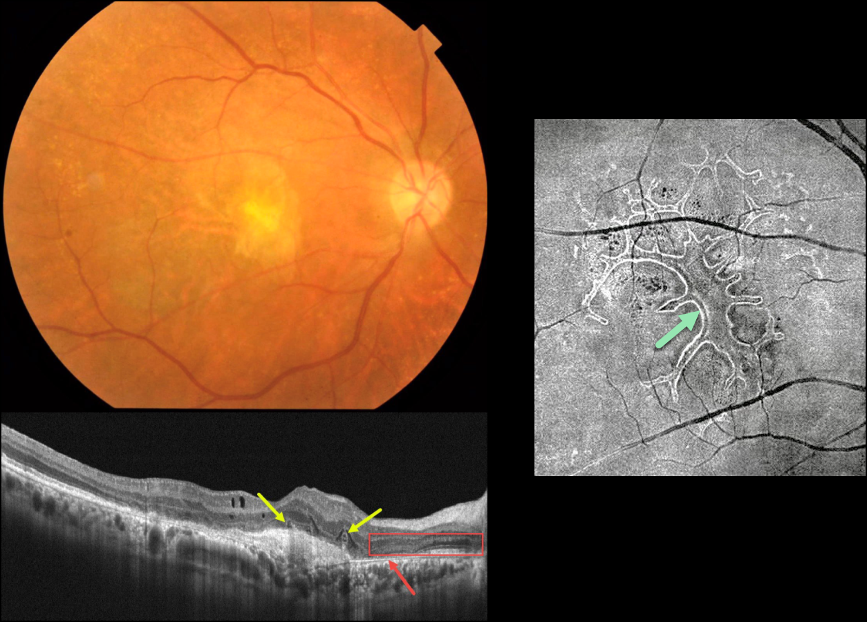
This 63YO male received several intravitreal anti-VEGF injections for neovascular AMD in his right eye, the most recent being 6 months earlier. Vision is counting fingers.
Color photography shows an inactive atrophic foveal scar. OCT scanning shows a hyperreflective macular neovascularization (MNV). The outer retina overlying the MNV is disorganized with several overlying hyporeflective lesions with surrounding hyperreflective rims. More nasally, there is complete RPE loss with bare Bruch’s membrane. The overlying retina is anomalous with additional hypo- and hyperreflective bands.
En face OCT scanning through the outer retina provides a more dramatic view of the extensive tubulation-branching network.
Learning Points:
Outer retinal tubulation (ORT) is often noted overlying inactive MNV with ongoing anti-VEGF therapy and should not be confused with exudative fluid or cysts, which lack a hyperreflective border. The outer hyperreflective band likely represents inner segment mitochondria undergoing fission and translocation toward the nucleus (Litts et al, Retina 2018;38:445-461).
ORT, initially described by Zweifel et al (Arch Ophthalmol 2009;127:1596-1602), is a neurodegenerative condition of the photoreceptors and Muller cells associated with atrophy affecting the outer retina and retinal pigment epithelium, including advanced AMD and inherited retinal diseases.
NEOVASCULAR AMD WITH PRECHOROIDAL CLEFT
Originally posted on @retina.rocks December 14, 2022
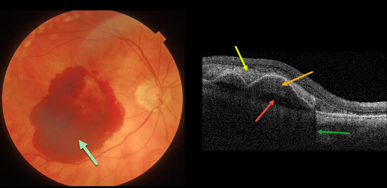
This 84 YO female with macular degeneration presented with acute vision loss of counting fingers vision in her right eye from neovascular AMD (nAMD).
Fundus photography shows a large central subretinal hemorrhage. Swept-source OCT shows the hyperreflective subretinal blood, which shadows the underlying choroid. Sub-RPE blood vs a type-1 (located beneath the RPE) macular neovascularization (MNV) overlies a prechoroidal cleft.
Anti-VEGF therapy was begun.
Learning Points:
A prechoroidal cleft is a hyporeflective space between the RPE and Bruch’s membrane. These are seen in up to about 20% of treated eyes with nAMD, particularly with type 3 neovascularization (intraretinal MNV) and polypoidal choroidal vasculopathy.
They are usually associated with worse visual acuity due to potential complications including RPE tear and subretinal hemorrhages (Kim et al, Retina 2017;37:2047-2055).
SUBMACULAR HEMORRHAGE
Akansha Sharma and Manish Nagpal
Originally posted on @retina.rocks September 6, 2022
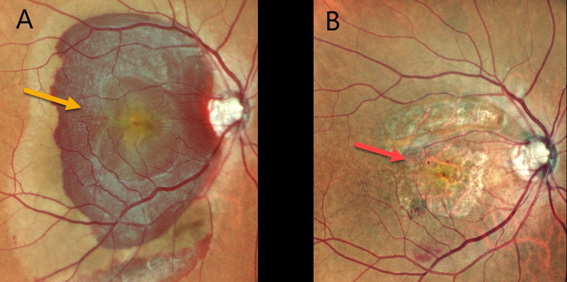
This 73YOF presented with 1 day of acute vision loss in her right eye. Vision was counting fingers OD and hand motion OS.
A large submacular hemorrhage from neovascular AMD occupied the entire right macula. A fibrosed macular scar was noted in her left eye (image not shown).
She was scheduled for vitrectomy with anti-VEGF injection, but unfortunately, was not taken to surgery for about a month from presentation.
Postoperatively, there is dry atrophic macular scarring with virtually complete resolution of all blood, but vision remains at counting fingers from macular atrophy.
NEOVASCULAR AMD
Originally posted on @retina.rocks July 20, 2022
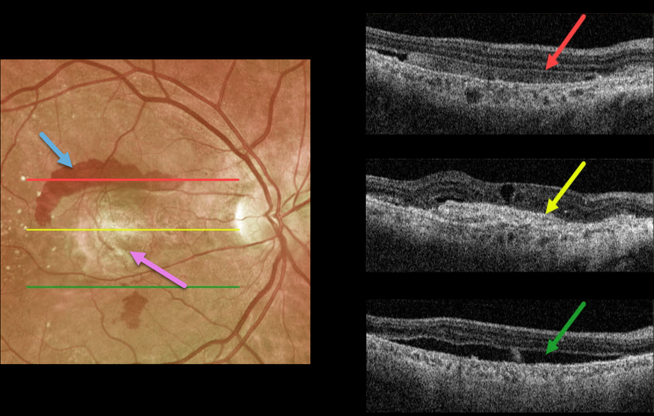
This 72YO male presented with longstanding decreased vision in his right eye. Vision was counting fingers due to a fibrosed scar from neovascular age-related macular degeneration (nAMD).
Optos imaging shows a central fibrosed macular scar with superior and inferior subretinal blood. Triton swept-source imaging shows multiple cool findings from this single neovascular lesion. Superiorly, the subretinal blood appears as hyperreflective material in the subretinal space. More centrally, the fibrotic scar/type 2 macular neovascularization is hyperreflective. There are also some overlying chronic cystic retinal changes. More inferiorly is shallow, subclinical subretinal fluid.
A therapeutic trial of monthly anti-VEGF injections was initiated, although we’re not sure this will have a visual effect given the subfoveal fibrosis.
NEOVASCULAR AMD + PRECHOROIDAL CLEFT
Originally posted on @retina.rocks July 8, 2022
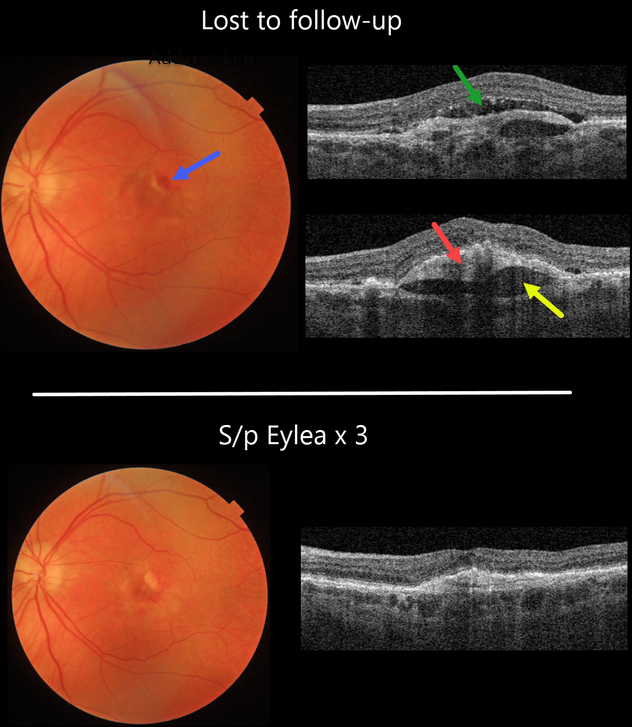
This 82YO female was maintaining 20/40 vision in her left eye with intravitreal Eylea every 8 weeks for neovascular AMD. She was then lost to follow-up, with new blood and 20/50 vision.
Swept source OCT shows new subretinal fluid. The hyperreflective sub-RPE type 1 macular neovascularization is elevated above a hyporeflective prechoroidal cleft.
Fortunately, vision returned to 20/30, with complete resolution of all exudative findings, following 3-monthly Eylea injections.
Learning Points:
A prechoroidal cleft is a hyporeflective space between the RPE and Bruch’s membrane. These are seen in up to about 20% of treated eyes with wet AMD, particularly with type 3 neovascularization and polypoidal choroidal vasculopathy. They are usually associated with worse visual acuity due to potential complications, including RPE tear and subretinal hemorrhages.
For a more detailed discussion of prechoroidal clefts, see Kim et al Retina 2017;37:2047-2055.
MACULAR NEOVASCULARIZATION (MNV)
Originally posted on @retina.rocks April 22, 2022
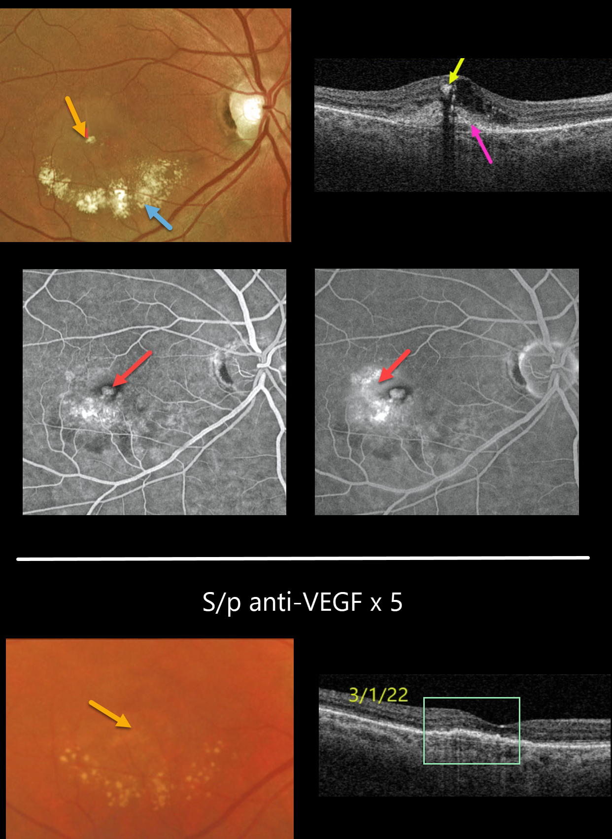
This 64YO female presented with this symptomatic unilateral exudative lesion in her right eye. Vision was 20/200.
Optos color imaging shows a small inner retinal yellow-white nodule with inferior subretinal lipid.
Triton swept-source OCT shows hyperreflectivity of this nodule with posterior shadowing, surrounding inner retinal fluid, and a hyperreflective macular neovascularization (MNV) located both below (type 1) and above (type 2) the RPE.
Optos fluorescein angiography shows a hyperfluorescent lesion with late leakage. The left eye showed mild macular drusen and pigmentary changes (not shown). This patient’s MNV is somewhat atypical given the extent of lipid exudation.
With monthly anti-VEGF therapy, the exudation resolved and vision improved to 20/60 following 5 intravitreal injections. The yellow nodular inner retinal lesion also completely resolved.
We therefore believe this most likely represented an unusual collection of inner macular lipids rather than a type 3 MNV.
NEOVASCULAR AMD
Originally posted on @retina.rocks March 22, 2022
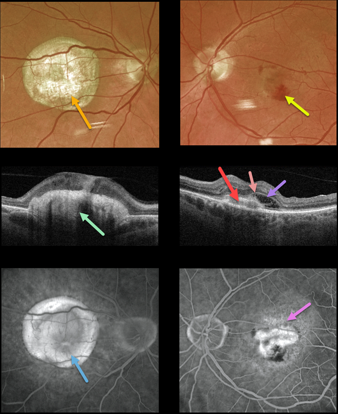
This 77YO male presented with counting fingers vision OD from a large fibrotic disciform scar and 20/200 vision OS from a new macular neovascularization.
The disciform scar is contracted and fibrotic, with marked hyperreflectivity on swept-source OCT. The MNV in his left eye is seen as opaque fluid with some subretinal blood. The type 2 MNV (located above the RPE) is noted on the OCT B-scan. There is also a bacillary detachment and a hyperreflective line indicating a possible pitchfork sign.
Optos fluorescein angiography shows diffuse staining of the subretinal scarring OD and leakage from the MNV OS.
Learning Points:
Disciform scars were the norm for untreated wet AMD before the anti-VEGF era that began in the mid-2000s. New-onset wet AMD diagnosed and treated with appropriate anti-VEGF dosing virtually never goes on to develop these lesions. There may be progressive macular atrophy following years of therapy, but not this type of fibrotic scarring.
An excellent review of wet AMD fibrotic lesions is in Souied et al’s AOS thesis (AJO 2020;214:151-171).
The pitchfork sign was originally described by Hoang et al (Retina 2013;33:1049-1055) as a unique OCT finding in inflammatory MNV. Falavarajani et al more recently noted that this finding is also associated with non-inflammatory MNV (Ophthalmic Surg Lasers Imaging Retina 2019;50:719-725).
The pathogenesis of the avascular pitchfork spikes is unknown, but may be related to outer retinal traction or Müller cell activation.
RPE TEAR
Originally posted on @retina.rocks February 2, 2022
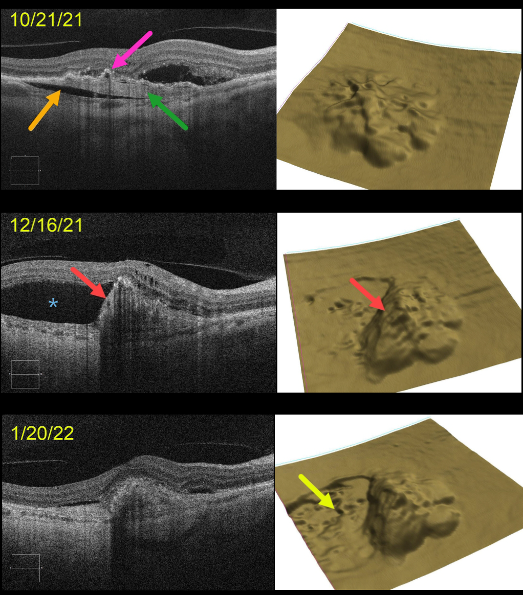
This 71YO female presented with a treatment-naive, age-related type 1 macular neovascularization (MNV). The already contracting MNV is elevated above the underlying choroid (prechoroidal cleft), and the overlying RPE is irregularly contracted and adherent to the MNV. The retinal pigment epithelium (RPE) map shows a 3-dimensional view of the RPE elevation. An anti-VEGF injection was given that day on 10/21/21.
On 12/16/21, vision was unchanged at 20/400 despite a new asymptomatic temporal RPE tear with marked overlying new subretinal fluid. An additional anti-VEGF injection was given.
On 1/20/22, vision was counting fingers. OCT scanning shows near-total resolution of all intra- and subretinal fluid. The RPE map nicely shows the crater of devoid RPE.
Learning Points:
A tear of the RPE is a fairly common occurrence in RPE detachment (PED) associated with type 1 MNV (located below the RPE). The neovascularization can spontaneously contract or involute following treatment. It then puts traction on the overlying RPE, which can then rip at the opposite edge of the PED.
This traction was already evident at our patient’s initial pre-injection visit, including the prechoroidal cleft, which is associated with a worse visual prognosis (see Kim et al, Retina 2017;37:2047-2055).
Like a cheap window blind, the torn RPE retracts and scrolls towards the MNV. This leaves a depigmented region where the RPE was and is now absent, along with a relatively straight, pigmented subretinal lesion extending from the scrolled RPE edge.

CHOROIDAL TRANSPLANT
Barbara Parolini and Veronika Matello
Originally posted on @retina.rocks December 21, 2021
This patient is a teacher who suffered from a wet AMD submacular hemorrhage. Despite six anti-VEGF injections, vision dropped to 20/200. She was 58 years old and unable to read.
After a long discussion regarding the pros vs cons of surgery, she underwent autologous choroidal transplantation in 2013.
The surgical procedure included:
1) Complete pars plana vitrectomy
2) Creation of a temporal retinal detachment
3) Peripheral 200-degree retinotomy
4) Macular neovascularization (MNV) removal
5) Feeder vessel endodiathermy
6) Designing the peripheral choroidal patch with endodiathermy
7) Cutting and isolation of a full-thickness autologous RPE and choroid transplant (under perfluorocarbon liquid, PFCL)
8) Translocation of the patch under the fovea (under PFCL)
9) Transfer of PFCL from under to over the retina with retinal reattachment
10) Peripheral laser
11) PFCL exchange for 1000cs silicone oil.
Eight years later, a Clarus 500 widefield photograph shows the intact submacular choroidal autograft and a white area of bare sclera in the temporal quadrant, which was the harvesting site for the autograft.
Canon Xephilio S1 widefield OCT shows a fairly normal retina overlying the graft. Xephilio A1 angioOCT shows the intact choroidal graft vessels arranged in a different direction compared to the native surrounding choroidal vasculature.
When last examined, near vision was 20/30. She recently developed an MNV in the contralateral eye, which is being treated with anti-VEGF injections.
MACULAR NEOVASCULARIZATION (MNV)
Originally posted on @retina.rocks September 27, 2021
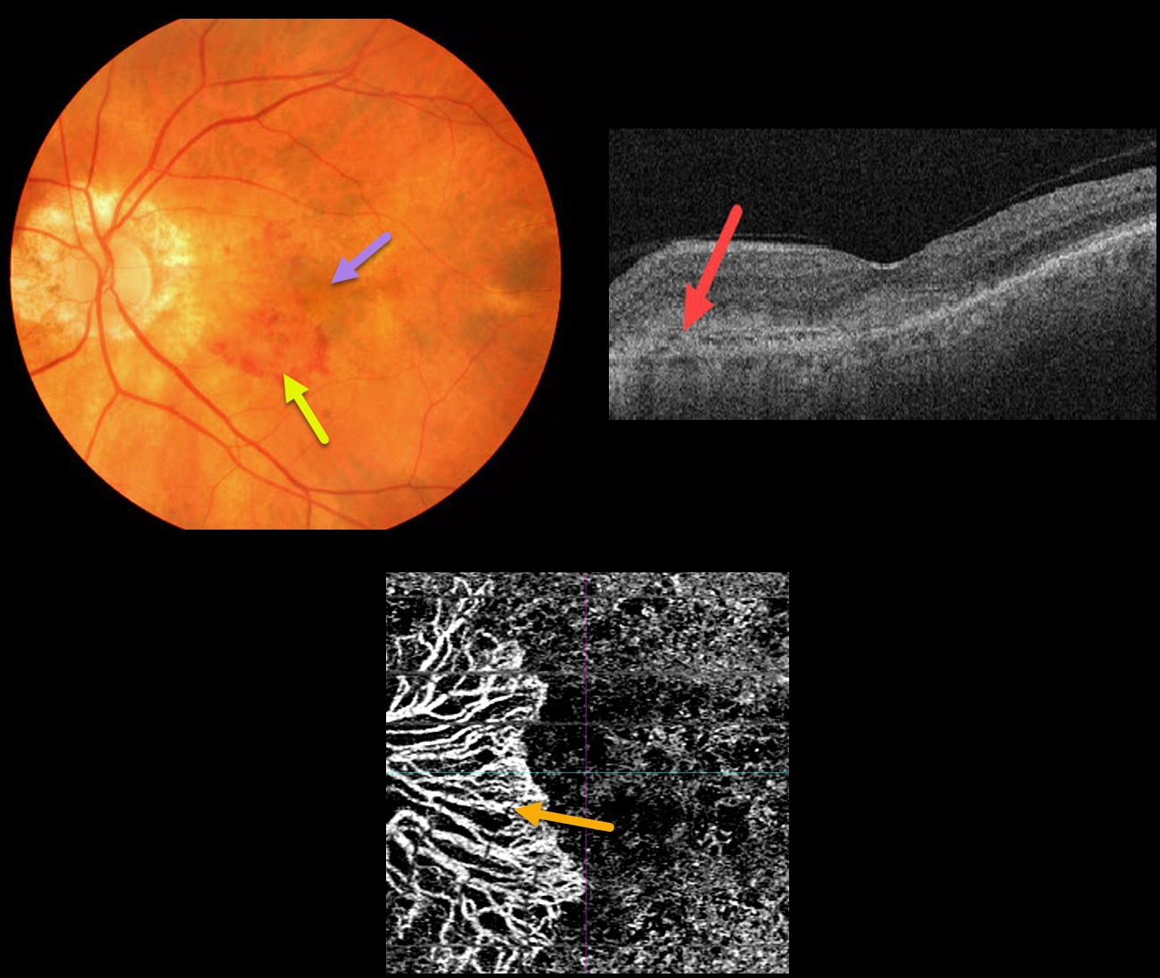
This 86YO male presented with symptomatic macular neovascularization (MNV).
Color imaging shows nasal patches of subretinal blood and more central subfoveal pigment. OCT scanning shows a hyperreflective MNV above the RPE (type 2 MNV). The peripapillary MNV is seen on OCT angiography.
Learning Points:
Macular neovascularization is also known by other terms, including choroidal neovascular membrane and subretinal neovascular membrane.
However, since these vessels are now known not to necessarily arise just from the choroid, the proper term is macular neovascularization (see Consensus on Neovascular Age-Related Macular Degeneration Nomenclature Study Group Ophthalmology 2020;127;616-636).
MNV is classified according to the location of the new vessels in relation to the RPE and neurosensory retina: type 1 (below the RPE), type 2 (between the RPE and neurosensory retina, and type 3 (within the neurosensory retina).
OUTER RETINAL TUBULATION FOLLOWING ONGOING ANTI-VEGF FOR NEOVASCULAR AMD
Originally posted on @retina.rocks September 23, 2021
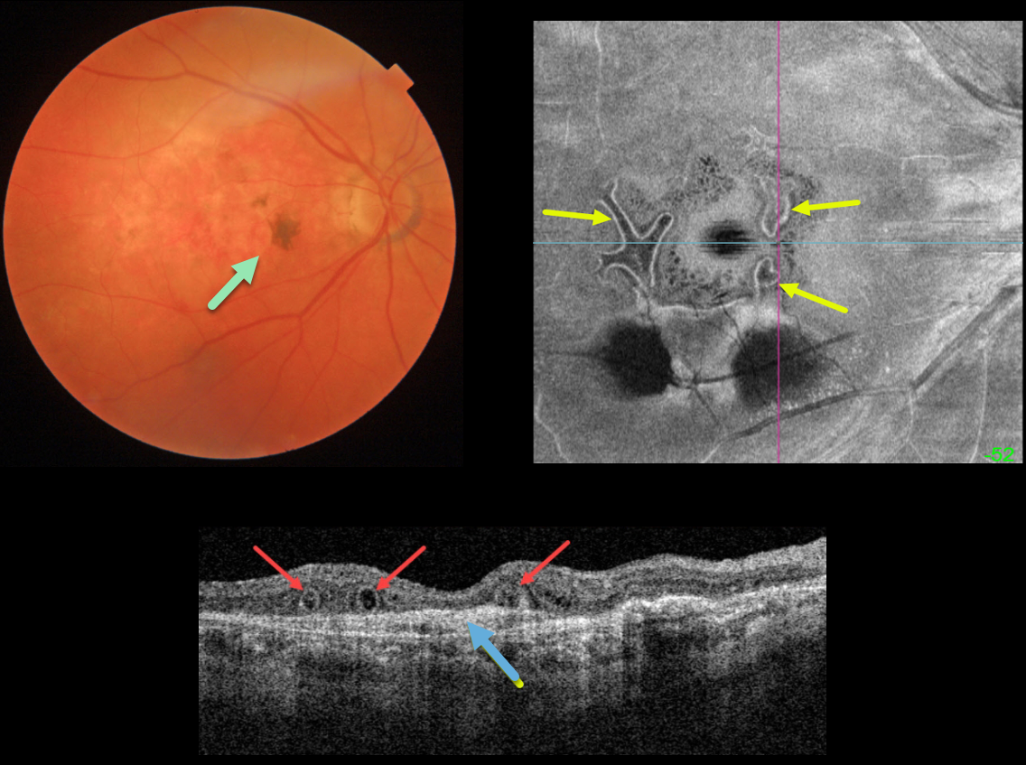
This is an 86YO female who has stable dry macular scarring from wet age-related macular degeneration with ongoing anti-VEGF injections.
Triton swept-source OCT shows numerous outer retinal tubulations (ORT) overlying an inactive type 1 macular neovascularization (MNV). These tubules are more dramatically seen with en face imaging of the outer retina.
Learning Points:
Outer retinal tubulation is often noted overlying inactive MNV with ongoing anti-VEGF therapy and should not be confused with exudative fluid or cysts, which lack a hyperreflective border.
ORT, initially described by Zweifel et al (Arch Ophthalmol 2009;127:1596-1602), is a neurodegenerative condition of the photoreceptors and Muller cells associated with atrophy affecting the outer retina and retinal pigment epithelium, including advanced AMD and inherited retinal diseases.
The outer hyperreflective band likely represents inner segment mitochondria undergoing fission and translocation toward the nucleus (Litts et al, Retina 2018;38:445-461).
MACULAR NEOVASCULARIZATION (MNV)
Originally posted on @retina.rocks September 14, 2021
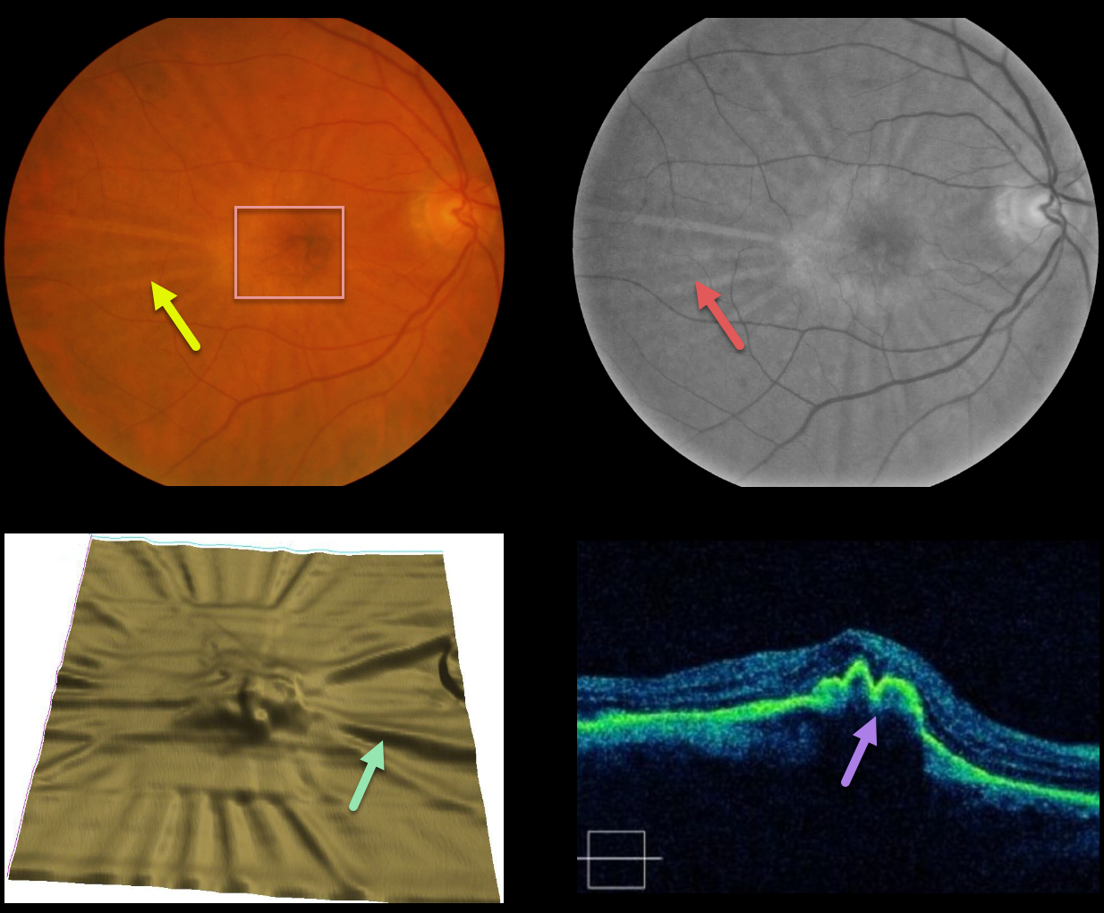
This patient developed radiating chorioretinal folds from an involuted type 1 macular neovascularization (MNV, located below the RPE) following multiple intravitreal Avastin injections.
These folds are imaged on the color and red-free photos, as well as on the OCT thickness map and B-scans. Remarkably, vision has been stable at 20/30 for years.
Learning Points:
Macular neovascularization is also known by other terms, including choroidal neovascular membrane and subretinal neovascular membrane.
However, since these vessels are now known not to necessarily arise just from the choroid, the proper term is macular neovascularization (see Consensus on Neovascular Age-Related Macular Degeneration Nomenclature Study Group Ophthalmology 2020;127;616-636).
MNV is classified according to the location of the new vessels in relation to the RPE and neurosensory retina: type 1 (below the RPE), type 2 (between the RPE and neurosensory retina, and type 3 (within the neurosensory retina).
RETINAL ANGIOMATOUS PROLIFERATION (RAP)
Originally posted on @retina.rocks August 18, 2021
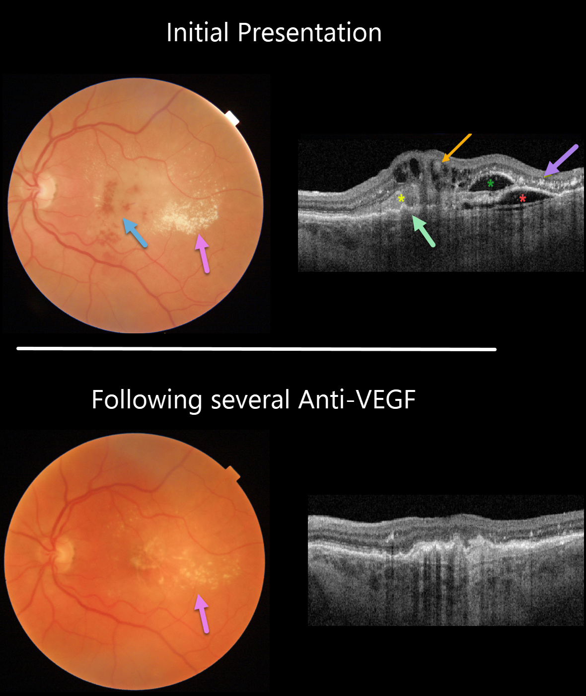
This 74YO man presented with counting fingers vision from previously untreated wet age-related macular degeneration (AMD).
Triton color imaging and swept-source OCT beautifully highlight this complex macular neovascularization (MNV). There are scattered inner and deep retinal hemorrhages, opaque fluid, and temporal lipid.
OCT shows an irregular shallow elevation of the RPE more nasally, possibly indicating a sub-RPE (type 1 MNV), with overlying subretinal hyperreflective material. More temporally, there is a more prominent area of RPE elevation that is more hyporeflective with an overlying pocket of intraretinal fluid vs a bacillary layer detachment. Finally, central inner retinal thickening and cysts are seen, indicating intraretinal neovascularization (type 3 MNV), also known as a retinal angiomatous proliferation (RAP) lesion. The lipid appears as hyperreflective clumps in the outer plexiform and outer nuclear layers.
The fluid and blood regressed, with significantly decreased lipid, after several monthly Avastin injections (image 2). Vision improved to 20/400.
Learning Points:
Unlike more typical age-related MNV that start either below (type 1 MNV) or occasionally above (type 2 MNV) the RPE, RAP lesions originate in the retina.
These incompetent vessels leak fluid and blood into the surrounding tissue. Their intraretinal location is a biomarker for RAP lesions.
NEOVASCULAR AMD
Originally posted on @retina.rocks January 14, 2021
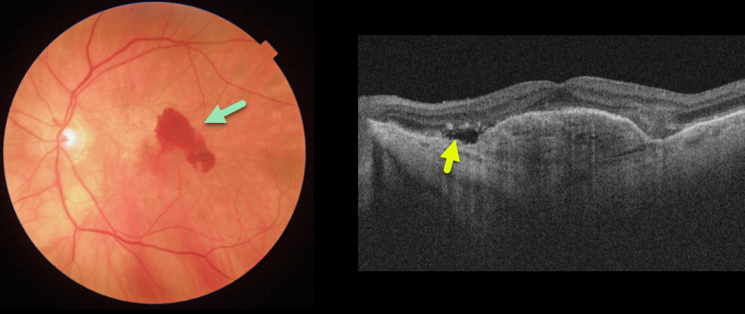
This patient with wet age-related macular degeneration (AMD) was being treated with PRN anti-VEGF injections. She came in for her regular examination without new symptoms and with 20/70 vision.
Although the OCT line scan shows some fairly subtle new sub-retinal fluid, the color photo dramatically shows new blood.
Learning Points:
There is currently some debate about the utility of performing full dilated examinations when patients are being followed with ongoing anti-VEGF therapy.
New blood is quite easy to miss on an OCT without a fundus photograph or examination. In our hands, having a high-quality fundus photograph at each visit, regardless of whether the patient is in for just an injection or a full examination, is invaluable.
RETINAL ANGIOMATOUS PROLIFERATION (RAP)
Originally posted on @retina.rocks January 5, 2021
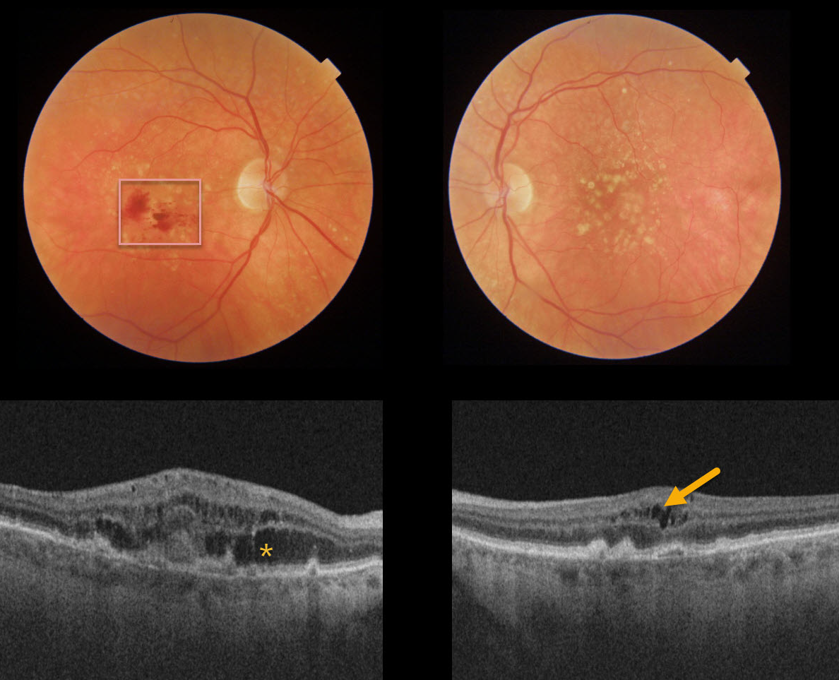
Our patient has age-related macular degeneration with a type 3 macular neovascularization (MNV), also known as retinal angiomatous proliferation (RAP).
Her fundus photo reveals inner retinal hemorrhages of her right eye and variable intraretinal fluid on OCT. Our patient showed no obvious exudation clinically, but the small amount of intraretinal fluid on OCT in the left eye indicates a subclinical RAP in this eye as well.
Bilateral anti-VEGF therapy was started, and we expect that she will do well.
Learning Points:
Unlike more typical age-related MNV that start either below (type 1 MNV) or occasionally above (type 2 MNV) the RPE, RAP lesions originate within the retina.
These incompetent vessels leak fluid and blood into the surrounding tissue.


