

AMD Peripheral Reticular Degeneration of the RPE (PRDRPE)
<< Back to Cases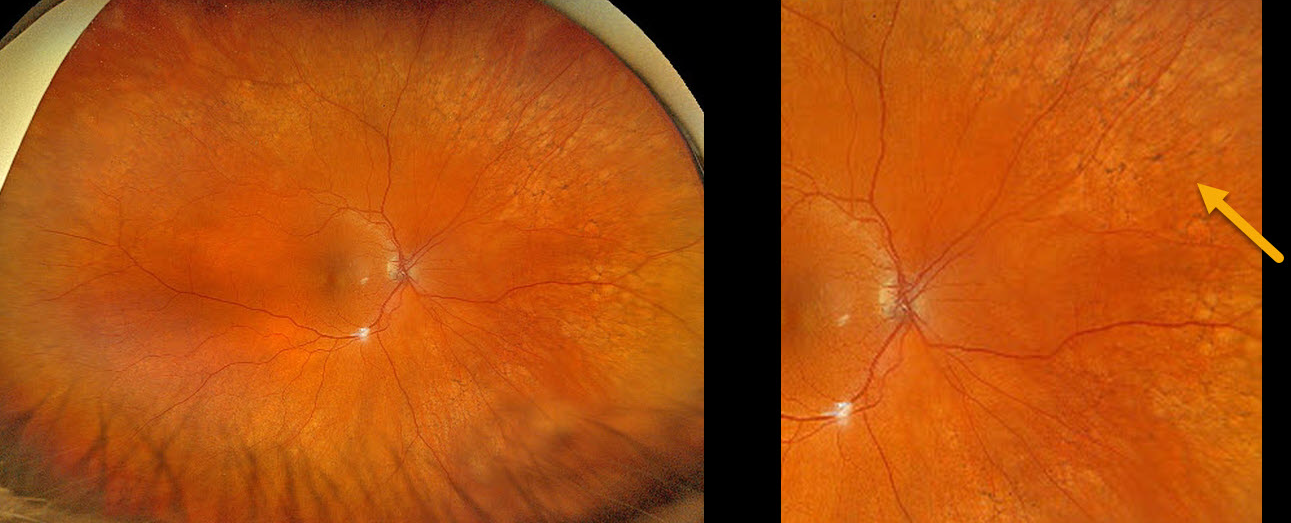
PERIPHERAL RETICULAR DEGENERATION OF THE RPE
Originally posted on @retina.rocks January 2, 2025
This 67YO female shows prominent bilateral peripheral reticular degeneration of the RPE (PRDRPE) with Optos ultra-widefield color RGB imaging.
Learning Points:
The reticular changes at first glance may appear like the ‘bone spicules’ found with retinitis pigmentosa, which are caused by disinhibited RPE cells migrating along retinal capillaries following photoreceptor degeneration. The findings in PRDRPE are part of the age-related macular degeneration (AMD) spectrum, are subretinal and located in the periphery.
A recent meta-analysis by Forshaw et al found PRDRPE to be present in 68-83% of eyes with AMD (Ophthalmology Retina 2019;3:734-743). It is associated with decreased dark adaptation, which may help explain why many patients with AMD have trouble with night vision (Lains et al, AJO 2018;190:113-124).
HRVO
Originally posted on @retina.rocks January 13, 2022
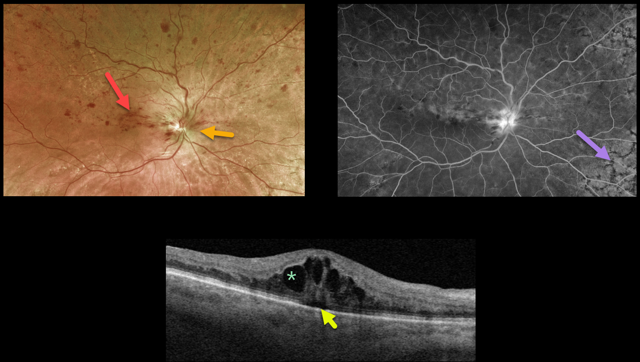
This 81YO female presented with 20/60 vision from a symptomatic superior hemiretinal vein occlusion (HRVO). Optos imaging shows foveal and superior inner retinal hemorrhages with a swollen optic nerve, and fluorescein angiography shows a well-perfused occlusion.
Swept-source OCT confirms moderate cystic macular edema with a trace amount of underlying subretinal fluid.
Age-related peripheral reticular degeneration of the RPE is best seen angiographically as an incidental finding.
The patient is receiving ongoing anti-VEGF injections to control her macular edema. Intravitreal injections (anti-VEGF or steroids) are the treatment of choice, although grid laser may help decrease the treatment burden.
Learning Points:
Branch retinal vein occlusions are virtually always caused by a retinal artery compressing a retinal vein within its common adventitial sheath.
Some people are born with an anomalous two-trunked central retinal vein that independently drains the superior and inferior retinal hemispheres.
HRVO, similar to central retinal vein occlusion, is caused by compression of the retinal vein posterior to the lamina cribrosa (see Hayreh, Arch Ophthalmol 1980;98:1600-1609).
PERIPHERAL RETICULAR DEGENERATION OF THE RPE
Originally posted on @retina.rocks June 22, 2021
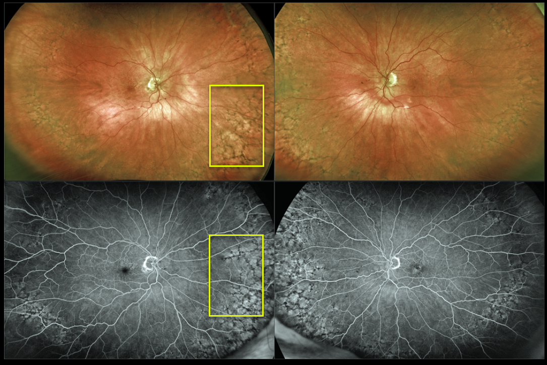
This 87YO female shows prominent bilateral peripheral reticular degeneration of the RPE (PRDRPE) with Optos ultra-widefield imaging and fluoroscein angiography.
The reticular changes at first glance may appear similar to the ‘bone spicules’ found with retinitis pigmentosa, which are caused by disinhibited RPE cells migrating along retinal capillaries following photoreceptor degeneration.
Learning Points:
The findings in PRDRPE are part of the age-related macular degeneration (AMD) spectrum and are subretinal and located in the periphery. A recent meta-analysis by Forshaw et al found PRDRPE to be present in 68-83% of eyes with AMD (Ophthalmology Retina 2019;734-743).
It is associated with decreased dark adaptation, which may help explain why many patients with AMD experience difficulty with night vision (see Lains et al, AJO 2018;190;113-124).


