

AMD RPE Detachment
<< Back to Cases
FLATTENED DRUSENOID PED CAUSING SECONDARY MACULAR ATROPHY
Originally posted on @retina.rocks June 23, 2026
This 72YO female has been followed for years with intermediate AMD. On 3/26/25, she remained asymptomatic with 20/40 vision.
Color photography shows confluent central drusen creating a drusenoid PED with a radiating pigment figure, surrounded by large soft drusen. The drusenoid RPE detachment (PED) is confirmed on swept-source OCT. The RPE and outer retinal bands are obscured by subretinal hyperreflective material. She returned 6 months later with stable vision and no new symptoms. The PED is mostly flattened with some resolution of the hyperreflective material.
Learning Points:
Drusen commonly remodel over time and their collapse, as in our patient, can lead to drusenoid atrophy of the RPE (Filho et al, Retina 2013;33;1558-1566). Vision may or may not decrease following drusen regression. In our experience, drusenoid atrophy remains stable, unlike primary age-related macular atrophy, whose radius enlarges linearly over time (Shen et al, Ophthalmology Retina 2020;4;899-910). We therefore do not consider intravitreal complement inhibitors for these patients.
INTERMEDIATE AMD WITH DRUSENOID PEDS
Originally posted on @retina.rocks January 8, 2026
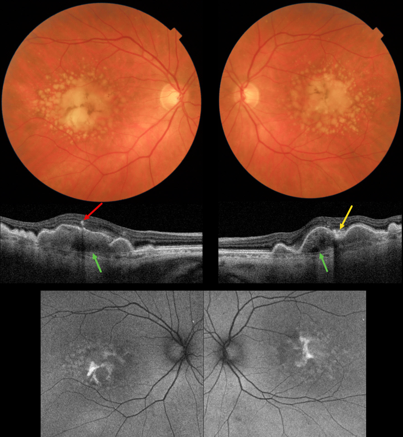
This 72YO female presented for her 6-month AMD examination. She was visually asymptomatic, and vision was 20/40 OU.
Triton color imaging shows bilateral chronic foveal RPE detachments (PEDs) with surrounding soft drusen. A radiating pigment figure overlies each PED. Swept-source OCT shows hyperreflective material beneath each PED, with some overlying hyperreflective foci and hyperreflective material likely representing lipofuscin. Bruch membrane is visible beneath the PED and surrounding soft drusen. Fundus autofluorescence (FAF) shows variable hyper-FAF from the pigment figures and lipofuscin.
Learning Points:
RPE detachments are most commonly associated with AMD and the central serous-pachychoroid spectrum. In cases of AMD, lesions can be avascular (as in our patient) or vascularized as part of a type 1 neovascular complex (below the RPE). AMD-related PEDs can form primarily or as a confluence of soft drusen. When they collapse, they may lead to secondary macular atrophy (Yu et al, Ophthalmology 2019;126:261-273).
The radiating pigment figure, described originally by Dr. Gass, is pathognomonic for a chronic PED and persists even after a PED flattens. Avascular PEDs are typically not treated. In contrast, anti-VEGF therapy is both safe and effective for vascularized PEDs, despite the risk of a RPE tear (Khanani et al, Retina 2018;38;2103-2117).
OCCULT MNV
Asma Samsudeen and Ashish Sharma
Originally posted on @retina.rocks November 23, 2023
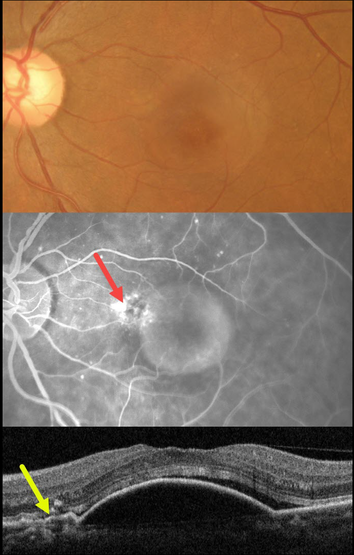
This 62YO female presented with 3 weeks of vision loss in her left eye. Vision was 20/30.
Color imaging shows a large central macular retinal pigment epithelial detachment (PED). Fluorescein angiography shows a somewhat unevenly filling PED, with staining vs leakage along its superonasal edge. OCT scanning shows a large PED, shallow overlying temporal subretinal fluid, and shallow irregular elevation of the RPE nasally indicative of a type 1 macular neovascularization (MNV).
The uneven angiographic pooling is also suggestive of an occult MNV. Anti-VEGF therapy was advised.
This case was submitted by Asma Samsudeen and Ashish Sharma.
Learning Points:
Originally described by Gass (Retina 1984;4:205-220), a notched PED is a sign of an occult MNV. The notch represents a type 1 MNV, which tethers the overlying RPE from the surrounding more highly elevated PED. Hydrostatic leakage from the MNV may contribute to the larger PED.
Although now considered a classic clinical and angiographic finding, in 1984 this was evidently not the case, as the reviewers for this article advised against publication. Based solely on who the contributing author was, the journal’s editor overruled his reviewers, published Dr. Gass’ manuscript, and the rest was literally history!
NOTCHED PED
Originally posted on @retina.rocks September 27, 2023
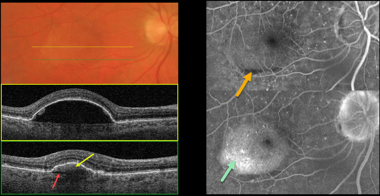
This 64YO male presented with recent central vision loss in his right eye. Vision was 20/40.
Color imaging shows a large central macular retinal pigment epithelial detachment (PED). Triton swept-source OCT through the central portion of the PED shows underlying hyporeflectivity. Scanning through the inferior portion shows a hyperreflective type 1 macular neovascularization (MNV) with a thin underlying prechoroidal cleft.
Optos fluorescein angiography shows an inferior linear filling defect within the MNV. The PED also shows late uneven pooling.
Learning Points:
Originally described by Gass (Retina 1984;4:205-220), a notched PED is a sign of an occult MNV. The notch represents a type 1 MNV, which tethers the overlying RPE from the surrounding more highly elevated PED. Hydrostatic leakage from the MNV may contribute to the larger PED.
Although now considered a classic clinical and angiographic finding, in 1984 this was evidently not the case, as the reviewers for this article advised against publication.
Based solely on who the contributing author was, the journal’s editor overruled his reviewers, published Dr. Gass’ manuscript, and the rest was literally history! The uneven angiographic pooling is also suggestive of an occult MNV.
A prechoroidal cleft is a hyporeflective space between the RPE and Bruch’s membrane. These are seen in up to about 20% of treated eyes with nAMD, particularly with type 3 neovascularization (intraretinal MNV) and polypoidal choroidal vasculopathy.
They are usually associated with worse visual acuity due to potential complications, including RPE tears and subretinal hemorrhage (Kim et al, Retina 2017;37:2047-2055).
Anti-VEGF therapy was recommended, and the patient was warned of the possibility of RPE development with or without treatment (Ersoz et al., Surv Ophthalmology 2017;62:493-505).
MACULAR HOLE AFTER COLLAPSE OF PED
Originally posted on @retina.rocks July 25, 2023
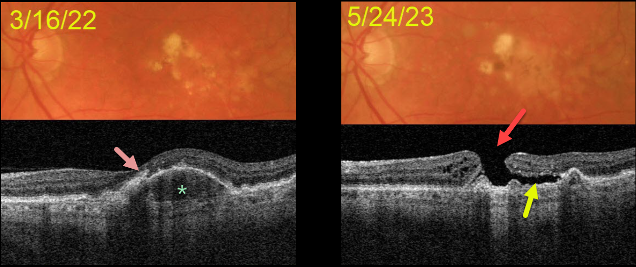
This 72YOM was examined with asymptomatic intermediate AMD bilaterally. Vision was 20/40 OD and 20/50 OS.
Color imaging of his left eye initially shows mostly large, soft drusen and pigment clumps. Swept source OCT shows a RPE detachment (PED) with overlying hyperreflective foci.
He returned over a year later without new symptoms, despite vision dropping to counting fingers. OCT shows a large macular hole overlying the collapsed PED. Shallow subretinal fluid lies below the temporal aspect of the hole.
The development of a macular hole following spontaneous collapse of a PED is quite unusual. Fortunately, vision in his right eye remains at 20/40 with intermediate dry AMD.
Since our patient was completely asymptomatic, he did not want macular hole surgery, and we will monitor him twice yearly.
LAMELLAR HOLE AFTER AMD PIGMENT EPITHELIAL DETACHMENT (PED) COLLAPSE
Originally posted on @retina.rocks December 30, 2022
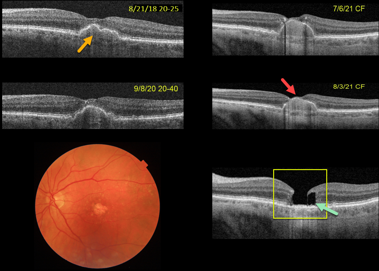
This 75YOF was seen in 2018 with non-neovascular AMD and a central pigment epithelial detachment (PED). Vision was 20/25. She returned on 9/8/20 with 20/40 vision with mild enlargement of the PED.
She complained of decreased vision when seen nearly one year later. Vision had dropped to 20/80. The PED had enlarged to involve full-thickness retina. She elected for an Eylea injection in hopes of possibly treating an occult macular neovascularization vs an avascular PED.
On 7/6/21, vision decreased to counting fingers although the OCT looked fairly stable. An additional Eylea injection followed by half-fluence PDT was given. On 8/3/21, the dome of the PED now extended through the inner retina and vision remained at counting fingers.
On 1/4/22, the PED had completely collapsed, with secondary retinal atrophy and a degenerative lamellar macular hole (LMH).
The progression of her PED through full-thickness retina and subsequent collapse with secondary retinal/RPE atrophy and a LMH are all quite unusual. Fortunately her right eye is 20/20 with intermediate dry AMD.
PACHYCHOROID PIGMENT EPITHELIAL DETACHMENT (PED)
Originally posted on @retina.rocks September 13, 2022
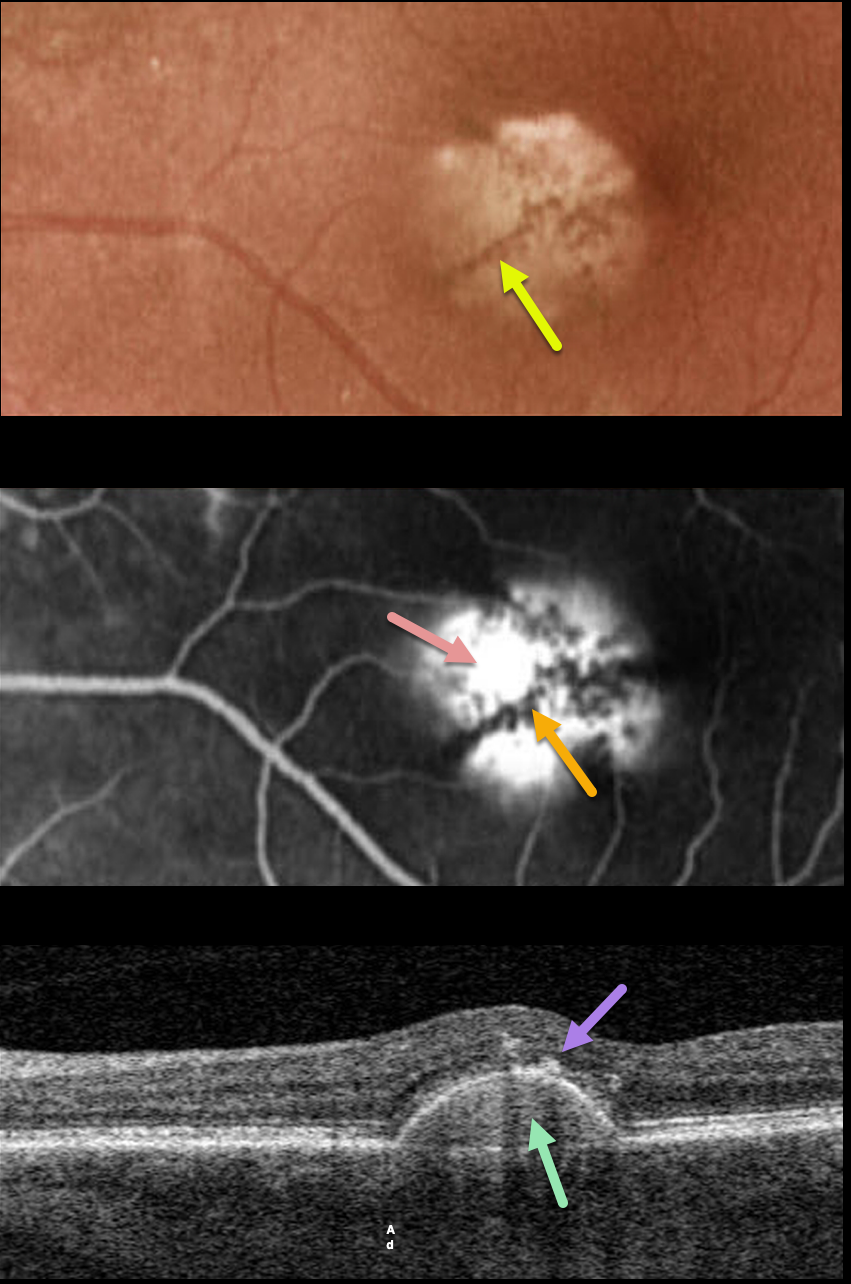
This 75YO female has been followed for several years with a stable, asymptomatic chronic retinal pigment epithelial detachment (PED) and 20/40 vision.
Optos color imaging shows a solid, variably depigmented, round PED in the inferonasal region of the left fovea. Radiating linear pigmentation is noted.
Fluorescein angiography shows hyperfluorescent pooling beneath the PED, with blockage from the overlying radiating pigment.
Swept-source OCT shows the PED with a hyperreflective core, overlying hyperreflective foci (representing the clinical radiating pigment), and a relatively thickened choroid for a patient of this age.
Learning Points:
Retinal pigment epithelial detachments are common in patients with AMD and are often associated with macular neovascularization. Non-vascularized PED is also seen as part of the pachychoroid spectrum.
The overlying pigment figure is pathognomonic for a chronic PED, and is seen even when these lesions flatten. Isolated avascular PEDs such as these do not require treatment.
Although our patient was in her mid-70s, there were no drusen and each fundus was otherwise normal. The chronicity of the PED, the lack of other signs of AMD, and the relatively thickened choroid all point to this being an avascular, pachychoroid PED.
PIGMENT EPITHELIAL DETACHMENT (PED)
Originally posted on @retina.rocks April 26, 2021
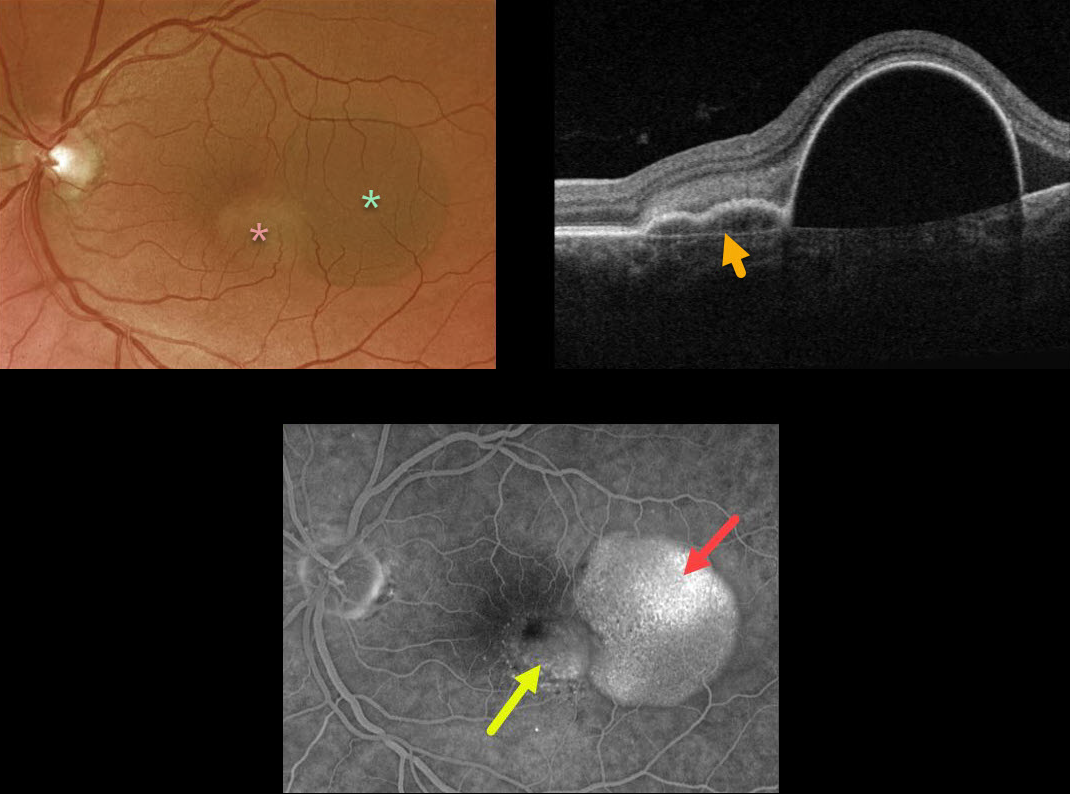
This 57YO female presented with counting-finger vision from a large RPE detachment (PED). The PED was located in the temporal macula, with a smaller notch of irregular RPE elevation more centrally.
Triton swept-source OCT shows a shallow PED at the notch overlying Bruch’s membrane. Optos fluorescein angiography shows late uneven filling of the PED and late speckled staining of the notch.
Anti-VEGF therapy was recommended.
Learning Points:
Originally described by Gass (Retina 1984;4:205-220), a notched PED is a sign of an occult macular neovascularization (MNV). The notch represents a type 1 MNV, which tethers the overlying RPE from the surrounding more highly elevated PED. Hydrostatic leakage from the MNV may contribute to the larger PED.
PIGMENT EPITHELIAL DETACHMENT (PED)
Originally posted on @retina.rocks October 13, 2020
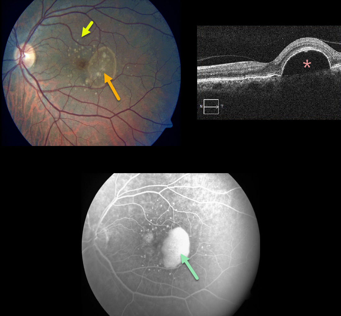
This patient presented with 20/25 vision in her left eye with significant Amsler grid changes. There is an irregular RPE detachment (PED) with variably sized drusen, and OCT confirms a large PED.
Fluorescein angiography shows a slightly uneven pattern of pooling beneath the PED.
We discussed starting treatment due to her symptoms, but for now, we have elected for close observation. There is a risk for an RPE tear with or without treatment
Learning Points:
PEDs associated with AMD are often associated with macular neovascularization (MNV).
Angiographic clues for MNV include a notched PED or uneven angiographic filling. Fluorescein angiography is probably still better than OCT angiography for detecting MNV.
Anti-VEGF therapy can effectively flatten these PEDs even when they are presumably avascular.
PIGMENT EPITHELIAL DETACHMENT (PED)
Originally posted on @retina.rocks September 30, 2020
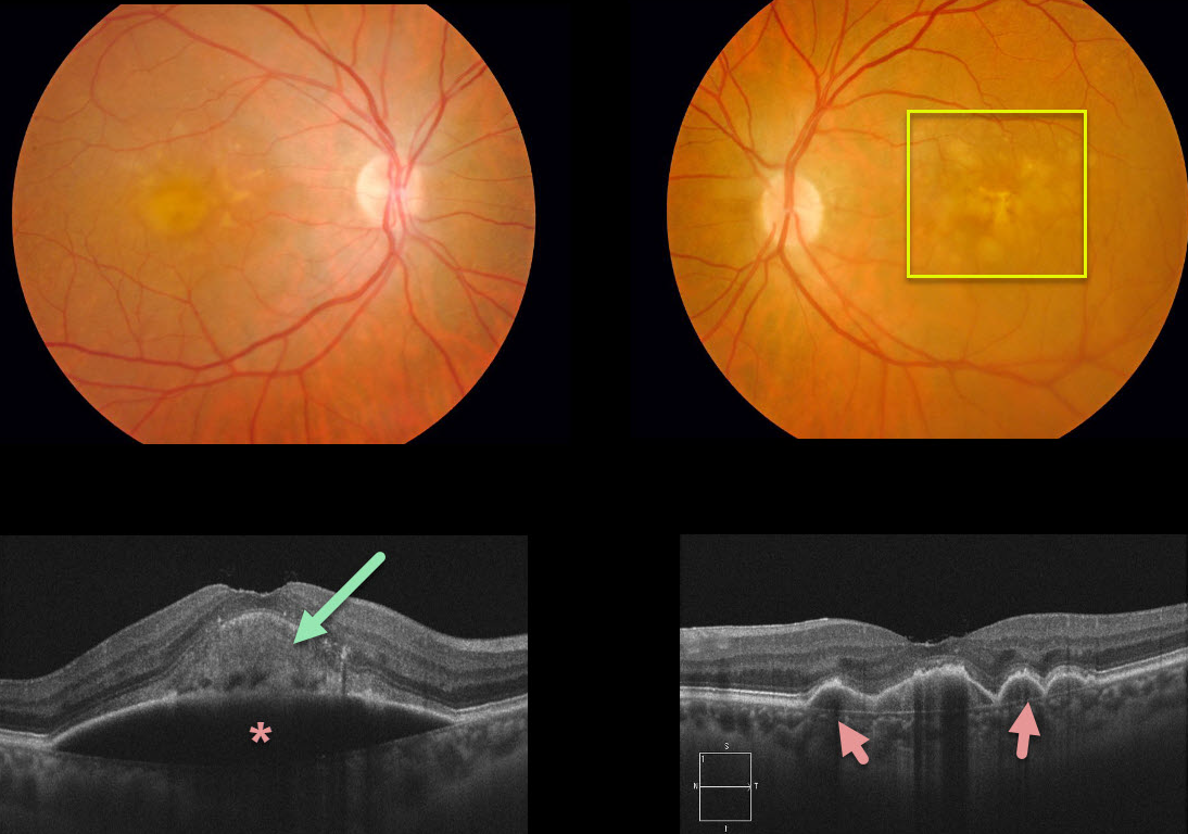
This patient had been followed for age-related macular degeneration (AMD) with bilateral confluent soft drusen. She then developed a large acquired vitelliform lesion overlying a retinal pigment epithelial detachment (PED) in her right eye. There are several PEDs also shown in the left eye.
Learning Points:
Vitelliform lesions are commonly found with other disorders, including retinal dystrophies (Best’s disease and macular dystrophies), cuticular drusen, AMD, and vitreomacular traction.
The yellow subretinal material is felt to be debris from shed photoreceptor outer segments. These lesions markedly autofluoresce on fundus autofluorescence.
A great reference to learn more about AVLs is Freund et al Retina 2011;31:13-25.


