

AMD RPE Tear
<< Back to Cases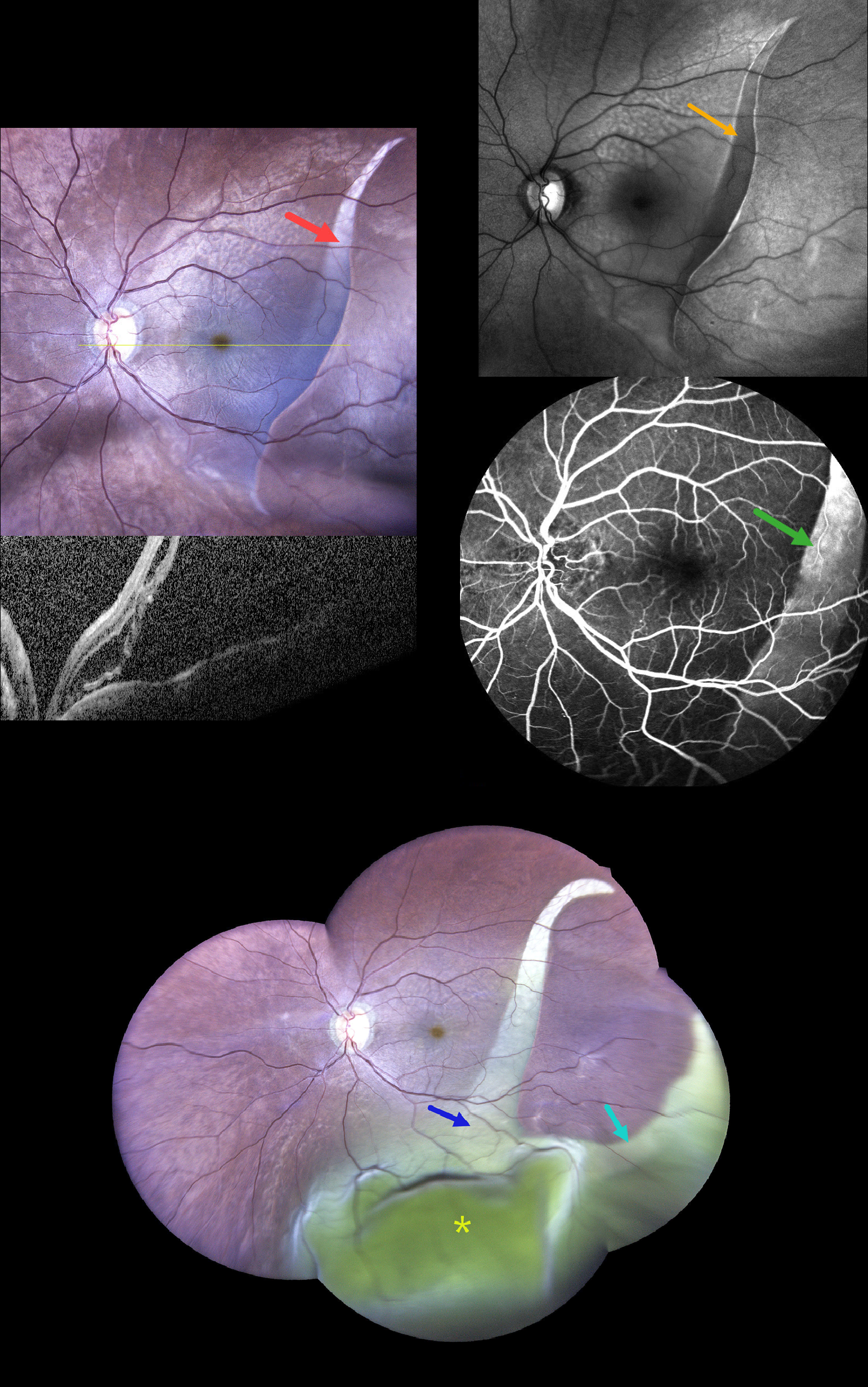
SEQUENTIAL GIANT RPE TEAR WITH SECONDARY EXUDATIVE RETINAL DETACHMENT
Rohan Suresh Ninan
Originally posted on @retina.rocks August 21, 2026
This 68YO male presented with sudden, painless decreased vision in his left eye 2 days ago. He gave a history of type 2 diabetes. Vision was 20/30 (6/9) in his normal OD and hand motion OS.
Widefield photography shows a large curvilinear area of pigment loss (giant RPE tear) extending from the superotemporal into the inferior midperiphery. OCT scanning shows marked macular subretinal fluid. On fundus autofluorescence (FAF), the lesion is hypo-FAF with a corresponding window defect on fluorescein angiography.
He returned 6 days later without new symptoms and stable vision. The prior RPE tear is enlarged in width, extends more inferiorly, and has developed a new branch extending into the inferotemporal midperiphery. A large inferior exudative retinal detachment is noted. Unfortunately, he was immediately lost to follow-up.
Learning Points:
A tear of the RPE is most often seen in RPE detachments associated with neovascular AMD, but can also occur in numerous other etiologies, including idiopathic central serous chorioretinopathy (ICSC) and polypoidal choroidal vasculopathy. The tear leaves a depigmented region where the RPE was and is now absent, along with a pigmented, relatively straight subretinal lesion from the scrolled RPE edge. Our patient’s RPE tear is atypical due to the lack of this hyperpigmented edge, indicating it is more of an RPE dehiscence than a scrolled tear, as well as the multibranched extension that developed 1 week later.
Giant retinal pigment epithelial (RPE) tears occurring outside the macula, defined as tears exceeding 3 clock hours, are most common following trauma or in bullous ICSC and polypoidal choroidal vasculopathy. We have seen these giant tears in previously clinically normal eyes, which later showed pachychoroid-related findings on ICG angiography and en face OCT.
RPE TEAR
Originally posted on @retina.rocks February 2, 2026
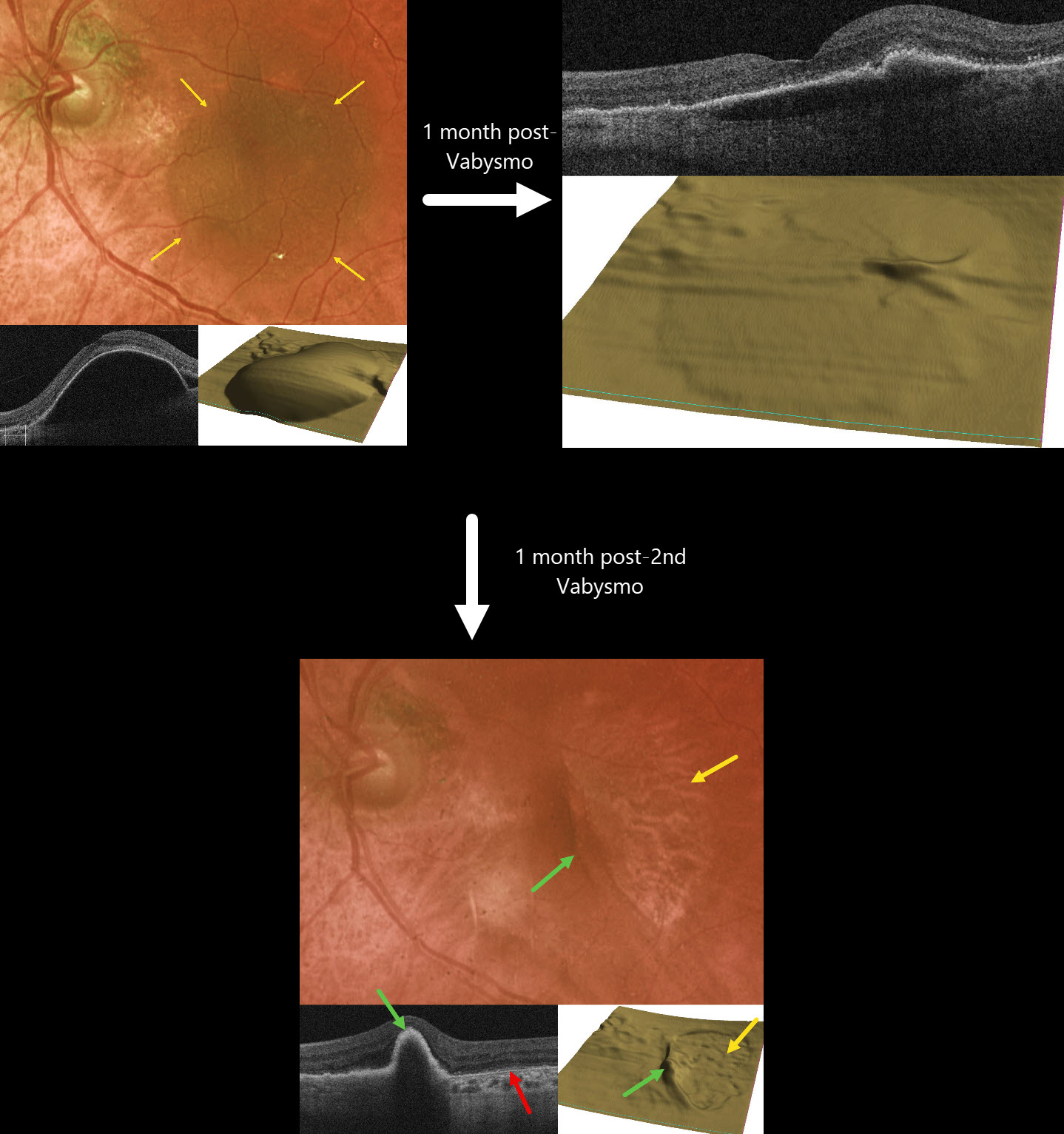
This 77YO female presented with recent vision loss in her left eye from new neovascular AMD. Vision was 20/400.
Optos color RG imaging shows a large pigmented retinal pigment epithelial detachment (PED) occupying most of the left macula. OCT scanning, B-scan, and RPE map confirm this large PED. Intravitreal Vabysmo was injected, and one month later, vision improved to 20/40. The PED nearly flattened.
Another Vabysmo injection was given, and one month later, she complained of some new vision loss. Vision was 20/80. A new RPE tear is noted, with bare RPE temporally and the retracted RPE nasally. Bare Bruch membrane is seen within the bed of the torn RPE. Vabysmo was injected, and treat-and-extend was begun.
Learning Points:
A tear of the RPE is a fairly common finding in RPE detachment (PED) associated with type 1 macular neovascularization (MNV, located below the RPE). Neovascularization can spontaneously contract or involute following treatment. It then puts traction on the overlying RPE, which can then rip at the opposite edge of the PED. Like a cheap window blind, the torn RPE retracts and scrolls towards the MNV. This leaves a depigmented region where the RPE was and is now absent, along with a pigmented, relatively straight subretinal lesion from the scrolled RPE edge. Anti-VEGF therapy can safely be continued following an RPE tear, with long-term visual outcome more closely related to the patient’s response to therapy than to the tear itself (Ophthalmology 2018;125:236-244), provided the tear is outside the foveal center.
RPE TEAR
Originally posted on @retina.rocks October 14, 2025
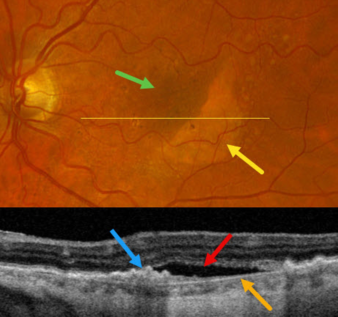
This 71YO female complained of one month of decreased vision in her left eye. Vision was 20/30 OD and 20/200 OS.
Moderate larger drusen with pigment clumps were noted in her right eye (not shown). Optos color RGB imaging of her left eye shows a crescent of subretinal pigment loss in the inferotemporal macula with increased subretinal pigment superonasally. OCT scanning within the area of pigment loss shows bare Bruch membrane with overlying subretinal fluid. The RPE nasally is retracted and bunched onto itself.
Learning Points:
A tear of the RPE is most seen in RPE detachments (PEDs) associated with neovascular AMD, but can also occur in numerous other etiologies, including central serous chorioretinopathy and polypoidal choroidal vasculopathy. The tear leaves a depigmented region where the RPE was and is now absent, along with a pigmented, relatively straight subretinal lesion from the scrolled RPE edge.
Our patient may harbor a macular neovascularization beneath the retracted RPE. A therapeutic trial of monthly intravitreal Avastin was thus started.
NEOVASCULAR AMD WITH RAPIDLY RESOLVED SHRM FOLLOWING RPE TEAR
Ayushi Gupta and Vishal Agrawal
Originally posted on @retina.rocks August 18, 2025
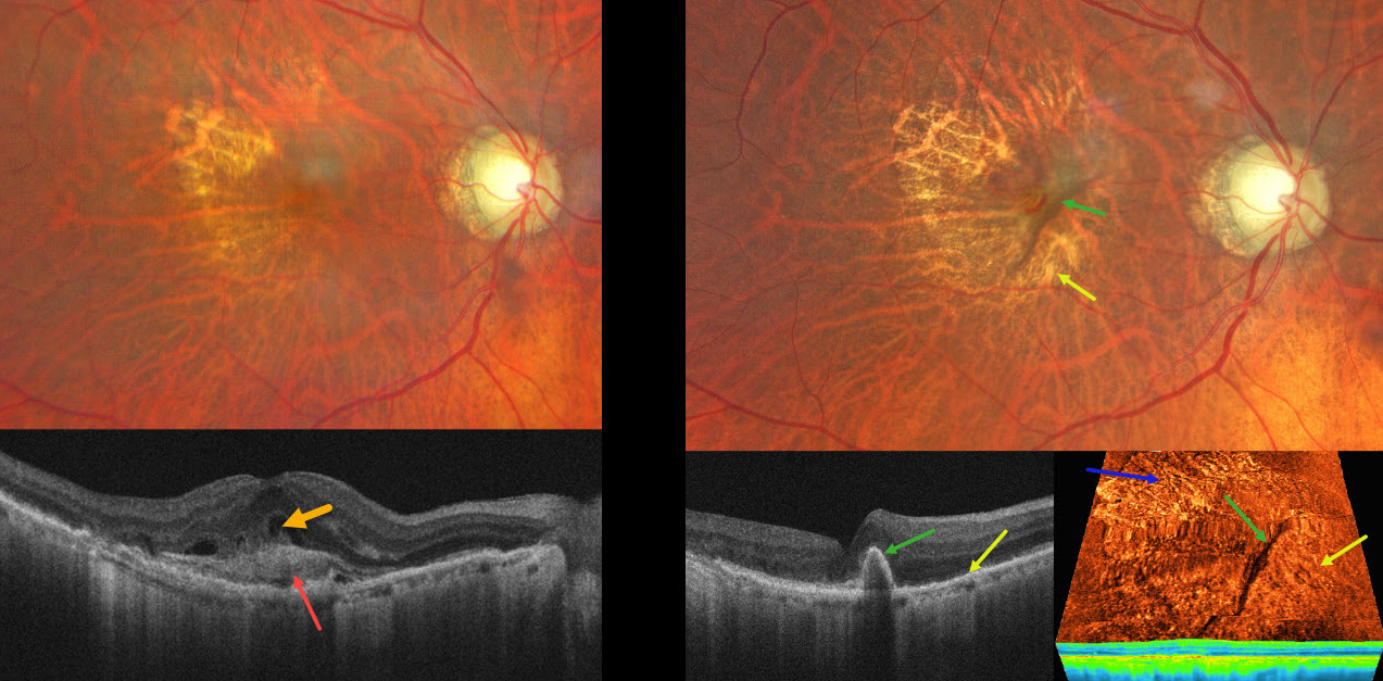
This 82YO male presented with recent vision loss in his right eye. Vision was 20/200.
Color photography shows a poor foveal reflex with superotemporal geographic atrophy. OCT scanning shows subretinal hyperreflective material (SHRM) with overlying retinal fluid. A possible bacillary layer detachment is also noted. About 6 weeks following a single intravitreal anti-VEGF injection, vision improved to 20/50. A new RPE tear is noted, with the bed of the tear inferonasal to the retracted hyperpigmented RPE. Despite persistent flow signals on OCT angiography, there is no fluid on structural OCT. The SHRM has completely resolved. A treat-and-extend anti-VEGF regimen was begun.
Learning Points:
A tear in the RPE is a fairly common finding in RPE detachment (PED) associated with type 1 macular neovascularization (MNV) located below the RPE. The neovascularization can spontaneously contract or involute following treatment. It then puts traction on the overlying RPE, which can then rip at the opposite edge of the PED. Like a cheap window blind, the torn RPE retracts and scrolls towards the MNV. This leaves a depigmented region where the RPE was and is now absent, along with a pigmented, relatively straight subretinal lesion from the scrolled RPE edge.
SHRM is an OCT finding that is invisible clinically. This material, located between the neurosensory retina and RPE, is thought to consist of fluid, fibrin, blood, and other fibrovascular tissues. SHRM is a biomarker of active disease that decreases with successful anti-VEGF therapy (Willoughby et al, Ophthalmology 2015;122:1846-1853). Persistent SHRM is associated with an increased incidence of scar formation and worse vision.
It is impossible to determine whether the anti-VEGF injection alone or in combination with the RPE tear caused such a rapid resolution of the SHRM. Fortunately, our patient’s vision remained quite good since the RPE tear just spared the foveal center.
RPE TEAR
Originally posted on @retina.rocks February 26, 2024
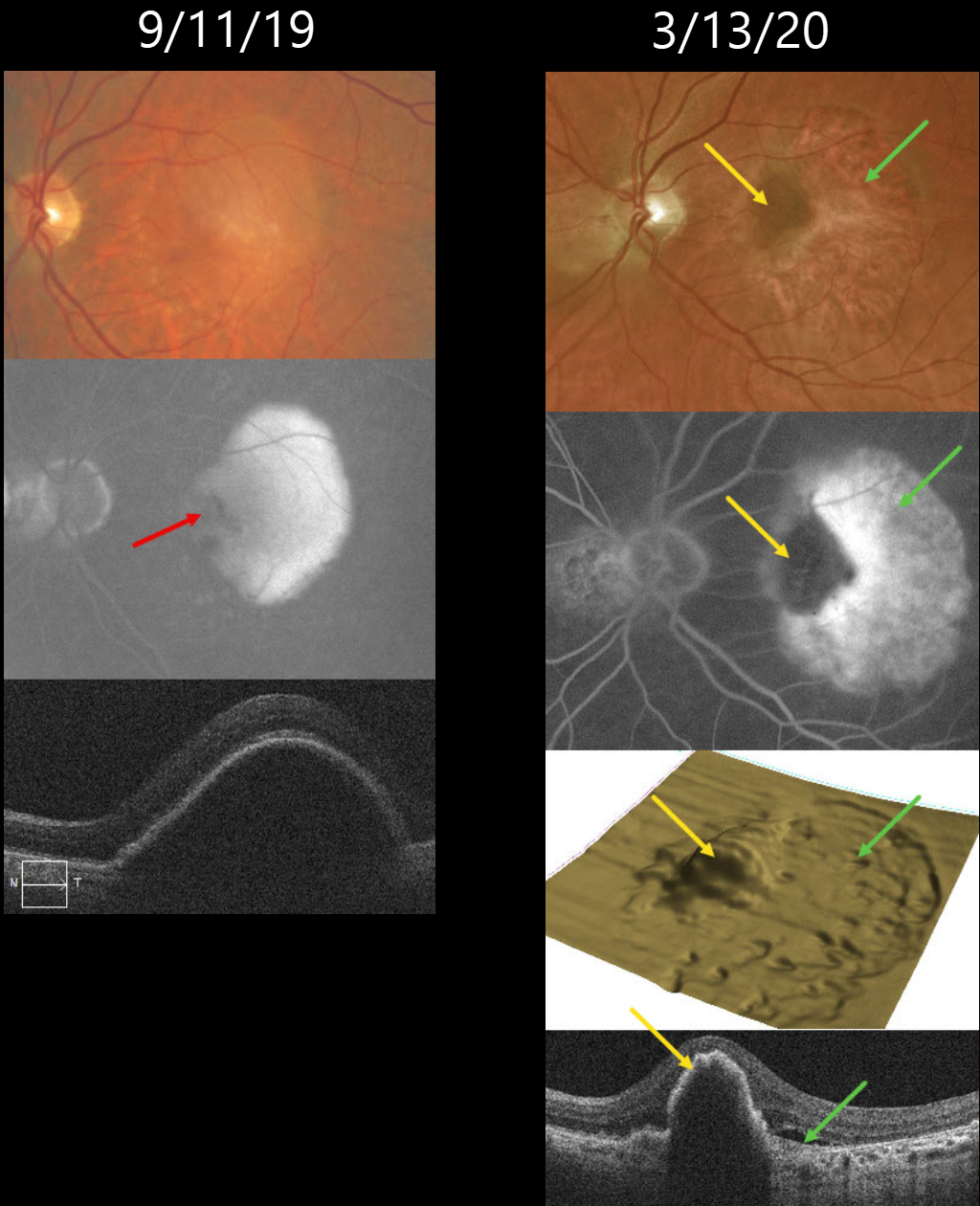
This 72YO male was followed for many years with stable intermediate AMD. On 9/11/19, he presented with a new symptomatic retinal pigment epithelial detachment (PED) with 20/30 vision. Fluorescein angiography shows an uneven-filling PED with a nasal hypofluorescent notch. A large PED is confirmed on OCT scanning.
He initially did well with monthly Eylea injections. While on treat-and-extend therapy, he presented on 3/13/20 without new visual symptoms, although his vision had decreased to 20/200.
Optos color RG imaging shows a new, large temporal RPE tear. On multimodal imaging, the bed of the tear is depigmented on color imaging, hyperfluorescent on fluorescein angiography (window defect), and shows a crater on the 3D OCT RPE map, and shows bare Bruch’s membrane on B-scan.
The retracted edge of the tear is hyperpigmented on color imaging, hypofluorescent on fluorescein angiography (blockage), an elevated mound on the 3D OCT RPE map, and a rippled, elevated PED on B-scan.
We subsequently decided on PRN therapy, with the last injection given on 3/12/21. When last examined, vision was stable at 20/400 with a stable dry lesion.
Learning Points:
A tear in the RPE is a fairly common occurrence in PED associated with type 1 macular neovascularization (MNV) located below the RPE. The neovascularization can spontaneously contract or involute following treatment. It then puts traction on the overlying RPE, which can then rip at the opposite edge of the PED.
Like a cheap window blind, the torn RPE retracts and scrolls towards the MNV. This leaves a depigmented region where the RPE was and is now absent, along with a pigmented, relatively straight subretinal lesion from the scrolled RPE edge.
Originally described by Gass (Retina 1984;4:205-220), a notched PED is a sign of an occult MNV. The notch represents a type 1 MNV, which tethers the overlying RPE from the surrounding more highly elevated PED. Hydrostatic leakage from the MNV may contribute to the larger PED.
RPE TEAR
Originally posted on @retina.rocks June 14, 2023
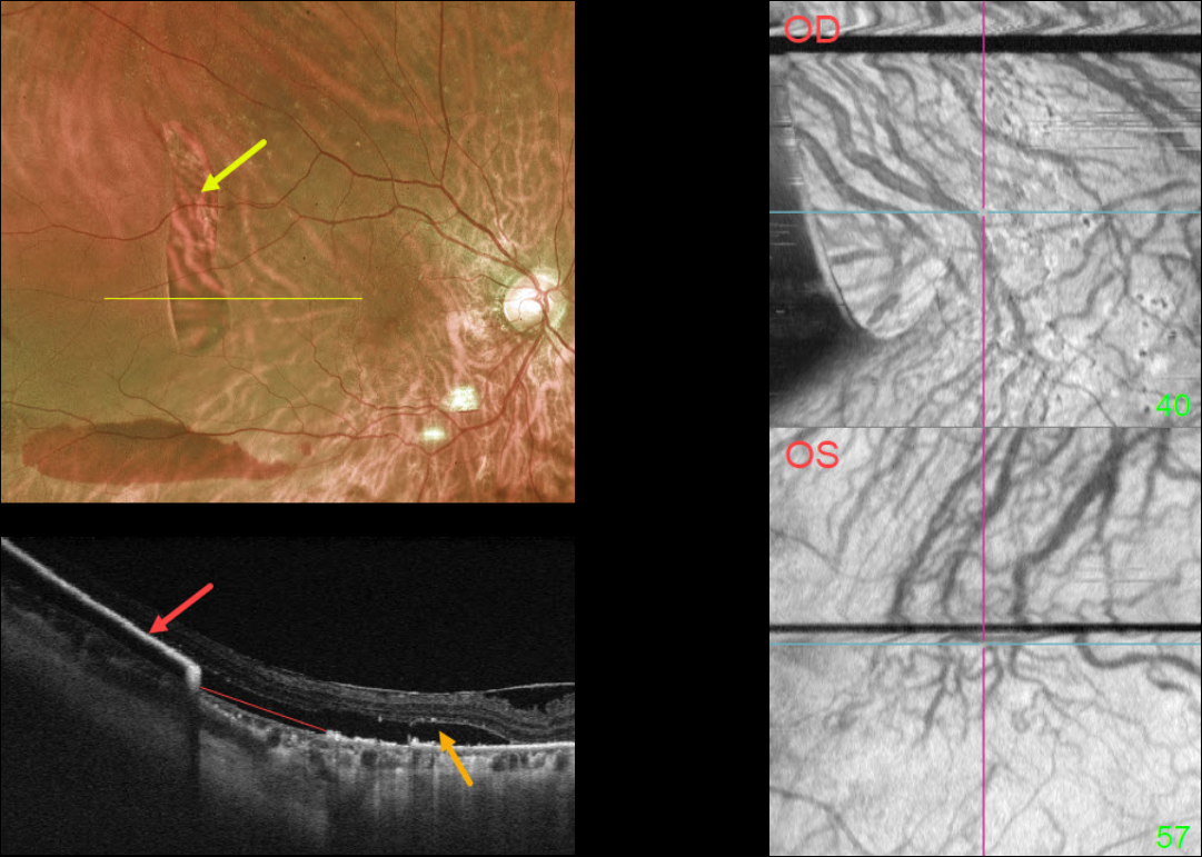
This 73YO female presented with acute vision loss in her right eye. Vision was 20/40 OD and 20/30 OS. There was a several-year history of type 2 diabetes with mild bilateral nonproliferative diabetic retinopathy.
Optos imaging shows a large vertical area of depigmentation in the distal superotemporal macula. There is a serous detachment surrounding this region, along with a far inferior subretinal hemorrhage.
Swept-source OCT through this lesion shows bare Bruch’s membrane from an RPE tear. The retracted RPE is markedly hyperreflective more temporally. Subretinal fluid overlies this region.
Macular choroidal en face imaging shows bilateral anomalous vasculature with choroidal anastomoses.
Learning Points:
A tear of the RPE is most commonly seen in RPE detachments (PEDs) associated with neovascular AMD, but can also occur in numerous other etiologies, including central serous chorioretinopathy and polypoidal choroidal vasculopathy.
The tear leaves a depigmented region where the RPE was and is now absent, along with a pigmented, relatively straight subretinal lesion from the scrolled RPE edge.
The choroidal en face OCT findings are more consistent with the pachychoroid spectrum. RPE tears are found in polypoidal choroidal vasculopathy, another pachychoroid disorder (Zhao et al, Retina 2020;40:477-489).
See Ersoz et al for a great review on RPE tear classification, pathogenesis, predictors, and management (Surv Ophthalmology 2017;62:493-505).
GIANT RPE TEAR
Anjana Mirajkar, Manish Nagpal, Navneet Mehrotr, and Akansha Sharma
Originally posted on @retina.rocks January 20, 2023
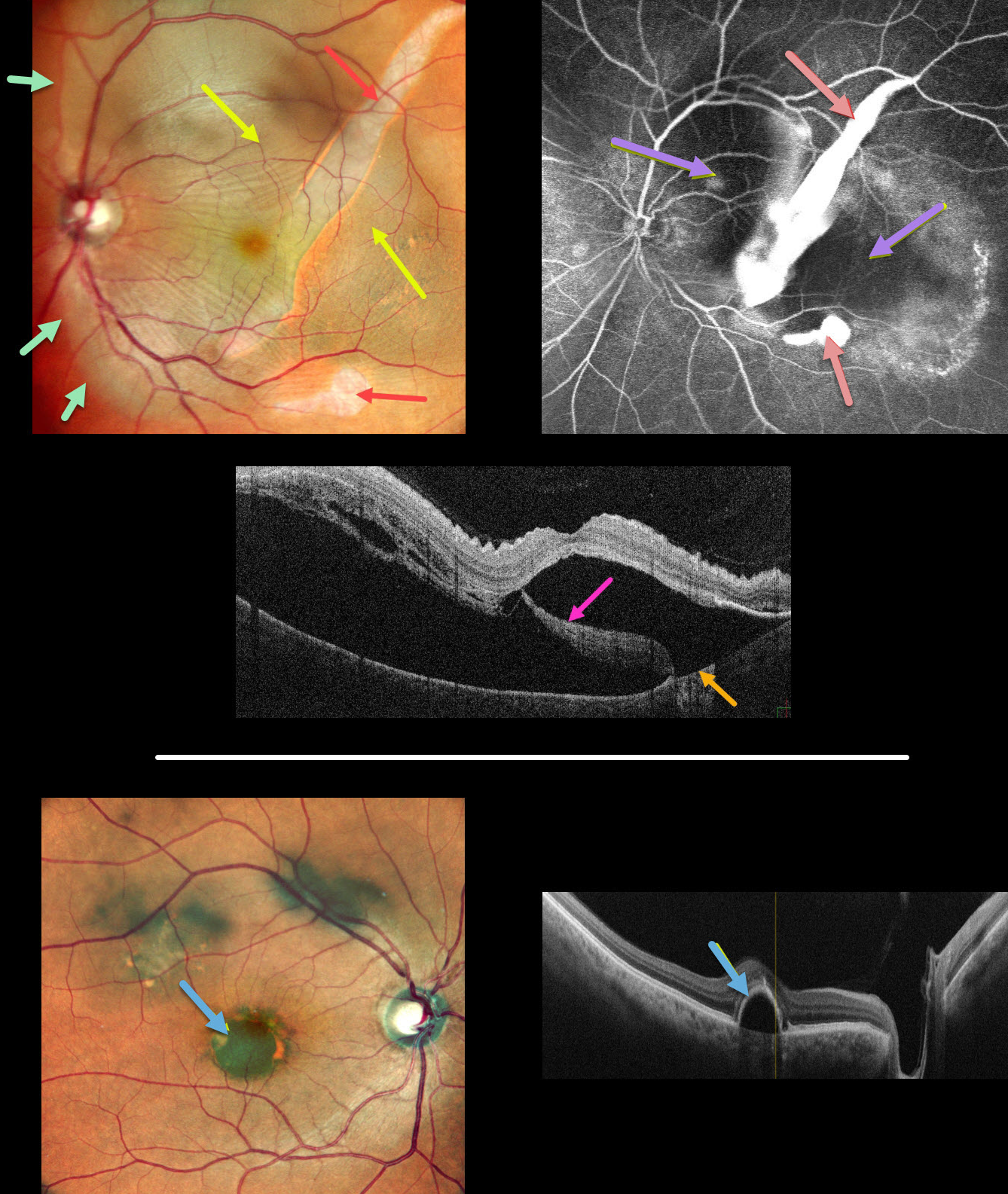
This 62YO male presented with 1 month of vision loss in his left eye. Vision was 20/30 OD and 20/60 OS.
Multicolor imaging of his left eye shows a giant RPE tear extending from the inferior macula into the superotemporal midperiphery, along with a much smaller tear just inferior to the macula. The beds of the RPE tears are depigmented. Increased subretinal pigmentation is noted on either edge of the giant tear. Subretinal fluid variably elevates the macula with inner retinal folds radiating from the optic nerve temporally. This subretinal fluid extends into the superior and temporal midperipheries.
B-scan OCT shows marked subretinal fluid. Bare Bruch’s membrane is noted temporally, and the RPE tear is lifted nasally and floating within the serous detachment.
Fluorescein angiography shows marked hyperfluorescent window defects from the absent RPE with blockage on either side of the giant tear from the redundant retracted RPE. He was placed on a 5-day tapering course of oral steroids but was subsequently lost to follow-up.
An asymptomatic retinal pigment epithelial detachment (PED) is noted in his right macula. The choroid does not appear thickened on OCT B-scan.
Learning Points:
A tear of the RPE is most commonly seen in RPE detachments (PEDs) associated with neovascular AMD, but can also occur in numerous other etiologies, including central serous chorioretinopathy and polypoidal choroidal vasculopathy. The tear leaves a depigmented region where the RPE was and is now absent, along with a pigmented, relatively straight subretinal lesion from the scrolled RPE edge.
Our patient’s presentation is unique in that RPE tears are usually much smaller and tend to rip in just one direction. The edges of our patient’s RPE retracted both nasally and temporally. RPE tears are usually singular, and ours had an additional, much smaller secondary tear more inferiorly.
Although our patient’s findings seem to be in the pachychoroid spectrum, the lack of a thickened choroid on OCT scanning makes the etiology less certain.
See Ersoz et al for a great review on RPE tear classification, pathogenesis, predictors and management (Surv Ophthalmology 2017;62:493-505).
RPE TEAR
Originally posted on @retina.rocks February 2, 2022
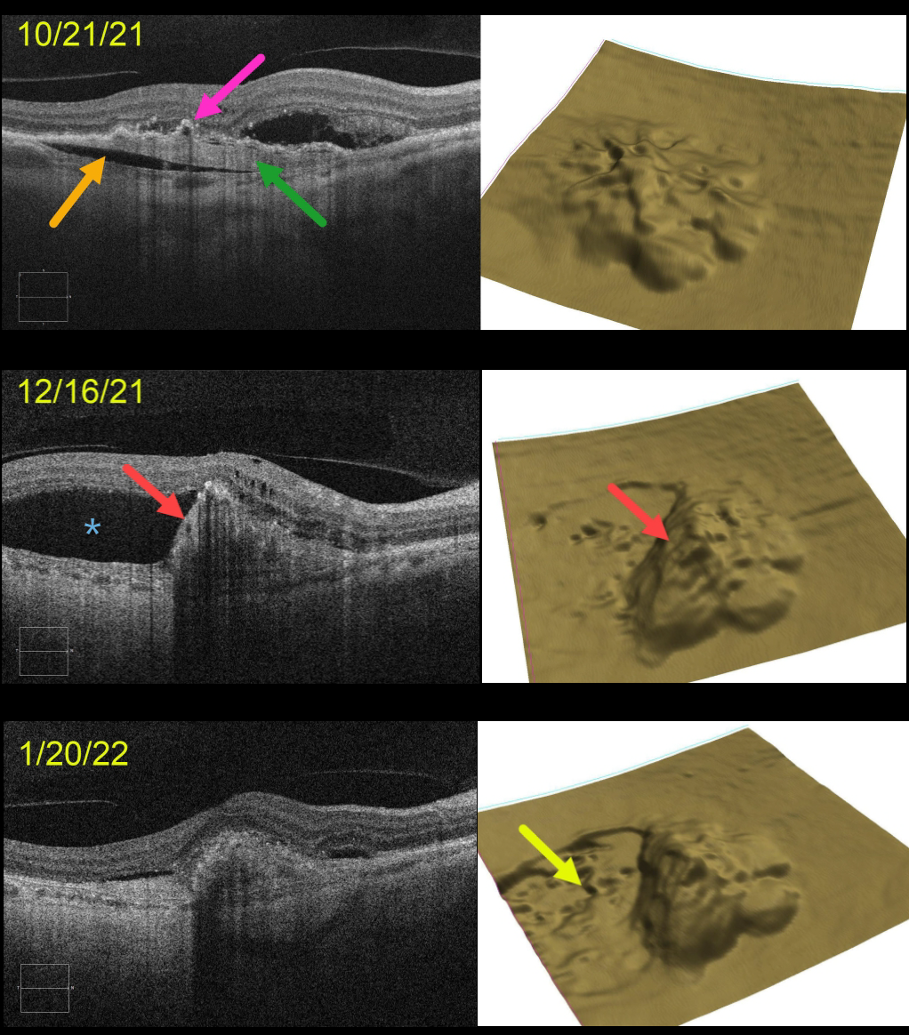
This 71YO female presented with a treatment-naive, age-related type 1 macular neovascularization (MNV). The already contracting MNV is elevated above the underlying choroid (prechoroidal cleft), and the overlying RPE is irregularly contracted and adherent to the MNV. The retinal pigment epithelium (RPE) map shows a 3-dimensional view of the RPE elevation. An anti-VEGF injection was given that day on 10/21/21.
On 12/16/21, vision was unchanged at 20/400 despite a new asymptomatic temporal RPE tear with marked overlying new subretinal fluid. An additional anti-VEGF injection was given.
On 1/20/22, vision was counting fingers. OCT scanning shows near-total resolution of all intra- and subretinal fluid. The RPE map nicely shows the crater of devoid RPE.
Learning Points:
A tear of the RPE is a fairly common occurrence in RPE detachment (PED) associated with type 1 MNV (located below the RPE). The neovascularization can spontaneously contract or involute following treatment. It then puts traction on the overlying RPE, which can then rip at the opposite edge of the PED.
This traction was already evident at our patient’s initial pre-injection visit, including the prechoroidal cleft, which is associated with a worse visual prognosis (see Kim et al, Retina 2017;37:2047-2055).
Like a cheap window blind, the torn RPE retracts and scrolls towards the MNV. This leaves a depigmented region where the RPE was and is now absent, along with a relatively straight, pigmented subretinal lesion extending from the scrolled RPE edge.
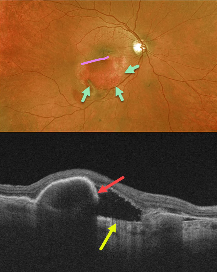
RPE TEAR
Originally posted on @retina.rocks August 11, 2020
Our patient had a retinal pigment epithelial (RPE) tear 2 weeks following an injection for wet age-related macular degeneration. The edges of the bare choroid and the edge of the retracted RPE can be visualized.
OCT shows bare Bruch’s membrane and the retracted, torn RPE.
Learning Points:
A tear of the RPE is a fairly common finding in RPE detachment (PED) associated with type 1 macular neovascularization (MNV), which is located below the RPE.
The neovascularization can spontaneously contract or involute following treatment. It then puts traction on the overlying RPE, which can then rip at the opposite edge of the PED. Like a cheap window blind, the torn RPE retracts and scrolls towards the MNV. This leaves a depigmented region where the RPE was and is now absent, along with a pigmented, relatively straight subretinal lesion extending from the scrolled RPE edge.


