

Anemia
<< Back to Cases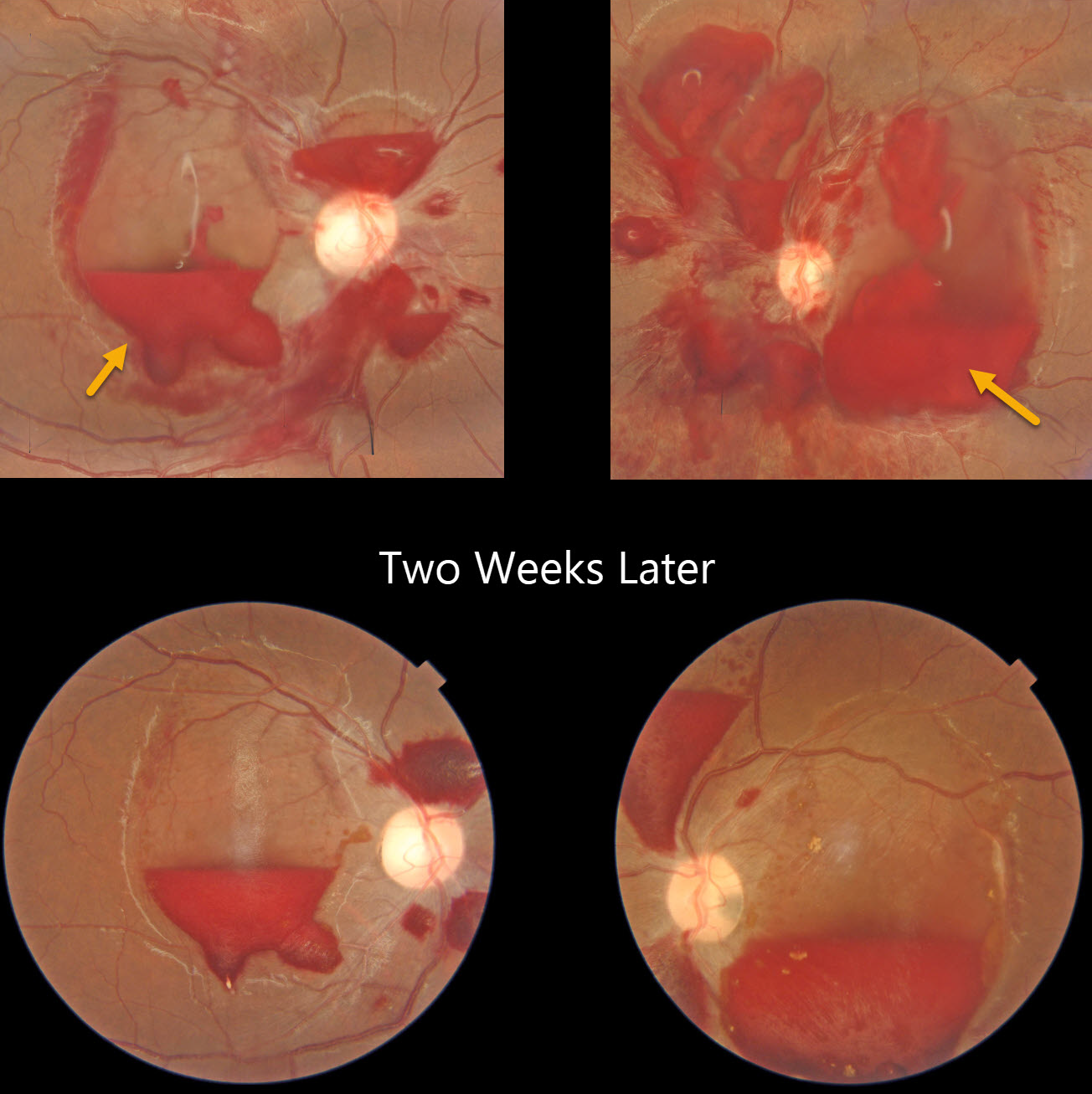
ANEMIC PRERETINAL HEMORRHAGES
The European VitreoRetina Society (EVRS) and Rajiv Gandhi
Originally posted on @retina.rocks August 15, 2025
This healthy 16YO female presented with 2 days of acute bilateral vision loss. She denied any trauma or Valsalva. Vision was 20/400 OU.
Color photography shows extensive bilateral preretinal and sub-internal limiting membrane hemorrhages. Some intraretinal white-centered hemorrhages are also noted. She was found to have profound iron-deficiency anemia due to very heavy menstruation with a hemoglobin of 5.2 gm/dl.
The blood was improving bilaterally two weeks later. Following multiple blood transfusions, the posterior segment hemorrhages spontaneously resolved (not shown).
ANEMIA WITH BILATERAL OPTIC NERVE EDEMA
Fraser McKay
Originally posted on @retina.rocks March 27, 2025
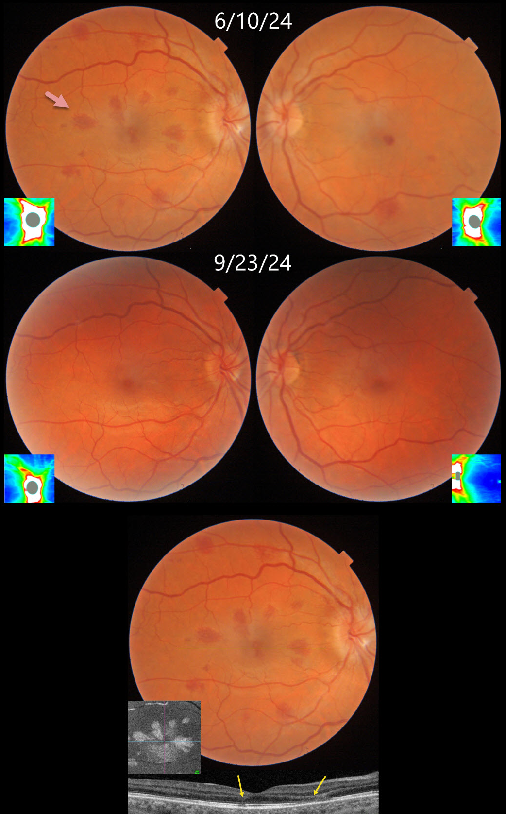
This 34YO female underwent uterine ablation 2 weeks earlier for heavy menstruation and severe secondary iron deficiency anemia. Although we don’t know what her hemoglobin level was prior to the procedure, it had risen to 10.7 postoperatively. Vision was 20/30 OD and 20/50 OS.
Triton color photography shows bilateral teardrop-shaped macular outer plexiform layer (OPL) hemorrhages, with greater involvement in her right eye. Numerous more peripheral deep and white-centered hemorrhages are also noted. Swept-source OCT confirms the location of the macular blood. En face OCT shows a more dramatic view of their petaloid shape. There is also mild optic nerve edema, especially in the right eye. The hemorrhages and nerve swelling mostly resolved with iron supplementation several months later.
Learning Points:
Anemia-related retinal findings most commonly include hemorrhages in the retina and sub-internal limiting membrane. Severe anemia is a rare cause for optic nerve swelling and should always be considered in patients with atypical idiopathic intracranial hypertension (Biousse et al, AJO 2003;135:437-446). The underlying pathogenesis of anemia-induced optic nerve swelling remains unknown.
Radiating deep macular hemorrhages are seen in multiple conditions, including Valsalva and trauma (Baumal et al, Br J Ophthalmol 2021;105:374-380). We have not previously seen these hemorrhages in isolated anemia. Their unique radiating appearance is caused by blood accumulating in Henle’s layer. Kon Graversen et al described hemorrhagic unilateral retinopathy, an idiopathic disorder with a similar appearance (Retina 2014;34:483-489).
ANEMIA
Anand Temkar and Manish Nagpal
Originally posted on @retina.rocks October 24, 2024
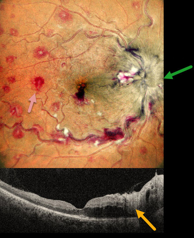
This 35YO male presented with 1 month of vision loss in his right eye. There was no past medical history. Vision was 20/40 OD and 20/20 in his normal OS.
MultiColor imaging shows a markedly swollen optic nerve with extensive multifocal deep, white-centered retinal hemorrhages. OCT shows marked mostly outer retinal edema extending from the nerve towards the macular center.
Extensive laboratory evaluation was negative except for a microcytic anemia with a hemoglobin of 2.9 g/dl. This was felt to be caused by an iron deficiency due to poor diet. Intravitreal anti-VEGF injections were recommended, along with oral prednisone for the optic nerve swelling. Unfortunately, he was immediately lost to follow-up.
Learning Points:
Anemia-related retinal findings most commonly include hemorrhages in the retina and sub-internal limiting membrane. Severe anemia is a rare cause for papilledema and should always be considered in patients with atypical idiopathic intracranial hypertension (Biousse et al, AJO 2003;135:437-446). The underlying pathogenesis of anemia-induced optic nerve swelling remains unknown. It is unclear why our patient had unilateral findings from a systemic disorder.
MEGALOBLASTIC ANEMIA
Alay Banker and Mauli Shah
Originally posted on @retina.rocks October 17, 2024
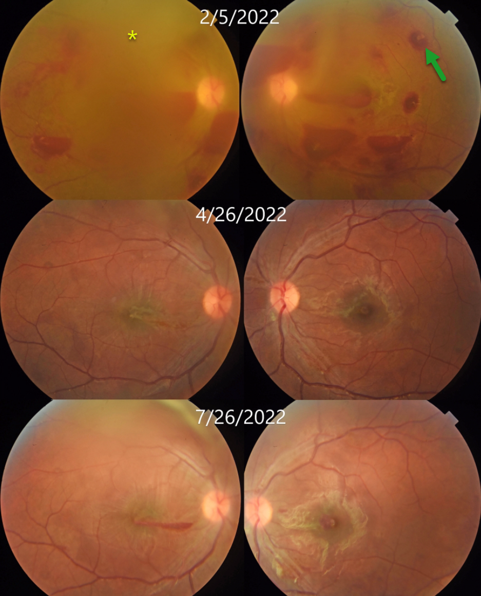
This 17YO male presented with 1 week of bilateral vision loss. Vision was counting fingers OU.
Bilateral white-centered and preretinal hemorrhages are noted. The fundus detail is decreased bilaterally due to breakthrough vitreous blood. Bloodwork revealed severe anemia with a hemoglobin of 5.3 g/dl. Vitamin B12 level was decreased at 160 pg/ml (normal 187-833). Based on the peripheral blood smear, which showed macrocytes, anisocytosis, poikilocytosis, target cells, and nucleated red blood cells, he was diagnosed with megaloblastic anemia.
His B12 deficiency resulted from a poorly balanced vegetarian diet. Following several blood transfusions and injectable vitamin B12 supplementation, as well as oral sublingual B12 and dietary modification, his anemia improved.
Two months later, vision improved to 20/200 OD and 20/80 OS; the hemoglobin rose to 12 g/dL; and funduscopic findings were improving. Five months following presentation, vision improved to 20/40 OU, and the retinal findings continued to normalize.
Learning Points:
Megaloblastic anemia is characterized by low hemoglobin in the presence of megaloblasts (large and immature nucleated red blood cells). It is caused by B12 or B9 (folate) deficiency, either from poor dietary intake (as in our patient) or from structural/functional gastrointestinal disorders that impair vitamin B malabsorption.
White-centered retinal hemorrhages and sub-internal limiting membrane/preretinal blood are commonly associated with anemia and blood dyscrasias. Other causes include bacterial endocarditis, hypertension, diabetic retinopathy, HIV retinopathy, and shaken baby syndrome. Hence, comorbidity must be questioned in such cases, and the patient should be screened for systemic diseases.
ANEMIA
Originally posted on @retina.rocks February 28, 2023
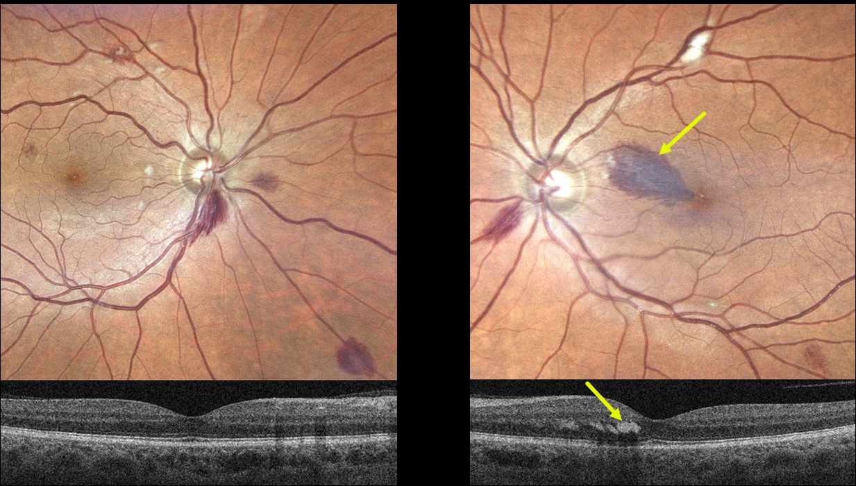
This 55YO male presented with a one-week history of a paracentral scotoma in his left eye. Vision was 20/40 OD and 20/60 OS. He gave a history of aplastic anemia, and his hemoglobin was 6.2 when he developed these symptoms. Blood transfusion brought his hemoglobin to over 8. He told us that his blood pressure runs low to normal.
Optos imaging shows scattered bilateral deep and superficial retinal hemorrhages with a few cotton wool spots. A tear-drop-shaped, larger hemorrhage is noted in the superonasal left macula. This hemorrhage localizes to the outer plexiform layer on swept-source OCT.
Learning Points:
The central retinal artery supplies the inner two-thirds of the retina, while the choroid supplies the outer third (the photoreceptor layers), the RPE, and the choroid. The outer plexiform layer (OPL) is the ‘end of the line’ for the inner retinal circulation.
In severe anemia, the more proximal inner retina removes whatever oxygen is available, leaving the deeper layers relatively ischemic. This can cause dysfunction of the retinal vascular endothelial tight junctions, which allows red blood cells to enter and stain the OPL, appearing clinically as deep retinal blot hemorrhages.
Deep blot OPL hemorrhages also appear for similar reasons in the temporal retinal periphery in ocular ischemia.
We advised observation, and expect his findings and symptoms to improve as long as his anemia is controlled.
ANEMIA
Originally posted on @retina.rocks November 30, 2020
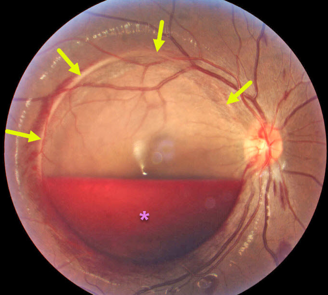
This 29YO 4-month pregnant female presented with acute vision loss. The internal limiting membrane (ILM) was detached throughout the entire macula, and an inferior-layered sub-ILM hemorrhage spared the macular center. Her hemoglobin was later found to be 11.
Learning Points:
Acute sub-ILM hemorrhage is most common following a Valsalva maneuver or trauma (which our patient denied), but can also happen spontaneously with anemia.
These hemorrhages virtually always resolve without sequelae, although the blood can be drained into the inferior vitreous by creating a small opening in the ILM using either a thermal or Nd:YAG laser.
ANEMIA
Originally posted on @retina.rocks August 5, 2020
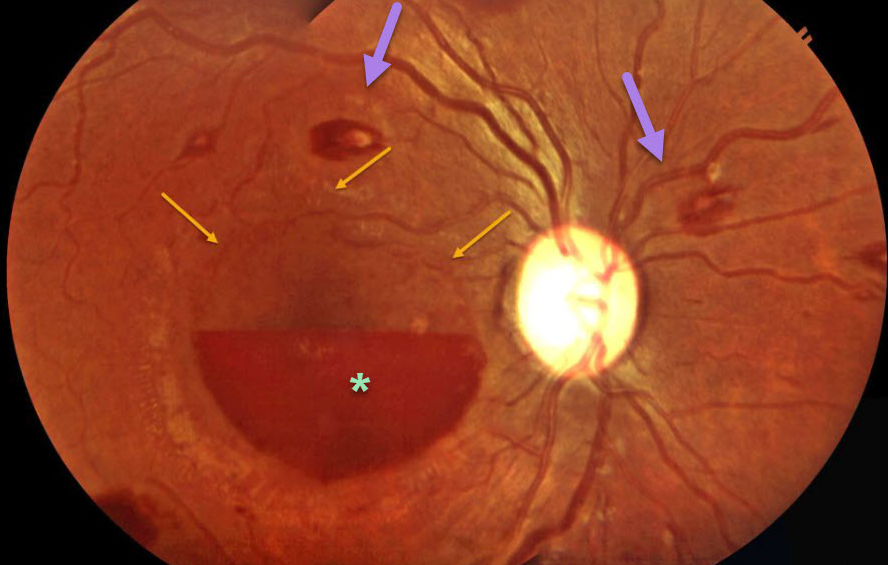
This patient presented with multiple white-centered flame-shaped retinal hemorrhages and sub-ILM hemorrhages consistent with anemic retinopathy. ILM detachment can be seen superior to the hemorrhage anterior to the fovea on the right eye.
Learning Points:
The term “Roth spot” was named after Dr. Moritz Roth, who, in 1872, noted white-centered hemorrhages in patients with bacterial endocarditis. He felt that these white spots were septic emboli that originated from an infected cardiac valve. However, similar lesions can be seen in many other disorders, including leukemia, anemia, diabetes, and hypertension.
The white material may represent a variety of causes, including fibrin. So these lesions are best called white-centered hemorrhages, unless one is specifically referring to a lesion in a patient with bacterial endocarditis. See Duane et al Ophthalmology 1980;87;66-69.


