

Angioid Streaks
<< Back to Cases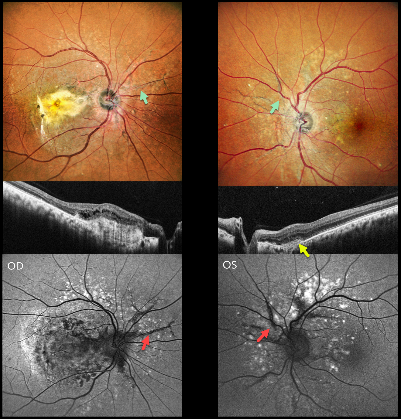
ANGIOID STREAKS WITH MNV AND PATTERN DYSTROPHY PHENOTYPE
Shraddha Raj Shrivastava and Manish Nagpal
Originally posted on @retina.rocks January 6, 2026
This healthy 42YO male presented with 10 months of vision loss in his right eye and 1 month of mild blurred vision in his left eye. Vision was counting fingers OD and 20/20 OS.
Pseudocolor SLO imaging shows angioid streaks extending around and radiating outwards from each optic nerve. There are drusen-like changes around each posterior pole, along with clumps of subretinal pigment temporally.
OCT scanning shows hyperreflective subretinal fibrosis with mild overlying cystic retinal edema OD. A much smaller area of subretinal hyperreflectivity (indicating a type 2 macular neovascularization, MNV) with overlying subretinal hyperreflective material is noted OS. The angioid streaks on fundus autofluorescence (FAF) are hypo-FAF due to RPE atrophy. The drusen-like changes are hyper-FAF.
Learning Points:
Retinal findings in pseudoxanthoma elasticum (PXE) most commonly include angioid streaks and a subretinal peaux d’orange appearance, most pronounced in each temporal macula. However, pattern dystrophy-like changes, as in our patient, can sometimes be the predominant phenotype (Murro et al, Graefe’s 2020;258:1881-1892). As with typical pattern dystrophies, the findings are dynamic and best captured with multimodal imaging.
PXE is caused by a mutation in the ABCA6 protein and is virtually always transmitted as an autosomal recessive disorder, although autosomal dominant transmission may rarely occur (Plomp et al, Am J Med Genet A 2004;126:403-412).
Our patient had no clinical signs of PXE and denied any family history of this disease. He was referred to his primary care doctor for further evaluation. Intravitreal anti-VEGF therapy was started OS.
ANGOID STREAKS WITH TRAUMATIC CHOROIDAL RUPTURE AND SUBRETINAL BLOOD
Neelam Khatwani
Originally posted on @retina.rocks July 2, 2025
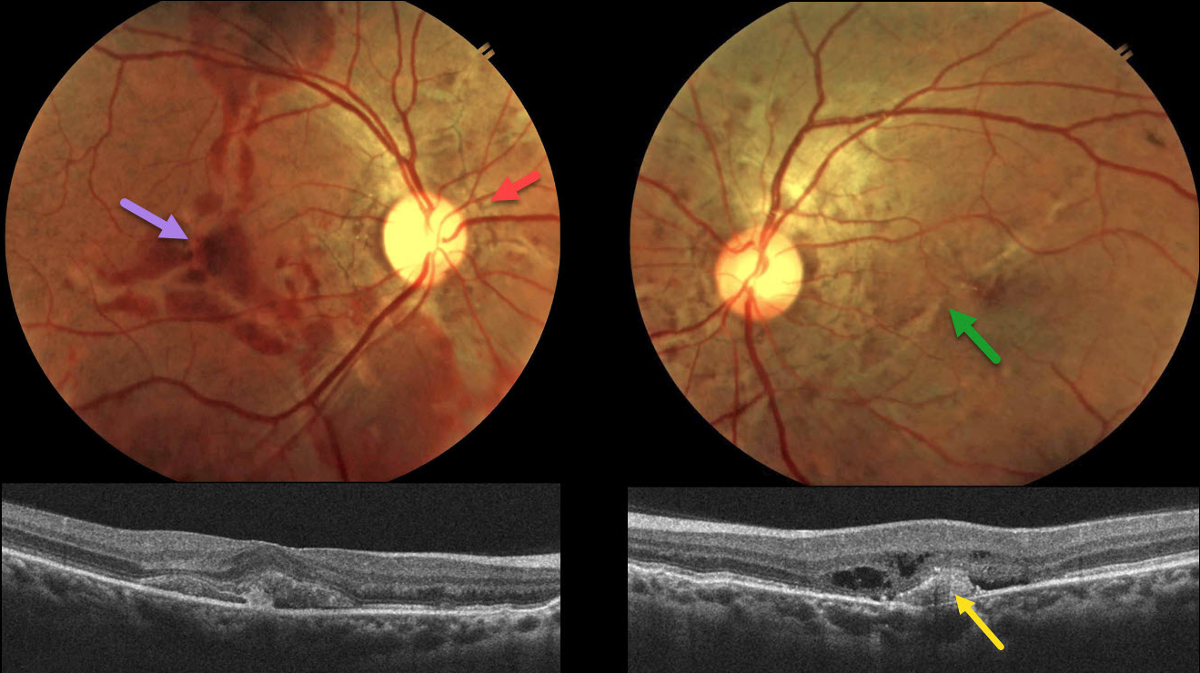
This 38YO male presented with 10 days of bilateral vision loss after being hit in the right eye with a fist. He denied any past medical history, and family history was negative for eye disease. Vision was 20/200 OD and 20/80 OS.
Color fundus photography shows irregular orange-gray lines radiating outwards from each nerve. Variable areas of subretinal blood are noted in the right macula, extending into the superior and inferior midperipheries. There is milder subretinal blood OS along with an irregular oblique macular angioid streak vs choroidal rupture. OCT OD shows subretinal hyperreflective material, likely blood. A possible type 1 (below the RPE) MNV with some outer retinal fluid is seen OS.
An anti-VEGF injection was given bilaterally, but unfortunately, the patient was immediately lost to follow-up.
Learning Points:
Initially described by Doyne in 1889, angioid streaks are orange-yellow, irregular, crack-like dehiscences in Bruch’s membrane associated with atrophic degeneration of the overlying RPE. The term “angioid” derives from its resemblance to blood vessels. Although they are historically mistakenly associated with numerous conditions, the only real association is with pseudoxanthoma elasticum (PXE, Nadelmann et al, Eye 2023;37:1596-1601). Patients are at significant risk for vision loss from MNV or pattern dystrophy-like changes (Murro et al, Graefe’s 2020;258:1881-1892).
Even minor trauma can cause choroidal ruptures with subretinal blood (Agrawal et al, JAMA Ophthalmology 2017;135(3):e165466). Unlike typical choroidal ruptures that are usually curvilinear with the nerve, the lesions from the brittle Bruch’s membrane in PXE appear as numerous irregular ruptures usually radiating from the nerve. Even though our patient sustained a direct injury to his right globe, the trauma was significant enough to cause choroidal ruptures in his left eye. The bilateral subretinal hemorrhages may be purely avascular, as their multifocality suggests.
PSEUDOXANTHUM ELASTICUM WITH ANGIOID STREAKS AND PATTERN DYSTROPHY PHENOTYPE
Mattie Adams
Originally posted on @retina.rocks June 4, 2025
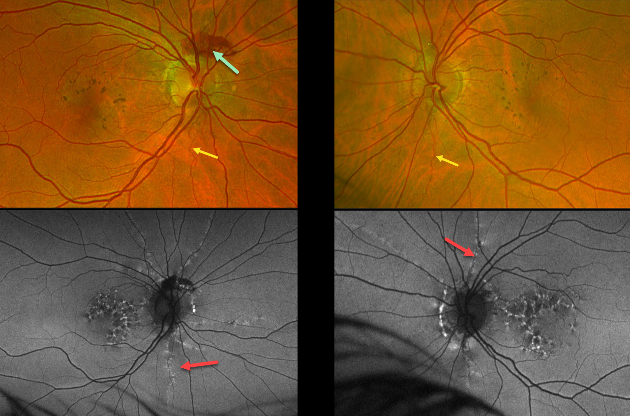
This 53YO female was referred for asymptomatic macular findings. Her mother has a history of pseudoxanthoma elasticum (PXE). Vision was 20/25 OD and 20/40 OS.
Optos color RG imaging shows coarse subretinal pigment clumps bilaterally. Subretinal blood is noted along the superior aspect of the right nerve from a peripapillary choroidal neovascularization. A few barely visible angioid streaks radiate from each nerve.
Fundus autofluorescence (FAF) shows variable hyper- and hypo-FAF flecks. The angioid streaks appear as hyper-FAF lesions radiating outwards from each nerve.
Learning Points:
Retinal findings in PXE most commonly include angioid streaks and a subretinal peaux d’orange appearance, most pronounced in each temporal macula. However, pattern dystrophy-like changes, as in our patient, can sometimes be the predominant phenotype (Murro et al, Graefe’s 2020;258:1881-1892). As with typical pattern dystrophies, the findings are dynamic and best captured with multimodal imaging.
PXE is caused by mutations in the ABCA6 gene, and our patient tested positive for homozygous pathogenic ABCA6 mutations. PXE is virtually always transmitted as an autosomal recessive disorder, although autosomal dominant transmission may rarely occur (Plomp et al, Am J Med Genet A 2004;126A:403-412).
ANGIOID STREAKS
Anand Temkar and Manish Nagpal
Originally posted on @retina.rocks November 21, 2024
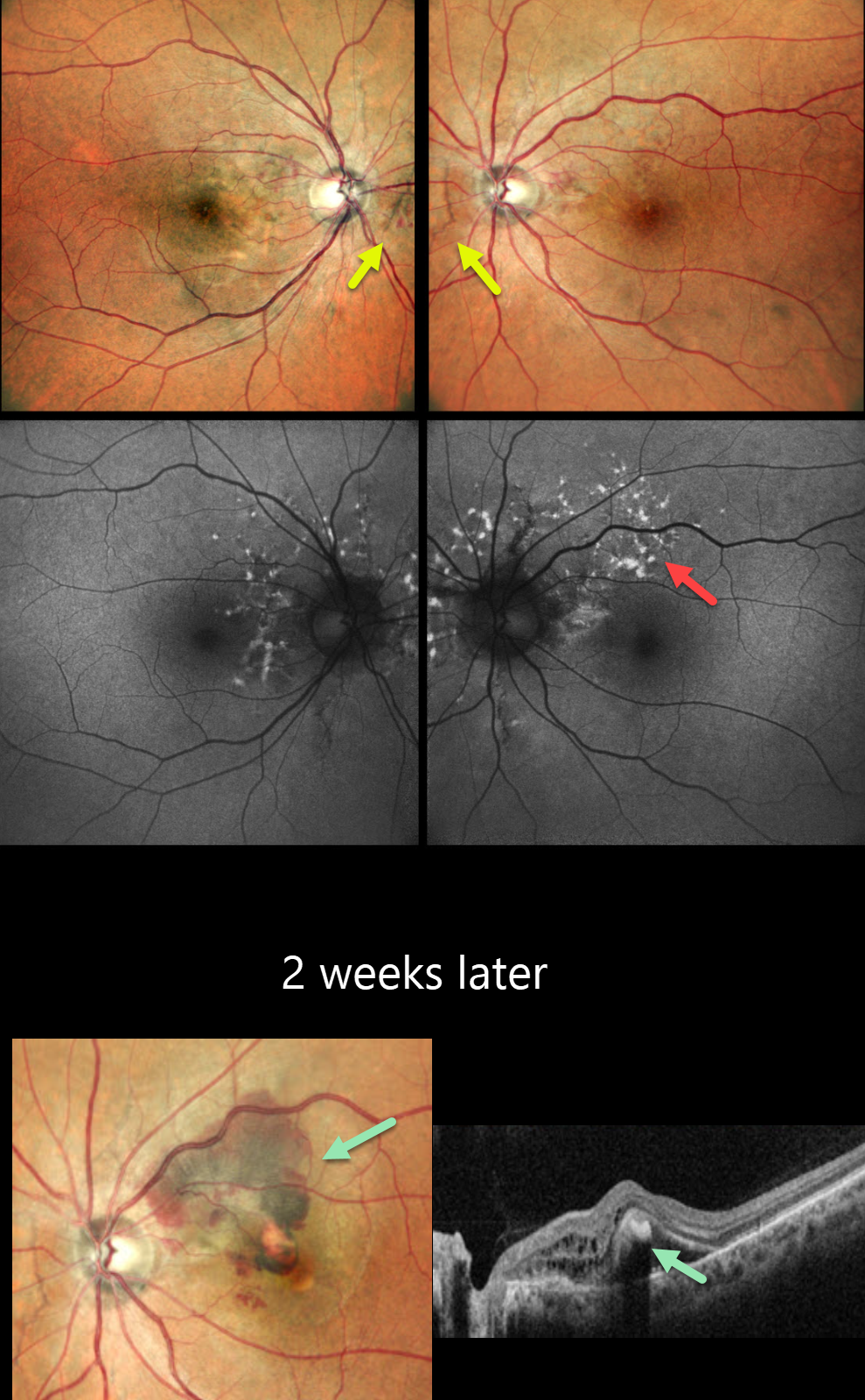
This 45YO male presented for a second opinion for bilateral wavy vision for 2 months. He was diagnosed elsewhere with macular neovascularization (MNV) and received two prior anti-VEGF injections (last 10 days earlier). Vision was 20/20 OU.
Pseudocolor SLO imaging shows subtle bilateral peripapillary angioid streaks associated with a few retinal hemorrhages. Fundus autofluorescence (FAF) shows numerous mostly peripapillary hyper-FAF flecks. Initial observation was recommended.
He returned two weeks later with new symptoms OS, and vision decreased to 20/120. New submacular fluid and blood are noted clinically and on OCT, and monthly anti-VEGF therapy was restarted. Unfortunately, he was lost to follow-up after 3 injections.
Learning Points:
Angioid streaks are orange-yellow, irregular, crack-like dehiscences in Bruch’s membrane associated with atrophic degeneration of the overlying RPE. These patients, especially those with pseudoxanthoma elasticum (PXE), are at significant risk for MNV as occurred in each of our patient’s eyes. MNV responds best to anti-VEGF therapy, and treatment is usually ongoing as with the typical neovascular AMD protocols. However, long-term vision is often poor due to RPE and choroidal atrophy (Mori et al, Graefe’s 2020;258:1863-1869).
Our patient had no family history of PXE or suggestive skin lesions, so skin biopsy or genetic testing was not performed. However, the funduscopic changes and macular dystrophy-like FAF findings are fairly pathognomonic for PXE.
ANGIOID STREAKS
Seif Anwar
Originally posted on @retina.rocks September 19, 2024
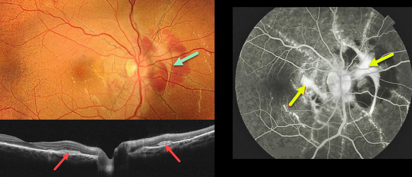
This healthy 18YO female presented with recent vision loss in her right eye. Vision was 20/40 OD and 20/30 OS.
Fundus photography shows angioid streaks extending around and radiating outwards from each optic nerve. The posterior pole has a diffuse orange skin (peaux d’orange) appearance. A fibrosed macular neovascularization (MNV) encircles the right nerve and extends into the nasal macula.
On OCT scanning, the hyperreflective MNV is located above the RPE (type 2 lesion) and is associated with minimal exudation. The MNV stains on fluorescein angiography. Intravitreal Lucentis therapy was started in her right eye.
Learning Points:
Angioid streaks are orange-yellow, irregular, crack-like dehiscences in Bruch’s membrane associated with atrophic degeneration of the overlying RPE. The orange skin (peaux d’orange) appearance is caused by a calcified Bruch’s membrane giving a yellow mottled ‘coquille d’oeuf’ (eggshell) appearance that contrasts with the normal orange RPE and choroidal pigmentation (Spaide, Retina 2015;35:392-397). These patients are at significant risk for MNV, as occurred in our patient’s right eye.
Angioid streaks are often missed clinically because their color and orientation blend with the overlying retinal vessels. Individuals with the extensive streaks and peaux d’orange, as seen in our patient, are pathognomonic for PXE, although less extensive streaks can also be seen in sickle cell and age-related macular degeneration (Eye 2023;37:1596-1601).
Our patient gave no family history of PXE. Genetic testing was not possible due to financial reasons, but we will refer her to dermatology for a skin biopsy to definitively establish this diagnosis.
PSEUDOXANTHUM ELASTICUM WITH ANGIOID STREAKS AND PATTERN DYSTROPHY PHENOTYPE
Originally posted on @retina.rocks July 11, 2024
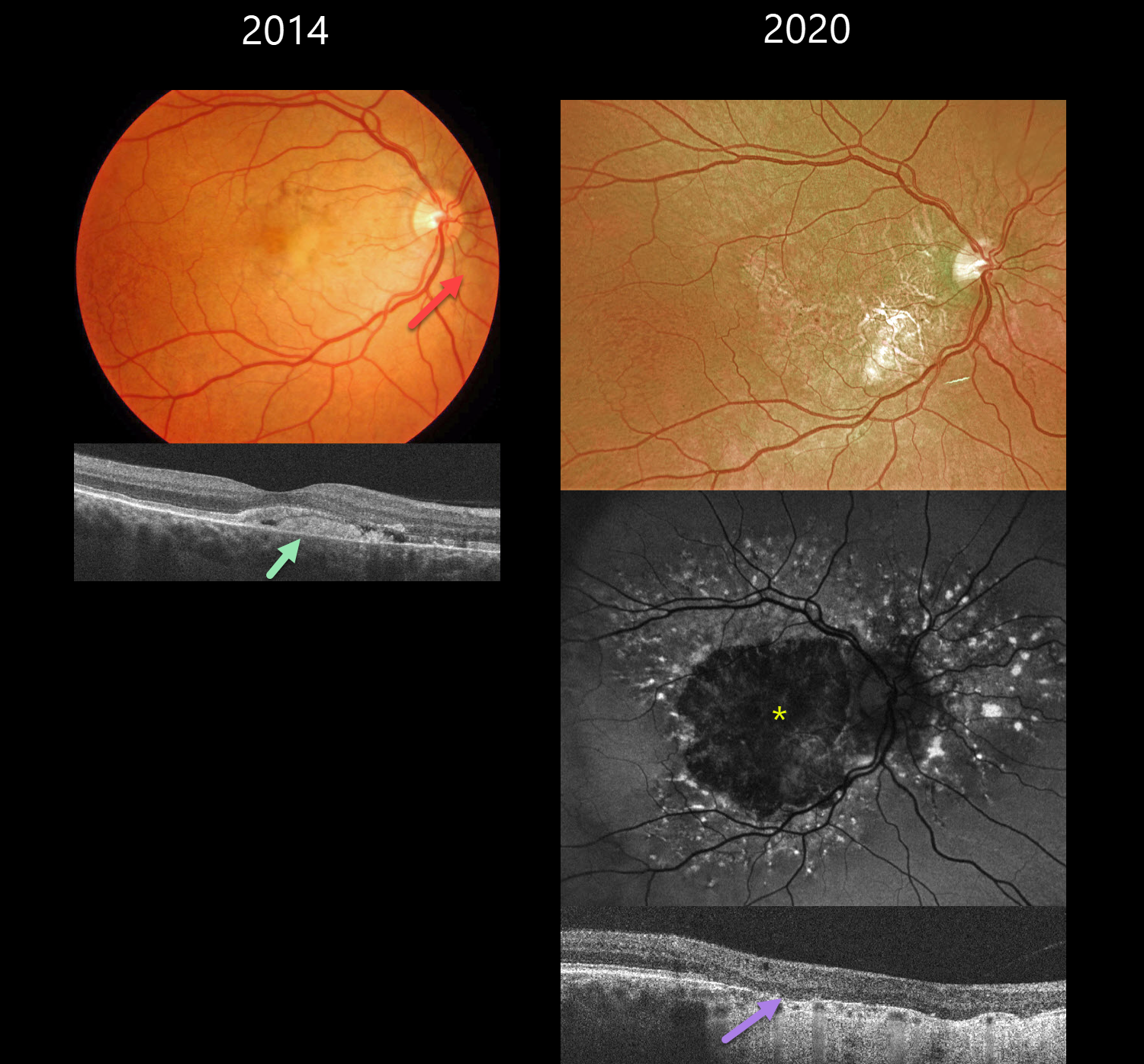
This 39YO male with a known history of pseudoxanthoma elasticum presented on 2/19/14. Findings were bilateral at this, and subsequent visits, so only his right eye images will be shown and discussed.
Color photography shows nasal yellowish subfoveal pigmentary changes with a peaux d’orange appearance temporally. Some peripapillary angioid streaks are noted. OCT shows hyperreflective vitelliform material and fluid below the retina, with some of this material also infiltrating the outer retina.
He has been followed without treatment, and when examined on 7/17/20, vision was 20/200 OD and 20/80 OS. Optos color RG imaging shows a variably atrophic macula. The atrophy on fundus autofluorescence is hypo-FAF. There is a fairly dramatic pattern of hyper-FAF spots radiating out from this atrophy. OCT shows outer retinal atrophy and no fluid. When last examined on 7/6/23, vision was 20/200 OU.
Learning Points:
Retinal findings in PXE most commonly include angioid streaks and a subretinal orange skin (peaux d’orange) appearance, most pronounced in each temporal macula. However, pattern dystrophy-like changes, as in our patient, can sometimes be the predominant phenotype (Murro et al, Graefe’s 2020;258:1881-1892). As with typical pattern dystrophies, the findings are dynamic and best captured with multimodal imaging.
ANGIOID STREAKS
Originally posted on @retina.rocks September 5, 2023
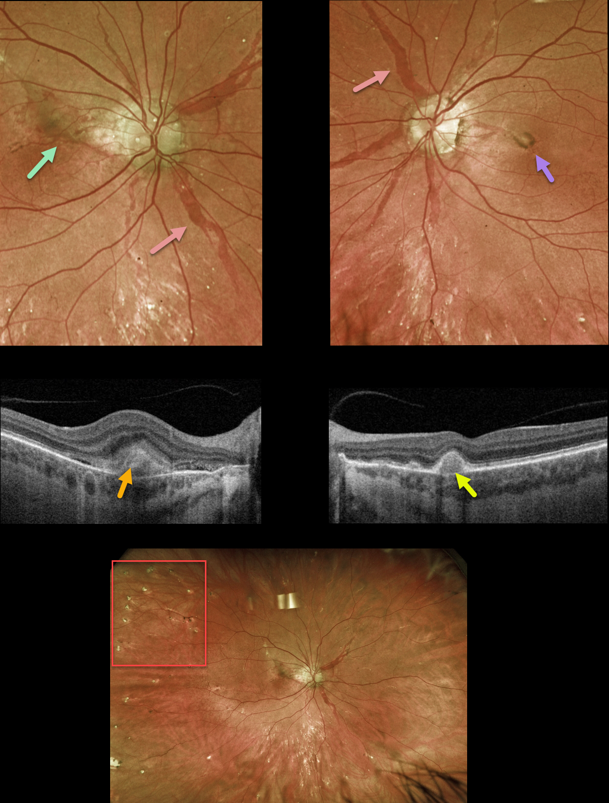
This 42YO female with a known history of pseudoxanthoma elasticum (PXE) presented with new central flashes in her right eye. Vision was 20/25 OD and 20/30 OS.
Optos color RG imaging shows large, irregularly dilated’ angioid streaks’ radiating outwards from each optic nerve. A pigmented macular neovascularization (MNV) with some subretinal blood is noted in the superior right macula, and a small inactive MNV is noted in the left nasal fovea. Innumerable small subretinal comet-shaped lesions (‘comet rain’) are noted.
Swept-source OCT through the right MNV shows a hyperreflective, thickened type 2 lesion (above the RPE). OCT through the left MNV shows a small, nodular, hyperreflective type 1 lesion (below the RPE).
Learning Points:
Angioid streaks are orange-yellow, irregular, crack-like dehiscences in Bruch’s membrane associated with atrophic degeneration of the overlying RPE. They are often missed clinically because their color and orientation blend with the overlying retinal vessels.
Our patient’s lesions are about the most dramatic and red angioid streaks we’ve ever seen and are pathognomonic for PXE, but less extensive streaks can also be seen in sickle cell and age-related macular degeneration.
The comet lesions are also pathognomonic for PXE. These lesions develop from the outer retina as a neurodegenerative process and strongly resemble the hyporeflective core with a surrounding hyperreflective rim found in outer retinal tubulation (see Murro et al., Graefe’s 2018;256:1605-1614).
These patients are at significant risk for macular neovascularization (MNV), as occurred in each of our patients’ eyes. Intravitreal Avastin therapy was started in her right eye. The left MNV will be observed, as it involuted spontaneously without prior symptoms.
PSEUDOXANTHUM ELASTICUM WITH ANGIOID STREAKS AND PATTERN DYSTROPHY PHENOTYPE
Originally posted on @retina.rocks March 20, 2023
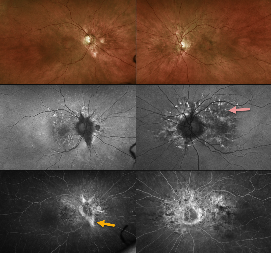
This 55YO female has been followed for years with macular pattern dystrophy-type changes due to pseudoxanthoma elasticum (PXD). Vision is 20/25 OU.
Color imaging shows variable mild pigmentary changes. Optos fundus autofluorescence imaging shows variable hyperautofluorescent flecks with variable angiographic staining. Some staining angioid streaks are noted in her right eye.
Learning Points:
Retinal findings in PXE most commonly include angioid streaks and a subretinal orange skin (peaux d’orange) appearance, most pronounced in each temporal macula.
However, pattern dystrophy-like changes, as in our patient, can sometimes be the predominant phenotype in some PXE patients (Murro et al, Graefe’s 2020;258:1881-1892). As with typical pattern dystrophies, the findings are dynamic and best captured with multimodal imaging.
ANGIOID STREAKS
Asma Samsudeen and Ashish Sharma
Originally posted on @retina.rocks February 17, 2023
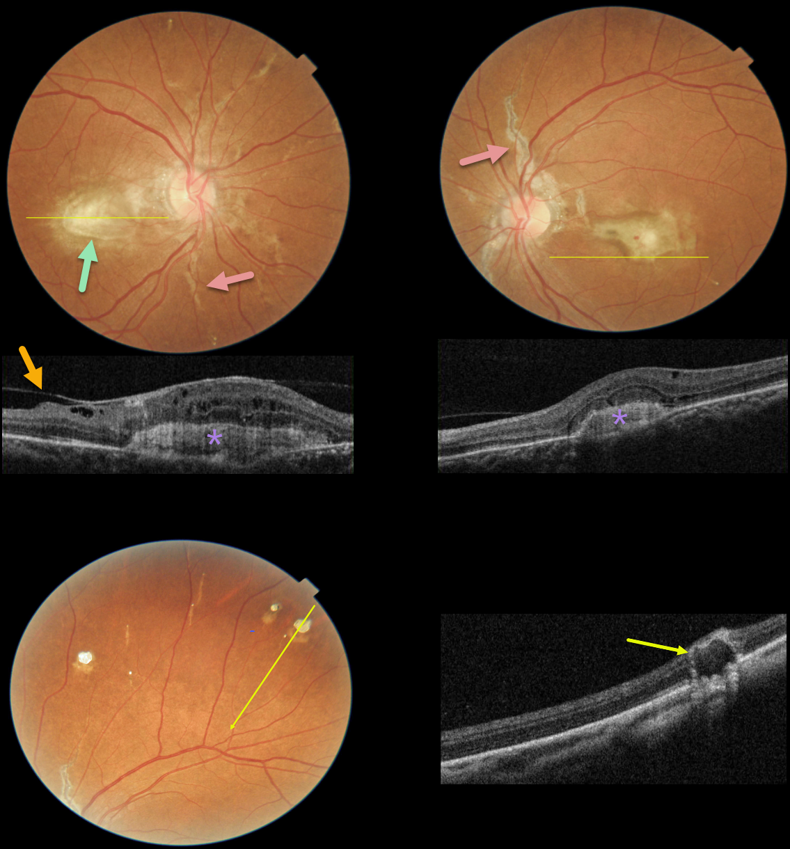
This 49YO female presented with one month of blurred vision. There was no past medical history except for a seizure disorder. Vision was 20/80 OD and 20/120 OS.
Fundus photographs show bilateral angioid streaks radiating from each optic nerve. Bilateral fibrosed subfoveal macular neovascularization (MNV) is present, and a small dot of blood is seen overlying the left macular lesion.
OCT scanning through the central right macula shows a hyperreflective, laminated MNV with overlying cystic retinal edema and a partially detached hyaloid vs macular pucker. OCT scanning through the inferior left macula also shows a hyperreflective MNV below the RPE associated with some overlying retinal thickening.
Numerous small subretinal comet-shaped lesions (‘comet rain’) are noted bilaterally. OCT scanning through one of these lesions shows a full-thickness hyporeflective lesion with a surrounding hyperreflective rim.
Learning Points:
Angioid streaks are orange-yellow, irregular, crack-like dehiscences in Bruch’s membrane associated with atrophic degeneration of the overlying RPE. They are often missed clinically since their color and orientation often blend in with the overlying retinal vessels.
Our patients’ lesions are easier to diagnose due to significant overlying RPE changes. The extent and prominence of the angioid streaks in our patient are pathognomonic for PXE, but less extensive streaks are seen in sickle cell disease and age-related macular degeneration.
These patients are at significant risk for macular neovascularization (MNV), which can be successfully treated with intravitreal anti-VEGF therapy.
The comet lesions are pathognomonic for PXE. These lesions develop from the outer retina as a neurodegenerative process and strongly resemble the hyporeflective core with a surrounding hyperreflective rim found in outer retinal tubulation (see Murro et al, Graefe’s 2018;256:1605-1614). Full-thickness lesions, as in our patient, were reported by Barteselli and Viola (Retina 2015;35:1051-1052).
Although our patient has no known personal or family history of PXE, her retinal findings indicate she has this disease. Her MNV appears chronic and fibrosed, and it is unclear why her symptoms were only present for one month. However, due to her recent symptoms and presence of overlying retinal fluid and blood, anti-VEGF therapy will be started.
ANGIOID STREAKS
Originally posted on @retina.rocks September 26, 2022
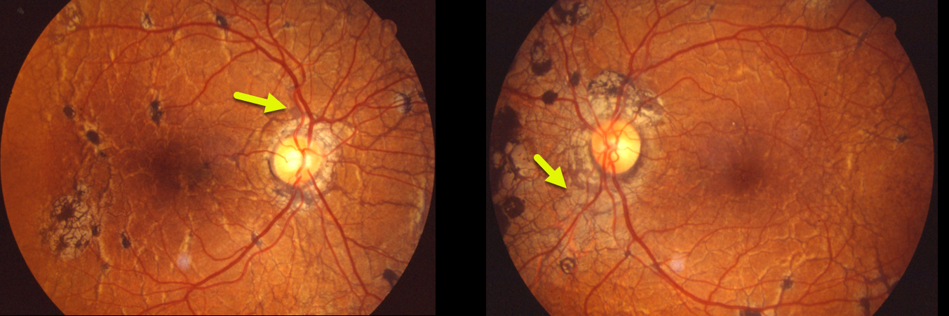
This patient was examined in 1989 and had classic findings of pseudoxanthoma elasticum. Extensive angioid streaks encircle and radiate from the nerves, extending into the peripheral maculas and beyond the arcades.
Angioid streaks are usually less dramatic and are often quite easy to miss since their color and orientation mimic the normal retinal vasculature, hence the term “angioid”.
The orange skin (peaux d’orange) appearance, most pronounced in each temporal macula, is caused by the calcified Bruch’s membrane giving a yellow mottled ‘coquille d’oeuf’ (eggshell) appearance that contrasts with the normal orange RPE and choroidal pigmentation (Spaide, Retina 2015;35:392-397).
Learning Points:
Angioid streaks are known to be associated with systemic conditions such as pseudoxanthoma elasticum, Ehlers-Danlos syndrome, Paget’s disease, sickle cells, or idiopathic (PEPSI).
These patients are at significant risk for macular neovascularization and need to be followed and treated as needed with anti-VEGF agents, as in patients with neovascular AMD.
Genetic testing with pseudoxanthoma would test positive for a pathogenic ABCC6 mutation.
ANGIOID STREAKS
Originally posted on @retina.rocks September 1, 2021
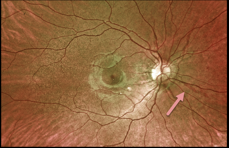
This 19YO female presented with these asymptomatic fundoscopic changes. Vision was 20/25 OU. Optos imaging shows angioid streaks radiating from each optic nerve.
Our patient denied any past medical or family history of pseudoxanthoma elasticum. Genetic testing for the ABCC6 mutation is pending.
Learning Points:
Angioid streaks are often quite easy to miss since their color and orientation mimic the normal retinal vasculature, hence the term ‘angioid.’
The orange skin (peaux d’orange) appearance, most pronounced in each temporal macula, is caused by the calcified Bruch’s membrane giving a yellow mottled ‘coquille d’oeuf’ (eggshell) appearance that contrasts with the normal orange RPE and choroidal pigmentation (Spaide, Retina 2015;35;392-397).
These patients are at significant risk for macular neovascularization (MNV) and need to be followed and treated as needed with anti-VEGF agents, as in patients with AMD.
ANGIOID STREAKS
Originally posted on @retina.rocks April 1, 2021
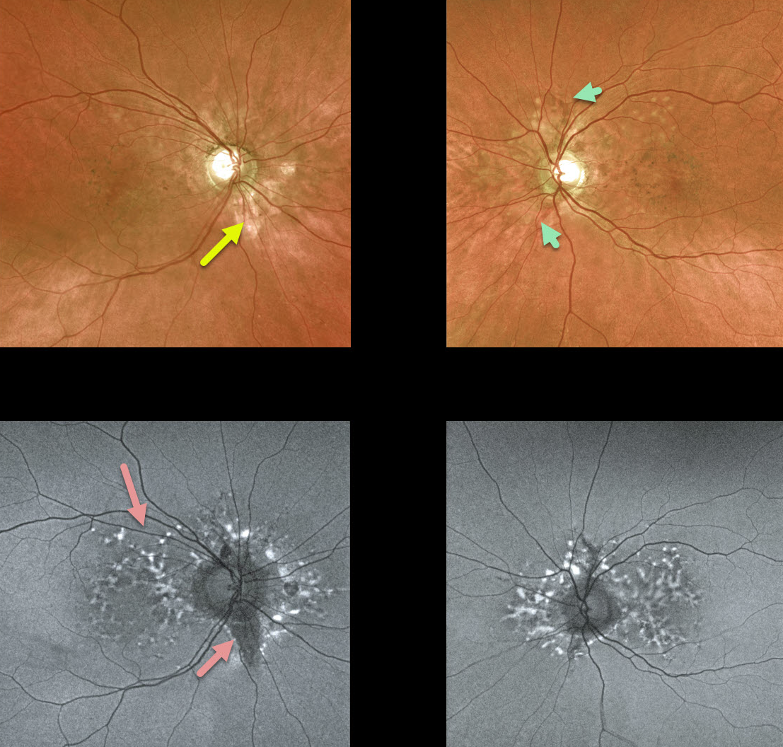
We have been following this 54YO female with pseudoxanthoma elasticum (PXE) for many years. Vision is 20/20 bilaterally, and fortunately, she remains asymptomatic.
She has bilateral macular peripapillary pigmentary changes. Angioid streaks, always tough to find unless the clinician actively looks for them, are not prominent, although a few are visible in her left eye.
Fundus autofluorescence (FAF) shows more dramatic changes, including scattered hyper- and hypo-FAF peripapillary and macular spots.
Her sister became legally blind many years ago from bilateral macular neovascularization (MNV).
Learning Points:
Patients with angioid streaks and pseudoxanthoma elasticum (PXE) can present with retinal findings similar to those of pattern dystrophies.
These patients are at significant risk for macular neovascularization and need to be followed and treated as needed with anti-VEGF agents, as in patients with AMD.
ANGIOID STREAKS
Originally posted on @retina.rocks November 26, 2020
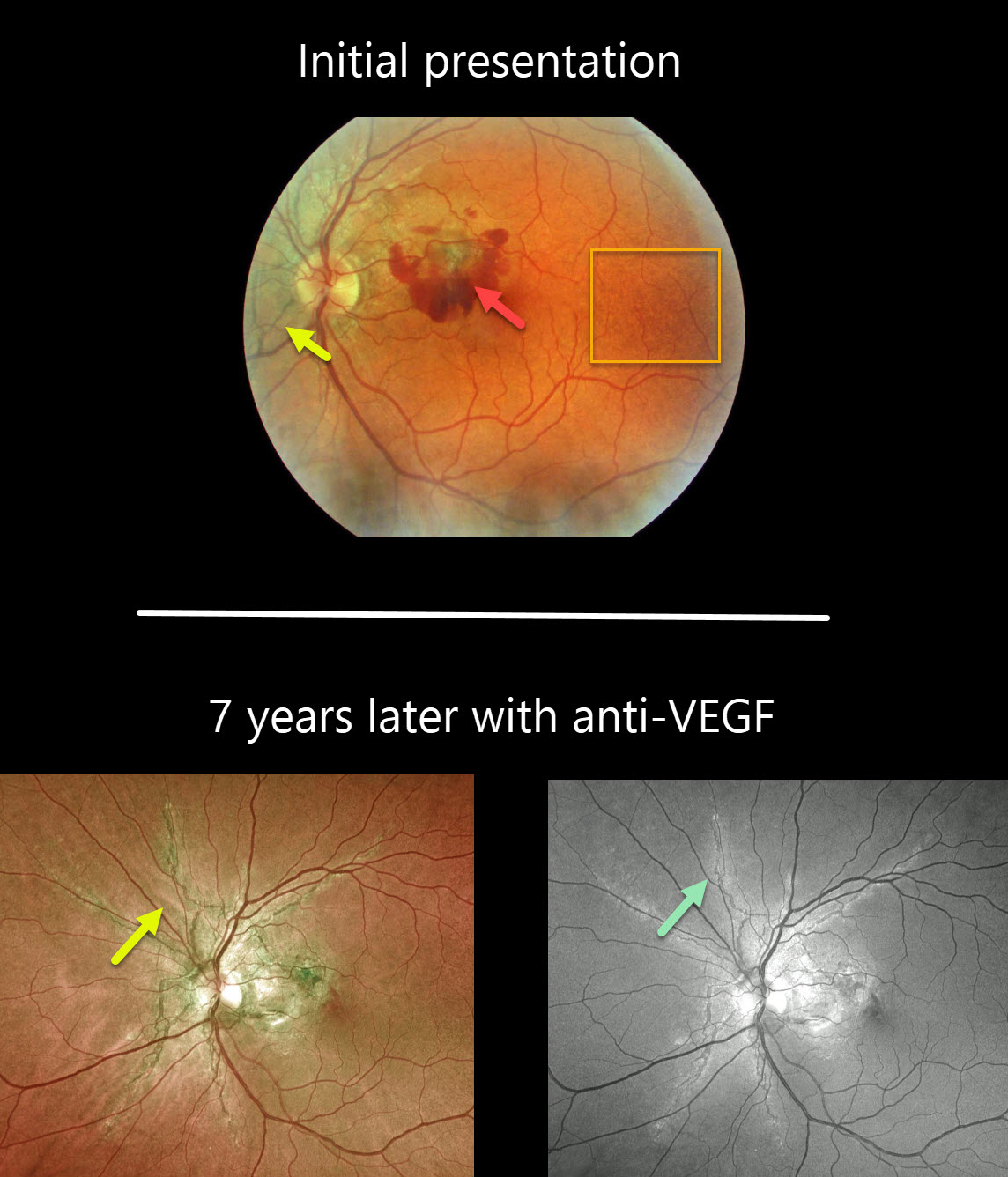
This patient initially presented with dramatic pigmented angioid streaks and acute vision loss from macular neovascularization.
The classic peau d’orange (orange skin) pigmentary changes are seen in the temporal macula.
Seven years later, vision is 20/25 with ongoing anti-VEGF therapy. The angioid streaks are best imaged with the Optos green channel.
Learning Points:
Macular neovascularization is a complication of virtually any disorder that affects Bruch’s membrane, the most common being age-related macular degeneration, ocular histoplasmosis, idiopathic, degenerative myopia (lacquer cracks), trauma (choroidal rupture), and angioid streaks.
ANGIOID STREAKS
Originally posted on @retina.rocks August 21, 2020
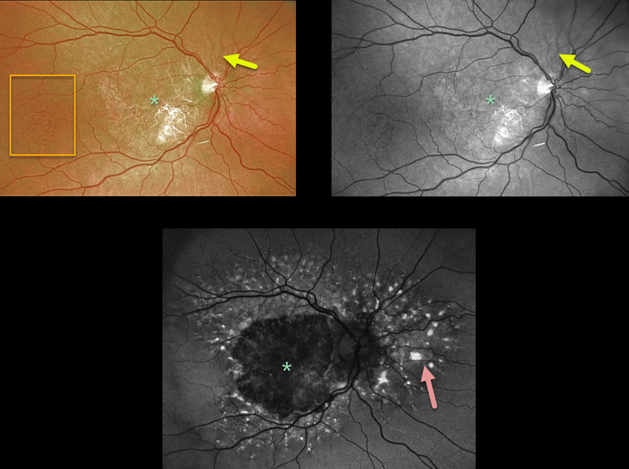
This patient has angioid streaks that radiate from the optic nerve. These are very easy to miss unless they are explicitly looked for.
Optos color and green-channel photographs of the right eye show central macular atrophy with more temporal peaux d’orange changes. The left eye (not pictured) showed identical findings.
Fundus autofluorescence (FAF) shows widespread hyper-FAF lesions surrounding the central hypo-FAF macula. These multifocal hyper-FAF lesions are typical of pattern dystrophies and can somewhat simulate fundus flavimaculatus if they extend outside the macula.
This patient is monitored regularly due to the risk of developing macular neovascularization, although that is fairly unlikely given the macular atrophy.
Learning Points:
Patients with angioid streaks and pseudoxanthoma elasticum (PXE) can present with retinal findings similar to those of pattern dystrophies.
These patients are at significant risk for macular neovascularization (MNV) and need to be followed and treated as needed with anti-VEGF agents, as in patients with AMD.
ANGIOID STREAKS
Originally posted on @retina.rocks April 7, 2020
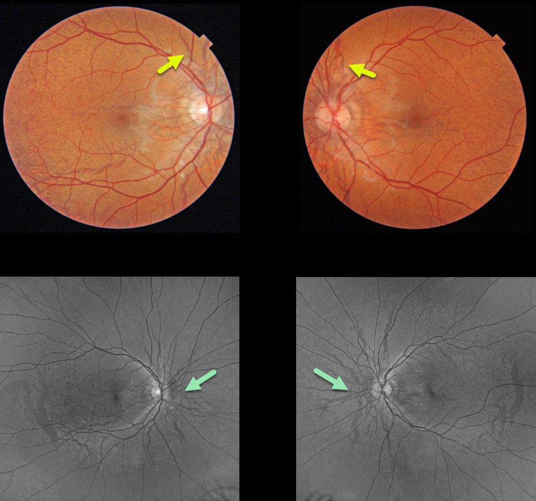
This patient with pseudoxanthoma elasticum (PXE) has classic extensive angioid streaks with peaux d’orange (orange skin). The orange skin appearance is caused by the calcified Bruch’s membrane giving a yellow mottled appearance that contrasts with the normal orange RPE and choroidal pigmentation. The green channel (532 nm) Optos images best show these findings in the RPE/Bruch’s membrane.
Learning Points:
Although angioid streaks can be seen in other disorders, including age-related and sickle-cell disease, the findings in our patient are pathognomonic for PXE.
These patients are at significant risk for macular neovascularization (MNV) and need to be followed and treated as needed with anti-VEGF agents, as in patients with AMD. Fortunately our patient has no MNV with normal macular OCTs (not shown).


