

Autosomal Dominant Radial Drusen (Doyne Honeycomb Retinal Dystrophy)
<< Back to Cases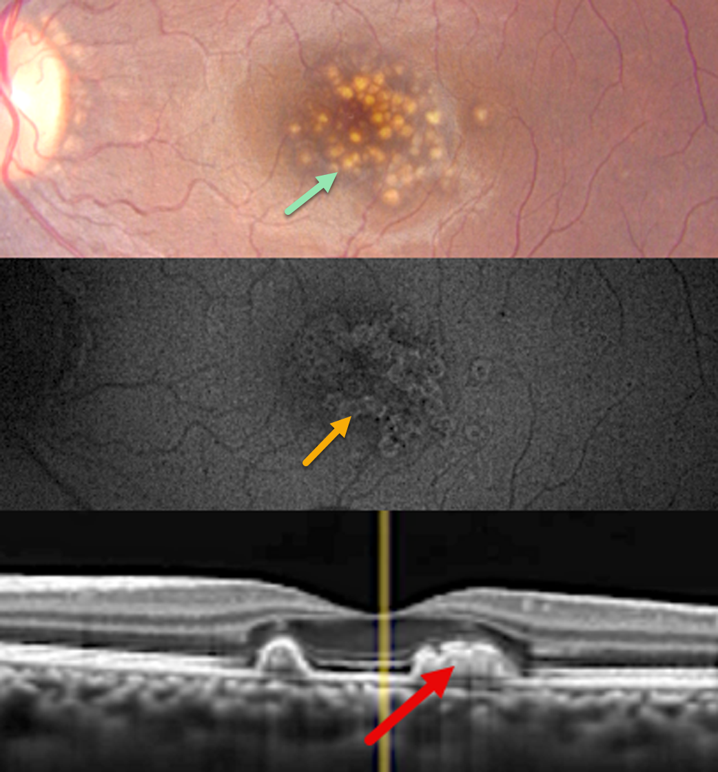
DOMINANTLY INHERITED RADIAL BASAL LAMINAR DRUSEN
Abhishek Karra and Ravindra Karra
Originally posted on @retina.rocks July 14, 2026
This healthy asymptomatic 27YO female presented for a routine eye examination. Ocular family history was negative. Vision was 20/20 OU.
Color photography of her left eye shows extensive, larger, well-defined drusen throughout the central macula. These drusen show a rim of hyper-fundus autofluorescence (FAF) with a central hypo-FAF core. The temporal foveal hyperreflective drusen are confluent, indenting the overlying retina into the outer nuclear layer. Similar findings were noted in her right eye (not shown).
Learning Points:
Doyne’s, also known as dominantly inherited radial basal laminar drusen or Malattia Leventinese, is a rare macular disorder caused by a mutation in the EFEMP1 gene on chromosome 2p16, although we have seen similar phenotypes with negative genetic testing. The EFEMP1 protein is a member of the fibulin family of extracellular matrix glycoproteins. The defective protein creates an abnormally thickened RPE basement membrane. Centrally large, nodular, and confluent drusen develop, along with a temporal radiating pattern of smaller cuticular drusen. Later, there may be variable amounts of RPE hyperplasia and fibrous metaplasia. Macular neovascularization may also occur.
Clues to the likely diagnosis of Doyne’s in our patient include extensive central, symmetrical drusen in a 27YO and pathognomonic drusen that indent the overlying retina on OCT. In addition, FAF usually shows hypo-FAF in age-related drusen. The lesions in Doyne’s, as in this patient, usually show hyper-FAF, likely from unmasking of the RPE by overlying outer retinal thinning.
RADIAL BASAL LAMINAR DRUSEN
Akansha Sharma, Manish Nagpal and Navneet Mehrotra
Originally posted on @retina.rocks March 30, 2026
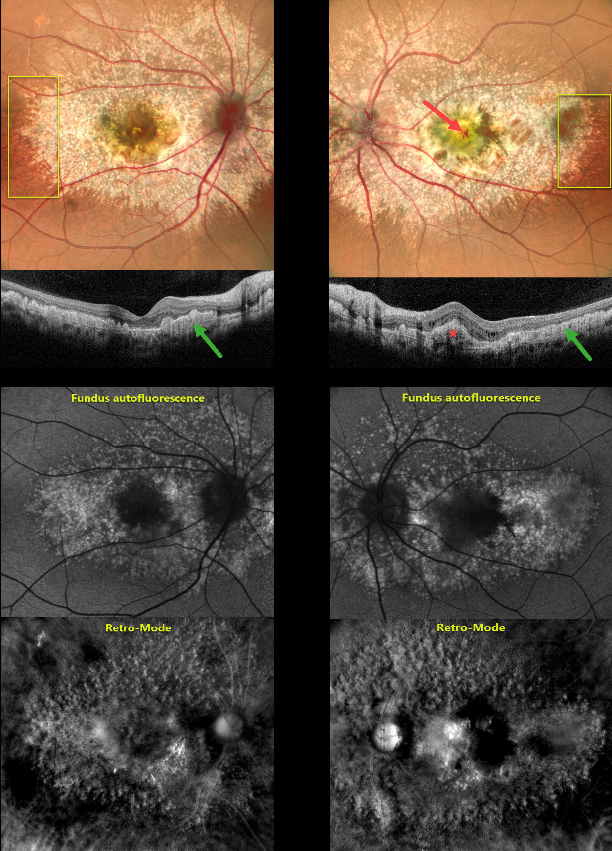
This 29YO male presented with a 1-month history of decreased vision in his left eye. Vision was 20/20 OD and counting fingers OS.
Pseudocolor SLO imaging shows extensive mixed and confluent drusen extending throughout the posterior pole in both eyes. A radiating linear pattern of fine drusen is present in each distal temporal macula. OCT scanning shows drusen indenting the overlying retina, extending into the outer nuclear layer and, in some places, up to the outer plexiform layer. Macular neovascularization (MNV) is present in the left fovea.
Fundus autofluorescence (FAF) shows macular hyper-FAF with surrounding relative hypo-FAF. Retro-Mode imaging reveals a dramatic, almost 3-dimensional appearance of depressions and mounds resembling the surface of the moon. Anti-VEGF therapy was started for the left eye.
Learning Points:
Doyne’s, also known as dominantly inherited radial basal laminar drusen or Malattia Leventinese, is a rare macular disorder caused by a mutation in the EFEMP1 gene on chromosome 2p16, although we have observed similar phenotypes in the absence of genetic testing. The EFEMP1 protein is a member of the fibulin family of extracellular matrix glycoproteins. The defective protein creates an abnormally thickened RPE basement membrane. Centrally large, nodular and confluent drusen develop, along with a temporal radiating pattern of smaller cuticular drusen. Later, there may be variable amounts of RPE hyperplasia and fibrous metaplasia. MNV may also occur.
Fundus autofluorescence (FAF) usually shows hypo-FAF in age-related drusen. The lesions in Doyne’s, as in this patient, usually show hyper-FAF, likely from unmasking of the RPE by overlying outer retinal thinning.
DOMINANTLY INHERITED RADIAL BASAL LAMINAR DRUSEN
Rohan Jain and Manish Nagpal
Originally posted on @retina.rocks October 28, 2025
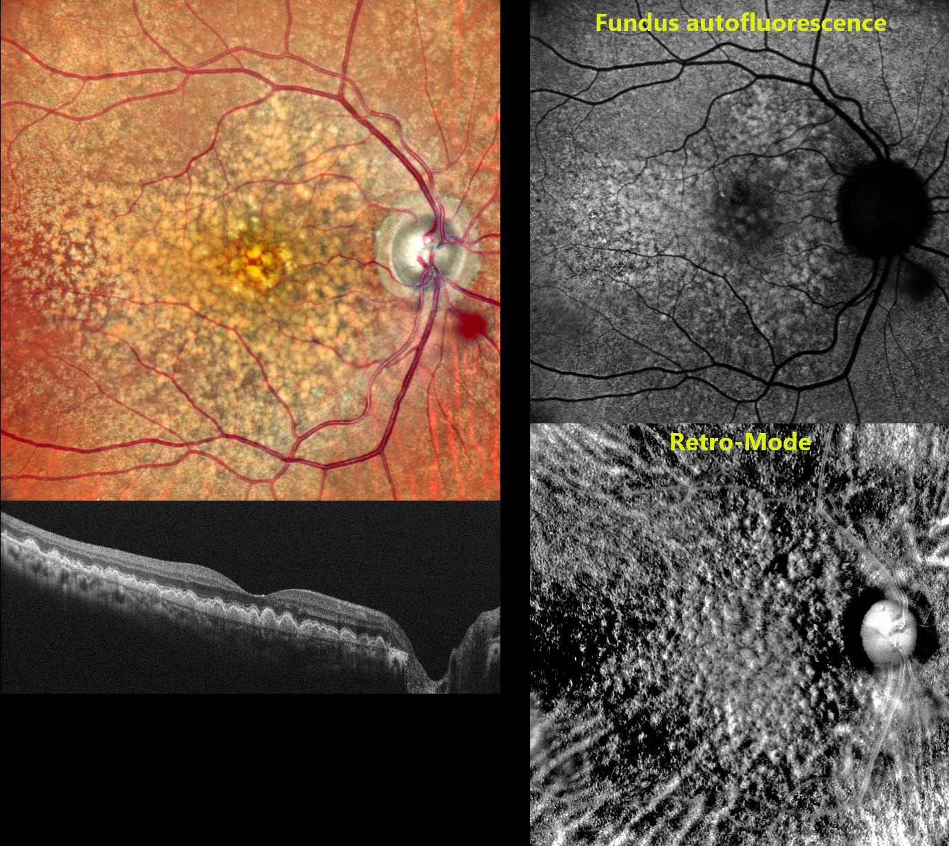
This 58 YO female presented to us with 2 to 3 years of bilateral vision loss. There was no family history of eye disease. Vision was 20/20 OU.
Pseudocolor SLO imaging shows extensive mixed and confluent drusen extending throughout the right posterior pole. OCT scanning shows drusen indenting the overlying retina, extending into the outer nuclear layer and, in some areas, up to the outer plexiform layer. Fundus autofluorescence (FAF) shows macular hyper-FAF with surrounding relative hypo-FAF. Retro-Mode imaging reveals a dramatic, almost 3-dimensional appearance of depressions and mounds resembling the surface of the moon. Identical findings were noted in her left eye (not shown).
Learning Points:
Doyne’s, also known as dominantly inherited radial basal laminar drusen or Malattia Leventinese, is a rare macular disorder caused by a mutation in the EFEMP1 gene on chromosome 2p16, although we have seen similar phenotypes with negative genetic testing. The EFEMP1 protein is a member of the fibulin family of extracellular matrix glycoproteins. The defective protein creates an abnormally thickened RPE basement membrane. Centrally large, nodular, and confluent drusen develop, along with a temporal radiating pattern of smaller cuticular drusen. Later, there may be variable amounts of RPE hyperplasia and fibrous metaplasia. Macular neovascularization may also occur.
Fundus autofluorescence (FAF) usually shows hypo-FAF in age-related drusen. The lesions in Doyne’s, as in this patient, usually show hyper-FAF, likely from unmasking of the RPE by overlying outer retinal thinning.
DOMINANTLY INHERITED RADIAL BASAL LAMINAR DRUSEN WITH POSSIBLE POLYPOIDAL CHOROIDAL VASCULOPATHY
Malvika Singh and Manish Nagpal
Originally posted on @retina.rocks June 25, 2025
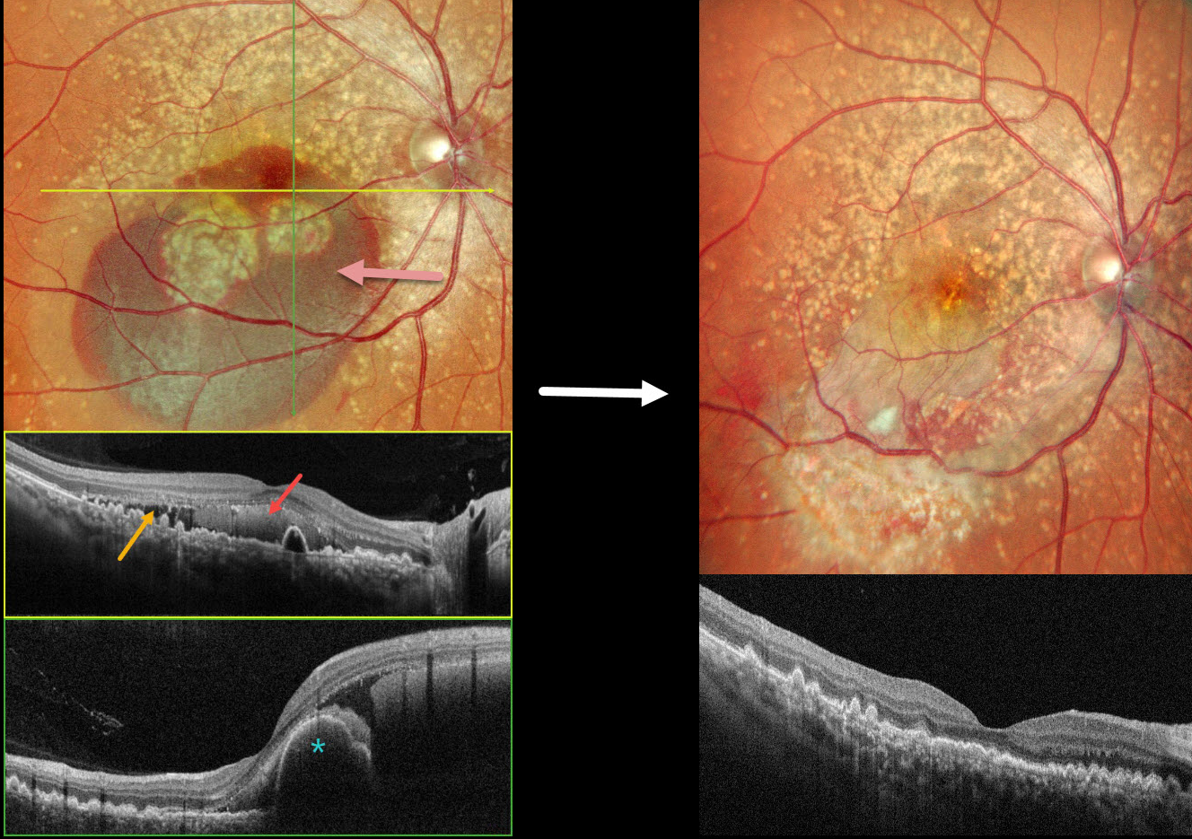
This 49YO female presented with 3 days of decreased vision in her OD. Vision was 20/70 OD and 20/15 OS. There was no family history of eye disease.
Pseudocolor SLO imaging shows extensive mixed and confluent drusen extending through each macula into the midperipheries. Inferior submacular blood extends into the right foveal center. OCT scanning shows extensive conically shaped drusen. The subretinal blood in her right eye is hyperreflective, with additional temporal subretinal fluid. A bilobed nodular RPE detachment is present within the nasal blood.
She underwent pars plana vitrectomy, fluid-air exchange, and intravitreal Avastin. One month postoperatively, vision improved to 20/15 OD with resolved fluid and trace residual blood.
Learning Points:
Doyne’s honeycomb macular dystrophy, also known as dominantly inherited radial basal laminar drusen or Malattia Leventinese, is a rare macular disorder caused by a mutation in the EFEMP1 gene on chromosome 2p16. The EFEMP1 protein is a member of the fibulin family of extracellular matrix glycoproteins. The defective protein creates an abnormally thickened RPE basement membrane.
Centrally large, nodular, and confluent drusen are noted, along with a temporal radiating pattern of smaller cuticular drusen. Later, there may be variable amounts of RPE hyperplasia and fibrous metaplasia. Macular neovascularization, as in this case, may also develop. Our patient’s pre-op right OCT was suggestive of polypoidal choroidal vasculopathy, although this is not a known association with Doyne’s.
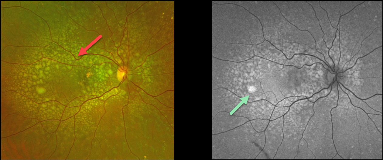
DOMINANTLY INHERITED RADIAL BASAL LAMINAR DRUSEN
Originally posted on @retina.rocks August 3, 2023
This 53YO female presented with these asymptomatic macular changes. She denied any family history of eye disease. Vision was 20/30 OD and 20/25 OS.
Optos color RG imaging shows extensive mixed drusen throughout the right posterior pole. The drusen hyper-autofluoresce.
OCT B-scan shows the sub-RPE drusen indenting and extending into the outer nuclear layer and up to the outer plexiform layer in some places. Similar findings were noted in her left eye (images not shown).
Learning Points:
Doynes, also known as dominantly inherited radial basal laminar drusen or Malattia Leventinese, is a rare macular disorder caused by a mutation in the EFEMP1 gene on chromosome 2p16. The EFEMP1 protein is a member of the fibulin family of extracellular matrix glycoproteins. The defective protein creates an abnormally thickened RPE basement membrane.
Centrally large, nodular, and confluent drusen develop, along with a temporal radiating pattern of smaller cuticular drusen. Later, variable amounts of RPE hyperplasia and fibrous metaplasia may be present. Macular neovascularization may also occur.
Fundus autofluorescence (FAF) usually shows hypo-FAF in age-related drusen. The lesions in Doyne’s, as in this patient, usually show hyper-FAF, likely from unmasking of the RPE by overlying outer retinal thinning.
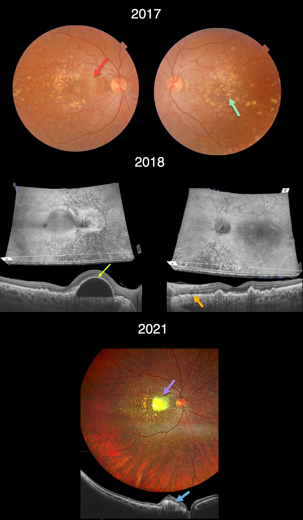
DOMINANTLY INHERITED RADIAL BASAL LAMINAR DRUSEN
Barbara Parolini and Veronika Matello
Originally posted on @retina.rocks September 16, 2022
This 33YOF presented in 2017 with a recent history of vision loss in her right eye. She had no past ocular or medical history, nor family history of eye disease. Vision of 20/70 OD and 20/20 OS.
Triton color imaging shows extensive, mostly large drusen scattered throughout each macula, extending into the midperipheries. A large serous retinal pigment epithelial detachment (PED) is noted in the nasal right macula.
Triton swept-source OCT analysis in 2018 shows numerous unique perspectives. 3D reconstruction reveals extensive lumpy drusen extending throughout the posterior poles, especially outside each macula. B-scans show extensive variably elevated drusen. The large PED is evident in the nasal right macula. The retina external to the ellipsoid zone is thickened and hyperreflective.
Three years later vision decreased to 20/200 OD and was stable at 20/20 OS. The PED became clinically yellow and markedly hyperreflective on OCT (Canon Xephilio OCT-S1). The lesion failed to respond to anti-VEGF therapy.
Learning Points:
Doynes, also known as dominantly inherited radial basal laminar drusen or Malattia Leventinese, is a rare macular disorder caused by a mutation in the EFEMP1 gene on chromosome 2p16.
The EFEMP1 protein is a member of the fibulin family of extracellular matrix glycoproteins. The defective protein creates an abnormally thickened RPE basement membrane.
Centrally large, nodular,and confluent drusen are noted, along with a temporal radiating pattern of smaller cuticular drusen.
Later, there may be variable amounts of RPE hyperplasia and fibrous metaplasia, which is likely the cause of the vision loss in our patient’s right eye. Macular neovascularization may also develop.
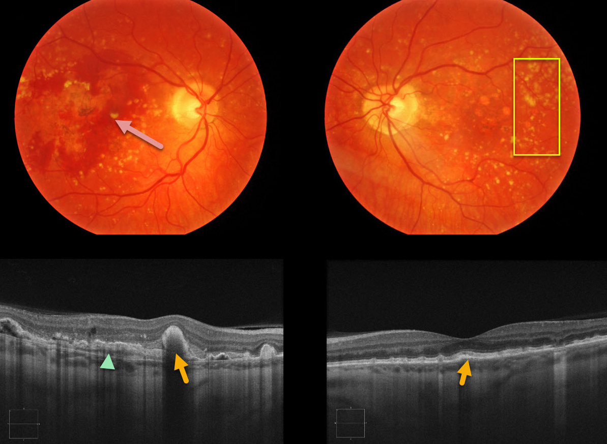
DOMINANTLY INHERITED RADIAL BASAL LAMINAR DRUSEN
Originally posted on @retina.rocks January 29, 2021
This 66YO female presented with acute vision loss of her right eye. There was no family history of eye disease. Extensive variably sized drusen were noted in both maculae, with subretinal blood from macular neovascularization (MNV) in her right eye.
OCT scanning shows variably sized and peaked drusen and subretinal hyperreflective material (SHRM) reminiscent of Doyne’s honeycomb macular dystrophy.
Learning Points:
Doyne’s honeycomb macular dystrophy, also known as dominantly inherited radial basal laminar drusen or Malattia Levantinese, is a rare macular disorder caused by a mutation in the EFEMP1 gene on chromosome 2p16. The EFEMP1 protein is a member of the fibulin family of extracellular matrix glycoproteins. The defective protein creates an abnormally thickened RPE basement membrane.
Centrally large, nodular, and confluent drusen are noted, along with a temporal radiating pattern of smaller cuticular drusen. Later, there may be variable amounts of RPE hyperplasia and fibrous metaplasia. MNV, as in this case, may also develop.
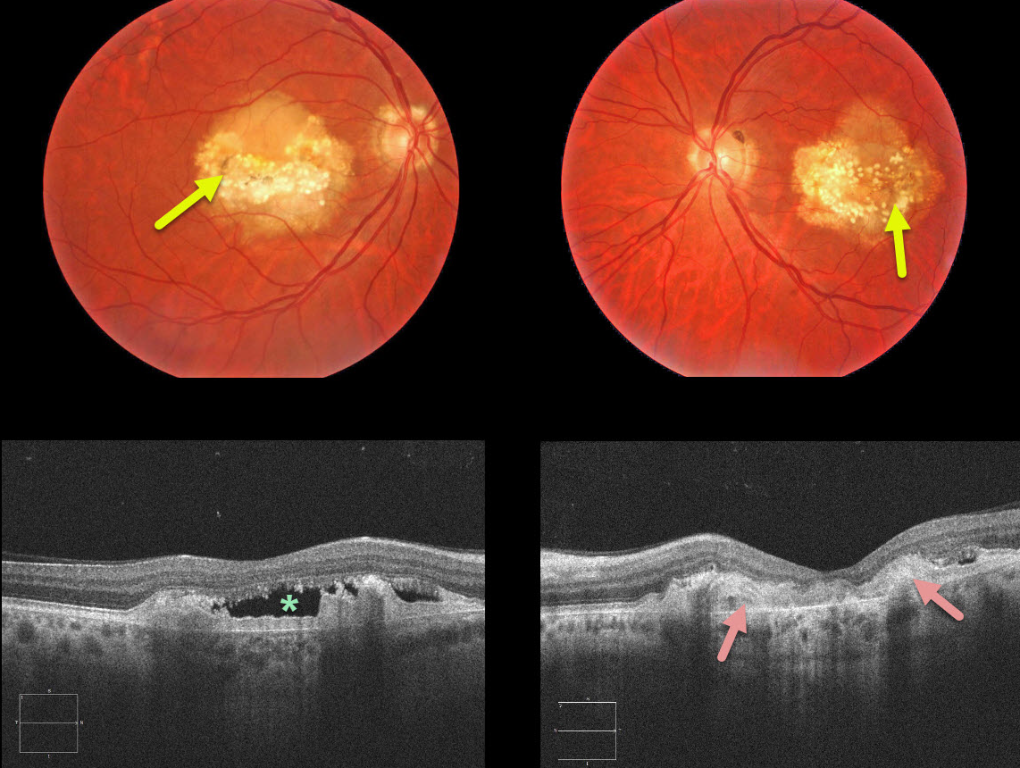
DOMINANTLY INHERITED RADIAL BASAL LAMINAR DRUSEN
Originally posted on @retina.rocks May 18, 2020
Our 54YO patient had a strong family history for macular problems. The fundus photos demonstrate radial cuticular drusen.
The OCT scans are even more dramatic with a saw-tooth pattern of drusen. Subretinal hyperreflective material (SHRM) is noted between Bruch’s membrane and the RPE and subretinal fluid is present in the right eye.
Learning Points:
Dominantly inherited radial basal laminar drusen (Doyne’s honeycomb macular dystrophy, Malattia Levantinese) is a rare genetic macular disorder. A defect in the EFEMP1 gene on chromosome 2p16 is thought to cause an abnormally thickened RPE basement membrane.
Centrally large, nodular and confluent drusen are noted, along with a temporal radiating pattern of smaller cuticular drusen. Later there may be variable amounts of RPE hyperplasia and fibrous metaplasia. Macular neovascularization may also develop.


