

Autosomal Recessive Bestrophinopathy
<< Back to Cases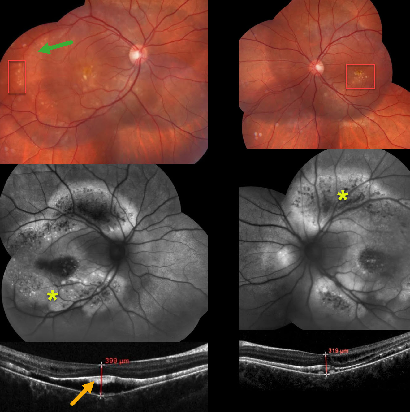
AUTOSOMAL RECESSIVE BESTROPHINOPATHY
The European VitreoRetina Society (EVRS) and Isha Acharya
Originally posted on @retina.rocks March 13, 2026
This healthy 17YO female presented with asymptomatic funduscopic findings. Family history was negative for eye disease. Vision was 20/40 OD and 20/50 OS.
Color imaging shows a rim of RPE pigment loss in the peripheral right temporal macula, along with bilateral yellow drusen-like changes. Fundus autofluorescence (FAF) shows much more pronounced findings with peripheral macular/midperipheral central hypo-FAF with surround hyper-FAF. Hyperreflective material is noted along the back surface of the detached foveal retina. Genetic testing was not performed due to cost concerns. Observation was recommended.
Learning Points:
Burgess et al first described autosomal recessive bestrophinopathy as a distinct BEST1-related disorder (American Journal of Human Genetics 2008;82:19-31). Unlike other BEST1 autosomal dominant disorders, patients have 2 abnormal alleles and thus no normally functioning bestrophin1 protein.
It is characterized by posterior pole vitelliform deposits and pigmentary changes that extend beyond the macula, subretinal fluid and vitelliform material, and intraretinal fluid (Casalino et al, Ophthalmology 2021;128:706-718). Vision gradually declines with age.
AUTOSOMAL RECESSIVE BESTROPHINOPATHY
Yuenpang Cheung and Stephen Tsang
Originally posted on @retina.rocks May 7, 2024
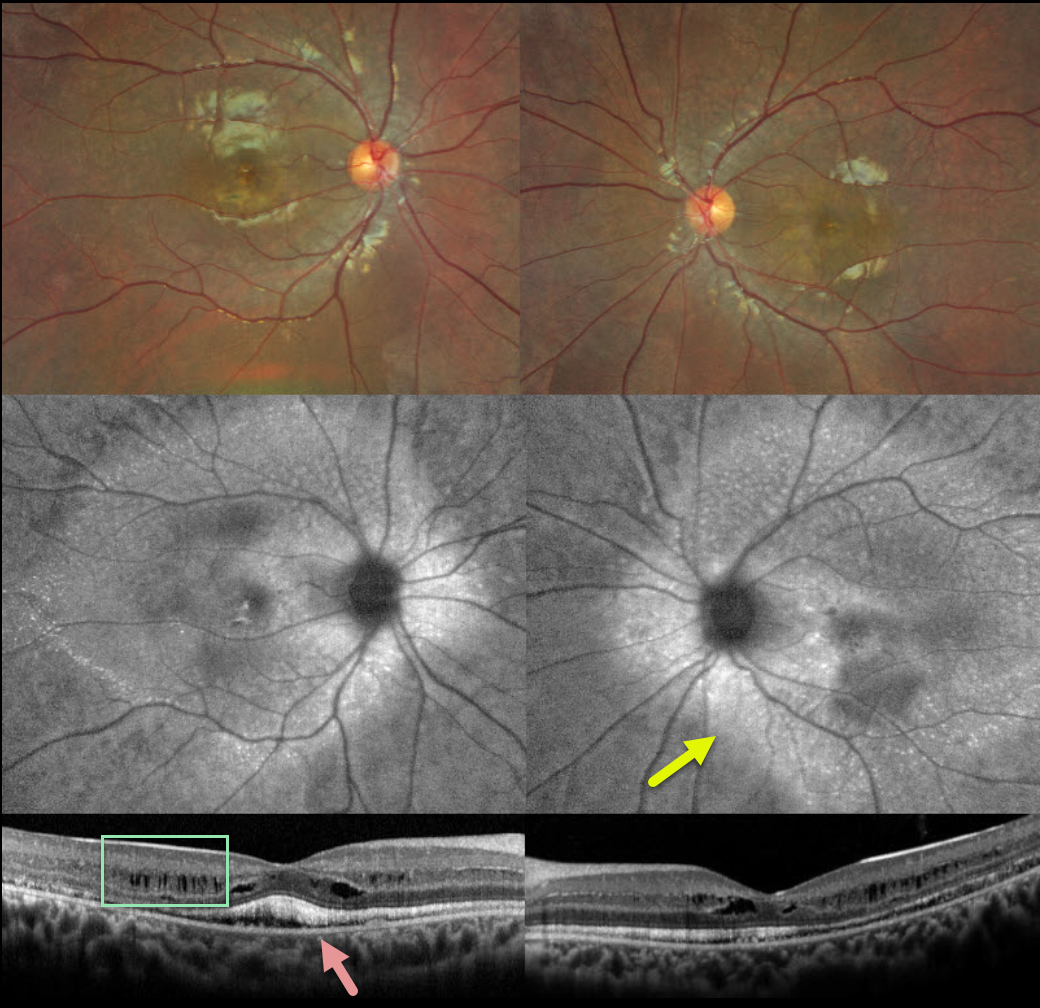
This 12YO boy has a known history of Best disease, which is being treated with topical dorzolamide for macular edema. He’s noticed some bilateral vision loss over the past 3 years. Vision is 20/25 OU.
Color imaging shows yellowing macular pigmentary changes. Fundus autofluorescence (FAF) shows much more pronounced changes with hyper-FAF extending beyond the arcades. OCT shows predominantly inner plexiform fluid versus schisis and hyperreflective subretinal vitelliform material.
Genetic testing revealed a heterozygous pathogenic BEST1 c.763C>T mutation, a heterozygous BEST1 c.113T>G mutation, and a single heterozygous PDE6B mutation. Oral acetazolamide 500mg daily was started.
Learning Points:
Burgess et al first described autosomal recessive bestrophinopathy as a distinct BEST1-related disorder (American Journal of Human Genetics 2008;82:19-31). Unlike other BEST1 autosomal dominant disorders, patients have 2 abnormal alleles and thus no normally functioning bestrophin1 protein.
It is characterized by posterior pole vitelliform deposits and pigmentary changes that extend beyond the macula, subretinal fluid and vitelliform material, and intraretinal fluid (Casalino et al, Ophthalmology 2021;128:706-718). Vision gradually declines with age.


