

Bacillary Layer Detachment
<< Back to Cases
DENGUE RETINITIS
Rohan Jain and Manish Nagpal
Originally posted on @retina.rocks September 9, 2026
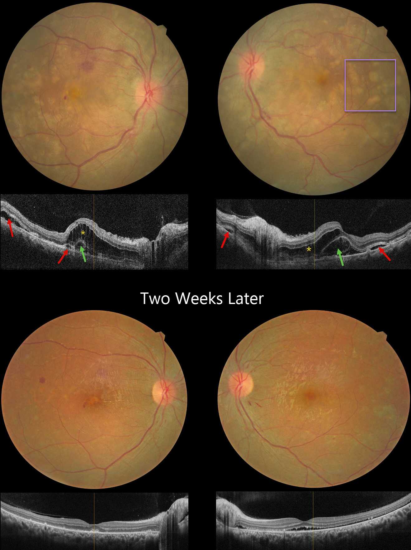
This 63YO male presented with 1 month of bilateral decreased vision following dengue fever, with positive Dengue NS1 antigen during the acute illness. Vision was 20/200 OD and counting fingers OS.
Pseudocolor SLO imaging shows extensive peripapillary cotton wool spots, retinal whitening, and significant macular edema. The fundus detail is partially obscured by overlying vitreous debris from vitritis. OCT scanning neurosensory retinal detachment with subretinal hyperreflective material, intraretinal cystoid edema, and subretinal fluid involving the macula in both eyes, consistent with post-dengue inflammatory retinitis. A possible bacillary layer detachment is also noted in the left eye.
Topical steroids and a 9-week course of tapering oral steroids, as well as a 3-week course of oral doxycycline, were started. Four months later, vision improved to 20/20 OD and 20/30 OS. The patches of white inner retinal ischemia are fading, and some lipid flecks have precipitated out in the mid-retina.
Learning Points:
Epidemic (post-fever) retinitis is an infectious or para-infectious entity that develops in immunocompetent individuals 2-6 weeks following a bacterial, viral, or protozoal infection. Rickettsiosis is the most identified cause, though a large proportion of cases remain idiopathic despite serologic testing; dengue, chikungunya, West Nile, Zika, leptospirosis, typhoid, and Ebola have also been implicated. Patients present with sudden painless vision loss. Fundus findings include unilateral or bilateral unifocal or multifocal patches of retinitis with possible nerve involvement, neuroretinitis with macular star, serous macular detachment, cystoid macular edema, and vasculitis or frosted branch angiitis.
Management is stratified by etiology and lacks controlled data: bacterial causes are treated with systemic antibiotics with or without corticosteroids, viral causes with corticosteroids alone, and many authors advocate observation, as the disease often resolves on its own. Corticosteroids should not be used as sole therapy while infectious causes (including TB, syphilis, and toxoplasmosis) remain in the differential. See Mahendradas et al (Indian J Ophthalmol 2020;68:1775-1786) for an excellent post-fever retinitis review.
HARADA DISEASE
Rohan Jain and Manish Nagpal
Originally posted on @retina.rocks August 20, 2026
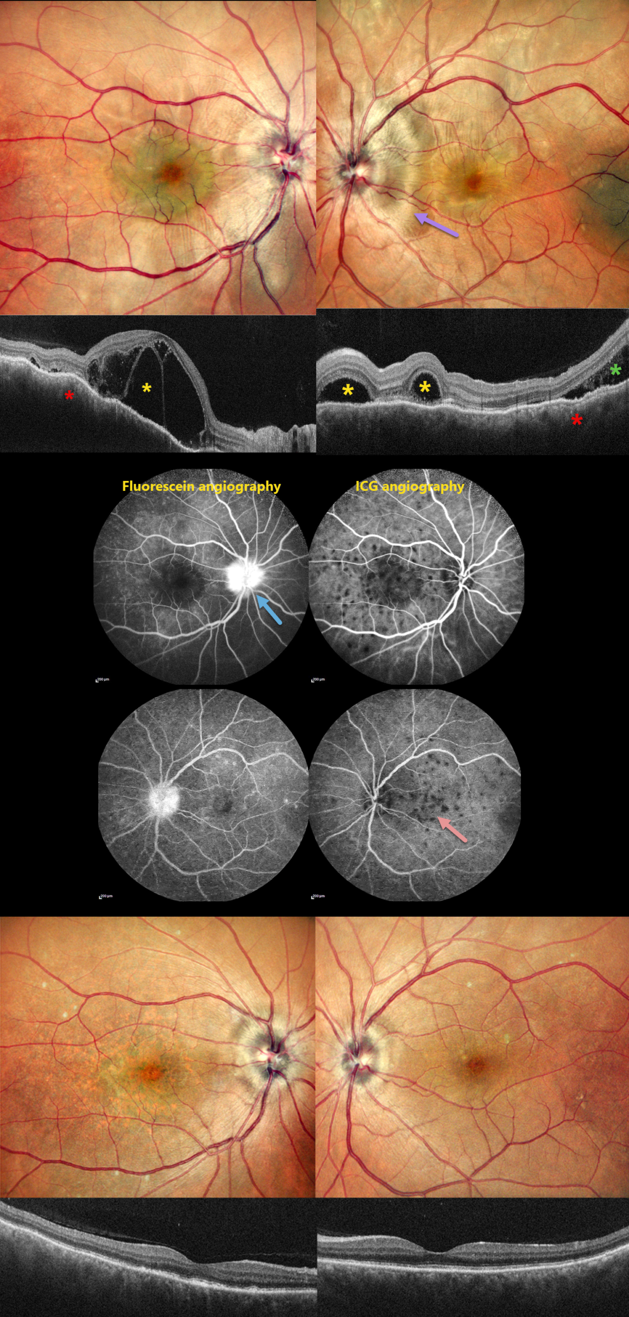
This 46YO male presented with 10 days of bilateral blurred vision with a left-sided headache. Vision was 20/200 OD and 20/20 OS. Anterior segments were normal.
Pseudocolor SLO imaging shows bilateral swollen nerves and irregular macular chorioretinal folds. OCT scanning shows multifocal bacillary layer detachments, subretinal fluid, and an undulating, thickened choroid. Fluorescein angiography shows bilateral multifocal areas of subretinal leakage and disc leakage, and indocyanine green angiography shows multiple hypofluorescent choroidal spots.
Based on the clinical and multimodal imaging findings, a diagnosis of acute Harada disease was established. He received a 3-day course of intravenous methylprednisolone followed by oral corticosteroids.
At the 2-month follow-up, the patient’s symptoms had completely resolved. Vision was 20/30 OD and 20/20 OS. The exudative findings are completely resolved on photography and OCT, and the choroidal thickening with overlying chorioretinal folds is also resolved.
Learning Points:
Harada disease is characterized by relatively acute bilateral panuveitis, optic nerve swelling, thickened choroid, chorioretinal folds, multifocal serous exudative retinal detachments, and bacillary layer detachments. Patients with only ocular findings have Harada disease (as in our patient), whereas those with additional systemic findings (including vitiligo, poliosis, headache, vertigo, and hearing loss) have Vogt-Koyanagi-Harada disease.
SYMPATHETIC OPHTHALMIA
The European VitreoRetinal Society (EVRS), Deepanshu Agrawal and Arun Bhargava
Originally posted on @retina.rocks February 20, 2026
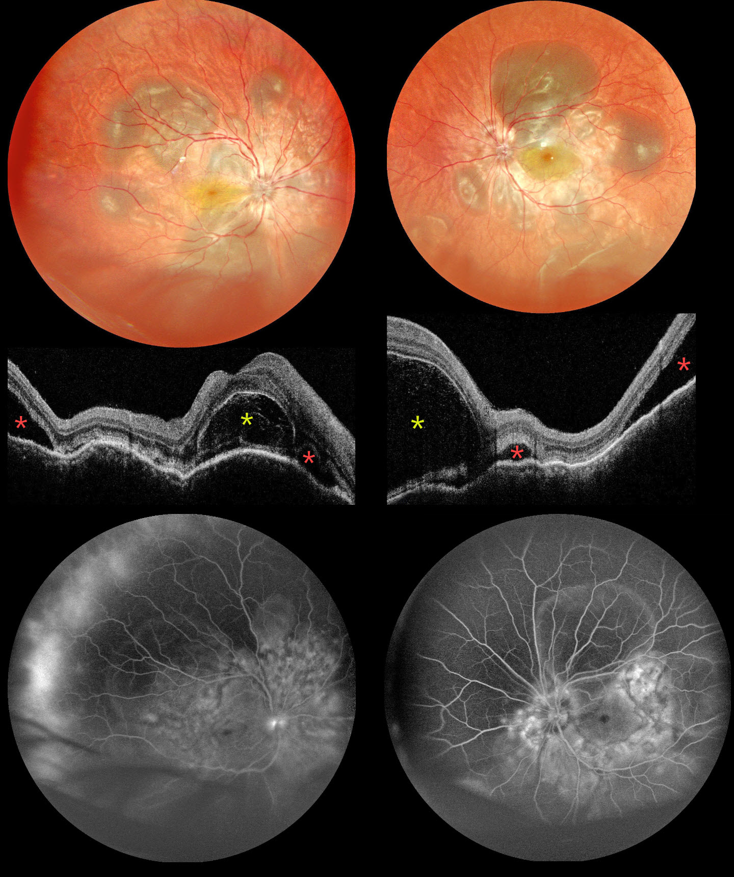
This 30YO male suffered an open globe injury from an iron nail OD on 11/2/25. Three days later, the corneo-limbal laceration was repaired elsewhere. He presented to us on 11/25 with 1 week of bilateral vision loss. Vision was hand motion OD and 20/400 OS. Mild anterior chamber cells were noted OU.
Multicolor SLO imaging shows bilateral multifocal pockets of subretinal fluid (SRF) along with inferior exudate retinal detachments. OCT scanning shows bilateral bacillary layer detachments (BALADs), SRF, and choroidal thickening and undulation. Fluorescein angiography shows multifocal subretinal leakage with disc leakage.
Learning Points:
Sympathetic ophthalmia is a rare, bilateral granulomatous panuveitis that develops following penetrating trauma or intraocular surgery to one eye, with an incidence of approximately 0.19% after open-globe injury (He et al, AJO 2022;234:117-125). It typically presents within the first year after the inciting event but can occur days to years later, presenting with decreased vision, pain, and photophobia. Findings mirror those seen in Vogt-Koyanagi-Harada disease, including granulomatous anterior uveitis, vitritis, exudative retinal detachment with BALAD, choroidal thickening, and Dalen-Fuchs nodules. Systemic corticosteroids remain the first-line treatment, with most patients requiring long-term corticosteroid-sparing immunosuppressive agents to prevent recurrences. Visual prognosis is generally good with treatment. See Parchand et al for a recent comprehensive update (Ind J Ophthalmol 2022;70:1931-1944).
Our patient was started on intravenous methylprednisolone 1g for 5 days, followed by oral steroids and azathioprine. All fluid resolved 3 weeks after starting treatment (not shown). Vision improved to 20/63 OU. A slow steroid taper was planned.
HARADA DISEASE
The European VitreoRetina Society (EVRS) and Ridham Nanda
Originally posted on @retina.rocks January 23, 2026
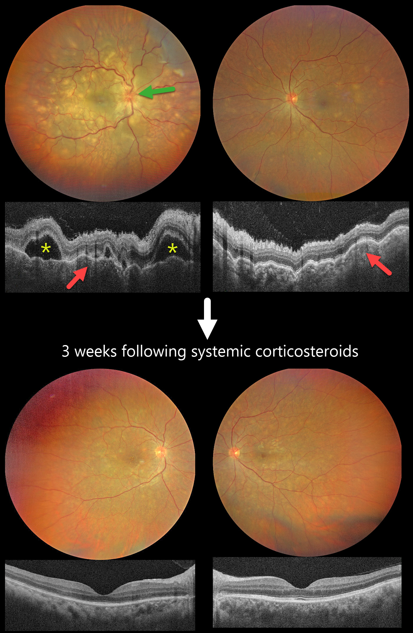
This previously healthy 40YO female presented with 2 days of blurred vision. She denied any systemic symptoms. Vision was 20/200 OD and 20/60 OS.
Color photography shows multifocal yellow-white subretinal lesions scattered throughout each posterior pole, with the right eye greater than the left, associated with overlying multifocal exudative retinal detachments. The right nerve is also edematous. OCT scanning shows a bilateral thickened choroid with overlying chorioretinal folds and multifocal serous detachments OD.
Intravenous methylprednisolone (1000mg/day) was administered for five days, followed by a 2-week course of oral prednisone (1mg/kg). Three weeks later, the funduscopic and OCT findings normalized. Vision improved to 20/30 OD and 20/20 OS.
Learning Points:
Harada disease is characterized by relatively acute bilateral panuveitis, optic nerve swelling, thickened choroid, chorioretinal folds, multifocal serous exudative retinal detachments, and bacillary layer detachments. Patients with just ocular findings have Harada disease (our patient), whereas those with additional systemic findings (including vitiligo, poliosis, headache, vertigo, and hearing loss) have Vogt-Koyanagi-Harada disease.
TUBERCULAR SERPIGINOUS-LIKE CHOROIDITIS
The European VitreoRetinal Society (EVRS) and Vaibhav Sethi
Originally posted on @retina.rocks January 2, 2026
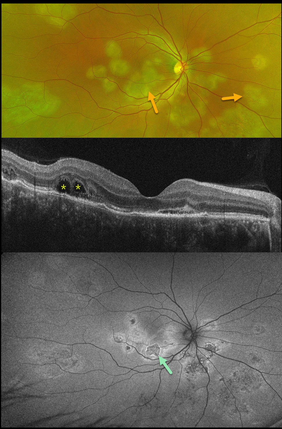
This previously healthy 41YO male presented with 4 days of blurred vision in his right eye. Vision was 20/80 OD and 20/20 in his normal OS.
Optos color RG imaging shows multifocal subretinal creamy white placoid lesions in the macula, peripapillary retina, and midperiphery. OCT scanning shows variable disruption of the outer retinal bands, as well as bacillary layer detachments (BALAD) temporally. The placoid lesions on fundus autofluorescence (FAF) show variable central hypo-FAF with more hyper-FAF borders.
Mantoux skin testing was positive at 24mm, and chest CT revealed left lower lobe ground glass opacities with hilar adenopathy. He was started on anti-tubercular therapy and oral prednisone.
Although tuberculosis most commonly presents with pulmonary involvement, extrapulmonary sites can include the gastrointestinal, skin, cardiovascular, genitourinary, and central nervous systems, as well as the eyes. Ocular involvement, like syphilis, can mimic virtually any type of uveitis, including anterior, intermediate, posterior, and panuveitis, retinitis, and retinal vasculitis, neuroretinitis, optic neuropathy, choroidal granuloma, choroiditis, and scleritis.
Learning Points:
Tubercular serpiginous-like choroiditis mimics serpiginous but affects younger patients with more multifocal and peripheral recurrences and progression if untreated (Agarwal et al, AJO 2020;220:160-169). Although the exact mechanism is not yet clear, a direct or indirect infectious trigger by the Mycobacterium tuberculosis is thought to cause choroiditis. Its origin is probably immunogenic since it seems to respond to corticosteroids and other immunosuppressants.
BALAD appears as a unique dome-shaped collection of intraretinal fluid from photoreceptor splitting. These detachments can be observed in an increasing number of conditions, most classically Vogt-Koyanagi-Harada disease (Cicinelli et al, Ophthalmology Retina 2020;4:454-456).
HARADA DISEASE
Originally posted on @retina.rocks December 16, 2025
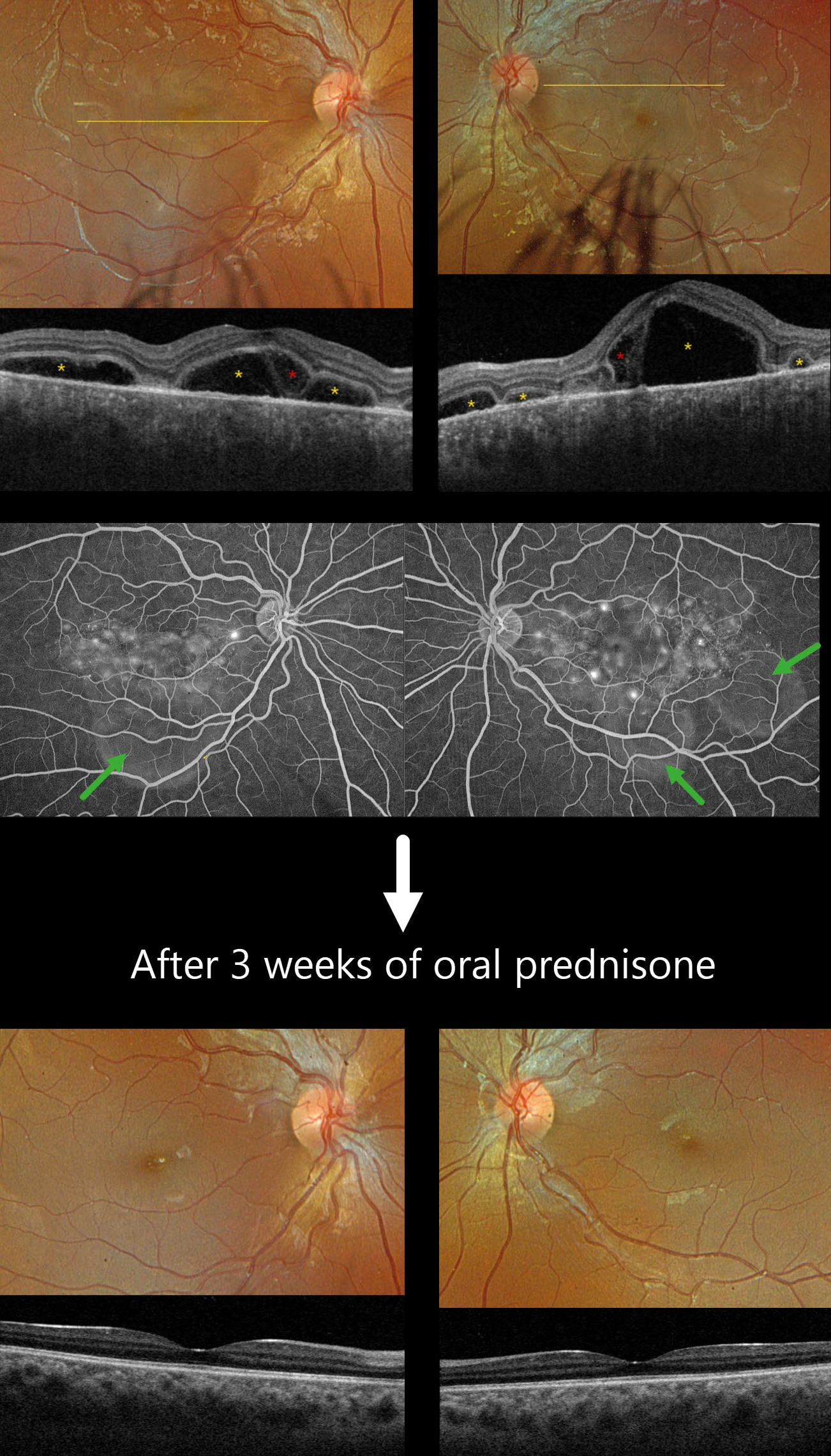
This previously healthy 13YO girl presented with several days of bilateral vision loss. Vision was 20/100 OD and 20/400 OS.
Optos color RGB imaging shows bilateral multifocal serous exudative retinal detachments. Triton swept-source OCT shows a complex pattern of multifocal pockets of subretinal fluid and bacillary layer detachments (BALAD). The choroid is markedly thickened. Fluorescein angiography shows multifocal pinpoint areas of subretinal leakage with pooling in the subretinal space and BALADs.
Learning Points:
Harada disease is characterized by relatively acute bilateral panuveitis, optic nerve swelling, thickened choroid, chorioretinal folds, multifocal serous exudative retinal detachments, and bacillary layer detachments. Patients with just ocular findings have Harada disease, whereas those with additional systemic findings (including vitiligo, poliosis, headache, vertigo, and hearing loss) have Vogt-Koyanagi-Harada disease.
Our patient was started on oral prednisone 40mg daily, and one week later, vision improved to 20/60 OU with resolution of all exudation (not shown). Three weeks later, the fundus appeared normal, and the choroid was approaching normal thickness. A slow steroid taper was started.
HARADA DISEASE
The European VitreoRetina Society (EVRS), Malvika Singh and Manish Nagpal
Originally posted on @retina.rocks October 3, 2025
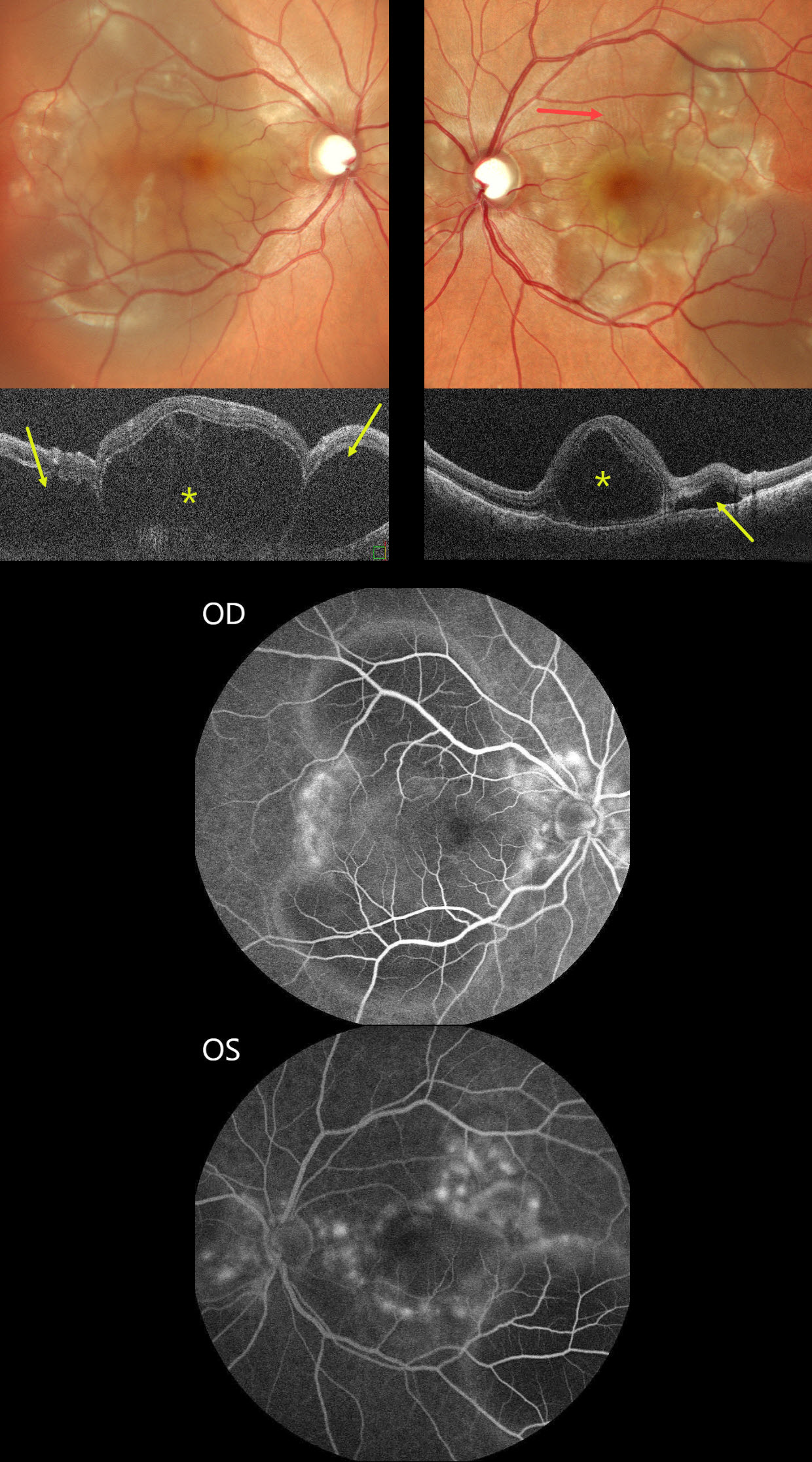
This previously healthy 30YO female presented with sudden bilateral vision loss for 5 days. Vision was counting fingers OU.
Color SLO imaging shows bilateral variably confluent serous detachments involving the posterior poles. Retinal folds are also noted in the left superior macula. OCT scanning shows a complex pattern of bacillary layer detachments (asterisks) and subretinal fluid (arrows). Fluorescein angiography shows multifocal subretinal leaks with pooling in the pockets of subretinal and intraretinal spaces.
She was started on oral steroids and immunomodulators in conjunction with a rheumatologist. One month later, vision returned to 20/20 OU with normalization of the funduscopic findings (not shown).
Learning Points:
Harada disease is characterized by relatively acute bilateral panuveitis, optic nerve swelling, thickened choroid, chorioretinal folds, multifocal serous exudative retinal detachments, and bacillary layer detachments. Patients with only ocular findings (as in our patient) have Harada disease, whereas those with additional systemic findings (including vitiligo, poliosis, headache, vertigo, and hearing loss) have Vogt-Koyanagi-Harada disease.
HARADA DISEASE
Nilesh Kumar
Originally posted on @retina.rocks August 20, 2025
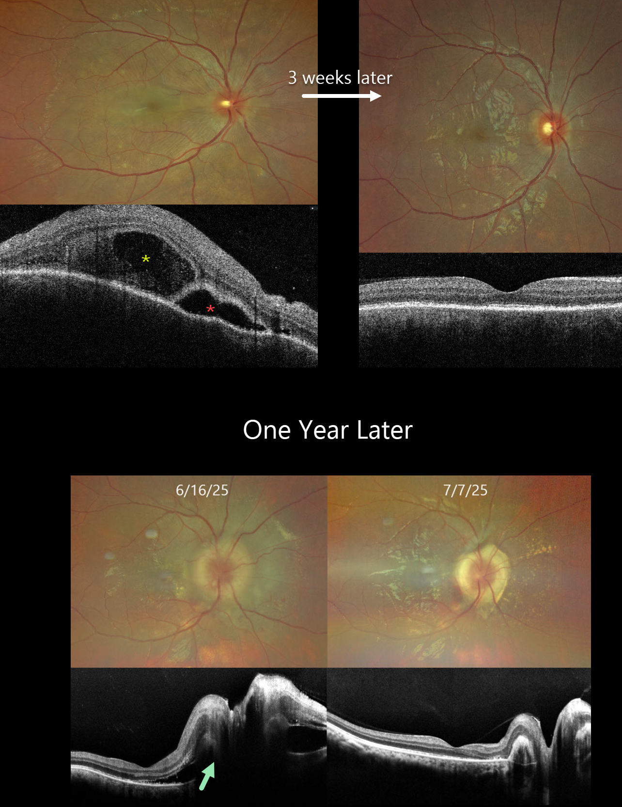
This previously healthy 24YO male presented with acute vision loss in his right eye. Vision was 20/200 OD and 20/20 in his normal OS. There were no neurologic or systemic symptoms.
Color photography shows an exudative retinal detachment involving the posterior pole. OCT shows a bacillary layer detachment, subretinal fluid with thickened retina and choroid. He was diagnosed with unilateral Harada disease and started on oral prednisone. Three weeks later, vision improved to 20/25 OD with marked clinical improvement. The oral prednisolone was tapered weekly over the next 6 weeks and stopped.
He returned one year later with recurrent vision loss. Vision was 20/200 OD. Exudative fluid with distal lipid flecks and a few small retinal hemorrhages surrounded the right nerve. OCT confirmed marked disk and peripapillary thickening with subretinal fluid. Oral prednisone was restarted, and oral azathioprine therapy was added. Three weeks later, the vision improved to 20/30. The fluid completely resolved with a ring of subretinal fibrosis surrounding the disc. The oral prednisone was tapered weekly over the next 6 weeks, and the oral azathioprine is planned to continue for the next year with regular monitoring.
Learning Points:
Harada disease is characterized by relatively acute bilateral panuveitis, optic nerve swelling, thickened choroid, chorioretinal folds, multifocal serous exudative retinal detachments, and bacillary layer detachments. Patients with just ocular findings have Harada disease, whereas those with additional systemic findings (including vitiligo, poliosis, headache, vertigo, and hearing loss) have Vogt-Koyanagi-Harada (VKH) disease. Although classically bilateral, early stages may present unilaterally before involving the fellow eye.
The disease generally progresses through four clinical stages: Prodromal, resembling a viral illness with systemic symptoms (fever, headache, meningismus); Uveitic, characterized by blurred vision, photophobia, optic disc edema, posterior choroidal thickening, and multifocal serous retinal detachments; Chronic/Convalescent, where depigmentation of the choroid leads to a “sunset-glow fundus” along with cutaneous signs like vitiligo, poliosis, and alopecia; and Recurrent, marked by repeated bouts of anterior uveitis and complications such as cataract, glaucoma, or macular neovascularization.
Management centers on high-dose systemic corticosteroids, often followed by steroid-sparing immunosuppressive agents to control inflammation and prevent recurrences. Early, aggressive therapy improves visual outcomes.
CHOROIDAL METASTASES WITH EXUDATIVE RETINAL DETACHMENT
Ayushi Gupta and Vishal Agrawal
Originally posted on @retina.rocks June 18, 2025
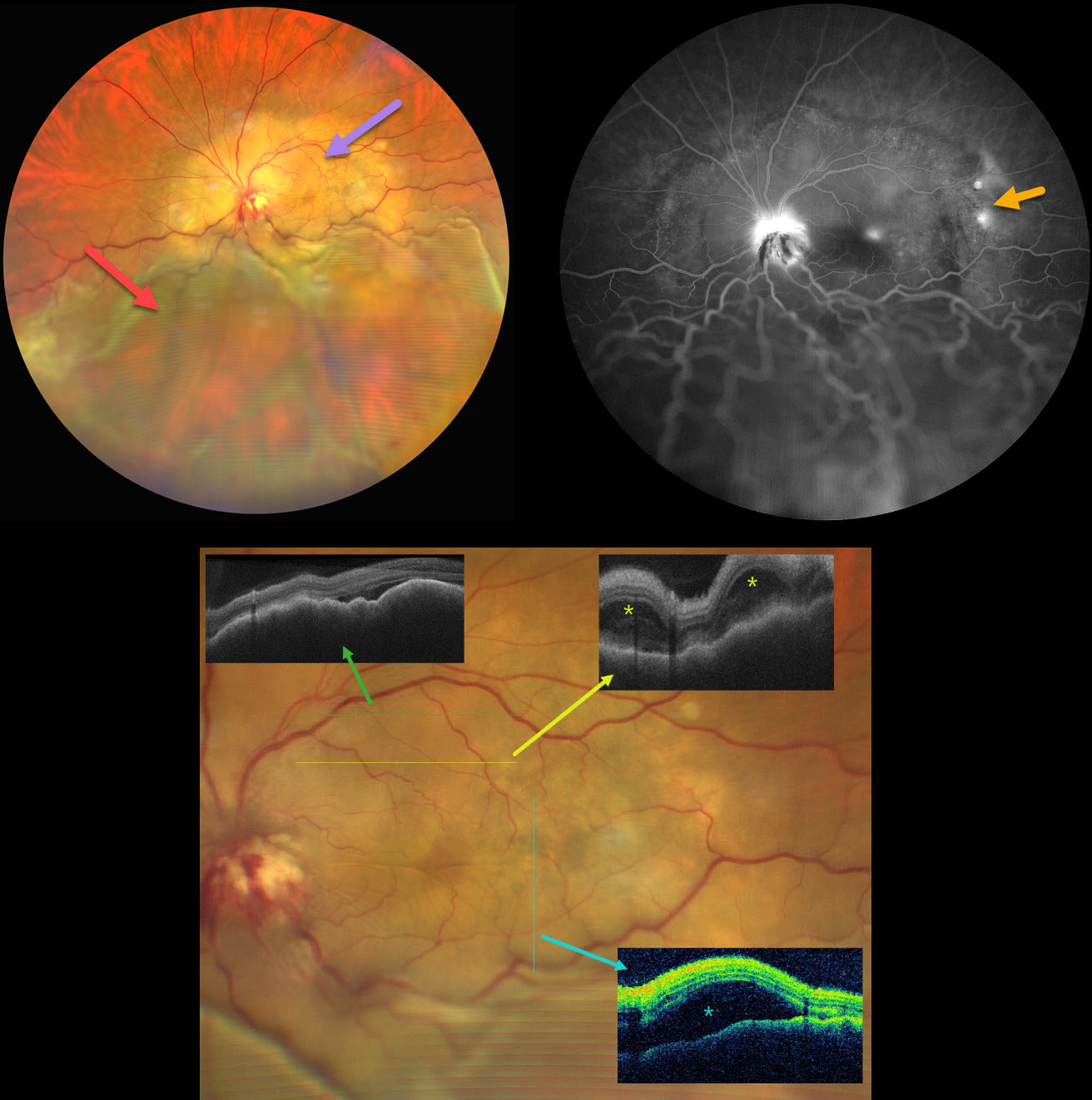
This 47YO female with a known history of metastatic breast carcinoma presented with several weeks of decreased vision in her left eye. She was on active chemotherapy and hormonal therapy. Vision was no light perception OD following trauma 30 years earlier, and hand motion OS.
Ultrawidefield color photography shows a large, amelanotic yellow choroidal lesion involving the entire macula and peripapillary choroid, which extends into the midperiphery. A large secondary inferior exudative retinal detachment involves the entire inferior hemisphere. This fluid shifted when laid supine. The optic disc is hyperemic with indistinct margins and associated blood.
OCT scanning shows a thickened, undulating choroid with variable overlying subretinal fluid and bacillary layer detachments. Fluorescein angiography shows multifocal subretinal leaks, choroidal lesion staining with possible overlying pooling, and intense nerve leakage.
Learning Points:
Although uveal melanoma is the most common primary intraocular malignancy, metastatic choroidal tumors are the most common intraocular malignancies. Lung and breast cancer are the most common sources.
Our patient had a known history of metastatic disease when she presented with this large lesion occupying her entire posterior pole. The extent of the metastasis and the overlying exudation are atypical in our experience. She was immediately referred to her oncologist for external-beam radiation, but was lost to follow-up.
HARADA DISEASE
Navneet Mehrotra and Tamanna Patel
Originally posted on @retina.rocks January 23, 2025
This 24YO female presented with several days of bilateral vision loss. She has a history of anti-glomerular basement membrane disease (anti-GBM) and is on hemodialysis. Her renal disease was quiescent, and there were no acute systemic complaints. There was no anterior segment inflammation, although mild vitreous cells were present bilaterally. Vision was 20/200 OD and 20/80 OS.
Fundus photography shows bilateral multifocal yellow subretinal lesions, inner retinal folds, some retinal hemorrhages, and mild disc edema. OCT scanning shows pockets of subretinal fluid, outer nuclear layer edema, and bacillary layer detachments. The choroid was thickened in both eyes on ultrasonography (not shown).
Oral prednisone 60mg/day was started in a tapering dose. Two weeks later, vision improved to 20/40 OD and 20/80 OS. The acute fundus findings were mostly resolved bilaterally.
Learning Points:
Harada disease is characterized by relatively acute bilateral panuveitis, optic nerve swelling, thickened choroid, chorioretinal folds, multifocal serous exudative retinal detachments, and bacillary layer detachments. Patients with just ocular findings have Harada disease, whereas those with additional systemic findings (including vitiligo, poliosis, headache, vertigo, and hearing loss) have Vogt-Koyanagi-Harada disease.
Anti-GBM disease is a rare autoimmune disorder characterized by autoantibodies that primarily target the basement membranes of the capillaries in the kidneys and lungs. Retinal findings include drusen occurring at an early age (Mansour et al, AJO Case Reports 2017;7:83-90). Acute fundus findings are rarely reported and consist of possibly steroid-induced central serous-like changes with multifocal retinal pigment epithelial detachments and exudative retinal detachments.
HARADA DISEASE
Anjana Mirajkar, Manish Nagpal and Navneet Mehrotra
Originally posted on @retina.rocks October 18, 2023
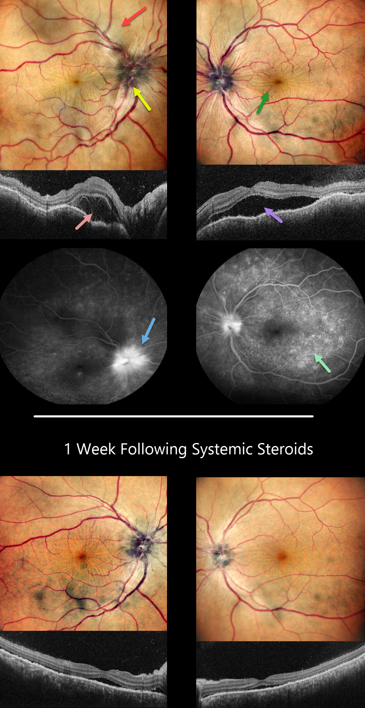
This 41YO female presented with headaches, redness, and pain OD for 1.5 months and OS for 2 weeks. She was diagnosed elsewhere with angle closure glaucoma, received a YAG peripheral iridotomy, and started on glaucoma drops. She then saw multiple other doctors who diagnosed her with papilledema.
Her CSF opening pressure was 21, and she was started on oral acetazolamide for a presumed diagnosis of idiopathic intracranial hypertension (IIH). In our office, vision was 20/100 OD and 20/60 OS. Intraocular pressure (IOP) was 30mmHG.
Anterior segments showed shallow anterior chambers with retrolental vitreous cells. Multicolor imaging shows bilateral irregular chorioretinal folds, multifocal serous detachments, and disc hyperemia.
OCT scanning shows a bacillary layer detachment OD, subretinal fluid OS, and a bilateral undulating thickened choroid. Fluorescein angiography shows bilateral optic nerve involvement and pinpoint subretinal leakage.
She was diagnosed with Harada’s disease and started on intravenous methylprednisolone for 3 days, followed by 60mg prednisone PO daily. She was also referred to rheumatology to start immunosuppressants. The acetazolamide was tapered and discontinued for the misdiagnosed IIH.
One week later, vision improved to 20/40 OU. IOP was 10mmHG OU with marked bilateral improvement in the chorioretinal folds, macular fluid, and choroidal thickening. A slow steroid taper was initiated pending initiation of immunosuppressants.
Learning Points:
This case has many classic findings for Harada’s disease, including bilateral panuveitis, optic nerve swelling, thickened choroid, chorioretinal folds, and multifocal serous exudative retinal detachments.
Patients with just ocular findings have Harada’s disease, whereas those with additional systemic findings (including vitiligo, poliosis, headache, vertigo, and hearing loss) have Vogt-Koyanagi-Harada (VKH) disease.
This case also reminds us that it is easy to misdiagnose these complex uveitis cases, especially when not all ocular findings are taken into full consideration in an attempt to give the patient a single, unifying diagnosis.
CHOROIDAL RUPTURE
Asma Samsudeen and Ashish Sharma
Originally posted on @retina.rocks July 6, 2023
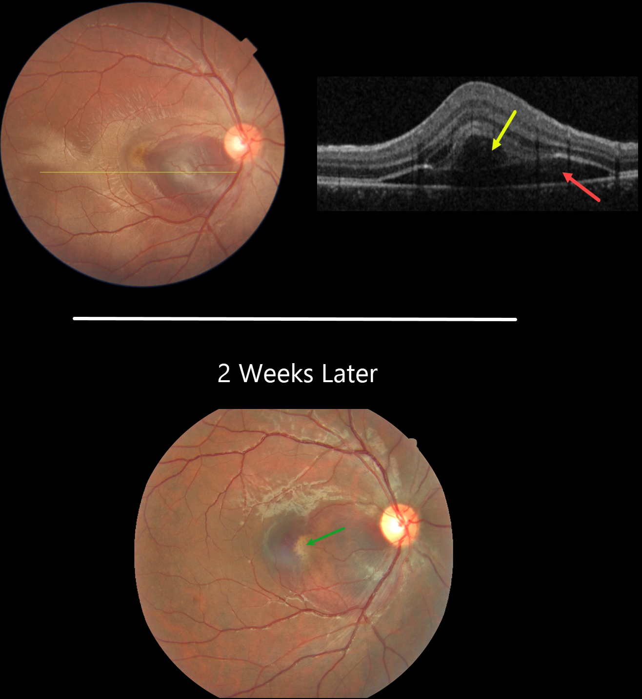
This 21YO male presented with immediate vision loss after being hit with a cricket ball in his right eye one day earlier. Vision was 20/60.
Fundus photography shows a subretinal hemorrhage in the nasal macula, a small rim of surrounding subretinal fluid, and a more temporal ill-defined commotio retina.
OCT scanning shows hyporeflective retinal elevation from the subretinal blood and fluid, along with an overlying bacillary layer detachment.
Our patient shows several findings classic for acute blunt trauma, including commotio retinae and subretinal blood. This subretinal blood almost always indicates an underlying choroidal rupture, which did, in fact, reveal itself on follow-up 2 weeks later, when vision returned to 20/20. The visual prognosis for these choroidal ruptures is good as long as the rupture spares the foveal center, although patients need lifelong surveillance due to the risk of secondary macular neovascularization.
Learning Points:
A bacillary layer (cone and rod inner and outer segments) detachment (BLD) appears as a unique dome-shaped collection of intraretinal fluid from photoreceptor splitting. These detachments can be observed in an increasing number of conditions, most classically Vogt-Koyanagi-Harada disease (Cicinelli et al, Ophthalmology Retina 2020;4:454-456).
About 8% of eyes with acute non-penetrating ocular trauma have a BLD (Venkatesh et al, Can J Ophthalmol 2022;57:328-336). These resolve within 2-10 days without visual or anatomic sequelae.
NEOVASCULAR AMD PRECHOROIDAL CLEFT + BACILLARY DETACHMENT
Originally posted on @retina.rocks November 29, 2022
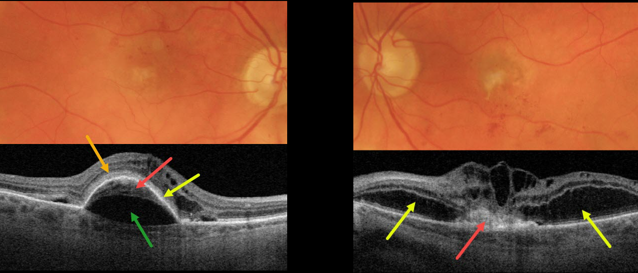
This 87YO patient presented with vision of 20/200 OD and counting finger vision OS from bilateral active neovascular AMD (nAMD).
The right macula has a few dots of central and superior macular blood. OCT scanning shows a type 1 macular neovascularization (MNV, located below the RPE) adherent to an overlying RPE detachment. A prechoroidal cleft separates the MNV from the underlying choroid. Subretinal hyperreflective material (SRHM) lies above the RPE detachment (orange arrow). Intraretinal fluid is noted nasally with trace subretinal fluid on either side of the RPE detachment.
The left macula has faint subfoveal fibrosis with some scattered more inferior subretinal blood. OCT scanning shows a type 2 MNV (located above the RPE), which is tethered to the overlying edematous retina. Bacillary layer detachments are noted nasal and temporal to the MNV.
Learning Points:
A prechoroidal cleft is a hyporeflective space between the RPE and Bruch’s membrane. These are seen in up to about 20% of treated eyes with nAMD, particularly with type 3 neovascularization (intraretinal MNV) and polypoidal choroidal vasculopathy.
They are usually associated with worse visual acuity due to potential complications, including RPE tear and subretinal hemorrhages (Kim et al, Retina 2017;37:2047-2055).
SHRM is an OCT finding that is invisible clinically. This material, located between the neurosensory retina and retinal pigment epithelium (RPE), is thought to consist of fluid, fibrin, blood, and other fibrovascular tissues.
SHRM is a biomarker of active disease that decreases with successful anti-VEGF therapy (Willoughby et al., Ophthalmology 2015;122:1846-1853). Persistent SHRM is associated with an increased incidence of scar formation and worse VA.
Bacillary layer (cone and rod inner and outer segments) detachment appears as a unique dome-shaped collection of intraretinal fluid from photoreceptor splitting. These detachments can be observed in an increasing number of conditions, most classically Vogt-Koyanagi-Harada disease (Cicinelli et al, Ophthalmology Retina 2020;4:454-456).
NEOVASCULAR AMD
Originally posted on @retina.rocks March 22, 2022
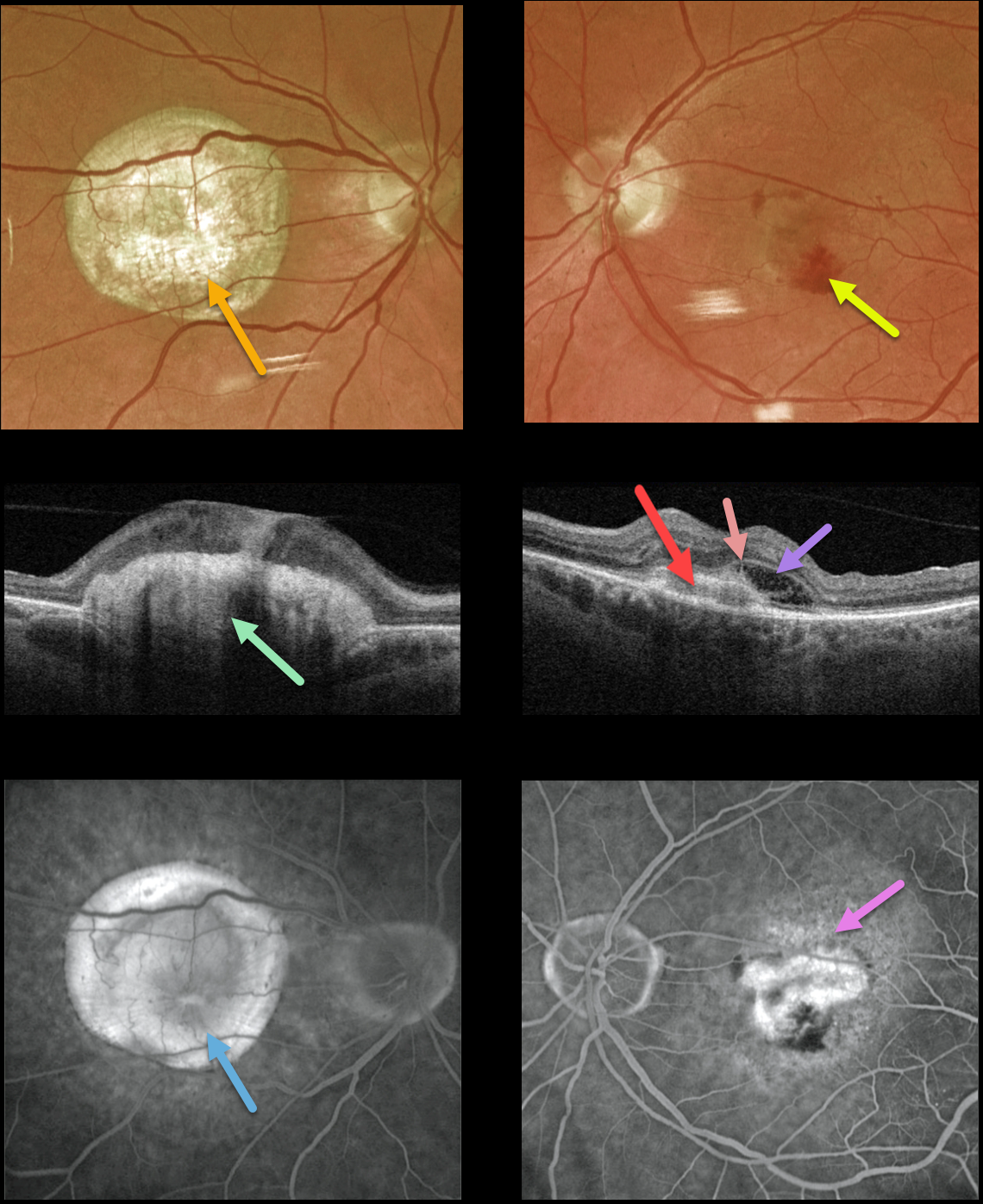
This 77YO male presented with counting fingers vision OD from a large fibrotic disciform scar and 20/200 vision OS from a new macular neovascularization.
The disciform scar is contracted and fibrotic, with marked hyperreflectivity on swept-source OCT. The MNV in his left eye is seen as opaque fluid with some subretinal blood. The type 2 MNV (located above the RPE) is noted on the OCT B-scan. There is also a bacillary detachment and a hyperreflective line indicating a possible pitchfork sign.
Optos fluorescein angiography shows diffuse staining of the subretinal scarring OD and leakage from the MNV OS.
Learning Points:
Disciform scars were the norm for untreated wet AMD before the anti-VEGF era that began in the mid-2000s. New-onset wet AMD diagnosed and treated with appropriate anti-VEGF dosing virtually never goes on to develop these lesions. There may be progressive macular atrophy following years of therapy, but not this type of fibrotic scarring.
An excellent review of wet AMD fibrotic lesions is in Souied et al’s AOS thesis (AJO 2020;214:151-171).
The pitchfork sign was originally described by Hoang et al (Retina 2013;33:1049-1055) as a unique OCT finding in inflammatory MNV. Falavarajani et al more recently noted that this finding is also associated with non-inflammatory MNV (Ophthalmic Surg Lasers Imaging Retina 2019;50:719-725).
The pathogenesis of the avascular pitchfork spikes is unknown, but may be related to outer retinal traction or Müller cell activation.
HARADA DISEASE
Originally posted on @retina.rocks February 7, 2022
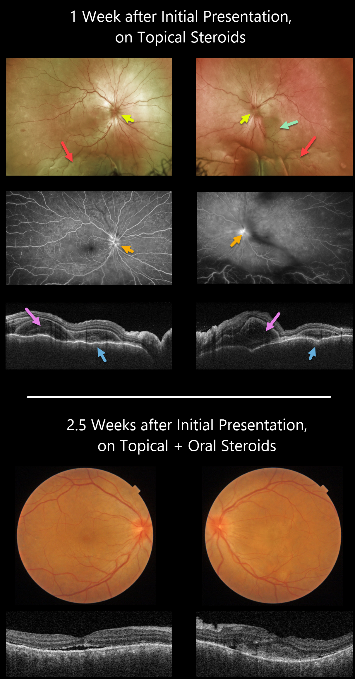
This 35YO female presented with a 2-week history of severe bilateral vision loss, photophobia, and red eyes. There was no significant past medical history.
Vision was counting fingers OU. There was moderate bilateral conjunctival injection, severe cells with moderate flare, moderate fibrin, and 360-degree posterior synechiae. Topical cycloplegia and steroids were prescribed.
One week later, she was referred to our retinal service. There was marked improvement of her anterior uveitis and the posterior synechiae were mostly broken throughout. There were moderate bilateral vitreous cells, markedly swollen optic nerves, irregular diffuse macular exudative fluid with inferior exudative retinal detachments.
Fluorescein angiography showed bilateral optic nerve leakage. Triton swept-source OCT showed bilateral markedly thickened and rippled choroid with multiple bacillary pockets of outer retinal thickening and fluid. She started oral prednisone 80mg daily.
Around 10 days later, vision improved to 20/200 OU, with improvement in optic nerve edema, macular fluid, bacillary detachments, and choroidal thickening. The inferior exudative detachments were also resolving (not imaged).
Learning Points:
This case has many classic findings for Harada disease, including bilateral panuveitis, optic nerve swelling, thickened choroid, chorioretinal folds, and multifocal serous exudative retinal detachments.
Given the severity of the panuveitis and exudative subretinal fluid, we were surprised that angiography did not show the more classic multifocal subretinal leaks with pooling in the overlying serous detachments and bacillary cavities.
Patients with just ocular findings have Harada disease, whereas those with additional systemic findings (including vitiligo, poliosis, headache, vertigo, and hearing loss) have Vogt-Koyanagi-Harada disease.
RETINAL ANGIOMATOUS PROLIFERATION (RAP)
Originally posted on @retina.rocks August 18, 2021
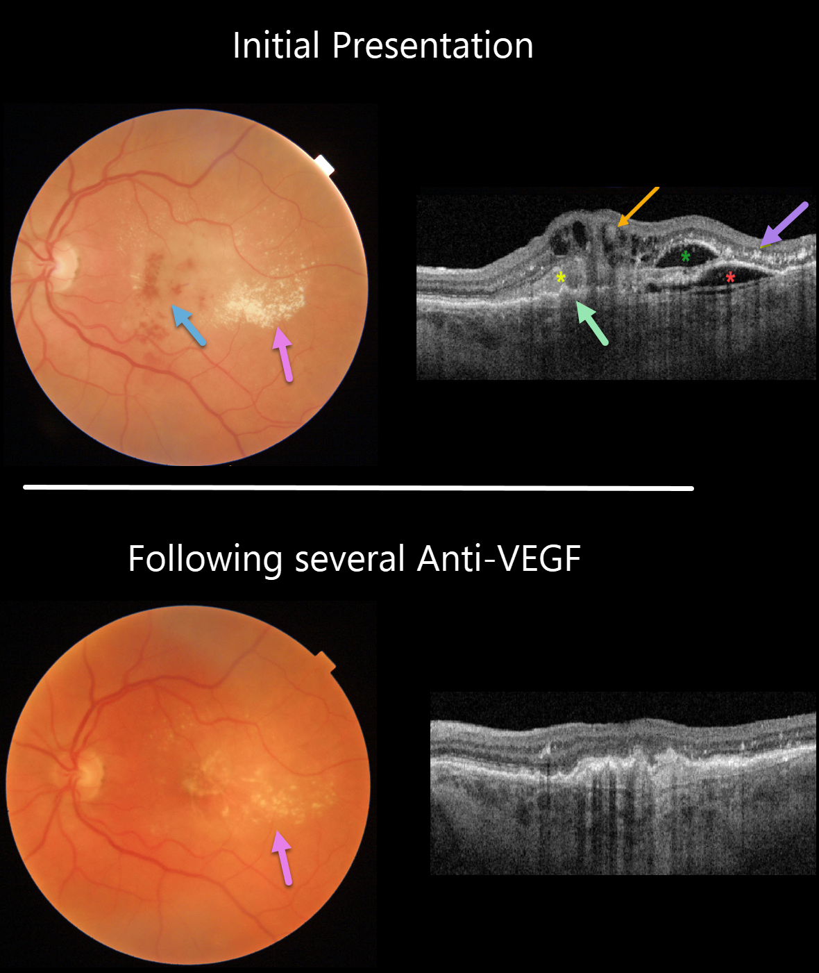
This 74YO man presented with counting fingers vision from previously untreated wet age-related macular degeneration (AMD).
Triton color imaging and swept-source OCT beautifully highlight this complex macular neovascularization (MNV). There are scattered inner and deep retinal hemorrhages, opaque fluid, and temporal lipid.
OCT shows an irregular shallow elevation of the RPE more nasally, possibly indicating a sub-RPE (type 1 MNV), with overlying subretinal hyperreflective material. More temporally, there is a more prominent area of RPE elevation that is more hyporeflective with an overlying pocket of intraretinal fluid vs a bacillary layer detachment. Finally, central inner retinal thickening and cysts are seen, indicating intraretinal neovascularization (type 3 MNV), also known as a retinal angiomatous proliferation (RAP) lesion. The lipid appears as hyperreflective clumps in the outer plexiform and outer nuclear layers.
The fluid and blood regressed, with significantly decreased lipid, after several monthly Avastin injections (image 2). Vision improved to 20/400.
Learning Points:
Unlike more typical age-related MNV that start either below (type 1 MNV) or occasionally above (type 2 MNV) the RPE, RAP lesions originate in the retina.
These incompetent vessels leak fluid and blood into the surrounding tissue. Their intraretinal location is a biomarker for RAP lesions.
SYSTEMIC LUPUS ERYTHEMATOSUS (SLE)
Originally posted on @retina.rocks December 29, 2020
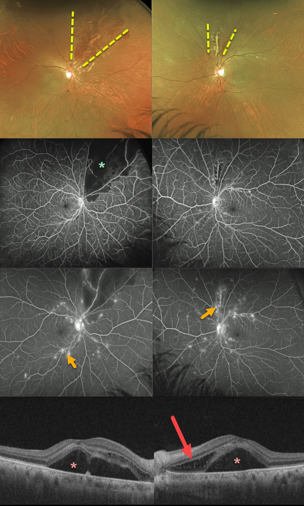
This 52YO female presented with severe bilateral vision loss (20/400 OD, counting fingers OS) following a several-week gastrointestinal illness with 60-pound weight loss.
She has bilateral areas of hemorrhagic retinal vasculitis, with areas of ischemia and leakage seen on fluorescein angiography. OCT shows macular subretinal fluid and a bacillary detachment in the left macula.
She had been hospitalized recently for hypertension and kidney failure and was diagnosed with a previously asymptomatic stroke. Extensive blood work was negative, and she denied symptoms of Behcet’s.
We suspected an underlying rheumatologic cause, and she was subsequently diagnosed with severe systemic lupus erythematosus (SLE) vasculitis.
She was started on high-dose oral prednisone followed by Cytoxan and hydroxychloroquine. Her ocular findings rapidly improved, and at her last examination 4 months later, vision had improved to 20/40 OD and 20/100 OS.
Learning Points:
SLE-associated retinopathy more typically appears as hypertensive retinopathy (bilateral nerve fiber layer infarcts and retinal hemorrhages), often in patients with cerebral vasculitis. Purtscher-like retinopathy can also be seen.
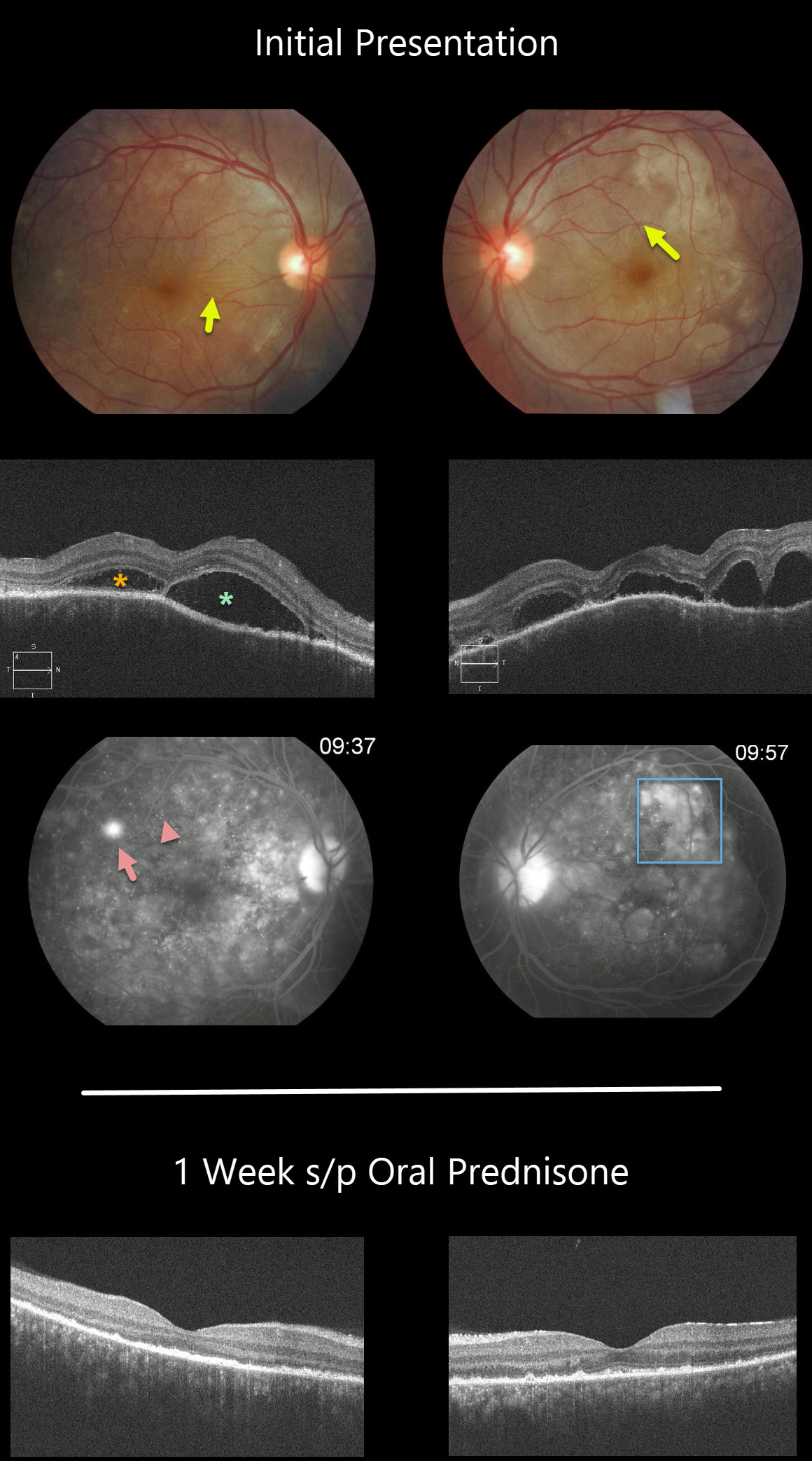
HARADA DISEASE
Originally posted on @retina.rocks September 23, 2020
This patient has classic posterior segment findings of Harada disease, including multifocal exudative serous retinal detachments.
Choroidal inflammation produces a markedly thickened choroid with overlying chorioretinal folds (seen in both eyes as variably radiating outer retinal folds).
The OCT scans show multifocal serous detachments with bacillary detachments. Fluorescein angiography shows multiple subretinal leaks with pooling of fluorescein within the various subretinal and bacillary spaces.
The patient started 60mg of oral prednisone daily. One week later, there was a dramatic improvement, with complete resolution of all intra- and subretinal fluid and normalization of choroidal thickness.
Learning Points:
Harada disease is a bilateral granulomatous panuveitis. These patients often need extended treatment with oral prednisone. Most can be successfully weaned off of steroids, although chronic treatment is sometimes necessary.
Choroidal thickening is a good biomarker for disease activity and thins with controlled disease activity.
Remember that in Harada disease, there are only ocular findings. Vogt-Koyanagi-Harada disease occurs when there are associated auditory, integumentary or neurologic symptoms.
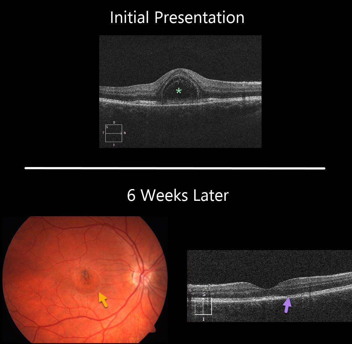
ACUTE IDIOPATHIC MACULOPATHY (AIM)
Originally posted on @retina.rocks July 23, 2020
This 22YO female presented with 1 week of unilateral vision loss and 20/200 vision in her right eye.
The macular OCT line scan demonstrates a bacillary layer (cone and rod inner and outer segments) detachment. This appears as a unique dome shaped collection of intraretinal fluid from photoreceptor myoid splitting.
Six weeks later vision improved to 20/40. The macular fluid resolved, with the development of a dry area of macular hyperpigmentation surrounded by a rim of pigment loss. OCT shows some subtle outer retinal foveal thinning with ellipsoid zone irregularities.
Learning Points:
Acute idiopathic maculopathy (AIM) is a usually unilateral idiopathic condition usually found in young people, often following a viral illness. It is usually self-limited, resolving within several weeks with significant visual improvement. The appearance of a bulls-eye lesion, as occurred in our patient, is classic for this disease.
Bacillary detachments can be observed in multiple conditions including Vogt-Koyanagi-Harada (VKH) disease, AIM, blunt trauma, or APMPPE.
See Fernandez-Avellaneda et al Retinal Cases & Brief Reports 2019 (published ahead of print) for a discussion of bacillary layer detachment in AIM.


