

Bartonella
<< Back to Cases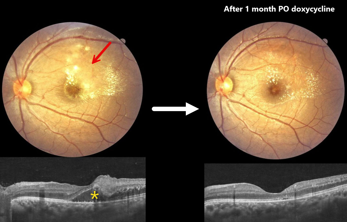
NEURORETINITIS
Anand Temkar, Surendra Pal and Prasanna Suresh
Originally posted on @retina.rocks February 25, 2026
This 21YO male presented with one month of intermittent headaches and vision loss in his left eye. Vision was 20/20 in his normal OD and 20/60 OS.
Color photography of the macula shows patches of yellow-white inner retinal opacification with superior and temporal lipid. OCT shows outer nuclear layer fluid with hyperreflective dots of lipid in the outer plexiform layer. Mild vitreous cells are present.
A presumed clinical diagnosis of Bartonella neuroretinitis was made, although serology could not be performed due to cost concerns. He was started on oral doxycycline 100mg PO BID and oral corticosteroids. Two weeks later, vision was 20/40 with resolving retinal opacification and fluid (not shown). Following a full month of oral therapy, vision improved to 20/20 with decreasing lipid and no fluid.
Learning Points:
The diagnosis for a unilateral swollen nerve is extensive. However, it becomes much smaller when associated with acute inner retinal ischemia and later lipid exudation within Henle’s layer (neuroretinitis), including cat scratch (Bartonella) and syphilis. When no underlying cause is found, the entity is called Leber’s idiopathic stellate neuroretinitis.
BARTONELLA
Originally posted on @retina.rocks February 20, 2025
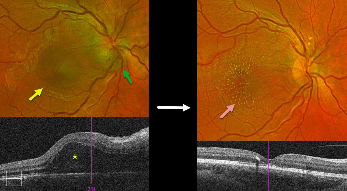
This 18YO female presented with a several-day history of vision loss in her right eye. Vision was 20/70 OD and 20/40 in her normal OS.
Optos color RG imaging shows inferior disc edema with a serous macular detachment. Triton swept-source OCT confirms the serous detachment extending towards the nerve.
Bartonella serology was positive, and she was placed on a 6-week course of oral antibiotics. Two months following presentation, vision improved to 20/40. The disc edema and serous detachment resolved, with the development of a lipid star.
Learning Points:
The diagnosis for a unilateral swollen nerve is extensive. However, it becomes much smaller when associated with acute inner retinal ischemia (not seen in our case) and, later, with lipid exudation within Henle’s layer (neuroretinitis), including cat-scratch (Bartonella) and syphilis. When no underlying cause is found, the entity is called Leber’s idiopathic stellate neuroretinitis
BARTONELLA
Originally posted on @retina.rocks July 3, 2024
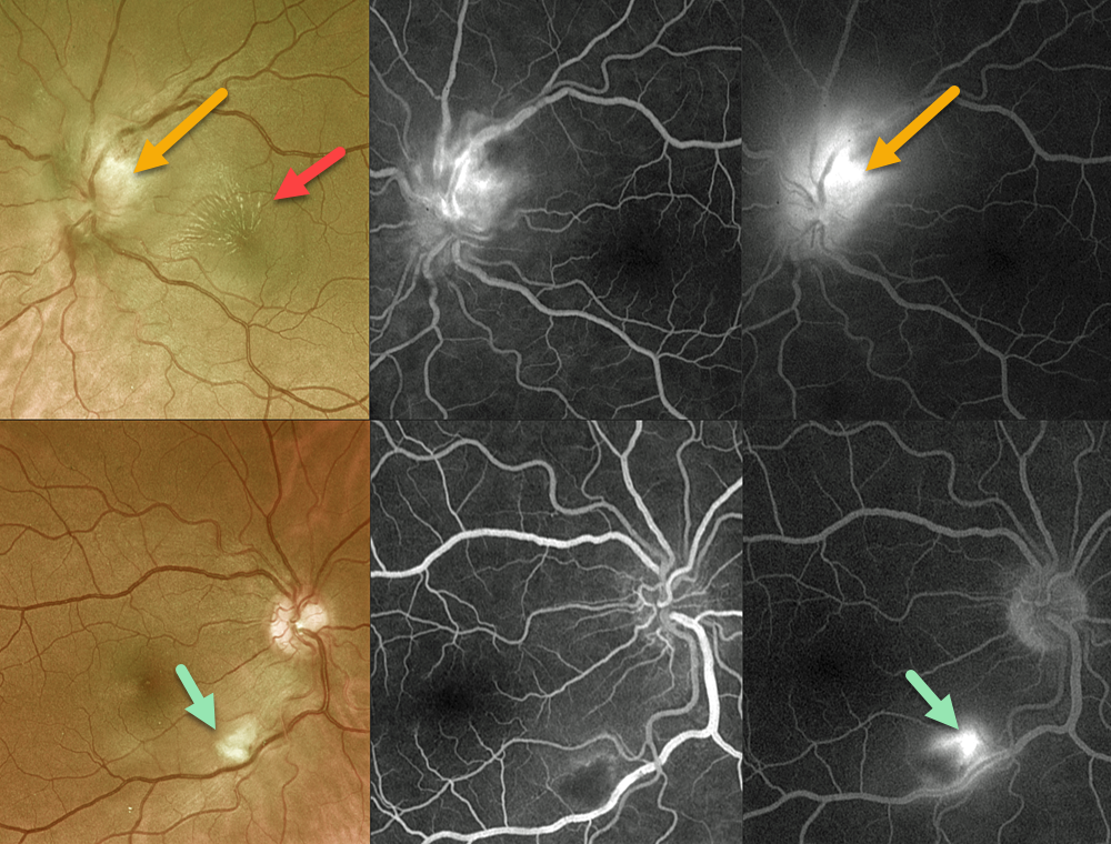
This 52YO female presented with one week of bilateral vision loss. She has multiple cats at home. Vision was 20/80 OD and counting fingers OS.
Optos color RG imaging shows an area of inferonasal macular inner retinal opacification OD, and a superiorly swollen left optic nerve with a superonasal macular lipid star.
Fluorescein angiography shows early blockage with late staining of the retinal lesion OD with an adjacent area of vasculitis. Marked optic nerve leakage is seen OS.
Bartonella henselae antibody testing was positive for both IgG >1:1024 and IgM 1:126. She was subsequently lost to follow-up.
Learning Points:
Bartonella henselae is the bacterium associated with cat scratch disease. It is transmitted from cat fleas carrying the bacterium or from flea feces that can be present on the cat’s claws/teeth, and transferred to humans from a cat scratch, lick, or bite. Common systemic findings include fever, malaise, and progressive lymphadenopathy. These findings usually occur within 1-2 weeks of a cat scratch/bite. Ocular effects primarily include neuroretinitis with rare cases of Parinaud’s ocular glandular syndrome, or, as in our patient’s case, chorioretinitis with vasculitis.
BARTONELLA
Originally posted on @retina.rocks July 13, 2023
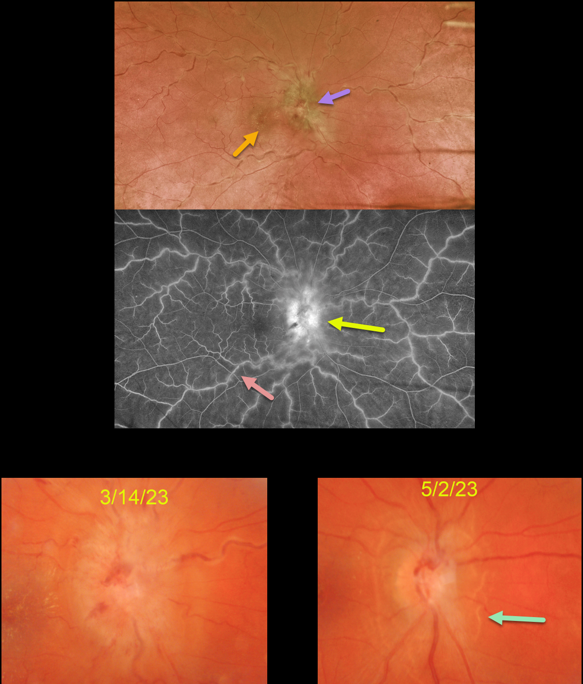
This 36YO female presented with unilateral vision loss in her left eye. She denied any systemic symptoms except for headaches for 3 months. Vision was 20/70 in her right eye and 20/25 in her left eye.
Optos color imaging shows severe optic nerve head edema, a foveal lipid star, dilated and tortuous vessels, and a variable frosted branch appearance to the retinal veins. Fluorescein angiography shows diffuse venous staining with optic nerve leakage.
Extensive blood work was positive for both Bartonella henselae (IgG 1:64) and Bartonella quintana (IgG 1:128), and negative for Bartonella IgM antibodies. Although she has cats at home, she denied cat scratches or exposure to cat fleas. She was referred to an infectious disease specialist who started her on doxycycline 100mg PO BID.
When she returned 6 weeks later, her headaches had resolved, and her vision was 20/80. The optic nerve swelling was dramatically improved with secondary peripapillary pigmentary changes.
Learning Points:
Bartonella henselae is the bacterium associated with cat scratch disease. It is transmitted from cat fleas carrying the bacterium or from flea feces that can be present on the cat’s claws/teeth, and transferred to humans from a cat scratch, lick, or bite.
Common systemic findings include fever, malaise, and progressive lymphadenopathy. These findings usually occur within 1-2 weeks of a cat scratch/bite.
Ocular effects primarily include neuroretinitis, with rare cases of Parinaud’s ocular glandular syndrome and, as in our patient’s case, chorioretinitis with vasculitis.
BARTONELLA
Originally posted on @retina.rocks January 24, 2023
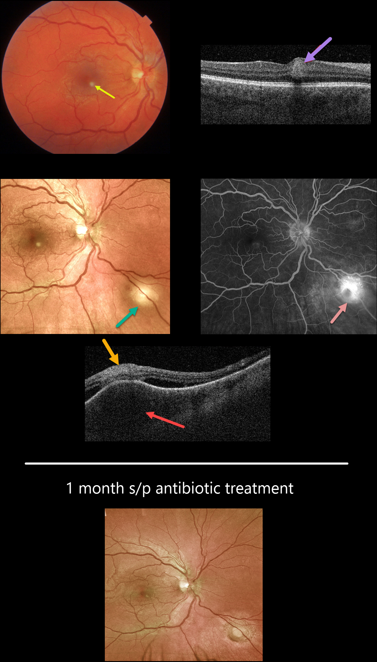
This healthy 27YO male presented with a few days of a paracentral scotoma in his right eye. Vision was 20/25 OD and 20/20 OS.
Color imaging shows a small inferonasal foveal area of inner retinal opacification. Swept-source OCT through this region shows inner retinal hyperreflectivity. An additional, larger subretinal lesion is noted in the inferonasal midperiphery. OCT scanning through this lesion shows marked choroidal thickening, overlying retinal disorganization and hyperreflectivity, and some surrounding subretinal fluid. Fluorescein angiography shows moderate leakage from this choroidal lesion.
On further questioning, the patient told us that he recently adopted 2 stray cats. He didn’t recall any cat scratches, rashes, or adenopathy, but Bartonella titers were markedly elevated (IgG 1:1280, IgM 1:400).
Following infectious disease consultation, he started a 6-week course of oral doxycycline and rifampin. Four weeks later, vision was 20/20, and both lesions had significantly improved.
Learning Points:
Bartonella Henselae is the bacterium associated with cat scratch disease. It is transmitted from cat fleas (carrying the bacterium) or from flea feces that can be present on the cat’s claws/teeth, and transferred to humans from a cat scratch or bite.
Common systemic findings include fever, malaise, and progressive lymphadenopathy. These findings usually occur within 1-2 weeks of a cat scratch/bite.
Ocular effects primarily include neuroretinitis with rare cases of Parinaud’s ocular glandular syndrome, or, as in our patient’s case, chorioretinitis.
In our practice, posterior segment Bartonella involvement most commonly presents with neuroretinitis. Our patient’s inflammation spared the nerve, with multifocal involvement including the inner retina and choroid.
NEURORETINITIS
Originally posted on @retina.rocks August 24, 2020
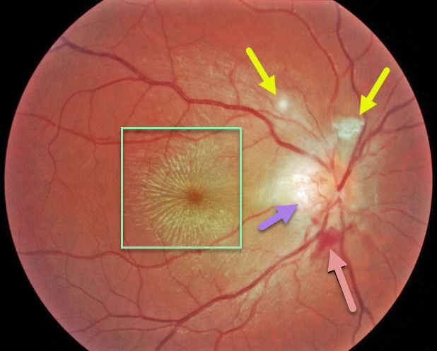
This 17YO presented with 20/200 vision OD and diffuse optic disc edema with temporal peripapillary retinitis, peripapillary inner retinal blood, a few areas of focal inner retinitis, and a prominent macular lipid star. The left eye was normal.
Our patient tested positive for Bartonella and had a cat (should’ve gotten a dog and opted for a Toxocara worm instead…).
Patients will usually improve without treatment, although antibiotic therapy may sometimes be recommended.
Learning Points:
Optic nerve inflammation, often from malignant hypertension or neuroretinitis, can cause fluid and blood to leak into the optic nerve and peripapillary tissues. When the fluid absorbs, the lipid can precipitate in Henle’s layer, creating the lipid star.
There are numerous causes for neuroretinitis, including cat scratch (Bartonella), Lyme disease, syphilis, and idiopathic (Leber’s stellate neuroretinitis).


