

Benign Familial Fleck Retina
<< Back to Cases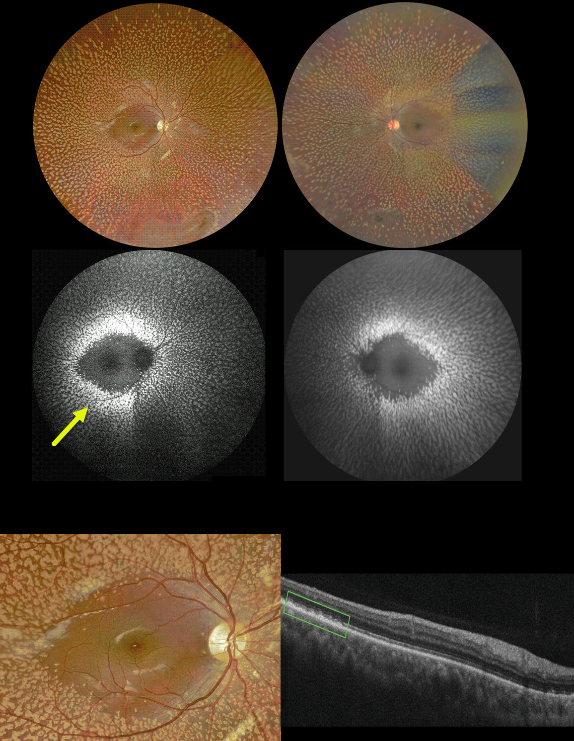
BENIGN FAMILIAL FLECK RETINA
Ayushi Gupta and Vishal Agrawa
Originally posted on @retina.rocks February 1, 2024
This 10YO boy complained of needing frequent changes of his glasses. Vision was 20/20 OD and 20/30 OS. He was a -8D myope OD and -6D myope OS. Corneal topography was consistent with keratoconus.
Color imaging shows symmetric, multiple, discrete yellow-white fleck lesions involving the entire fundus except the maculae. A patch of inferior lattice is also noted OD.
On fundus autofluorescence, these flecks show marked hyper-FAF. On OCT, the flecks appear as hyperreflective outer retinal deposits that extend into and distort the overlying ellipsoid zone.
Learning Points:
Initially reported by Aish and Dajani (BJO 1980;64:652-659), benign familial fleck retina (BFFR) is an extremely rare autosomal recessive disorder that belongs to a heterogeneous group of flecked retina syndromes.
The BFFR gene encodes for a group V phospholipase A2 (PLA2G5). Despite the dramatic fundus appearance, affected individuals have normal acuity, visual fields, and retinal function, including electrophysiology.
Other flecked retinal disorders include fundus albipunctatus, fundus flavimaculatus, familial drusen, retinitis punctata albescens, and fleck retina of Kandori. These disorders can be associated with night blindness, central visual problems, abnormal ERG, and abnormal perimetry.
The ERG helps distinguish BFFR from other flecked retina syndromes, such as retinitis punctata albescens and fundus albipunctatus, as scotopic ERG responses are decreased in the latter conditions but normal in BFFR.


