

Best Disease
<< Back to Cases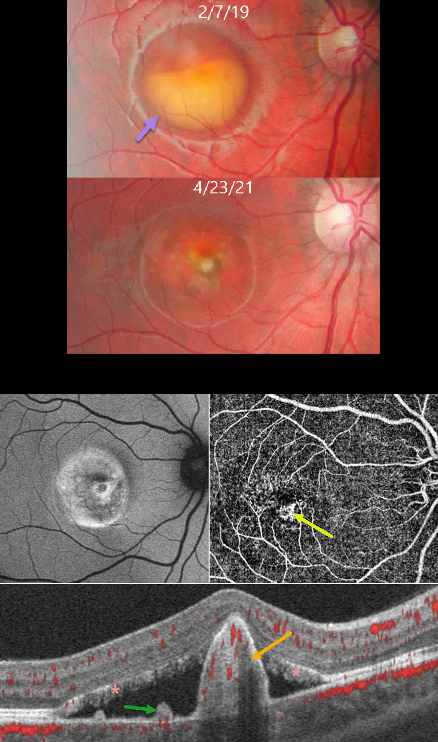
BEST DISEASE WITH MACULAR NEOVASCULARIZATION
João Pedro Marques
Originally posted on @retina.rocks August 26, 2026
This healthy 11YO boy presented to us on 2/7/19 with asymptomatic retinal findings. Vision was 20/25 OD and 20/20 OS.
Color photography of his right eye shows a partially scrambled vitelliform lesion consistent with Best vitelliform macular dystrophy (BVMD). A smaller, scarred, and atrophic lesion was noted in his left eye (not shown).
He returned 2 years later without visual complaints. Vision was 20/40 OD. The vitelliform lesion in his right macula is atrophic, with a central depigmented nodule. Fundus autofluorescence (FAF) shows variable hyper- and hypo-FAF. OCTA en face shows a central flow signal. OCTA B-scan shows a nodular subretinal hyperreflective pillar containing flow signals, subretinal fluid, and vitelliform material layered on the RPE and lining the outer retina. The vision and OCT findings did not change following 2 intravitreal Lucentis injections, so observation was recommended. We continue to follow him with stable findings 5 years later.
Learning Points:
BVMD is associated with a mutation in the BEST1 gene, which encodes the bestrophin-1 protein. Bestrophin-1, a calcium-activated chloride channel, is primarily found in the basolateral plasma membrane of the RPE. BEST1 mutations cause a variety of phenotypes, including autosomal recessive bestrophinopathy, best vitelliform macular dystrophy, and autosomal dominant vitreoretinochoroidopathy.
Secondary macular neovascularization (MNV) develops in up to half of eyes, depending on the imaging modality used, and can develop at any stage of the disease. MNV exists in two forms: the less common exudative form, which presents with acute vision loss, subretinal hemorrhage, and intraretinal/subretinal fluid, and the more common non-exudative form seen in more advanced (vitelliruptive and atrophic) stages (Parodi et al, Invest Ophthalmol Vis Sci 2020;61(6);61).
These subclinical MNVs are best detected with swept-source OCTA. Structural OCT findings that suggest non-exudative MNV include focal choroidal excavations and nodular subretinal pillars (as in our patient). Exudative MNV responds well to standard anti-VEGF injections, whereas non-exudative lesions tend to remain stable without treatment.
BEST DISEASE WITH MYELINATED NERVE FIBER LAYER
The European Society (EVRS) and Vaibhav Sethi
Originally posted on @retina.rocks July 25, 2025
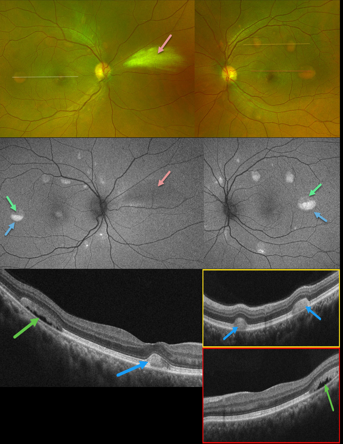
This 39YO male presented with asymptomatic retinal lesions. Family history was negative. Vision was 20/20 OU.
Optos color RG imaging shows bilateral partially scrambled vitelliform lesions in each posterior pole. On OCT, the superior, scrambled portion of these lesions is hyporeflective, and the inferior, yellow portion is hyperreflective. On fundus autofluorescence (FAF), the superior portions are hypo-FAF, and the inferior vitelliform material is hyper-FAF. The incidental area of the nasal myelinated nerve fiber layer OD is hypo-FAF from blocking the underlying RPE.
Learning Points:
Vitelliform lesions are characterized by the accumulation of yellow subretinal material thought to be caused by impaired metabolism of photoreceptors or retinal pigment epithelium. They can be associated with a wide variety of retinal disorders, including Best vitelliform macular dystrophy (BVD, our patient), adult-onset foveomacular dystrophy, acute exudative polymorphous vitelliform maculopathy, as well as paraneoplastic, toxic, tractional, and degenerative etiologies (Iovino et al, Surv Ophthalmology 2023;68:361-379).
BVD is usually transmitted as an autosomal-dominant disorder due to a mutation in the BEST1 gene, which encodes bestrophin-1. Bestrophin-1, a calcium-activated chloride channel, is primarily found in the basolateral plasma membrane of the RPE. Unfortunately, our patient denied genetic testing due to cost.
BEST DISEASE
Evgenii Chernov
Originally posted on @retina.rocks September 17, 2024
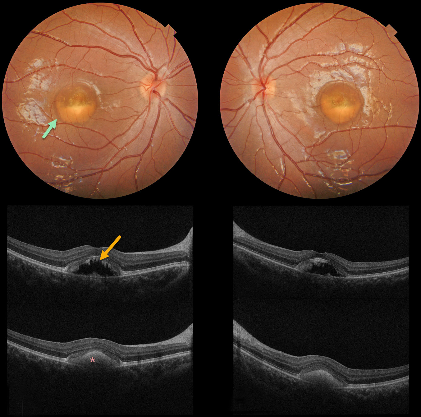
Color photography of our patient’s eyes shows partially scrambled Best lesions. OCT scanning through the superior lesion shows a mostly hyporeflective elevation of the neurosensory retina, with shaggy hyperreflective material along the posterior edge of the outer segments. OCT scanning through the inferior lesion shows hyperreflective layering of the vitelliform material.
Learning Points:
Best disease is associated with a mutation in the BEST1 gene, which encodes bestrophin-1. Bestrophin-1, a calcium-activated chloride channel, is primarily found in the basolateral plasma membrane of the RPE. BEST1 mutations cause a variety of phenotypes, including autosomal recessive bestrophinopathy, best vitelliform macular dystrophy, and autosomal dominant vitreoretinochoroidopathy.
Our patient has progressed to the ‘scrambled egg’ appearance. The remaining vitelliform material gravitates inferiorly, often leaving an optically empty, hyporeflective space between the RPE and outer retina. Eventually, the retina flattens, with secondary outer retinal atrophy.
ADULT-ONSET FOVEOMACULAR VITELLIFORM DYSTROPHY
Originally posted on @retina.rocks April 29, 2024
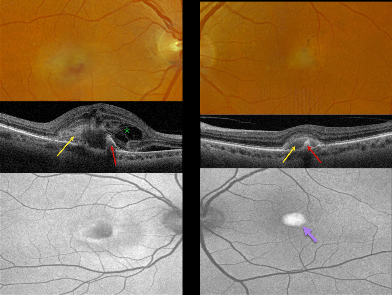
This 71YO female was referred for neovascular AMD. She noticed recent vision loss in her right eye and was fairly asymptomatic in her left eye. Vision was counting fingers OD and 20/80 OS.
Optos color RGB imaging shows somewhat turbid, yellowish foveal fluid OD and a vitelliform lesion OS. Triton swept-source OCT shows hyperreflective subretinal vitelliform material and hyperreflectivity from a subretinal pigment clump. Subretinal fluid and a complex pattern of loculated pockets of intraretinal fluid are noted OD.
On Optos fundus autofluorescence (FAF), the lesion shows central hypo-FAF and peripheral hyper-FAF OD, and marked hyper-FAF OS.
Anti-VEGF therapy was started for macular neovascularization (MNV) in her right eye.
Learning Points:
Best disease is associated with a mutation in the BEST1 gene, which encodes bestrophin-1. Bestrophin-1, a calcium-activated chloride channel, is primarily found in the basolateral plasma membrane of the RPE.
BEST1 mutations cause a variety of phenotypes, including adult-onset foveomacular vitelliform dystrophy (as in our patient), best vitelliform macular dystrophy, autosomal recessive bestrophinopathy, and autosomal dominant vitreoretinochoroidopathy.
Although our patient had a Best phenotype, genetic testing revealed uncertain significant heterozygous variants SAG, SLC7A14, and VCAN.
In our experience, MNV is rarely associated with Best lesions, although Miyagi et al identified MNV in 18% of eyes in their retrospective Asian population (Graefe’s 2022;260:1125-1137).
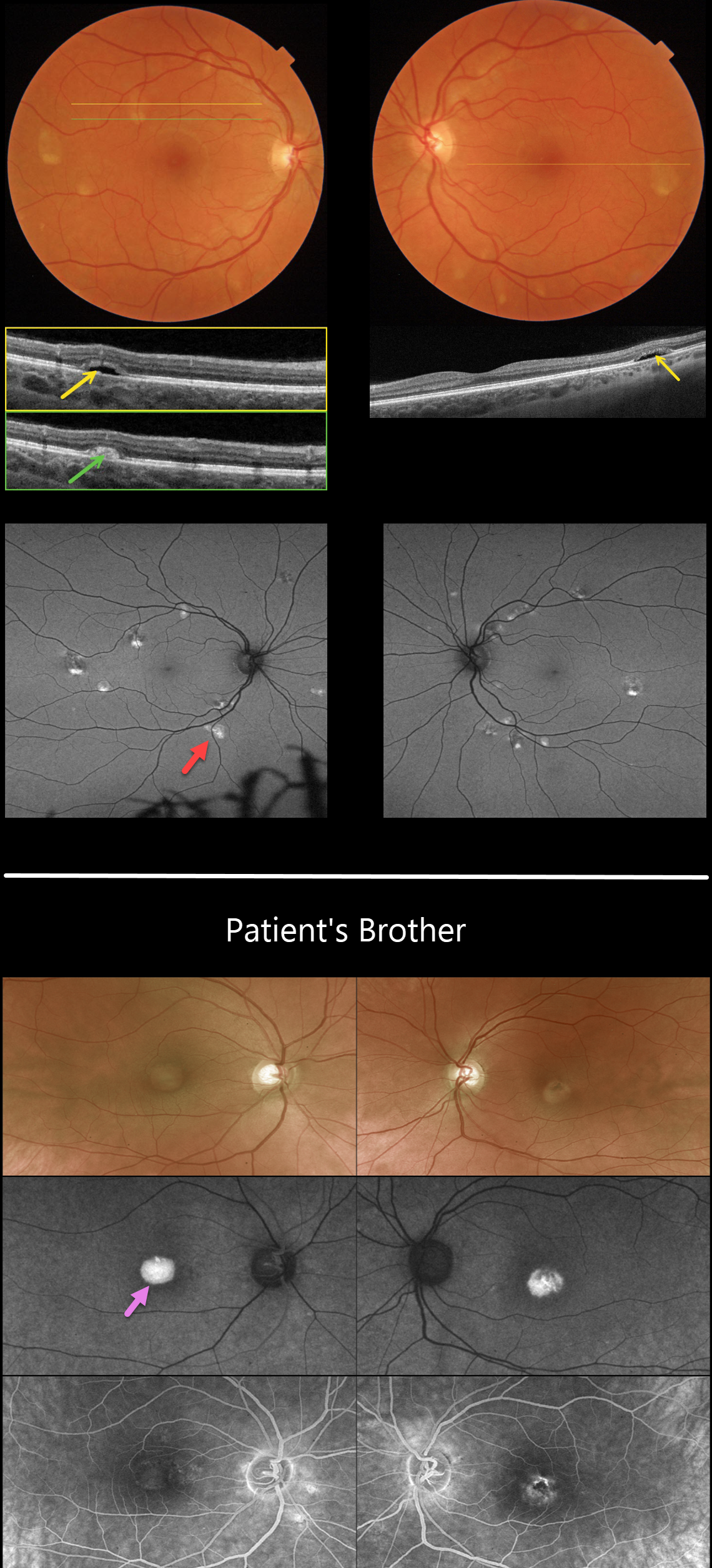
IMPG1 BEST DISEASE WITH DIFFERENT PHENOTYPES IN SIBLINGS
Jessica Johnson and Mattie Adams
Originally posted on @retina.rocks February 5, 2024
This 41YO female complained of increasing difficulties with dark adaptation and night vision. Vision was 20/25 OU.
Color imaging shows bilateral, multifocal, partially scrambled vitelliform lesions. The superior scrambled aspect of each lesion on OCT shows subretinal hyporeflectivity, and the inferiorly layered vitelliform material is hyperreflective. The inferior vitelliform material is markedly hyper-autofluorescent with Optos imaging.
Genetic testing was positive for a heterozygous pathogenic IMPG1 deletion in exon 7.
Interestingly, her brother was recently diagnosed with Best disease, having more typical single, subfoveal yellow vitelliform lesions. The brother’s genetic testing showed a similar IMPG1 mutation.
Learning Points:
Best disease is most often associated with a mutation in the BEST1 gene, which encodes bestrophin-1. Bestrophin-1, a calcium-activated chloride channel, is primarily found in the basolateral plasma membrane of the RPE.
BEST1 mutations cause a variety of phenotypes, including autosomal recessive bestrophinopathy, best vitelliform macular dystrophy, and autosomal dominant vitreoretinochoroidopathy.
Multifocal lesions are a rare phenotype and are not usually associated with subfoveal lesions. Multifocal Best needs to be differentiated from other causes of multifocal serous detachments, including multifocal central serous, autosomal recessive bestrophinopathy (see Boon et al, Ophthalmology 2013;120:809-820), and idiopathic or paraneoplastic acute exudative polymorphous vitelliform maculopathy (see Barbazetto et al, Ophthalmology 2018;125:75-88).
Vitelliform macular dystrophies (VMD) are most commonly caused by BEST1, PRPH2, or VMD2 mutations. IMPG1 and IMPG2, which encode proteins that are components of the interphotoreceptor matrix, have also been implicated in both autosomal dominant and recessive VMD.
Manes et al. reported a wide variety of IMPG1 VMD phenotypes, including multifocal lesions and more classic single subfoveal lesions, as seen in our two patients (Am J Hum Genet 2013;93:571-578).
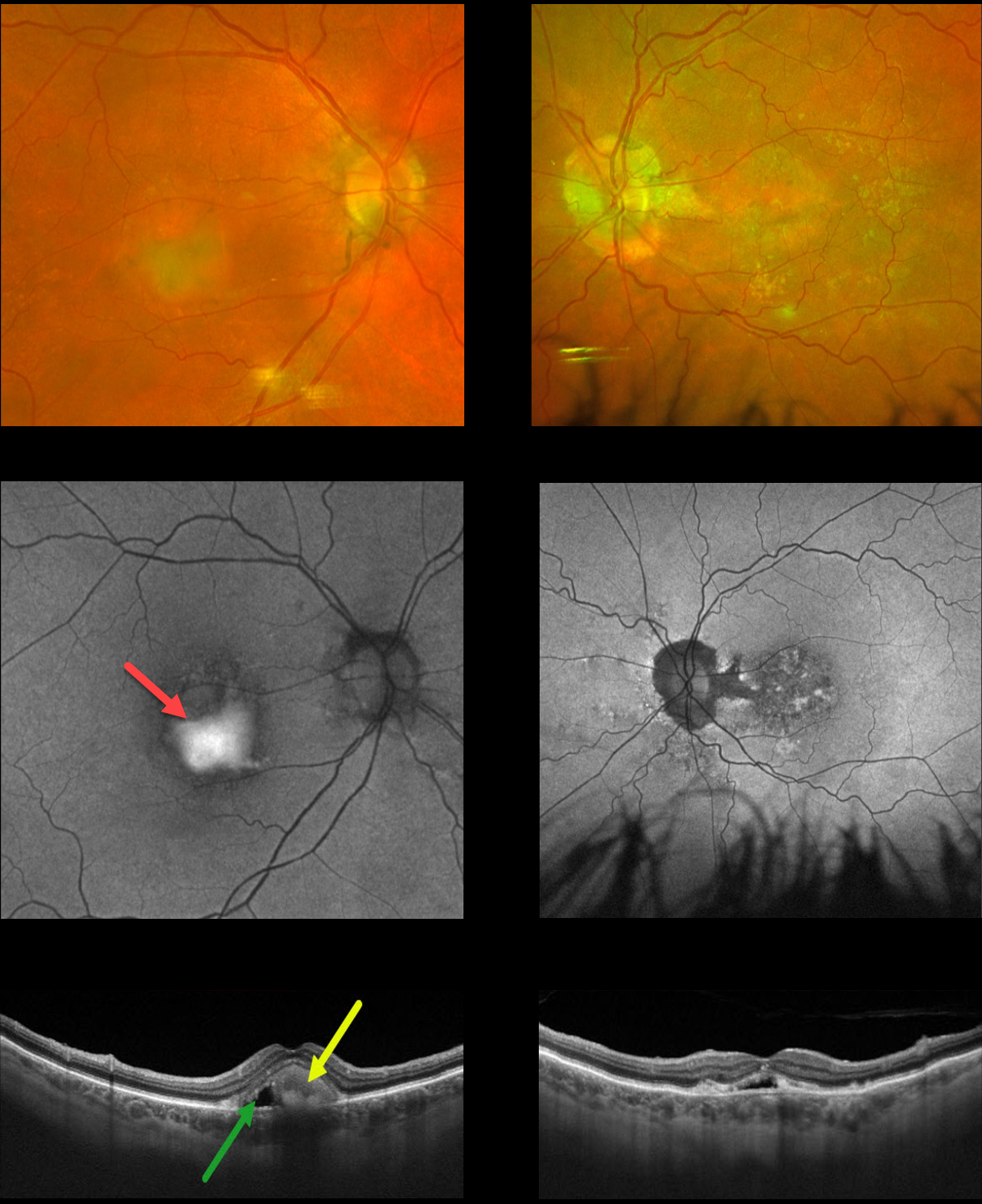
BEST DISEASE
Originally posted on @retina.rocks January 19, 2023
This 75YO male presented with chronic stable central blurriness. Vision was 20/150 OU.
Color photography of his right eye shows superior foveal pigment loss with inferior yellowish, vitelliform material, with more atrophic foveal changes in his left eye.
Fundus autofluorescence (FAF) shows the vitelliform material to be dramatically hyper-autofluorescent OD, with a more speckled hypo-FAF appearance OS.
Swept-source OCT scanning of his right eye shows hyperreflective inferior layering of the vitelliform material with a hyporeflective space more superiorly. Similar OCT findings are also seen in his left eye.
Our patient has progressed to a more atrophic, ‘scrambled egg’ appearance, most evident in his right eye. The remaining vitelliform material gravitates inferiorly, often leaving an optically empty, hyporeflective space between the RPE and outer retina. Eventually, the retina flattens over time with secondary atrophy, which is more pronounced in his left eye.
Learning Points:
Best disease is associated with a mutation in the BEST1 gene, which encodes the bestrophin-1 protein. Bestrophin-1, a calcium-activated chloride channel, is primarily found in the basolateral plasma membrane of the RPE.
BEST1 mutations cause a variety of phenotypes, including autosomal recessive bestrophinopathy, best vitelliform macular dystrophy, and autosomal dominant vitreoretinochoroidopathy.
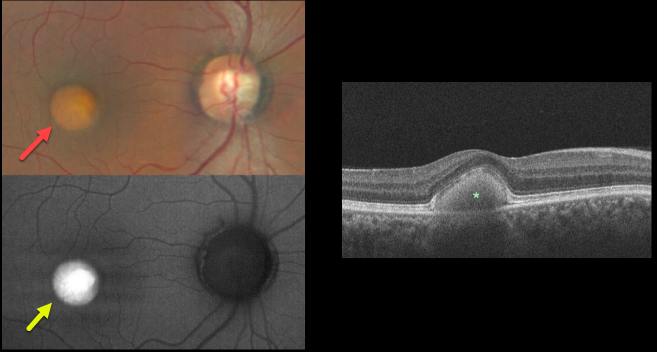
BEST DISEASE
Will Gibson
Originally posted on @retina.rocks November 9, 2022
This 39YO male presented with these asymptomatic subretinal yellow posterior pole lesions. Vision was 20/30 OU.
The sharply demarcated yellowish lesions are markedly hyper-autofluorescent. This vitelliform material is hyperreflective on OCT. The subretinal material extends into and elevates the central retina.
Learning Points:
Best disease is associated with a mutation in the BEST1 gene, which encodes the bestrophin-1 protein.
Bestrophin-1, a calcium-activated chloride channel, is primarily found in the basolateral plasma membrane of the RPE. BEST1 mutations cause a variety of phenotypes, also including autosomal recessive bestrophinopathy, best vitelliform macular dystrophy, and autosomal dominant vitreoretinochoroidopathy.
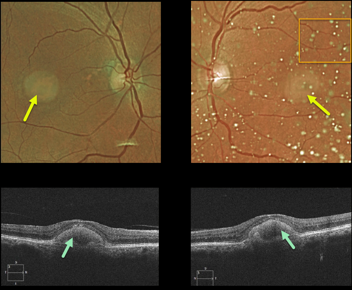
ADULT-ONSET FOVEOMACULAR VITELLIFORM DYSTROPHY
Originally posted on @retina.rocks January 18, 2022
This 87YO female shows classic findings for adult-onset foveomacular vitelliform dystrophy (Adult Best). Vision was 20/60 OD and 20/80 OS Optos imaging shows small, round, symmetrical subfoveal yellow lesions. Moderate asteroid hyalosis is noted as an incidental finding on the OS.
Triton swept-source OCT shows subretinal hyperreflective material above the RPE that extends into and indents the overlying retina.
Learning Points:
Adult Best is associated with a mutation in the BEST1 gene, which encodes the bestrophin-1 protein. Bestrophin-1, a calcium-activated chloride channel, is primarily found in the basolateral plasma membrane of the RPE.
BEST1 mutations cause a variety of phenotypes, including autosomal recessive bestrophinopathy, best vitelliform macular dystrophy, and autosomal dominant vitreoretinochoroidopathy.
These patients tend to maintain fairly good vision. Outer retinal and RPE atrophy can cause central vision loss, as can the rare development of macular neovascularization. Patients should therefore regularly monitor their vision with an Amsler grid.
BEST DISEASE
Originally posted on @retina.rocks November 5, 2021
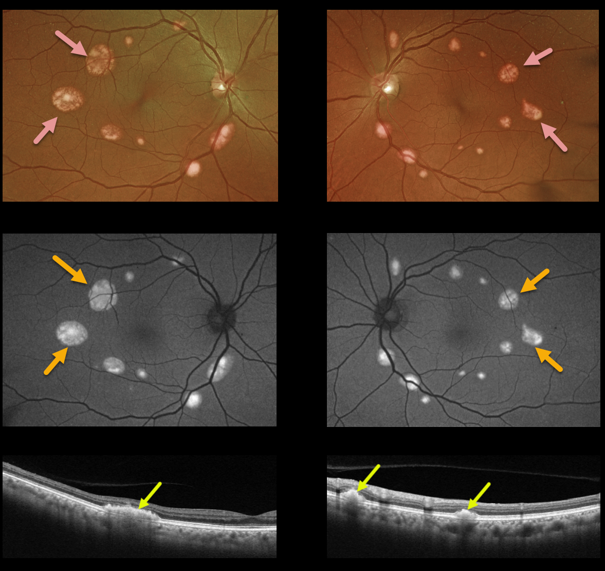
This 68YO female presented with these asymptomatic multifocal subretinal yellow posterior pole lesions. Vision is 20/20 OU.
The sharply demarcated yellowish lesions are markedly hyper-autofluorescent and are hyperreflective on Triton swept-source OCT. There was no known family history, but genetic testing revealed a heterozygous pathogenic BEST1 mutation.
Learning Points:
Best disease is associated with a mutation in the BEST1 gene, which encodes the bestrophin-1 protein. Bestrophin-1, a calcium-activated chloride channel, is primarily found in the basolateral plasma membrane of the RPE.
BEST1 mutations cause a variety of phenotypes, including autosomal recessive bestrophinopathy, best vitelliform macular dystrophy, and autosomal dominant vitreoretinochoroidopathy. Multifocal lesions are a rare phenotype and are not usually associated with subfoveal lesions.
Multifocal Best needs to be differentiated from other causes of multifocal serous detachments, including autosomal recessive bestrophinopathy (see Boon et al, Ophthalmology 2013;120:809-820) and idiopathic or paraneoplastic acute exudative polymorphous vitelliform maculopathy (see Barbazetto et al, Ophthalmology 2018;125:75-88).
BEST DISEASE
Originally posted on @retina.rocks September 14, 2020
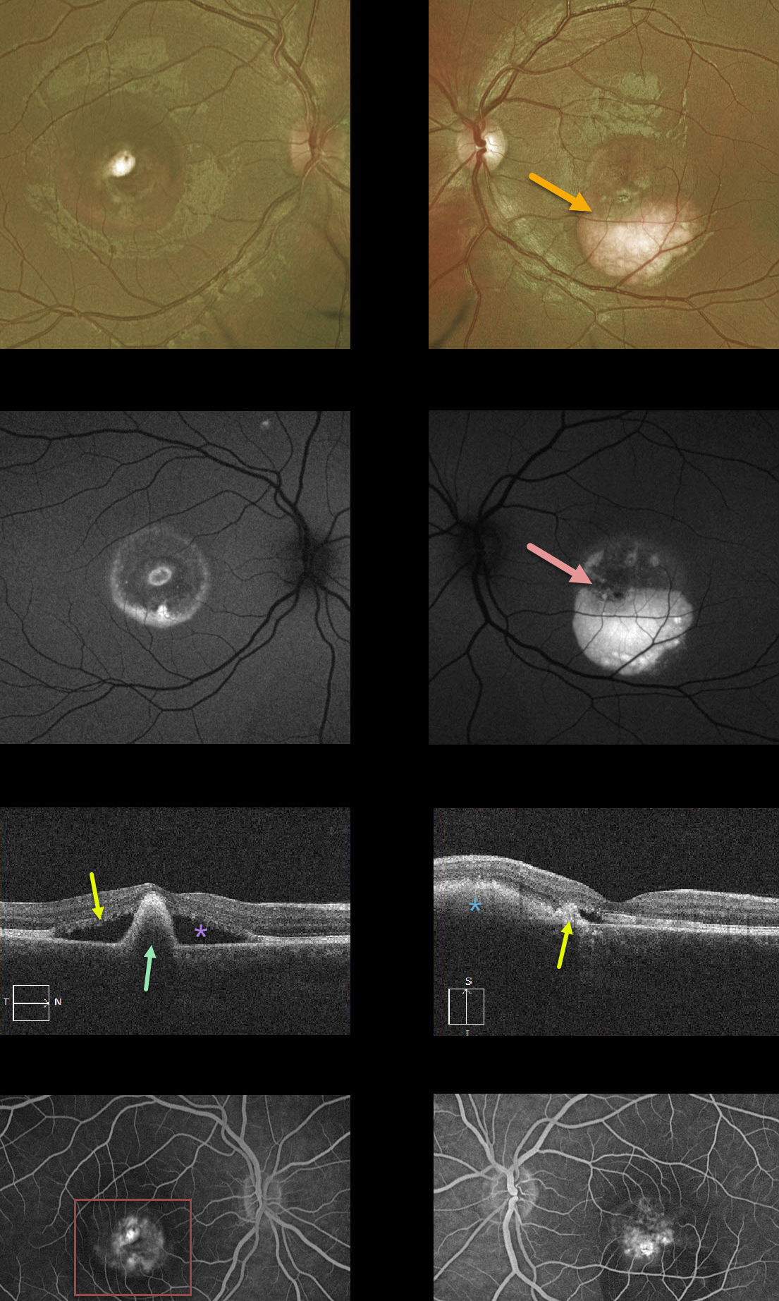
This patient has Best’s disease and has progressed to the more atrophic, ‘scrambled egg’ appearance. The fundus photos show asymmetric layering of the residual yellow-white subretinal vitelliform material.
This material is highly autofluorescent and can also appear as hyperreflective on OCT scans. The right eye has a large subfoveal pigment clump with surrounding non-exudative subretinal fluid. The left eye has a smaller subfoveal scar with a flattened vitelliform lesion without subretinal fluid. Fluorescein angiography (FA) shows staining without active leakage.
Learning Points:
Best vitelliform macular dystrophy (Best’s disease) is associated with a mutation in the BEST1 gene. This inherited disorder usually presents in childhood or early adulthood with a bilateral foveal “egg-yolk” appearance.


