

Bilateral Diffuse Uveal Melanocytic Proliferation (BDUMP)
<< Back to Cases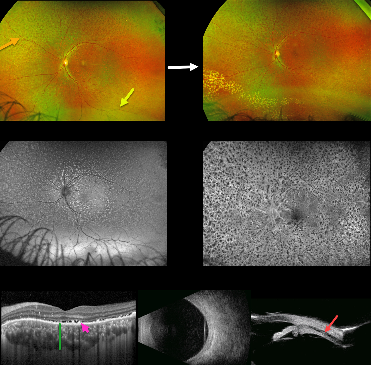
IDIOPATHIC UVEAL EFFUSION SYNDROME vs UNILATERAL BDUMP
Carl-Joe Mehanna
Originally posted on @retina.rocks July 17, 2024
This 48YO healthy male presented with two months of progressive loss of vision in his left eye. Vision was 20/50 OD due to a subcapsular cataract and 20/100 OS with a clear lens. Anterior segments were normal.
Optos color RG imaging of the left eye shows a diffuse orange-red leopard spot appearance to the fundus with an inferior exudative retinal detachment. Fundus autofluorescence (FAF) shows these lesions to be markedly hyper-FAF and block on fluorescein angiography.
Macular OCT shows a thickened choroid, shallow subretinal fluid, and multiple hyperreflective lipofuscin deposits on top of the RPE. B-scan ultrasonography shows a diffusely thickened choroid with overlying retinal detachment, and ultrasound biomicroscopy shows a ciliary body effusion.
Extensive medical workup, including bloodwork and brain MRI, was negative for malignancy or infectious processes (including syphilis, HIV, and TB). He was lost to follow-up for a year, when he presented with resolved subretinal but with new peripheral lipid. He has since been lost to follow-up.
Learning Points:
The idiopathic uveal effusion syndrome (non-nanophthalmic) is a rare entity presenting with idiopathic exudative choroidal, ciliary body, and shifting retinal detachment due to impaired transscleral suprachoroidal drainage caused by an abnormal thickened sclera (Elagouz et al, Surv Ophthalmology 2010;55:134-145). Chronic detachment can cause leopard-spot changes and vision loss.
The differential diagnosis includes leukemic infiltration, central nervous system lymphoma, and bilateral diffuse uveal melanocytic proliferation (BDUMP). The most likely cause of these red subretinal lesions is RPE-engorged, subretinal lipofuscin, which was noted on our patient’s OCT.
The five cardinal signs of idiopathic uveal effusion syndrome include 1) multifocal round/oval red patches at the level of the RPE, 2) early hypofluorescence of these patches, 3) multiple uveal melanocytic tumors with diffuse uveal thickening, 4) exudative retinal detachment, and 5) rapidly progressive cataract (Gass et al, Arch Ophthalmol 1990;108:427-533).
In our patient’s case, we feel the most likely diagnosis is idiopathic uveal effusion syndrome. However, we have seen similar findings in BDUMP, which can present unilaterally (Spaide, Retinal Cases & Brief Reports 2018;12:263-165). However, BDUMP is usually a paraneoplastic process, and our patient’s metastatic workup was negative.
BILATERAL DIFFUSE UVEAL MELANOCYTIC PROLIFERATION (BDUMP)
Originally posted on @retina.rocks September 29, 2021
This is a 62YO female presented with bilateral vision loss over the past 3 months. Her vision was 20/200 OD and counting fingers OS compared to 20/20 OU at baseline. She was diagnosed with stage 4 ovarian cancer one month earlier.
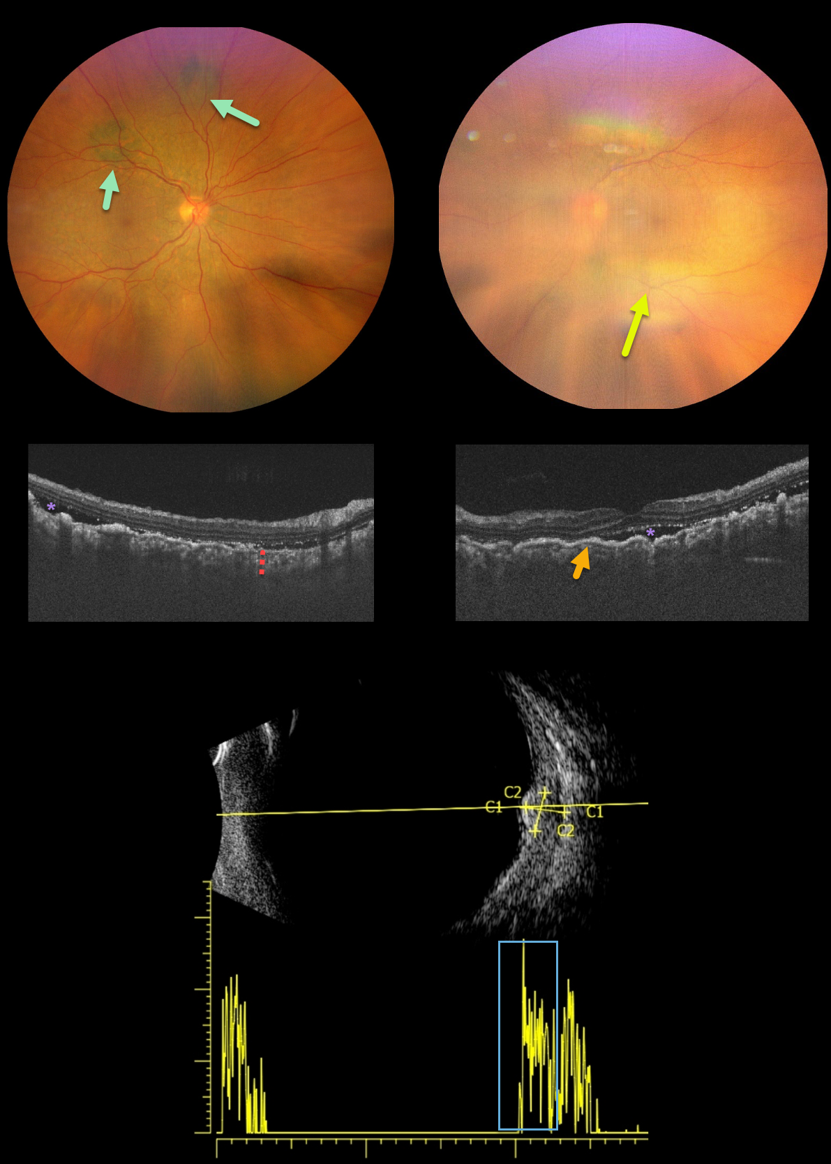
New multifocal, elevated, pigmented choroidal lesions are noted superonasally in the right eye. View of the left fundus is limited due to a significant progressive cataract.
OCT reveals bilateral choroidal thickening with overlying folds and some subretinal fluid. B-scan ultrasonography through one of the choroidal lesions in the right eye shows moderate homogenous internal reflectivity.
Learning Points:
Initially described by Machemer in 1966, the five cardinal features of bilateral diffuse uveal melanocytic proliferation (BDUMP) include:
(1) multiple round or oval subtle red patches at the level of the RPE
(2) a striking pattern of multifocal areas of fluorescein angiographic hyperfluorescence from these lesions
(3) multiple slightly elevated pigmented and nonpigmented uveal melanocytic tumors with diffuse uveal thickening
(4) exudative retinal detachment
(5) rapidly progressive cataracts (Gass et al, Arch Ophthalmology 1990;108:527-533).
The thickened choroid and pigmentary changes are secondary to infiltration by benign uveal melanocytes, possibly in response to a primary visceral tumor (ovarian and uterine carcinomas in women, lung carcinoma in men) that produces melanocytic growth factors. Vision typically deteriorates to near blindness within a year of presentation. The serous detachments can resolve with plasmapheresis, although the overall prognosis is generally poor, with a mean survival of about 1 year from the time of ocular diagnosis.
PERIPAPILLARY PACHYCHOROID SYNDROME WITH UNILATERAL BDUMP
Originally posted on @retina.rocks October 15, 2020
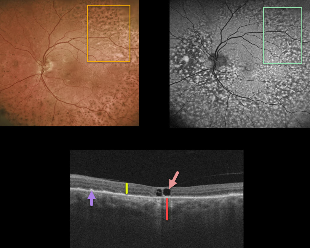
While being followed for peripapillary pachychroid syndrome (PPS) with periodic photodynamic therapy for symptomatic foveal fluid OS, this patient presented with new diffuse pigmentary deposits in the left eye only.
Fundus autofluorescence shows a dramatic pattern of hyperautofluorescent and hypofluorescent spots scattered throughout the fundus. These findings, although unilateral, look very similar to those in the paraneoplastic condition, bilateral diffuse uveal melanocytic proliferation (BDUMP).
Macular OCT scanning shows a diffusely thickened choroid when compared to the neurosensory retina. Other OCT findings here include a few large central foveal cysts, outer retinal atrophy, and RPE changes.
Our patient has no known prior cancer diagnoses, and recently had a negative CT abdominal scan and chest x-ray. He continues to be followed closely by his internist since BDUMP often precedes the diagnosis of a systemic malignancy by months to years.
Learning Points:
Typical PPS findings include thickened choroid, chorioretinal folds, shorter axial lengths, and hyperopia.
In BDUMP, the thickened choroid and pigmentary changes are secondary to infiltration by benign uveal melanocytes, possibly in response to a primary visceral tumor that produces melanocytic growth factors.


