

Birdshot Chorioretinopathy
<< Back to Cases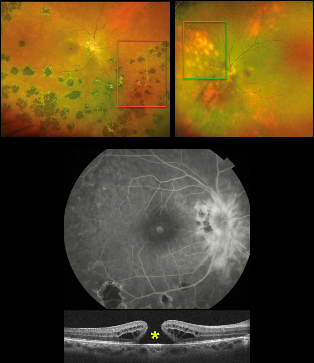
MULTIFOCAL CHOROIDITIS VS BIRDSHOT
Seif Anwar
Originally posted on @retina.rocks June 17, 2026
This healthy 41YO female presented with 6 months of bilateral vision loss. Vision was 20/240 OD and 20/200 OS.
Optos color RG imaging OD shows peripapillary scarring, multifocal large hyperpigmented chorioretinal scars scattered inferiorly and temporally, and some vascular sheathing. A full-thickness macular hole is noted and confirmed on OCT. Fluorescein angiography shows peripapillary leakage.
Her left eye’s Optos shows markedly different findings, with multifocal, creamy-white, depigmented lesions in a pattern radiating away from the disc.
Medical workup was negative, including HLA-A29, QuantiFERON TB Gold, chest X-ray, and serologies for toxoplasmosis and CMV.
Learning Points:
Birdshot chorioretinopathy was appropriately described and named by Drs. Ryan and Maumenee (Am J Ophthlamol 1980;89:31-45) since the lesions look like shotgun birdshot scattered throughout the fundus. Birdshot is typically chronic and bilateral, with about 90-95% of patients being HLA-A29 positive and usually affects women in their 40s to 60s.
Multifocal choroiditis (MFC) is the current preferred term for a group of historically heterogeneous disorders, including multifocal choroiditis, recurrent multifocal choroiditis, multifocal choroiditis and panuveitis, punctate inner choroidopathy, progressive subretinal fibrosis, and pseudo-histoplasmosis (Essex et al, Retina 2013;33:1-4 and Spaide et al, Retina 2013;33:1315-1324). MFC occurs in otherwise healthy adults, often myopic and female. Findings include variably pigmented multifocal scars throughout the fundus, panuveitis, subretinal fibrosis, and macular neovascularization (MNV).
Our patient’s diagnosis is unclear because her right eye shows features suggestive of MFC, while her left eye is fairly pathognomonic for birdshot. In our experience, birdshot lesions never become hyperpigmented, as in our patient’s right eye, although the lesions in her left eye are virtually pathognomic for birdshot.
Following several Remicade treatments, vision remained stable at 20/240 OD and improved to 20/50 OS. We continue to follow her closely.
What’s your diagnosis???
BIRDSHOT CHORIORETINOPATHY WITH MNV
Yuenpang Cheung
Originally posted on @retina.rocks January 22, 2025
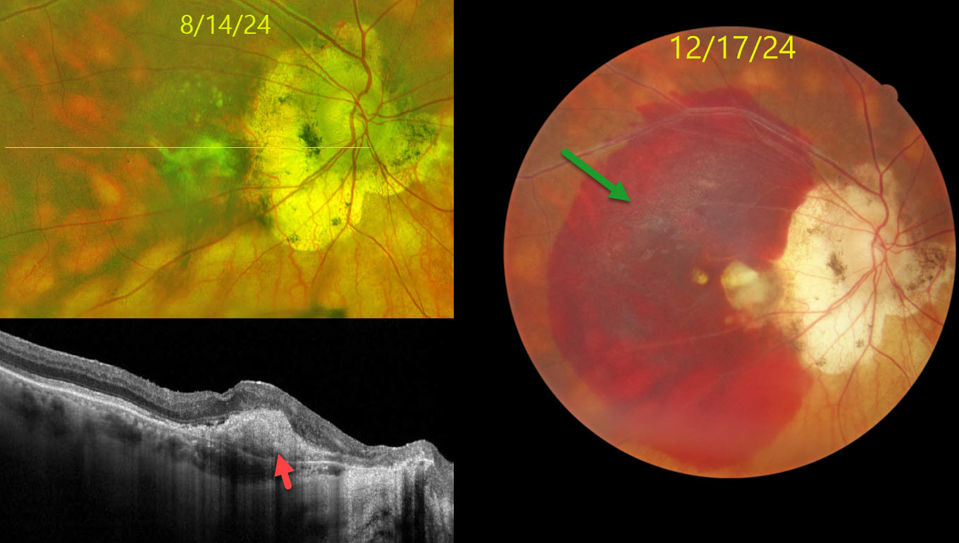
This 73YO male has a 40-year history of birdshot chorioretinopathy which has been managed most recently with PRN intravitreal Ozurdex and Yutiq. He has also been treated with q4 month Vabysmo injections for macular neovascularization (MNV) OD. He presented with recent decreased vision OD. Vision was counting fingers OD and 20/20 OS.
Color photography shows a large submacular hemorrhage, peripapillary scarring, and multiple creamy birdshot lesions more peripherally. Optos RG imaging 4 months earlier shows bilateral peripapillary scarring and scattered birdshot lesions. OCT scanning shows a hyperreflective type 1 (below the RPE) MNV OD.
Learning Points:
Birdshot chorioretinopathy was appropriately described and named by Drs. Ryan and Maumenee (Am J Ophthlamol 1980;89:31-45) since the lesions look like shotgun birdshot scattered throughout the fundus. Birdshot is typically chronic and bilateral with a very high HLA-A29 association. It usually affects women in their 40s to 60s. Vision loss is usually caused by cystoid macular edema (CME) and retinal atrophy, although MNV, as in our patient, can rarely occur. Asymptomatic patients can be observed. CME and symptomatic uveitis can be treated with oral steroids although many will still require immunomodulatory therapy.
POSSIBLE BIRDSHOT CHORIORETINOPATHY
Originally posted on @retina.rocks January 11, 2023
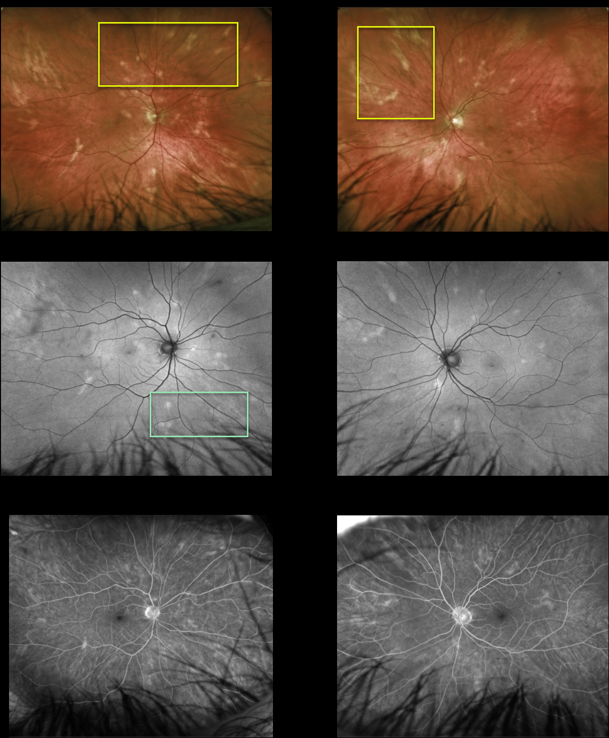
This healthy 51YO male presented with asymptomatic lesions in each fundus. Vision was 20/25 OU.
Multimodal imaging findings were symmetrical bilaterally. Optos color imaging shows amelanotic subretinal lesions that seem to radiate outwards from each disc. These lesions are mostly hyperautofluorescent with window-type defects on fluorescein angiography. OCT scanning was normal bilaterally (not shown). HLA-A29 was negative.
Learning Points:
Birdshot chorioretinopathy was appropriately named and described by Drs. Ryan and Maumenee (AJO 1980;89:31-45), since the lesions look like shotgun birdshot scattered throughout the fundus. Birdshot is typically chronic and bilateral, with a very high HLA-A29 association.
We are not certain that our patient has birdshot. The pattern of lesions and less creamy coloration is somewhat atypical of what we’ve normally seen in our practice.
Although HLA-A29 is positive in about 90% of cases, it is not required for the diagnosis in the setting of other clinical findings (SUN Working Group AJO 2021;228:65-71). Finally, birdshot lesions are typically hypofluorescent (Koizumi et al., Ophthalmology 2008;115:e15-e20), unlike the hyperfluorescence observed in our patient.
Since he is asymptomatic, has no signs of active uveitis, and no macular edema, we will initially follow him twice yearly.
BIRDSHOT CHORIORETINOPATHY
Will Gibson
Originally posted on @retina.rocks August 3, 2022
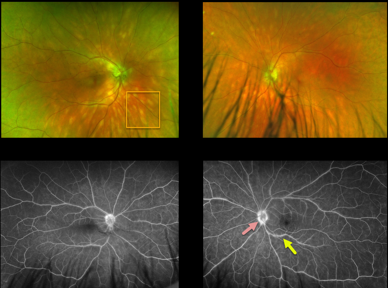
This 48YO male presented with these creamy yellow-white choroidal lesions scattered throughout each posterior pole. Vision was 20/25 OU.
Fluorescein angiography shows mild optic nerve leakage, particularly in the left eye, and mild scattered venous staining/leakage in the left eye.
Learning Points:
This disease was appropriately described and named by Drs. Ryan and Maumenee (Am J Ophthlamol 1980;89:31-45) since the lesions look like shotgun birdshot scattered throughout the fundus.
Birdshot is typically chronic and bilateral, with a very high association with HLA-A29. It usually affects women in their 40s to 60s. Vision loss is usually caused by cystoid macular edema (CME) and retinal atrophy, although macular neovascularization can rarely occur. Asymptomatic patients can be observed. CME and symptomatic uveitis can be treated with oral steroids, although many will still require immunomodulatory therapy.
BIRDSHOT CHORIORETINOPATHY
Originally posted on @retina.rocks November 3, 2020
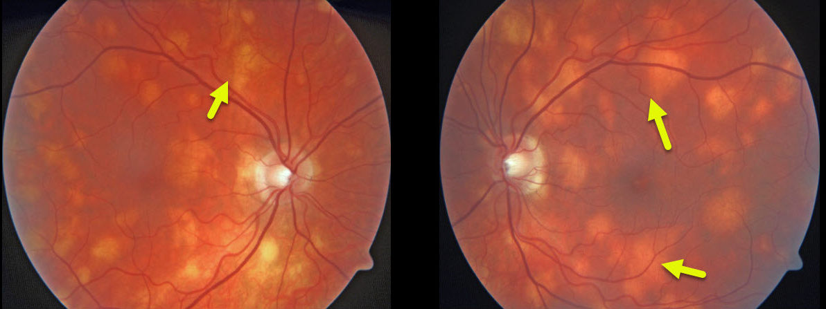
These creamy yellow-white choroidal lesions scattered throughout the posterior pole are characteristic of birdshot chorioretinopathy.
Learning Points:
This disease was appropriately described and named by Drs. Ryan and Maumenee (Am J Ophthalmol 1980;89:31-45), since the lesions look like shotgun birdshot scattered throughout the fundus.
Birdshot is typically chronic and bilateral, with a very high HLA-A29 association. It usually affects women in their 40s to 60s. Vision loss is usually caused by cystoid macular edema (CME) and retinal atrophy, although macular neovascularization can rarely occur.
Asymptomatic patients can be observed. CME and symptomatic uveitis can be treated with oral steroids, although many will still require immunomodulatory therapy.


