

BRAO
<< Back to Cases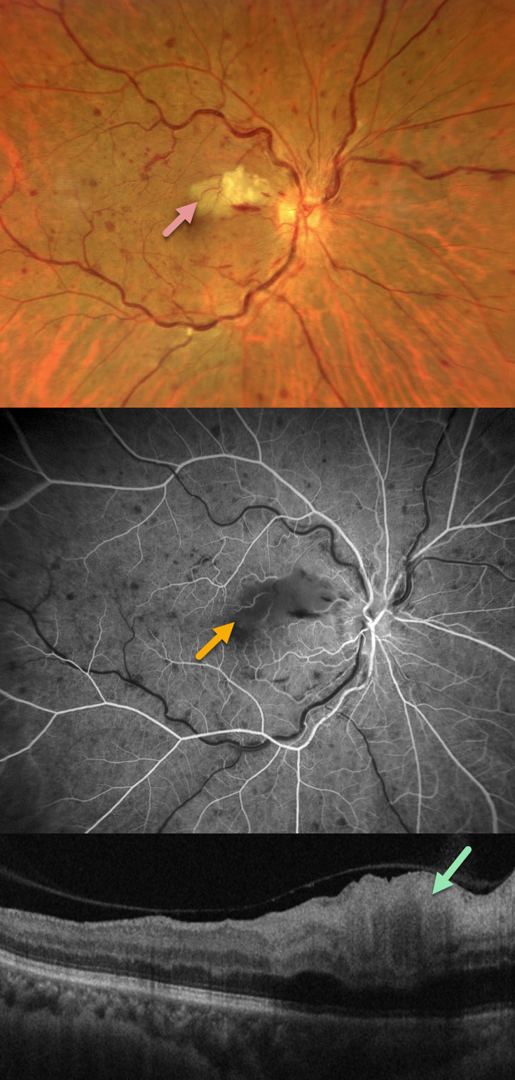
CRVO WITH SECONDARY CILIORETINAL ARTERY OCCLUSION
Francis Ezra Laxamana, Bianca Teresa Suarez, Jesus Jacinto Bautista and Carlo Ladores.
Originally posted on @retina.rocks September 7, 2026
This 58YO female with a history of hypertension and diabetes presented with sudden painless vision loss in her right eye. Vision was 20/25 OD and 20/20 in her normal OS.
Fundus photography shows a non-ischemic central retinal vein occlusion (CRVO) with dilated tortuous veins, mild scattered retinal hemorrhages, and a superonasal macular branch retinal artery occlusion (BRAO). The BRAO shows inner retinal hyperreflectivity on OCT. Fluorescein angiography shows good perfusion throughout the distribution of the CRVO, with a filling defect from the BRAO.
Learning Points:
Some patients with CRVO will develop a secondary non-embolic cilioretinal artery occlusion caused by either compression from optic nerve edema or from a sudden rise in intraluminal retinal capillary bed pressure (Hayreh et al, Retina 2008;28;581-594).
CILIORETINAL ARTERY OCCLUSION
Malvika Singh and Manish Nagpal
Originally posted on @retina.rocks December 30, 2025
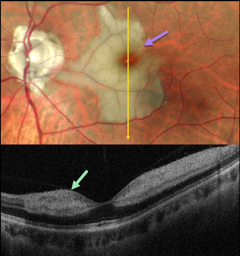
This healthy 46YO male presented with an 8-day history of a central scotoma in his left eye. Vision was 20/20 in his normal OD and 20/400 OS.
Pseudocolor SLO imaging shows an acute cilioretinal artery occlusion with foveal sparing. OCT scanning shows marked inner retinal hyperreflectivity with posterior shadowing.
Learning Points:
The inner two-thirds of the neurosensory retina is supplied by the central retinal artery, with the choroid supplying the RPE and photoreceptor layer. Cilioretinal arteries, which arise from the posterior choroidal circulation, are present in up to 50% of individuals, and bilateral in about 25% (Schneider et al, Acta Ophthalmologica 2021;99:e310-e318).
Their clinical significance usually arises from foveal preservation in eyes with concomitant retinal artery occlusions (Brown and Shields, Arch Ophthalmology 1979;97:84-92). However, they can also be a rare cause of primary branch retinal artery occlusion, either in isolation (our patient) or in association with other entities, including central retinal vein occlusions (Hayreh et al, Retina 2008;28:581-594).
RETINAL ARTERIAL MACROANEURYSM (RAM) WITH BRAO
Originally posted on @retina.rocks August 13, 2024
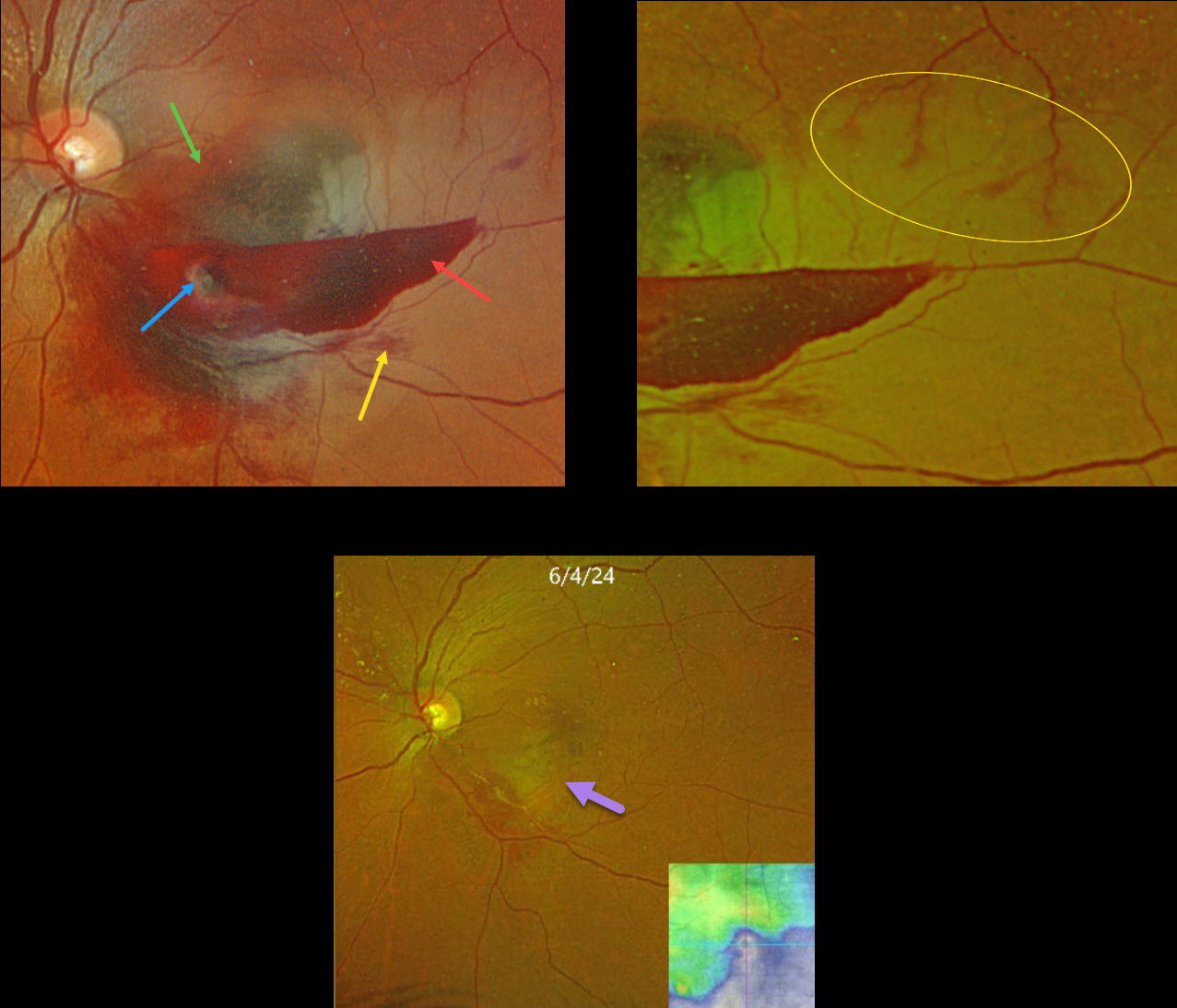
This 59YO female presented with acute vision loss in her left eye. Vision was counting fingers.
Optos color RGB shows a multi-layered macular hemorrhage, including preretinal, intraretinal, and subretinal blood. The causative ruptured retinal arterial macroaneurysm (RAM) is noted within the preretinal blood inferonasally. The surrounding neurosensory retina is somewhat opaque from a secondary inferior branch retinal artery occlusion (BRAO).
Optos RG color imaging shows some periarterial sparing along the superotemporal edge of the BRAO. The perfused superior retinal arteriole, as it enters the region of the occlusion, still has some oxygen that is diffusing into the perivascular tissue.
The patient was treated with several monthly Avastin injections in hopes of more rapidly resolving the surrounding fluid to help relieve arterial compression. Although the blood and fluid resolved, secondary central and inferotemporal macular atrophy resulted from the prior BRAO. Vision was 20/400.
Learning Points:
A BRAO can be a rare complication of direct thermal laser of a RAM (Russell and Folk, AJO 1987;104:186-187), which is one reason it is almost never performed anymore. In this case, we believe the BRAO was secondary to mechanical arterial compression from the surrounding blood.
INTRAOCULAR FOREIGN BODY WITH SECONDARY BRAO
Anjana Mirajkar and Manish Nagpal
Originally posted on @retina.rocks August 5, 2024
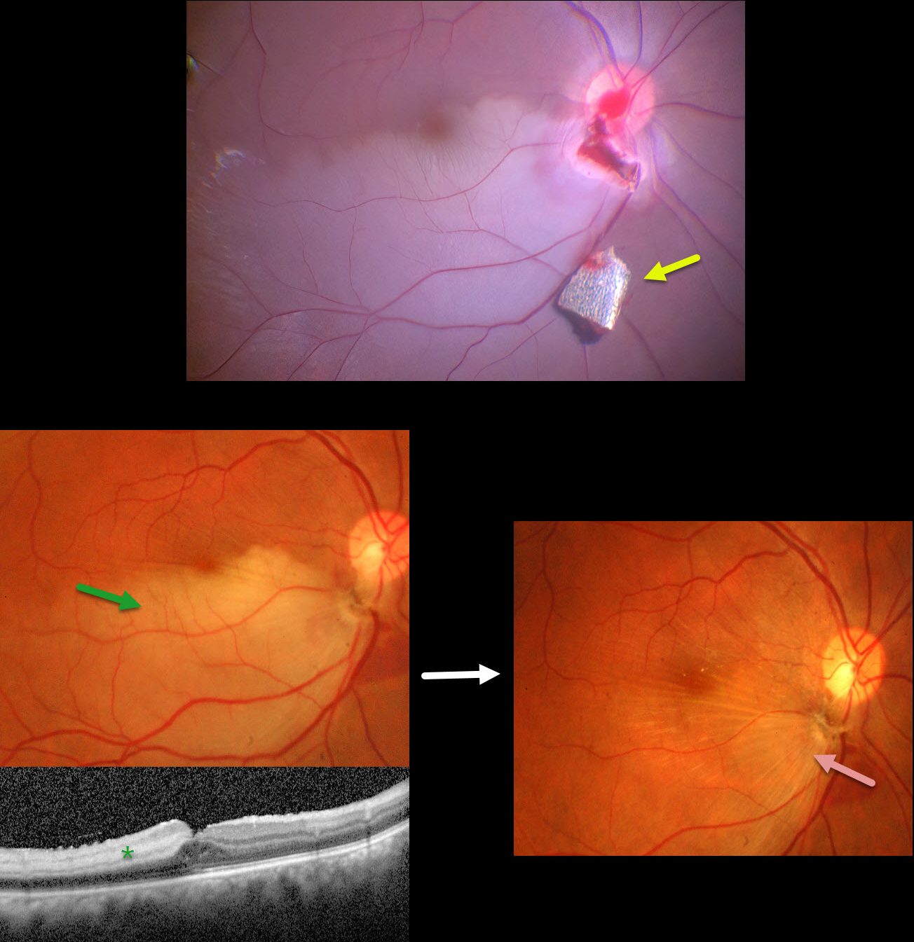
This 34YO male presented with pain and vision loss immediately following a metal-on-metal injury at work. Vision was light perception. Slit lamp examination revealed a corneal laceration and traumatic cataract. B-scan ultrasonography and CT scanning revealed an intraocular foreign body (IOFB). The patient was immediately taken to surgery for corneal wound repair, lensectomy, and vitrectomy.
Intraoperative photography shows a metallic intraocular foreign body (IOFB) resting just inferior to the optic nerve. The impact site is just inferior to the optic nerve, with an inferotemporal branch retinal artery occlusion (BRAO) distally. The IOFB was removed with a rare-earth magnet.
Two weeks following surgery, vision was 20/120 with an aphakic correction. The inferior retina remains opaque, and the OCT shows inner retinal hyperreflectivity from the BRAO. A secondary intraocular lens implantation was performed 3 months postoperatively. Eleven months following the initial surgery, the BRAO is fully resolved, and secondary retinal striae extend outwards from the impact site. Vision remarkably improved to 20/30.
This case was published in the Indian Journal of Ophthalmology (Nagpal M et al, Ind J Ophthalmol 2018;66;146-148).
CRVO + CILIORETINAL ARTERY OCCLUSION
Amit Nandan Tripathi and Vipin Rana
Originally posted on @retina.rocks June 18, 2024
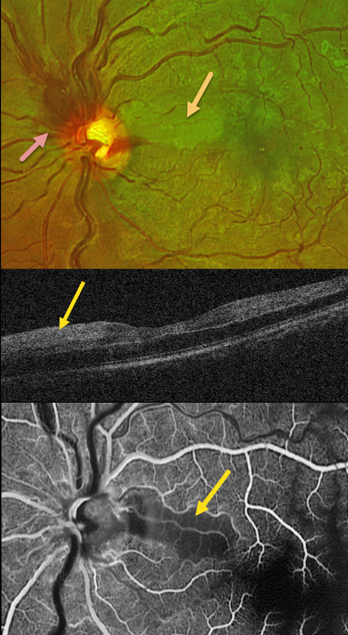
This healthy 30YO male presented with sudden vision loss in his left eye. Vision was 20/20 in his healthy right eye and counting fingers in his left eye.
Optos color RG imaging shows an acute non-ischemic central retinal vein occlusion (CRVO) with mild inner retinal hemorrhages scattered in all quadrants. The optic nerve is somewhat swollen superiorly, and there is an acute cilioretinal artery occlusion in the superior papillomacular bundle.
This area is hyperreflective on OCT. Fluorescein angiography shows a well-perfused CRVO except for a localized area of ischemia within the nasal occlusion.
Learning Points:
Some patients with CRVO will develop a secondary non-embolic cilioretinal artery occlusion caused by either compression from optic nerve edema or from a sudden rise in intraluminal retinal capillary bed pressure (Hayreh et al, Retina 2008;28:581-594). Observation was recommended, and vision improved to 20/60 five days after symptom onset.
CRVO + SECONDARY BRAO
Will Gibson
Originally posted on @retina.rocks April 15, 2024
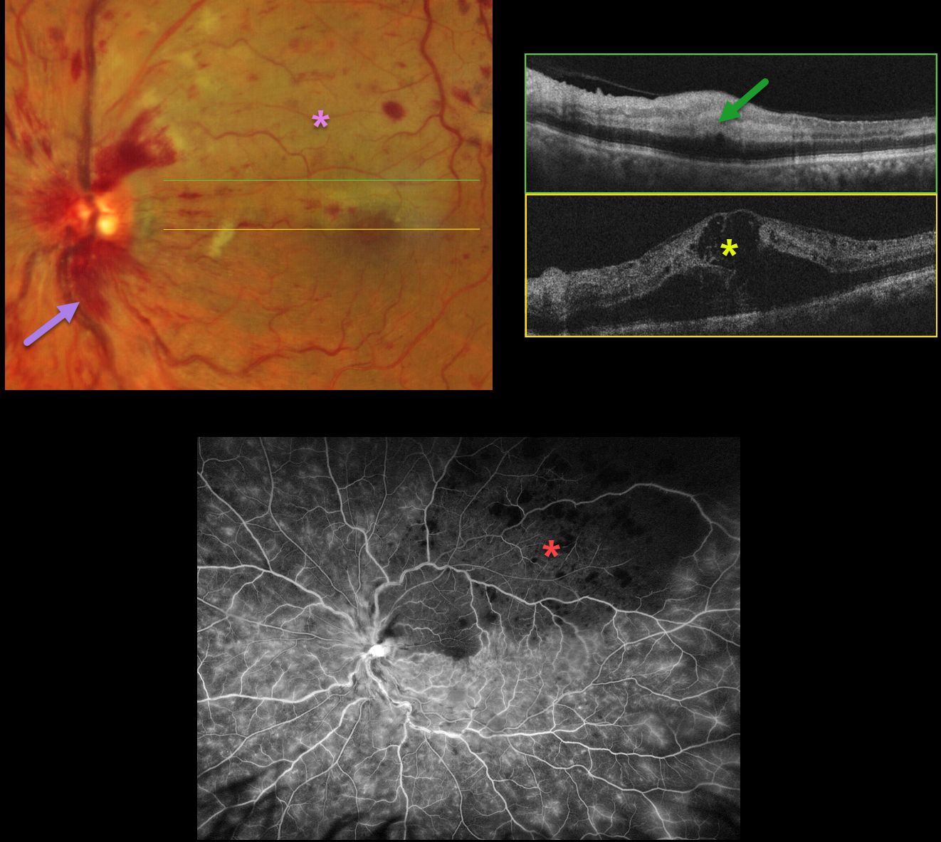
This 66YO male presented with sudden vision loss in his left eye. Vision was 20/30 in his normal right eye and counting fingers in his left eye.
Optos color RG imaging shows a central retinal vein occlusion (CRVO) with mild retinal hemorrhages in all quadrants, except for more moderate hemorrhages superotemporally. A secondary superior branch retinal artery occlusion (BRAO) bisects the fovea.
OCT scanning shows marked central edema, with hyperreflective and thickened inner retina within the BRAO. Fluorescein angiography shows a well-perfused CRVO, except for delayed arterial filling and ischemia within the BRAO.
Learning Points:
Some patients with CRVO will develop a secondary non-embolic BRAO caused by compression from optic nerve edema. However, our patient’s optic nerve did not appear that swollen. Our patient was started on anti-VEGF therapy for the severe macular edema.
BRAO
Originally posted on @retina.rocks December 26, 2022
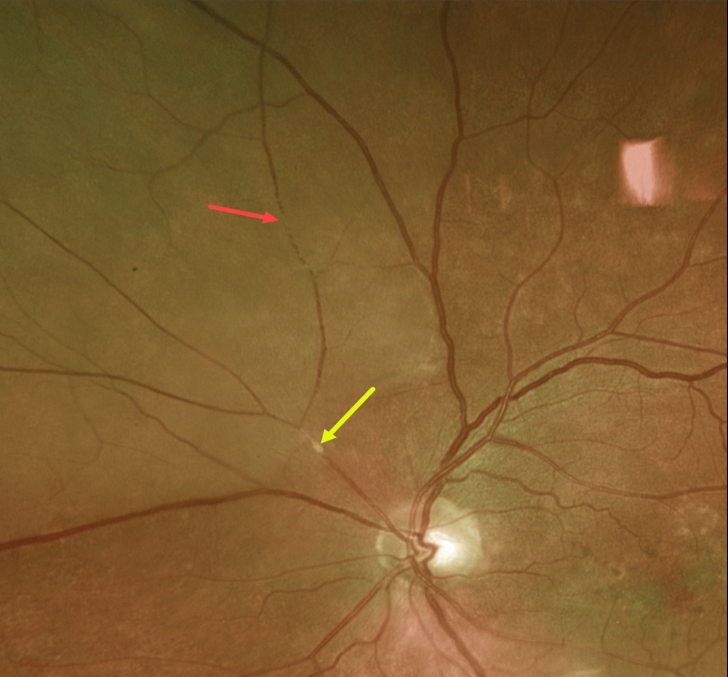
This 73YO male presented with acute inferotemporal visual field loss.
Optos imaging shows an acute extramacular branch retinal artery occlusion (BRAO) caused by a likely platelet-fibrin embolus just superonasal to the optic nerve. The distal retina is somewhat opaque, and the blood column is stagnant superiorly.
This patient had acute retinal ischemia (defined as acute BRAO, central retinal artery occlusion or transient monocular vision loss), and was immediately referred to the nearest stroke center (Biousse et al, Ophthalmology 2018;125:1597-1607).
RETINAL ARTERIAL MACROANEURYSM WITH SECONDARY BRAO
Originally posted on @retina.rocks December 8, 2022
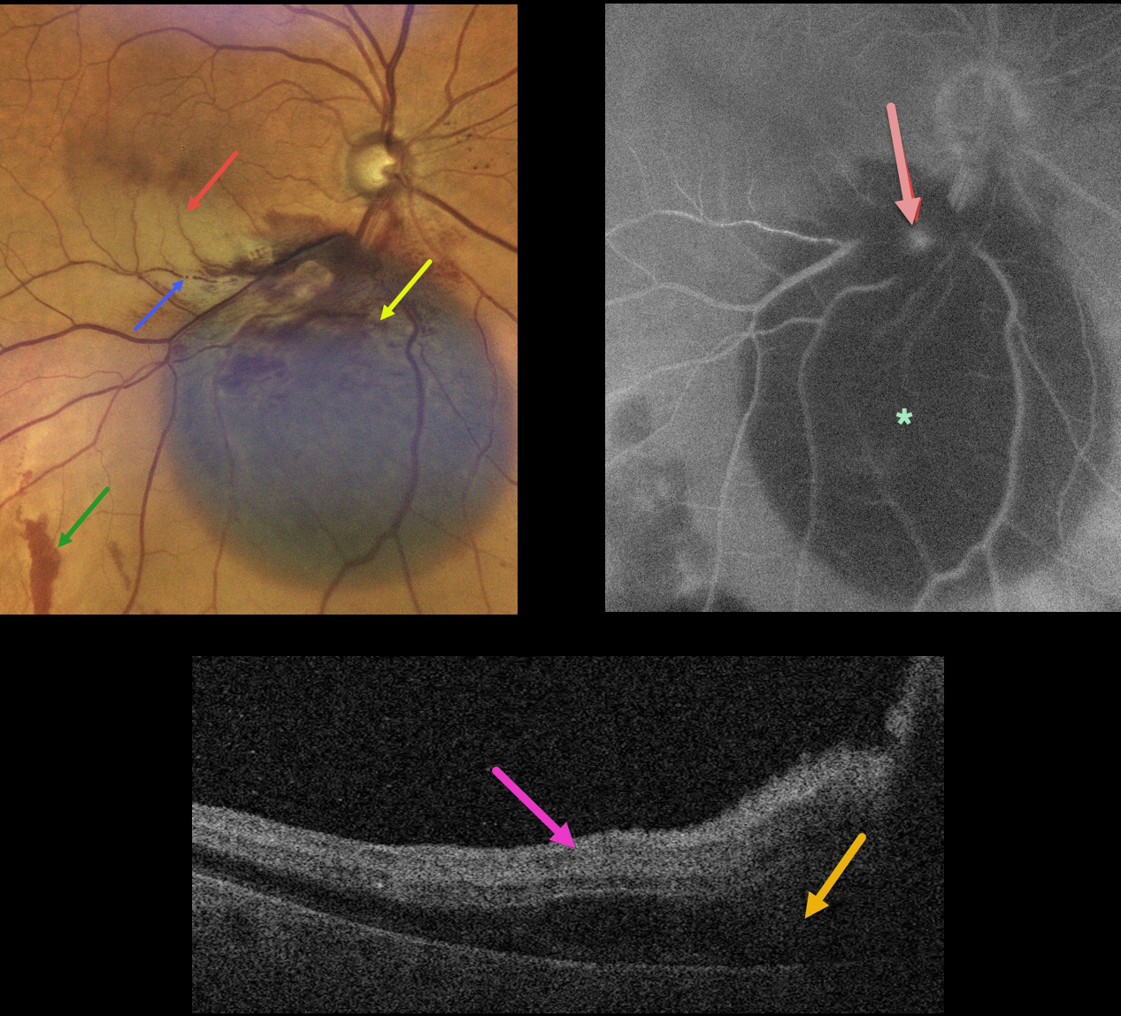
This 79YO female presented with acute inferior paracentral vision loss in her right eye. Vision was 20/50.
A large blister of dark subretinal blood was noted just inferior to the optic nerve. A few small areas of more distal red subretinal blood were seen. An inferior macular branch retinal artery occlusion likely accounted for her symptoms. The blood column is stagnant within the inferior portion of the acute occlusion.
OCT scanning through the BRAO shows hyperreflective inner retina with subretinal fluid.
Fluorescein angiography shows blockage from the subretinal blood, along with a leaking retinal arterial macroaneurysm (RAM).
Learning Points:
A BRAO can be a rare complication from direct thermal laser of a RAM (Russell and Folk, AJO 1987;104:186-187), which is one reason why this is almost never performed anymore. In this case, we believe the BRAO was secondary to mechanical arterial compression from the surrounding blood.
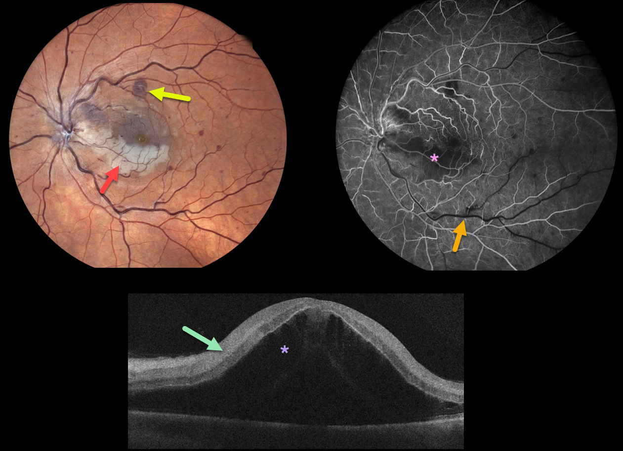
CRVO WITH SECONDARY BRAO
Originally posted on @retina.rocks October 18, 2022
This 49YO male woke with counting finger vision in his left eye. Fundus photography shows a fresh central retinal vein occlusion (CRVO) with mild retinal hemorrhages. An acute inferior macular branch retinal arterial occlusion (BRAO) is also noted.
OCT B-scan shows marked inner retinal hyperreflectivity and severe macula edema.
Fluorescein angiography in the venous laminar phase shows inferior macular hypofluorescence from combined blockage from the ischemic white retina, along with a filling defect from the BRAO. The retinal perfusion elsewhere is good.
Some patients with CRVO will develop a secondary non-embolic BRAO caused by compression from optic nerve edema. However, our patient’s optic nerve did not appear that swollen. Anti-VEGF therapy was started for the severe macular edema.
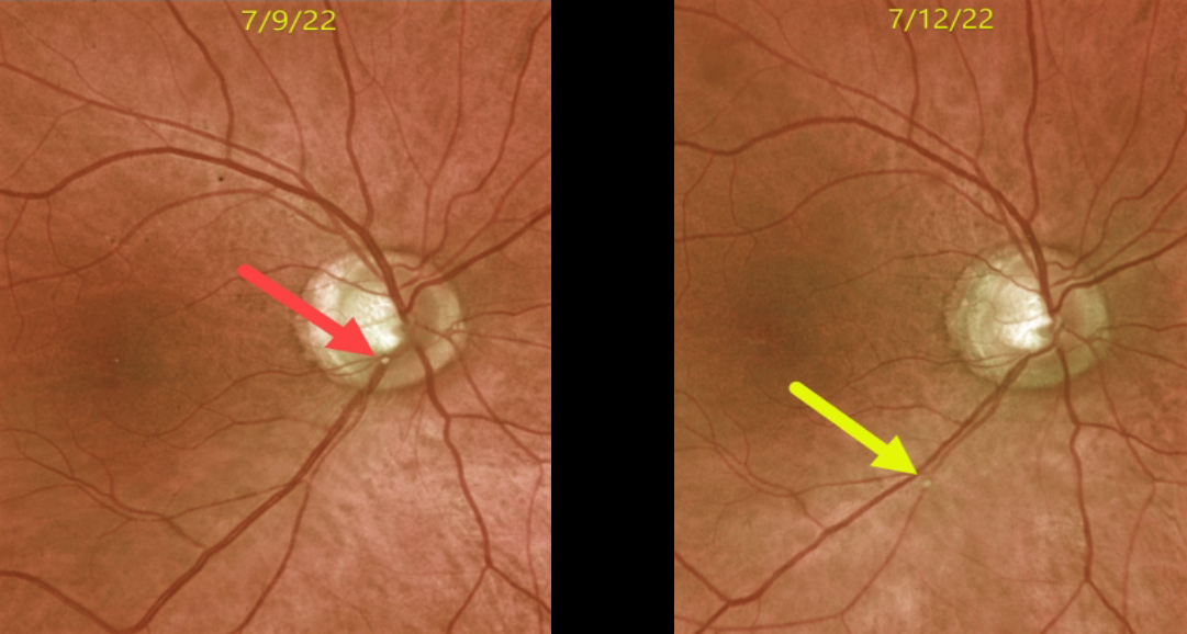
RETINAL EMBOLUS
Originally posted on @retina.rocks July 22, 2022
This 72YO male presented with acute superior field loss from an acute branch retinal artery occlusion (BRAO) in his right eye.
The causative calcific embolus is seen originally along the inferior optic nerve margin. Three days later, the plaque moved further downstream.
This patient had acute retinal ischemia (defined as acute BRAO, central retinal artery occlusion, or transient monocular vision loss), and was immediately referred to the nearest stroke center, which is the standard of care.
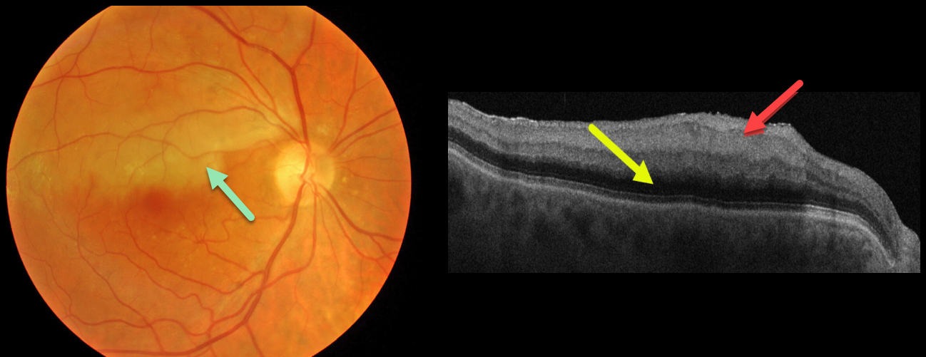
BRAO
Originally posted on @retina.rocks April 29, 2022
This 80YO male presented with an acute branch retinal artery occlusion (BRAO) and 20/25 vision. Triton color imaging shows an opaque retina in the superior macula.
Swept-source OCT shows a thickened, hyperreflective inner retina with posterior shadowing due to decreased light transmittance through the opacified inner retinal layers. There are no emboli.
This patient had acute retinal ischemia (defined as acute BRAO, central retinal artery occlusion or transient monocular vision loss), and was immediately referred to the nearest stroke center.
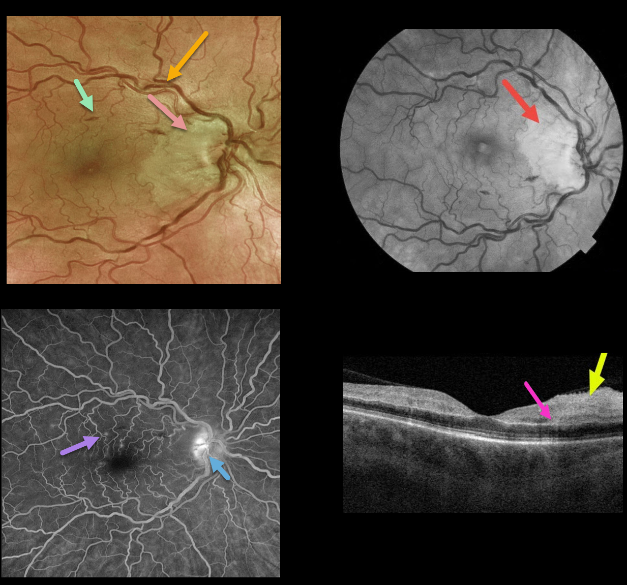
CRVO + RAO + PAMM
Originally posted on @retina.rocks March 4, 2022
This 50YO female presented with counting finger vision in her right eye due to an acute central retinal vein occlusion (CRVO) with a nasal macular retinal artery occlusion (RAO).
Optos fundus photography shows retinal vascular tortuosity, mild retinal hemorrhages, a mildly swollen nerve, and white ischemic nasal macular retina. The ischemic retinal changes are most apparent on the red-free photo.
Fluorescein angiography (FA) shows some mild focal areas of macular leakage with late staining of the temporal disc.
OCT shows areas of inner retinal edema and hyperreflectivity. There is also a small area of mid-retinal hyperreflectivity consistent with paracentral acute middle maculopathy (PAMM).
Learning Points:
Cilioretinal artery occlusion is usually non-embolic and seen in association with giant cell arteritis, secondary to a CRVO, or as an isolated event. The occlusion may be due in part to arterial compression from a swollen nerve.
Pichi et al felt that CRVO PAMM-type lesions, as seen in our patient, are due to hypoperfusion from increased downstream intraluminal pressure (Br J Ophthalmol 2019;103:1137-1145).
We couldn’t find a definite cilioretinal artery in our patient, although the ischemia was within the distribution of one if one were present. Interestingly, the nasal macular capillaries appeared perfused on FA. However, FA captures just the inner retinal capillary plexus.
Although the OCT shows evidence of acute inner capillary plexus ischemia, the angiographically perfused inner retina supports a more PAMM-type mechanism in our case.
BRAO
Originally posted on @retina.rocks January 6, 2022
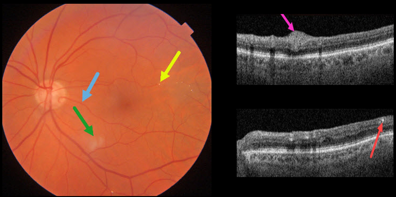
This 85YO male has 20/25 vision despite multiple retinal emboli and a resolving branch retinal artery occlusion (BRAO).
Triton swept-source OCT shows residual inner retinal edema and opacification. The embolus on Triton photography is also imaged in the OCT B-scan.
A cilioretinal artery may be responsible for the good vision.
BRVO
Originally posted on @retina.rocks September 7, 2021
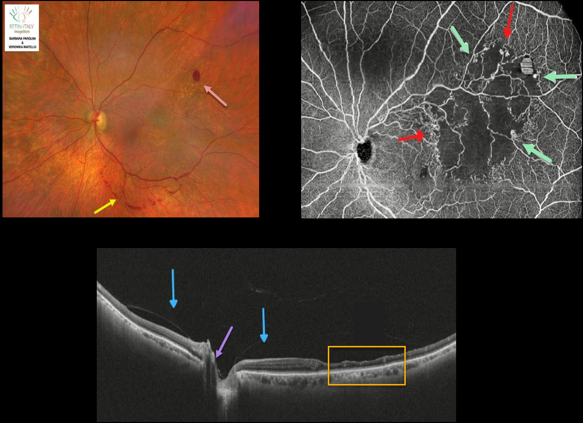
This 76YO female was referred for recent floaters. Wide-field color photography shows some retinal hemorrhages in the superotemporal retina, along with a band of preretinal blood below the inferotemporal arcade.
Wide-field SD-OCT shows inner retinal thinning within the area of occlusion temporally. The posterior hyaloid is detached over the posterior pole with a persistent adherence to the optic nerve.
Wide-field OCT angiography (OCTA) shows a broad area of capillary ischemia within the distribution of the branch retinal vein occlusion (BRVO), along with areas of retinal neovascularization.
Learning Points:
The endothelial cells in retinal neovascularization lack the tight junctions found in the normal retinal vasculature and therefore leak profusely on fluorescein angiography.
OCTA highlights retinal vessels not with a traditional fluorescing dye, but by detecting the movement of red blood cells within the vessel. This is why OCTA images offer beautiful detail but no leakage.
The role of OCTA versus traditional fluorescein angiography is still being determined, but growing evidence suggests it may provide similar clinical information without the need for dye injection (see Spaide et al, Progress Retinal Eye Research 2018;54;1-55).
SUSAC SYNDROME
Originally posted on @retina.rocks July 20, 2021
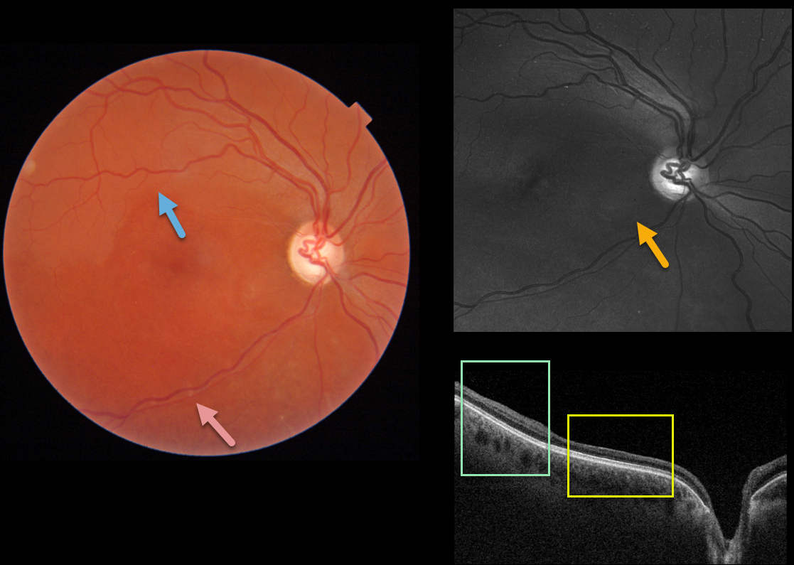
This healthy 30YO female was referred for poor vision in her right eye. She mentioned losing vision in this eye during high school, but couldn’t provide any further details. She also gave a history of hearing loss, which had never been evaluated. Vision was HM OD and 20/30 in her normal left eye.
Triton imaging shows faint retinal whitening in the superior macula. The retinal arterioles are narrowed and somewhat sheathed inferiorly.
Swept-source OCT shows normal inner retinal architecture temporally, with inner retinal atrophy and disorganization of the normal architecture more nasally. Right inferotemporal nerve fiber loss is quite dramatic on Optos green imaging.
We diagnosed her with a resolved inferior branch retinal artery occlusion (BRAO), although this may represent Susac’s syndrome. The involved retina appeared redder than the uninvolved superior retina, accounting for the whitish superior retina noted above.
Subsequent work-up, including hypercoagulable labs and MRI, was negative. She denied prior intravenous drug use. To complete the workup, we will be obtaining carotid Doppler and cardiac ultrasonography.
Learning Points:
Susac’s syndrome, also known as retinocochleocerebral vasculopathy, is a rare microangiopathic disease characterized by a triad of encephalopathy, sensorineural hearing loss, and branch retinal artery occlusions with a predilection for younger females.
In the presence of Susac’s syndrome, MRI findings should show corpus callosum involvement. However, the full triad commonly takes months to develop, complicating diagnosis.
BRAO
Originally posted on @retina.rocks October 19, 2020
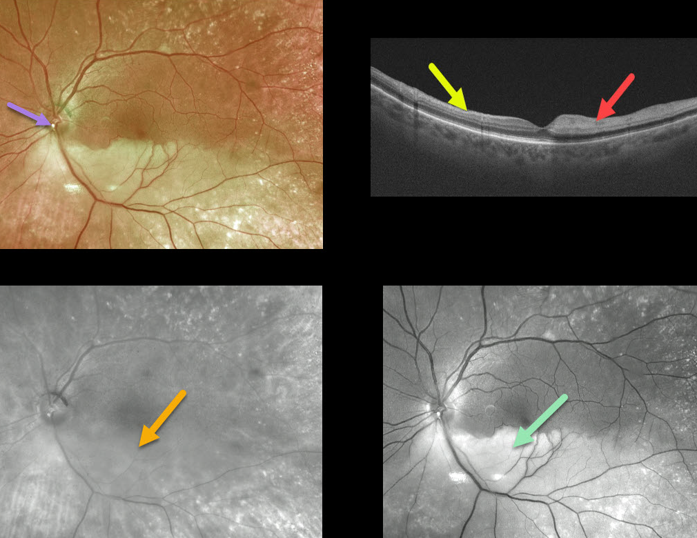
This is an 80YO female who presented with a branch retinal arterial occlusion (BRAO) and 20/40 vision.
The causative calcific embolus is seen overlying the inferior optic nerve. The opaque retina is especially well visualized with the Optos green channel but virtually invisible in the Optos red channel.
The vertical B-scan contrasts the normal superior retina with the ischemic inferior retina (marked hyperreflectivity of the inner retina, causing shadowing of the outer retina and choroid).
Learning Points:
This patient had acute retinal ischemia (defined as acute BRAO, central retinal artery occlusion, or transient monocular vision loss) and was immediately referred to the nearest stroke center, which is the standard of care.
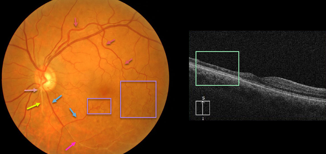
RETINAL ARTERIOVENOUS MALFORMATION
Originally posted on @retina.rocks July 28, 2020
At first or quick glance, this looks like an old inferotemporal branch retinal vein occlusion (BRVO).
On closer inspection, there is a white-sheathed vessel arising from the nerve inferiorly. Numerous collateral vessels are noted inferior and temporal to the fovea, and there is a sheathed vessel in the inferior arcade.
OCT shows marked inner retinal atrophy with preservation of the photoreceptor layers, consistent with a prior branch retinal artery occlusion. So the collateral vessels (virtually always a sign of a prior venous occlusion) indicate an old BRVO, and the atrophic and sheathed vessel indicates an old BRAO, both in the same quadrant. Right?
Tracing the large branch of the superotemporal arcade inferotemporally, we see that the collaterals appear to be arterial. Inferior to the nerve, we see an arteriole that crosses the sheathed vessel, indicating that the sheathed vessel is not an artery but a vein. Moreover, the large vessel off the nerve, inferotemporal in location, appears to be a vein that eventually connects to the collateral vessels we previously identified as arterial.
Our best guess is that this may be a low-grade arteriovenous malformation, possibly with a prior secondary BRAO/BRVO. Unfortunately, we don’t have a fluorescein angiogram, which may help better define the vascularization.
Learning Points:
Remember that arteries don’t cross arteries and veins don’t cross veins. Tracing vessels can be a helpful key to identifying retinal vascular disorders.
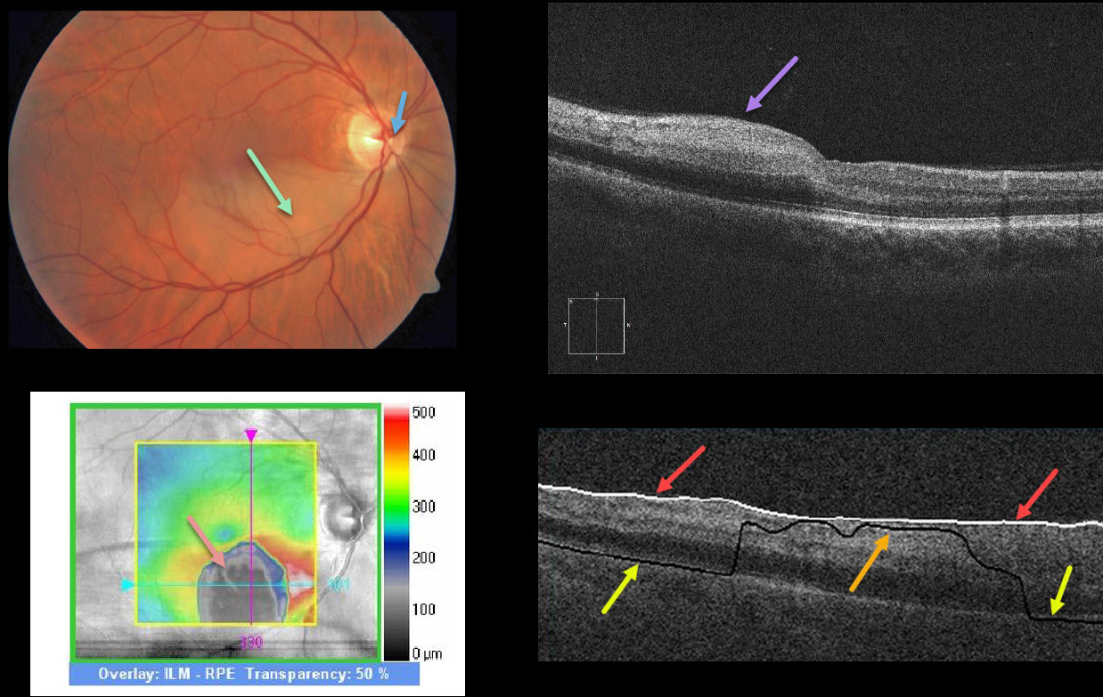
BRAO
Originally posted on @retina.rocks June 30, 2020
This patient has a subtle white, opaque retina in the inferior macula due to an acute branch retinal artery occlusion (BRAO). The causative embolus is the tiny white lesion just nasal to the nerve’s center.
OCT shows hyperreflectivity from the acutely ischemic inner retinal layers. The black blob on the retinal thickness map reflects an incorrectly identified RPE detachment (PED) caused by the algorithm misidentifying the RPE due to the marked hyperreflectivity of the inner retinal layers.
This phenomenon is demonstrated by the B-scan, which shows that the ILM is correctly identified by the white line. The black line is supposed to identify the RPE, which is correctly identified nasally and temporally but misidentified centrally due to said hyperreflectivity.
Learning Points:
An acute BRAO can be subtle clinically but more dramatic on OCT. Acute inner retinal OCT hyperreflectivity can lead to misidentification of retinal layers, resulting in artifacts.


