

BRVO
<< Back to Cases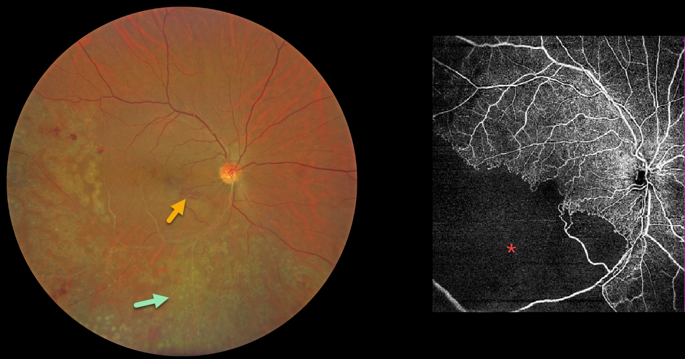
BRVO WITH RETINAL NEOVASCULARIZATION
Abhishek Karra and Ravindra Karra
Originally posted on @retina.rocks August 10, 2026
This 42YO male presented with a 3-month history of decreased vision in his right eye. He had received laser treatment in this eye elsewhere. Vision was 20/60 OD.
Color photography shows an old ischemic inferotemporal branch retinal vein occlusion (BRVO) with heavy scatter laser throughout its distribution. Collateral vessels are noted in the inferonasal macula. OCT angiography shows profound capillary loss. There was no macular edema, and observation was recommended.
Learning Points:
In 1986, the Branch Vein Occlusion Study Group reported that scatter laser decreased the risk of vitreous hemorrhage from retinal neovascularization in BRVO and suggested that laser should be applied once neovascularization develops (Arch Ophthalmol 1986;104:34-41). Thirty-five years later, scatter laser remains the gold standard, although some may prefer to stabilize the neovascularization with anti-VEGF therapy before laser.
BRVO WITH RETINAL NEOVASCULARIZATION AND COLLATERALS
Originally posted on @retina.rocks February 11, 2026
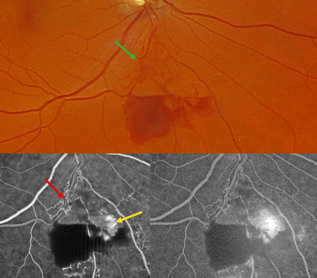
This 59YO male was referred for asymptomatic retinal findings in his right eye. Vision was 20/30.
Optos color RG imaging shows an inferior branch retinal vein occlusion with midperipheral preretinal blood. Collateral vessels are noted at the site of the occlusion. These collaterals show no leakage angiographically, although the adjacent retinal neovascularization does leak.
Learning Points:
Collateral vessels are dilated, pre-existing vessels that bypass an occlusion. These are most commonly seen in the posterior pole following retinal venous occlusions and are exceedingly rare with arterial occlusions. The endothelial tight junctions are intact, so they don’t leak angiographically. This helps differentiate them from neovascularization, which does leak. Our patient’s eye shows us a wonderful example of both.
BRVO WITH RETINAL NEOVASCULARIZATION
Originally posted on @retina.rocks November 3, 2025
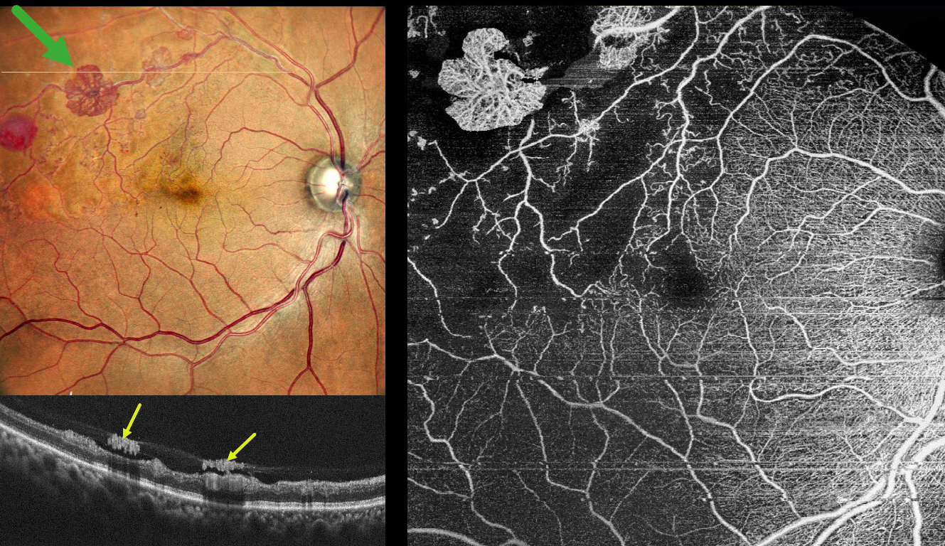
This 55YO hypertensive male presented for a routine eye examination. He gave a history of a resolved superotemporal branch retinal vein occlusion (BRVO) with macular edema in his right eye that was successfully treated with a single anti-VEGF injection in 2022. Vision was 20/15 OD and 20/15 in his normal OS.
Pseudocolor SLO imaging shows an ischemic superotemporal BRVO with several fronds of active red and variably fibrosed retinal neovascularization. A round deep retinal hemorrhage is noted distally. On OCT, the neovascularization appears as hyperreflective plaques along the posterior edge of the partially detached posterior hyaloid. OCT angiography confirms diffuse capillary nonperfusion, a relatively preserved foveal avascular zone, and flow signals within the areas of neovascularization.
Learning Points:
An attached vitreous provides the scaffold for neovascularization to grow onto and spread. Retinal neovascularization grows from the inner retinal surface and proliferates along the outer posterior hyaloid face (Vaz-Pereira et al, Retina 2017;37:1287-1296). This tethering to a partially detached and contracting hyaloid is what causes tractional retinal detachment and vitreous hemorrhage in proliferative retinopathies.
In 1986, the Branch Vein Occlusion Study Group reported that scatter laser decreased the risk of vitreous hemorrhage from retinal neovascularization in BRVO and suggested that laser should be applied once neovascularization develops (Arch Ophthalmol 1986;104:34-41). Almost 40 years later, scatter laser remains the gold standard, although some may prefer to stabilize neovascularization with anti-VEGF therapy before laser. An anti-VEGF injection, followed by a superotemporal scatter laser, was therefore recommended for our patient.
BRVO WITH RED AND YELLOW DEVITALIZED BLOOD
Originally posted on @retina.rocks September 29, 2025
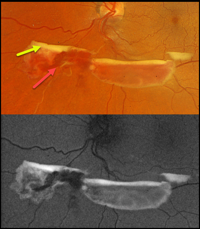
This 48YO female was referred for asymptomatic retinal findings. Vision was 20/20 in the normal OD and 20/20 OS.
Optos color RGB imaging shows red and yellow preretinal blood just inferior to the nerve due to an ischemic extramacular inferonasal branch retinal vein occlusion (BRVO). On fundus autofluorescence (FAF), the red blood is hypo-FAF, and the devitalized yellow blood is markedly hyper-FAF. Intravitreal Avastin was given, and scatter laser was scheduled, although she was immediately lost to follow-up.
Learning Points:
Porphyrins are organic molecules that are part of the heme molecule in hemoglobin. When these compounds break down, the devitalized blood appears yellow and becomes intensely hyperautofluorescent (Bloom and Spaide, Retinal Cases & Brief Reports 2022;16:401-402).
BRVO WITH COLLATERALS AND DRIL
Malvika Singh, Akansha Sharma and Manish Nagpal
Originally posted on @retina.rocks August 19, 2025
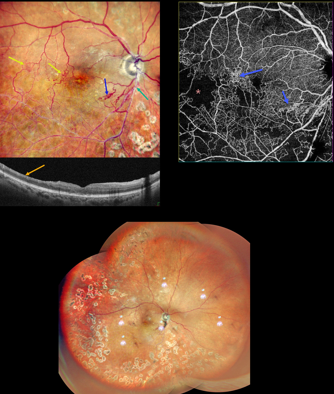
This 65YO male presented with a branch retinal vein occlusion (BRVO) in his right eye and 20/40 vision. He was treated elsewhere about a year ago with 3 intravitreal injections and scatter laser 7 years earlier.
Pseudocolor SLO shows an old major inferotemporal ischemic BRVO, which arises from an arteriovenous crossing just inferior to the disc. Extensive collateral vessels bypass the occlusion and cross the temporal horizontal raphe. OCT scanning shows thinned, featureless inner retina (disorganization of retinal inner layers, DRIL). Scatter laser is noted temporally and inferiorly. OCT angiography shows variable capillary loss throughout the BRVO distribution and highlights the collateral vessels.
Learning Points:
Collateral vessels are dilated, pre-existing vessels that bypass an occlusion. These are most commonly seen in the posterior pole following retinal venous occlusions and are exceedingly rare with arterial occlusions. The endothelial tight junctions are intact, so they don’t leak angiographically. This helps differentiate them from neovascularization, which does leak.
DRIL is seen in various retinal vascular disorders, including diabetic macular edema and retinal vein occlusions. It is generally associated with worse vision and increased anti-VEGF treatment burden (see Babiuch et al, JAMA Ophthalmol 2019;137:38-46).
BRVO WITH RETINAL VENOUS MACROANEURYSM
Originally posted on @retina.rocks April 14, 2025
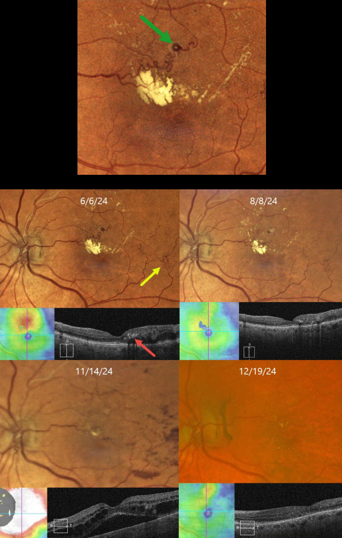
This 72YO female has been followed since 2017 for a branch retinal vein occlusion in her left eye. She has required ongoing anti-VEGF therapy for macular edema and failed macular laser for treatment burden several times. Although her eye was stable for years, on 6/6/24, there was new macular lipid with increased superior macular edema. Vision was stable at 20/25.
Color imaging shows a major superotemporal branch retinal vein occlusion with extensive collateral vessels and superior macular edema. Confluent lipid exudates threaten the macular center and appear as hyperreflective deposits in the mid-retina on OCT. A retinal venous macroaneurysm (RVM) is noted in the superior macula.
An intravitreal Eylea injection was administered, followed by focal thermal laser treatment of the RVM. Two months later, on 8/8/24, vision was 20/30. The lipid was decreasing, and the edema resolved. On 11/14/24, vision decreased to 20/60. Although the lipid was almost resolved, recurrent macular fluid persisted. Following a single Avastin injection, vision improved to 20/30, and the edema resolved. Macular grid laser was applied in hopes of reducing the treatment burden. We will continue to follow her closely and treat her as needed.
Learning Points:
In 1990, Cousins et al reported that RVMs can be an additional finding in occlusive venous disease, always in association with collateral vessels or capillary or arterial macroaneurysms (AJO 1990;109:567-570). These vascular abnormalities, also known as retinal capillary macroaneurysms or telangiectatic capillaries (TelCaps), tend to have a higher rate of intraretinal lipid (40%) but are less likely to affect central vision since they are usually outside the macula. They tend to have a higher association with retinal ischemia, thus being at a higher rate for developing retinal neovascularization (32%). Although these lesions will usually involute spontaneously, we treated our patient out of concern that the lipid might extend into the foveal center.
BRVO AND PERIPHLEBITIS FROM LIKELY SARCOIDOSIS
Originally posted on @retina.rocks October 8, 2024
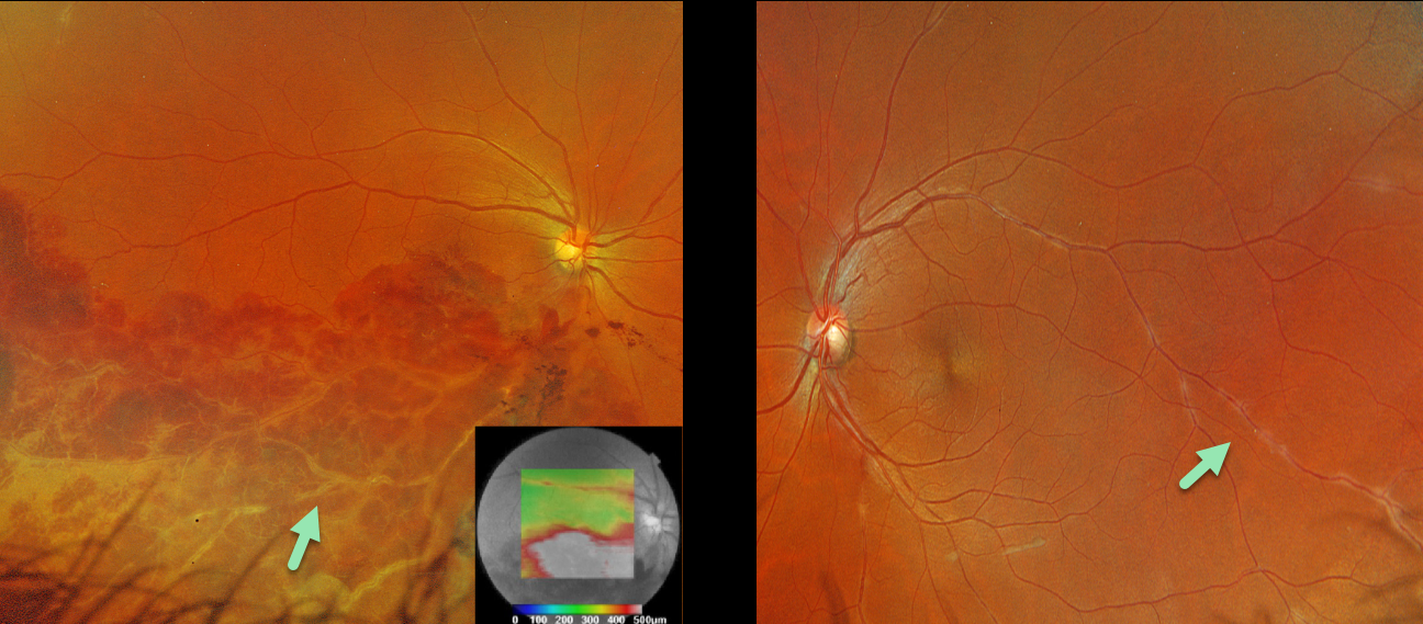
This healthy 36YO male presented with 1 week of unilateral vision loss. Vision was 20/400 OD and 20/20 OS.
Optos color RGB imaging OD shows a major inferotemporal branch retinal vein occlusion (BRVO) with severe confluent retinal hemorrhages and a frosted appearance to the major vessels. The occlusion does not seem to arise from a more proximal arteriovenous (AV) crossing. A small area of peripheral superotemporal periphlebitis is noted OD, with more extensive periphlebitis OS. Triton swept-source OCT shows inferior macular fluid.
BRVOs nearly always arise at an AV crossing, where a thickened arterial vessel compresses the underlying vein. Sarcoidosis is the classic exception where a BRVO is not caused by an AV crossing, but by an inflammatory narrowing of the involved vein. In addition, our patients’ segmental periphlebitis (‘candle wax drippings’) is virtually pathognomonic for sarcoidosis as well.
A sarcoidosis workup was recommended, although the patient failed to show for his testing and was immediately lost to follow-up.
BRVO + MYELINATED NERVE FIBER LAYER
Evgenii Chernov
Originally posted on @retina.rocks September 30, 2024
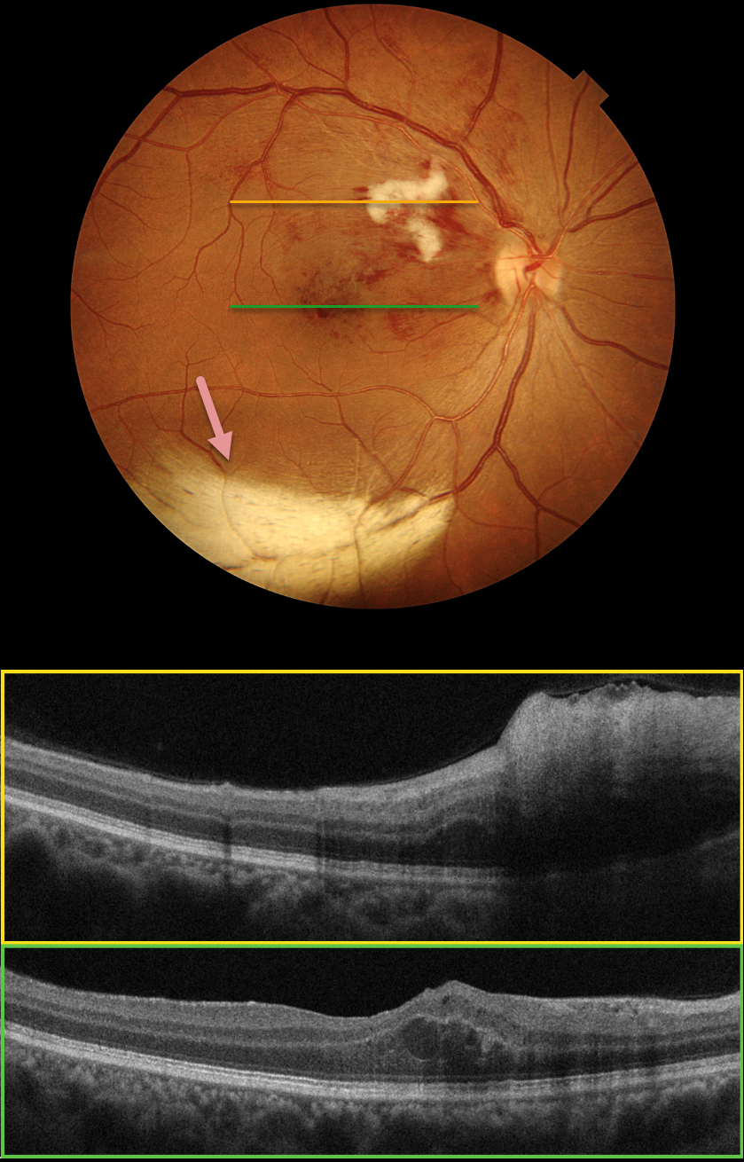
Color photography of this 48YO female’s right eye shows a major superotemporal branch retinal vein occlusion (BRVO) with foveal blood. A prominent nerve fiber layer infarct (cotton-wool spot, CWS) is present in the superonasal macula. OCT scanning shows diffuse hyperreflective inner retinal thickening through the CWS and some central foveal fluid in the outer nuclear layer. An unrelated patch of myelinated nerve fiber layer (NFL) is noted along the inferotemporal arcade. The macular edema resolved following 3 monthly intravitreal Lucentis injections (not shown).
Our patient’s eye shows two different pathologies that each involve the NFL. Myelinated NFL occurs when retinal nerve fibers develop a myelin sheath, which usually stops posterior to the lamina cribrosa. The white myelin beautifully displays the normally transparent NFL. Myelination may represent an oligodendrocytic choristoma (see Rao et al Retina 2019;39:1125-1132). Nerve fiber layer infarcts cause a more fluffy, white ischemic opacification of the NFL and are a relatively nonspecific finding in numerous disorders, including diabetes, retinal venous occlusion, hypertension, and HIV/AIDS.
BRVO WITH RETINAL VENOUS MACROANEURYSM
Seif Anwar
Originally posted on @retina.rocks August 22, 2024
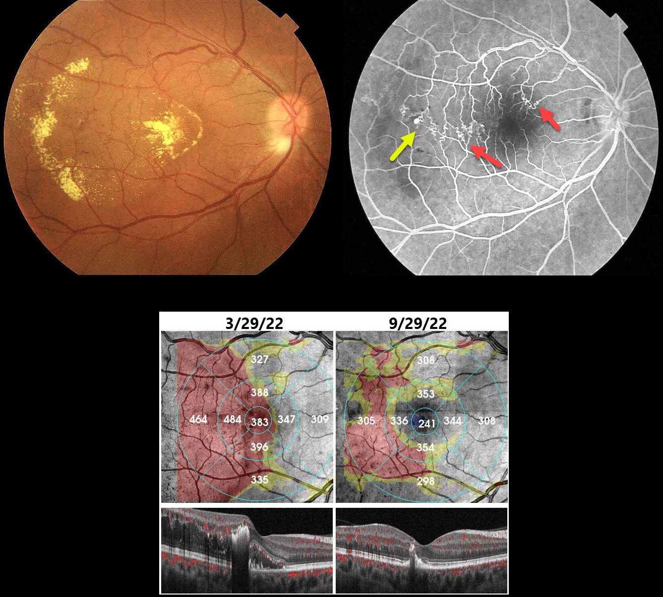
This 40YO hypertensive male presented with gradual vision loss in his right eye. Vision was 20/200 OD and 20/20 in his normal OS.
Color imaging shows central and temporal macular lipid and telangiectasia. Fundus fluorescein angiography shows a superotemporal branch retinal vein occlusion (BRVO) with superonasal and temporal collateral vessels. A retinal venous macroaneurysm (RVM) is noted distally.
Our patient was initially treated unsuccessfully with several monthly Lucentis injections. The macular edema and lipid improved following macular grid laser with focal treatment of the RVM.
Learning Points:
In 1990 Cousins et al reported that RVMs can be an additional finding in occlusive venous disease, always in association with collateral vessels or capillary or arterial macroaneurysms (AJO 1990;109:567-570). These vascular abnormalities tend to have a higher rate of intraretinal lipid (40%) but are less likely to affect central vision since they are usually outside the macula. They tend to be associated with retinal ischemia, thus increasing the risk of developing retinal neovascularization (32%).
BRVO
Originally posted on @retina.rocks July 18, 2024
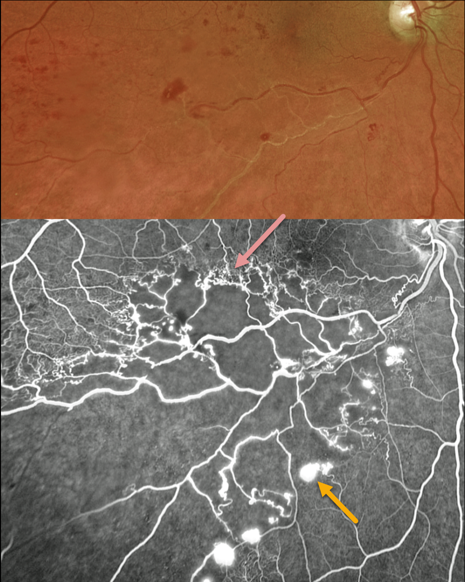
This 56YO male presented with a major inferotemporal ischemic branch retinal vein occlusion (BRVO) in his right eye and 20/40 vision. Despite the deceptively good vision, Optos ultra-widefield imaging showed marked ischemia throughout the distribution of the occlusion, which fortunately spared the central macula. A row of collateral vessels extends along the superior edge of the occlusion, with retinal neovascularization extending along its inferior extent. Leaking disc neovascularization is also present. A scatter laser throughout the area of ischemia was performed, but unfortunately, he was then lost to follow-up.
Learning Points:
In 1986, the Branch Vein Occlusion Study Group reported that scatter laser decreased the risk of vitreous hemorrhage from retinal neovascularization in BRVO, and suggested that laser should be applied once neovascularization develops (Arch Ophthalmol 1986;104:34-41). Thirty-five years later, scatter laser remains the gold standard, although some may prefer to first stabilize neovascularization with anti-VEGF therapy before laser.
Collateral vessels are dilated, pre-existing vessels that bypass an occlusion. These are most commonly seen at the posterior pole following retinal venous occlusions and are exceedingly rare with arterial occlusions. The endothelial tight junctions are intact, so they don’t leak angiographically. This helps differentiate them from neovascularization, which does leak.
BRVO
Nilesh Kumar
Originally posted on @retina.rocks April 11, 2024
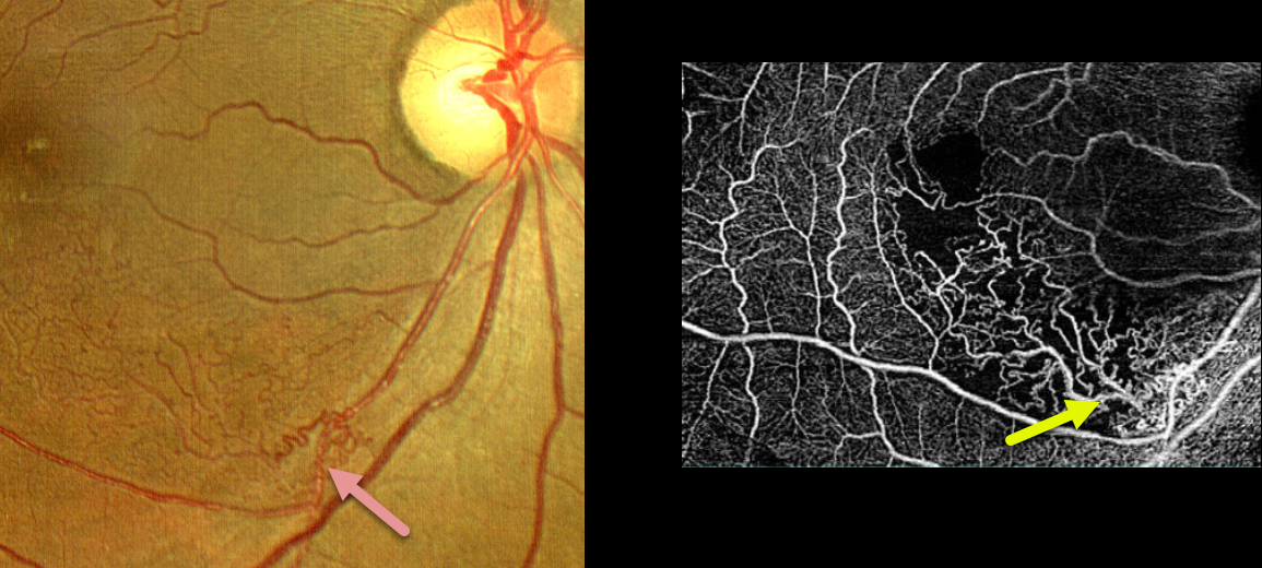
This 53YO female presented with 20/80 vision in her right eye from a macular branch retinal vein occlusion (BRVO) with macular edema. The edema resolved following a single injection of a Lucentis biosimilar. She was then lost to follow-up for 8 months, when she returned with 20/20 vision.
Extensive inferonasal macular collateral vessels are seen on fundus photo and OCTA. OCT showed no edema (not shown).
Learning Points:
Collateral vessels are dilated, pre-existing vessels that bypass an occlusion. These are most commonly seen at the posterior pole following retinal venous occlusions, but can also occur at the optic disc with optic nerve meningiomas. The endothelial tight junctions are intact, so they don’t leak on fluorescein angiography. This helps differentiate them from neovascularization, which does leak.
Collaterals most commonly form in the deep vascular plexus, which represents the primary venous drainage for the inner retina (Freund et al, JAMA Ophthalmology 2018;1262-1270).
BRVO
Originally posted on @retina.rocks February 27, 2024
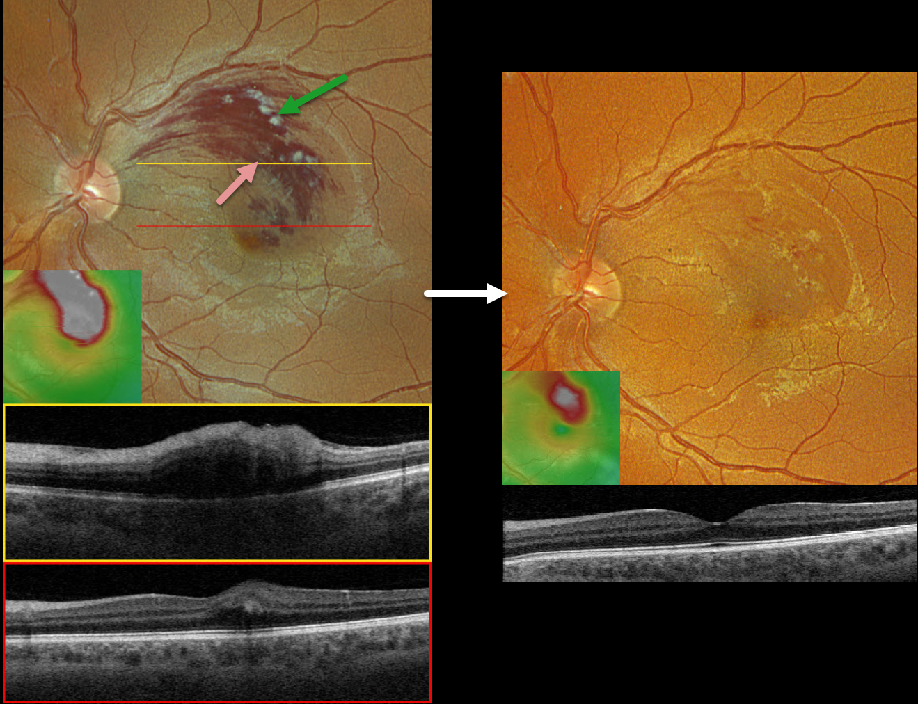
This healthy 33YO male presented with a 1-week history of vision loss in his left eye. Vision was 20/20 in his normal right eye and 20/60 in his left eye.
Optos color RGB imaging shows a superior macular branch retinal vein occlusion (BRVO) with relatively confluent inner retinal hemorrhages and superiorly located nerve fiber layer infarcts. Retinal fluid extends through the fovea.
Triton swept-source OCT through the superior macula shows retinal thickening and inner hyperreflectivity from the blood. Scanning through the macular center shows very shallow central edema. The macular cube shows the extent of the retinal thickening.
Following several monthly Avastin injections, vision improved to 20/30, and the macula was virtually free of fluid.
Learning Points:
Macular grid laser photocoagulation (Branch Vein Occlusion Study Group, AJO 1984;96:271-282) was the mainstay for treating persistent vision loss due to BRVO macular edema until the advent of intravitreal steroid and anti-VEGF therapy in the early-to-mid 2000s.
Although we were initially ‘WOW’ed by the near immediate and dramatic edema improvement that was never seen with thermal laser, the SCORE Study Research Group in 2009 suggested that grid laser should “remain the benchmark against which other treatments are compared” due to similar vision between intravitreal triamcinolone and laser but with fewer laser adverse events (Arch Ophthalmol 2009;127:1115-1128).
Intravitreal anti-VEGF injections have subsequently become the standard of care for initial therapy. A recent systematic review and meta-analysis showed that a treat-and-extend protocol is as effective as monthly and PRN regimens (OSLI Retina 2023;54:131-138).
Adding macular laser probably does not reduce the treatment burden or further improve vision (Ojima et al, Retina 2023;43:999-1004).
BRVO WITH RETINAL VENOUS MACROANEURYSM
Originally posted on @retina.rocks October 2, 2023
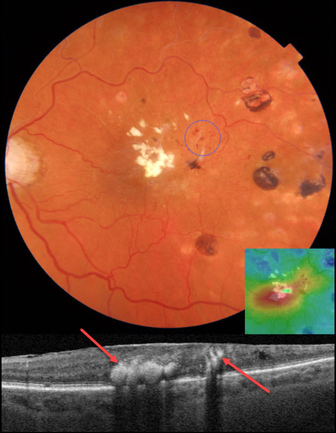
This 83YO female presented with recent vision loss in her left eye. There was a prior history of a branch retinal vein occlusion (BRVO), for which she received scatter laser elsewhere 8 years earlier. Vision was 20/60 OS.
Color photography shows superotemporal macular telangiectasia with likely retinal venous macroaneurysms (RVMs). Fairly dense lipid is noted in the fovea along with temporal laser scarring.
On OCT scanning, the lipid is markedly hyperreflective with posterior shadowing. There is central macular edema on the thickness map. Focal thermal laser was recommended to the RVMs.
Learning Points:
In 1990, Cousins et al reported that RVMs can be an additional finding in occlusive venous disease, always in association with collateral vessels or capillary or arterial macroaneurysms (AJO 1990;109:567-570).
These vascular abnormalities tend to have a higher rate of intraretinal lipid (40%) but are less likely to affect central vision since they are usually outside the macula. They tend to have a higher rate of developing retinal neovascularization (32% of eyes).
BRVO
Originally posted on @retina.rocks August 1, 2023
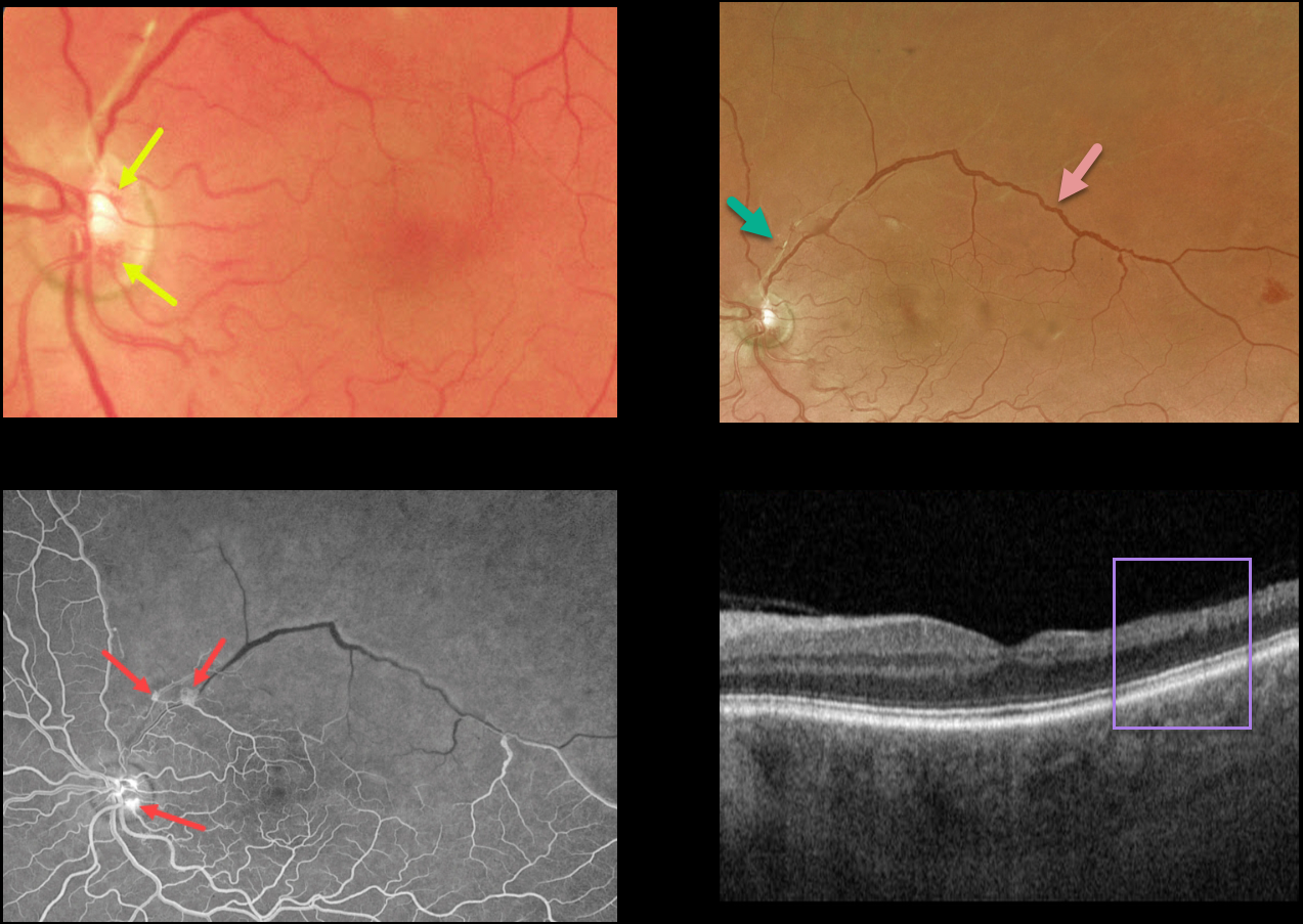
This 55YO male presented with relatively asymptomatic inferior field loss in his left eye.
Color imaging of the left nerve and macula shows a few small areas of disc neovascularization (NVD) and a fibrosed arteriole superiorly. Optos color RG imaging shows an irregularly dilated superotemporal major retinal vein and a featureless superior retina.
Fluorescein angiography shows severe retinal ischemia throughout the entire distribution of the branch retinal vein occlusion (BRVO) with leaking NVD and neovascularization elsewhere.
OCT scanning shows temporal inner retinal atrophy with the inner retinal layers replaced by a single hyperreflective band.
Scatter laser photocoagulation was recommended.
Learning Points:
In 1986, the Branch Vein Occlusion Study Group reported that scatter laser decreased the risk of vitreous hemorrhage from retinal neovascularization in BRVO, and suggested that laser should be applied once neovascularization develops (Arch Ophthalmol 1986;104:34-41).
Decades later, scatter laser remains the gold standard, although some may prefer to first stabilize neovascularization with anti-VEGF therapy prior to laser.
RETINAL TEAR + BRVO
Originally posted on @retina.rocks June 5, 2023
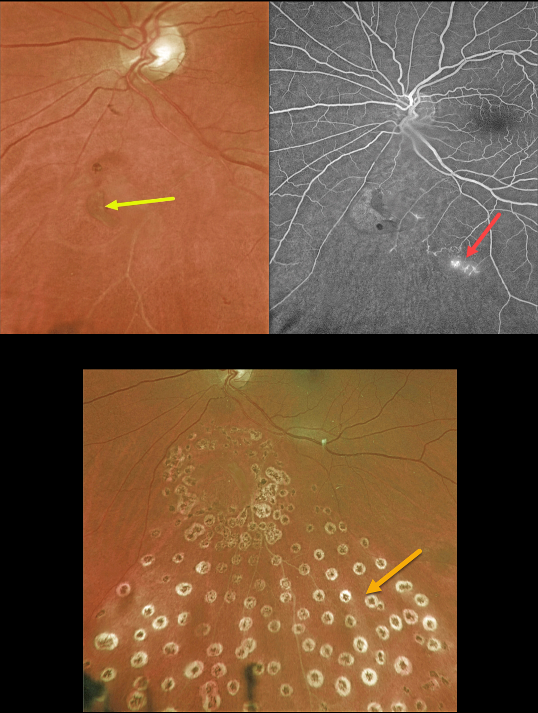
This 63YO female presented with symptomatic flashes and floaters in her left eye.
Optos color imaging shows a retinal tear just inferior to the nerve. An ischemic branch retinal vein occlusion (BRVO) is noted inferiorly. A posterior vitreous detachment (PVD) was noted clinically.
Fluorescein angiography confirmed the inferior ischemic BRVO with some small areas of retinal neovascularization. The tear appeared to be completely independent of the venous occlusion.
The retinal tear was surrounded by a few nearly confluent rows of laser photocoagulation burns, and inferior scatter laser was applied into the distribution of the BRVO. Fundus photography, a few months later, shows the laser scarring.
Learning Points:
Retinal tears are virtually always seen in the retinal periphery and are caused by an adherent vitreous pulling the retina anteriorly towards the vitreous base, most commonly during an acute PVD.
Our patient’s tear is unusual due to its quite posterior location and the flap occurring along its posterior edge. This implies that the vitreous was tugging towards the optic nerve. The optic nerve is usually the last site for the vitreous to release from during an acute PVD (Johnson, AJO 2010;149:371-382).
We hypothesize that the vitreous was completely detached except at the optic nerve and just inferiorly, and the tear was created from anterior to posterior traction when the vitreous separated from the nerve.
BRVO
Originally posted on @retina.rocks May 8, 2023
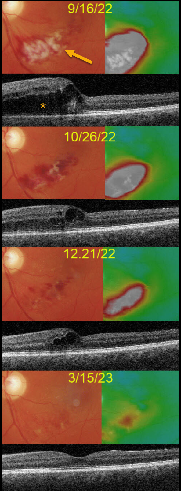
This 50YO male presented on 9/16/22 with 20/50 vision in his left eye from a macular branch retinal vein occlusion with edema.
Although we recommended anti-VEGF injections, he reported that a similar episode improved without treatment about 10 years earlier. He therefore asked if we could initially refrain from treatment.
The occlusion and edema slowly and spontaneously regressed. When last examined on 3/15/23, vision was 20/20 and just a small amount of non-central macular edema remained.
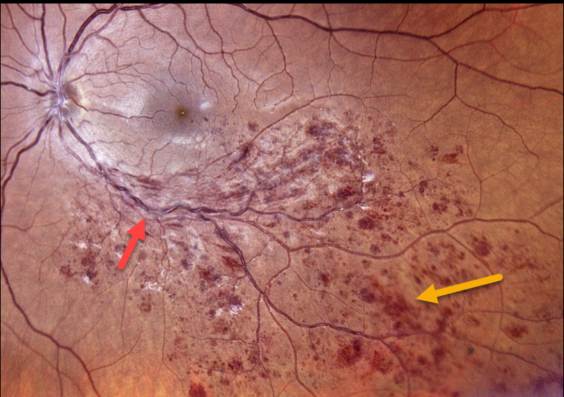
BRVO
Originally posted on @retina.rocks February 2, 2023
This 42YO female was referred for a completely asymptomatic branch retinal vein occlusion (BRVO) in her left eye. Vision was 20/20 due to a lack of macular edema or ischemia.
Color photography shows classic findings, including a quadrant of dilated tortuous veins with mostly inner retinal hemorrhages.
We will initially follow her every 3-4 months and recommend treatment if macular edema or retinal neovascularization develops.
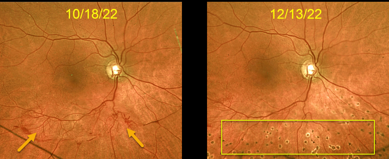
BRVO
Originally posted on @retina.rocks January 25, 2023
This 57YO female was referred for an asymptomatic branch retinal vein occlusion (BRVO) in her right eye. Vision was 20/30.
Optos color imaging shows a normal right macula, with large active retinal neovascularization extending along and distal to the inferotemporal arcade.
Six weeks after the inferior scatter laser, the neovascularization regressed dramatically and virtually completely.
Learning Points:
The results of the Branch Vein Occlusion Study Group (Arch Ophthalmology 1986;104:34-41) still remain the gold standard for treating BRVO-related retinal neovascularization.
Once neovascularization develops, scatter laser into the involved quadrant significantly reduces the risk for vitreous hemorrhage.
Although anti-VEGF therapy can initially be given to rapidly stabilize the neovascularization, laser is usually all that’s needed, as illustrated by our patient.
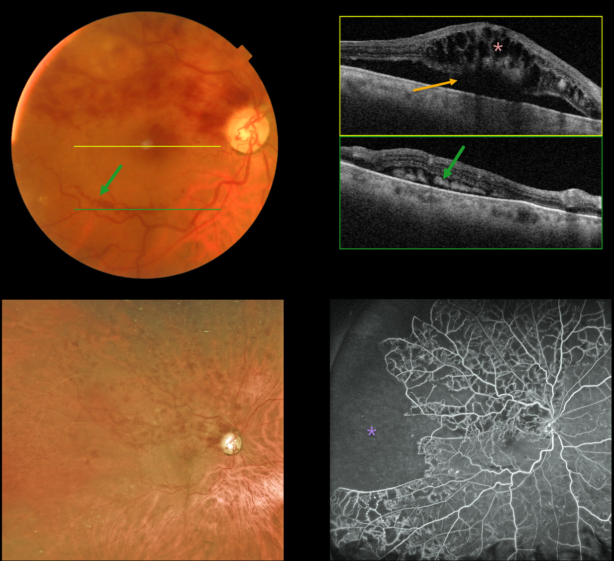
BRVO
Originally posted on @retina.rocks January 9, 2023
This 79YO male presented with recent vision loss of 20/400 in his right eye from a fresh branch retinal vein occlusion (BRVO). Layered subretinal blood is noted in the inferior macula.
Swept-source OCT through the macular center shows marked cystoid macular edema with underlying subretinal fluid. OCT through the inferior macular subretinal blood shows subretinal hyperreflectivity. Optos fluorescein angiography shows marked peripheral ischemia without neovascularization.
Learning Points:
Layered preretinal blood is a common occurrence with proliferative retinopathies, including proliferative diabetic retinopathy and BRVO. The retinal neovascularization grows from the retinal surface onto the back of the posterior hyaloid, and blood accumulates in the subhyaloid space.
The appearance of layered inferior macula blood in our patient is a ‘fake-out’ because the blood was neither neovascular nor preretinal. Blood, likely due to hydrostatic pressure, was forced into the subretinal space, and the subretinal fluid allowed it to settle inferiorly.
The blood and fluid completely resolved following several monthly intravitreal anti-VEGF injections.
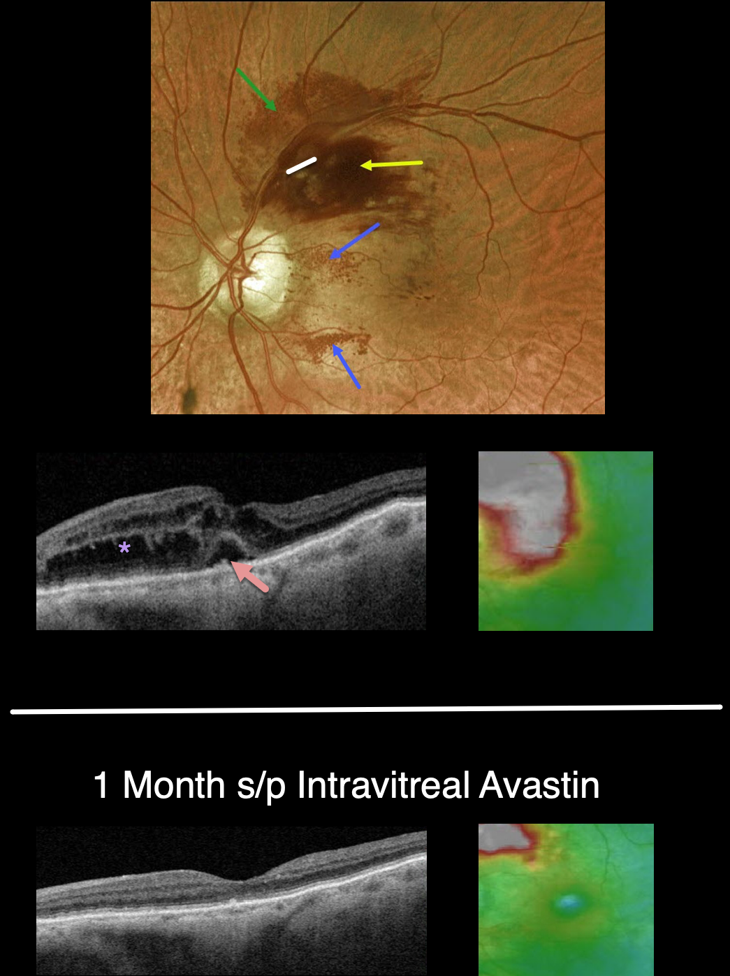
BRVO
Originally posted on @retina.rocks December 28, 2022
This 88YO female presented with recent vision loss in her right eye. Vision was 20/50.
Optos color imaging shows an unusual appearing superotemporal branch retinal vein occlusion (BRVO). Confluent inner retinal hemorrhages are noted in the superonasal macula, with milder deep retinal blood more superiorly. Unusual dots of intraretinal blood are also noted nasally and inferonasally.
Swept-source OCT shows moderately severe central and superonasal macular edema with some foveal subretinal fluid.
An intravitreal Avastin injection was given. One month later, vision improved to 20/30. The retinal hemorrhages dramatically improved. OCT scanning shows complete resolution of all central fluid, with some residual distal superonasal edema.
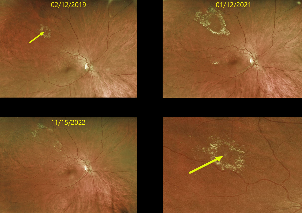
RETINAL VENOUS MACROANEURYSM
Originally posted on @retina.rocks December 15, 2022
This 63 YO female originally presented in 2019 with a history of a major superotemporal branch retinal vein occlusion (BRVO). Successful macular laser was performed for macular edema.
When initially examined, a midperipheral retinal venous macroaneurysm (RVM) with some surrounding lipid was noted. This RVM has been observed due to its distant location from the fovea. The series of photos over several years shows the exudation to wax and wane. At the last examination on 11/15/22, there was almost complete lipid resolution. Vision remains at 20/25.
Learning Points:
In 1990 Cousins et al reported that RVMs can be an additional finding in occlusive venous disease, always in association with collateral vessels or capillary or arterial macroaneurysms (AJO 1990;109;567-1700).
These vascular abnormalities tend to have a higher rate of intraretinal lipid (40%) but are less likely to affect central vision since they are usually outside the macula.
They tend to have a higher association with retinal ischemia, thus being at a higher rate of developing retinal neovascularization (32%). The lesions will usually involute, as was the case in our patient.
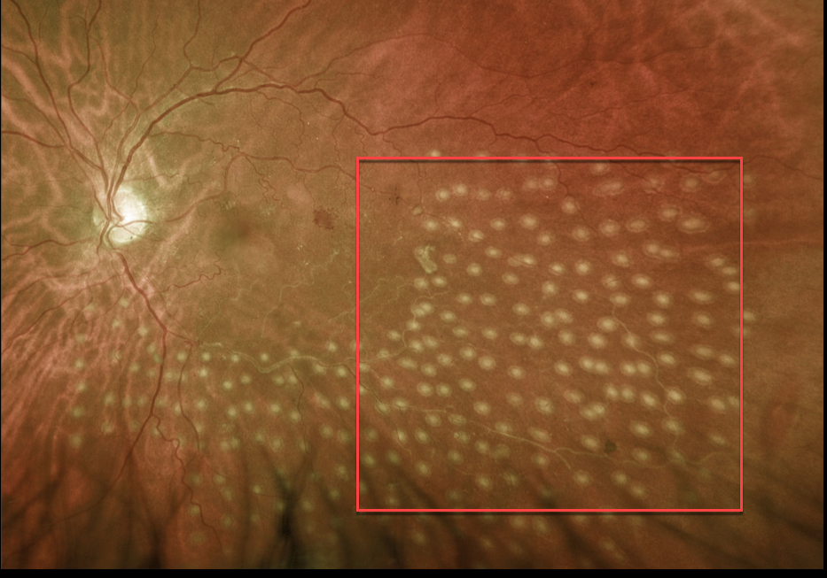
BRVO
Originally posted on @retina.rocks October 5, 2022
This 64YO female presented with one month of symptomatic vitreous floaters and 20/70 vision in her left eye.
Clinical examination and Optos photography with fluorescein angiography (not shown) confirmed an inferotemporal ischemic branch retinal vein occlusion (BRVO) with severe ischemia and scattered areas of retinal neovascularization.
She was scheduled to return for scatter laser photocoagulation, but was lost to follow-up. She presented 6 months later with one day of severe vision loss OS, now at 20/200. A significant vitreous hemorrhage precluded laser, so two monthly intravitreal Avastin injections were given.
Following this, the hemorrhage decreased enough to allow for scatter laser into the involved quadrant.
Learning Points:
In 1986, the Branch Vein Occlusion Study Group reported that scatter laser decreased the risk of vitreous hemorrhage from retinal neovascularization in BRVO, and suggested that laser should be applied once neovascularization develops (Arch Ophthalmol 1986;104:34-41).
Decades later, scatter laser remains the gold standard, although some may prefer to first stabilize neovascularization with anti-VEGF therapy before laser.
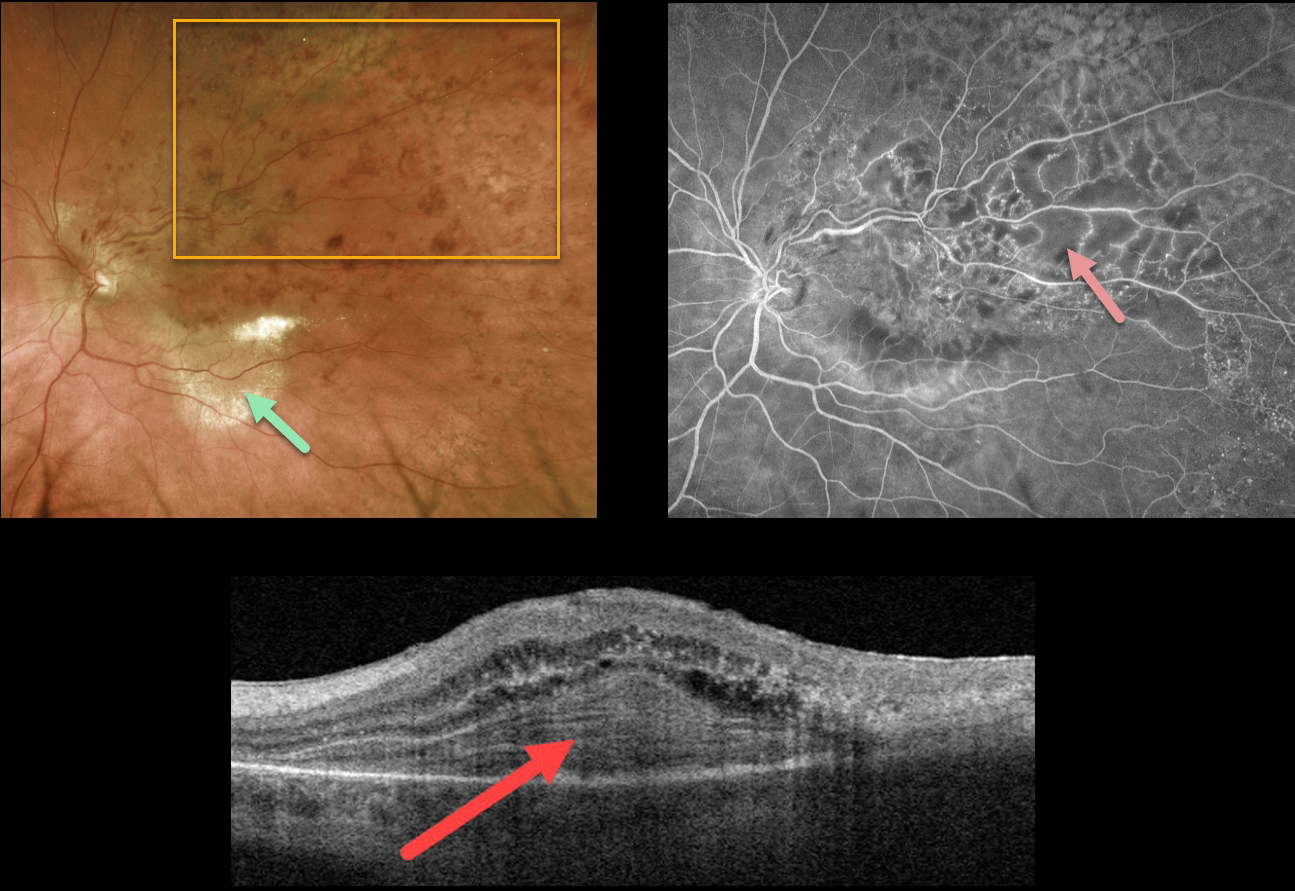
BRVO
Originally posted on @retina.rocks August 8, 2022
This 82YO female presented with a major superotemporal branch retinal vein occlusion (BRVO) and hand motion vision. She reported decreased vision for over a year.
Optos photography shows a major superotemporal BRVO. A plaque of subretinal lipid is seen in the inferotemporal macula. Swept-source OCT through this lipid shows an unusual laminated, subretinal, hyperreflective mound. Fluorescein angiography shows some macular leakage and some areas of more peripheral ischemia.
Although the patient has a poor prognosis due to the chronicity of the disease, monthly intravitreal Avastin for the central edema was initiated in hopes that it may improve her vision.
Learning Points:
Lipid exudates are rarely seen in BRVO, which may be due to a physiologic defect in the inner blood-retinal barrier (the endothelial cell tight junctions) that are too small to allow larger lipoproteins to escape into the extracellular space.
When present, lipid is often associated with a retinal venous macroaneurysm (Cousins et al, AJO 1990;109:567-170), which was not visualized in our patient.
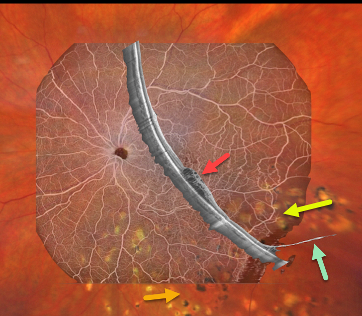
BRVO WITH DEGENERATIVE RETINOSCHISIS
Barbara Parolini and Veronika Matello
Originally posted on @retina.rocks June 13, 2022
This 67YO female has a history of branch retinal vein occlusion (BRVO) in her left eye. Asymptomatic degenerative retinoschisis was also present bilaterally. Vision was 20/33. She had received prior scatter laser and intravitreal injections about 3 years earlier.
Fortunately, all findings remained stable despite loss to follow-up due to the COVID pandemic.
Clarus 500 wide field imaging shows mild inferotemporal tortuosity with collateral vessels within the involved quadrant. Prior scatter laser scarring for secondary retinal neovascularization, along with prior barrier laser around the posterior edge of the schisis cavity, is noted.
Canon Xephilio S1 OCT angiography highlights the collateral vessels and more peripheral retinal nonperfusion. Xephilio B-scan shows some inferotemporal macular edema, and just catches the more distal inferotemporal schisis.
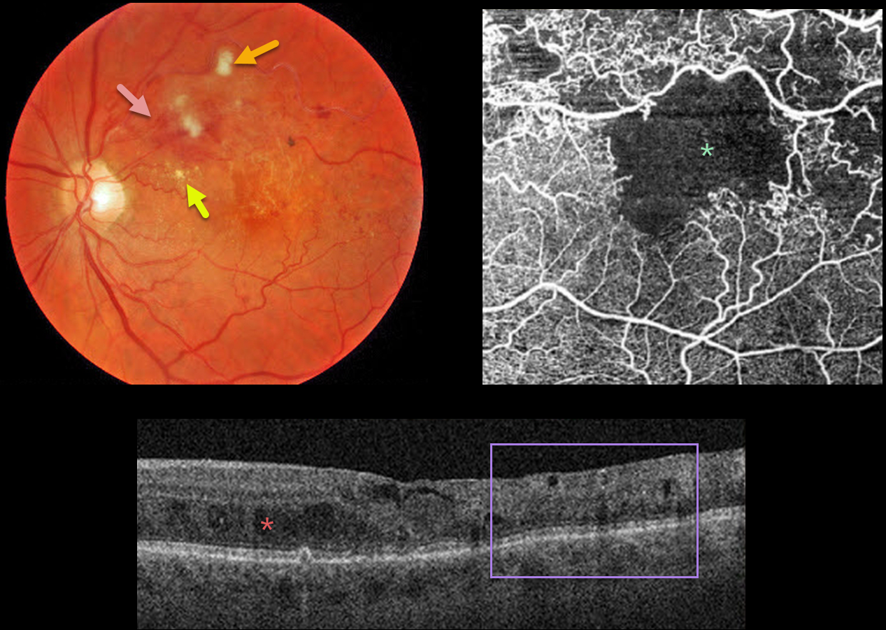
BRVO WITH MACULAR ISCHEMIA
Originally posted on @retina.rocks June 10, 2022
This 79YO male presented with a major superotemporal branch retinal vein occlusion (BRVO) and counting fingers vision.
Fundus photography shows superior macular hemorrhages with inner retinal ischemia (cotton-wool spots) and some nasal lipid. The vessels, especially around the superotemporal fovea, are nonperfused.
The clinically suspected foveal ischemia is confirmed on OCT angiography. Spectral-domain OCT shows disorganization of the inner retinal layers (DRIL), particularly within the clinically ischemic temporal macula, along with scattered inner and outer retinal cysts.
Learning Points:
DRIL is seen in various retinal vascular disorders, including diabetic macular edema and retinal vein occlusions. It is generally associated with worse vision and increased anti-VEGF treatment burden (see Babiuch et al, JAMA Ophthalmol 2019;137:38-46).
Since the patient has no central foveal thickening or retinal neovascularization, we are observing for now. Anti-VEGF therapy will not restore the foveal circulation.
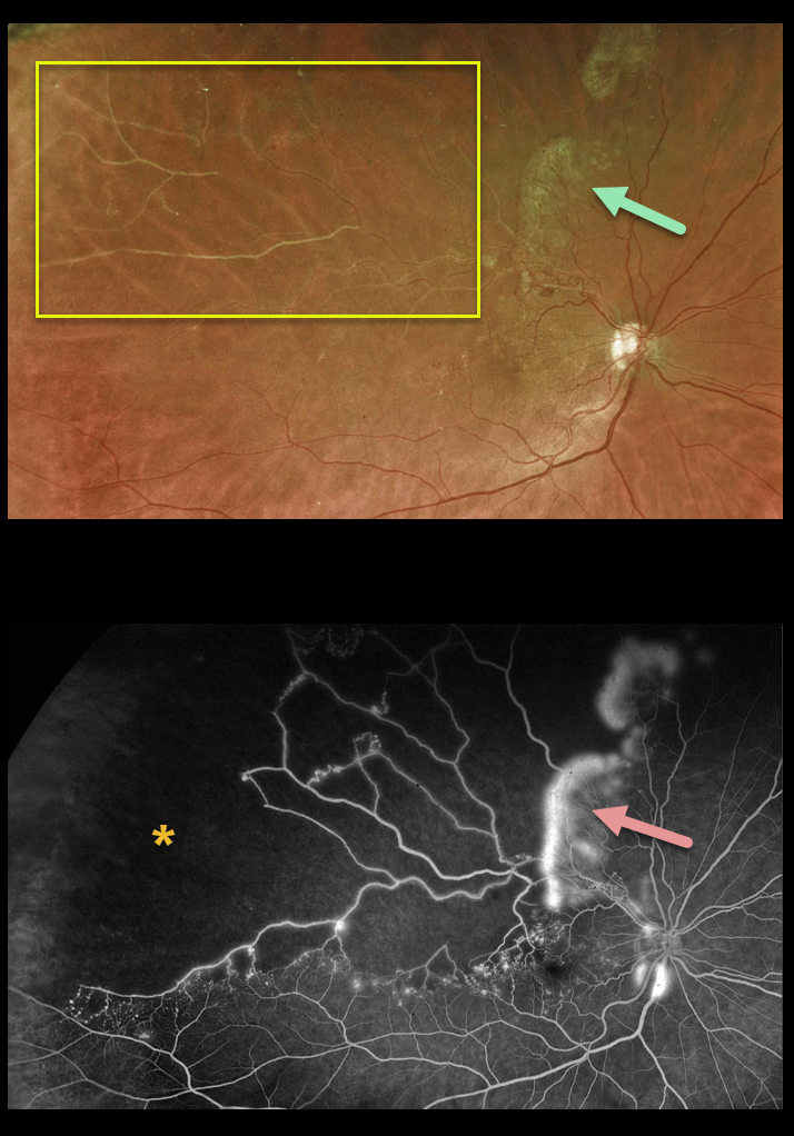
BRVO
Originally posted on @retina.rocks June 14, 2021
This 53YO male presented with a major superotemporal branch retinal vein occlusion (BRVO) and 20/40 vision.
Despite the deceptively good vision, Optos ultra-widefield imaging shows marked ischemia throughout the distribution of the occlusion, which fortunately spares the central macula. There is a broad zone of mostly fibrosed retinal neovascularization at the border of the occlusion superiorly.
Angiography demonstrates ischemia and leakage corresponding to the fibrosed retinal neovascularization.
Scatter laser throughout the area of ischemia was recommended.
Learning Points:
In 1986, the Branch Vein Occlusion Study Group reported that scatter laser decreased the risk of vitreous hemorrhage from retinal neovascularization in BRVO, and suggested that laser should be applied once neovascularization develops (Arch Ophthalmol 1986;104:34-41).
Thirty-five years later, scatter laser remains the gold standard, although some may prefer to stabilize neovascularization with anti-VEGF therapy before laser.
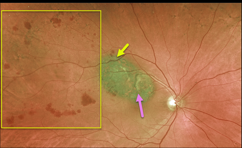
UVEAL MELANOMA + BRVO
Originally posted on @retina.rocks May 18, 2021
This 78YO male has a history of uveal malignant melanoma (MM), which was successfully treated with initial photodynamic therapy (PDT) (due to the presence of subretinal fluid) followed by transpupillary thermotherapy (TTT) once dry.
He developed a secondary branch retinal vein occlusion (BRVO), likely due to thermal damage from the TTT.
Vision is 20/30, and we continue to follow him closely. A scatter laser will be needed if he develops increasing ischemia with neovascularization from the BRVO.
Learning Points:
BRVOs are virtually always caused by an artery compressing a vein at a shared arteriovenous crossing.
However, rarely, they can occur due to other causes, including inflammation (most commonly sarcoidosis) or secondary to interventions such as TTT.
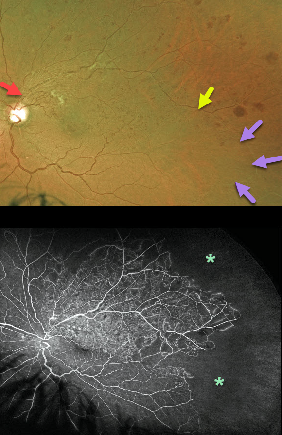
BRVO
Originally posted on @retina.rocks April 30, 2021
This is a 54YO male presented with a major branch retinal vein occlusion (BRVO). Vision was 20/80, and Eylea injections were started for associated macular edema.
The Optos color image shows a major superotemporal BRVO with moderate retinal hemorrhages, some nerve fiber layer infarcts, and collateral vessels. The involved retinal vein is red in the midperiphery. However, it becomes white as it continues into the inferotemporal periphery.
Optos fluorescein angiography shows extensive peripheral capillary dropout.
Usually, one would expect a BRVO to involve one quadrant (retinal vessels virtually always respect the horizontal meridian).
The occluded vein in our patient most likely represents an atypical retinal venous malformation (RVM), which is a congenital anomalous vessel that, by definition, crosses the horizontal.
Learning Points:
Retinal venous malformations were originally described by Gary Brown et al as congenital retinal macrovessels (Arch Ophthalmology 1982;100:1430-1436). These vessels are usually located in the macular region and are virtually always venous.
These lesions are now called retinal venous malformations (RVMs) since about one-quarter of patients have associated venous anomalies in the brain (Pichi et al., JAMA Ophthalmol 2018;136;372-379).
BRVO
Originally posted on @retina.rocks March 8, 2021
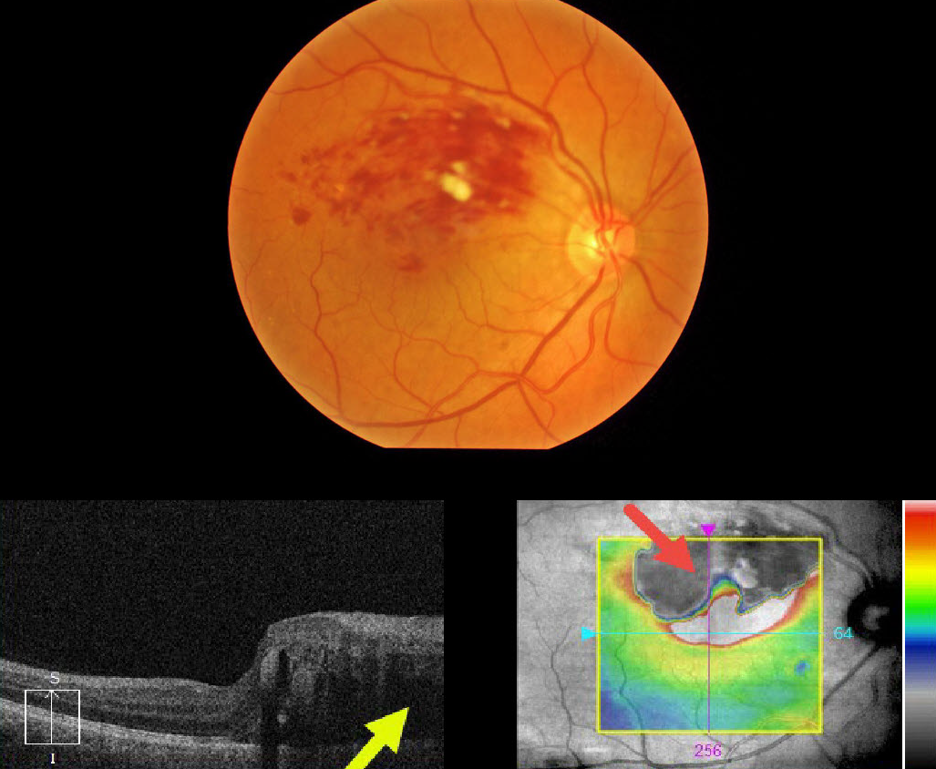
This 57YO male presented with 20/400 vision in his right eye from a superior macular branch retinal vein occlusion.
The OCT at first glance seems to just show superior macular edema extending through the macular center, but why is the superior retinal thickness map black, and what does that mean?
The retinal thickness map is supposed to represent the retinal thickness from the internal limiting membrane (ILM) to the tip of the outer segments just above the apical retinal pigment epithelium (RPE).
Our very expensive OCT machines usually accurately identify the ILM, but often don’t do such a great job of finding the RPE, especially when the OCT beam is attenuated as it passes through edematous retina or increased inner retinal reflectivity. In this case, the confluent inner retinal blood is highly reflective, causing posterior shadowing of the OCT signal.
Learning Points:
Intuitively, most of us would realize the black portion of the thickness map is an artifact, but now we know exactly what is going on. These artifacts are quite common if one looks for them.
The OCT cannot accurately locate the RPE signal and therefore incorrectly localizes it to the outer margin of the inner retinal blood.
BRVO
Originally posted on @retina.rocks August 31, 2020
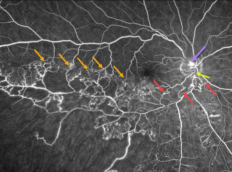
This patient has an ischemic branch retinal vein occlusion (BRVO) with extensive collateral vessels.
Occlusion occurs when an artery compresses the underlying vein where the vessels share a common adventitial sheath. Extensive collateral vessels cross the temporal horizontal raphe as well as around the occlusion more posteriorly. A small area of disc neovascularization leaks.
Learning Points:
Collateral vessels are dilated, pre-existing vessels that bypass an occlusion. These are most commonly seen in the posterior pole following retinal venous occlusions, but can also occur on the optic disc with optic nerve meningiomas.
The endothelial tight junctions are intact, so there’s no leakage on angiography. This helps differentiate them from neovascularization, which does leak.
RETINAL ARTERIOVENOUS MALFORMATION
Originally posted on @retina.rocks July 28, 2020
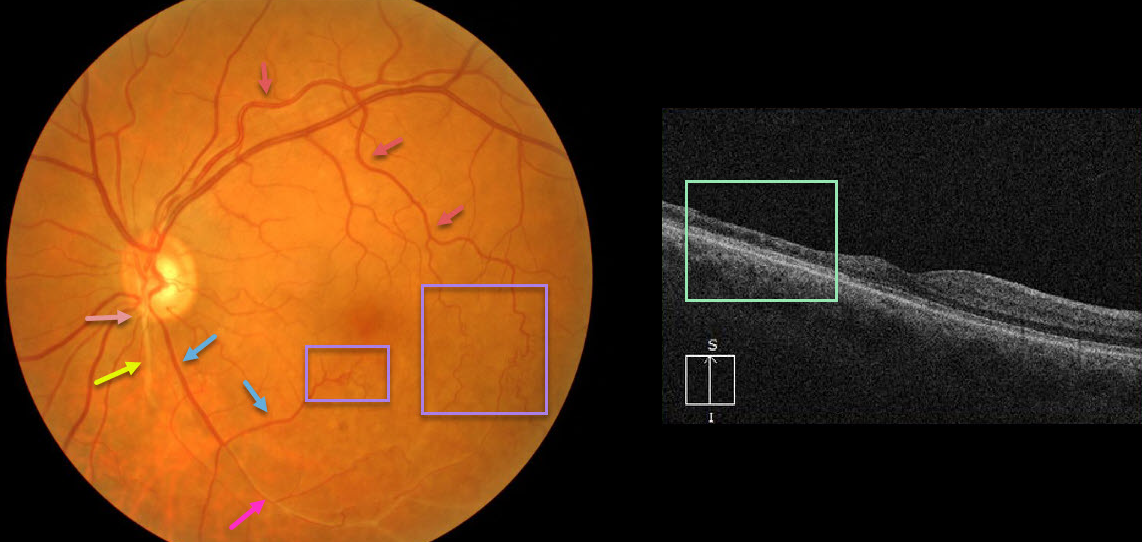
At first or quick glance, this looks like an old inferotemporal branch retinal vein occlusion (BRVO).
On closer inspection, there is a white-sheathed vessel arising from the nerve inferiorly. Numerous collateral vessels are noted inferior and temporal to the fovea, and there is a sheathed vessel in the inferior arcade.
OCT shows marked inner retinal atrophy with preservation of the photoreceptor layers, consistent with a prior branch retinal artery occlusion. So the collateral vessels (virtually always a sign of a prior venous occlusion) indicate an old BRVO, and the atrophic and sheathed vessel indicates an old BRAO, both in the same quadrant. Right?
Tracing the large branch of the superotemporal arcade inferotemporally, we see that the collaterals appear to be arterial. Inferior to the nerve, we see an arteriole that crosses the sheathed vessel, indicating that the sheathed vessel is not an artery but a vein. Moreover, the large vessel off the nerve, inferotemporal in location, appears to be a vein that eventually connects to the collateral vessels we previously identified as arterial.
Our best guess is that this may be a low-grade arteriovenous malformation, possibly with a prior secondary BRAO/BRVO. Unfortunately, we don’t have a fluorescein angiogram, which may help better define the vascularization.
Learning Points:
Remember that arteries don’t cross arteries and veins don’t cross veins. Tracing vessels can be a helpful key to identifying retinal vascular disorders.
BRVO
Originally posted on @retina.rocks January 27, 2020
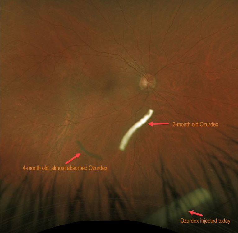
This patient is receiving Ozurdex (dexamethasone 0.7%) intravitreal injections every 2 months to control macular edema from a branch retinal vein occlusion (BRVO).
The Ozurdex implant slowly releases the steroid and eventually totally absorbs, with the clinical effect lasting about 2 to 3 months. This Optos image shows 3 Ozurdex implants in various stages of their life cycles.
Learning Points:
In addition to Ozurdex, other possible treatment modalities for BRVO macular edema include anti-VEGF or triamcinolone injections, and macular laser photocoagulation.


