

Cataract, Dislocated IOL + Retained Lens Fragments
<< Back to Cases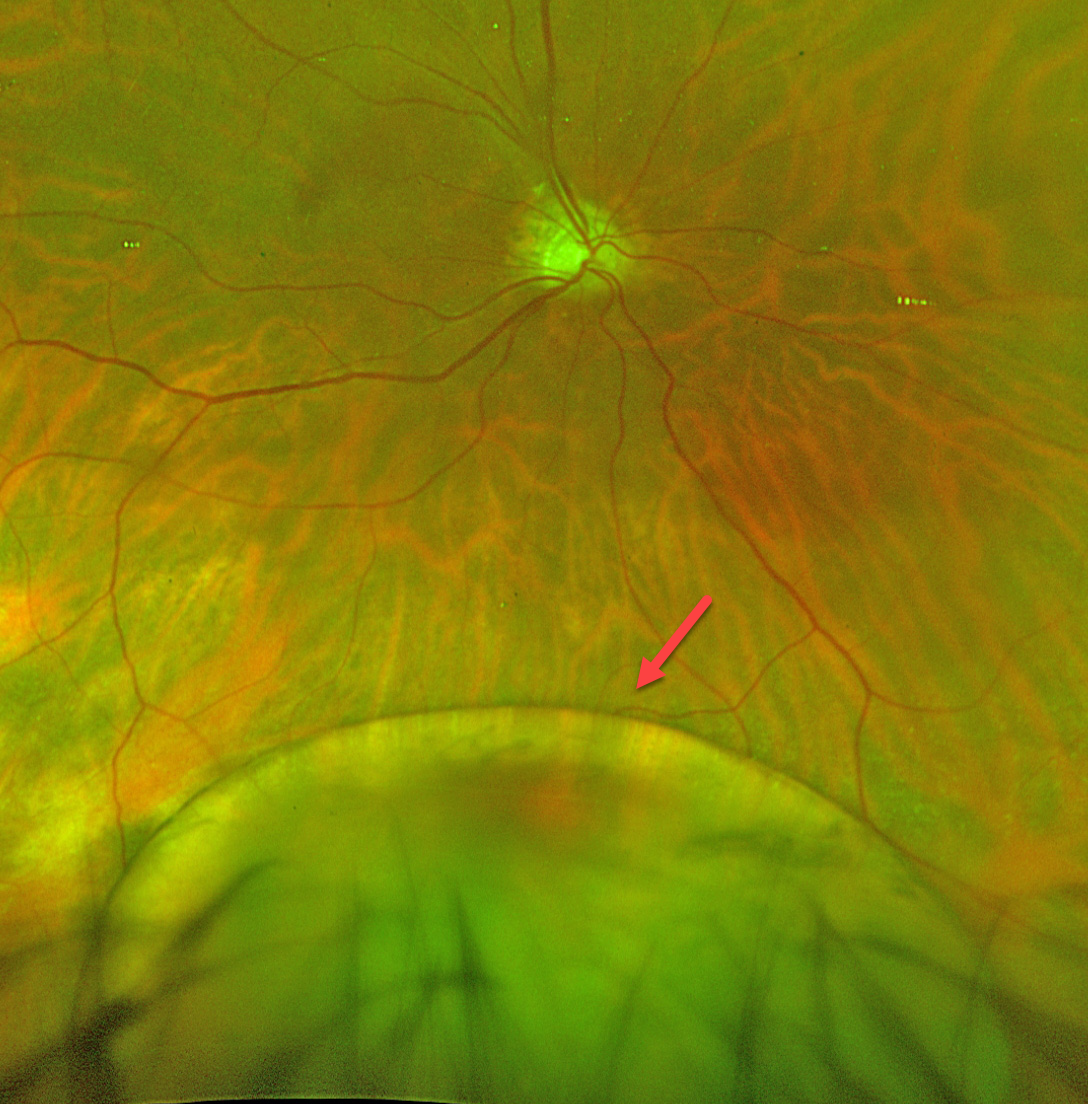
SPONTANEOUSLY DISLOCATED CATARACT
Originally posted on @retina.rocks July 23, 2026
This 56YO developmentally delayed male presented with seeing a “black spot” in his right eye. He was monocular with a phthisical left eye from unclear etiology. Vision in his right eye was decreased to 20/400 from his baseline of 20/70 at his prior visit.
He was largely nonverbal and difficult to examine due to extreme photosensitivity. Optos color RG imaging shows an inferiorly dislocated crystalline lens.
Urgent pars plana vitrectomy and lensectomy were performed. After discussion with the patient’s guardian, the decision was made to leave the patient aphakic rather than to place a secondary scleral-fixated or scleral-sutured lens due to a history of frequent eye rubbing. The patient was fitted with aphakic glasses, which restored his vision to baseline.
Learning Points:
Dislocation or subluxation of the crystalline lens can be caused by trauma, ocular conditions such as ectopia lentis et pupillae, pseudoexfoliation, aniridia, high myopia, or hereditary disorders such as Marfan syndrome, homocystinuria, Weill-Marchesani syndrome, Ehlers-Danlos syndrome, or sulfite oxidase deficiency. Our patient had a known cardiofaciocutaneous (CFC) syndrome secondary to BRAF mutation, but this has not been associated with ectopia lentis.
DISLOCATED IOL
Originally posted on @retina.rocks May 6, 2026
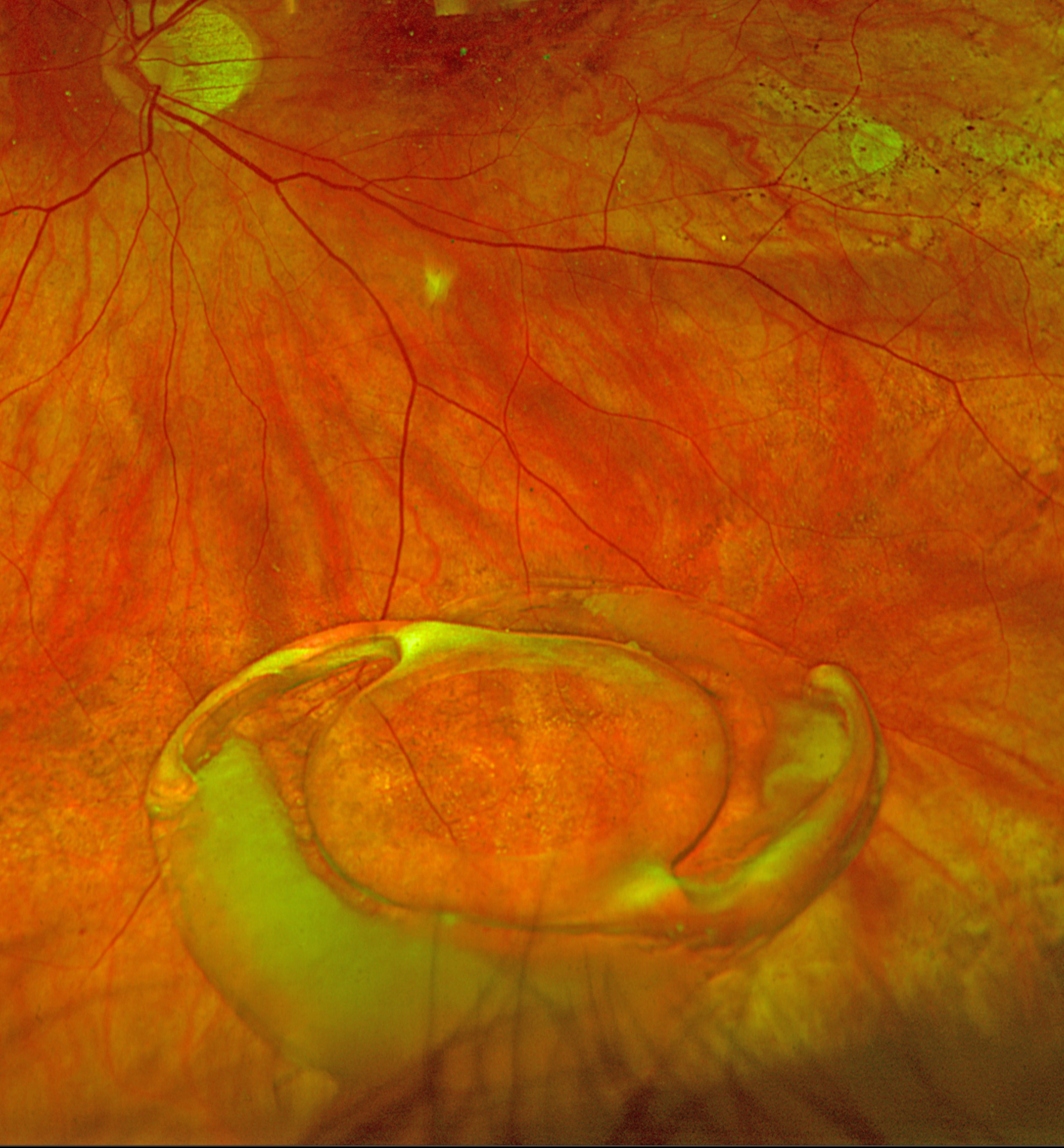
This 80YO female noticed sudden vision loss in her left eye. When examined the next morning, vision was 20/400.
Optos color RG imaging shows an inferiorly dislocated intraocular lens (IOL) with surrounding capsular material settled over the inferior retinal periphery. She underwent successful IOL explantation with a secondary anterior chamber IOL.
DISLOCATED LENS
The European VitreoRetina Society (EVRS), Akansha Sharma, Manish Nagpal, Malvika Singh and Tejaswita Verma
Originally posted on @retina.rocks October 10, 2025
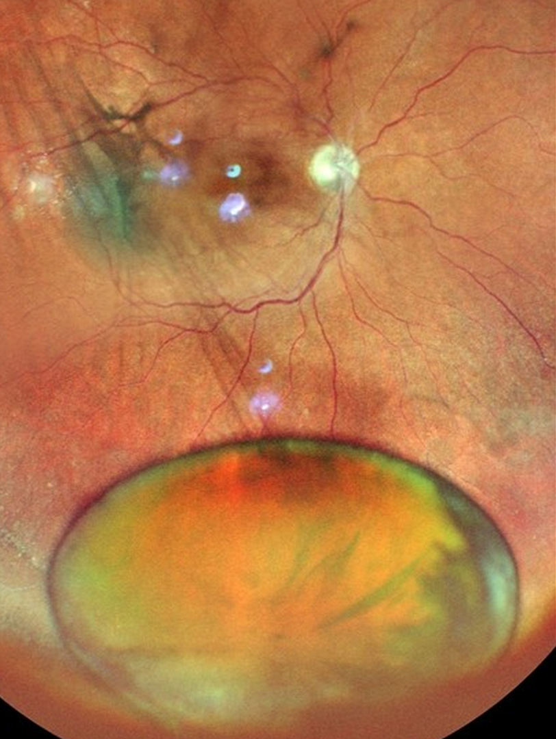
This 67YO male presented with 3 weeks of decreased vision in his right eye. Vision was 20/120 OD with a +10 diopter sphere and 20/30 in his normal OS. Color photography shows a cataractous lens fully dislocated into the inferior vitreous cavity. A shallow macula-off rhegmatogenous retinal detachment is noted in the posterior pole without apparent breaks. Pars plana vitrectomy was performed the following day, which included lens extraction and retinal detachment repair.
Learning Points:
Posterior dislocation of the crystalline lens can be caused by trauma or by systemic disorders such as Marfan syndrome, homocystinuria, or Weill-Marchesani syndrome. Pars plana vitrectomy is needed for a completely dislocated lens, as with this patient, while lenses that are subluxed may be managed with phacoemulsification or other means.
Our patient’s lens spontaneously dislocated with no history of trauma. Pseudoexfoliation was noted on slit lamp examination of her left lens, which may have predisposed her to lens dislocation.
DISLOCATED CATARACT
Anjana Mirajkar and Manish Nagpal
Originally posted on @retina.rocks September 3, 2024
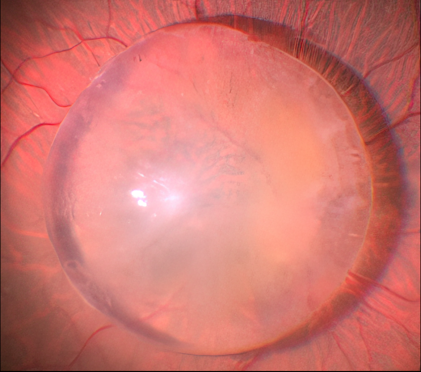
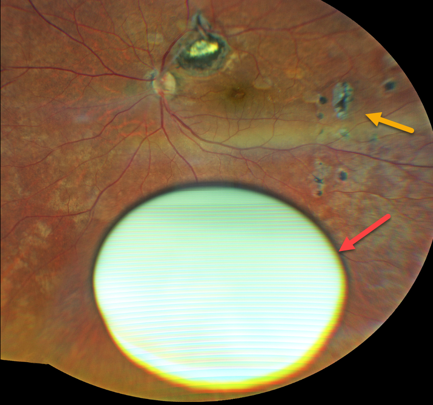
Just a cool intraoperative photo of a posteriorly dislocated cataract.
Unfortunately, we don’t have any further clinical information for this patient.
COUCHED CATARACT
Ogugua Okonkwo, Adekunle Olubola Hassan, and Zainab Ogunsakin
Originally posted on @retina.rocks August 8, 2024
This 67YO female underwent a couching procedure for a left cataract in 2018. Cataract surgery with a posterior chamber IOL OD was performed in 2020, and 2 months later, the cataract surgeon referred her to us for evaluation of the couched lens OS.
Vision was 20/40 OS with a +10.00 diopter lens. She was lost to follow-up until she returned in March 2024 with counting-fingers vision OS. Her intraocular pressure was slightly elevated, and she had new focal lesions in the peripheral retina, which were felt to represent inflammation from leaking lens protein.
Fundus photography shows a dislocated intumescent hypermature cataract in the inferior vitreous. Multifocal chorioretinal scars are scattered throughout the fundus. Pars plana vitrectomy, lensectomy, and anterior chamber lens implantation were performed. Vision without correction was 20/200 OS one month postoperatively.
Learning Points:
Couching is the earliest documented form of cataract surgery and was widely practiced worldwide (Leffler et al, Ann Transl Med 2020;8(22):1551). It was described in 600-800 BC by an ancient Indian surgeon. This practice, though now largely abandoned with the advent of modern cataract surgery, is still rarely practiced in rural areas of some African countries, including Nigeria.
Couching is performed in the ‘comfort’ of the patient’s home by the ‘coucher’ who moves around in the rural community where their service is needed. The procedure involves using a sharp instrument to push the cataractous lens backward into the vitreous. If all goes well. A high hyperopic spectacle correction is prescribed. It can, however, be complicated by glaucoma, lens dislocation into the anterior chamber, cornea decompensation, uveitis, retinal detachment, vitreous hemorrhage, and intraocular infection.
DISLOCATED IOL
Anjana Mirajkar and Manish Nagpal
Originally posted on @retina.rocks July 30, 2024
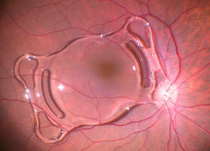
This patient presented with a spontaneously posteriorly dislocated intraocular lens (IOL). The lens was explanted, and a secondary scleral-fixated lens was placed. Unfortunately, we don’t have any further clinical information.
RETAINED LENS FRAGMENT
Originally posted on @retina.rocks June 24, 2022
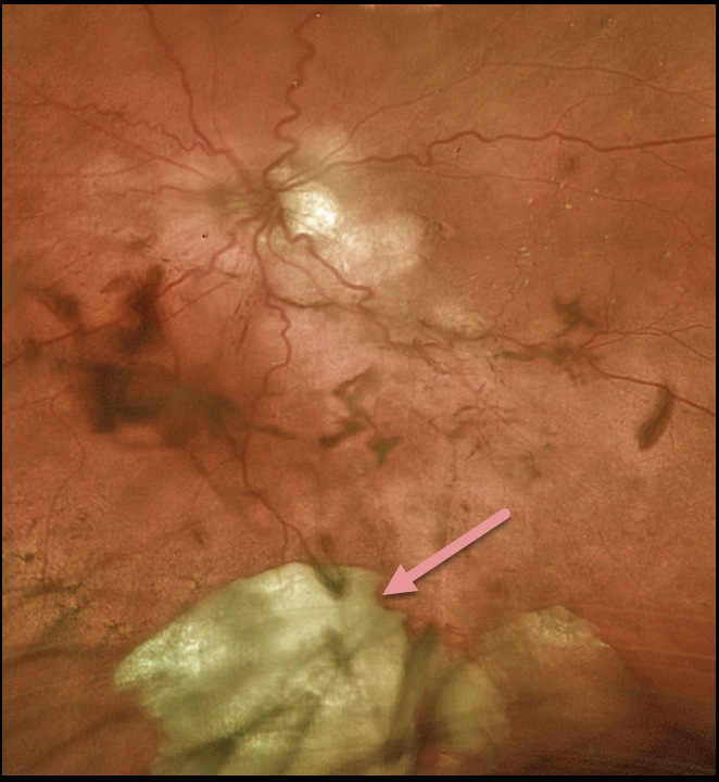
This 83YO female underwent cataract extraction for a hypermature lens. The posterior capsule tore, and a large lens fragment fell posteriorly. A 3 piece intraocular lens was successfully placed in the sulcus at the time of surgery.
She had previously received multiple intravitreal injections from multiple outside providers for exudative macular degeneration. We have no documentation if the capsule had ever been compromised.
Learning Points:
Posteriorly dislocated lens fragments are a known complication of cataract surgery. The crystalline lens is an immune privileged tissue. When lens fragments get exposed following cataract surgery, severe inflammation and increased eye pressure usually follow.
Rarely, permanent vision loss occurs from glaucoma or cystoid macular edema. Small lens fragments can be managed conservatively, but larger fragments like this one need to be surgically removed.
DISLOCATED IOL
Originally posted on @retina.rocks May 24, 2022
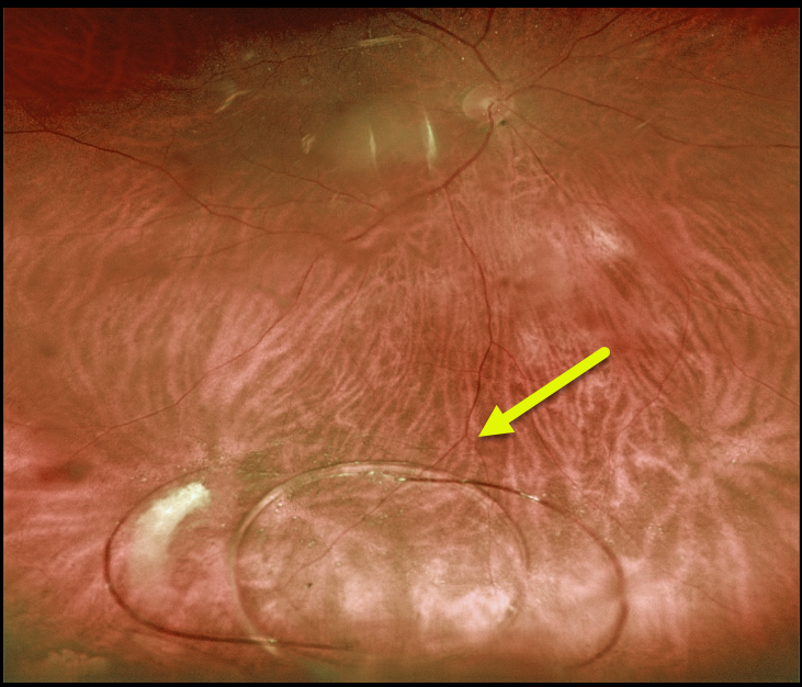
This 78YO male presented with a several-month history of decreased vision in his right eye. He had prior bilateral cataract surgery years earlier at an outside practice. Vision was 20/25 OU. His right posterior chamber implant was decentered inferiorly, but the optic remained in the visual axis.
The lens subsequently completely dislocated into the inferior vitreous before the anterior segment surgeon could perform scleral fixation.
He subsequently underwent successful vitrectomy, lens explantation with placement of an anterior chamber implant.
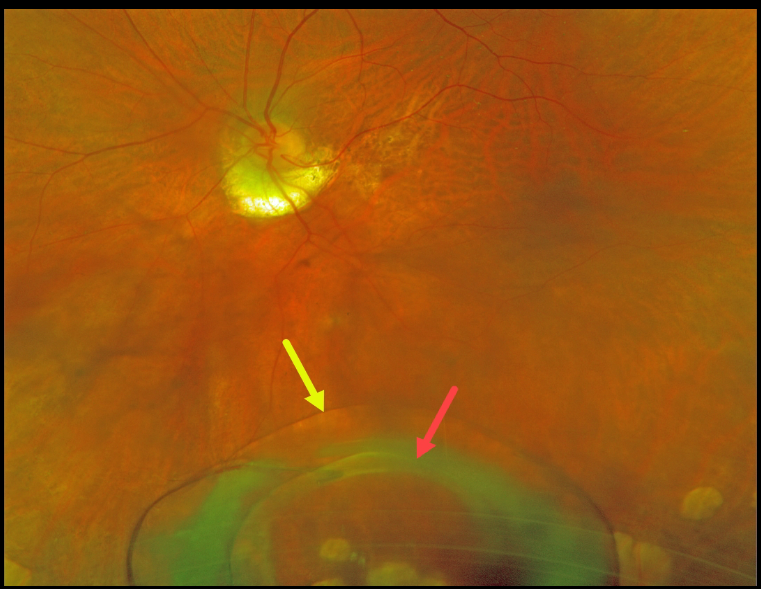
DISLOCATED LENS
Originally posted on @retina.rocks January 24, 2022
This 80YO female presented with subacute, painless vision loss in her left eye. Previously, she had been treated for macular neovascularization from ocular histoplasmosis but had been lost to follow-up.
Optos imaging shows a 3-piece intraocular lens with an intact capsule settled in the inferior midperiphery.
She denied any trauma, and previous exams showed no zonular dehiscence. The cataract surgery had been performed nearly 25 years earlier.
The lens will be explanted during vitrectomy with placement of a scleral-fixated lens.
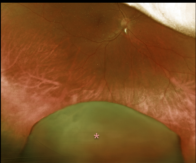
DISLOCATED LENS
Originally posted on @retina.rocks August 26, 2021
This 51YO female presented with a 6-month history of vision loss in her right eye. She remembered falling 7 months earlier, then woke a few weeks later with severe unilateral vision loss. Vision was counting fingers from a total dislocation of her crystalline lens into the inferior vitreous.
Vitrectomy surgery to remove the dislocated cataract, along with placement of an anterior chamber intraocular lens, was performed, resulting in a return to 20/30 vision.
Learning Points:
Posterior dislocation of the crystalline lens can be caused by trauma or by systemic disorders such as Marfan syndrome, homocystinuria, or Weill-Marchesani syndrome. Pars plana vitrectomy is needed for a completely dislocated lens, as in this patient, whereas subluxed lenses may be managed with phacoemulsification or other means.
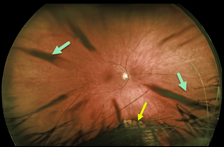
CORTICAL CATARACT
Originally posted on @retina.rocks June 1, 2021
This patient’s Optos image shows beautiful cortical cataract spoke artifacts along with congenital hypertrophy of the RPE (CHRPE) inferiorly, which is mostly obstructed by the patient’s hair and eyelashes.
Learning Points:
Cataracts or other anterior segment findings can create artifacts in fundus photography or optical coherence tomography.
Superior eyelashes often obscure the inferior retinal periphery on Optos imaging, so the technician needs to remind the patient to open their eyes wide and sometimes manually elevate the lid.
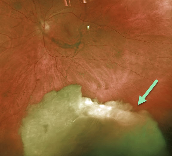
RETAINED LENS FRAGMENT
Originally posted on @retina.rocks November 2, 2020
This patient came to see us for a second opinion following cataract surgery elsewhere six weeks earlier. He has been on topical steroid drops since his surgery. His pressure was 42 mmHg with significant anterior chamber cell and flare.
Virtually the entire cataract was floating in the inferior vitreous. He was scheduled for immediate pars plana vitrectomy and lensectomy.
Learning Points:
Posteriorly dislocated lens fragments are a known complication of cataract surgery. The crystalline lens is an immunoprecipitated tissue.
When lens fragments are exposed after cataract surgery, severe inflammation and increased intraocular pressure usually follow. Rarely, permanent vision loss occurs from glaucoma or cystoid macular edema.
Small lens fragments can be managed conservatively, but larger fragments require surgical removal.


