

Central Areolar Choroidal Dystrophy
<< Back to Cases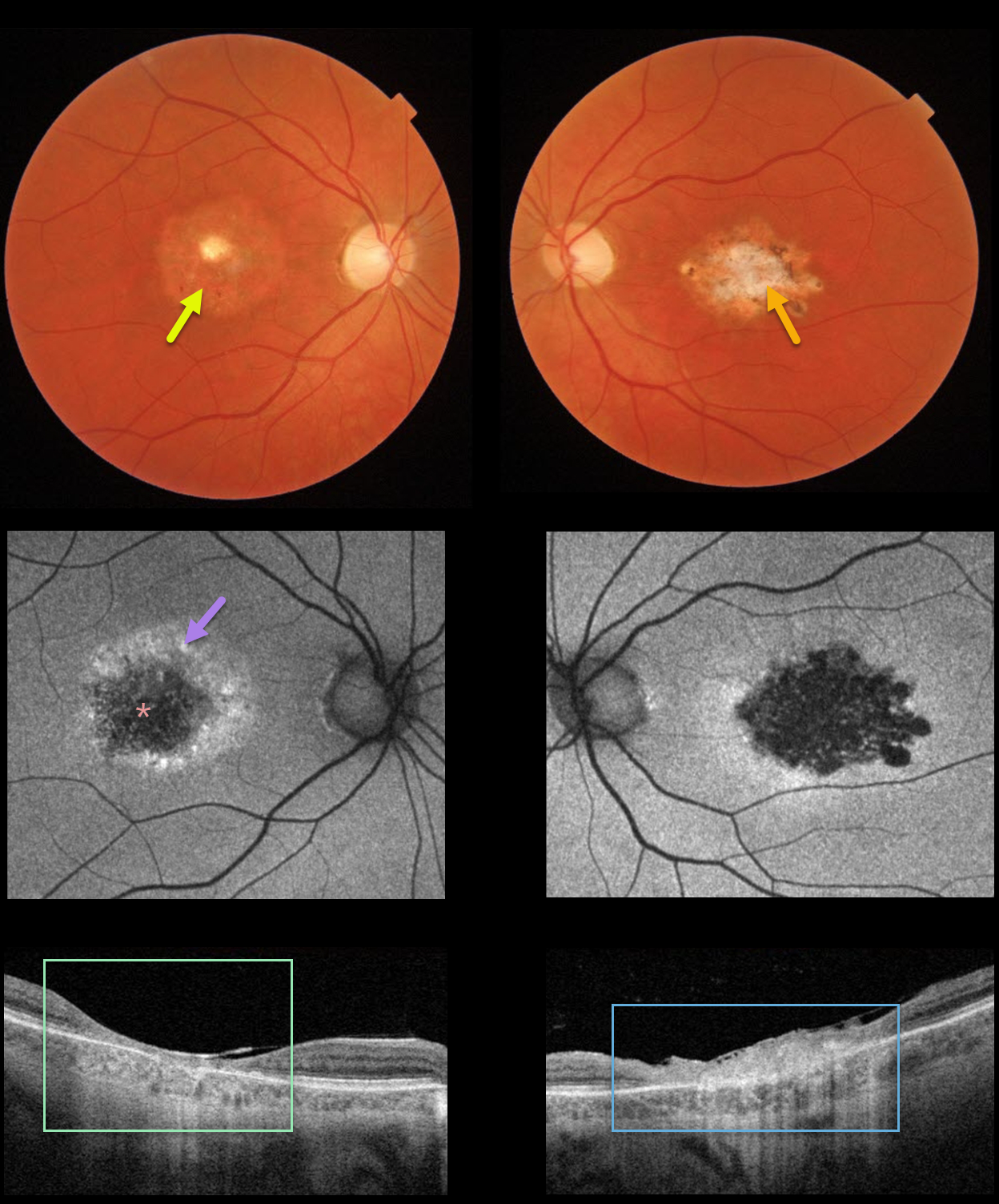
CENTRAL AREOLAR CHOROIDAL DYSTROPHY
Originally posted on @retina.rocks May 11, 2022
This 40YO female gave a 4-5 year history of gradual bilateral vision loss. Vision was 20/70 OD and 20/200 OS. There was no family history of retinal disease.
Color imaging shows a round area of central pigment loss OD and a more irregular area of atrophic pigmentary changes OS.
Optos fundus autofluorescence (FAF) showed central hypo-FAF with surrounding hyper-FAF.
Swept-source OCT showed a markedly thinned and disorganized central macula OD, with a more thickened, hyperreflective, disorganized central macula OS.
Genetic testing revealed a heterozygous pathogenic mutation in IQCB1, a defect commonly associated with autosomal recessive Senior-Løken Syndrome. We felt the clinical picture was most consistent with central areolar choroidal dystrophy (CACD).
Learning Points:
CACD is usually autosomal dominant and genetically heterogeneous, caused by mutations in the peripherin/RDS (PRPH2) gene. The current genetic testing panel results, as this case illustrates, must still be approached with caution (see Rodriguez-Munoz et al, Retina 2021;41:1966-1975).
See Smailhodzic et al Invest Ophthalmol Vis Sci 2011;52;8908-8918 for how to differentiate CACD from age-related macular degeneration best clinically.
MACULAR DYSTROPHY
Originally posted on @retina.rocks August 19, 2020
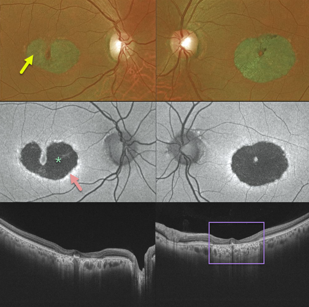
Our patient has a bilateral bull’s-eye maculopathy with central foveal sparing. Vision was 20/30 OD and 20/60 OS.
Fundus autofluorescence shows central hypo-FAF from loss of the central RPE. The thin surrounding hyper-FAF ring likely represents diseased, at-risk RPE cells. The OCT scans show pericentral outer retinal and RPE loss.
Learning Points:
Our patient no doubt has some type of inherited macular disorder. Some possibilities include central areolar chorioretinal dystrophy or a cone-rod dystrophy.
Central areolar chorioretinal dystrophy is a genetic condition associated with mutations in the RDS/peripherin gene, which encodes for a cell surface glycoprotein found in the rod and cone outer segments. The RPE and choriocapillaris atrophy over time, eventually forming a round, atrophic lesion in the macula. Vision can vary from 20/20 to 20/200.


