

Choroidal Detachment
<< Back to Cases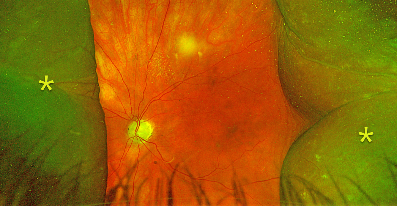
CHOROIDAL DETACHMENT
Originally posted on @retina.rocks March 19, 2026
This 81YO female presented with counting fingers vision in her right eye 3 weeks following XEN Gel stent glaucoma surgery. Intraocular pressure was 11 mmHg.
Optos color RG imaging shows nasal and temporal choroidal detachments extending toward, but not into, the macula. Observation was recommended, and we expect the choroidals to resolve as she extends postoperatively.
Learning Points:
A choroidal detachment is defined by the abnormal presence of fluid or blood in the suprachoroidal space. Serous choroidal detachments, also known as choroidal effusions, are a frequent complication of glaucoma surgery. Several causative mechanisms are believed to be at play: hypotony allows fluid to accumulate in the suprachoroidal space, and inflammation increases the choroidal permeability. The detached ciliary body may also produce less aqueous humor, thus creating a self-perpetuating cycle. Often, observation is sufficient, but topical steroids and cycloplegics can help expedite resolution.
PSEUDO-UVEAL MELANOMA FROM CHOROIDAL DETACHMENT
Originally posted on @retina.rocks December 25, 2025
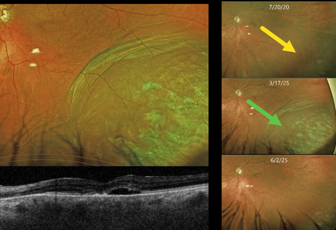
This 85YO female presented with an elevated pigmented choroidal mass in her inferotemporal periphery, which extended into her distal macula (Optos color RG). This mass was absent 5 years earlier. Circumferential choroidal folds extend along the posterior edge of the lesion. Triton swept-source OCT shows a blister of subretinal fluid and a thickened choroid.
She has a history of uncontrolled glaucoma and underwent a XEN Gel Stent procedure 8 days earlier. Intraocular pressure (IOP) was 8 mmHg.
Learning Points:
A choroidal detachment is defined by the abnormal presence of fluid or blood in the suprachoroidal space. Serous choroidal detachments, also known as choroidal effusions, are a frequent complication of glaucoma surgery. Several causative mechanisms are believed to be at play: hypotony allows fluid to accumulate in the suprachoroidal space, and inflammation increases the choroidal permeability. The detached ciliary body may also produce less aqueous, thus creating a self-perpetuating cycle. Often, observation is sufficient, but topical steroids and cycloplegics can be helpful in promoting resolution.
Other than the obvious appearance of this lesion immediately following glaucoma surgery, clues to this not being a uveal MM include the choroidal folds along the posterior extent of the choroidal detachment and an OCT consistent with hypotony maculopathy. About 10 weeks later, the IOP was 9 mmHg, and the choroidal effusion had completely resolved.
ESCITALOPRAM-INDUCED UVEAL EFFUSIONS WITH ANGLE CLOSURE GLAUCOMA
Nikolaos Zagorianos, Mattie Adams, Jessica Haycraft and Brett Abney
Originally posted on @retina.rocks June 3, 2025
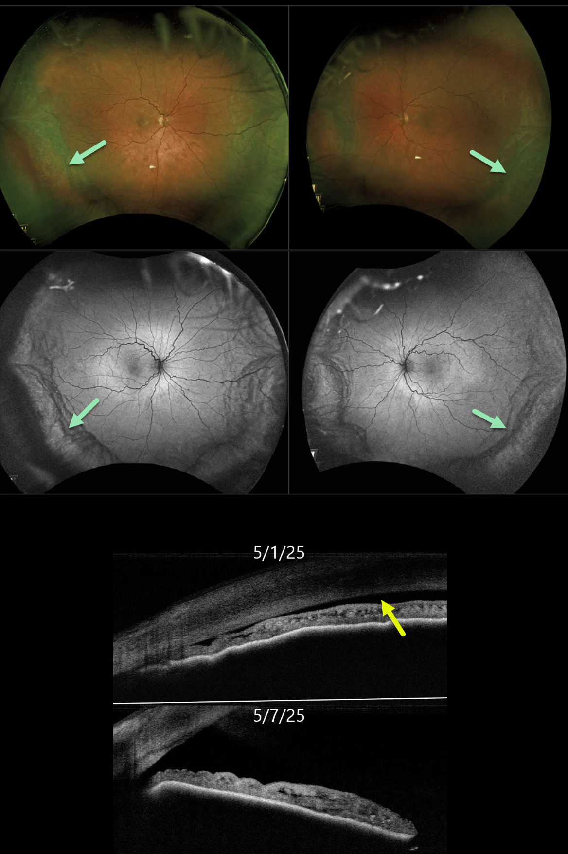
This 67YO female woke with bilateral blurred vision and severe eye pain. She was started on Lexapro (escitalopram) six months earlier. Vision was 20/400 OD and 20/100 OS.
Slit lamp examinations showed bilateral shallow anterior chambers, with no angle structures visible in either eye on gonioscopy and anterior segment OCT. Intraocular pressures (IOP) were 50 mmHg OD and 44 mmHg OS.
Optos color RG and green channel imaging shows bilateral peripheral encircling choroidal detachments. Anterior segment OCT confirmed a flat anterior chamber. Rocklatan, Simbrinza, Alphagan, Atropine, and Diamox were administered in the office, with the IOP decreasing to 38 OD and 36 OS. We contacted her primary care doctor to discontinue the Lexapro. One week later, vision improved to 20/30 OD and 20/25 OS. IOP was 16 OD and 13 OS, the anterior chamber angles were open clinically and on OCT, and the choroidal detachments were decreasing.
Learning Points:
Drug-induced ciliochoroidal effusions with secondary angle closure glaucoma (ACG) are a known complication of topiramate therapy, but have also rarely been reported with selective serotonin reuptake inhibitors, including escitalopram (Zelefsky et al, AJO 2006;141:1144-1147). Unlike typical ACG, which is cured with a peripheral iridotomy, these drug-induced ACG attacks are treated with dilation. Atropine paralyzes the ciliary body, which allows the lens/Iris diaphragm to return to its natural state, rotating backward and increasing the anterior chamber depth. Topical steroids help decrease uveal vascular permeability, thereby reducing choroidal effusion.
CHOROIDAL DETACHMENT
Mattie Adams
Originally posted on @retina.rocks January 27, 2025
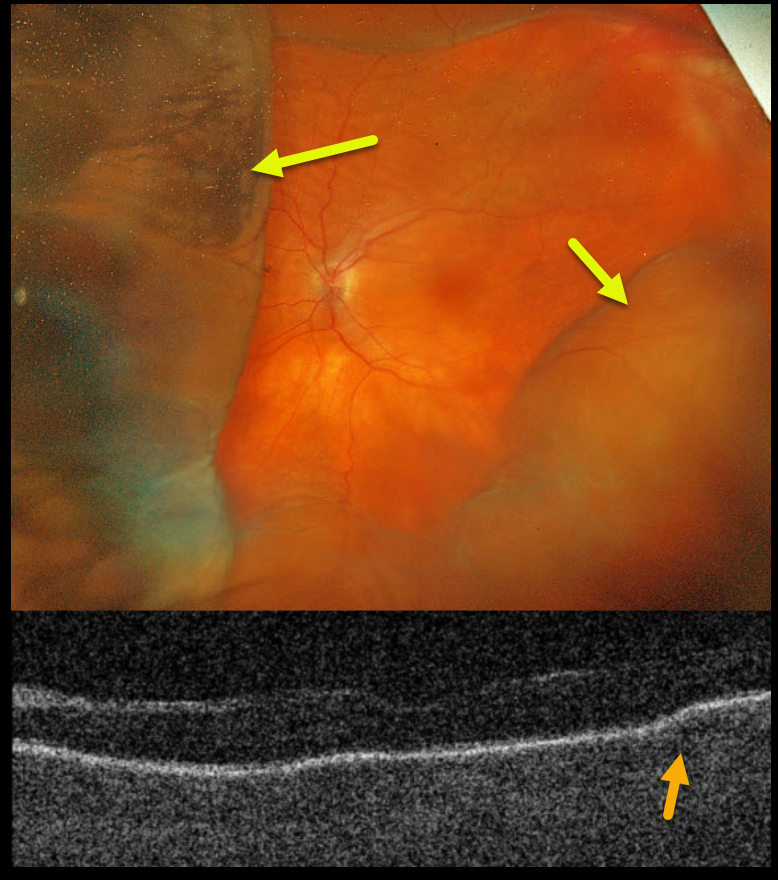
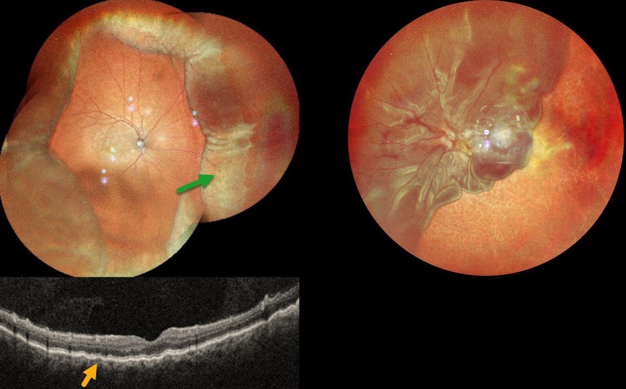
This 78YO female presented with acute vision loss in her left eye. Penetrating keratoplasty (PKP) was performed 9 months earlier. When seen on 8/26/24, vision was counting fingers in part due to irregular astigmatism from the residual PKP sutures, which were then removed. The cornea was Seidel negative after removal. She returned one week later complaining of a curtain in her visual field. Her anterior chamber was flat, and her corneal graft was dehisced for 3 clock hours.
Optos color RGB imaging shows a variably elevated choroidal detachment. Triton swept-source OCT shows chorioretinal folds.
Learning Points:
A choroidal detachment is defined by the abnormal presence of fluid or blood in the suprachoroidal space. Serous choroidal detachments, also known as choroidal effusions, are a frequent complication of glaucoma surgery. Several causative mechanisms are believed to be at play: hypotony allows fluid to accumulate in the suprachoroidal space and inflammation increases the choroidal permeability. The detached ciliary body may also produce less aqueous, thus creating a self-perpetuating cycle. Often, observation is sufficient but topical steroids and cycloplegics can be helpful to aid in resolution.
Hypotony maculopathy is characterized by low IOP and chorioretinal folds, most commonly following glaucoma filtration surgery. Optic nerve edema can also be seen due to decreased axoplasmic transport.
Our patient was immediately taken to surgery for suturing of the dehisced PKP. The choroidals and chorioretinal folds immediately improved and completely resolved two months later (not shown).
CHOROIDAL DETACHMENT
Anand Temkar and Manish Nagpal
Originally posted on @retina.rocks October 31, 2024
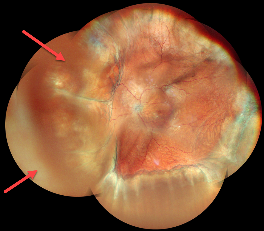
This 52YO male presented with 2 weeks of vision loss OD. He had a complicated ocular history of multiple prior surgeries elsewhere, including cataract surgery OD 2 months earlier and multiple retinal detachment surgeries OS. Vision was 20/30 OD and LP OS. Pressure was 8mmHg with no wound leaks on slit lamp OD.
MultiColor imaging shows encircling peripheral choroidal detachments OD, and OCT shows shallow macular chorioretinal folds. A total retinal detachment is noted OS with bare choroid nasally and inferiorly.
Learning Points:
A choroidal detachment is defined by the abnormal presence of fluid or blood in the suprachoroidal space. Serous choroidal detachments, also known as choroidal effusions, are a frequent complication of glaucoma surgery or following any intraocular procedure. Frank wound leaks usually require surgical repair. Otherwise, conservative management, often with topical/systemic steroids, is recommended.
Since our patient’s surgical wound was secure and the anterior chamber was formed, topical and oral steroids were prescribed.
CHOROIDAL DETACHMENT
Anand Temkar and Manish Nagpal
Originally posted on @retina.rocks May 23, 2024
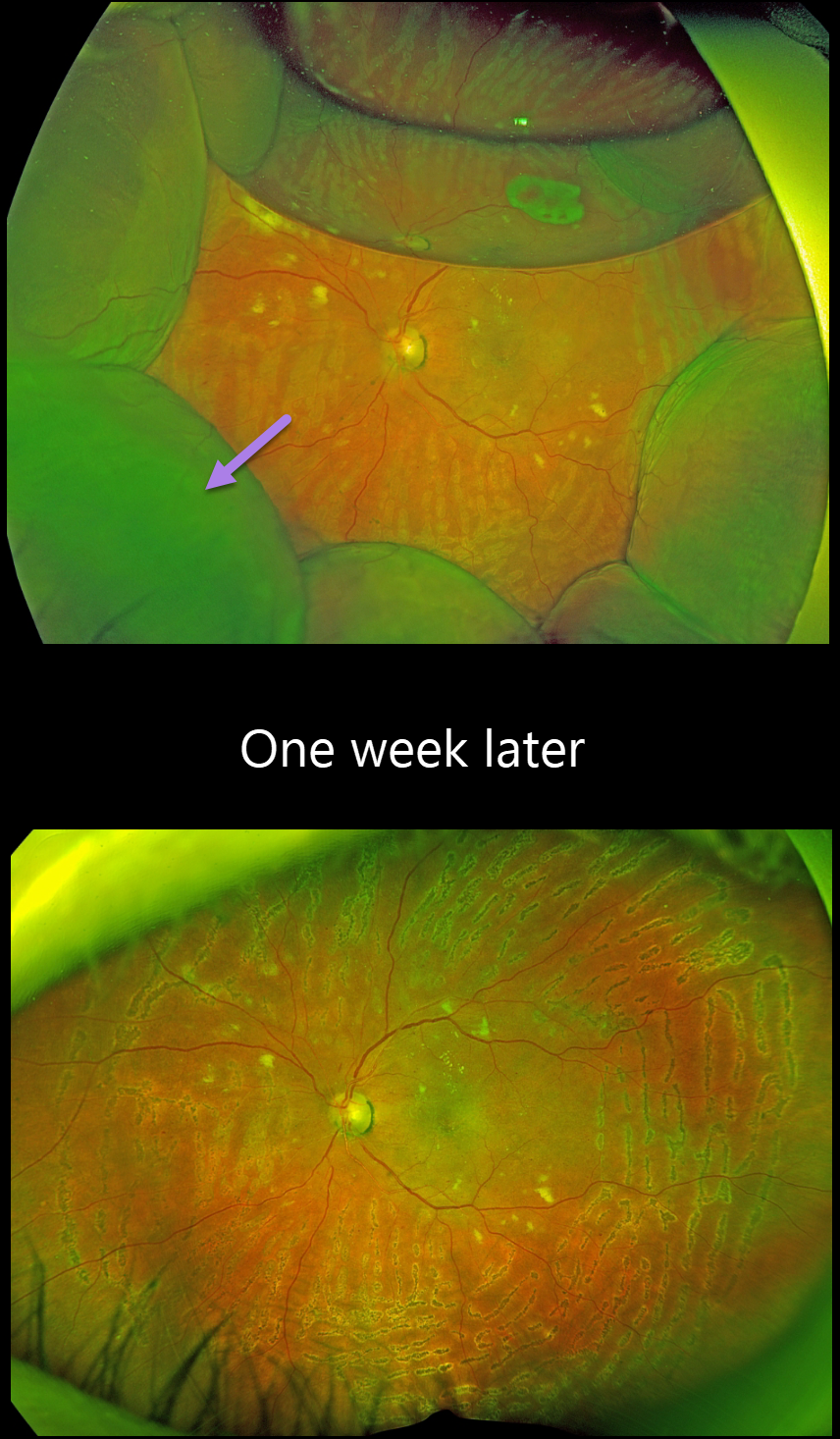
This 63YO male underwent glaucoma filtering surgery with mitomycin C one month earlier at an outside practice. Vision was 20/30, and intraocular pressure was 4. Montage color imaging shows extensive choroidal detachments. He was immediately lost to follow-up.
Learning Points:
A choroidal detachment is defined by the abnormal presence of fluid or blood in the suprachoroidal space. Serous choroidal detachments, also known as choroidal effusions, are a frequent complication of glaucoma surgery.
Several causative mechanisms are believed to be at play: hypotony allows fluid to accumulate in the suprachoroidal space, and inflammation increases the choroidal permeability. The detached ciliary body may also produce less aqueous humor, thus creating a self-perpetuating cycle. Often, observation is sufficient, but topical steroids and cycloplegics can be helpful in facilitating resolution.
CHOROIDAL DETACHMENT
Originally posted on @retina.rocks December 11, 2023
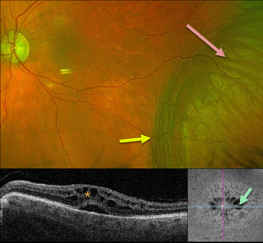
This 66YO female presented for her postoperative exam following an uneventful pars plana vitrectomy one day earlier for a diabetic traction retinal detachment.
Optos RG imaging shows 360 degrees of peripheral choroidal detachments with a 30% air bubble. Fresh panretinal photocoagulation burns are noted, and the retina is attached. All wounds were secure, and the intraocular pressure was 20mmHG.
We are not sure why our patient presented with these choroidals 1 day postoperatively, since her pressure was normal and there were no wound leaks.
Uveal effusion is a rare transient complication following heavy panretinal photocoagulation (Gentile et al, Ophthalmology 1996;103:827-832), although our patient’s endolaser did not appear overly intense.
When examined one week later, vision was 20/400, and the IOP was 17. The choroidals had spontaneously and completely resolved.
CHOROIDAL DETACHMENT
Originally posted on @retina.rocks November 30, 2023
This 84YO male has a long history of advanced chronic open-angle glaucoma with numerous surgical procedures. For several years, his left eye has had stable hypotony, with an intraocular pressure (IOP) of about 3-6 mmHg. Vision is 20/60.
Optos color RG imaging shows an inferotemporal choroidal detachment with chorioretinal folds along its posterior margin. Triton swept-source OCT shows cystoid edema with subretinal fluid, and en face imaging shows radiating mid-retina foveal cysts.
Since his symptoms, vision, IOP, and retinal findings remain stable, we are continuing to follow him without further intervention.
CHOROIDAL DETACHMENT
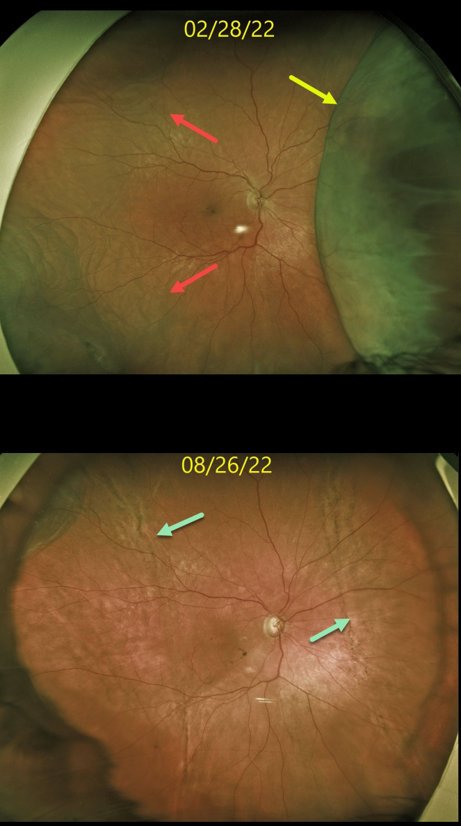
Originally posted on @retina.rocks October 7, 2022
This 74YO female was receiving periocular and intravitreal steroids for persistent pseudophakic cystoid macular edema (CME). Steroid response prevented further treatment, and she eventually required XenGel glaucoma surgery to control her pressure and allow for continued CME treatment.
Four days following glaucoma surgery, vision was 20/200, and the intraocular pressure (IOP) was 6 mmHg. Optos imaging shows a large nasal choroidal detachment with a shallow temporal choroidal detachment.
Over the next 3 months, her intraocular pressure normalized with total resolution of the choroidals. A ‘high-water’ mark of subretinal pigmentation is seen along the posterior extent of both the prior nasal and temporal choroidal detachments.
Learning Points:
Choroidal detachment is relatively common immediately following glaucoma surgery.
Two mechanisms are believed to be at play: hypotony allows fluid to accumulate in the suprachoroidal space, and inflammation increases the choroidal permeability. The detached ciliary body may also produce less aqueous humor, thus creating a self-perpetuating cycle.
Often, observation is sufficient, but topical steroids and cycloplegics can be helpful in promoting resolution.
CHOROIDAL DETACHMENT
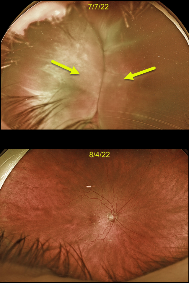
Originally posted on @retina.rocks August 15, 2022
This 88YO female developed kissing choroidal detachments immediately following Ahmed glaucoma valve placement.
The choroidal detachment spontaneously resolved after 1 month once the intraocular pressure normalized.
Learning Points:
Choroidal detachment is relatively common immediately following glaucoma surgery.
Two mechanisms are believed to be at play: hypotony allows fluid to accumulate in the suprachoroidal space and inflammation increases the choroidal permeability. The detached ciliary body may also produce less aqueous, thus creating a self-perpetuating cycle.
Often, observation is sufficient, but topical steroids and cycloplegics can be helpful to aid in resolution.
CHOROIDAL DETACHMENT
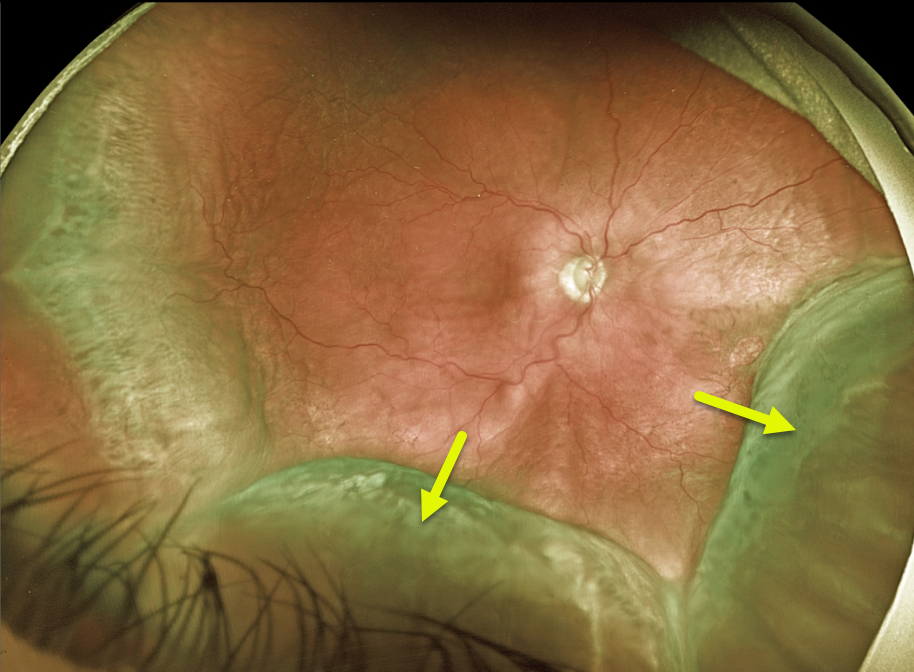
Originally posted on @retina.rocks July 14, 2022
This 68YO male patient presented with peripheral choroidal detachments following placement of a Baerveldt shunt 1 week earlier. Vision was counting fingers, and pressure was 1 mmHg. All wounds were secure, and the anterior chamber was formed.
Learning Points:
Choroidal detachment is relatively common immediately following glaucoma surgery. Two mechanisms are believed to be at play:
1) hypotony allows fluid to accumulate in the suprachoroidal space, and 2) inflammation increases the choroidal permeability. The detached ciliary body may also produce less aqueous, thus creating a self-perpetuating cycle.
Often, observation is sufficient, but topical steroids and cycloplegics can be helpful in promoting resolution. We are following this patient closely and expect the choroidals to resolve as postoperative pressure increases.
CHOROIDAL DETACHMENT
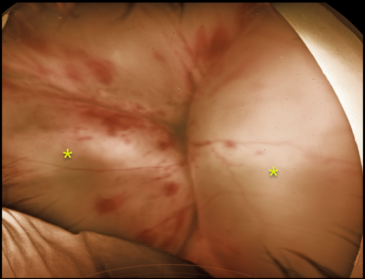
Originally posted on @retina.rocks December 23, 2021
This 25YO male patient presented to our practice with no light perception vision in his left eye. He had lost vision following an injury from a cat three years prior.
Optos imaging shows large kissing choroidal detachments with scattered intraretinal hemorrhages.
He has severe neovascular glaucoma with significant pain, and is considering cyclophotocoagulation vs enucleation.
CHOROIDAL DETACHMENT
Originally posted on @retina.rocks November 24, 2021
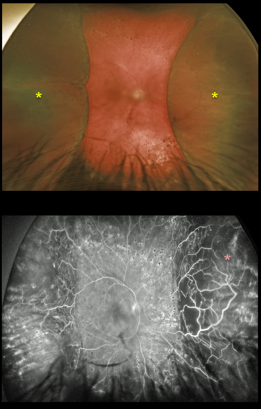
This 49YOM underwent trabeculectomy surgery elsewhere 6 weeks earlier.
Peripheral choroidal detachments are noted on Optos imaging. Fluorescein angiography shows significant peripheral ischemia.
Recent panretinal photocoagulation was started due to proliferative diabetic retinopathy (neovascularization not visualized due to the choroidals).
Learning Points:
Choroidal detachment is relatively common after glaucoma surgery. Two mechanisms are believed to be at play: hypotony allows fluid to accumulate in the suprachoroidal space, and inflammation increases the choroidal permeability. The detached ciliary body may also produce less aqueous humor, thus creating a self-perpetuating cycle.
Often, observation is sufficient, but topical steroids and cycloplegics can help promote resolution.
CHOROIDAL DETACHMENT
Originally posted on @retina.rocks August 4, 2021
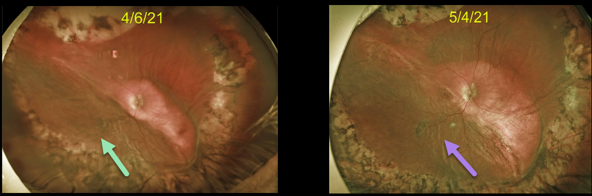
This 75YO male developed a large choroidal detachment 2 months following a complex vitrectomy for an intraocular lens exchange. There was no wound leak, and the intraocular pressure was 5.
He returned one month later with a spontaneously improving choroidal detachment.
Learning Points:
Choroidal detachment is relatively common after surgery with hypotony. Two mechanisms are believed to be at play: hypotony allows fluid to accumulate in the suprachoroidal space, and inflammation increases the choroidal permeability.
The detached ciliary body may also produce less aqueous humor, thus creating a self-perpetuating cycle.
CHOROIDAL DETACHMENT
Originally posted on @retina.rocks March 30, 2020
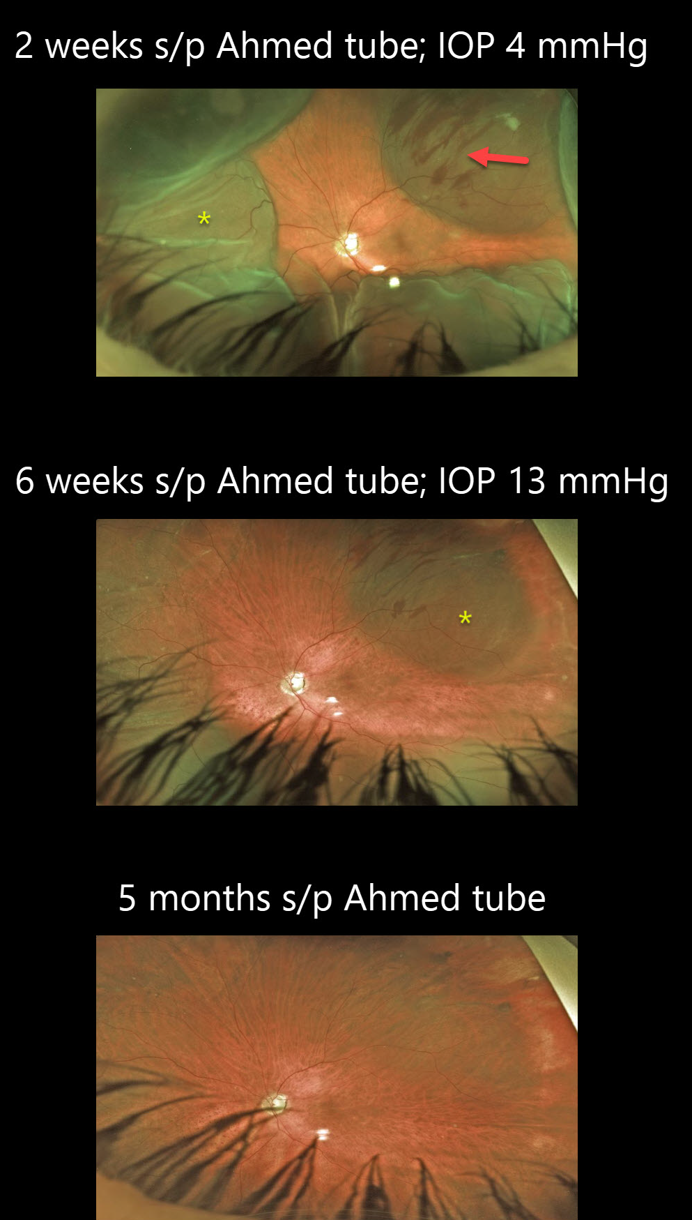
This patient had an uncomplicated Ahmed tube surgery 2 weeks prior, but he did not stop his IOP-lowering medication nor take his prescribed anti-inflammatories and atropine.
His IOP was 4 mmHg, which led to choroidal detachments. The subretinal blood noted superotemporally is likely iatrogenic from an inadvertent overly deep suture used to secure the Ahmed explant.
Aggressive anti-inflammatories and atropine drops were started, and he was asked to stop all IOP-lowering drops.
The choroidals continued to resolve at 6 weeks as IOP continued to climb to 13mmHg. Five months post-op the choroidal detachment and subretinal blood completely resolved.
Learning Points:
Choroidal detachment is relatively common after glaucoma filtration surgery. Two mechanisms are believed to be at play:
(1) Hypotony allows fluid to accumulate in the suprachoroidal space
(2) Inflammation increases the choroidal capillary permeability.
The detached ciliary body may also produce less aqueous, thus creating a self-perpetuating hypotony.


