

Choroidal Excavation
<< Back to Cases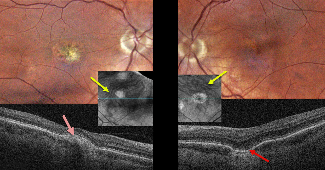
PACHYCHOROID SPECTRUM PREVIOUSLY MISDIAGNOSED AS OCULAR HISTOPLASMOSIS
Steven Bloom
Originally posted on @retina.rocks April 22, 2026
This healthy 39YO female gave a history 18 years earlier of prior anti-VEGF therapy OD for ocular histoplasmosis. She was visually asymptomatic, and vision was 20/40 OD and 20/30 OS.
Color photography of her left eye shows a patchy area of pigment loss in the superonasal fovea. OCT scanning through this area shows a choroidal excavation. The right macula has an overall mostly depigmented area of scarring, which appears as a mound of subretinal hyperreflectivity on OCT. Choroidal en face OCT shows dilated pachyvessels OU that cross the horizontal meridian (vortex anastomoses). There were no areas of peripheral chorioretinal scarring in either eye. Observation was recommended.
Learning Points:
Pachychoroid, meaning “thick” choroid, is secondary to large, dilated outer choroidal vessels. Increased oncotic pressure from these dilated vessels can cause RPE dysfunction, serous detachment, and macular neovascularization (MNV). Pachychoroid disease spectrum is a growing list of findings, including idiopathic central serous chorioretinopathy (ICSC), pachychoroid neovascularization, polypoidal choroidal vasculopathy, pachychoroid pigment epitheliopathy, pachydrusen, peripapillary pachychoroid syndrome, choroidal excavation, and pachyvitelliform maculopathy.
Our guess is that our patient had active ICSC when she was treated for her ‘ocular histoplasmosis.’ The lesion in the right macula is atypical for ICSC. But the other findings, including dilated choroidal vessels with vortex anastomoses and a choroidal excavation in her left eye, argue for pachychoroid as her unifying diagnosis.
CHOROIDAL EXCAVATION WITH MACULAR NEOVASCULARIZATION
Originally posted on @retina.rocks September 30, 2025
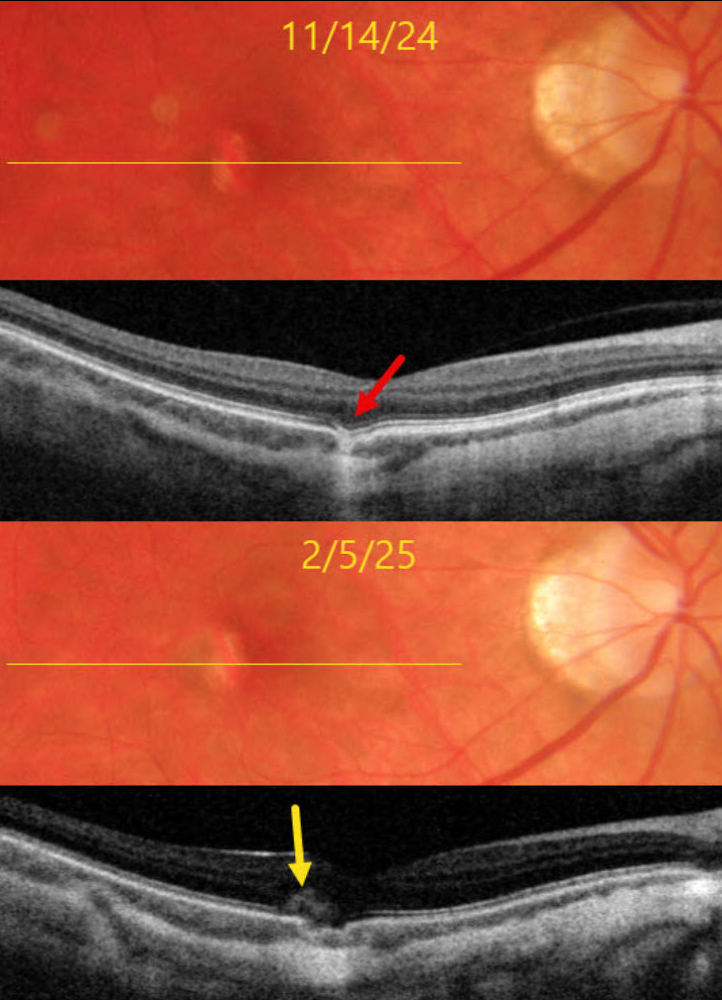
This 39YO myopic female was referred on 11/14/24 for an asymptomatic lesion in her right macula. Vision was 20/25 OD and 20/25 in her normal OS.
Triton color imaging shows a small vertically oval area of subretinal pigment loss temporal to the right fovea. Swept-source OCT shows a focal choroidal excavation (FCE) within this region.
She returned on 2/5/25 with recent blurred vision in this eye. Vision was 20/30. A new type 2 (above the RPE) macular neovascularization (MNV) is noted along the temporal edge of the FCE.
Learning Points:
FCE, originally described by Jampol et al. in 2006, is likely part of the pachychoroid spectrum, as it is usually associated with a thickened choroid and pachyvessels (not noted in our myopic patient). The FCE can be observed, although symptomatic manifestations, including active central serous retinopathy or MNV (as in our patient), often require treatment. An excellent review is by Verma et al, Br J Ophthalmol 2021;105:1043-1048.
One month after a single intravitreal Avastin injection, vision was 20/30, her symptoms had resolved, and the lesion was completely dry on funduscopy and OCT. We are following her closely with PRN therapy.
CHOROIDAL EXCAVATION
Originally posted on @retina.rocks August 15, 2023
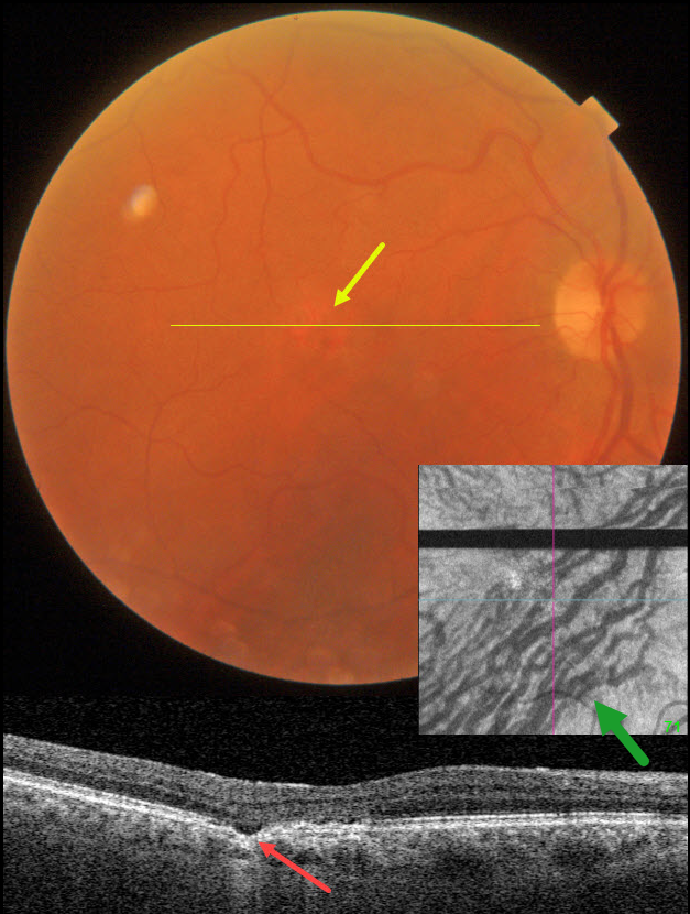
This 74YO male was seen for a routine eye examination. He told us he had a laser procedure on his right eye 18 years ago. Based on this image, what type of laser was it?
Fundus photography shows a normal macula except for an area of depigmentation in the temporal fovea.
Swept-source OCT through this area shows a small focal choroidal excavation. Although the choroid appears to have normal thickness, large outer choroidal vessels spanning virtually the entire choroid are noted nasally.
Choroidal en face OCT is markedly abnormal with dilated vessels and vortex anastomoses.
Our patient shows several pachychoroid-related findings, including a thick choroid for their age, large choroidal vessels, FCE, and vortex anastomoses. Our patient currently has asymptomatic pachychoroid despite having a ‘normal’ choroidal thickness on OCT.
So what type of laser did our patient have? Photodynamic therapy for central serous retinopathy, which has remained in remission since treatment in 2005
Learning Points:
Pachychoroid, meaning “thick” choroid, is secondary to large dilated Haller vessels. Increased oncotic pressure from these dilated vessels can cause RPE dysfunction, serous detachment, and macular neovascularization.
The choroidal thickness depends on numerous factors, including refractive error and age. A high myope’s choroid should be quite thin, but if it appears as a ‘normal’ thickness, it is thick for that eye. We also often see pachychoroid disease in elderly patients whose choroid is abnormally thickened for their age, but appears ‘normally’ thick when not accounting for this.
FCE, originally described by Jampol et al in 2006, is likely part of the pachychoroid spectrum, as it is usually associated with a thickened choroid and pachyvessels. The FCE can be observed, although symptomatic manifestations, including active central serous retinopathy or macular neovascularization, often require treatment. An excellent review is by Verma et al, Br J Ophthalmol 2021;105:1043-1048.
The choroidal venous system, as with the neurosensory retinal vasculature, usually respects the horizontal meridian. With pachychoroid disorders, these anastomoses develop, and in this case, the inferior vortex system drains nearly the entire macula.
For a wonderful review highlighting the concept of venous overload choroidopathy, see Spaide et al, Progress Retinal Eye Research 2022;86:100973.
CHOROIDAL EXCAVATION
Will Gibson
Originally posted on @retina.rocks February 9, 2022
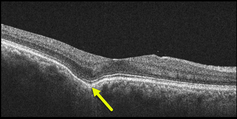
This 56YO highly myopic male has 20/20 vision in his right eye with an asymptomatic temporal foveal focal choroidal excavation (FCE). No treatment was recommended.
Learning Points:
Focal choroidal excavation, originally described by Jampol et al. in 2006, is likely part of the pachychoroid spectrum, as it is usually associated with a thickened choroid and pachyvessels.
The FCE can be observed, although symptomatic manifestations, including active central serous retinopathy or macular neovascularization, often require treatment.
An excellent review is by Verma et al, Br J Ophthalmol 2021;105:1043-1048.
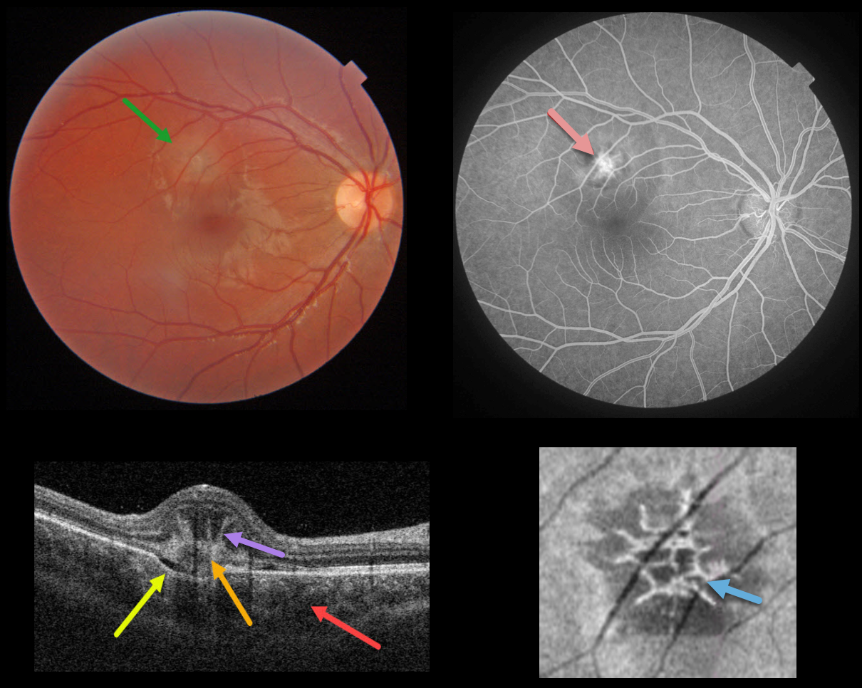
IDIOPATHIC MACULAR NEOVASCULARIZATION (MNV)
Originally posted on @retina.rocks December 20, 2021
This healthy 14YO female presented with acute vision loss in her right eye. Vision was 20/40. Triton color photography shows a depigmented subretinal lesion in the superior macula.
Swept-source OCT shows a constellation of fascinating findings. A type 2 macular neovascularization is seen overlying a choroidal excavation. The choroid is thickened.
The most dramatic findings are the unusual linear, radiating, hyperreflective lesions extending anteriorly from the MNV, also known as the “pitchfork sign.” En face OCT through this lesion shows a spiky, wreath-like pattern of hyperreflectivity.
The MNV was confirmed on fluorescein angiography. Intravitreal anti-VEGF was begun.
Learning Points:
The pitchfork sign was originally described by Hoang et al (Retina 2013;33:1049-1055) as a unique OCT finding in inflammatory MNV. Falavarajani et al more recently noted that this finding is also associated with non-inflammatory MNV (Ophthalmic Surg Lasers Imaging Retina 2019;50:719-725). The pathogenesis of the avascular pitchfork spikes is unknown, but may be related to outer retinal traction or Müller cell activation.
Focal choroidal excavation, originally described by Jampol et al in 2006, is likely part of the pachychoroid spectrum and is associated with type 2 MNV. An excellent review is by Chung et al (Retina 2017;37;199-221).
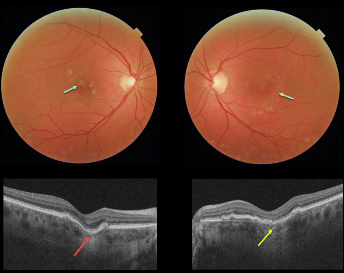
FOCAL CHOROIDAL EXCAVATION
Originally posted on @retina.rocks October 6, 2021
This 60YO female has a history of idiopathic central serous chorioretinopathy that was previously treated successfully with photodynamic therapy in her left eye. Bilateral macular drusen are noted.
OCT shows a definite focal choroidal excavation (FCE) in her right eye, and a possible FCE in her left eye.
Learning Points:
Focal choroidal excavation, originally described by Jampol et al. in 2006, is likely part of the pachychoroid spectrum, as it is usually associated with a thickened choroid and pachyvessels.
The FCE can be observed, although symptomatic manifestations, including active central serous retinopathy or macular neovascularization, often require treatment.
Spaide and Ryan noted this finding in about two-thirds of patients with central serous retinopathy (Am J Ophthalmology 2015;160;1211-1216). An excellent review is by Chung et al (Retina 2017;37;199-221).
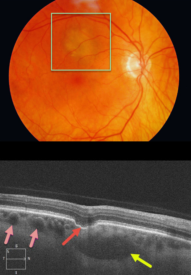
CHOROIDAL EXCAVATION
Originally posted on @retina.rocks February 17, 2021
This patient was referred for an asymptomatic, slightly depigmented subretinal lesion in the superior right macula. OCT through this lesion reveals a focal choroidal excavation, choroidal pachyvessels, and probable loculated outer choroidal fluid.
Learning Points:
Focal choroidal excavation (FCE), originally described by Jampol et al in 2006, is likely part of the pachychoroid spectrum since it is usually associated with a thickened choroid and pachyvessels. The FCE can be observed, although symptomatic manifestations, including active central serous retinopathy or macular neovascularization, often require treatment.
Our patient also showed accumulation of fluid in the outer choroid. Spaide and Ryan noted this finding in about two-thirds of patients with central serous retinopathy (Am J Ophthalmology 2015;160;1211-1216). The presence of an FCE, pachyvessels, and loculated posterior choroidal fluid in our patient is intriguing and supports the association between pachychoroid disorders and FCE.


