

Choroidal Granuloma
<< Back to Cases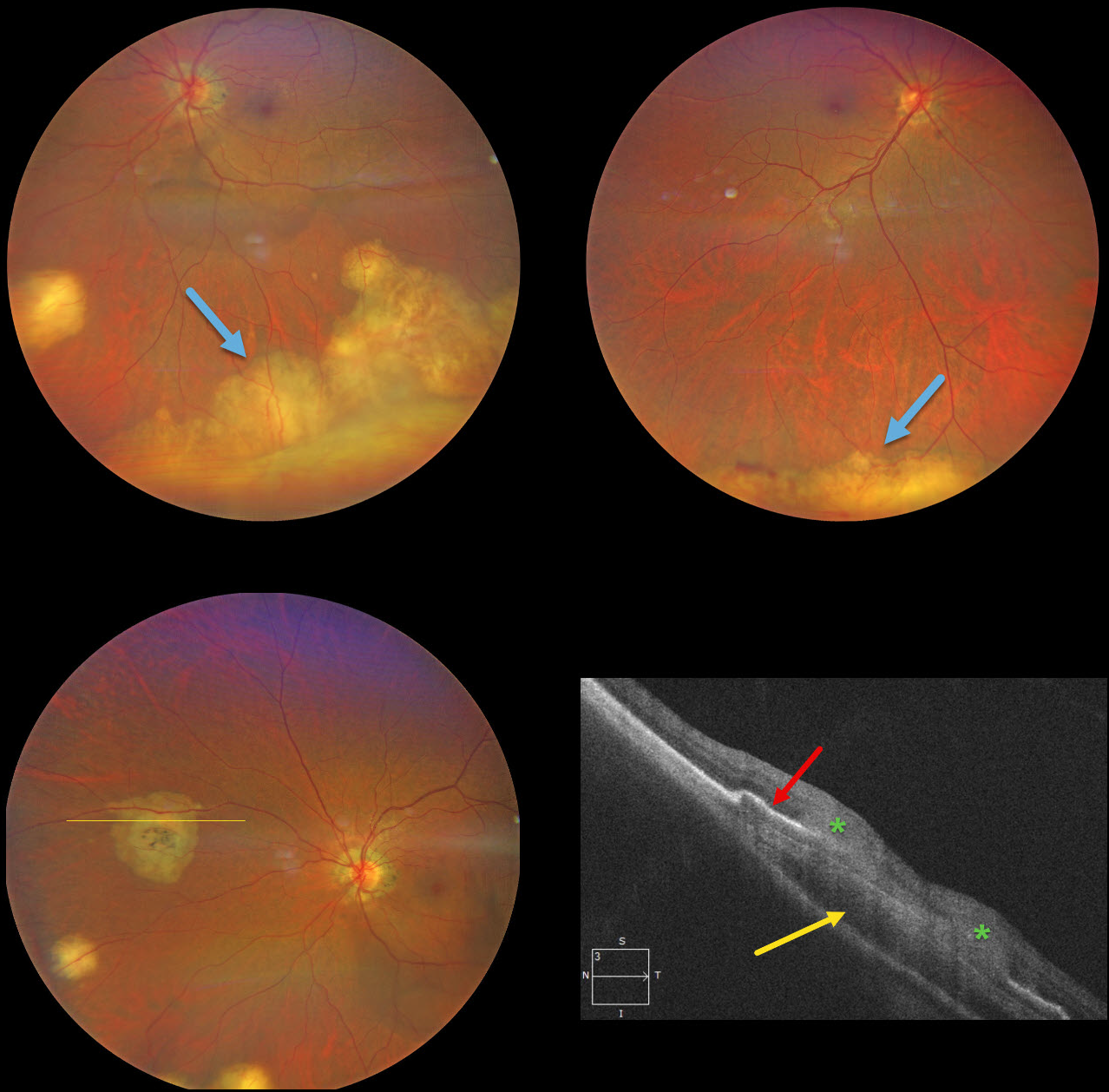
MULTIFOCAL TUBERCULOSIS CHOROIDAL GRANULOMAS
Will Gibson
Originally posted on @retina.rocks July 3, 2025
This previously healthy 49YO Indian female was visiting the United States and presented with new floaters in her left eye. Vision was 20/25 OD and 20/40 OS. There was no anterior uveitis, and the vitreous was clear bilaterally.
Color photography shows multifocal bilateral amelanotic choroidal lesions, some isolated and others confluent in the inferior periphery. OCT scanning through a nasal lesion OS shows a relatively hyporeflective choroidal lesion, irregular overlying RPE elevation, and probable infiltration of the overlying retina.
QuantiFERON-TB testing was positive with a negative chest X-ray. The patient was referred to the infectious disease department and started on anti-tuberculosis treatment (ATT).
Learning Points:
Although TB most commonly presents with pulmonary involvement, extrapulmonary sites can include the gastrointestinal, skin, cardiovascular, genitourinary, and central nervous systems, as well as the eyes. Ocular involvement, like syphilis, can mimic virtually any type of uveitis, including anterior, intermediate, posterior, and panuveitis, retinitis and retinal vasculitis, neuroretinitis, optic neuropathy, choroidal granuloma (our patient), choroiditis, and scleritis. For an excellent review of intraocular tuberculosis, see Bupta et al, Survey Ophthalmology 2007;52:561-587.
Tubercular granulomas can be distinguished from sarcoid lesions by being solitary, yellow, larger, and vascularized (Agarwal et al, AJO 2021;226:42-55). Our patient is therefore somewhat unusual with bilateral multifocal lesions. Sarcoid workup was negative.
CHOROIDAL GRANULOMA
Evgenii Chernov
Originally posted on @retina.rocks April 28, 2025
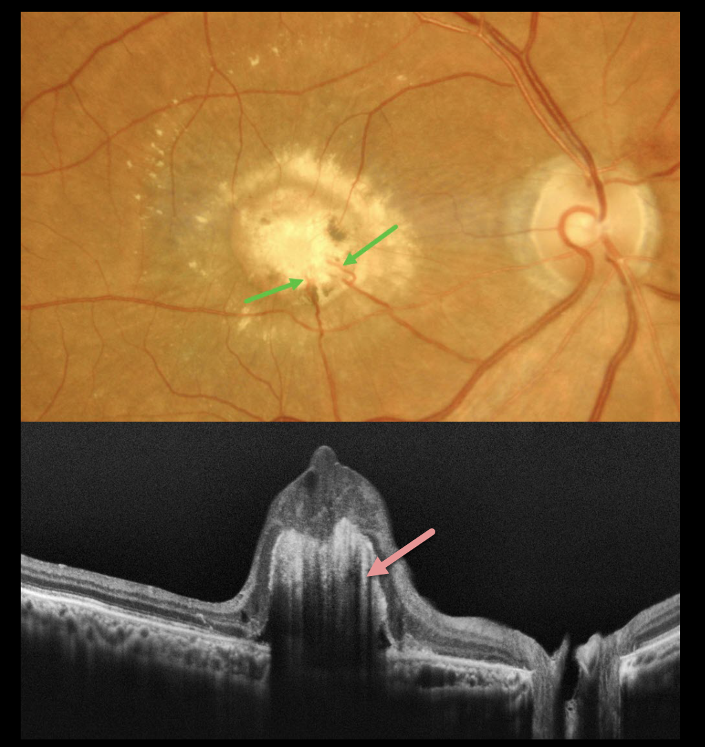
This healthy 64YO male presented with gradual vision loss in his right eye. Vision was 20/2000 OD and 20/20 in his normal OS.
Color imaging shows a yellow-white subfoveal nodular lesion with surrounding subretinal lipid. Several retinal choroidal anastomoses are noted within the lesion inferiorly. OCT scanning shows a hyperreflective elevated deep retinal lesion with overlying disorganized retina. There is marked posterior shadowing obscuring all deep retinal and choroidal detail.
Learning Points:
Choroidal granulomas usually appear as solitary, creamy white, round subretinal lesions. They are generally hyporeflective on OCT imaging (Invernizzi et al, Retina 2015;35:525-531) and can extend into the subretinal space, as in our patient. Causes include sarcoidosis, tuberculosis, Bartonella, and VKH.
Under normal conditions, the retinal and choroidal circulations remain separate and distinct. Rarely, the two circulations directly connect via a chorioretinal anastomosis. These can occur in chorioretinal scars, in macular telangiectasia, and in disciform scars.
Our best guess is that this lesion represents a vascularized choroidal granuloma. Due to its chronicity and lack of significant overlying retinal exudation, observation was recommended.
TUBERCULOSIS CHOROIDAL GRANULOMA
Divya Nair
Originally posted on @retina.rocks July 22, 2024
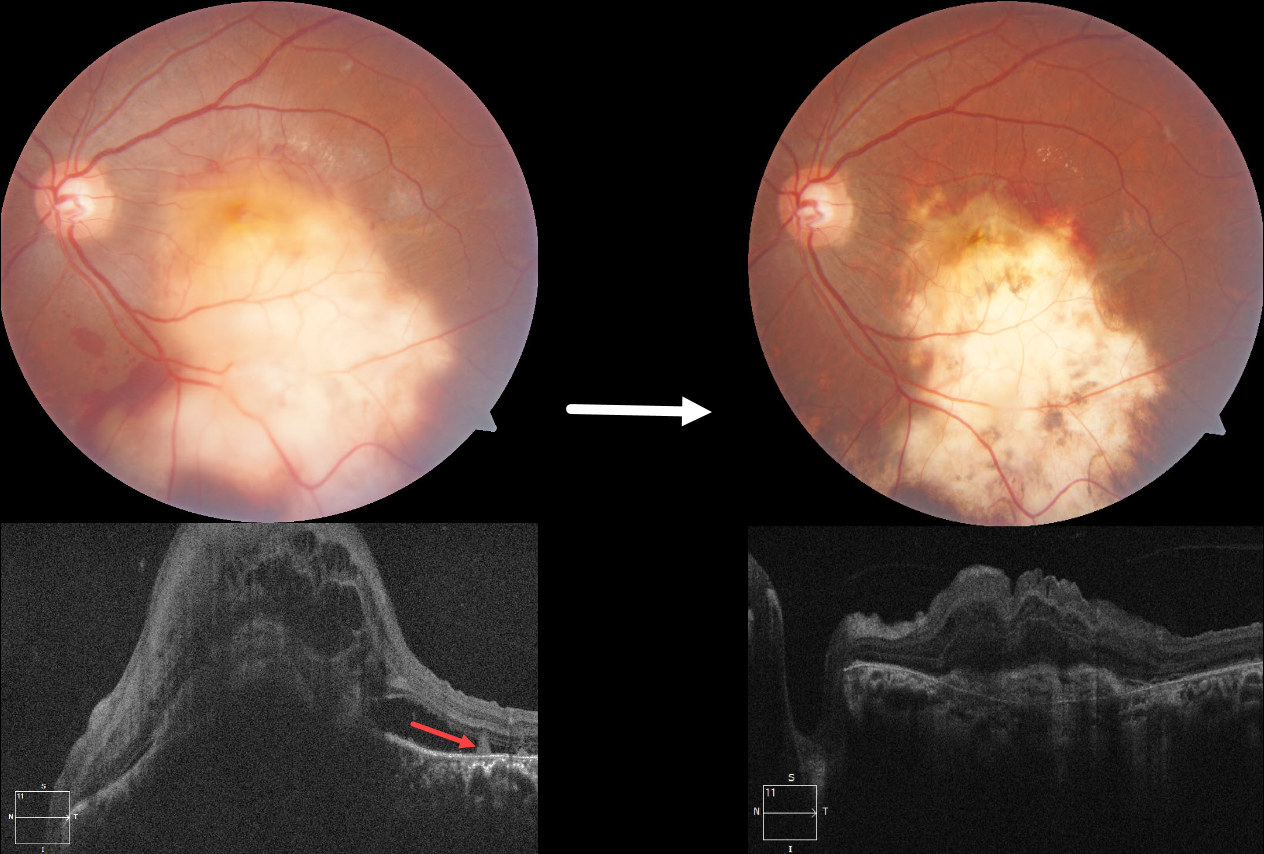
This 36YO male presented with 10 days of vision loss in his left eye. One month earlier, he was diagnosed with disseminated tuberculosis (TB), peritonitis, and pleural effusion, and was placed on systemic antitubercular therapy (ATT). Vision was 20/20 in his normal right eye and counting fingers in his left eye.
Fundus photography shows a large elevated yellowish submacular lesion that extends into the inferior midperiphery. Subretinal blood is noted along its superior and inferior margins. Faint lipid is present superotemporally, and some retinal striae are seen temporally. Spectral domain OCT shows a large dome-shaped hyporeflective choroidal lesion with overlying cystic retinal thickening, subretinal fluid, and subretinal hyperreflective material. A few hyperreflective pyramidal lesions are noted above the RPE temporally.
We started him on oral corticosteroids. In view of the vascularized lesion with subretinal blood, an intravitreal injection of Avastin was also given. Six weeks later, there is a relatively flat scar with near complete resolution of the blood. The choroidal lesion has resolved with an overlying hyperreflective subretinal scar and decreased retinal thickening. Unfortunately, vision remained at counting fingers.
Learning Points:
Although TB most commonly presents with pulmonary involvement, extrapulmonary sites can include the gastrointestinal, skin, cardiovascular, genitourinary, and central nervous systems, including the eyes. Ocular involvement, like syphilis, can mimic virtually any type of uveitis, including anterior, intermediate, posterior, and panuveitis, retinitis and retinal vasculitis, neuroretinitis, optic neuropathy, choroidal granuloma, choroiditis, and scleritis.
For an excellent review of intraocular tuberculosis, see Bupta et al, Survey Ophthalmology 2007;52:561-587.
Tubercular granulomas can be distinguished from sarcoid lesions by their solitary nature, yellow color, larger size, and vascularization (Agarwal et al., AJO 2021;226:42-55). All these features were found in our patient. Vascularization is thought to arise from localized hypoxia, leading to increased levels of vascular endothelial growth factor. Anti-VEGF injections and systemic corticosteroids, in addition to ATT, are helpful in hastening granuloma resolution and reducing secondary exudation (Lee et al, Graefe’s 2022;260:1641-1650).
CHOROIDAL GRANULOMA
Originally posted on @retina.rocks May 20, 2024
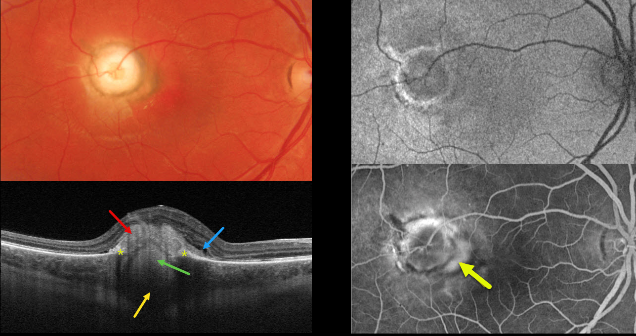
This healthy 26YO male presented with a 4-month history of decreased vision in his right eye. Vision was 20/200 OD and 20/25 in his normal OS.
Optos color RG imaging shows a well-defined, 1800-micron, pale, subretinal nodular lesion with surrounding pigment. Triton swept-source OCT shows a hyperreflective subretinal mound, a hyperreflective lesion extending from the choroid through a break in the RPE, posterior choroidal thickening and shadowing, and trace outer retinal fluid.
Optos fundus autofluorescence (FAF) shows the lesion to be hypo-FAF with a rim of hyper-FAF.
On fluorescein angiography, the nodule stains along its edges, with a leaking macular neovascularization along its nasal edge. Anti-VEGF therapy was begun.
Learning Points:
Choroidal granulomas usually appear as solitary, creamy white, round subretinal lesions. They are generally hyporeflective on OCT imaging (Invernizzi et al, Retina 2015;35:525-531) and can extend into the subretinal space, as in our patient. Causes include sarcoidosis, tuberculosis, and VKH. Testing results for our patient were completely negative, including sarcoidosis, tuberculosis, Bartonella, and syphilis.
CHOROIDAL GRANULOMA
Originally posted on @retina.rocks June 3, 2022
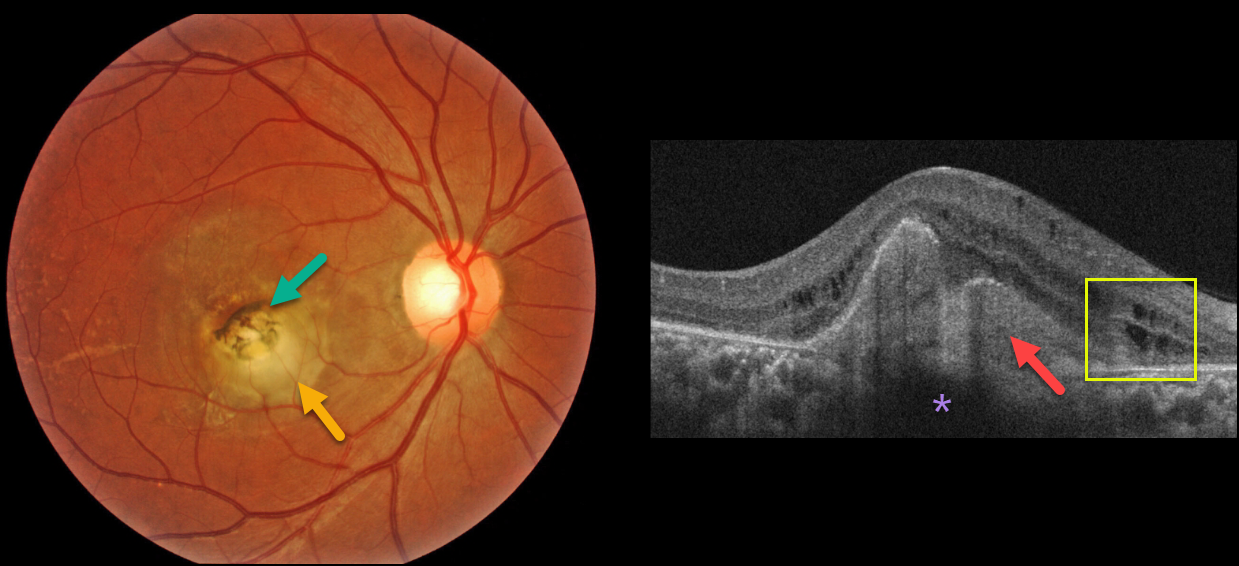
A healthy 32YO female presented with a 6-month history of vision loss in her right eye. Vision was counting fingers OD and 20/20 in her healthy left eye.
Fundus imaging shows a slightly elevated, creamy white, subfoveal lesion surrounded by slightly hyperpigmented macular scarring.
OCT shows a hyperreflective subretinal lesion that shadows the underlying choroid. There are some mild cystic changes in the inner and outer nuclear layers.
Learning Points:
Choroidal granulomas usually appear as solitary, creamy white, round subretinal lesions. They are hyporeflective on OCT imaging (Invernizzi et al, Retina 2015;35:525-531). They can extend into the subretinal space, as in our patient. Causes include sarcoidosis, tuberculosis, and VKH. Testing results for sarcoidosis and tuberculosis are still pending.
CHOROIDAL GRANULOMA
Originally posted on @retina.rocks December 8, 2021
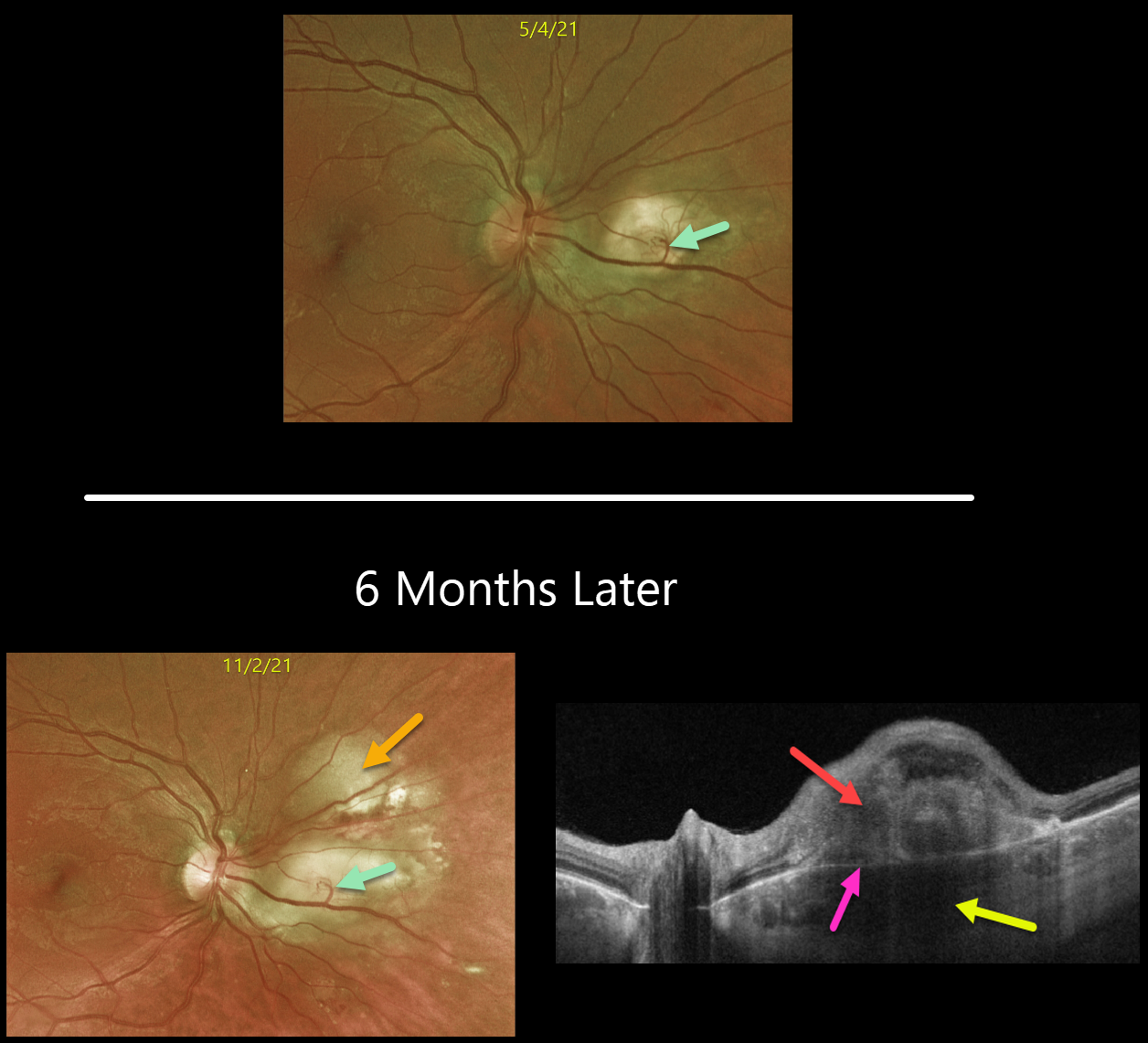
This is a follow-up from our 6/7/21 post of a choroidal granuloma, likely representing the initial manifestation of systemic sarcoidosis. The patient was sent to pulmonology to confirm the diagnosis, but he did not keep the appointment.
Six months later, the choroidal granuloma had significantly enlarged.
A retinochoroidal anastomosis is noted between the overlying retinal vein and the lesion. Triton swept-source OCT shows a thickened, hyporeflective choroidal lesion, a hyperreflective outer retinal lesion with overlying inner retinal disorganization, and visible Bruch’s membrane separating the choroidal and retinal components.
Learning Points:
Choroidal granulomas are aggregations of inflammatory cells that appear clinically as a solitary, creamy-white, round subretinal lesion. Causes include sarcoidosis, tuberculosis, and VKH.
They are hyporeflective with increased transmission on OCT imaging (see Invernizzi et al, Retina 2015;35:525-531).
CHOROIDAL GRANULOMA
Originally posted on @retina.rocks June 7, 2021
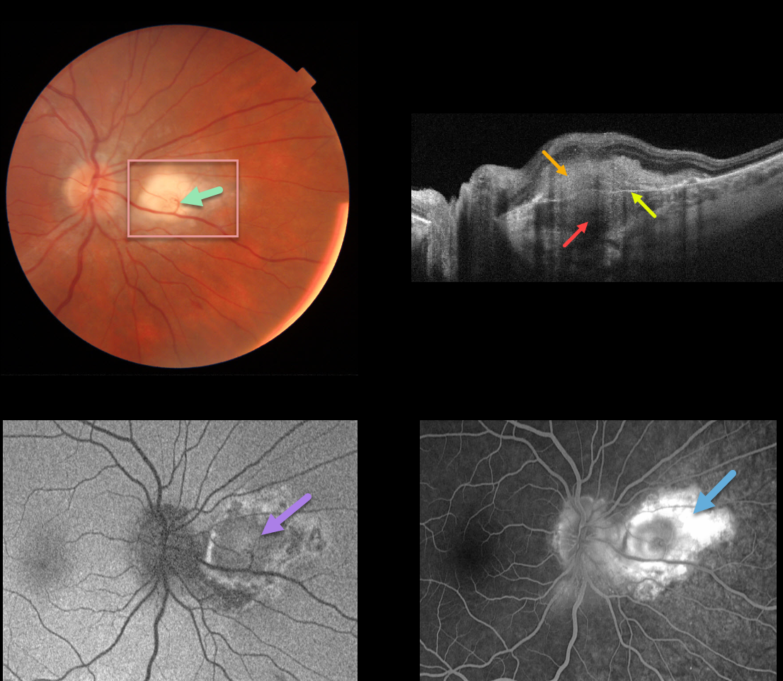
This 30YO male was referred for an asymptomatic lesion in his right eye. Vision was 20/20 OU, and the left fundus was healthy.
There is a nodular, creamy white, deep retinal/subretinal lesion just nasal to the right optic nerve. A retinochoroidal anastomosis was noted between the overlying retinal vein and the lesion.
Triton swept-source OCT shows a thickened hyporeflective choroidal lesion, a hyperreflective outer retinal lesion with overlying inner retinal disorganization, and visible Bruch’s membrane separating the choroidal and retinal components.
Optos fundus autofluorescence (FAF) shows relatively diffuse hypo-FAF, and fluorescein angiography shows diffuse staining of the lesion.
Our patient’s bloodwork and chest x-ray were suggestive of sarcoid. Quantiferon gold, ACE, and toxoplasmosis IgG/IgM were all negative. His primary care provider was alerted to the probable diagnosis of sarcoidosis.
Learning Points:
Choroidal granulomas usually appear as solitary, creamy white, round subretinal lesions. They are hyporeflective on OCT imaging (Invernizzi et al, Retina 2015;35:525-531). Causes include sarcoidosis, tuberculosis, and VKH.
The choroidal and retinal circulations normally coexist without connecting to each other. A chorioretinal anastomosis breaks this rule when a retinal vessel directly connects with the choroid.
Clinically obvious chorioretinal anastomosis is most common in disciform lesions and toxoplasmosis scars. They are also a subclinical finding noted in 65% of eyes with MacTel 2 using OCT and OCT angiography (see Spaide et al, Retina 2018;38:1920-1929).
A laser-induced chorioretinal anastomosis was originally proposed as a possible treatment for central retinal vein occlusion in 1995, and a recent randomized trial showed it could reduce the anti-VEGF treatment burden (see McAllister et al., JAMA Ophthalmol 2018;136;1391-1397). However, it is still rarely used in current clinical practice.
CHOROIDAL GRANULOMA
Originally posted on @retina.rocks April 15, 2021
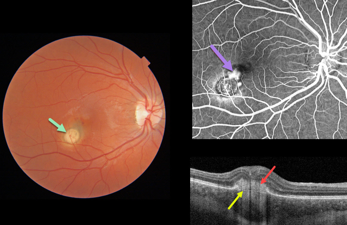
This 22YO female presented with 20/400 vision from a macular neovascularization (MNV) associated with a choroidal granuloma.
The Triton image shows a focal yellow-white nodular subretinal lesion with adjacent subretinal pigment and blood along its foveal edge.
Swept-source OCT through the MNV shows the hyperreflective MNV above Bruch’s membrane. Fluorescein angiography shows leakage from the MNV.
Learning Points:
Choroidal granulomas usually appear as solitary, creamy white, round subretinal lesions. They are hyporeflective on OCT imaging (Invernizzi et al, Retina 2015;35:525-531). Causes include sarcoidosis, tuberculosis, and VKH. They can occasionally cause exudation that responds to anti-VEGF injections or photodynamic therapy.
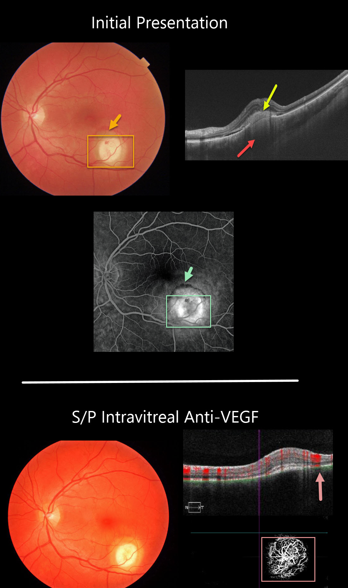
CHOROIDAL GRANULOMA
Originally posted on @retina.rocks October 16, 2020
The initial photo in this series shows a focal yellow-white nodular lesion with adjacent subretinal blood. The OCT shows subretinal hyperreflective material, some shallow subretinal fluid, and an amelanotic choroidal granuloma.
Fluorescein angiography shows staining of the lesion, with some blood blockage along its superior edge.
A later fundus photo shows complete resolution of the hemorrhage following treatment with a single intravitreal anti-VEGF injection.
Despite the resolved exudation, OCTA angiography still shows flow signals within the lesion. We continue to follow her closely for recurrent exudation.
Learning Points:
Choroidal granulomas usually appear as solitary, creamy white, round subretinal lesions. They are hyporeflective on OCT imaging (Invernizzi et al, Retina 2015;35:525-531). Causes include sarcoidosis, tuberculosis and VKH. They can occasionally cause exudation that responds to anti-VEGF injections or photodynamic therapy.
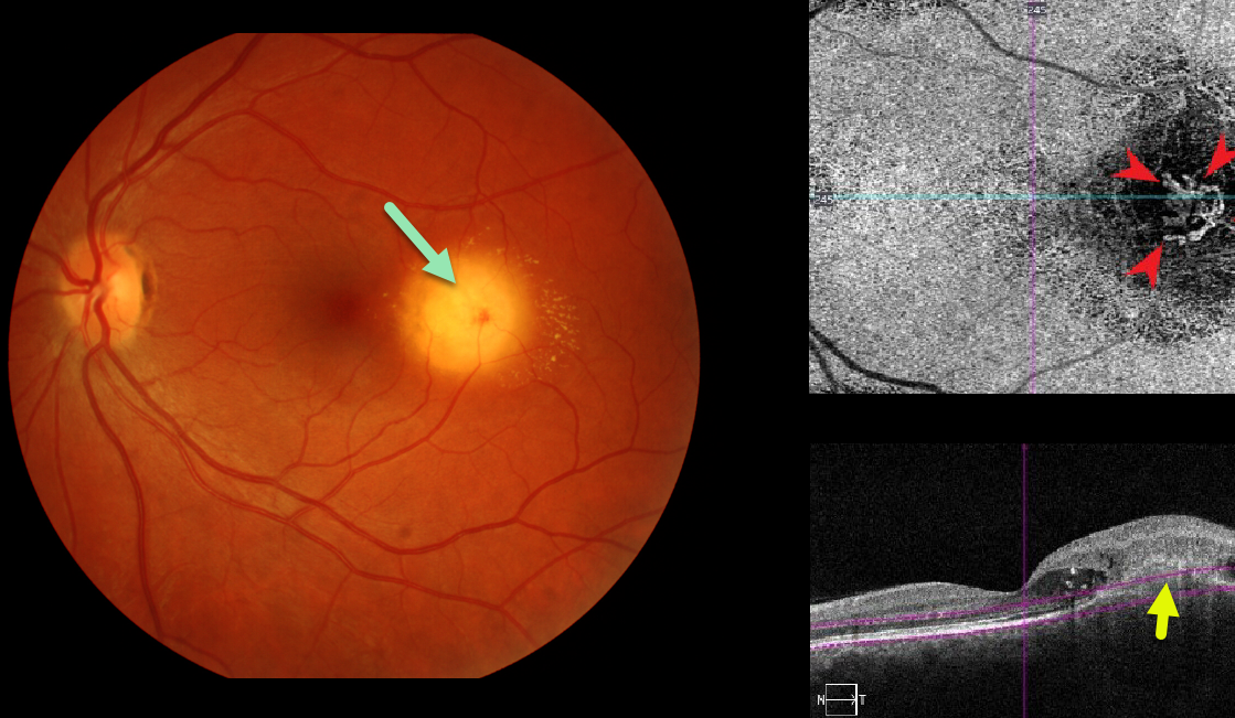
CHOROIDAL GRANULOMA
Originally posted on @retina.rocks May 13, 2020
Our patient was seen recently with asymptomatic macular neovascularization (MNV) associated with a choroidal granuloma. Clinically there was new blood, fluid and lipid.
OCT angiography confirms an active MNV. This lesion is deep to the RPE which is consistent with a focal scleral nodule.
Learning Points:
Choroidal granulomas usually appear as solitary creamy white, round subretinal lesions. They are hyporeflective on OCT imaging (Invernizzi et al, Retina 2015;35:525-531). Causes include sarcoidosis, tuberculosis and VKH. They can occasionally cause exudation that responds to anti-VEGF injections or photodynamic therapy.
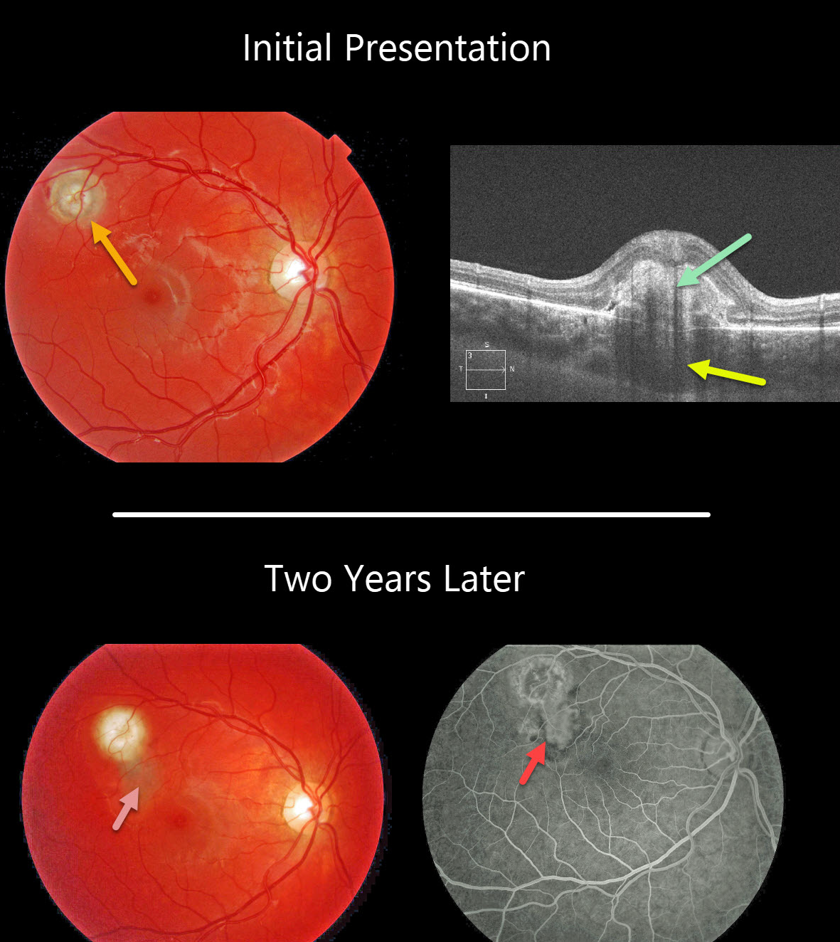
CHOROIDAL GRANULOMA
Originally posted on @retina.rocks April 20, 2020
This patient’s fundus photo shows a focal yellow-white nodular lesion of the right eye. OCT shows the subretinal hyperreflective material and an underlying choroidal granuloma.
Two years later, the patient subsequently developed an active macular neovascularization along the inferior edge of the original lesion, seen better on fluorescein angiography.
Learning Points:
Choroidal granulomas usually appear as solitary creamy white, round subretinal lesions. They are hyporeflective on OCT imaging (Invernizzi et al, Retina 2015;35:525-531). Causes include sarcoidosis, tuberculosis and VKH. They can occasionally cause exudation that responds to anti-VEGF injections or photodynamic therapy.


