

Choroidal Hemangioma
<< Back to Cases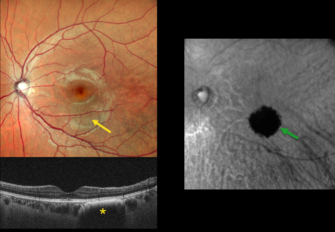
SUBCLINICAL CHOROIDAL NEVUS VS HEMANGIOMA
Shraddha Raj Shrivastava, Steven Bloom, and Manish Nagpal
Originally posted on @retina.rocks May 13, 2026
This asymptomatic 26YO male presented for a routine eye examination. Vision was 20/20.
Clinical biomicroscopy and MultiColor scanning laser ophthalmoscope (SLO) imaging are normal except for faint hyperpigmentation in the inferior macula. OCT in this region shows a dome-shaped hyporeflective lesion with an anterior hyperreflective border. Retromode imaging shows a well-defined hyporetroreflective lesion.
Learning Points:
Traditional color photography uses white light to image the posterior segment. Newer imaging devices utilize LED or SLO technology to illuminate the fundus with blue, green, and infrared light, which best image the vitreoretinal interface/inner retina, mid-retina to RPE, and choroid, respectively. Many of these technologies create a ‘color’ fundus image that is not true to life yet contains clinically useful depth information in a flat image. In our patient’s case, Nidek Mirante Retromode imaging best detected this subclinical lesion using a 790 nm SLO. For a recent review of Retromode imaging, see Sukkarieh et al, Surv Ophthalmology 2023;68:1027-1037.
So does our patient have a choroidal nevus or hemangioma? Internally, the authors are split between a nevus (SRS and MN) and a hemangioma (SB). Findings supporting a choroidal hemangioma include its orange color blending with the surrounding RPE/choroid and round shape on Retromode imaging. However, we have not seen a hemangioma with an anterior hyperreflective margin on OCT. Jonna et al described 5 OCT patterns of flat choroidal nevi (Ophthalmology Retina 2019;3:270-277), including a subtype with “anteriorly bowed hyperreflectivity with discrete borders and cascading edges,” which appears identical to our patient’s OCT.
CHOROIDAL HEMANGIOMA
Moazzam Parvez and Krishnendu Nandi
Originally posted on @retina.rocks July 14, 2025
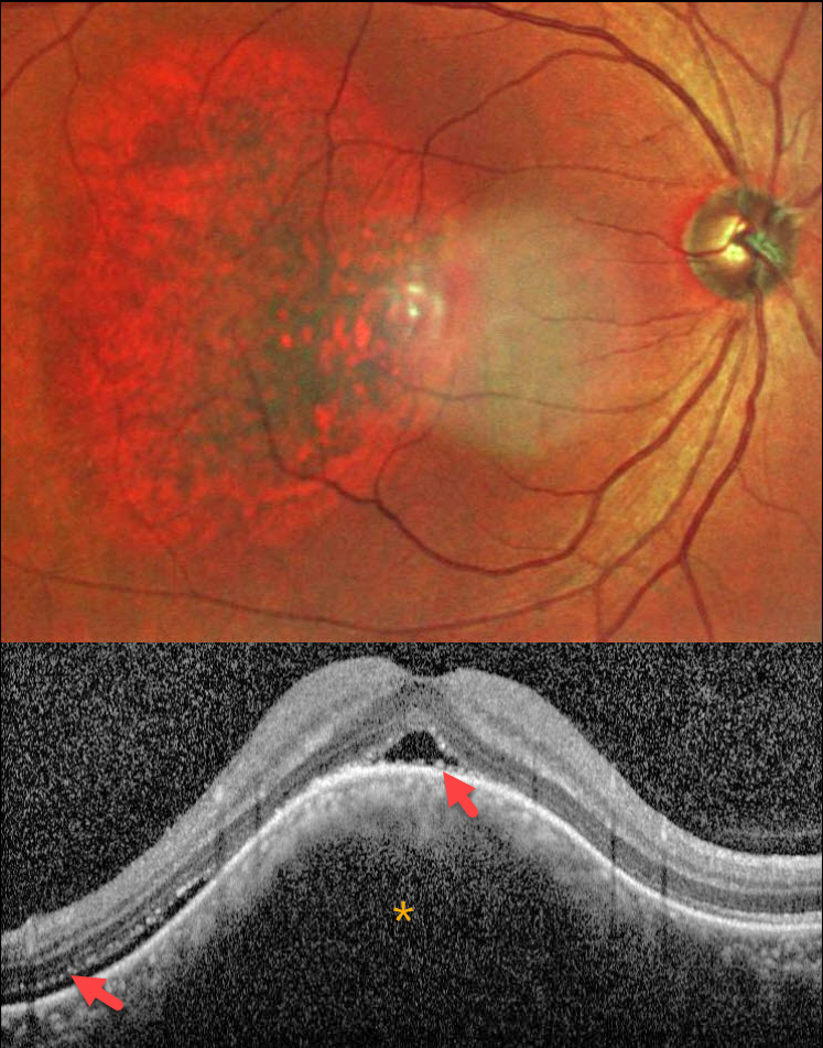
This healthy 42YO male presented with blurred and distorted vision in his right eye. Vision was 20/80 OD and 20/20 in his normal OS.
MultiColor SLO imaging shows an oval orange-red choroidal lesion in the central and temporal macula. OCT scanning shows an elevated choroidal lesion with overlying foveal and distal temporal subretinal fluid. The outer retinal layers are thinned and somewhat disorganized. Shaggy hyperreflective material overlies the central RPE.
Learning Points:
Choroidal hemangioma is a benign vascular choroidal tumor. Circumscribed tumors are isolated and non-syndromic, and appear as orange-red, dome-shaped posterior pole lesions. Diffuse hemangiomas are usually seen in association with the Sturge-Weber syndrome. The lesions are difficult to see funduscopically because the orange tumor blends with the surrounding choroid. In our patient, the MultiColor imaging more dramatically highlights this lesion.
Although there is no malignant potential, it can cause vision loss from exudation with subretinal and intraretinal fluid, which is best treated with photodynamic therapy (Yang et al, Surv Ophthalmology 2025;70:389-400).
CHOROIDAL HEMANGIOMA
Ayushi Gupta and Vishal Agrawal
Originally posted on @retina.rocks March 25, 2025
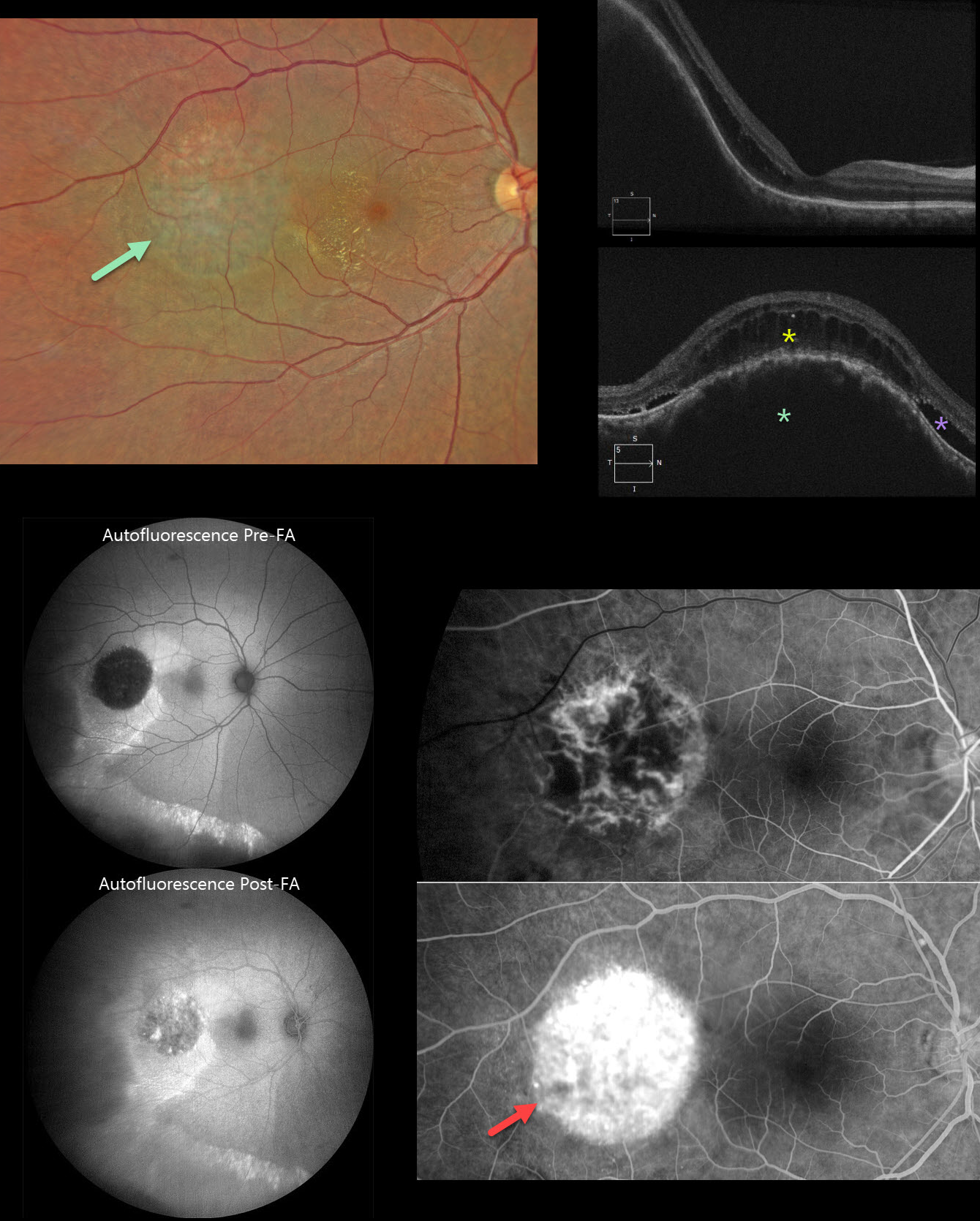
This 37YO male presented with a 3-week history of decreased vision in his right eye. Vision was 20/70 OD and 20/20 in his normal OS.
Color photography shows a subretinal lesion with a somewhat orange hue in the temporal macula. Lipid extends into the temporal fovea. Some subtle pigmentary changes extend inferiorly. OCT scanning shows marked choroidal thickening within the lesion with overlying cystic retinal edema and some subretinal fluid.
On fundus autofluorescence (FAF), the lesion is hypo-FAF with surrounding hyper-FAF and a hyper-FAF gutter extending inferiorly. The lesion becomes hyperfluorescent following fundus fluorescein angiography (FFA). The lesion shows intrinsic vasculature with late leakage on FFA. Unfortunately, our patient was lost to follow-up immediately.
Learning Points:
Choroidal hemangioma is an uncommon benign hamartomatous disorder that presents as either a circumscribed, orange-red, round choroidal lesion (our patient), which is almost always isolated and nonsyndromic, or a diffuse form, which is usually part of Sturge-Weber syndrome. Although there is no malignant potential, it can cause vision loss due to exudation of subretinal and intraretinal fluid, which is best treated with photodynamic therapy (see Yang et al, Surv Ophthalmology 2025;70:389-400). Lipid exudate is a rare finding.
Our case also reminds us why fundus imaging is performed in a specific order: color photography, followed by FAF, then FFA. In FAF, lipofuscin within the RPE is the intrinsic pigment that generates the FAF signal. In FFA, fluorescence is generated by sodium fluorescein that is injected intravenously. Our patient’s hemangioma is hypo-FAF, but when fluorescein stains the lesion, it no longer generates an ‘autofluorescent’ signal due to the fluorescein dye.
CHOROIDAL HEMANGIOMA
Anand Temkar, Navneet Mehrotra, and Manish Nagpal
Originally posted on @retina.rocks December 31, 2024
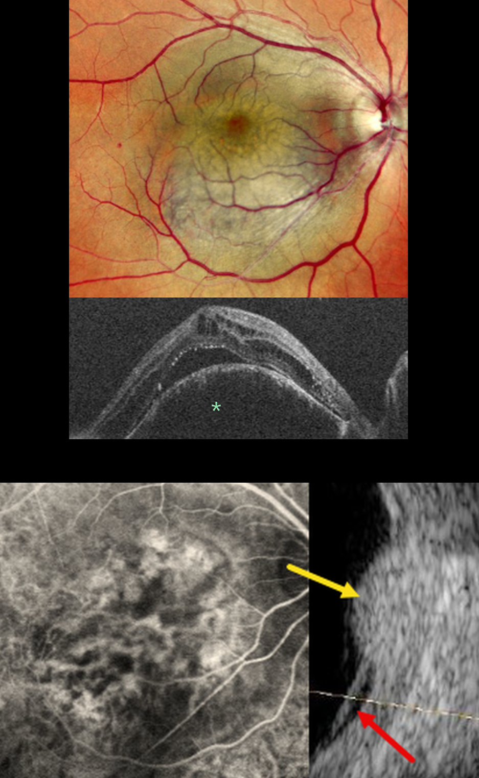
This 42YO male presented with two months of vision loss in his right eye. Vision was 20/200 OD and 20/20 in his normal left eye.
Mirante pseudocolor SLO imaging shows a pigmented subretinal lesion occupying the entire macula. OCT shows a markedly thickened and elevated choroid with overlying cystic retinal edema and subretinal fluid. The lesion is hyperfluorescent on angiography. B-scan ultrasonography shows it to have homogeneous high internal reflectivity with localized subretinal fluid inferiorly.
Learning Points:
Choroidal hemangioma is an uncommon benign hamartomatous disorder that presents as either a circumscribed, orange-red, round choroidal lesion (as in our patient), which is almost always isolated and nonsyndromic, or a diffuse form that is usually part of Sturge-Weber syndrome.
Although it has no malignant potential, it can cause vision loss due to exudation of subretinal and intraretinal fluid, which can be treated with photodynamic therapy (see Tsipursky et al., Surv Ophthalmology 2011;56:68-85).
The Mirante SLO uses blue (488nm), green (532nm), and red (670nm) confocal lasers to generate a fundus image. The blue laser best images the vitreoretinal interface and inner retina, the green images the mid retina, and the infrared images the outer retina, RPE, and choroid (Roy et al, Surv Ophthalmology 2024;69:378-402). Combining these 3 wavelengths into a single file produces a pseudocolor image that often provides the clinician with more information than a standard color photo. However, in some disorders, including choroidal hemangioma, this technology creates a false hyperpigmented appearance, which can lead to the misdiagnosis of uveal melanoma (Becker et al, OSLIR 2023;54:292-296).
CHOROIDAL HEMANGIOMA MISDIAGNOSED AS UVEAL MELANOMA
Originally posted on @retina.rocks November 19, 2024
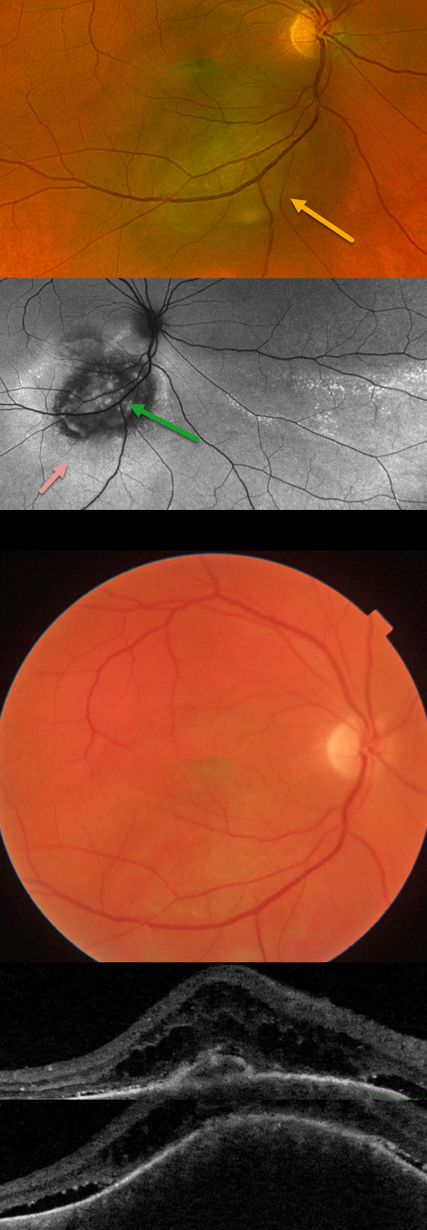
This 44YO male was referred for recent vision loss from a possible uveal melanoma. Vision was 20/150 in his right eye and 20/20 in his healthy left eye.
Optos color RG imaging shows an elevated, pigmented subretinal lesion extending from the inferior macula below the inferotemporal arcade. Fundus autofluorescence (FAF) shows this lesion to be hypo-FAF, with surrounding hyper-FAF and areas of hyper-FAF extending into the inferior and inferotemporal midperiphery, indicating prior extramacular subretinal fluid.
Triton color imaging, however, shows the choroidal lesion as orange. Swept-source OCT shows a thickened choroidal lesion with overlying subretinal fluid and cystic retinal edema. The lesion measured 9.6 x 9.0mm in basal diameter and 3.4mm thick with B-scan ultrasonography (not shown). There was moderate-to-high internal reflectivity, consistent with a choroidal hemangioma. Photodynamic therapy was recommended.
Learning Points:
Isolated choroidal hemangiomas are benign vascular lesions. Although they have no malignant potential, they can cause vision loss due to exudation of subretinal and intraretinal fluid, which can be treated with photodynamic therapy (see Tsipursky et al, Surv Ophthalmology 2011;56:68-85).
This lesion was initially misdiagnosed as a uveal malignant melanoma based on the hyperpigmented appearance on Optos RG imaging. This has also been reported in the literature (Becker et al, OSLIR 2023;54:292-296). These images are generated from a red (635nm) and green (532nm) laser. This creates a greenish tint, which distorts the true fundus color. In our patient, this was quite dramatic, with the hemangioma’s true orange color evident only in the Triton image. A newer Optos true-color RGB unit has recently been introduced, which maintains the ability to view the separate RG channels while also generating a true-to-life color image.
CHOROIDAL HEMANGIOMA
Originally posted on @retina.rocks September 18, 2023
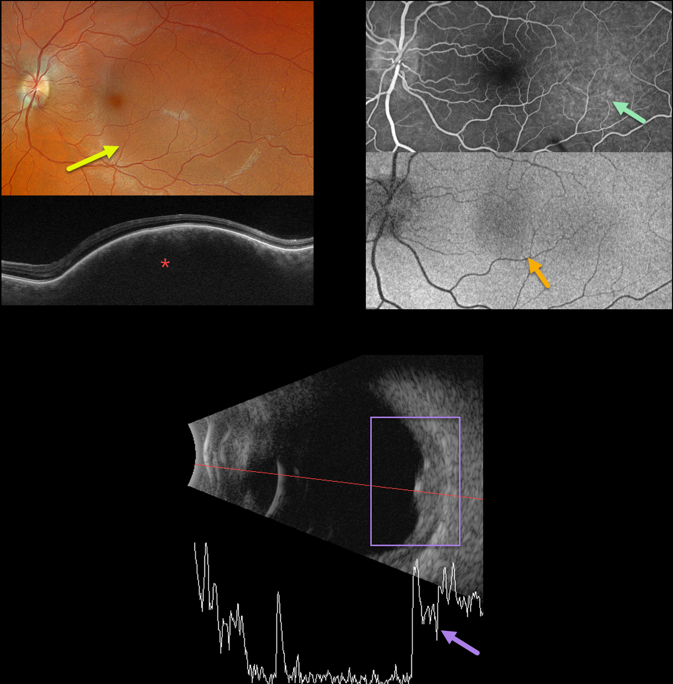
This 35YO female was referred for an asymptomatic left macular lesion. Vision was 20/20 in her normal right eye and 20/30 in her left eye.
Optos color RGB imaging shows a perfectly round subretinal lesion centered in the temporal macula, extending just beneath the macular center.
Swept-source OCT shows a thickened choroidal lesion without overlying subretinal fluid. Fluorescein angiography shows filling of this lesion without leakage, and fundus autofluorescence (FAF) shows faint hypo-FAF.
B-scan ultrasonography shows homogeneous high internal reflectivity.
Learning Points:
Isolated choroidal hemangiomas are benign vascular lesions. Although they have no malignant potential, they can cause vision loss due to exudation of subretinal and intraretinal fluid, which can be treated with photodynamic therapy (see Tsipursky et al, Surv Ophthalmology 2011;56:68-85).
Since our patient’s lesion was asymptomatic and without exudation, we will follow her several times yearly.
CHOROIDAL HEMANGIOMA
Barbara Parolini and Veronika Matello
Originally posted on @retina.rocks September 12, 2022
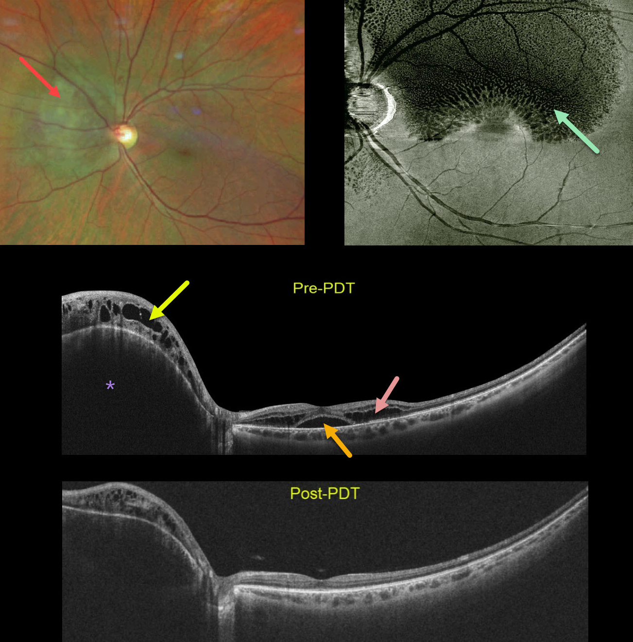
This healthy 45YO female was referred for macular schisis. Vision was 20/50. Color photography shows an orange-red subretinal lesion nasal to the nerve.
Widefield OCT shows an elevated hyporeflective choroidal mass with overlying cystic retinal edema. Subfoveal fluid with cystic fluid in the outer retina is noted in the macula. An en face OCT of the outer retina more dramatically shows the cystic changes.
The choroidal lesion with overlying retinal edema dramatically improved following photodynamic therapy. Vision improved to 20/20.
Learning Points:
Widefield imaging is revolutionizing the way we diagnose and treat retinal disease. This case is a perfect example in which macular imaging alone could lead to an incorrect diagnosis.
In this case, the macular findings in isolation suggest either optic pit maculopathy (Imamura et al, Retina 2010;30:1104-1112) or paraproteinemic maculopathy (Mansour et al, Ophthalmology 2014;121:1925-1932). This widefield technology led to a correct diagnosis, allowing for appropriate treatment with excellent anatomic and visual results.
CHOROIDAL HEMANGIOMA
Originally posted on @retina.rocks August 11, 2021
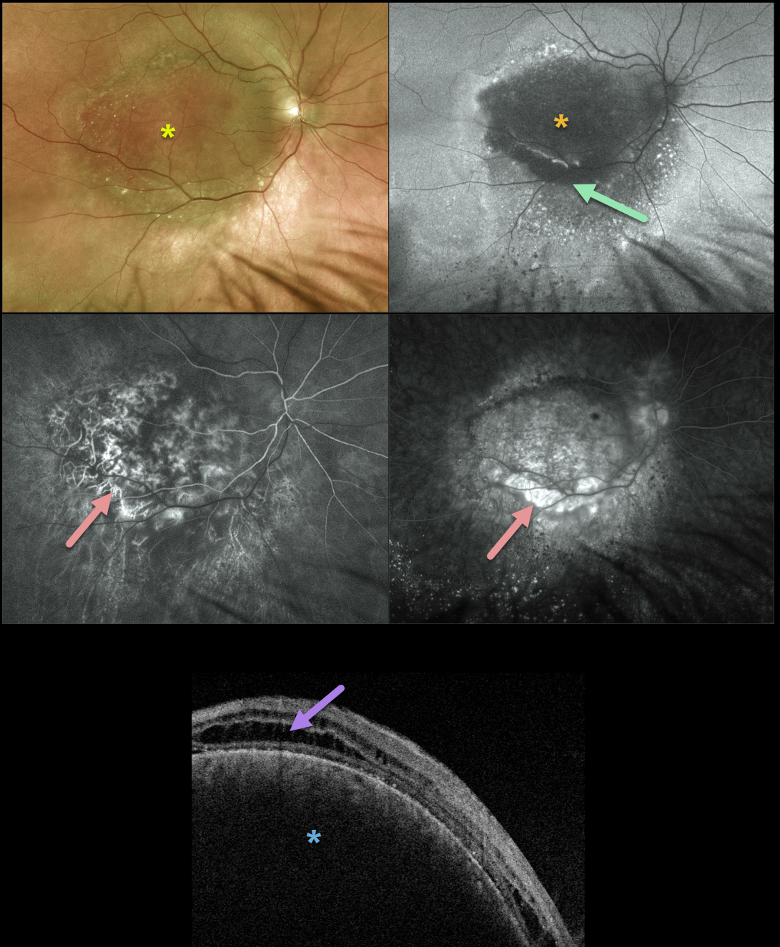
We have been following this 71YO male since 2014 with a choroidal hemangioma. Vision without correction was counting fingers, but improved with a +8.00 correction to 20/70. He tried a trial with a contact lens, but preferred to remain uncorrected due to visually disabling metamorphopsia.
The lesion has remained relatively stable over the years, and he continues to prefer observation over possible photodynamic therapy (PDT).
Multifocal Optos imaging on 8/3/21 shows a fairly stable tumor. Color imaging shows the hemangioma to be mostly orange-red with some more peripheral brownish coloration.
Fundus autofluorescence (FAF) reveals that the tumor is mostly hypo-FAF, indicating damage to the overlying RPE. There is a surrounding variable hyper- and hypo-FAF with a gutter of changes extending more inferiorly due to overlying exudative subretinal fluid.
Fluorescein angiography shows minimal leakage with mostly variable window-type defects. Triton swept-source OCT shows cystic edema overlying the markedly elevated choroidal tumor.
Learning Points:
Isolated choroidal hemangiomas are benign vascular lesions. Although they have no malignant potential, they can cause vision loss from exudation leading to subretinal and intraretinal fluid, which can be treated with PDT.
CHOROIDAL HEMANGIOMA
Originally posted on @retina.rocks January 8, 2021
This 44yo female presented with 20/20 vision and no visual complaints.
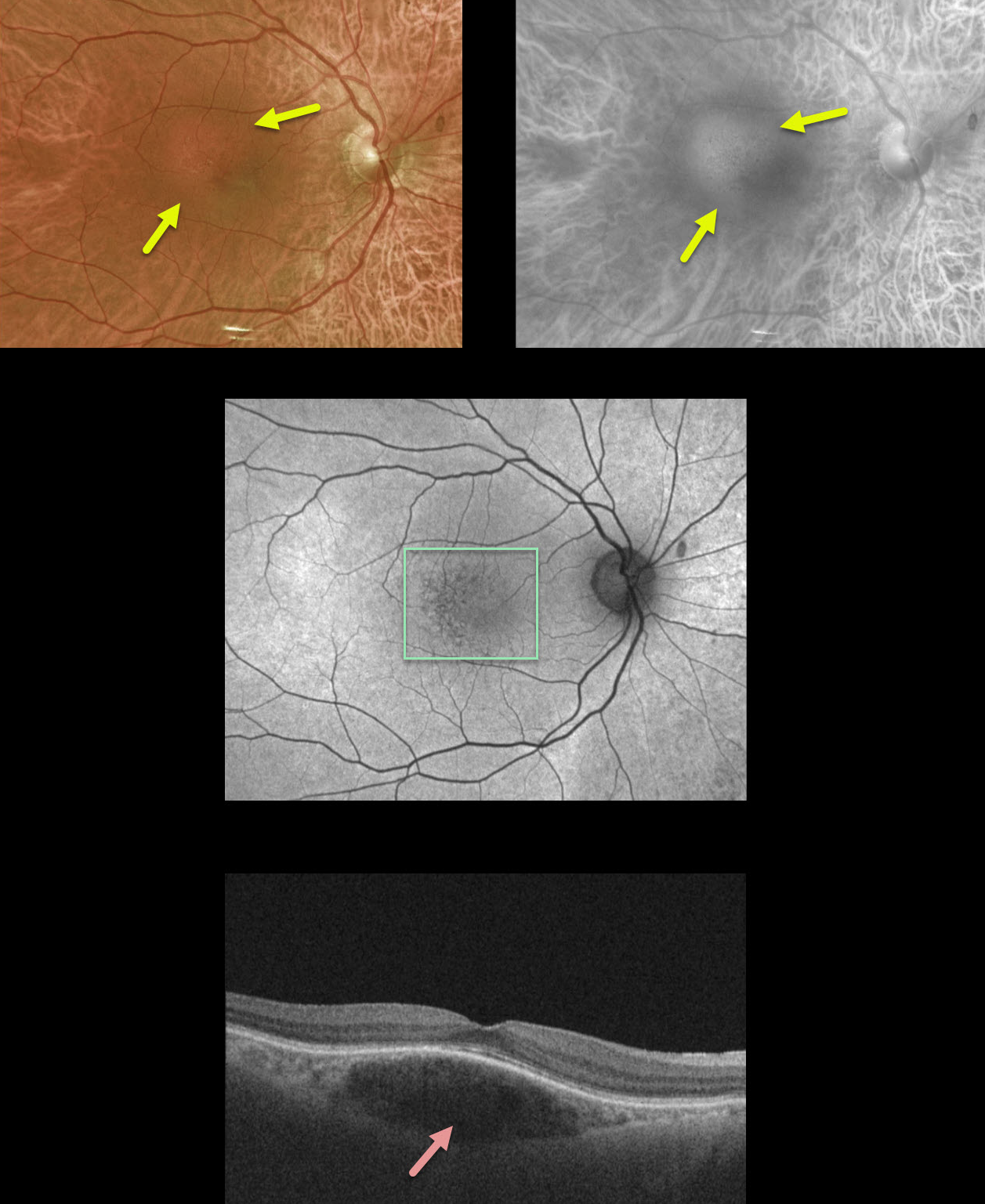
If you look closely at the color Optos image, you can see a small 1.5 disc diameter subretinal lesion in the superotemporal macula with mild overlying pigmentary changes. The Optos red channel best illuminates the lesion, indicating its choroidal location.
Fundus autofluorescence shows very mild overlying pigmentary changes. OCT shows a somewhat fusiform-shaped hyporeflective choroidal tumor without overlying exudation.
Since our patient was asymptomatic without fluid, we are following her twice yearly and will consider treatment if she develops symptomatic fluid.
Learning Points:
Isolated choroidal hemangiomas are benign vascular lesions. Although they have no malignant potential, they can cause vision loss due to exudation and subretinal and intraretinal fluid, which can be treated with photodynamic therapy.
CHOROIDAL HEMANGIOMA
Originally posted on @retina.rocks March 2, 2020
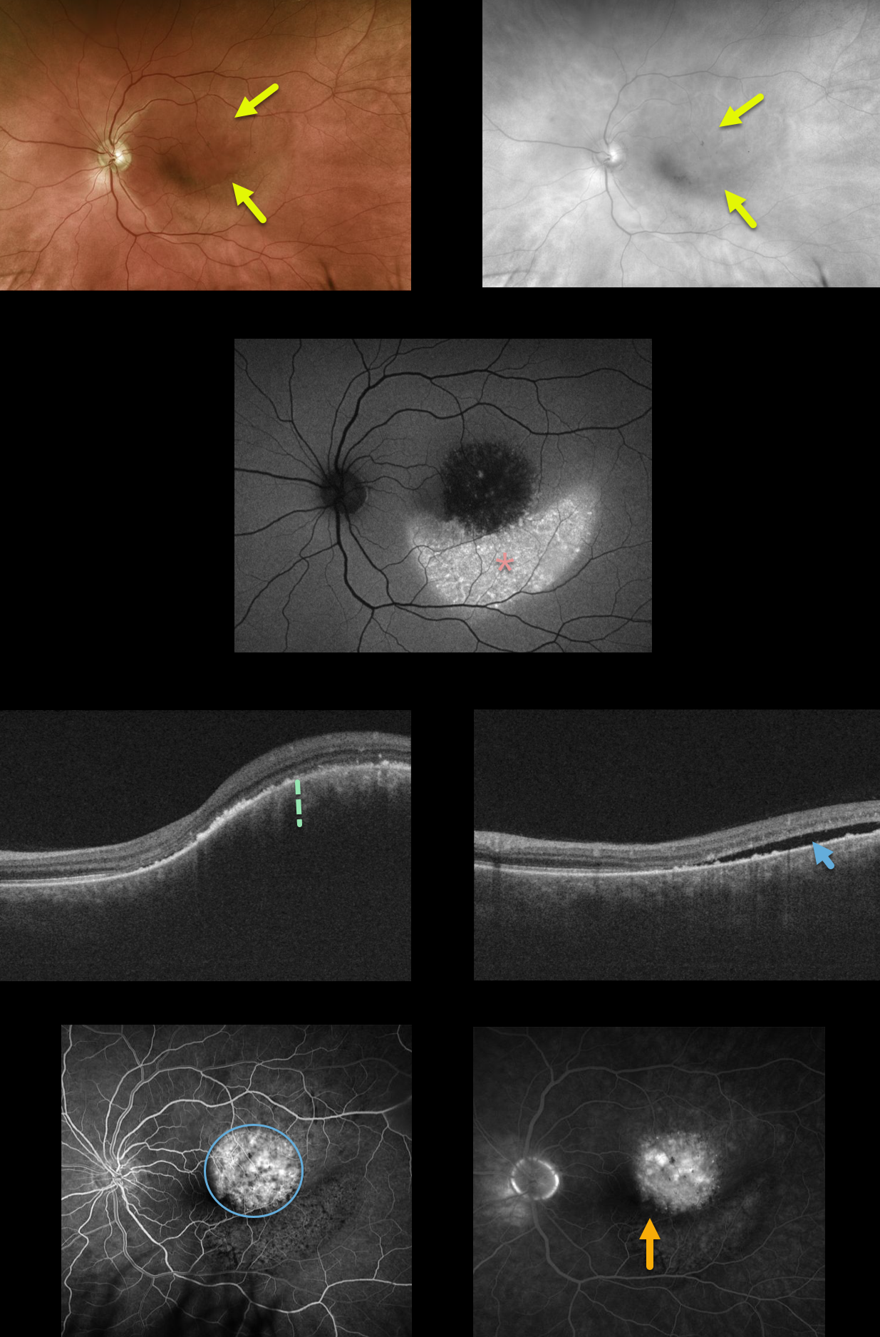
Our patient has a choroidal hemangioma which is difficult to see in the Optos color photograph, but somewhat better defined in the red channel image.
There is marked increased fundus autofluorescence inferior to the tumor, due to migrating subretinal fluid causing RPE alterations. OCT shows marked choroidal thickening within the tumor with subretinal fluid.
Fluoroscein angiography shows mild leakage from the tumor, with faint pigmentary changes inferiorly. Vision is 20/40 and he is minimally symptomatic due to the lack of foveal fluid. We are currently carefully observing this lesion and will consider photodynamic therapy if increased fluid and vision loss develop.
Learning Points:
Choroidal hemangiomas are benign choroidal vascular tumors that can become symptomatic later in life from overlying exudation. The usually round tumors are variably elevated and are often difficult to see clinically since their orange color blends in with the normal surrounding fundus.
CHOROIDAL HEMANGIOMA
Originally posted on @retina.rocks January 1, 2020
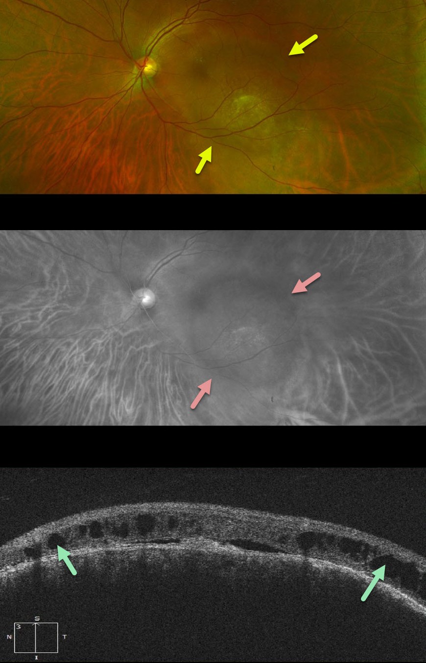
This patient has an isolated choroidal hemangioma. The extent of the lesion is difficult to appreciate on the Optos color photograph, but becomes more obvious on the red Optos channel (633nm) which best visualizes the choroid.
The OCT shows overlying cystic changes which is causing increasing visual symptoms.
Photodynamic therapy was performed in hopes of decreasing the macular leakage.
Learning Points:
Isolated choroidal hemangiomas are benign vascular tumors. The lesion can be difficult to see with the color photograph and is better visualized with the red channel, fundus autofluorescence, and fluoroscein angiography.


